2. Results and Discussion
In the design of our PFN antibiotic, we chose to exploit fluoroquinolone because it has been extensively used as an inherently fluorescent antibiotic [
10,
35,
36] and has demonstrated activity against a variety of clinically important pathogens, including
Pseudomonas aeruginosa [
37],
Escherichia coli [
38],
Staphylococcus aureus [
39], and
Enterococcus faecalis [
40]. Furthermore, we have previously worked with the related molecule ciprofloxacin [
41,
42]. In order to utilize the fluoroquinolone core to generate a profluorescent nitroxide which would retain antimicrobial activity, we proposed incorporating the nitroxide moiety via an amine linker at the C-7 position (see compound
1 for numbering) of the fluoroquinolone. Functionalization at this position with amino-nitrogen heterocycles has been shown to facilitate DNA gyrase or topoisomerase IV binding and subsequently enhance potency against both Gram-positive and Gram-negative bacteria [
43]. Hence, the amino-functionalized heterocyclic nitroxides
2–
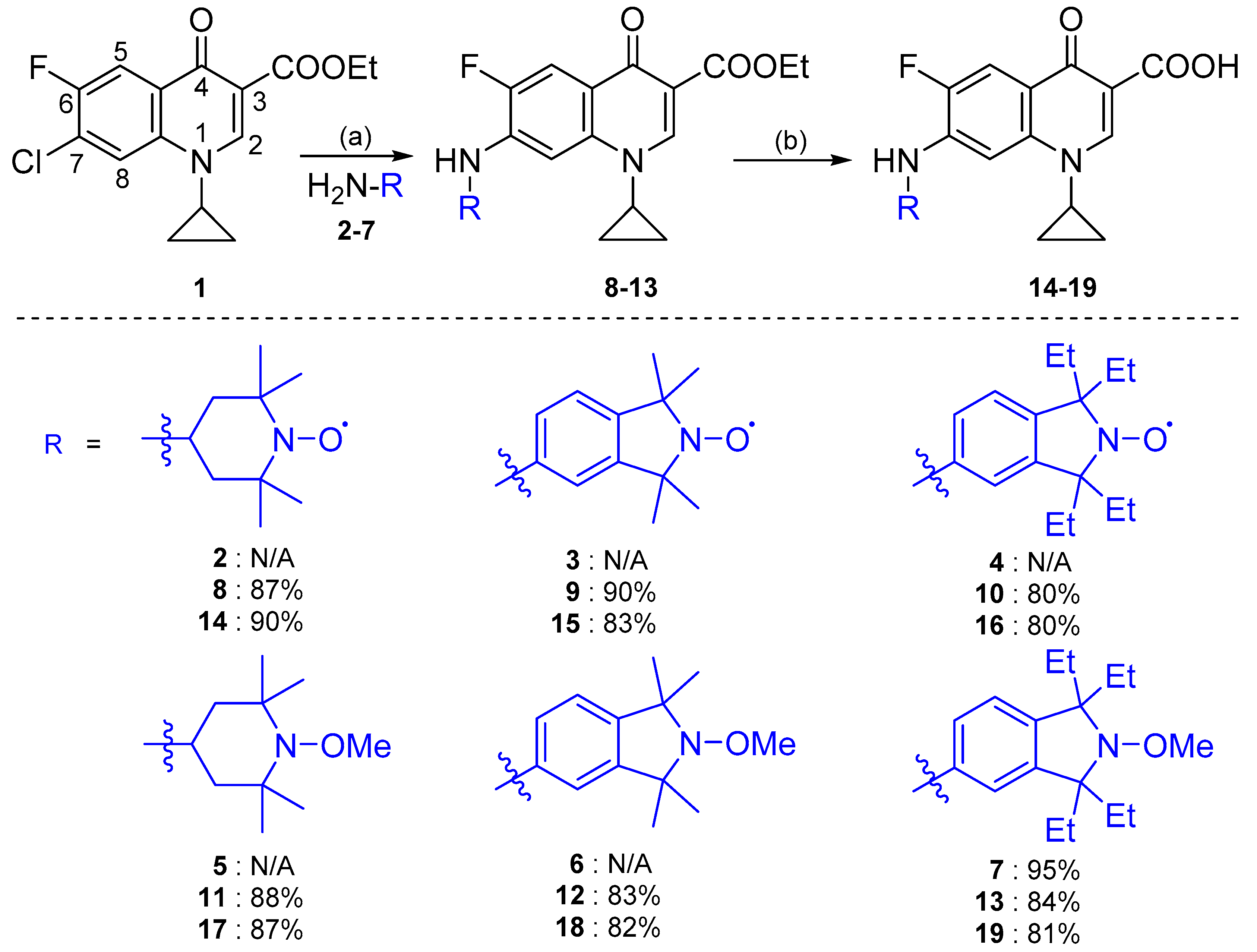
4 were selected to generate the desired profluorescent fluoroquinolone–nitroxides. The synthesis of fluoroquinolone–nitroxides (FNs)
15–
17 began by utilizing a Buchwald–Hartwig amination of fluoroquinolone
1 with 4-amino-2,2,6,6-tetramethylpiperidin-1-yloxyl (amino-TEMPO)
2, 5-amino-1,1,3,3-tetramethylisoindolin-2-yloxyl (amino-TMIO)
3 or 5-amino-1,1,3,3-tetraethylisoindolin-2-yloxyl (amino-TEIO)
4 using Pd(OAc)
2 and BINAP in THF to afford the ethyl ester fluoroquinolone–nitroxides
8–
10 in high yield (80–90%) (
Scheme 1). Final deprotection of
8–
10 via base mediated hydrolysis gave the desired FNs
14–
16 in excellent yield (80–90%).
In addition to the FNs
14–
16, their corresponding fluoroquinolone–methoxyamine (FM) derivatives
17–
19 were also synthesized, to examine the specific effect of the nitroxide moiety on fluoroquinolone fluorescence suppression. Furthermore, they could also serve as controls in assessing any antibacterial activity of the nitroxide moiety, and enable the intermediates to be well characterized by NMR spectroscopy (nitroxides are paramagnetic and typically display significantly broadened NMR spectroscopy signals). Utilizing Fenton conditions, the nitroxides
2–
4 were reacted with hydrogen peroxide, iron (II) sulfate heptahydrate and DMSO to furnish methoxyamines
5–
7 in moderate to excellent yield (77–95%). Subsequent amination, followed by base mediated deprotection, as described previously, afforded FMs
17–
19 in high yield (81–87%) (
Scheme 1).
With the new FNs
14–
16 and their corresponding FMs
17–
19 in hand, we proceeded to evaluate their photophysical properties. All FNs
14–
16 and their corresponding FMs
17–
19 displayed absorbance spectra, fluorescence spectra and extinction coefficients (
Table 1) characteristic of fluoroquinolones [
44]. However, FNs
15 and
16, and FMs
18 and
19, which contained aromatic isoindoline-based functionality, exhibited substantially reduced quantum yields (ՓF) (>10-fold lower) when compared to the compounds FN
14 and FM
17 (piperidine-based functionality). This reduction in fluorescence potentially arises from a disruption to the delocalized π-electron system of the quinolone core, by the amine linked aromatic system of the isoindoline. Consequently, these results indicate that the addition of an aromatic ring via an amine linkage to the fluoroquinolone core at the C-7 position negatively impacts the fluorescent intensity of the fluorophore.
A comparison of the fluorescence arising from solutions of FNs
14–
16 and their corresponding FMs
17–
19 in chloroform identified a substantial fluorescence suppression in the presence of the nitroxide moieties (calculated from the ratio between the quantum yields of the corresponding methoxyamine and nitroxide conjugates) (
Table 1). Fluorescence suppression was greatest in FNs
15 and
16 (67.5- and 75.0-fold, respectively), both of which bear the isoindoline core. FN
14 also demonstrated significant fluorescence suppression (27.7-fold), albeit lower than the other two nitroxide conjugates (FNs
15 and
16). While these findings confirm that the physical properties of FNs
14–
16 are suitable for profluorescent probe applications, these specific PFNs were designed for biological applications, and thus testing was repeated in an aqueous solution. Unfortunately, FNs
15–
16 and their FM derivatives
18–
19 displayed no measurable fluorescence in the aqueous solution, indicating water-induced fluorescence quenching. Thus, the photophysical properties of these compounds would not be optimal for aqueous biological applications (removal of the nitroxide would restore fluorescence, but no signal would be detectable due to water-induced fluorescence quenching). Conversely, FN
14 and FM
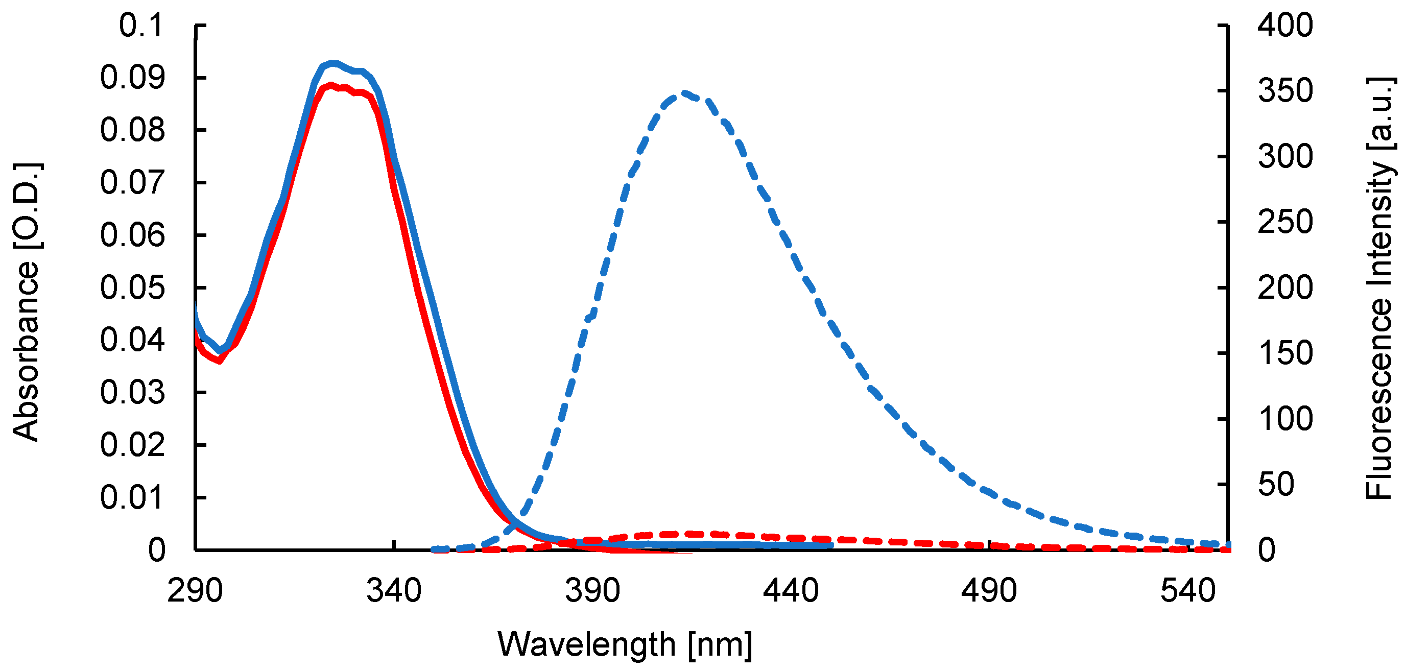
17 not only retained their fluorescence in the aqueous solution (
Figure 2), but FM
17 actually produced a higher fluorescence quantum yield, which resulted in an improved suppression ratio in the aqueous solution compared to chloroform (
Table 1). Subsequently, FN
14 and FM
17 were identified as possessing optimal photophysical properties for antibiotic–bacterial interaction studies.
To assess the dynamic emission range of FN
14, the reduction of FN
14 with an excess (1000 equivalents) of sodium ascorbate (
Scheme 2), chosen due to its aqueous solubility, was conducted and monitored via fluorescence spectroscopy. Following treatment of FN
14 with sodium ascorbate in aqueous solution (distilled water, pH 7), reduction of the nitroxide (quenched species) to the corresponding hydroxylamine (fluorescent species), resulted in a steady increase in the fluorescence emission over time (measured every minute for 30 minutes) (
Figure 3). Importantly, complete restoration of fluorescence was achieved (>99% switch on) compared to methoxyamine derivative
17 at the same concentration after approximately 30 minutes. This result suggests that the fluorescence quantum yield of FM
17 and the corresponding hydroxylamine derivative
20 of FN
14 are similar, and thus FM
17 is a true and accurate fluorescent control for FN
14. Furthermore, the fact that the fluorescence of FN
14 was completely restored by removal of the free radical nitroxide highlights its profluorescent nature and demonstrates its potential value as a biological probe for visualizing free radical and redox processes.
With the intended use of FN
14 and FM
17 in biological systems, we pondered the effect of pH on the fluorescence emission of FN
14 and FM
17. Thus, we decided to examine the effect of pH on fluorescence intensity for FM
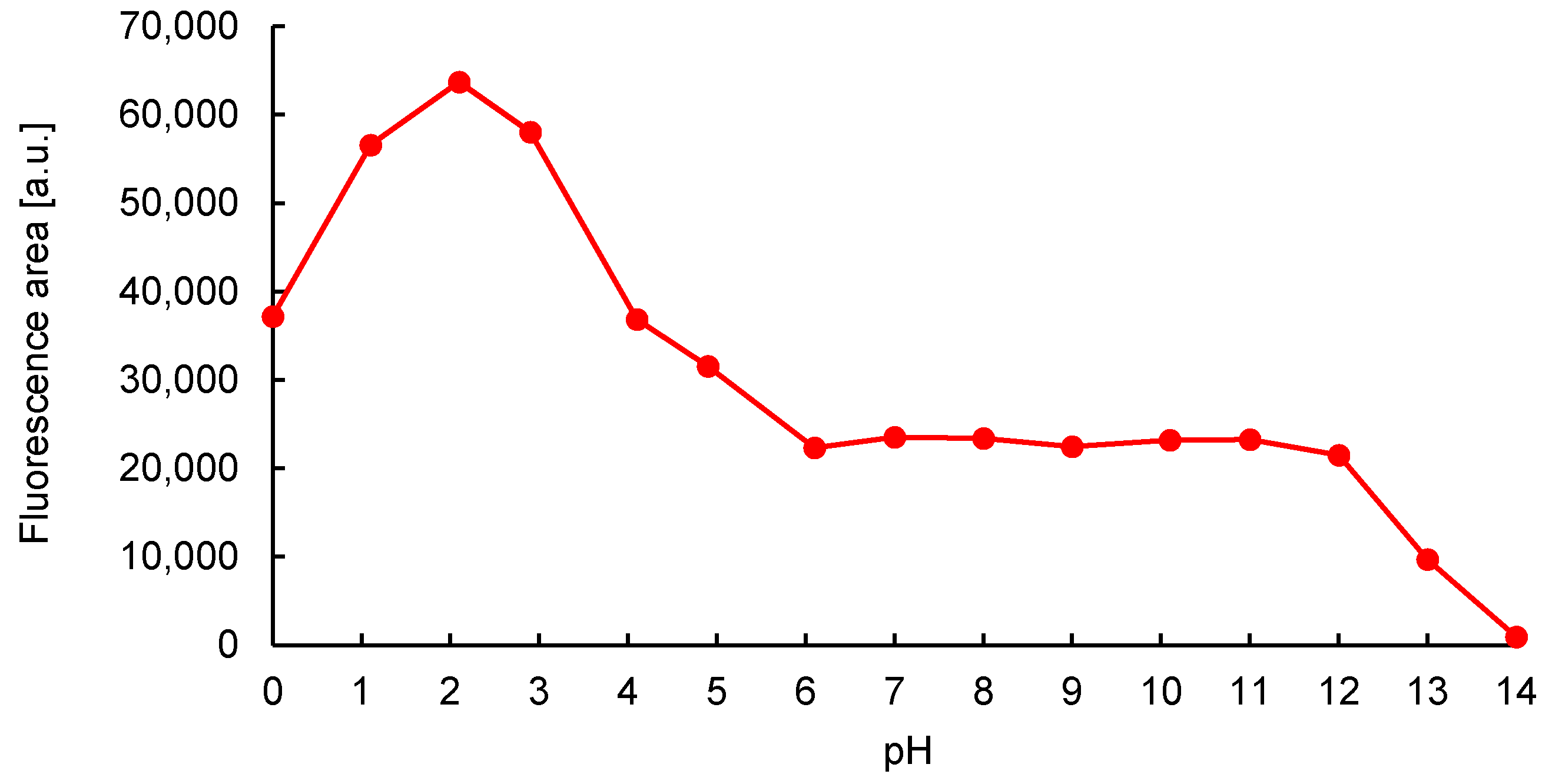
17 between the pH range of 0–14 (
Figure 4). FM
17 demonstrated fluorescence intensity stability between pH 6–12, supporting its suitability as a bacteriological probe. Interestingly, the fluorescence intensity of FM
17 significantly increased (>2.5-fold) at lower pH (<6), indicating that protonation of the heteroatoms within the conjugated system of the fluorophore drastically increases the fluorescence output of the compound. Conversely, at higher pH (>12) the fluorescence of FM
17 was considerably reduced and eventually completely quenched (pH 14). While compounds FN
14 and FM
17 were not specifically designed as fluorescent pH probes, their pH-dependent fluorescence could certainly be exploited for this purpose in suitable applications.
Following examination of the photophysical properties of FNs
14–
16 and FMs
17–
19, we proceeded to determine whether these compounds retained antimicrobial activity. Our biological investigations were initiated with the screening of FNs
14–
16 and FMs
17–
19 in minimum inhibitory concentration (MIC) assays against the common Gram-negative pathogens
P. aeruginosa and
E. coli, and Gram-positive pathogens
S. aureus and
E. faecalis (
Table 2). FNs
14 and
16 exhibited the highest activity against
S. aureus (MIC ≤ 20 µM). Interestingly, their corresponding FMs
17 and
19 both demonstrated no activity against this species (MIC > 1200 µM), suggesting that the presence of the free radical nitroxide may mediate
S. aureus antibacterial activity. This same trend was also observed against
E. faecalis with FN
16 (MIC ≤ 310 µM) and the corresponding FM
19 (MIC > 600 µM). As this trend was not observed for the Gram-negative species tested (
P. aeruginosa and
E. coli), it suggests that this activity may be specific against Gram-positive bacteria.
Furthermore, considering that nitroxides possess no inherent antibacterial activity (
Supplementary Material,
Table S1), the difference between the nitroxide-containing conjugate and its corresponding methoxyamine derivative against Gram-positive species was surprising. While the activity of FNs
14 and
16 was highest against
S. aureus, these two conjugates also exhibited Gram-negative antimicrobial activity, with FN
14 being most active against
E. coli (MIC ≤ 100 µM) and FN
16 against
P. aeruginosa (MIC ≤ 160 µM). The aqueous fluorescent properties of FN
14 combined with its antibacterial activity, suggesting this compound would be useful for monitoring antibiotic–bacterial interactions in both Gram-positive and Gram-negative bacteria.
As FN
14 and the corresponding FM
17 demonstrated optimal photophysical and biological properties, these compounds were subsequently evaluated for use as bacteriological probes. FN
14 and FM
17 were administered at two concentrations (150 µM and 600 µM) to
P. aeruginosa,
E. coli,
S. aureus, and
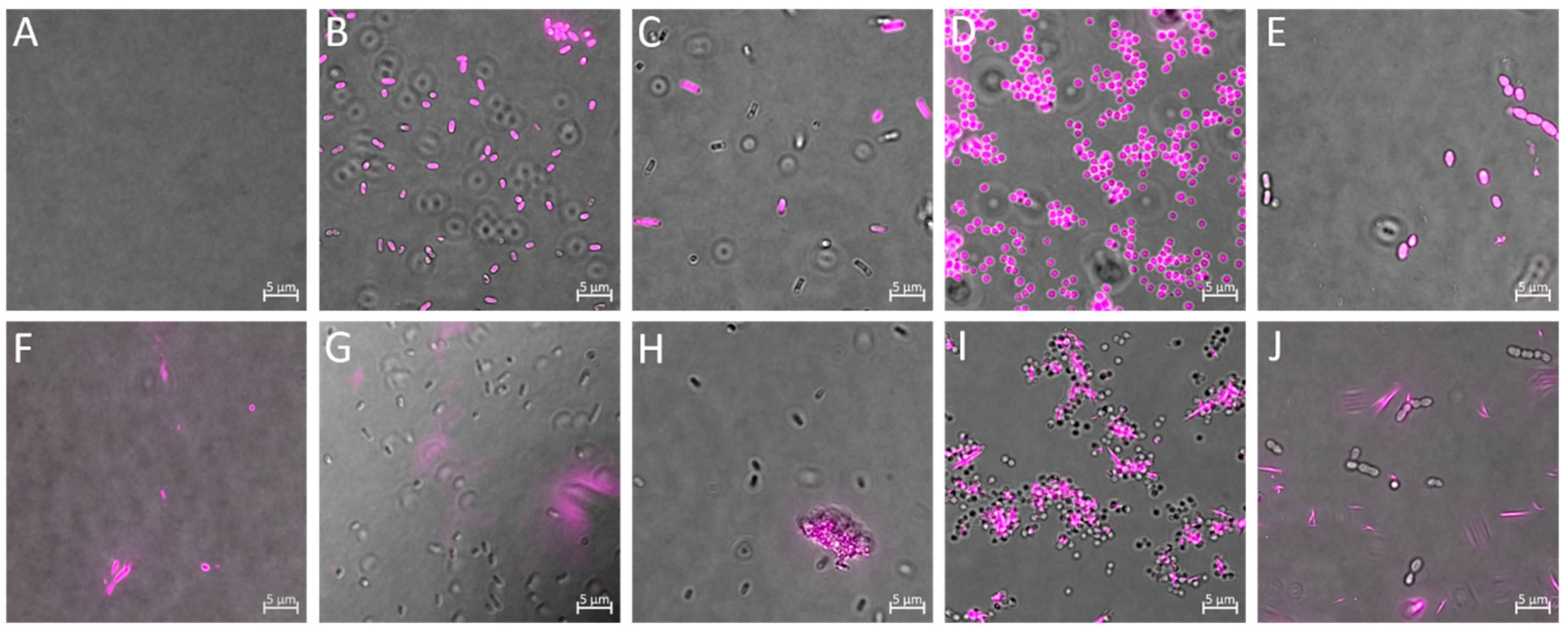
E. faecalis cells for 90 minutes, then visualized via fluorescence microscopy. FN
14 emitted bright fluorescence upon cell entry in all species tested (
Figure 5B–E). However, FN
14 bacterial cell entry and fluorescence was found to be both concentration and species specific. When FN
14 was administered to
P. aeruginosa at 150 µM, very few (~10%) cells fluoresced (
Supplementary Material,
Figure S1); however, when the concentration of FN
14 was increased to 600 µM, nearly all bacterial cells fluoresced (~90%) (
Figure 5B). A similar pattern was observed with
E. coli cells treated with FN
14 (
Figure 5C). Interestingly, this concentration-dependent fluorescence output was not observed in Gram-positive bacteria (
S. aureus and
E. faecalis). In fact, when either
S. aureus or
E. faecalis were treated with FN
14 (150 µM), almost every bacterial cell emitted measurable fluorescence (~99%) (
Figure 5D,E, respectively), suggesting that the process by which fluorescence is activated for FN
14 occurs more readily and/or more frequently in Gram-positive species.
Intriguingly, despite FM
17 being a fluorescence activated derivative of FN
14, and hence always fluorescent, it did not emit a measurable cell-associated fluorescence signal. Instead, fluorescence was only detected in the liquid medium, where FM
17 formed fluorescent aggregates (
Figure 5G–J). The lack of any bacterial-associated fluorescence for FM
17 suggests that it either does not enter bacterial cells or its fluorescence is quenched intracellularly. The inability of FM
17–
19 to translocate through the bacterial cell envelope could potentially explains their lack of antimicrobial activity against the Gram-positive pathogens
S. aureus and
E. faecalis (
Table 1). However, this possibility would not explain their activity against Gram-negative pathogens
P. aeruginosa and
E. coli, where the potency of both the FNs
14–
16 and their corresponding FMs
17–
19 derivatives is conserved.
To test the hypothesis that fluorescence activation of FN
14 occurs intracellularly, we examined the fluorescence properties of FN
14 and FM
17 in medium only (no bacterial cells present). Here, we treated the medium with either 150 or 600 µM of FN
14 or FM
17 for 90 minutes. Our findings indicated that FN
14 in medium emits no measurable fluorescence (
Figure 5A) (FN
14 also emitted no measurable fluorescence in PBS, LB, and MH). However, when bacterial cells were present in a medium containing FN
14, they became highly fluorescent while the surrounding medium still exhibited no fluorescence (
Figure 5B–E). Interestingly, in similar assays, FM
17 became immediately fluorescent in medium alone (
Figure 5F) and could be seen to form small aggregates and crystals (
Figure 5F–J) that were highly fluorescent. As FN
14 was not visible in medium but clearly visible inside bacterial cells, while FM
17 was only visible in medium, we can conclude that FN
14’s fluorescence is activated via an intracellular process, and thus, FN
14 can function as a true intracellular bacteriological probe with the potential to simultaneously monitor antibiotic–bacterial interactions and intracellular free radical and redox processes.
Importantly, FN 14 exhibited a fluorescence signal which did not require background subtraction or correction for bacteria autofluorescence (bacteria autofluorescence was not detected under these conditions). Furthermore, while the experiments reported here utilized an excitation wavelength of ~365 nm, FN 14 was also efficiently excited by a 405 nm laser or a multiphoton laser set at 720 nm. Taken together these results demonstrate the utility of FN 14 and support its use as a potential live-cell imaging probe.
3. Materials and Methods
3.1. General Methods
Synthetic reactions of an air-sensitive nature were carried out under an atmosphere of ultra-high purity argon. Anhydrous THF was obtained from the solvent purification system, Pure Solv
TM Micro, by Innovative Technologies. Anhydrous toluene was dried by storage over sodium wire. All other reagents were purchased from commercial suppliers and used without further purification. Ciprofloxacin, 7-chloro-1-cyclopropyl-6-fluoro-4-oxo-1,4-dihydroquinoline-3-carboxylic acid (Q-Acid), 4-carboxy-2,2,6,6-tetramethylpiperidin-1-yloxyl (CTEMPO), 2,2,6,6-tetramethylpiperidin-1-yloxyl (TEMPO), and 4-amino-2,2,6,6-tetramethylpiperidin-1-yloxyl (amino-TEMPO)
2 were purchased from Sigma-Aldrich Chemical Company. Ethyl 7-chloro-1-cyclopropyl-6-fluoro-4-oxo-1,4-dihydroquinoline-3-carboxylate
1 [
45], 5-amino-1,1,3,3-tetramethylisoindolin-2-yloxyl (amino-TMIO)
3 [
26], 5-nitro-1,1,3,3-tetraethylisoindolin-2-yloxyl
4 [
46], 4-amino-1-methoxy-2,2,6,6-tetramethylpiperidine (amino-TEMPOMe)
5 [
47], 5-amino-2-methoxy-1,1,3,3-tetramethylisoindoline (amino-TMIOMe)
6 [
48], 5-nitro-1,1,3,3-tetraethylisoindolin-2-yloxyl (nitro-TEIO) [
49], 1,1,3,3-tetramethylisoindolin-2-yloxyl (TMIO), [
50] and 1,1,3,3-tetraethylisoindolin-2-yloxyl (TEIO) [
51] were prepared in house by previously documented procedures. The analytical data obtained for each compound was consistent with that previously reported in the literature. All
1H NMR spectra were recorded at 600 MHz on a Bruker Avance 600 instrument. All
13C NMR spectra were recorded at 150 MHz on a Bruker Avance 600 instrument. Spectra were obtained in the following solvents: CDCl
3 (reference peaks:
1H NMR: 7.26 ppm;
13C NMR: 77.19 ppm), CD
2Cl
2 (reference peaks:
1H NMR: 5.32 ppm;
13C NMR: 53.84 ppm),
d6-DMSO (reference peaks:
1H NMR: 2.50 ppm;
13C NMR: 39.52 ppm) and CD
3OD (reference peaks:
1H NMR: 3.31 ppm;
13C NMR: 49.00 ppm). All NMR experiments were performed at room temperature. Chemical shift values (δ) are reported in parts per million (ppm) for all
1H NMR and
13C NMR spectral assignments.
1H NMR spectroscopy multiplicities are reported as: s = singlet, br. s = broad singlet, d = doublet, dd = doublet of doublets, m = multiplet. Coupling constants are reported in Hz. All spectra are presented using MestReNova 9.0. High-resolution ESI mass spectra were obtained with a Thermo Fisher Scientific Orbitrap Elite mass spectrometer (Thermo Fisher Scientific, Waltham, MA, USA) or an Agilent Q-TOF LC high-resolution mass spectrometer, which utilized electrospray ionization in positive ion mode. Analytical HPLC was carried out on an Agilent Technologies HP 1100 Series HPLC system using an Agilent C18 column (250 mm × 4.6 mm × 5 μm) with a flow rate of 1 mL min
−1. The purity of all final compounds was determined to be 95% or higher using HPLC analysis. EPR spectra were obtained with the aid of a miniscope MS 400 Magnettech EPR spectrometer. Column chromatography was performed using LC60A 40–63 Micron DAVISIL silica gel. Thin-layer chromatography (TLC) was performed on Merck Silica Gel 60 F254 plates. TLC plates were visualized under a UV lamp (254 nm) and/or by development with phosphomolybdic acid (PMA). Melting points were measured with a variable temperature apparatus by the capillary method and are uncorrected. Samples were separated by HPLC (Dionex Ultimate 3000) on a Phenomenex Luna C18 column (250 mm × 2.0 mm × 5 μm) held at 40 °C. Mobile phase A was 20% acetonitrile (ACN), and mobile phase B was 90% ACN, both containing 10 mM ammonium acetate, flowing at 0.2 mL min
−1. The gradient commenced at 57% B for 3 minutes, increasing to 100% B over 7 minutes, and holding at 100% B for a further 5 minutes before returning to initial conditions for 5 minutes. Post-column, the eluent was split (~9:1) for both UV and MS detection. High-resolution mass spectra were acquired on an LTQ Orbitrap Elite mass spectrometer (Thermo Fisher Scientific, Bremen, Germany) equipped with a heated electrospray ionization source, operating in the positive ion mode with a mass resolution of 120,000 (FWHM at
m/
z 400). This method was used for nitro-TEIOMe with the following modifications: Isocratic run where mobile phase A was 20% ACN/80% water, containing 10 mM ammonium acetate, and mobile phase B was 100% MeOH, both flowing at 0.2 mL·min
−1. All UV-Vis spectra were recorded on a single beam Varian Cary 50 UV-Vis spectrophotometer. Fluorescence measurements were performed on a Varian Cary 54 Eclipse fluorescence spectrophotometer equipped with a standard single-cell sample holder. Fluorescence microscopy was conducted on a Zeiss Axio Vert.A1 FL-LED equipped with filter sets, 49 (excitation: G 365 nm, beamsplitter: FT 395 nm, emission: BP 445/50 nm) used for FN 14 and FM 17. All microscopy experiments utilized a 100× oil immersion objective.
3.2. Synthesis of 5-Nitro-2-methoxy-1,1,3,3-tetraethylisoindoline (Nitro-TEIOMe)
Iron (II) sulfate heptahydrate (0.72 g, 2.58 mmol, 2.5 equiv) was added to a solution of nitro-TEIO (300 mg, 1.03 mmol, 1 equiv) in DMSO (10 mL). The mixture was cooled to 0 °C, and 35% aqueous hydrogen peroxide (276 µL, 4.12 mmol, 4 equiv) was added in a dropwise manner. The resulting mixture was stirred at 0 °C for 10 minutes and then at room temperature for an additional 1.5 hours. The reaction mixture was diluted with deionized water (40 mL) before being extracted with diethyl ether (3 × 20 mL). The combined organic extracts were washed with deionized water (200 mL) and dried over anhydrous sodium sulfate. The solvent was removed in vacuo to afford product nitro-TEIOMe as a light beige solid (285 mg, 0.93 mmol, 90%). 1H NMR (600 MHz, CDCl3): (*note the signals for the four ethyl groups were not observed on this NMR timescale) δ = 8.15 (dd, J = 8.3, 2.4 Hz, 1H, Ar-H), 7.98 (d, J = 2.4 Hz, 1H, Ar-H), 7.27 (dd, J = 8.3, 2.6 Hz, 1H, Ar-H), 3.80 (s, 3H, NOCH3). 13C NMR (150 MHz, CDCl3): δ = 152.8, 148.0, 147.2, 123.2, 122.6, 117.4, 67.4, 67.2, 65.8, 29.8, 24.8. HRMS (ESI): m/z calculated for C7H27N2O3 + H+ [M+H+]: 307.2022; found 307.2027. LC-MS Analysis: Rt = 10.9 min; area 99%. MP: 78.5–79.5 °C.
3.3. Synthesis of 5-Amino-2-methoxy-1,1,3,3-tetraethylisoindoline (Amino-TEIOMe) 7
Palladium on carbon 10% wt. loading (87 mg, 0.082 mmol, 10 mol %) was added to a solution of nitro-TEIOMe (250 mg, 0.82 mmol, 1 equiv) in methanol (30 mL). The solution was placed in a Parr hydrogenator under an atmosphere of hydrogen gas (25 psi), with shaking, for 3.5 hours. The resulting solution was filtered through Celite, then acidified (pH 1) with aqueous hydrochloric acid (2 M) and extracted with diethyl ether (3 × 20 mL). The remaining aqueous solution was basified (pH 12) with sodium hydroxide (2 M) and extracted with diethyl ether (3 × 20 mL). The combined organic extracts were dried over anhydrous sodium sulfate, and the solvent was removed in vacuo to afford product 7 as a light yellow oil (215 mg, 0.78 mmol, 95%). 1H NMR (600 MHz, CDCl3): δ = 6.80 (d, J = 7.9 Hz, 1H, Ar-H), 6.56 (dd, J = 8.0, 2.3 Hz, 1H, Ar-H), 6.37 (d, J = 2.2 Hz, 1H, Ar-H), 3.67 (s, 3H, NOCH3), 2.03–1.93 (m, br, 4H, 2 × CH2), 1.72-1.67 (m, br, 4H, 2 × CH2), 0.92 (s, 6H, 2 × CH3), 0.78 (s, 6H, 2 × CH3). 13C NMR (150 MHz, CDCl3): δ = 144.5, 144.0, 133.2, 124.2, 114.0, 110.3, 72.8,72.4, 63.6, 30.1, 29.6, 9.6, 9.1. HRMS (ESI): m/z calculated for C17H29N2O + H+ [M+H+]: 277.2280; found 277.2282. LC-MS: Rt = 14.1 min; area 99%.
3.4. General Procedure for the Synthesis of Fluoroquinolone–Nitroxides 14–16 and Fluoroquinolone–Methoxyamines 17–19
Cesium carbonate (3 equiv), palladium acetate (6 mol %), BINAP (10 mol %), Q-Ester 1 (2 equiv) and the specific primary amine (1 equiv) were added to a Schlenk vessel under an atmosphere of argon. THF (60 mL), which had been degassed with argon, was then added. The vessel was sealed and heated at 65 °C for 72 hours. The reaction was allowed to cool to room temperature, and the solvent was removed via rotary evaporation. The resulting residue was washed three times with aqueous hydrochloric acid (2 M, 3 × 20 mL) and the combined filtrates were extracted with diethyl ether (3 × 10 mL). The aqueous phase was neutralized with saturated sodium carbonate and extracted with dichloromethane (3 × 50 mL). The combined extracts were dried over anhydrous sodium sulfate and the solvent removed in vacuo. Purification was achieved via column chromatography (SiO2, chloroform 98%, methanol 2%).
3.5. Ethyl 1-Cyclopropyl-6-fluoro-7-(2,2,6,6-tetramethyl-1-oxy-piperidine-4-yl)amino)-4-oxo-1,4-dihydroquinoline-3-carboxylate 8
Reagents: Cesium carbonate (860 mg, 2.64 mmol, 3 equiv), palladium acetate (12 mg, 0.053 mmol, 6 mol %), BINAP (55 mg, 0.088 mmol, 10 mol %), Q-Ester 1 (545 mg, 1.76 mmol, 2 equiv), amino-TEMPO 2 (150 mg, 0.88 mmol, 1 equiv). Product: Orange solid (340 mg, 0.77 mmol, 87%). 1H NMR (600 MHz, CDCl3): (*note compound is a free-radical, some signals appear broadened, and other signals are missing) δ = 8.38 (s, 1H, NCH=C), 7.95 (s, 1H, Ar-H), 7.09 (s, 1H, Ar-H), 4.27 (d, J = 6.8 Hz, 1H, OCH2CH3), 3.28 (s, 1H, C=CHNCH), 1.30 (t, J = 6.5 Hz, 3H, OCH2CH3), 1.10 (s, br, 4H, 2 × NCHCH2). 13C NMR (150 MHz, CDCl3): (*note compound is a free-radical, some signals appear broadened, and other signals are missing) δ = 172.2, 165.0, 147.0, 139.5, 138.5, 118.2, 111.0, 109.6, 94.1, 60.0, 33.9, 13.6, 9.2. MP: 214.5–215.4 °C. HRMS (ESI): m/z calculated for C24H32FN3O4 + H+ [M+H+]: 445.2377; found 445.2379. LC-MS: Rt = 5.0min; area 100%. EPR: g = 2.00009, aN = 1.384 mT.
3.6. Ethyl 1-Cyclopropyl-6-fluoro-7-((1-methoxy-2,2,6,6-tetramethylpiperidine-4-yl)amino)-4-oxo-1,4-dihydroquinoline-3-carboxylate 11
Reagents: Cesium carbonate (860 mg, 2.64 mmol, 3 equiv), palladium acetate (12 mg, 0.053 mmol, 6 mol %), BINAP (55 mg, 0.088 mmol, 10 mol %), Q-Ester 1 (545 mg, 1.76 mmol, 2 equiv), amino-TEMPOMe 5 (165 mg, 0.88 mmol, 1 equiv). Product: Light yellow solid (354 mg, 0.77 mmol, 88%). 1H NMR (600 MHz, CDCl3): δ = 8.44 (s, 1H, NCH=C), 7.94 (d, J = 12.1 Hz, 1H, Ar-H), 6.94 (d, J = 7.0 Hz, 1H, Ar-H), 4.36 (q, J = 7.1 Hz, 3H, OCH2CH3), 4.31 (m, 1H, NH), 3.72 (m, 1H, C=CHNCH), 3.37 (m, 1H, NHCH), 1.96 (d, br, t, J = 12.2 Hz, 2H, NHCHCH2), 1.49 (t, J = 12.2 Hz, 2H, NHCHCH2), 1.39 (t, J = 7.1 Hz, 3H, OCH2CH3), 1.26–1.24 (s, 12H, 4 × CH3), 1.26–1.24 (m, 2H, NCHCH2), 1.14 (m, 2H, NCHCH2). 13C NMR (150 MHz, CDCl3): δ = 173.2, 166.2, 151.0, 148.6, 147.9, 104.2, 140.0, 139.1, 118.9, 111.6, 111.4, 110.4, 96.1, 65.7, 60.9, 60.0, 45.3, 44.0, 34.5, 33.0, 20.9, 14.6, 8.3. MP: 238.1–240.0 °C. HRMS (ESI): m/z calculated for C25H35FN3O4 + H+ [M+H+]: 460.2612; found 460.2611. LC-MS: Rt = 12.0 min; area 97%.
3.7. Ethyl 1-Cyclopropyl-6-fluoro-7-((1,1,3,3-tetramethylisoindolin-2-yloxyl-5-yl)amino)-4-oxo-1,4-dihydroquinoline-3-carboxylate 9
Reagents: Cesium carbonate (860 mg, 2.64 mmol, 3 equiv), palladium acetate (12 mg, 0.053 mmol, 6 mol %), BINAP (55 mg, 0.088 mmol, 10 mol %), Q-Ester 1 (545 mg, 1.76 mmol, 2 equiv), amino-TMIO 3 (180 mg, 0.88 mmol, 1 equiv). Product: Yellow solid (378 mg, 0.79 mmol, 90%). 1H NMR (600 MHz, CDCl3): (*note compound is a free-radical, some signals appear broadened, and other signals are missing) δ = 8.51 (s, 1H, NCH=C), 8.17 (d, J = 9.2 Hz, 1H, Ar-H), 7.60 (s, br, 1H, Ar-H), 6.45 (s, br, 1H, Ar-NH), 4.41 (q, J = 6.9 Hz, 2H, OCH2CH3), 3.30 (s, br, 1H, C=CHNCH), 1.43 (t, J = 6.9 Hz, 3H, OCH2CH3), 1.30–0.93 (m, br, 4H, 2 × NCHCH2). 13C NMR (150 MHz, CDCl3): (*note compound is a free-radical, some signals appear broadened, and other signals are missing) δ = 172.7, 165.8, 147.9, 138.0, 121.4, 112.2, 112.0, 110.4, 99.5, 60.8, 34.6, 14.3, 8.6. MP: 216.5–217.7 °C. HRMS (ESI): m/z calculated for C27H30FN3O4 + H+ [M+H+]: 479.2220; found 479.2227. LC-MS: Rt = 5.7 min; area 98%. EPR: g = 2.00003, aN = 1.433 mT.
3.8. Ethyl 1-Cyclopropyl-6-fluoro-7-((2-methoxy-1,1,3,3-tetramethylisoindoline-5-yl)amino)-4-oxo-1,4-dihydroquinoline-3-carboxylate 12
Reagents: Cesium carbonate (860 mg, 2.64 mmol, 3 equiv), palladium acetate (12 mg, 0.053 mmol, 6 mol %), BINAP (55 mg, 0.088 mmol, 10 mol %), Q-Ester 1 (545 mg, 1.76 mmol, 2 equiv), amino-TMIOMe 6 (182 mg, 0.88 mmol, 1 equiv). Product: Pale yellow solid (360 mg, 0.73 mmol, 83%). 1H NMR (600 MHz, CDCl3): δ = 8.48 (s, 1H, NCH=C), 8.11 (d, J = 11.8 Hz, 1H, Ar-H), 7.54 (d, J = 6.9 Hz, 2H, Ar-H), 7.20–7.11 (m, 2H, 2 × Ar-H), 7.07 (d, J = 1.8 Hz, 2H, Ar-H), 6.32 (d, J = 3.7 Hz, 1H, Ar-NH), 4.38 (q, J = 7.1 Hz, 2H, OCH2CH3), 3.79 (s, 3H, NOCH3), 3.25 (tt, J = 7.1, 4.0 Hz, 1H, C=CHNCH), 1.45–1.39 (s, br, 12H, 4 × CH3), 1.40 (t, J = 7.1 Hz, 3H, OCH2CH3), 1.19–1.12 (m, 2H, NCHCH2), 1.10–1.05 (m, 2H, NCHCH2). 13C NMR (150 MHz, CDCl3): δ = 173.2, 166.2, 151.0, 149.4, 148.2, 147.1, 138.7, 138.6, 138.1, 123.0, 121.4, 121.0, 121.9, 115.4, 112.4, 112.3, 110.5, 99.0, 67.2, 67.1, 65.7, 61.0, 34.6, 14.6, 8.3. MP: 242.8–243.6 °C. HRMS (ESI): m/z calculated for C28H33FN3O4 + H+ [M+H+]: 494.2455; found 494.2456. LC-MS: Rt = 13.7 min; area 98%.
3.9. Ethyl 1-Cyclopropyl-6-fluoro-7-((1,1,3,3-tetraethylisoindolin-2-yloxyl-5-yl)amino)-4-oxo-1,4-dihydroquinoline-3-carboxylate 10
Reagents: Cesium carbonate (860 mg, 2.64 mmol, 3 equiv), palladium acetate (12 mg, 0.053 mmol, 6 mol %), BINAP (55 mg, 0.088 mmol, 10 mol %), Q-Ester 1 (545 mg, 1.76 mmol, 2 equiv), amino-TEIO 4 (230 mg, 0.88 mmol, 1 equiv). Product: Yellow solid (376 mg, 0.70 mmol, 80%). 1H NMR (600 MHz, CDCl3): (*note compound is a free-radical, some signals appear broadened, and other signals are missing) δ = 8.50 (s, 1H, NCH=C), 8.16 (d, J = 9.6 Hz, 1H, Ar-H), 7.53 (s, br, 1H, Ar-H), 6.39 (s, 1H, Ar-NH), 4.40 (q, J = 7.0 Hz, 2H, OCH2CH3), 3.27 (s, 1H, C=CHNCH), 1.42 (t, J = 7.0 Hz, 3H, OCH2CH3), 1.10 (s, br, 4H, 2 × NCHCH2). 13C NMR (150 MHz, CDCl3): (*note compound is a free-radical, some signals appear broadened, and other signals are missing) δ = 172.8, 165.9, 148.0, 138.2, 121.3, 112.3, 110.5, 99.3, 60.9, 34.7, 14.4, 8.5. MP: 178.5–180.0 °C (decomposed). HRMS (ESI): m/z calculated for C31H38FN3O4 + H+ [M+H+]: 534.2849; found 534.2850. LC-MS: Rt = 12.1 min; area 97%. EPR: g = 2.00012, aN = 1.394 mT.
3.10. Ethyl 1-Cyclopropyl-6-fluoro-7-((2-methoxy-1,1,3,3-tetraethylisoindoline-5-yl)amino)-4-oxo-1,4-dihydroquinoline-3-carboxylate 13
Reagents: Cesium carbonate (860 mg, 2.64 mmol, 3 equiv), palladium acetate (12 mg, 0.053 mmol, 6 mol %), BINAP (55 mg, 0.088 mmol, 10 mol %), Q-Ester 1 (545 mg, 1.76 mmol, 2 equiv), amino-TEIOMe 7 (243 mg, 0.88 mmol, 1 equiv). Product: Light yellow solid (406 mg, 0.74 mmol, 84%). 1H NMR (600 MHz, CDCl3): δ = 8.47 (s, 1H, NCH=C), 8.12 (d, J = 11.8 Hz, 1H, Ar-H), 7.41 (d, J = 7.0 Hz, 1H, Ar-H), 7.17 (dd, J = 8.1, 2.1 Hz, 1H, Ar-H), 7.08 (d, J = 8.1 Hz, 1H, Ar-H), 6.96 (d, J = 2.1 Hz, 1H, Ar-H), 6.30 (d, J = 3.7 Hz, 1H, Ar-NH), 4.38 (q, J = 7.1 Hz, 2H, OCH2CH3), 3.70 (s, 3H, NOCH3), 3.22 (td, J = 7.1, 3.6 Hz, 1H, C=CHNCH), 2.17–1.89 (m, br, 4H, 2 × CCH2CH3), 1.89–1.65 (m, br, 4H, 2 × CCH2CH3), 1.40 (t, J = 7.1 Hz, 3H, OCH2CH3), 1.32–1.19 (m, br, 2H, NCHCH2), 1.11–1.04 (m, br, 2H, NCHCH2), 1.04–0.91 (s, br, 6H, 3 × CCH2CH3), 0.87–0.70 (s, br, 6H, 3 × CCH2CH3). 13C NMR (150 MHz, CDCl3): δ = 173.2, 166.3, 151.0, 149.4, 148.1, 144.8, 139.8, 138.8, 137.7, 124.8, 121.3, 117.7, 112.4, 112.3, 110.5, 98.9, 72.9, 72.8, 63.7, 51.0, 34.6, 29.5, 14.6, 9.2, 8.2. MP: 214.8–217.6 °C. HRMS (ESI): m/z calculated for C32H41FN3O4 + H+ [M+H+]: 550.3081; found 550.3081. LC-MS: Rt = 19.6 min; area 96%.
3.11. General Procedure for the Synthesis of FNs 14–19 via Base Mediated Ester Hydrolysis
Aqueous sodium hydroxide (2 M, 7 equiv) was added to a solution of the specific ethyl ester (1 equiv) in HPLC grade methanol (50 mL), and the resulting solution was stirred at 50 °C for 5 hours. The reaction mixture was cooled to room temperature and diluted with deionized water (50 mL). The pH was adjusted to approximately 6 using aqueous hydrochloric acid (2 M) and the mixture extracted with dichloromethane (3 × 20 mL). The combined organic extracts were dried over anhydrous sodium sulfate, and the solvent was removed in vacuo. Purification was achieved via column chromatography (SiO2, chloroform 98%, methanol 2%).
3.12. 1-Cyclopropyl-6-fluoro-7-(2,2,6,6-tetramethyl-1-oxy-piperidine-4-yl)amino)-4-oxo-1,4-dihydroquinoline-3-carboxylic acid 14
Reagents: 8 (49 mg, 0.11 mmol, 1 equiv), aqueous sodium hydroxide (2 M, 0.39 mL, 0.77 mmol, 7 equiv) and HPLC grade methanol (10 mL). Product: Orange powdery solid (41 mg, 0.10 mmol, 90%). 1H NMR (600 MHz, CDCl3): (*note compound is a free-radical, some signals appear broadened, and other signals are missing) δ = 15.20 (s, 1H, COOH), 8.74 (s, 1H, NCH=C), 8.05 (s, 1H, Ar-H), 7.78–7.29 (s, br, 1H, Ar-H), 4.86–4.20 (s, br, 1H Ar-NH), 3.52 (s, 1H, C=CHNCH), 1.39–1.09 (m, br, 4H, 2 × NCHCH2). 13C NMR (150 MHz, CDCl3): (*note compound is a free-radical, some signals appear broadened, and other signals are missing) δ = 190.7, 177.0, 168.9, 167.2, 147.2, 140.6, 110.9, 108.1, 35.5, 22.2. MP: 253.8–255.0 °C. HRMS (ESI): m/z calculated for C22H28FN3O4 + H+ [M+H+]: 417.2064; found 417.2068. LC-MS: Rt = 4.98 min; area 100%. HPLC analysis: Retention time = 2.174 min; peak area = 99%; eluent A, Acetonitrile; eluent B, H2O (TFA 0.1%); isocratic (99:1) over 20 min with a flow rate of 1 mL min−1 and detected at 254 nm; C18 column; column temperature, rt. EPR: g = 2.00029, aN = 1.547 mT.
3.13. 1-Cyclopropyl-6-fluoro-7-((1-methoxy-2,2,6,6-tetramethylpiperidine-4-yl)amino)-4-oxo-1,4-dihydroquinoline-3-carboxylic acid 17
Reagents: 11 (50 mg, 0.11 mmol, 1 equiv), aqueous sodium hydroxide (2 M, 0.39 mL, 0.77 mmol, 7 equiv) and HPLC grade methanol (10 mL). Product: White powdery solid (43 mg, 0.10 mmol, 87%). 1H NMR (600 MHz, CDCl3): δ = 15.31 (s, 1H, COOH), 8.73 (s, 1H, NCH=C), 7.97 (d, J = 11.6 Hz, 1H, Ar-H), 7.06 (d, J = 6.9 Hz, 1H, Ar-H), 4.56 (s, 1H, Ar-NH), 3.80–3.72 (m, 1H, C=CHNCH), 3.65 (s, 3H, NOCH3), 3.49 (tt, J = 7.2, 4.1 Hz, 1H, NHCH), 1.99 (d, J = 13.3 Hz, 2H, NHCHCH2), 1.53 (d, J = 12.5 Hz, 2H, NHCHCH2), 1.36–1.31 (m, 2H, NCHCH2), 1.28 (s, 12H, 4 × CH3), 1.23–1.15 (m, 2H, NCHCH2). 13C NMR (150 MHz, CDCl3): δ = 177.1, 167.5, 147.2, 104.4, 137.4, 117.3, 115.9, 110.7, 110.5, 108.1, 104.6, 100.1, 96.0, 65.8, 60.0, 45.1, 44.2, 35.3, 33.0, 21.0, 8.4. MP: 283.7–284.9 °C. HRMS (ESI): m/z calculated for C23H31FN3O4 + H+ [M+H+]: 432.2299; found 432.2299. LC-MS: Rt = 12.19 min; area 96%. HPLC analysis: Retention time = 2.612 min; peak area = 95%; eluent A, Acetonitrile; eluent B, H2O (TFA 0.1%); isocratic (99:1) over 20 min with a flow rate of 1 mL min−1 and detected at 254 nm; C18 column; column temperature, rt.
3.14. 1-Cyclopropyl-6-fluoro-7-((1,1,3,3-tetramethylisoindolin-2-yloxyl-5-yl)amino)-4-oxo-1,4-dihydroquinoline-3-carboxylic acid 15
Reagents: 9 (52 mg, 0.11 mmol, 1 equiv), aqueous sodium hydroxide (2 M, 0.39 mL, 0.77 mmol, 7 equiv) and HPLC grade methanol (10 mL). Product: Yellow powdery solid (40 mg, 0.09 mmol, 83%). 1H NMR (600 MHz, CDCl3): (*note compound is a free-radical, some signals appear broadened, and other signals are missing) δ = 15.07 (s, 1H, COOH), 8.75 (s, 1H, NCH=C), 8.15 (d, J = 10.1 Hz, 1H, Ar-H), 7.67 (s, 1H, Ar-H), 6.66 (s, 1H, Ar-NH), 3.39 (s, 1H, NHCH), 1.40 – 1.22 (m, br, 4H, 2 × NCHCH2). 13C NMR (150 MHz, CDCl3): (*note compound is a free-radical, some signals appear broadened, and other signals are missing) δ = 176.8, 166.9, 147.3, 139.3, 121.9, 118.3, 111.2, 108.1, 99.2, 35.5, 29.6, 8.8. MP: 253.3–254.6 °C. HRMS (ESI): m/z calculated for C25H26N3O4 + H+ [M+H+]: 451.1907; found 451.1913. LC-MS: Rt = 5.49 min; area 98%. HPLC analysis: Retention time = 2.225 min; peak area = 96%; eluent A, Acetonitrile; eluent B, H2O (TFA 0.1%); isocratic (99:1) over 20 min with a flow rate of 1 mL min−1 and detected at 254 nm; C18 column; column temperature, rt. EPR: g = 2.00006, aN = 1.433 mT.
3.15. 1-Cyclopropyl-6-fluoro-7-((2-methoxy-1,1,3,3-tetramethylisoindoline-5-yl)amino)-4-oxo-1,4-dihydroquinoline-3-carboxylic acid 18
Reagents: 12 (54 mg, 0.11 mmol, 1 equiv), aqueous sodium hydroxide (2 M, 0.39 mL, 0.77 mmol, 7 equiv) and HPLC grade methanol (10 mL). Product: Pale yellow powdery solid (42 mg, 0.09 mmol, 82%). 1H NMR (600 MHz, CDCl3): δ = 15.17 (s. 1H, COOH), 8.71 (s, 1H, NCH=C), 8.08 (d, J = 11.5 Hz, 1H, Ar-H), 7.60 (d, J = 7.0 Hz, 1H, Ar-H), 7.23–7.13 (m, 2H, Ar-H), 7.09 (d, J = 1.8 Hz, 1H, Ar-H), 6.51 (d, J = 3.8 Hz, 1H, Ar-NH), 3.80 (s, 3H, NOCH3), 3.36 (tt, J = 7.1, 4.0 Hz, 1H, NHCH), 1.46 (s, br, 12H, 4 × CH3), 1.24–1.17 (m, 2H, NCHCH2), 1.17–1.09 (m, 2H, NCHCH2). 13C NMR (150 MHz, CDCl3): δ = 177.1, 167.3, 151.3, 149.7, 147.5, 147.2, 143.1, 140.0, 139.9, 138.6, 137.7, 123.2, 123.0, 122.1, 121.1, 117.7, 116.1, 115.3, 111.4, 111.2, 108.1, 99.0, 98.8, 67.2, 67.1, 65.7, 52.2, 35.4, 34.7, 29.8, 24.9, 8.3. MP: 293.7–295.3 °C. HRMS (ESI): m/z calculated for C26H29N3O4 + H+ [M+H+]: 466.2142; found 466.2148. LC-MS: Rt = 13.64 min; area 98%. HPLC analysis: Retention time = 3.243 min; peak area = 98%; eluent A, Acetonitrile; eluent B, H2O (TFA 0.1%); isocratic (99:1) over 20 min with a flow rate of 1 mL min−1 and detected at 254 nm; C18 column; column temperature, rt.
3.16. 1-Cyclopropyl-6-fluoro-7-((1,1,3,3-tetraethylisoindolin-2-yloxyl-5-yl)amino)-4-oxo-1,4-dihydroquinoline-3-carboxylic acid 16
Reagents: 10 (59 mg, 0.11 mmol, 1 equiv), aqueous sodium hydroxide (2 M, 0.39 mL, 0.77 mmol, 7 equiv) and HPLC grade methanol (10 mL). Product: Pale yellow powdery solid (45 mg, 0.09 mmol, 80%). 1H NMR (600 MHz, CDCl3): (*note compound is a free-radical, some signals appear broadened, and other signals are missing) δ = 15.08 (s. 1H, COOH), 8.73 (s, 1H, NCH=C), 8.13 (d, J = 9.0 Hz, 1H, Ar-H), 7.59 (s, 1H, Ar-H), 6.60 (s, 1H, Ar-NH), 3.38 (s, 1H, NHCH), 1.39–1.22 (m, br, 2H, NCHCH2), 1.22–1.08 (m, br, 2H, NCHCH2). 13C NMR (150 MHz, CDCl3): (*note compound is a free-radical, some signals appear broadened, and other signals are missing) δ = 176.8, 166.9, 147.3, 139.4, 118.0, 111.3, 111.1, 108.0, 98.9, 35.4, 29.6, 8.5. MP: 245.1–246.2 °C. HRMS (ESI): m/z calculated for C29H34N3O4 + H+ [M+H+]: 507.2533; found 507.2537. LC-MS: Rt = 12.27 min; area 98%. HPLC analysis: Retention time = 3.140 min; peak area = 96%; eluent A, Acetonitrile; eluent B, H2O (TFA 0.1%); isocratic (80:20) over 20 min with a flow rate of 1 mL min−1 and detected at 254 nm; C18 column; column temperature, rt. EPR: g = 2.00009, aN = 1.379 mT.
3.17. 1-Cyclopropyl-6-fluoro-7-((2-methoxy-1,1,3,3-tetraethylisoindoline-5-yl)amino)-4-oxo-1,4-dihydroquinoline-3-carboxylic acid 19
Reagents: 13 (60 mg, 0.11 mmol, 1 equiv), aqueous sodium hydroxide (2 M, 0.39 mL, 0.77 mmol, 7 equiv) and HPLC grade methanol (10 mL). Product: Pale yellow powdery solid (47 mg, 0.09 mmol, 81%). 1H NMR (600 MHz, CDCl3): δ = 15.19 (s, 1H, COOH), 8.71 (s, 1H, NCH=C), 8.09 (d, J = 11.4 Hz, 1H, Ar-H), 7.45 (d, J = 7.0 Hz, 1H, Ar-H), 7.20 (dd, J = 8.0, 2.1 Hz, 1H, Ar-H), 7.12 (d, J = 8.0 Hz, 1H, Ar-H), 7.12 (d, J = 8.0 Hz, 1H, Ar-H), 6.49 (d, J = 3.8 Hz, 1H, Ar-NH), 3.70 (s, 3H, NOCH3), 3.32 (ddd, J = 10.7, 7.1, 4.1 Hz, 1H, NHCH), 2.19–1.92 (m, br, 4H, CCH2CH3), 1.88–1.66 (m, br, 4H, CCH2CH3), 1.17–1.09 (m, 2H, NCHCH2), 1.09–1.04 (m, 2H, NCHCH2), 1.03–0.88 (s, br, 6H, 2 × CCH2CH3), 0.88–0.70 (s, br, 6H, 2 × CCH2CH3). 13C NMR (150 MHz, CDCl3): δ = 177.1, 167.4, 151.3, 149.6, 147.3, 145.1, 140.9, 140.5, 140.4, 139.9, 124.9, 122.0, 118.6, 117.6, 111.4, 111.2, 108.1, 98.6, 73.0, 72.9, 63.7, 35.4, 30.1, 29.6, 9.6, 9.1, 8.3. MP: 270.5–272.0 °C. HRMS (ESI): m/z calculated for C30H37N3O4 + H+ [M+H+]: 522.2768; found 522.2771. LC-MS: Rt = 19.13 min; area 97%. HPLC analysis: Retention time = 7.328 min; peak area = 95%; eluent A, Acetonitrile; eluent B, H2O (TFA 0.1%); isocratic (80:20) over 20 min with a flow rate of 1 mL min−1 and detected at 254 nm; C18 column; column temperature, rt.
3.18. Fluorescence Quantum Yield and Extinction Coefficient Calculations
Quantum yield efficiencies of fluorescence for compounds
14–
19 were obtained from measurements at five different concentrations in water, ethanol, or chloroform using the following equation:
where
A and
F denote the absorbance and fluorescence intensity, respectively, Σ[
F] denotes the peak area of the fluorescence spectra, calculated by summation of the fluorescence intensity, and η denotes the refractive index of the solvent (chloroform = 1.444, ethanol = 1.362, water = 1.000). Anthracene (
ФF = 0.27 in ethanol) was used as the standard. Extinction coefficients were calculated from the obtained absorbance spectra. This is a standardised method and our values are consistent with the values reported for other profluorescent nitroxides using the same method [
17,
30,
48].
3.19. Fluorescence Spectroscopy Measurements: Reduction of FN 14 with Sodium Ascorbate
Sodium ascorbate (200 µM solution in water, 0.5 mL), was added to a solution of FN 14 (2 µM solution in water, 0.5 mL) in a 4-sided quartz cuvette, equipped with a magnetic stirrer bar. The resulting solution (1 µM of FN 14 1 equiv and 1000 µM sodium ascorbate 1000 equiv) was placed in the fluorescence spectrophotometer, equipped with a magnetic stirrer, and measurements were recorded every minute for 30 mins. The measurement of the blank sample (time = 0 min) was conducted similarly by adding water (0.5 mL) to a solution of FN 14 (2 μM solution in water, 0.5 mL).
3.20. Evaluating the Effect of pH on the Fluorescence Intensity of FM 17
Fifteen aqueous solutions (ranging from pH 0 to pH 14) containing the same concentration (500 µM) of FM 17 were prepared. Each respective solution was then analysed via fluorescence spectrophotometery (λex = 340 nm), and the total fluorescence area was calculated.
3.21. Bacterial Strains and Culture Conditions
Pseudomonas aeruginosa ATCC 27853, Escherichia coli ATCC 25922, Staphylococcus aureus ATCC 29213, and Enterococcus faecalis ATCC 19433 were grown routinely in Lysogeny broth (LB) medium with shaking (200 rpm) at 37 °C. Minimum inhibitory concentration (MIC) assays were conducted in Mueller Hinton (MH) medium (OXOID, Thermo Fisher).
3.22. MIC Susceptibility Assays for Compounds 14–19
The MIC for each fluoroquinolone-based adduct 14–19 were determined by the broth microdilution method, in accordance with the 2015 (M07-A10) Clinical and Laboratory Standards Institute (CLSI). In a 96-well plate, twelve two-fold serial dilutions of each compound were prepared to a final volume of 100 µL in MH medium. At the initial time of inoculation, each well was inoculated with 5 × 10⁵ bacterial cells, which had been prepared from fresh overnight cultures in MH. The MIC for a compound was defined as the lowest concentration of an agent that prevented visible bacterial growth after 18 hours of static incubation at 37 °C (MIC values were also confirmed by spectrophotometric analysis at OD600). Compounds 14–19 were tested between the concentration ranges of 1200 to 0.6 µM. Working solutions of compounds 14–19 were prepared in MH medium that had been inoculated with bacteria at approximately 5 × 10⁶ CFU mL−1. Negative controls containing DMSO at the highest concentration required to produce a 1200 µM final concentration for compounds 14–19 were also prepared and serially diluted (12 dilutions total) in the same method as the antimicrobial agents. MIC values for compounds 14–16 were obtained from at least two independent experiments, each consisting of at least three biological replicates and at least two technical replicates of each biological replicate. FNs 14–16 and their corresponding FMs 17–19 were prepared in DMSO at a concentration of 8 mM (stock solutions) and stored at −20 °C.
3.23. Fluorescence Microscopy of Bacterial Cells Treated with FN 14 or FM 17
Overnight bacterial cultures in LB (10 mL) were concentrated by centrifugation at 3000× g for 5 minutes. Cell pellets were washed twice in 10 mL saline (0.9%) and resuspended to approximately 109 CFU mL−1 in saline (0.9%). Cell suspensions were treated with FN 14 or FM 17 (150 or 600 µM) for 1.5 hour at 37 °C. Wet mounts (5 µL) of treated cell suspensions were prepared and immediately analyzed by fluorescence microscopy.


