Article

Synergistic Bactericidal Effects of R- and F-Type Pyocin Cocktails Against Clinical Pseudomonas aeruginosa Isolates from Central Taiwan


1
Division of Chest Medicine, Department of Internal Medicine, Asia University Hospital, Asia University, Taichung 413505, Taiwan
2
Doctoral Program in Translational Medicine, National Chung Hsing University, Taichung 402202, Taiwan
3
Graduate Institute of Biochemistry, College of Life Sciences, National Chung Hsing University, Taichung 402202, Taiwan
4
Taichung Chest Disease Care Association, Taichung 407219, Taiwan
5
Graduate Institute of Genomics and Bioinformatics, National Chung Hsing University, Taichung 402202, Taiwan
6
Institute of Molecular and Genomic Medicine, National Health Research Institutes, Miaoli 35053, Taiwan
7
Division of Laboratory Medicine, Asia University Hospital, Asia University, Taichung 413505, Taiwan
8
Department of Medical Laboratory Science and Biotechnology, Asia University, Taichung 413505, Taiwan
*
Author to whom correspondence should be addressed.
Antibiotics 2026, 15(6), 596; https://doi.org/10.3390/antibiotics15060596
Submission received: 4 April 2026 / Revised: 5 June 2026 / Accepted: 7 June 2026 / Published: 10 June 2026
(This article belongs to the Special Issue Antimicrobial Peptides (AMPs) Against Human Pathogens)

Abstract

Background/Objectives: Pseudomonas aeruginosa is a major cause of healthcare-associated infections, and the global rise of multidrug-resistant (MDR) strains has created an urgent need for alternative therapeutics. R- and F-type pyocins are phage tail-like bacteriocins that selectively kill P. aeruginosa by binding to lipopolysaccharide (LPS) receptors. We characterized O-serotype distribution and pyocin susceptibility among clinical isolates from central Taiwan to evaluate their therapeutic potential. Methods: A total of 109 ICU-derived P. aeruginosa isolates were analyzed. O-serotypes were determined by PCR, and pyocin gene carriage was confirmed by sequencing. Purified R1, R2, R5, F1, F2, F4, F7, and F12 pyocins were tested using spot assays. LPS profiles were examined by SDS-PAGE to explore structural correlates of resistance. Synergistic effects of combined R- and F-type pyocins were assessed in MDR isolates. Results: The most prevalent serotypes were O6 (23.9%), O2/O5/O16/O18/O20 (20.2%), O1 (16.5%), and O11/O17 (15.6%). Susceptibility was strongly serotype-dependent: O1 and O6 were highly sensitive to both pyocin types, whereas the O2/O5/O16/O18/O20 group showed marked resistance. SDS-PAGE demonstrated that resistant isolates possessed densely packed long-chain O-antigens, likely shielding LPS core receptors from pyocin binding. F-type pyocins exhibited bactericidal activity comparable to R-types, and R/F pyocin cocktails produced synergistic killing against MDR isolates. Conclusions: These findings provide an updated serotype profile of P. aeruginosa in Taiwan and highlight the importance of LPS structural variability in pyocin susceptibility. These results underscore the potential of pyocin-based cocktails as a promising precision-medicine strategy to inhibit the planktonic growth and biofilm formation of multidrug-resistant P. aeruginosa isolates.

1. Introduction

As a common nosocomial pathogen, the Gram-negative bacterium Pseudomonas aeruginosa frequently targets vulnerable populations, including those with immunodeficiency and cystic fibrosis [1]. Previous studies in Taiwan have identified P. aeruginosa as a major causative pathogen of healthcare-associated pneumonia, especially in severe cases requiring hospitalization or intensive care [2]. The rise and spread of multidrug-resistant (MDR) P. aeruginosa underscore the urgent need for novel therapeutic strategies that go beyond traditional antibiotics [3,4]. Beyond those novel therapeutics, pyocins present a highly specific and potent alternative for the precision treatment of bacterial infections, particularly against MDR P. aeruginosa strains [5,6,7].
Pyocins, a family of bacteriocins produced by P. aeruginosa, effectively kill bacterial strains of the same or closely related species [8]. Pyocins are generally classified into R-, F-, and S-type pyocins. R- and F-type pyocins are both phage tail-like bacteriocins (also called tailocins) [9]. They are structurally analogous to bacteriophages but have discarded the protective capsid and the encapsulated genetic materials. Both R- and F-type pyocins contain the receptor binding proteins (RBPs), also referred to as tail fibers, which specifically recognize the lipopolysaccharide (LPS) of target competitor bacteria [10,11,12]. In contrast, S-type pyocins are comparably smaller and water-soluble [8], consisting of a single polypeptide chain and function similarly to colicins, one type of narrow-spectrum bacteriocins produced by Enterobacteriaceae. S-type pyocins exert their bactericidal activities through DNase [13,14], tRNase [15], or pore-formation [16], and the molecular mechanisms of killing and immunity are relatively well-understood due to extensive studies on colicins.
R- and F-type pyocins can be further subtyped based on the amino acid sequence of their RBPs, with R-type pyocins classified into subtypes R1 to R5 [11], and F-type pyocins into subtypes F1 to F12 [12]. The killing mechanism of R-type pyocin action is better characterized compared to F-type pyocins. R-type pyocins are contractile bacterial nanomachines that share structural homology with the tails of Myoviridae bacteriophages. The headless particles kill target bacteria through a highly coordinated mechanism: tail fibers first recognize specific carbohydrate residues in the LPS core, triggering a sheath contraction that drives a central spike into the target cell. The physical puncture destroys the bacterial membrane potential and causes immediate cell death [10,17]. The killing mechanism of F-type pyocins remains under investigation, yet genetic and structural analyses suggest that they function similarly to Siphoviridae phage tails. Unlike their contractile R-type counterparts, F-type pyocins utilize a non-contractile mechanism to penetrate the bacterial membrane and dissipate the proton motive force [12].
LPS, a critical component of the outer membrane of Gram-negative bacteria, consists of three domains: lipid A, core oligosaccharides, and O-antigen polysaccharides [18]. While the lipid A and core regions are relatively conserved, the O-antigen is highly variable. P. aeruginosa synthesizes two types of O-antigens: the common polysaccharide antigen (CPA), composed of repeating units of D-rhamnose and conserved across most P. aeruginosa strains, and the O-specific antigen (OSA), which exhibits strain-specific variability in sugar composition [19,20]. The International Antigen Typing Scheme (IATS), the most widely used serotyping system for P. aeruginosa, classifies the bacteria into 20 serotypes (O1–O20) based on the structural diversity of OSA [18]. This classification plays a crucial role in epidemiological studies, clinical diagnostics, and prevalence tracking of specific P. aeruginosa strains. Previous studies have identified the LPS as the critical determinant for R-type pyocin activity, acting simultaneously as a protective shield and a target receptor [10,21]. However, there is a significant knowledge gap regarding whether F-type pyocins exhibit similar serotype-dependent specificity.
In this study, 109 clinical isolates of P. aeruginosa were collected from regional hospitals in Taichung, Taiwan. We employed PCR and DNA sequencing to identify their respective serotypes and characterized the specific pyocin profiles produced by each isolate using established LPS gene cluster data as a reference. These findings provide an updated epidemiological snapshot of the Taichung region in Taiwan and offer insights into the diversity of pyocin production among clinical strains. Furthermore, we purified three R-type and five F-type pyocins from the isolates and cross-mapped the susceptibility of the 109 isolates to these specific pyocins. The systematic study provides a comprehensive analysis of the correlation between the serotypes of clinical P. aeruginosa strains and susceptibility to R- and F-type pyocins. We also investigated the synergy between the R- and F-type pyocins against the planktonic growth and biofilm formation of MDR P. aeruginosa, highlighting the potential of phage tail-like pyocin-based cocktails to serve as precision antimicrobial agents with targeted killing activity against high-risk clinical strains.

2. Results

2.1. O-Serotype Distribution of the Clinically Isolated P. aeruginosa Strains in Central Taiwan

A total of 109 clinical P. aeruginosa isolates were collected from the sterile sites of patients at two regional hospitals in Taichung, Taiwan. These isolates were serotyped using PCR with the primers as previously described by Raymond et al. [22] (Figure S1) due to the lack of a commercial antibody-based serotyping kit in Taiwan (see Table S1 for the sequences of primer pairs). This study demonstrated that the twenty P. aeruginosa serotypes (O1–O20) defined by the International Antigenic Typing System (IATS) can be grouped into 11 highly divergent gene clusters responsible for O-antigen biosynthesis of the LPS. However, the primer pair for determining O12 serotype was inadvertently omitted in the study of Raymond et al. [22]. To complement this, we used the primer pair reported by Thrane et al. to identify the serotype of O12 [23]. Among the 109 isolates, nine out of the 11 O-serotype groups were detected (Figure 1A), with the O9 and O12 groups being notably absent from the clinical isolates. Four isolates could not be typed using PCR-based serotyping and were classified as “Others”.
The statistical results revealed that the O6 serotype was the most prevalent among the 109 isolates, accounting for 23.9% (n = 26) of all the isolates (Figure 1A). This was followed by the O2/O5/O16/O18/O20 group (20.2%), O1 (16.5%), and the O11/O17 group (15.6%); the four dominant groups of serotypes represented 76.0% of the total isolates. The findings are largely consistent with prior studies [24,25] that identified O1, O2, O6, and O11 as the most frequent clinical serotypes, albeit with varying relative frequencies.

2.2. Profiling of R- and F-Pyocins in the P. aeruginosa Isolates

We performed molecular typing on the 109 isolates to determine the production of R- and F-type pyocins. The presence of the pyocin clusters was confirmed by PCR targeting the genes PA0623 and PA0633, which encode the tube proteins of R- or F-type pyocins, respectively. The specific subtypes were then determined by PCR and DNA sequencing of the tail fiber genes PA0620 (R-type) and PA0641 (F-type) [10,12]. The amino acid sequences of the tail fibers for R2, R3, and R4 pyocins are extremely similar and are therefore referred to herein as R2 pyocin [26,27,28]. Among the 109 isolates, 33.9% (n = 37) carried genes for R-type pyocins exclusively, while 23.9% (n = 26) were restricted to F-type pyocins (Figure 1C). A substantial portion of the isolates (41.3%, n = 45) possessed both R- and F-type pyocin clusters. Notably, only one isolate (AUHPA48) lacked the genes producing any pyocin type and was labeled as “None” in Figure 1B. Among the 82 isolates carrying R-type pyocin gene clusters (Figure 1B), R2 pyocin was the most dominant subtype, found in 27.8% (n = 30) of the total collection (Figure 1D). This was followed by R1 and R5 pyocin subtypes, which were detected in 22.9% (n = 25) and 20.2% (n = 21) of the total isolates, respectively. These data indicate a relatively balanced distribution between the isolates carrying the various R-type pyocin subtypes and those lacking R-type pyocin genes (23.8%, n = 27). Consistent with the observations of Köhler et al. [10] and Mei et al. [5], our data revealed a high degree of specificity between pyocin subtypes and producer serotypes: 72.0% (18/25) of R1-producing isolates were serotype O6, 56.7% (17/30) of R2-producing isolates belonged to the serotype group O2/O5/O16/O18/O20, and 77.2% (17/22) of R5-producing isolates were O11/O17. Notably, five isolates (4.6%, n = 5) could not be typed using the primer pairs listed in Table S2, and these were classified as non-typable (NT) (Figure 1D). Interestingly, all five of these NT isolates belonged to the O1 serotype.
F-type pyocins comprise 11 subtypes (F1, F2, F4–F12), as described in a recent taxonomic study [12]. In contrast to the serotype distribution of R-pyocin producers, the prevalence of isolates carrying the F-pyocins exhibited a higher level of bias toward a specific pyocin subtype. Of the 71 isolates harboring F-type pyocin gene clusters (Figure 1B,C), the F2 subtype was predominant, accounting for 26.6% (n = 29) of the total collection, where 58.6% (17/29) of F2-producing isolates were serotype O1 (Figure 1E). This high prevalence is in line with previous reports identifying F2 as the most frequently occurring F-pyocin subtype [12]. This was followed by F11, which was identified in 11.0% (n = 12) of isolates. Other F-pyocins occurred at low frequencies, with each representing less than 10% of the isolates, or were entirely absent. Notably, one isolate (0.9%) failed to yield a product with the known F-type subtyping primers and was therefore classified as non-typable (NT) (Figure 1E).

2.3. The Correlation Between R- and F-Type Pyocin Susceptibility and O-Serotypes

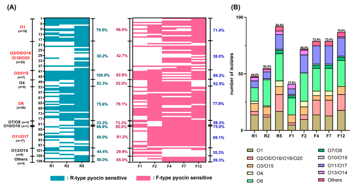
To investigate the correlation between pyocin susceptibility and P. aeruginosa O-antigen profile, we conducted systematic spot assays to evaluate the R- and F-type pyocins’ susceptibility of 109 clinical isolates. A panel of eight pyocins (R1, R2, R5, F1, F2, F4, F7, and F12) was purified from the clinical isolates, AUHPA312, AUHPA318, AUHPA325, AUHPA331, AUHPA343, AUHPA348, AUHPA32, and AUHPA326, respectively, each of which exclusively produces a single pyocin subtype (Table S3).
Among the four serotype groups with higher prevalence in this study (O1, O2/O5/O16/O18/O20, O6, and O11/O17), O2/O5/O16/O18/O20 revealed the lowest susceptibility to the eight pyocins (30.2% and 42.7% to R- and F-pyocins, respectively) (Figure 2A, Table 1). The other three serotype groups (O1, O6, and O11/O17) exhibited high overall pyocin susceptibility rates with 71.4%, 77.8% and 69.1%, respectively. Particularly, a divergence in susceptibility was observed within the O11/O17 serotype group, with relatively low sensitivity to R-type pyocins (49.0%) but higher sensitivity to F-type pyocins (81.2%) (Figure 2A, Table 1). The results align with previous findings indicating that serotypes O1 and O6 are susceptible to R1, R2, and R5 pyocins, whereas serotype O11 displays high resistance [10]. Notably, the killing capacities of F-pyocins against isolates within the four major serotype groups were found to be comparable to or even greater than those of R-pyocins (Figure 2A, Table 1), highlighting that F-pyocins represent an antimicrobial agent as potent as R-pyocins [12]. The susceptibility screening revealed that R5 pyocin possessed the highest bactericidal efficacy, with 84.4% (n = 92) of the isolates being susceptible. This was followed by F12 pyocin, which exhibited 79.8% (n = 87) of the isolates (Figure 2B).

2.4. LPS Structural Variations Drive Differential R-Type Pyocin Susceptibility

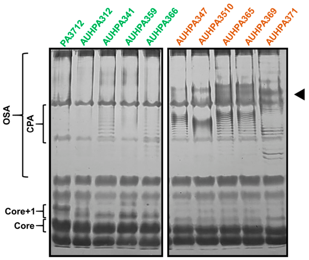
Given that LPS can also function as a protective shield against R-type pyocins [10], we sought to determine whether structural variations in LPS contribute to the reduced susceptibility observed in the O2/O5/O16/O18/O20 serotype group. To investigate this, we performed SDS-PAGE analysis to characterize the LPS profiles of representative strains. Our analysis included five clinical isolates from the O6 serotype and five isolates from the O2/O5/O16/O18/O20 serotype group for comparative morphological characterization of LPS.
Electrophoretic analysis revealed that the majority of isolates exhibited a characteristic ladder-like pattern in the upper region of the gel, representing B-band (OSA) chains with varying O-antigen repeat numbers (Figure 3). Bands corresponding to the LPS core were identified at the migration front, alongside ‘core+1’ species—representing the core substituted with a single O-repeating unit. Notably, isolates within the O2/O5/O16/O18/O20 serotype group displayed significantly denser LPS packing and an enrichment of high-molecular-weight O-antigen chains compared to the O6 group (Figure 3, black triangle). This structural profile correlates with the observed resistance of these serotypes to the eight tested pyocins (Figure 1A, Table 1). Interestingly, the O2/O5/O16/O18/O20 group exhibited reduced levels of core+1 LPS relative to the O6 serotype. This shift from core+1 toward long-chain oligomerization in the resistant group highlights a structural basis for reduced pyocin susceptibility.

2.5. Synergistic Efficacy of R- and F-Pyocin Cocktails Against MDR P. aeruginosa Clinical Isolates

In light of the increasing prevalence of antibiotic-resistant P. aeruginosa, we evaluated the therapeutic potential of R- and F-type pyocins specifically against clinical MDR isolates. Among the total 109 isolates, 50 underwent antibiotic susceptibility testing, with the highest resistance rate observed for ciprofloxacin/levofloxacin (16.0%/20.0%), followed by imipenem/meropenem (10.0%/12.0%) (Table 2 and Table S4), consistent with the global survey of antibiotic resistance [24,29]. The screening led to the identification of three MDR isolates: AUHPA312, AUHPA325, and AUHPA399 (Table 2 and Table S4). Notably, all three MDR isolates exhibited susceptibility to at least one R- or F-type pyocin (Table S3), highlighting the potential of these bacteriocins as targeted antimicrobial agents for resistant infections.
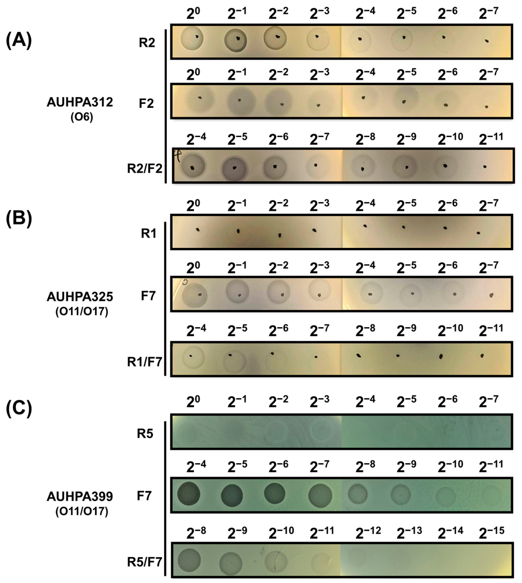
To assess the bactericidal efficacy of pyocin cocktail therapeutics against MDR P. aeruginosa, we evaluated combinations of R- and F-type pyocins against isolates AUHPA312 and AUHPA325. To maximize the likelihood of a potent effect, we restricted our cocktail components to individual pyocins that were independently active against both target strains. By systematically paring these R- and F-type variants (Figure 4), we were able to evaluate their interactions. The nature of these interactions—whether synergistic, additive, indifferent, or antagonistic—was characterized using fractional inhibitory concentration index (FICI) analysis (Table 3 and Table S5).
Isolate AUHPA312 was identified as O6 serotype and carried the R1 pyocin gene cluster. As expected, this isolate exhibited resistance to R1 pyocin (Table S3). This is consistent with the principle that producer strains are typically immune to their own pyocin type [8,10,11,12], although the underlying mechanism governing this self-immunity remains to be elucidated. Consequently, R1 pyocin was excluded from the cocktail formulations (Table 3). Specifically, the parings of R2/F1, R2/F2, or R2/F12, R5/F1, and R5/F2, demonstrated potent synergy, as evidenced by the FICI values ≤ 0.5 (Figure 4, Table 3). For isolate AUHPA325 (O11/O17 serotype, R5 pyocin carrier), R5 pyocin was excluded from synergy assays due to self-resistance (Table S3). Additionally, R2 was also omitted from testing because the strain was inherently resistant to R2 (Table S3). However, all of the tested cocktail formulations against AUHPA325 resulted only in additive interactions, with no synergistic effects detected (Figure 4, Table 3). Interestingly, although isolate AUHPA399 (O11/O17 serotype) is an R5 pyocin carrier, it showed susceptibility to R5 pyocin, which is inconsistent with the self-immunity dogma; however, this kind of self-sensitivity was observed by Mei et al., probably due to the mutations of the LPS receptor [5]. AUHPA399 was resistant to R2 and F1 (Table S3), and therefore, the two pyocins were excluded from synergy assays. The pairing of R5/F7 demonstrated a synergistic killing effect with FICI = 0.3002 (Figure 4, Table 3).
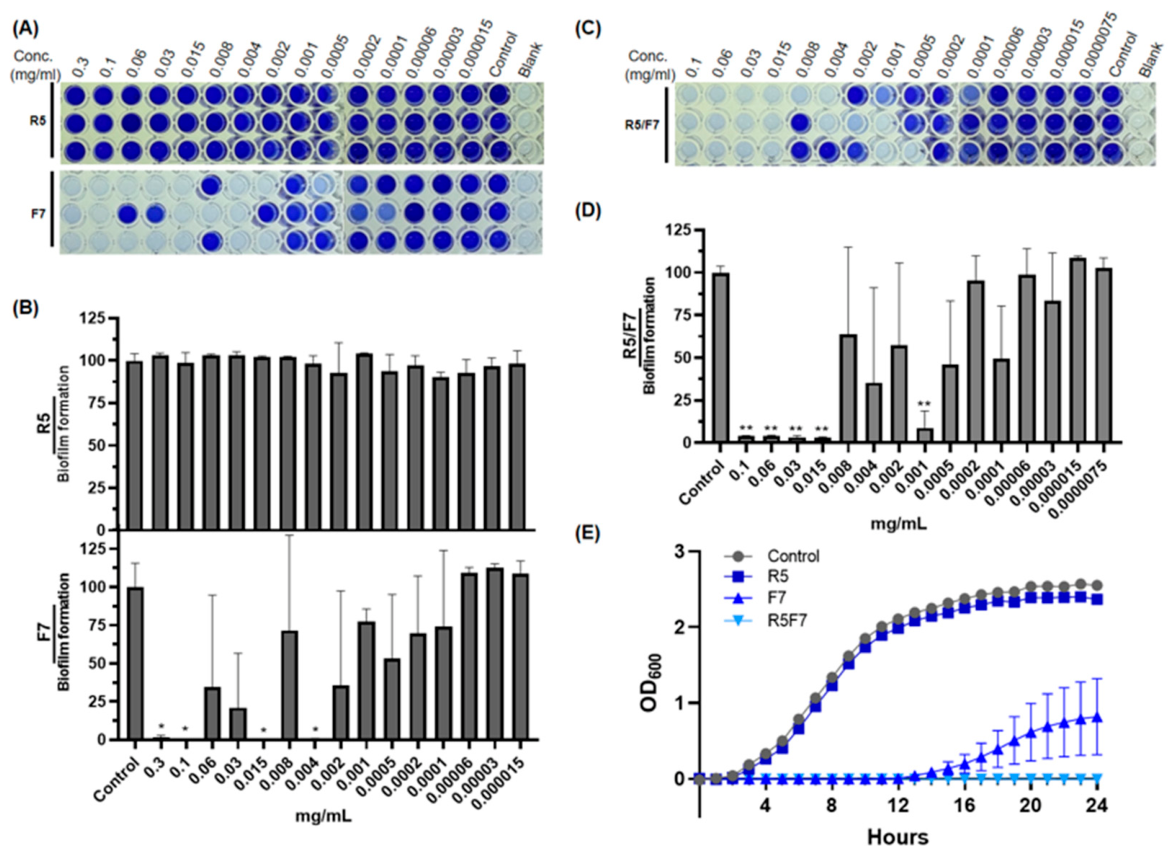
To evaluate the capability of the pyocins to inhibit biofilm formation, crystal violet assays were performed on the two multidrug-resistant (MDR) isolates, AUHPA312 and AUHPA399, following treatment with the R2/F2 and R5/F7 cocktails, respectively (Figure 5 and Figure S2). Unexpectedly, AUHPA312 exhibited no pyocin concentration-dependent reduction in biofilm formation when treated with individual R2, F2, or the combined R2/F2 cocktail (Figure S2). Conversely, AUHPA399 showed complete inhibition and a dose-dependent decrease in biofilm mass when treated with individual F7 or the R5/F7 cocktail (Figure 5A–D). Although some heterogeneity in biofilm development was noted across the three biological replicates—potentially due to pyocin self-aggregation or receptor competition—the MICs required to completely suppress biofilm development were determined to be 0.004 mg/mL for the F7 pyocin and 0.001 mg/mL for the R5/F7 cocktail (Figure 5B,D). Furthermore, growth kinetics experiments demonstrated that while R5 pyocin exhibited very mild bactericidal activity, F7 pyocin exerted a potent effect, and the combined R5/F7 cocktail completely suppressed bacterial growth throughout the 24-h observation period (Figure 5E).

3. Discussion

The O-antigen in P. aeruginosa LPS is a critical virulence factor that facilitates systemic dissemination [30,31,32]. Accordingly, characterizing serotype distribution is vital for optimizing clinical management and developing serotype-specific therapeutic interventions. Given that the most recent epidemiological data from Taiwan dates back to 1988 [33], predating the widespread adoption of the IATS classification system, in this study, we updated the regional epidemiological profile by serotyping 109 isolates from central Taiwan. Our serotyping analysis indicated that O6, O1, O2/O5/O16/O18/O20, and O11/O17 are the most predominant serotypes (Figure 1). These results largely align with the global epidemiological data reported by Pirnay et al. [34] and Nasrin et al. [29], which identified O11, O1, and O6 as the top three most prevalent serotypes via traditional antibody-based slide agglutination assays. Notably, when Nasrin et al. employed PCR to characterize isolates that were non-typable by agglutination assay [35], the O2/O5/O16/O18/O20 group emerged as the second most frequent. This underscores the value of PCR-based serotyping as a critical tool to complement traditional serotyping assays. Serotypes O2, O5, O16, O18, and O20 exhibit significant structural similarities in their O-antigen architecture [20,35,36]. As demonstrated by Raymond et al. [22], these five serotypes constitute a group due to a high degree of DNA genetic similarity. While current PCR-based serotyping lacks the resolution to differentiate individual members of this group, it remains a more accessible alternative for general laboratories. However, further investigation into the fine-scale genetic variations of the LPS biosynthesis gene cluster is required to refine PCR- or sequencing-platforms for higher-resolution serotyping.
We identified the O2/O5/O16/O18/O20 serogroup as the least susceptible to pyocin-mediated killing. This resistance is consistent with the model where long-chain O-antigens in strains like PAO1 (serotype O5) shield the LPS core receptors [10], a phenomenon previously visualized via electron microscopy and silver-stained SDS-PAGE as high-density LPS packing [37]. For the first time, we provide a systematic comparison of F- and R-pyocin spectra, demonstrating that both types exhibit a shared sensitivity profile (O1, O6, and O11) and resistance profile (O2/O5/O16/O18/O20). While these susceptibility patterns align with the findings on F-pyocins reported by Saha et al. [12], the precise molecular receptors for F-pyocins have yet to be determined.
The strategies employed by R- and F-type pyocins to prevent self-killing differ fundamentally from those of S-type pyocins [8]. S-type pyocins function analogously to colicins, killing target cells by degrading DNA (pyocins AP41, S1, S2, and S3), hydrolyzing tRNA (pyocin S4), or permeabilizing the cytoplasmic membrane (pyocin S5). To ensure self-protection, a short immunity gene is co-expressed alongside the cognate S-type pyocin; this immunity protein binds directly to the cytotoxic domain and neutralizes its bactericidal activity. Conversely, R- and F-type pyocins lack dedicated immunity proteins, and the precise mechanisms by which producing strains evade their own tail-like bacteriocins remain a subject of debate. Systematic mutational analyses involving the deletion of the O-specific antigen (OSA), common antigen (CPA), or core oligosaccharide have demonstrated that OSA and CPA polysaccharide chains sterically shield the cell from R-type pyocins, whereas the outer core oligosaccharide serves as the primary receptor [10]. In contrast, the specific receptor configuration required for F-type pyocin recognition remains less conclusive [12].
Standard empirical therapies, though critical for improving survival rates in severe P. aeruginosa infections, are increasingly undermined by the bacterium’s extensive resistance profile. This clinical crisis has led the World Health Organization to list carbapenem-resistant P. aeruginosa (CRPA) among the three species requiring the most urgent antibiotic development [38]. Recent surveillance data from Taiwan indicates a significant upward trend in the prevalence of CRPA (10.2%, 349 out of 3408 isolates) [39]. A primary molecular driver for this resistance is the carriage of carbapenemase and efflux pump-encoding genes [3]. Phage tail-like pyocins can bypass the microbial defense mechanisms by leveraging O-antigen specificity for precision targeting [40].
To the best of our knowledge, this work provides the first documented evidence of the bactericidal efficacy of combined R- and F-type pyocin formulations. In the present study, the three MDR isolates, AUHPA312 (serotype O6), AUHPA325 (serotype O11/O17), and AUHPA399 (serotype O11/O17), displayed susceptibility to at least one R- and F-pyocin, underscoring their therapeutic potential against P. aeruginosa infections. Furthermore, R- and F-pyocin cocktails yielded synergistic bactericidal activity against AUHPA312 but remained additive against AUHPA325. This discrepancy may be attributed to the susceptibility profile of AUHPA325, which showed resistance to both R2 and R5 pyocins, and weak susceptibility to R1 pyocin. Given that this isolate was strongly susceptible to F-pyocins (Table S3), the dominant killing activity of F-pyocins likely obscured the detection of synergy between the two pyocin classes. The high susceptibility of the O11/O17 group, including isolate AUHPA325, to F-pyocins (Figure 2A, Table 1) correlates with their genomic architecture. As illustrated in Figure 1B, these strains possess only R-type pyocin gene clusters. Because bacteria often evolve immunity primarily against the specific class of pyocins they produce themselves, the exclusive presence of R-pyocin genes in the O11/O17 group provides a plausible explanation for their vulnerability to exogenous F-pyocins (Figure 1B).
Although the R2/F2 pyocin cocktail produced discernible zones of inhibition on a lawn of AUHPA312 (Figure 4), substantial biofilm development was still observed in TSB medium (Supplementary Figure S2). Notably, the inhibition zones for AUHPA312 were less distinct than those for AUHPA399, suggesting incomplete bactericidal activity. We hypothesize that partially lysed cells enhance the release of extracellular DNA (eDNA), which then serves as a structural scaffold that facilitates the adherence and survival of remaining cells during biofilm development. A similar phenomenon has been reported under sub-lethal concentrations of conventional antibiotics [41]. To our knowledge, this is the first study to document such biofilm-promoting concerns regarding R- and F-type pyocins. Consequently, the risk of induced biofilm formation must be carefully evaluated alongside standard bactericidal assays when characterizing these tail-like bacteriocins for therapeutic development.

4. Materials and Methods

4.1. P. aeruginosa Strains

In this study, 101 clinical isolates of P. aeruginosa were obtained from Asia University Hospital, Taichung, Taiwan, and eight isolates were collected from Feng Yuan Hospital, Taichung, Taiwan [42,43]. A total of 109 isolates were independently collected from sterile sites. All isolates were preserved in Tryptic Soy Broth (TSB) containing 25% (v/v) glycerol and stored at −80 °C. According to the Clinical and Laboratory Standards Institute guidelines, isolates exhibiting resistance to antibiotics from three or more different classes with distinct mechanisms of action are defined as MDR.

4.2. O-Antigen Serotype Determination of P. aeruginosa

The O-antigen serotyping of P. aeruginosa was conducted using PCR followed by DNA sequencing as described by Raymond et al. [22] (Figure S1, Table S1). Genomic DNA from the isolates was extracted using the EasyPrep Genomic DNA Extraction Kit (Biotools, New Taipei City, Taiwan) following the manufacturer’s instructions. The primer sequences used for O-antigen serotyping are summarized in Table S1.

4.3. Identification of R- and F-Type Pyocin Subtypes

Identification and subtyping of R- and F-type pyocins were performed using PCR followed by sequencing where necessary. The primer pairs used for pyocin identification and subtyping are summarized in Table S2. The presence of R- and F-type pyocins in the isolates was confirmed by PCR amplification of the tube genes of R-type (PA0623) and F-type pyocins (PA0633). To determine the subtypes of R-type pyocin in the isolates, the tail fiber gene (PA0620) was specifically amplified using primer pairs targeting the R1, R2 group (comprising R2, R3, R4), and R5. For F-type pyocin subtyping, the DNA fragments from the gene encoding the central tail fiber (PA0641) to trpG (PA0649) were amplified, followed by Sanger sequencing. The sequenced data were mapped against the amino acid sequences of PyoF10 of various F-pyocins as reported by Saha et al. [12].

4.4. Purification of R- and F-Type Pyocin

Purification of R- and F-type pyocins was performed with modifications to the methods described by Ge et al. [44]. For pyocin susceptibility testing, strains AUHPA312, AUHPA318, AUHPA325, AUHPA331, AUHPA343, AUHPA348, AUHPA32, and AUHPA326 were used as the producers for R1, R2, R5, F1, F2, F4, F7, and F12 pyocins, respectively. A single colony of each pyocin-producing strain was inoculated into 20 mL of TSB and incubated in a shaker incubator overnight at 37 °C, 220 rpm. The culture was then transferred to 400 mL of fresh TSB and incubated for two hours at 37 °C. To induce the production of pyocins, mitomycin C (3 µg/mL) was then added to trigger the SOS response of P. aeruginosa, followed by incubation for another two and a half hours at 30 °C. DNase I was added to the final concentration of 3 μg/mL, with incubation for another 30 min. The culture was centrifuged, and the supernatant was filtered through a 0.22 µm filter. Ammonium sulfate was added to a final concentration of 50% (w/v) to precipitate the pyocins. After one hour at 4 °C, the precipitated protein was collected by centrifugation at 10,000× g for 15 min, resuspended in SM buffer (50 mM Tris-HCl, pH 7.5, 100 mM NaCl, and 8 mM MgCl2), and dialyzed against SM buffer overnight. The sample was then centrifuged at 150,000× g for 1 h at 4 °C. Pyocins were fractionized in the pellets and resuspended in 2 mL of SM buffer. The protein concentration was measured using the Pierce™ BCA Protein Assay Kit (Thermo Scientific, Waltham, MA, USA). The purified R- or F-type pyocins were stored at −80 °C.

4.5. Susceptibility Assay of P. aeruginosa to R- and F-Pyocin

A single colony of each test isolate was inoculated into TSB and incubated overnight at 37 °C, 220 rpm. An aliquot of 100 µL from the overnight culture was added to TSB soft agar (0.75% agar), thoroughly mixed, and poured into a Petri dish to solidify. Then, 10 µL of R- and F-type pyocins, each at a concentration of 0.5 mg/mL, was spotted onto the solidified soft agar surface. The plates were incubated overnight at 37 °C. To evaluate the efficacy of the pyocin cocktail combination against MDR strains, the bactericidal activity of individual pyocins was first assessed using twofold serial dilutions. Next, individual R- and F-type pyocins of different subtypes were mixed at a ratio of 1:1 (v/v), and the bactericidal activity of the combination was evaluated using the same method. The synergy of the cocktail combinations was assessed using the fractional inhibition concentration index (FICI). A clear zone of inhibition on the plate was considered indicative of sensitivity, while a faint zone indicated weak sensitivity. The formation of plaques was interpreted as phage contamination.

4.6. LPS Extraction and Visualization

The extraction and visualization of LPS were performed as described by Kulikov et al. [45] with minor modifications. In brief, one milliliter of overnight TSB culture of selected P. aeruginosa strains was pelleted by centrifugation at 10,000× g for 30 s using a benchtop microcentrifuge, washed three times with an identical volume of PBS and subsequently resuspended in 50 μL of lysis buffer (2% w/v of SDS, 4% v/v of 2-mercaptoethanol, 10% v/v glycerol, 1 M Tris-HCl (pH 6.8), and 0.01% bromophenol blue). After incubation at 95 °C for 10 min followed by cooling to room temperature, the lysate was treated with the final concentration of 2.5 mg/mL protease K in the lysate. The mixture was then incubated for 1 h in a thermomixer. Two microliters of the sample was loaded onto a 12% SDS-PAGE gel (29:1 acrylamide:bis-acrylamide) for electrophoresis. After electrophoresis, the gel was incubated for 15 min in fixer-oxidizer solution, followed by three washes with distilled water (7 min each). The gel was stained using the ProteoSilver™ Silver Stain Kit (Sigma, St. Louis, MO, USA).

4.7. Kinetic Assay

AUHPA399 was cultured overnight in TSB broth at 37 °C with shaking at 220 rpm. The overnight culture was diluted with fresh TSB and mixed with R5, F7, or the R5F7 combination in a 96-well microtiter plate, resulting in a final bacterial density of OD600 = 0.01 and a final pyocin concentration of 0.004 mg/mL. Bacterial growth was monitored using a microplate reader (BMG LABTECH SPECTROstar Nano, Ortenberg, Germany) at 37 °C with orbital shaking at 200 rpm for 24 h, and OD600 values were recorded every hour.

4.8. Microtiter Biofilm Inhibition Assay

The bacterial strain was cultured overnight (O/N) in TSB broth at 37 °C with shaking at 220 rpm. The overnight culture was diluted with fresh TSB and mixed with different concentrations of individual R-type and F-type pyocins or their cocktail in a 96-well microtiter plate, resulting in a final bacterial density of OD600 = 0.01. The plate was incubated statically at 37 °C for 24 h. After incubation, planktonic cells and culture medium were removed, and the wells were gently washed twice with phosphate-buffered saline (PBS) to remove non-adherent cells. The attached biofilms were stained with 0.1% crystal violet (CV) for 15 min, followed by two PBS washes to remove excess stain. The bound CV was then solubilized with 30% acetic acid for 15 min at room temperature, and the absorbance was measured at 570 nm (OD570) using a microplate reader to evaluate the inhibitory effects of individual R-type and F-type pyocins and their cocktail on biofilm formation.

5. Conclusions

In summary, this study provides an updated serotype distribution of P. aeruginosa serotypes in central Taiwan and identified F-type pyocins as potent antimicrobial candidates against clinical isolates, potentially surpassing R-type pyocins in efficacy. Our analysis characterized the distinct associations between serotype profiles and pyocin susceptibility, demonstrating that variations in LPS are primary drivers of the observed resistance patterns. While the precise mechanisms of receptor recognition and self-immunity require further investigation, these findings underscore the therapeutic potential of pyocin cocktails as a robust strategy for combating MDR P. aeruginosa.

Supplementary Materials

The following supporting information can be downloaded at: https://www.mdpi.com/article/10.3390/antibiotics15060596/s1, Figure S1: A schematic diagram of the 11 groups of gene clusters involved in OSA biosynthesis [46]; Figure S2: Biofilm development of AUHPA312 after pyocin treatments; Table S1: Primers used for serotyping; Table S2: Primers used for R- and F-type pyocin subtyping; Table S3: Serotype, R-, and F-type pyocin production and susceptibility of 109 isolates; Table S4: Antimicrobial susceptibility testing of 50 isolates; Table S5: Minimum inhibitory concentrations (MICs) of pyocin subtypes against AUHPA312 and AUHPA325.

Author Contributions

Conceptualization, N.-J.H. and C.-W.H.; bioinformatic analyses, W.-T.X., Z.-L.J., Y.-T.C. and T.-K.E.; investigation, Y.-L.S., W.-T.X., Z.-L.J., N.-J.H. and C.-W.H.; writing—original draft, Y.-L.S., W.-T.X. and Z.-L.J.; writing—review & editing, N.-J.H. and C.-W.H.; funding acquisition, C.-W.H.; supervision, N.-J.H. and C.-W.H. All authors have read and agreed to the published version of the manuscript.

Funding

This work was supported by grants from the National Science and Technology Council (NSTC), Taiwan (113-2314-B-468-007) and Asia University Hospital (no. 11451003). The funding agency had no role in the study design, data collection, analysis, interpretation, manuscript preparation, or decision to publish.

Institutional Review Board Statement

This study was conducted in accordance with the Declaration of Helsinki, and by the Institutional Ethics Review Board of Feng-Yuan Hospital (Approval Number: 113033).

Informed Consent Statement

Patient consent was waived by the Institutional Ethics Review Board of Feng-Yuan Hospital due to the retrospective nature of the study and the use of fully anonymized clinical bacterial isolates, with no access to identifiable personal data.

Data Availability Statement

The data supporting the findings of this study are available from the corresponding author, C.-W. Huang, upon reasonable request.

Acknowledgments

We sincerely thank Asia University Hospital and Feng-Yuan Hospital for providing the P. aeruginosa isolates. We also acknowledge the use of ChatGPT 5.0 to improve readability and language. After using this tool/service, the authors reviewed and edited the content as needed and take full responsibility for the content of the publication.

Conflicts of Interest

The authors declare no conflicts of interest.

Abbreviations

The following abbreviations are used in this manuscript:
CPACommon polysaccharide antigen
FICIFractional inhibition concentration index
OSAO-specific antigen
LPSLipopolysaccharide
MDRMultidrug resistant

References

  1. Reynolds, D.; Kollef, M. The Epidemiology and Pathogenesis and Treatment of Pseudomonas aeruginosa Infections: An Update. Drugs 2021, 81, 2117–2131. [Google Scholar] [CrossRef] [Scilit] [PubMed]
  2. Wu, C.-L.; Ku, S.-C.; Yang, K.-Y.; Fang, W.-F.; Tu, C.-Y.; Chen, C.-W.; Hsu, K.H.; Fan, W.C.; Lin, M.C.; Chen, W.; et al. Antimicrobial drug-resistant microbes associated with hospitalized community-acquired and healthcare-associated pneumonia: A multi-center study in Taiwan. J. Formos. Med. Assoc. 2013, 112, 31–40. [Google Scholar] [CrossRef] [Scilit]
  3. Pang, Z.; Raudonis, R.; Glick, B.R.; Lin, T.J.; Cheng, Z. Antibiotic resistance in Pseudomonas aeruginosa: Mechanisms and alternative therapeutic strategies. Biotechnol. Adv. 2019, 37, 177–192. [Google Scholar] [CrossRef] [Scilit]
  4. Alqahtani, A.; Kopel, J.; Hamood, A. The In Vivo and In Vitro Assessment of Pyocins in Treating Pseudomonas aeruginosa Infections. Antibiotics 2022, 11, 1366. [Google Scholar] [CrossRef] [Scilit] [PubMed]
  5. Mei, M.; Estrada, I.; Diggle, S.P.; Goldberg, J.B. R-pyocins as targeted antimicrobials against Pseudomonas aeruginosa. npj Antimicrob. Resist. 2025, 3, 17. [Google Scholar] [CrossRef] [Scilit]
  6. Estrada, I.; Smith, P.; Mei, M.; Goldberg, J.B.; Diggle, S.P. Microbial Primer: The R-pyocins of Pseudomonas aeruginosa. Microbiology 2025, 171, 001640. [Google Scholar] [CrossRef] [Scilit]
  7. Six, A.; Mosbahi, K.; Barge, M.; Kleanthous, C.; Evans, T.; Walker, D. Pyocin efficacy in a murine model of Pseudomonas aeruginosa sepsis. J. Antimicrob. Chemother. 2021, 76, 2317–2324. [Google Scholar] [CrossRef] [Scilit] [PubMed]
  8. Michel-Briand, Y.; Baysse, C. The pyocins of Pseudomonas aeruginosa. Biochimie 2002, 84, 499–510. [Google Scholar] [CrossRef] [Scilit]
  9. Scholl, D. Phage Tail-Like Bacteriocins. Annu. Rev. Virol. 2017, 4, 453–467. [Google Scholar] [CrossRef] [Scilit]
  10. Köhler, T.; Donner, V.; van Delden, C. Lipopolysaccharide as shield and receptor for R-pyocin-mediated killing in Pseudomonas aeruginosa. J. Bacteriol. 2010, 192, 1921–1928. [Google Scholar] [CrossRef] [Scilit]
  11. Ghequire, M.G.; De Mot, R. Ribosomally encoded antibacterial proteins and peptides from Pseudomonas. FEMS Microbiol. Rev. 2014, 38, 523–568. [Google Scholar] [CrossRef] [Scilit]
  12. Saha, S.; Ojobor, C.D.; Li, A.S.C.; Mackinnon, E.; North, O.I.; Bondy-Denomy, J.; Lam, J.S.; Ensminger, A.W.; Maxwell, K.L.; Davidson, A.R. F-Type Pyocins Are Diverse Noncontractile Phage Tail-Like Weapons for Killing Pseudomonas aeruginosa. J. Bacteriol. 2023, 205, e00029-23. [Google Scholar] [CrossRef] [Scilit] [PubMed]
  13. Duport, C.; Baysse, C.; Michel-Briand, Y. Molecular characterization of pyocin S3, a novel S-type pyocin from Pseudomonas aeruginosa. J. Biol. Chem. 1995, 270, 8920–8927. [Google Scholar] [CrossRef] [Scilit]
  14. Sano, Y.; Matsui, H.; Kobayashi, M.; Kageyama, M. Molecular structures and functions of pyocins S1 and S2 in Pseudomonas aeruginosa. J. Bacteriol. 1993, 175, 2907–2916. [Google Scholar] [CrossRef] [Scilit]
  15. Elfarash, A.; Wei, Q.; Cornelis, P. The soluble pyocins S2 and S4 from Pseudomonas aeruginosa bind to the same FpvAI receptor. MicrobiologyOpen 2012, 1, 268–275. [Google Scholar] [CrossRef] [Scilit] [PubMed]
  16. Behrens, H.M.; Lowe, E.D.; Gault, J.; Housden, N.G.; Kaminska, R.; Weber, T.M.; Thompson, C.M.; Mislin, G.L.; Schalk, I.J.; Walker, D.; et al. Pyocin S5 Import into Pseudomonas aeruginosa Reveals a Generic Mode of Bacteriocin Transport. mBio 2020, 11, e03230-19. [Google Scholar] [CrossRef] [Scilit]
  17. Ge, P.; Scholl, D.; Prokhorov, N.S.; Avaylon, J.; Shneider, M.M.; Browning, C.; Buth, S.A.; Plattner, M.; Chakraborty, U.; Ding, K.; et al. Action of a minimal contractile bactericidal nanomachine. Nature 2020, 580, 658–662. [Google Scholar] [CrossRef] [Scilit]
  18. King, J.D.; Kocíncová, D.; Westman, E.L.; Lam, J.S. Review: Lipopolysaccharide biosynthesis in Pseudomonas aeruginosa. Innate Immun. 2009, 15, 261–312. [Google Scholar] [CrossRef] [Scilit]
  19. Huszczynski, S.M.; Lam, J.S.; Khursigara, C.M. The Role of Pseudomonas aeruginosa Lipopolysaccharide in Bacterial Pathogenesis and Physiology. Pathogens 2019, 9, 6. [Google Scholar] [CrossRef] [Scilit] [PubMed]
  20. Lam, J.S.; Taylor, V.L.; Islam, S.T.; Hao, Y.; Kocincova, D. Genetic and Functional Diversity of Pseudomonas aeruginosa Lipopolysaccharide. Front. Microbiol. 2011, 2, 118. [Google Scholar] [CrossRef] [Scilit]
  21. Redero, M.; López-Causapé, C.; Aznar, J.; Oliver, A.; Blázquez, J.; Prieto, A.I. Susceptibility to R-pyocins of Pseudomonas aeruginosa clinical isolates from cystic fibrosis patients. J. Antimicrob. Chemother. 2018, 73, 2770–2776. [Google Scholar] [CrossRef] [Scilit]
  22. Raymond, C.K.; Sims, E.H.; Kas, A.; Spencer, D.H.; Kutyavin, T.V.; Ivey, R.G.; Zhou, Y.; Kaul, R.; Clendenning, J.B.; Olson, M.V. Genetic variation at the O-antigen biosynthetic locus in Pseudomonas aeruginosa. J. Bacteriol. 2002, 184, 3614–3622. [Google Scholar] [CrossRef] [Scilit]
  23. Thrane, S.W.; Taylor, V.L.; Freschi, L.; Kukavica-Ibrulj, I.; Boyle, B.; Laroche, J.; Pirnay, J.P.; Lévesque, R.C.; Lam, J.S.; Jelsbak, L. The Widespread Multidrug-Resistant Serotype O12 Pseudomonas aeruginosa Clone Emerged through Concomitant Horizontal Transfer of Serotype Antigen and Antibiotic Resistance Gene Clusters. mBio 2015, 6, e01396-15. [Google Scholar] [CrossRef] [Scilit]
  24. Jamasbi, R.J.; Proudfoot, E.M. Phenotypic and Genotypic Characteristics of Clinical Isolates of Pseudomonas aeruginosa: Rate of Occurrence and Distribution of Different Serotypes, Antimicrobial Susceptibility Profiles, and Molecular Typing. Lab. Med. 2008, 39, 155–161. [Google Scholar] [CrossRef] [Scilit]
  25. Lu, Q.; Eggimann, P.; Luyt, C.E.; Wolff, M.; Tamm, M.; François, B.; Mercier, E.; Garbino, J.; Laterre, P.F.; Koch, H.; et al. Pseudomonas aeruginosa serotypes in nosocomial pneumonia: Prevalence and clinical outcomes. Crit. Care 2014, 18, R17. [Google Scholar] [CrossRef] [Scilit]
  26. Buth, S.A.; Shneider, M.M.; Scholl, D.; Leiman, P.G. Structure and Analysis of R1 and R2 Pyocin Receptor-Binding Fibers. Viruses 2018, 10, 427. [Google Scholar] [CrossRef] [Scilit]
  27. Scholl, D.; Martin, D.W., Jr. Antibacterial efficacy of R-type pyocins towards Pseudomonas aeruginosa in a murine peritonitis model. Antimicrob. Agents Chemother. 2008, 52, 1647–1652. [Google Scholar] [CrossRef] [Scilit]
  28. Mei, M.; Thomas, J.; Diggle, S.P. Heterogenous Susceptibility to R-Pyocins in Populations of Pseudomonas aeruginosa Sourced from Cystic Fibrosis Lungs. mBio 2021, 12, e00458-21. [Google Scholar] [CrossRef] [Scilit] [PubMed]
  29. Nasrin, S.; Hegerle, N.; Sen, S.; Nkeze, J.; Sen, S.; Permala-Booth, J.; Choi, M.; Sinclair, J.; Tapia, M.D.; Johnson, J.K.; et al. Distribution of serotypes and antibiotic resistance of invasive Pseudomonas aeruginosa in a multi-country collection. BMC Microbiol. 2022, 22, 13. [Google Scholar] [CrossRef] [Scilit] [PubMed]
  30. Pier, G.B. Pseudomonas aeruginosa lipopolysaccharide: A major virulence factor, initiator of inflammation and target for effective immunity. Int. J. Med. Microbiol. 2007, 297, 277–295. [Google Scholar] [CrossRef] [Scilit]
  31. Hancock, R.E.; Mutharia, L.M.; Chan, L.; Darveau, R.P.; Speert, D.P.; Pier, G.B. Pseudomonas aeruginosa isolates from patients with cystic fibrosis: A class of serum-sensitive, nontypable strains deficient in lipopolysaccharide O side chains. Infect. Immun. 1983, 42, 170–177. [Google Scholar] [CrossRef] [Scilit]
  32. Pier, G.B.; Ames, P. Mediation of the killing of rough, mucoid isolates of Pseudomonas aeruginosa from patients with cystic fibrosis by the alternative pathway of complement. J. Infect. Dis. 1984, 150, 223–228. [Google Scholar] [CrossRef] [Scilit]
  33. Ding, M.J.; Wu, T.M. Serogrouping, plasmid and drug resistance of Pseudomonas aeruginosa in Taiwan. Zhonghua Minguo Wei Sheng Wu Ji Mian Yi Xue Za Zhi Chin. J. Microbiol. Immunol. 1988, 21, 165–178. [Google Scholar]
  34. Pirnay, J.P.; Bilocq, F.; Pot, B.; Cornelis, P.; Zizi, M.; Van Eldere, J.; Deschaght, P.; Vaneechoutte, M.; Jennes, S.; Pitt, T.; et al. Pseudomonas aeruginosa population structure revisited. PLoS ONE 2009, 4, e7740. [Google Scholar] [CrossRef] [Scilit] [PubMed]
  35. Newton, G.J.; Daniels, C.; Burrows, L.L.; Kropinski, A.M.; Clarke, A.J.; Lam, J.S. Three-component-mediated serotype conversion in Pseudomonas aeruginosa by bacteriophage D3. Mol. Microbiol. 2001, 39, 1237–1247. [Google Scholar] [CrossRef]
  36. Burrows, L.L.; Charter, D.F.; Lam, J.S. Molecular characterization of the Pseudomonas aeruginosa serotype O5 (PAO1) B-band lipopolysaccharide gene cluster. Mol. Microbiol. 1996, 22, 481–495. [Google Scholar] [CrossRef] [Scilit] [PubMed]
  37. Lam, J.S.; Graham, L.L.; Lightfoot, J.; Dasgupta, T.; Beveridge, T.J. Ultrastructural examination of the lipopolysaccharides of Pseudomonas aeruginosa strains and their isogenic rough mutants by freeze-substitution. J. Bacteriol. 1992, 174, 7159–7167. [Google Scholar] [CrossRef] [Scilit] [PubMed]
  38. World Health Organization. WHO Bacterial Priority Pathogens List, 2024: Bacterial Pathogens of Public Health Importance to Guide Research, Development and Strategies to Prevent and Control Antimicrobial Resistance; World Health Organization: Geneva, Switzerland, 2024. [Google Scholar]
  39. Lin, K.-Y.; Lauderdale, T.-L.; Wang, J.-T.; Chang, S.-C. Carbapenem-resistant Pseudomonas aeruginosa in Taiwan: Prevalence, risk factors, and impact on outcome of infections. J. Microbiol. Immunol. Infect. 2016, 49, 52–59. [Google Scholar] [CrossRef] [Scilit]
  40. Bhattacharjee, R.; Nandi, A.; Sinha, A.; Kumar, H.; Mitra, D.; Mojumdar, A.; Patel, P.; Jha, E.; Mishra, S.; Rout, P.K.; et al. Phage-tail-like bacteriocins as a biomedical platform to counter anti-microbial resistant pathogens. Biomed. Pharmacother. 2022, 155, 113720. [Google Scholar] [CrossRef] [Scilit]
  41. Penesyan, A.; Paulsen, I.T.; Gillings, M.R.; Kjelleberg, S.; Manefield, M.J. Secondary Effects of Antibiotics on Microbial Biofilms. Front. Microbiol. 2020, 11, 2109. [Google Scholar] [CrossRef] [Scilit]
  42. Huang, C.-W.; Lin, Y.-S.; Huang, W.-C.; Lai, C.-C.; Chien, H.-J.; Hu, N.-J.; Chen, J.H. Inhibition of the clinical isolates of Acinetobacter baumannii by Pseudomonas aeruginosa: In vitro assessment of a case-based study. J. Microbiol. Immunol. Infect. 2022, 55, 60–68. [Google Scholar] [CrossRef] [Scilit]
  43. Zheng, Y.F.; Lin, Y.S.; Huang, J.W.; Tang, K.T.; Kuo, C.Y.; Wang, W.C.; Chien, H.J.; Chang, C.J.; Hu, N.J.; Lai, C.C. SWATH-MS Based Secretome Proteomic Analysis of Pseudomonas aeruginosa Against MRSA. Proteomics 2024, 25, e202300649. [Google Scholar] [CrossRef] [Scilit] [PubMed]
  44. Ge, P.; Scholl, D.; Leiman, P.G.; Yu, X.; Miller, J.F.; Zhou, Z.H. Atomic structures of a bactericidal contractile nanotube in its pre- and postcontraction states. Nat. Struct. Mol. Biol. 2015, 22, 377–382. [Google Scholar] [CrossRef] [Scilit]
  45. Kulikov, E.E.; Golomidova, A.K.; Prokhorov, N.S.; Ivanov, P.A.; Letarov, A.V. High-throughput LPS profiling as a tool for revealing of bacteriophage infection strategies. Sci. Rep. 2019, 9, 2958. [Google Scholar] [CrossRef] [Scilit] [PubMed]
  46. van den Belt, M.; Gilchrist, C.; Booth, T.J.; Chooi, Y.H.; Medema, M.H.; Alanjary, M. CAGECAT: The CompArative GEne Cluster Analysis Toolbox for rapid search and visualisation of homologous gene clusters. BMC Bioinform. 2023, 24, 181. [Google Scholar] [CrossRef] [Scilit] [PubMed]
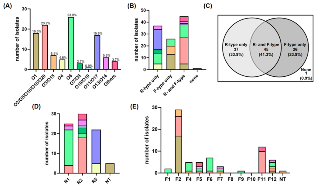
Figure 1. Distribution of O-antigen serotypes and pyocin subtypes among the clinical P. aeruginosa isolates. (A) Distribution of the 11 identified O-serotype groups, showing the absolute counts with prevalence rate determined via PCR-based serotyping. Pyocin production profiles across the isolate collection are presented by (B) a stacked histogram, and (C) a Venn diagram. The total number of isolates producing various (D) R-type and (E) F-type subtypes are shown. Each color in the stacked histogram corresponds to a specific O-antigen serotype as defined in (A). NT, non-typable.
Antibiotics 15 00596 g001
Figure 2. Bactericidal host range of pyocin subtypes against P. aeruginosa O-antigen serotypes. (A) Susceptibility profiles of 109 isolates to R-type (teal) and F-type (pink) pyocins, categorized by the eleven O-antigen serotype groups. The four predominant serotype groups are labeled red. The specific susceptibility rates to R- and F-pyocins are indicated in teal and pink, respectively, while combined susceptibility rates to both R- and F-type pyocins are highlighted in blue. (B) Prevalence of pyocin sensitivity across the collection. Histograms represent the total number of isolates, categorized as various serotypes, that are susceptible to each specific R- and F-type pyocin subtype. The color code is consistent with the scheme as defined in Figure 1.
Antibiotics 15 00596 g002
Figure 3. Comparative analysis of LPS profiles. Silver-stained SDS-PAGE of LPS extracted from clinical isolates belonging to the O2/O5/O16/O18/O20 serotype group (labeled in brown) and O6 serotype (labeled in green). The black triangle highlights the enrichment of high-molecular-weight OSA in the O2/O5/O16/O18/O20 serotype group. Brackets delineate the migration ranges of CPA and OSA. ‘Core’ denotes uncapped LPS, while ‘Core+1’ indicates the LPS core substituted with a single O-antigen repeating unit.
Antibiotics 15 00596 g003
Figure 4. Synergistic bactericidal activity of pyocin cocktails against MDR P. aeruginosa isolate. Comparative bactericidal activities of individual R- and F-type pyocins versus their combined cocktail pairs against (A) AUHPA312, (B) AUHPA325, and (C) AUHPA399. Assays were performed using individual pyocins or cocktail pairs followed by twofold serial dilution as indicated.
Antibiotics 15 00596 g004
Figure 5. Biofilm development and planktonic growth of AUHPA399 after pyocin treatments. (A,C) Crystal violet assays showing biofilm formation of AUHPA399 after treatments of (A) individual pyocins, R5 and F7, and (C) R5/F7 cocktail. The highest concentrations of added pyocin are indicated, followed by 2-fold serial dilution. Control: no pyocin added. (B,D) Normalized level of biofilm formation of AUHPA399 in 2-fold serially diluted (B) individual pyocins R5 and F7, and (D) R5/F7 cocktail. (E) Bacterial growth kinetics of AUHPA399 after pyocin treatments. All the OD570 values in each well are normalized against the control as 100%. The statistical significance between the OD570 values for the control and pyocin-treated cells was analyzed by one-way ANOVA using GraphPad Prism 9. Data represent the mean ± SD with n = 3. * p < 0.05, ** p < 0.01.
Antibiotics 15 00596 g005
Table 1. Susceptibility of 109 P. aeruginosa isolates, categorized as O-antigen serotype, to various pyocin subtypes.
GroupNumber of
Isolates
R1R2R5F1F2F4F7F12
O11814
(77.7%)
12
(66.6%)
17
(94.4%)
5
(27.7%)
10
(55.5%)
14
(77.7%)
13
(72.2%)
18
(100%)
O2/O5/O16/O18/O20223
(13.6%)
2
(9.1%)
15
(68.1%)
2
(9.1%)
4
(18.1%)
13
(59.1%)
14
(63.6%)
14
(63.6%)
O3/O1577
(100%)
7
(100%)
7
(100%)
7
(100%)
7
(100%)
4
(57.1%)
4
(57.1%)
7
(100%)
O452
(40%)
1
(20%)
5
(100%)
0
(0%)
5
(100%)
4
(80%)
4
(80%)
0
(0%)
O62612
(46.1%)
23
(88.4%)
24
(92.3%)
15
(57.6%)
25
(96.1%)
20
(76.9%)
20
(76.9%)
23
(88.4%)
O7/O830
(0%)
0
(0%)
3
(100%)
2
(66.6%)
0
(0%)
3
(100%)
3
(100%)
3
(100%)
O10/O1910
(0%)
1
(100%)
1
(100%)
1
(100%)
0
(0%)
1
(100%)
1
(100%)
1
(100%)
O11/O17176
(35.3%)
6
(35.3%)
13
(76.5%)
5
(29.4%)
16
(94.1%)
16
(94.1%)
16
(94.1%)
16
(94.1%)
O13/O1463
(50%)
2
(33.3%)
3
(50%)
4
(66.6%)
4
(66.6%)
0
(0%)
0
(0%)
1
(16.6%)
Others41
(25%)
1
(25%)
4
(100%)
0
(0%)
1
(25%)
4
(100%)
4
(100%)
4
(100%)
Table 2. Antibiotic resistance profiles of 50 P. aeruginosa isolates. The sensitivity (S) and resistance (R) status for the MDR isolates AUHPA312, AUHPA325, and AUHPA399 are indicated for comparison.
AntibioticsResistant/Total StrainsAUHPA312AUHPA325AUHPA399
Ciprofloxacin8/50 (16.0%)RRS
Levofloxacin10/50 (20.0%)RRR
Ceftazidime3/50 (6.0%)RSS
Cefepime3/50 (6.0%)RRS
Imipenem5/50 (10.0%)RSR
Meropenem6/50 (12.0%)RRR
Piperacillin/Tazobactam3/50 (6.0%)RSS
Table 3. A Synergy analysis of R- and F-pyocin cocktails against MDR P. aeruginosa isolates AUHPA312, AUHPA325, and AUHPA399 based on FICI.
AUHPA312 (O6)AUHPA325 (O11/O17)AUHPA399 (O11/O17)
Cocktail
Formula
FICIInterpretationCocktail
Formula
FICIInterpretationCocktail
Formula
FICIInterpretation
R2/F10.2125SR1/F13.33IndR1/F20.5003Add
R2/F20.09375SR1/F20.5025AddR1/F40.50005Add
R2/F40.50125AddR1/F40.52AddR1/F71.0006Ind
R2/F70.50125AddR1/F70.52AddR1/F123.3336Ind
R2/F120.283SR1/F120.50015AddR5/F25.003Anta
R5/F10.225S R5/F46.6673Anta
R5/F20.125S R5/F70.3002S
R5/F40.5025Add R5/F126.6673Anta
R5/F70.5025Add
R5/F120.533Add
FICI: fractional inhibitory concentration index. FICI ≤ 0.5, synergy, S; 0.5 < FICI ≤ 1, additive, Add; 1 < FICI ≤ 4, indifferent, Ind; FICI > 4, antagonism, Anta.
Disclaimer/Publisher’s Note: The statements, opinions and data contained in all publications are solely those of the individual author(s) and contributor(s) and not of MDPI and/or the editor(s). MDPI and/or the editor(s) disclaim responsibility for any injury to people or property resulting from any ideas, methods, instructions or products referred to in the content.