Article

Etiological and Epidemiological Characteristics of Severe Mastitis and the Outcomes Treatment Following a Single Dose of Fluoroquinolones Administered During On-Farm Veterinary Interventions

1
Veterinary Clinic of Haute Auvergne, 15100 Saint Flour, France
2
Veterinary Clinic of Tétras Lyre, 74520 Valleiry, France
3
Veterinary Clinic of the Souleuvre, 14350 Souleuvre en Bocage, France
4
Rumi Passion, 67270 Hochfelden, France
5
Optivet, 01190 Saint Bénigne, France
6
Veterinary Clinic EVA, 79150 Argentonnay, France
7
Veterinarians South Mayenne, 53400 Craon, France
8
Department of Livestock Diseases, National Veterinary School of Toulouse, 31300 Toulouse, France
*
Author to whom correspondence should be addressed.
Antibiotics 2026, 15(6), 538; https://doi.org/10.3390/antibiotics15060538
Submission received: 18 March 2026 / Revised: 10 May 2026 / Accepted: 20 May 2026 / Published: 25 May 2026
(This article belongs to the Special Issue Evidence in Antibiotic Mastitis Therapy)

Abstract

Background/objectives: severe mastitis is one of the leading causes of mortality in dairy cows. Its primary complication is shock, predominantly associated with systemic inflammatory response syndrome, which remains extremely challenging for practitioners to manage. The average mortality rate is estimated at approximately 25%. Many authors recommend the use of fluoroquinolones for this indication. However, these antibiotics are classified as critically important for human health, and their use requires strict compliance with specific guidelines (bacteriological analysis and antimicrobial susceptibility testing). In addition, some practitioners remain reluctant to use this class of antibiotics in field conditions. Therefore, the present study aimed to evaluate the outcomes of systematic antibiotic therapy using fluoroquinolones in cases of severe mastitis and to identify factors that may influence treatment success. Methods: a total of 323 cows with severe mastitis were enrolled by eight participating veterinary clinics located across different regions of France. The study design included: (i) clinical scoring based on a standardized grid developed by practitioners routinely managing this condition, (ii) bacteriological analysis of milk samples (with antimicrobial susceptibility testing performed when Gram-negative bacteria were isolated), and (iii) post-treatment follow-up consisting of telephone interviews conducted at 5 and 15 days after inclusion. Cows presenting with a clinical score ≥3 (scale 0–36) in association with local signs of mastitis were classified as having severe mastitis and received an injection of 10 mg/kg marbofloxacin along with 2.2 mg/kg flunixin (unless another NSAID had been administered within the previous 24 h). When the clinical score was ≥6, cows additionally received intravenous fluid therapy consisting of 3 L of 7.2% NaCl, supplemented by oral drenching if spontaneous water intake was insufficient. Results: a total of 43 cows died or were euthanized during the study period, corresponding to a mortality rate of 13.3%. The mean clinical score at inclusion was 12.6. The clinical signs most strongly associated with mortality were decubitus and hypothermia at admission. Escherichia coli was isolated in 67.0% of severe mastitis cases, either as a single pathogen (82.9%) or in mixed infections (17.1%). Overall, Gram-negative bacteria (Escherichia coli, Klebsiella spp., Pseudomonas aeruginosa, other Gram-negative organisms) were identified in 79.0% of cases. A total of 188 coliform isolates were tested for antimicrobial susceptibility. All isolates (100%) were susceptible to marbofloxacin, as were all tested Gram-negative strains, whereas only 79.9% of E. coli isolates were susceptible to sulfonamide/trimethoprim. Compared with previously published data, the observed mortality rate was lower despite the poor clinical condition of cows at admission. Conclusion: the timeliness of initiating effective antimicrobial therapy appears to be a critical determinant of survival in cows with severe mastitis.

1. Introduction

Severe mastitis represents the leading cause of intervention by cattle practitioners in the context of udder health. These conditions are life-threatening and result in substantial economic losses. They are often considered the primary cause of mortality in dairy herds [1,2]. Reported mortality rates range from 13.5 and 35.0%, with overall economic losses amounting to several billion euros. Multiple mechanisms contribute to the pathophysiology of severe mastitis, including intense local inflammation leading to the release of large quantities of inflammatory mediators [3], frequent endotoxemia [4,5] and, in the most severe cases, septicemia [5,6,7]. The use of antibiotics in the treatment of severe mastitis, particularly cases caused by E. coli, has long been debated [8,9]. However, parenteral antimicrobial therapy is now widely considered essential [7,10,11,12].
Severe mastitis cases observed under field conditions are difficult to reproduce through experimental inoculation. The severity of these cases is thought to be primarily determined by the rapidity of the host immune response within the udder following infection [3]. Consequently, early and effective treatment is likely to be critical in limiting disease progression and improving survival outcomes. The objectives of this observational study were, first, to provide a detailed description of the disease, including its etiological agents, the categories of cows most affected and the stages of lactation at higher risk, and second, to accurately assess its clinical severity using a standardized scoring system. An additional objective was to evaluate the clinical and zootechnical outcomes of a systematic treatment protocol combining a fluoroquinolone with a non-steroidal anti-inflammatory drug (NSAID), with adjunctive fluid therapy administered when indicated.

2. Results

A total of 324 cases of severe mastitis were initially enrolled in the study. However, only 323 cases were retained for the final analysis, as one case presented with a clinical score <3. Complete data were not available for all 323 cases; however, the majority of records were sufficiently complete for analysis. Consequently, the total number of observations varies depending on the parameter evaluated. The cases were recruited from 185 farms, of which 128 contributed a single case and 57 contributed multiple cases. Two farms contributed up to 12 cases each. At the time of inclusion, 83 cows (25.7%) were already receiving antimicrobial treatment. These treatments primarily consisted of penethamate, injectable combinations of sulfonamides and trimethoprim, penicillin G, or intramammary formulations combining neomycin, bacitracin and tetracycline.

2.1. Epidemiology of Severe Mastitis

Severe mastitis predominantly affects older cows. Cows in their third lactation or higher account for approximatively three-quarters of cases, despite typically representing less than one-third of the dairy herd (see Table 1). More than one-quarter of cases occurred during the peripartum period, whereas the majority (68.2%) developed during the first half of lactation (Table 2). Severe mastitis occurred in approximatively one out of six cases around calving, either immediately before or after parturition.

2.2. Clinical Signs

The prevalence of the main clinical signs affecting general condition is summarized in Table 3 (see Table S1). At the time of inclusion, eight cows presented with gangrene and eight with diarrhea. Clinical scores ranged from 3 to 31, with a mean value of 12.6. A total of 92.3% of cows had a clinical score ≥6 and therefore received fluid therapy. At inclusion, 26.3% of cows were recumbent.

2.3. Bacteriological Analyses

Bacteriological analyses performed in veterinary laboratories resulted in the isolation of a single bacterial species in 280 cases (Table 4), two species in 40 cases (Table 5) and more than two species in 3 cases. These latter samples were therefore considered contaminated. These results highlight the marked predominance of Enterobacteriaceae, with E. coli being by far the most frequently isolated organism in the milk samples. The combination of E. coli and S. uberis was the most common in cases involving dual bacterial isolation. Among the eight cows presenting with gangrenous mastitis, four yielded an ‘other Gram-positive’ bacteriological result, two had coliform bacteria isolated from the udder, one had a mixed infection with E. coli and S. uberis, and one sample was classified as ‘contaminated’.
Only antimicrobial susceptibility results for coliform bacteria are reported (see Table 6), as the number of isolates was sufficient to ensure representative findings.

2.4. Health and Economic Consequences

A total of 43 cows died or were euthanized within 15 days following inclusion for severe mastitis. This corresponding to a mortality rate of 13.3%.
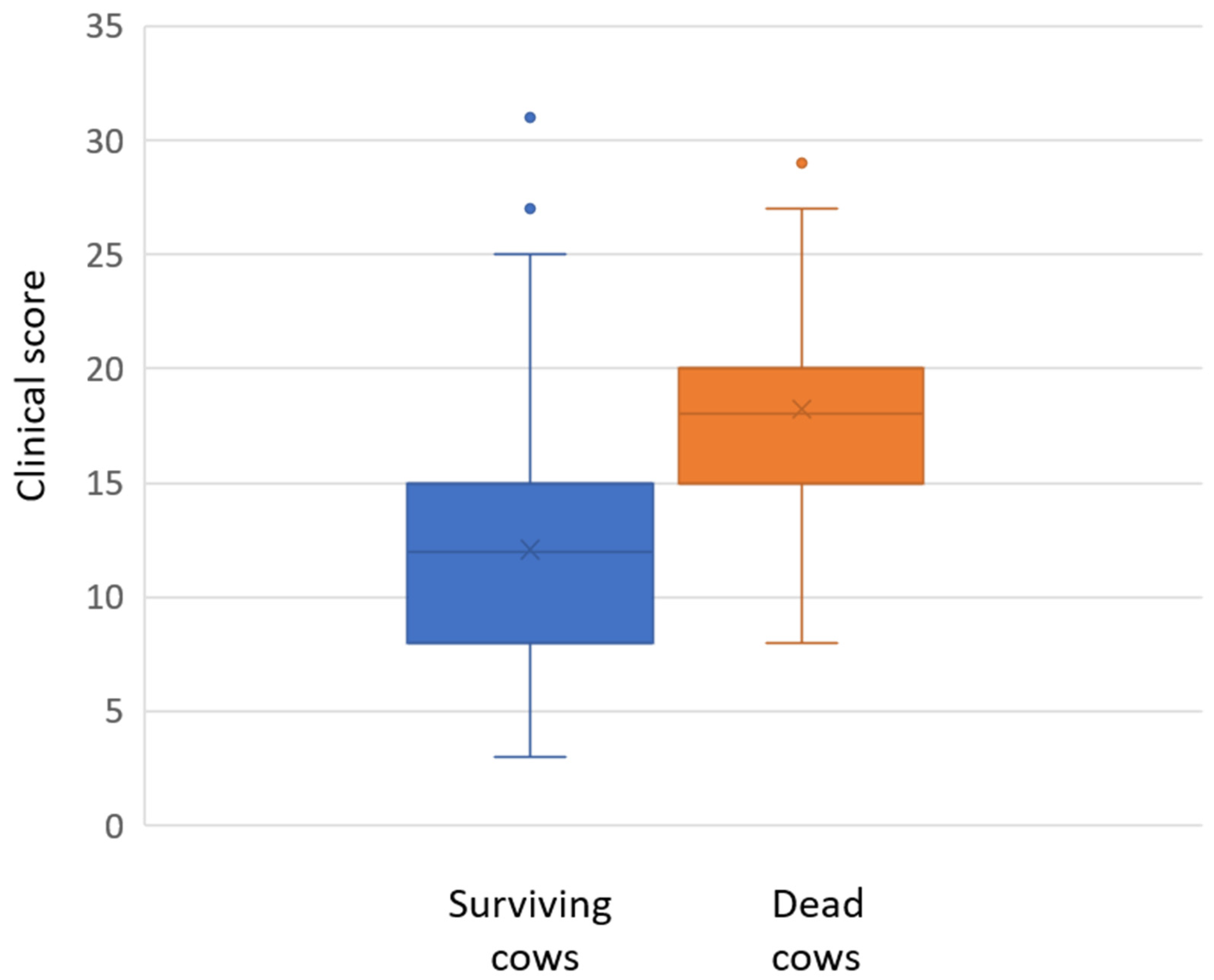
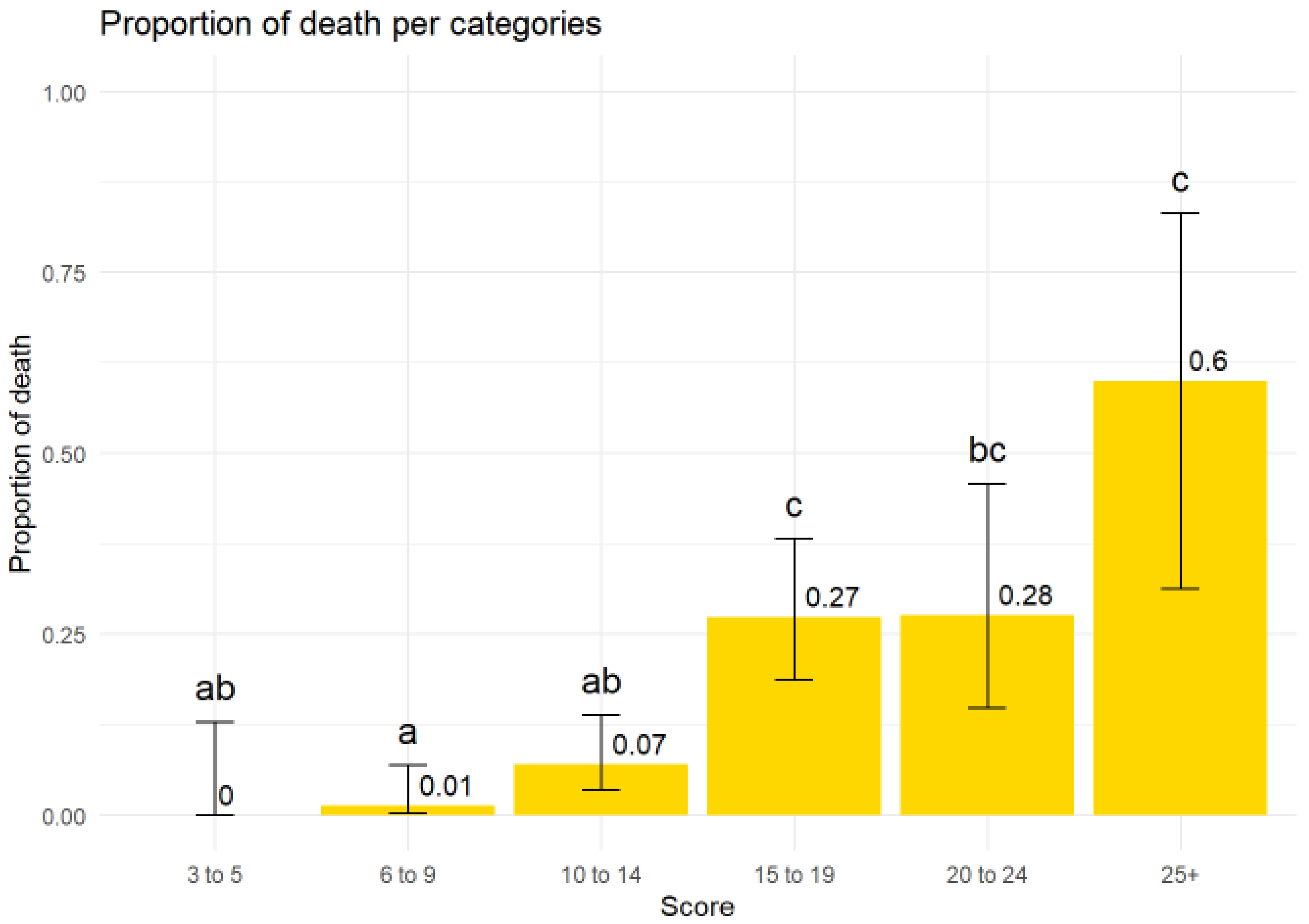
Complete clinical records were available for 320 cows (3 missing datasets), including 277 survivors and 43 cows that died or were euthanized. The mean clinical score in cows that died or were euthanized was 18.2, compared with 12.1 (p < 0.01) in cows that were alive and clinically healthy at 15 days post-inclusion (Table 7, Figure 1). The mortality rate increased with the clinical score at inclusion (see Table 8, Figure 2). Three of the eight cows presenting with gangrenous mastitis did not survive.
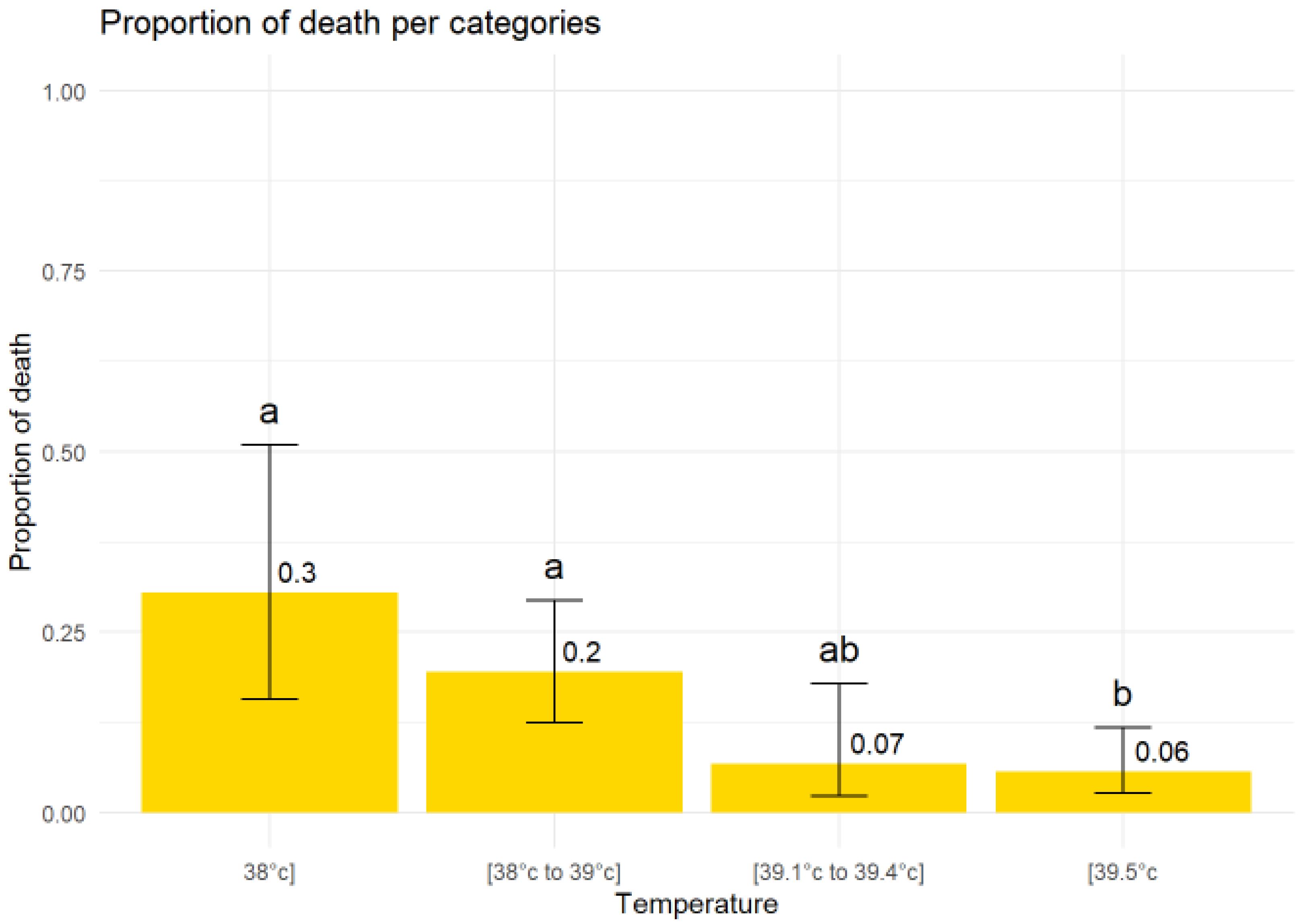
The clinical parameter most strongly associated with mortality was recumbency. Nearly 40% of cows that were recumbent at the time of inclusion died or were euthanized, whereas only 4.1% of cows that were standing at inclusion did not complete the study (p < 0.0001, Figure 3). The lowest mortality rate (5.6%) was observed in cows with hyperthermia at inclusion, compared with normothermic cows (19.5%) or hypothermic cows (30.4%) (Figure 4).
The pathogens isolated in association with these outcomes varied, although E. coli predominated (see Table 9).
Fifteen days after the clinical episode, the impact of severe mastitis on milk production (Table 10) and on the affected quarter (Table 11) were assessed.

3. Discussion

This study is based on an evaluation of the clinical consequences of severe mastitis and the outcomes of initial fluoroquinolone treatment. The success of antimicrobial therapy for intramammary infections is primarily assessed by bacteriological cure. This is generally the case for most forms of mastitis, except in severe cases, where the inflammatory response is particularly intense. In such cases, exacerbation of this inflammation represents a greater clinical challenge than pathogen elimination. Accordingly, most studies focusing on severe mastitis report clinical recovery as the primary endpoint [5,7,13,14]. Clinical and zootechnical outcomes were assessed via structured telephone interviews conducted 5 days and 15 days after inclusion. Although this method may introduce bias, data collection was strictly standardized, with predefined response categories. Quantitative measurements would have been more objective but were not feasible in a field setting. In particular, farmers were asked to estimate whether milk yield was above or below 50% of pre-mastitis production, which represents a potential source of classification bias; however, any misclassification is likely to be nondifferential.
The classification of severe mastitis used in this study is based on Wenz et al. [15], focusing on systemic clinical signs rather than local udder abnormalities. This approach is justified, as systemic signs are not consistently correlated with local lesions [3], particularly in the peripartum period, where diagnostic delays may occur if there are no obvious local signs. The clinical score was therefore constructed using systemic parameters (rectal temperature, enophthalmos, depression, ruminal motility), with additional indicators including ruminal fill, locomotion, ability to stand, skin tent duration as an additional indicator of dehydration, and scleral injection as a marker associated with septicemia [16].
The clinical signs observed and their frequency at inclusion were consistent with previous reports [17,18]. Overall, the clinical scores confirm the severity of the condition. The threshold for initiating fluid therapy (score ≥6) was supported by outcome data, as the lowest score among non-surviving cows was 8. However, clinical evolution over time could not be monitored due to logistical constraints. Cows were considered clinically recovered when they were standing and voluntarily eating, as assessed during follow-up calls.
Severe mastitis was more frequent in older cows, consistent with previous studies showing increased risk with parity [3,19,20,21]. Although a substantial proportion of cases occurred during the peripartum period (>25%), cases were distributed throughout lactation, with many occurring in mid- to late lactation. Approximately 5% of cases occurred shortly after drying off, potentially reflecting suboptimal hygiene during intramammary dry-cow therapy.
Gram-negative bacteria, particularly E. coli, predominated (≈80% of isolates), a proportion higher than previously reported [2,3,4,5,6,7,8,9,10,11,12,13,14,15,16,17,18,19,20,21,22,23,24]. This likely reflects case severity, as severe clinical presentations are more frequently associated with Enterobacteriaceae such as Escherichia coli or Klebsiella spp. [17,25]. Culture-negative results were rare (3.6%), in contrast to other studies [12,22], likely due to higher bacterial loads and case severity. A potential limitation is the inability of the culture method used to detect anaerobic organisms.
This study is, to our knowledge, the first to report antimicrobial susceptibility data specifically for Escherichia coli isolated from severe mastitis cases. Susceptibility patterns were broadly consistent with previous reports in mastitis populations not stratified by severity [26,27], although a higher proportion of resistance to sulfonamide/trimethoprim was observed. No ESBL-producing E. coli were detected.
Severe mastitis is associated with a marked systemic inflammatory response and may progress to sepsis, explaining the high mortality rates reported (approximately 25%) [28], with substantial variability across studies (13.5–35% or higher) [5,6,7,13,14,28]. These differences likely reflect variation in case definition, pathogen distribution (particularly Gram-negative involvement), clinician involvement (veterinarians [29] vs. technicians [13,22]), and treatment protocols. The use of a standardized clinical score in this study allowed objective severity stratification and uniform initial treatment.
Hypertonic saline infusion (3 L) was systematically administered in cases with a clinical score ≥6, in line with recommendations for hypovolemic shock management [30]. This protocol was applied uniformly during the first 24 h. Subsequent antimicrobial adjustments were made based on bacteriological results, mainly targeting Gram-positive infections (penicillin G or tylosin). These additional treatments were not considered treatment failures, as early outcome within the first 24 h was considered the most relevant prognostic window.
Designing a randomized field trial with a negative control group would be ethically and practically challenging given disease severity. Comparisons with the literature are further limited by heterogeneity in inclusion criteria and therapeutic protocols. Only one study presents partially comparable conditions [29], although important differences exist regarding antimicrobial choice, absence of fluoroquinolones, and lack of standardized clinical scoring. Compared with this study, our population included more recumbent cows (26.3% vs. 15.0%), more frequent use of fluid therapy (92.3% vs. 81.4%), and less calcium supplementation (25.1% vs. 53.0%), while mortality was approximately twofold lower.
Interestingly, cows with hyperthermia at inclusion showed lower mortality. Given that hyperthermia is typically transient in severe mastitis (12–24 h) [25], early intervention during this phase may improve survival outcomes.
Beyond mortality, severe mastitis had major economic consequences, with approximately 40% of cows experiencing a marked reduction in milk yield and two-thirds ceasing production. These findings are consistent with previous reports [28], which also describe high culling rates and quarter loss following severe mastitis episodes.
Finally, early fluid therapy guided by clinical scoring (≥6) appears justified and consistent with recommendations for sepsis management [31].
There is broad consensus that severe mastitis requires parenteral antimicrobial therapy [10,32]. Fluoroquinolones have demonstrated strong efficacy due to their pharmacokinetic properties and activity against major pathogens involved in severe mastitis [10,33]. Their use is particularly relevant in high bacterial load infections, where rapid reduction in pathogen burden is critical to limiting inflammation and preventing septicemia [3,5,23,34,35,36]. However, conflicting field studies exist [27,37], although both present important methodological limitations, including heterogeneity in severity classification, lack of randomization, and imbalance between treatment groups [38]. As such, their conclusions should be interpreted with caution. Overall, no robust evidence currently demonstrates the efficacy of non-critical antibiotics in severe mastitis, the only available study [39] also has significant biases, particularly regarding the severity of the conditions. While fluoroquinolones must be used judiciously due to concerns regarding antimicrobial resistance [40], this study provides field-based data on their use under real-world conditions. No fluoroquinolone-resistant Gram-negative isolates were detected. The absence of resistance emergence may be related to single-dose administration and restricted use to severe mastitis cases.

4. Material and Methods

Participating veterinary clinics: Eight veterinary practices participated in this study; all located in the main dairy regions of France. Each practice routinely performed on-site bacteriological analyses using a standardized culture protocol. As part of the study agreement, all clinics committed to systematically recording clinical parameters, applying a standardized treatment protocol, and ensuring follow-up of all included cases. A shared Excel database was used to record case-level data and monitor study progress in real time. The data were collected during farm visits conducted for the management of severe mastitis cases. For various reasons (oversights, farmer unavailability, delayed recording), some clinical records were incomplete. Whenever possible, they were completed retrospectively when the information was still available.
Case inclusion: With farmer consent, a veterinarian from one of the eight participating clinics included each case following a farm call reporting a sick cow. In cases where mastitis was suspected, a standardized clinical scoring sheet was completed based on field experience and the literature (Table S1) [15]. This included rectal temperature, rumen fill, ruminal motility, ocular congestion, enophthalmos, skin tent duration, behavior, and degree of depression. Cows were classified as severe mastitis cases when the clinical score was ≥3. A score ≥6 was considered indicative of shock. Animals already under treatment could be included provided that previous treatments were recorded. Antibiotic-treated cows at inclusion were accepted. None of the cows had been vaccinated against mastitis pathogens. The study period covered the year 2025, with a target inclusion of at least 250 cases.
Treatment: Cows with a clinical score ≥3 received a single intravenous injection of marbofloxacin (10 mg/kg) and flunixin (2.2 mg/kg), provided no other NSAID had been administered within the previous 24 h. No intramammary treatment was administered during the first 24 h post-inclusion. When the clinical score was ≥6, cows received 3 L of 7.2% hypertonic saline solution. If spontaneous water intake did not occur thereafter, oral drenching was performed. Calcium administration was left to the discretion of the attending veterinarian but was systematically recorded. Fluid therapy, with or without drenching, was repeated every 12 h until resolution of shock. Subsequent treatments were determined by the attending veterinarian based on bacteriological results and clinical evolution. No additional antibiotic or anti-inflammatory treatment was permitted within the first 24 h after inclusion. After 24 h, additional antimicrobial therapy was restricted to cases involving Gram-positive intramammary infections.
Additional samples: Microbiological procedures across the eight clinics followed guidelines inspired by the National Mastitis Council [41].
Briefly, 30 µL of well-mixed milk were streaked onto three culture media using a sterile calibrated loop:
  • 5% sheep blood agar (COS–bioMérieux, Lyon, France) for sample quality assessment,
  • 5% sheep blood agar supplemented with nalidixic acid (15 mg/L) and colistin sulfate (10 mg/L) (CNA–bioMérieux) for Gram-positive selection,
  • Hektoen enteric agar (HEKT–bioMérieux) for detection of Enterobacteriaceae.
Plates were incubated aerobically at 37 °C and read at 12, 24, and 48 h. Bacterial identification was performed using standard laboratory methods described elsewhere [20].
Catalase testing (3% H2O2) was performed on colonies grown on CNA. Colonies that were CNA-positive, catalase-positive, and exhibited double hemolysis, or were coagulase-positive, were identified as Staphylococcus aureus. Gram staining was performed on other CNA-positive catalase-positive colonies to distinguish non-aureus staphylococci (NAS) from Bacillus spp., Corynebacterium spp., yeasts, Prototheca spp., and fungi.
For Gram-positive catalase-negative colonies, aesculin hydrolysis was assessed. The Lancefield grouping test was performed when no reaction was observed after two hours of incubation. Aesculin-positive isolates were subcultured on bile aesculin agar to differentiate Streptococcus uberis from Enterococcus spp. Trueperella pyogenes was identified based on small, slow-growing colonies on CNA agar with delayed β-hemolysis, catalase negativity, aesculin negativity, pleomorphic Gram-positive rods, and characteristic V-shaped arrangements.
For organisms isolated on HEKT agar, subculture was performed on CPSO agar and in triple sugar iron (TSI) medium (bioMérieux). Colony color, growth pattern, and medium reactions were used for identification. Only Escherichia coli, Klebsiella spp., and Pseudomonas aeruginosa were differentiated from other Gram-negative bacteria to avoid misclassification.
E. coli appeared as yellow colonies on a salmon background on HEKT agar, fermented glucose and lactose in TSI medium, and formed pink colonies on CPSO agar. Klebsiella spp. showed similar biochemical behavior but produced blue colonies on CPSO agar. Pseudomonas aeruginosa appeared as green colonies on HEKT agar, with no glucose or lactose fermentation and no gas production in TSI medium; colonies appeared brown on CPSO agar.
Identification procedures were regularly validated against reference laboratories, with consistently high agreement (κ > 0.90).
Samples yielding two bacterial species were classified as “mixed culture,” whereas samples yielding three or more distinct species (≥1 CFU per species) were considered contaminated. A threshold of ≥1 CFU/10 µL was required for detection of major mastitis pathogens (Staphylococcus aureus, Streptococcus agalactiae, Streptococcus dysgalactiae, Streptococcus uberis, coliforms, and Enterococcus spp.) [42]. For other pathogens, the threshold was 2 CFU/10 µL.
Most isolates were subjected to antimicrobial susceptibility testing using the disc diffusion method according to EUCAST guidelines [43].
For Enterobacteriaceae, discs included: marbofloxacin, sulfamethoxazole/trimethoprim, nalidixic acid, gentamicin, amoxicillin/clavulanic acid, and cefquinome. For staphylococci, discs included penicillin, cefoxitin, marbofloxacin, erythromycin, and lincomycin. For enterococci, discs included ampicillin, cephalexin, erythromycin, and lincomycin. Streptococci were not tested, as they are considered uniformly susceptible to penicillin in France [44].
Clinical outcomes were recorded for all included cases. Follow-up was performed via two structured telephone interviews conducted 5 and 15 days after inclusion. Farmers were asked standardized questions regarding changes in general condition, milk production, and the status of the affected quarter (Table S2).
Statistical analysis: Descriptive statistics were first performed using R statistical software. The distribution of the variable of interest (score) was observed, and normality was rejected using the Shapiro–Wilk test. The median, range and interquartile range were therefore reported to describe their distribution. The association between this variable and cows’ mortality was therefore tested using a non-parametric Wilcoxon rank sum test. The level of statistical significance was set at p < 0.05.
The association between categorical data such as rectal temperature, clinical score, recumbency at inclusion day and mortality were explored using a Chi-squared or Fisher exact test (if one of the cells had a sample size <5). For contingency variables with 3 or more categories (temperature, score), post hoc tests were performed using Bonferroni correction to determine categories associated with different mortality probability.

5. Conclusions

The management of severe mastitis remains challenging for practitioners, with substantial economic and clinical losses still frequently observed. The use of fluoroquinolones raises a therapeutic and ethical dilemma. These critically important antimicrobials exhibit prolonged environmental persistence, and their use is therefore subject to strict regulatory restrictions and should be minimized whenever possible. However, severe mastitis is a life-threatening condition, and fluoroquinolones, due to their pharmacological and pharmacokinetic properties, are particularly well suited for its treatment. They demonstrate high efficacy against the main pathogens associated with septic shock, for which rapid bacterial elimination is essential. The use of fluoroquinolones, despite their critically important status and the strict framework governing their use, may prove valuable, particularly in a context where rapid bacteriological diagnosis is available, enabling targeted therapy and limitation of antibiotic use. This study is only observational: it does not constitute proof of the superiority of fluoroquinolones in the treatment of this type of mastitis, which could only be demonstrated by a properly randomized comparative study with a positive or negative control group.
Several strategies may help reduce mortality associated with severe mastitis. Optimization of immune function through balanced and well-supplemented nutrition, particularly with antioxidant support, as well as proper management of dietary transitions, is essential. Immunity may also be enhanced through vaccination strategies, including vaccines based on the J5 E. coli mutant, which have been shown to reduce the severity of Gram-negative infections. Early detection remains a critical factor in improving outcomes. In human medicine, even short delays in the management of sepsis have been associated with increased mortality risk, highlighting the importance of prompt intervention in severe systemic infections.


Supplementary Materials

The following supporting information can be downloaded at: https://www.mdpi.com/article/10.3390/antibiotics15060538/s1, Table S1: Shared Excell database; Table S2: Scoring of clinical cases.

Author Contributions

Conceptualization, O.S. and L.D.; Methodology, O.S.; Software, L.D.; Validation, O.S., P.P., N.P., C.L., A.N., V.H., A.S. and C.D.C.; Formal analysis, L.D.; Investigation, O.S., P.P., N.P., C.L., A.N., V.H., A.S., C.D.C. and L.D.; Resources, O.S.; Writing—original draft, O.S.; Writing—review & editing, O.S.; Supervision, O.S. All authors have read and agreed to the published version of the manuscript.

Funding

This research received no external funding. The participants in this study conducted the study during their work hours, as part of their daily routine.

Institutional Review Board Statement

This study was conducted within the usual setting of veterinary practice and included only clinical cases that required veterinary intervention. Apart from the collection of epidemiological and clinical data and the follow-up of the cows included in the study, the veterinary intervention was entirely consistent with routine practice.

Informed Consent Statement

We obtained verbal informed consent from the animal owners. This decision was based on the fact that we were treating cases of severe mastitis using an appropriate treatment authorized for this indication. The study focused solely on accurately recording the clinical, epidemiological, and disease progression parameters associated with this condition. All procedures were carried out as part of the routine course of our clinical practice.

Data Availability Statement

The data is organized into two Excel files (one shared with all participating veterinary facilities and another containing all available clinical data) and is available upon request.

Acknowledgments

Special thanks are expressed to Sebastien Buczinski for statistical analysis. Special thanks are expressed to all the veterinarians at the eight veterinary clinics who contributed clinical cases. Special thanks are also expressed to Cécile Enault for her help with the English translation.

Conflicts of Interest

The authors declare no conflicts of interest.

References

  1. Green, M.J.; Cripps, P.J.; Green, L.E. Prognostic indicators for toxic mastitis in dairy cows. Vet. Rec. 1998, 143, 127–130. [Google Scholar] [CrossRef]
  2. Hagner, K.A.; Nordgren, H.S.; Sarjokari, K.; Sukura, A.; Rajala-Schultz, P.J. Role of mastitis in on-farm deaths of Finnish dairy cows. J. Dairy Sci. 2024, 107, 5962–5973. [Google Scholar] [CrossRef] [PubMed]
  3. Burvenich, C.; Van Merris, V.; Mehrzad, J.; Diez-Fraile, A.; Duchateau, L. Severity of E. coli mastitis is mainly determined by cow factors. Vet. Res. 2003, 34, 521–564. [Google Scholar] [CrossRef]
  4. Katholm, J.; Andersen, P.H. Acute coliform mastitis in dairy cows: Endotoxin and biochemical changes in plasma and colony-forming units in milk. Vet. Rec. 1992, 131, 513–514. [Google Scholar] [CrossRef] [PubMed]
  5. Krebs, I.; Zhang, Y.; Wente, N.; Leimbach, S.; Krömker, V. Bacteremia in Severe Mastitis of Dairy Cows. Microorganisms 2023, 11, 1639. [Google Scholar] [CrossRef]
  6. Cebra, C.K.; Garry, F.B.; Dinsmore, R.P. Naturally occurring acute coliform mastitis in Holstein cattle. J. Vet. Int. Med. 1996, 10, 252–257. [Google Scholar] [CrossRef]
  7. Wenz, J.R.; Barrington, G.M.; Garry, F.B. Bacteremia associated with naturally occurring acute coliform mastitis in dairy cows. J. Am. Vet. Med. Assoc. 2001, 219, 976–981. [Google Scholar] [CrossRef]
  8. Guterbock, W.M.; Van Eenennaam, A.L.; Anderson, R.J.; Gardner, I.A.; Cullor, J.S.; Holmberg, C.A. Efficacy of intramammary antibiotic therapy for treatment of clinical mastitis caused by environmental pathogens. J. Dairy Sci. 1993, 76, 3437–3444. [Google Scholar] [CrossRef] [PubMed]
  9. Pyörälä, S.H.; Pyörälä, E.O. Efficacy of parenteral administration of three antimicrobial agents in treatment of clinical mastitis in lactating cows: 487 cases (1989–1995). J. Am. Vet. Med. Assoc. 1998, 212, 407–411. [Google Scholar] [CrossRef]
  10. Suojala, L.; Kaartinen, L.; Pyörälä, S. Treatment for bovine Escherichia coli mastitis—An evidence-based approach. J. Vet. Pharmacol. Ther. 2013, 36, 521–531. [Google Scholar] [CrossRef]
  11. Mansion-de Vries, E.M.; Hoedemaker, M.; Krömker, V. Evidence-based aspects of clinical mastitis treatment. Tierarztl. Prax. Ausg. G Grosstiere Nutztiere 2015, 43, 287–295. [Google Scholar] [CrossRef]
  12. Brennecke, J.; Falkenberg, U.; Wente, N.; Krömker, V. Are severe mastitis cases in dairy cows associated with bacteremia? Animals 2021, 11, 410. [Google Scholar] [CrossRef]
  13. Erskine, R.J.; Bartlett, P.C.; VanLente, J.L.; Phipps, C.R. Efficacy of systemic ceftiofur as a therapy for severe clinical mastitis in dairy cattle. J. Dairy Sci. 2002, 85, 2571–2575. [Google Scholar] [CrossRef] [PubMed]
  14. Hagiwara, S.; Mori, K.; Nagahata, H. Predictors of fatal outcomes resulting from acute Escherichia coli mastitis in dairy cows. J. Vet. Med. Sci. 2016, 78, 905–908. [Google Scholar] [CrossRef][Green Version]
  15. Wenz, J.R.; Barrington, G.M.; Garry, F.B.; Dinsmore, R.P.; Callan, R.J. Use of systemic disease signs to assess disease severity in dairy cows with acute coliform mastitis. J. Am. Vet. Med. Assoc. 2001, 218, 567–572. [Google Scholar] [CrossRef]
  16. Fecteau, G.; Smith, B.P.; George, L.W. Septicemia and meningitis in the newborn calf. Vet. Clin. N. Am. Food Anim. Pract. 2009, 25, 195–208. [Google Scholar] [CrossRef]
  17. Menzies, F.D.; Mc Bride, S.H.; Mc Dowell, S.W.; Mc Coy, M.A.; Mc Connell, W.; Bell, C. Clinical and laboratory findings in cases of toxic mastitis in cows in Northern Ireland. Vet. Rec. 2000, 147, 123–128. [Google Scholar] [CrossRef] [PubMed]
  18. Bleul, U.; Sacher, K.; Corti, S.; Braun, U. Clinical findings in 56 cows with toxic mastitis. Vet. Rec. 2006, 159, 677–680. [Google Scholar] [CrossRef] [PubMed]
  19. Van Werven, T.; Noordhuizen-Stassen, E.N.; Daemen, A.J.; Schukken, Y.H.; Brand, A.; Burvenich, C. Preinfection in vitro chemotaxis, phagocytosis, oxidative burst, and expression of CD11/CD18 receptors and their predictive capacity on the outcome of mastitis induced in dairy cows with Escherichia coli. J. Dairy Sci. 1997, 80, 67–74. [Google Scholar] [CrossRef] [PubMed]
  20. Mehrzad, J.; Dosogne, H.; Meyer, E.; Burvenich, C. Local and systemic effects of endotoxin mastitis on the chemiluminescence of milk and blood neutrophils in dairy cows. Vet. Res. 2001, 32, 131–144. [Google Scholar] [CrossRef]
  21. Vangroenweghe, F.; Duchateau, L.; Burvenich, C. Moderate inflammatory reaction during experimental Escherichia coli mastitis in primiparous cows. J. Dairy Sci. 2004, 87, 886–895. [Google Scholar] [CrossRef]
  22. Schmenger, A.; Krömker, V. Characterization, Cure Rates and Associated Risks of Clinical Mastitis in Northern Germany. Vet. Sci. 2020, 7, 170. [Google Scholar] [CrossRef] [PubMed]
  23. Krebs, I.; Zhang, Y.; Wente, N.; Leimbach, S.; Krömker, V. Severity of Clinical Mastitis and Bacterial Shedding. Pathogens 2023, 12, 1098. [Google Scholar] [CrossRef] [PubMed]
  24. Salat, O.; Lemaire, G.; Durel, L.; Perrot, F. Etiology of severe mastitis in French dairy herds. PLoS ONE 2023, 18, e0295614. [Google Scholar] [CrossRef] [PubMed]
  25. Poutrel, B.; Fromageau, A. Estimation de la prévalenec des pathogènes impliqués dans les mammites cliniques aiguës. Bull. GTV 2008, 43, 65–68. [Google Scholar]
  26. Botrel, M.A.; Haenni, M.; Morignat, E.; Sulpice, P.; Madec, J.Y.; Calavas, D. Distribution and antimicrobial resistance of clinical and subclinical mastitis pathogens in dairy cows in Rhône-Alpes, France. Foodborne Pathog. Dis. 2010, 5, 479–487. [Google Scholar] [CrossRef]
  27. Suojala, L.; Simojoki, H.; Mustonen, K.; Kaartinen, L.; Pyörälä, S. Efficacy of enrofloxacin in the treatment of naturally occurring acute clinical Escherichia coli mastitis. J. Dairy Sci. 2010, 93, 1960–1969. [Google Scholar] [CrossRef]
  28. Burvenich, C.; Bannerman, D.D.; Lippolis, J.D.; Peelman, L.; Nonnecke, B.J.; Kehrli, M.E., Jr.; Paape, M.J. Cumulative physiological events influence the inflammatory response of the bovine udder to Escherichia coli infections during the transition period. J. Dairy Sci. 2007, 90, E39–E54. [Google Scholar] [CrossRef]
  29. Le Page, T.; Buczinski, S.; Dubuc, J.; Labonté, J.; Roy, J.P. Development of a nomogram to estimate the 60-day probability of death or culling due to severe clinical mastitis in dairy cows at first veterinary clinical evaluation. Vet. Sci. 2023, 10, 268. [Google Scholar] [CrossRef]
  30. Minasyan, H. Sepsis and septic shock: Pathogenesis and treatment perspectives. J. Crit. Care 2017, 40, 229–242. [Google Scholar] [CrossRef]
  31. Oliveira, R.P.; Velasco, I.; Garcia Soriano, F.; Fridman, G. Clinical review: Hypertonic saline resuscitation in sepsis. Crit. Care 2002, 6, 418–423. [Google Scholar] [CrossRef] [PubMed]
  32. Piechl, S.; Urbantke, V.; Spergser, J.; Wehrend, A.; Wittek, T.; Baumgartner, M. Resistance patterns of bovine mastitis-associated Escherichia coli isolates to selected antimicrobial agents in Austria. Tierarztl. Prax. Ausg. G Grosstiere Nutztiere 2025, 53, 289–300. [Google Scholar] [CrossRef] [PubMed]
  33. Grandemange, E.; Perrin, P.A.; Schwab-Richards, R.; Woehrle, F. Efficacy of a single injection of marbofloxacin in the treatment of acute E. coli mastitis in lactating dairy cows. Rev. Med. Vet. 2017, 168, 219–228. [Google Scholar]
  34. Hirvonen, J.; Eklund, K.; Teppo, A.M.; Huszenica, G.; Kulcsar, M.; Saloniemi, H.; Pyorälä, S. Acute phase response in dairy cows with experimentally induced Escherichia coli mastitis. Acta Vet. Scand. 1999, 40, 35–46. [Google Scholar] [CrossRef] [PubMed]
  35. Hogan, J.; Smith, K.L. Coliform mastitis. Vet. Res. 2003, 34, 507–519. [Google Scholar] [CrossRef]
  36. Fredebeul-Krein, F.; Schmenger, A.; Wente, N.; Zhang, Y.; Krömker, V. Factors Associated with the Severity of Clinical Mastitis. Pathogens 2022, 11, 1089. [Google Scholar] [CrossRef]
  37. Persson, Y.; Katholm, J.; Landin, H.; Mörk, M.J. Efficacy of enrofloxacin for the treatment of acute clinical mastitis caused by Escherichia coli in dairy cows. Vet. Rec. 2015, 176, 673. [Google Scholar] [CrossRef]
  38. Roberson, J.R. Treatment of clinical mastitis. Vet. Clin. North Am. Food Anim. Pract. 2012, 28, 271–288. [Google Scholar] [CrossRef]
  39. Shpigel, N.Y.; Winkler, M.; Ziv, G.; Saran, A. Relationship between in vitro sensitivity of coliform pathogens in the udder and the outcome of treatment for clinical mastitis. Vet. Rec. 1998, 142, 135–137. [Google Scholar] [CrossRef]
  40. Aarestrup, F.M.; Jensen, N.E.; Jorsal, S.E.; Nielsen, T.K. Emergence of resistance to fluoroquinolones among bacteria causing infections in food animals in Denmark. Vet. Rec. 2000, 146, 76–78. [Google Scholar] [CrossRef]
  41. Middleton, J.; Fox, L.; Pihjetti, G.; Petersson-Wolfe, C. (Eds.) Laboratory Handbook on Bovine Mastitis, 3rd ed.; National Mastitis Council, Inc.: New Prague, MN, USA, 2017; 147p, Available online: https://www.academia.edu/50651208/Laboratory_Handbook_on_Bovine_Mastitis?email_work_card=view-paper (accessed on 15 May 2022).
  42. Dohoo, I.R.; Smith, J.; Andersen, S.; Kelton, D.F.; Godden, S. Diagnosing intramammary infections: Evaluation of definitions based on a single milk sample. J. Dairy Sci. 2011, 94, 250–261. [Google Scholar] [CrossRef]
  43. Matuschek, E.; Brown, D.F.; Kahlmeter, G. Development of the EUCAST disk diffusion antimicrobial susceptibility testing method and its implementation in routine microbiology laboratories. Clin. Microbiol. Infect. 2014, 20, O255–O266. [Google Scholar] [CrossRef]
  44. Haenni, M.; Lupo, A.; Madec, J.Y. Antimicrobial Resistance in Streptococcus spp. Microbiol. Spectr. 2018, 6, 25. [Google Scholar] [CrossRef]
Figure 1. Comparison of the distribution of clinical scores between survivors and non-survivors.
Antibiotics 15 00538 g001
Figure 2. Comparison of mortality rates according to clinical score at the inclusion (a ≠ b: p < 0.05; a ≠ c; p < 0.05, b ≠ c: p < 0.05).
Antibiotics 15 00538 g002
Figure 3. Comparison of the proportion of deaths among standing and recumbent cows at the start of the study (p < 0.0001).
Antibiotics 15 00538 g003
Figure 4. Comparison of mortality rates according to body temperature at admission (a ≠ b: p < 0.001). (38 °c] = <38.0 °c; [39.5 °c = >39.5 °c).
Antibiotics 15 00538 g004
Table 1. Distribution of lactation numbers of included cows (79 missing points of data).
Lactation Number
1234567>7
Number of cases234073422916129
Percentage 9.416.429.917.211.96.64.93.7
Table 2. Distribution of clinical cases by the stage of lactation at which they occurred (22 missing points of data).
Time Relative to Calving
−48 h to +48 h48 h to
3 weeks
3 weeks to 2 months2 months to 5 months>5 monthsDry off
Number of cases433945788313
Percentage14.30131525.9027.604.30
Table 3. Percentage of clinical cases exhibiting one or more of the clinical features associated with a deterioration in general condition (281 complete clinical records).
Clinical SignsPrevalence (%)
Impaired ruminal motility86.8
Depression73.3
Decreased or absent appetite70.7
Gait disturbance (other than recumbency)58.7
Recumbency26.3
Signs of dehydration52.0
Ocular congestion38.0
Hyperthermia (>39.4 °C)41.6
Mild hyperthermia (39.1 °C–39.4 °C)17.5
Normothermia (38.0 °C–39.0 °C)31.9
Hypothermia (<38.0 °C)9.0
Table 4. Distribution of bacteriological results when only one type of pathogen was isolated.
Isolated PathogensNumber of IsolatesPercentage
Escherichia coli17662.9
Gram-negative bacteriaKlebsiella spp.207.1
Pseudomonas93.2
Other Gram-negative51.8
Staphylococcus aureus155.4
Gram-positive bacteriaStreptococcus uberis196.8
Streptococcus dysgalactiae82.9
Trueperella pyogenes41.4
Other Gram-positive
(CNS, Enterococcus spp., Bacillus cereus)
124.3
yeast20.7
No growth103.6
Table 5. Distribution of bacteriological results when two pathogens were isolated.
Two Pathogens IsolatedNumber of Isolates
Escherichia coli + Streptococcus uberis18
Escherichia coli + Staphylococcus aureus4
Escherichia coli + other Gram-positive8
Escherichia coli + Streptococcus dysgalactiae2
Escherichia coli + Klebsiella2
Escherichia coli + Trueperella pyogenes1
Klebsiella + Streptococcus uberis1
Klebsiella + other Gram-positive1
Staphylococcus aureus + Streptococcus dysgalactiae2
Streptococcus uberis + other Gram-positive1
Table 6. Percentage of susceptibility of 188 strains of E. coli to commonly used antibiotics for this indication determined using the disc diffusion method.
SusceptibleIntermediateResistant
Amoxycillin + clavulanic acid35.247.817
Cefquinome98.5 1.5
Nalidixic acid93.72.93.4
Marbofloxacin100
Gentamycin99.30.7
Sulfamethoxazole + trimethoprim79.9 20.1
Table 7. Statistical comparison of the mean clinical scores between cows that survived and those that died or were euthanized (clinical score missing for 3 cows).
CharacteristicAlive
N = 277
Dead
N = 43
p-Value 2
Score12.1 1 (6.9–17.3)18.2 1 (13.5–22.9)<0.001
1 = meaning of the displayed value, 2 = statistical test used.
Table 8. Overview of the total number of cows and the number that died or were euthanized in each of the different clinical score classes (clinical score missing for 3 cows).
Clinical Score ClassesNumber of CasesDead/Euthanized CowsMortality Rate (%)
3 to 52600
6 to 97811.3
10 to 1410077
15 to 19772127.3
20 to 2429827.6
≥2510660
Table 9. Distribution of pathogens isolated from dead or euthanized cows.
Pathogen(s) IsolatedNumber of Dead/Euthanized Cows
Escherichia coli25
Escherichia coli + other pathogen6
Klebsiella2
Staphylococcus aureus2
Other Gram-positive4
Streptococcus uberis1
Streptococcus uberis + other Gram-positive1
No growth2
Table 10. Assessment of milk production in cows 15 days after they were included (251 valid records; the others had missing data or were beef cattle).
Maintaining Full Milk ProductionMaintaining over 50% of Milk ProductionMaintaining Under 50% of Milk ProductionLoss of Milk Production
Number of case56943467
Percentage 22.337.513.526.7
Table 11. Assessment of the condition of the affected quarter of the cows 15 days after they were included (253 valid records; the others had missing data or were beef cattle).
The Quarter Has Been Fully CuredThe Quarter Is Still Swollen/Hard, but the Milk Is NormalThe Quarter and the Milk Are Still Being ModifiedThe Quarter Is Dry
Number of cases112352086
Percentage44.313.87.934.0
Disclaimer/Publisher’s Note: The statements, opinions and data contained in all publications are solely those of the individual author(s) and contributor(s) and not of MDPI and/or the editor(s). MDPI and/or the editor(s) disclaim responsibility for any injury to people or property resulting from any ideas, methods, instructions or products referred to in the content.