


Abstract
Background/Objectives: Biofilms are a form of defense that enables bacteria to withstand antibiotic pressure and demonstrate antibiotic resistance. It is crucial to develop anti-biofilm strategies in order to combat chronic and persistent multidrug-resistant (MDR) infections. Methods: In this study, we developed 3D biofilms of single-, dual-, and poly-species MDR ESKAPE components, including the pathogens P. aeruginosa S. aureus and K. pneumoniae, in CF Mu3Gel. We evaluated the efficacy of using a phage, a di-hetero phage cocktail or a poly-hetero phage cocktail in combination with ciprofloxacin to eliminate mature biofilm biomass after 72 h or one week in a single treatment. Results: The phage components mostly exhibited synergistic behavior when combined with ciprofloxacin and with each other in di- and poly-hetero-cocktails. The reduction in 72-h dual- and poly-species biofilms was one log higher than that of one-week biofilms treated with the phage–antibiotic combination. The greatest reductions were observed in the 72-h single-species biofilm with combination therapy, at 1.4–3.0 log. Reductions of 2.16 and 1.6 log were observed in the dual-species P. aeruginosa and S. aureus biofilm and the poly-species biofilm, respectively. Conclusions: This study examined how a single application of phages or phage cocktails, either alone or in combination with ciprofloxacin, impacted established biofilm models, and how this affected the proportion of microcolonies of different species within each model. These insights will facilitate the development of strategies for multiple follow-up treatments, as well as the reordering of phages, phage cocktails, and combinations with antibiotics, to improve outcomes. The 3D biofilm models developed here could be used to screen phages or phage cocktails either on their own or alongside other therapies. This would facilitate the application of in vitro findings to real physiological settings.
1. Introduction
Antibiotic resistance poses a significant challenge to global public health. One of the most significant factors contributing to the emergence of antibiotic resistance is the ability of biofilms to increase tolerance to antimicrobial agents. Biofilms increase bacteria’s resistance to a wide range of environmental factors, making them difficult to treat with antimicrobials. Up to 80% of clinical bacterial infections are associated with biofilms [1,2]. Bacteria embedded in biofilms can be 1000 times more resistant to antimicrobials than planktonic bacteria [3]. Biofilm formation is associated with most chronic human infections [4,5] and is crucial for the long-term survival of pathogens. Natural biofilms in the environment and in hosts are often multispecies communities that differ greatly from mono-species biofilm systems in composition, structure, and antimicrobial resistance [6,7].
Polymicrobial infections involving Pseudomonas aeruginosa, Staphylococcus aureus, and Klebsiella pneumoniae are frequently reported in clinical settings [5], including in patients with cystic fibrosis (CF) [7,8,9,10], urinary tract infections, and wound infections [11,12]. These bacteria exhibit strong virulence factors and the ability to produce biofilms, enabling them to initiate infectious diseases [13]. These infections are characterized by poor clinical outcomes and increased antibiotic resistance [12,14]. The dynamics of lung colonization are defined by the co-colonization of P. aeruginosa by different species, including S. aureus and K. pneumoniae, and their interaction through competition and/or cooperation [15]. This results in a longer duration and more complicated infection [5,16]. Therefore, studying prevalent combinations of bacterial infections is important yet challenging. Importantly, P. aeruginosa, S. aureus, and K. pneumoniae are multidrug-resistant (MDR) ESKAPE pathogens. Carbapenem-resistant K. pneumoniae is classified as a critical priority pathogen [17]. Meanwhile, carbapenem-resistant P. aeruginosa and methicillin-resistant S. aureus are higher-priority pathogens on the 2024 World Health Organization [18] list of priority bacterial pathogens. These pathogens pose a significant threat to public health due to the limited treatment options available.
MDR pathogens are a cause for concern and necessitate the development of unconventional therapies. Among these therapies, phage therapy is a promising treatment strategy [17,19], as evidenced by its successful use in recent compassionate cases in Europe and the United States. Phages have several advantages over antibiotics. They typically target only one species or strain of bacteria, leaving the host’s commensal microbiota largely unharmed. Phages can effectively lyse MDR pathogens. They replicate at the active infection site, providing a sustained therapeutic effect [20]. Since the 1990s, phages have been considered a potential treatment for multidrug-resistant (MDR) diseases [21]. Several studies have demonstrated the effectiveness of using phages to treat MDR bacterial infections in vitro [22,23,24,25]. Recently, Pirnay et al. published an analysis of successful clinical outcomes with personalized phages. They reported clinical improvement and eradication of the targeted bacteria in 77.2% and 61.3% of infections, respectively. In vivo bacteriophage resistance selection and in vitro phage–antibiotic synergy were documented in 43.8% and 90% of the evaluated patients, respectively [26]. Schooley et al. published significant findings regarding the efficacy and health benefits of personalized phage treatment [27].
Phages can be used alongside standard antibiotics without affecting their antibacterial pharmacodynamic activity. The phage–antibiotic combination is a promising complementary therapy for treating MDR bacterial infections [19,28,29,30]. Gharieb et al. demonstrated the anti-biofilm potential of lytic phages against MDR S. aureus biofilms [31]. Bacteriophages can infect metabolically inactive persister cells embedded in biofilms, which are highly resistant to antibiotics [32]. Developing potential strategies to combat persister cells is highly significant from a clinical standpoint [33]. A combination of phages and antibiotics has been shown to significantly reduce the biofilms of different pathogens [34,35,36,37,38,39,40,41].
Phage cocktails are an encouraging approach for treating MDR infections and biofilms. Forti et al. treated biofilms with a phage cocktail, demonstrating that the phages could reach and reduce the biomass of bacteria embedded within the biofilms. Furthermore, they demonstrated the potential of phage cocktail therapy by effectively reducing infections in mice and treating bacteriemia in wax moths (Galeria mellonella) [20]. Several studies have demonstrated the successful use of phage cocktails in treating biofilms [42,43].
In order to study biofilm structure and formation, exopolysaccharide (EPS) production, metabolic activity, microbial behavior, and antibiotic resistance, as well as to screen different antimicrobials and translate their effects in vivo [4], it is important to develop a biofilm pattern that mimics the physiologically relevant conditions of the host. The main models of bacterial biofilms include in vitro static models (e.g., microtiter plates), dynamic systems (e.g., flow cells, continuous-flow reactors, 3D biofilms, and microcosms), specialized models, and ex vivo models [21,44]. Pacheco et al. developed and tested mucin-containing hydrogels (CF-Mu3Gel), which are dynamic, heterogeneous, three-dimensional (3D) structures that mimic the human body’s physiological setting. These structures include crosslinking and oxygen gradients. Specifically, the CF-Mu3Gel mimics the pattern of cystic fibrosis (CF) airway mucus, facilitating the study of interactions between different bacterial species [45].
In this study, we developed single-, dual- and poly-species 3D biofilms of P. aeruginosa, S. aureus and K. pneumoniae in CF Mu3Gel. These biofilms matured over periods of 72 h and one week. Our goals were threefold: first, to evaluate the efficacy of using a phage, a phage di-hetero-cocktail, or a phage poly-hetero-cocktail in combination with an antibiotic for the one-shot elimination of mature biofilm biomass; second, to demonstrate the synergy between the phages in the hetero-cocktail and the antibiotic; and third, to demonstrate how phages can modify the composition of biofilms, which could counteract the persistent and resilient nature of biofilm-associated pathogens.
We studied the relative abundance of different species in biofilms of combined models, both with and without exposure to an antibiotic, a phage, or a phage cocktail, either alone or in combination. Consequently, we performed a comparative analysis of the results from the different biofilm models. Additionally, we evaluated the synergistic interactions of each component and their persistence in dual- and poly-species biofilms. We also examined how the proportion of persistence of the different species changes under antimicrobial exposure. The results showed that P. aeruginosa can become sensitive to ciprofloxacin again when treated with a combination of phage cocktails and antibiotics. These findings provide insight into how the proportion of different species in microcolonies changes in each biofilm model. These findings will help further develop strategies for multiple follow-up treatments and reordering phage, phage cocktail, and antibiotic combinations to improve outcomes.
In the future, these 3D biofilm models in CF Mu3Gel could be used to screen phages or phage cocktails, either alone or in combination with other therapies. This would facilitate the translating of in vitro experience into real physiological settings, and would be useful for studying the interaction between different species, as well as variations within the same species, in settings that are as similar to nature as possible.
2. Results
2.1. Phage Lytic Activity on Planktonic Cultures and MIC
The S. aureus ATCC 6538, P. aeruginosa PAO1K and K. pneumoniae ATCC 27736 bacterial strains used in this study had previously been tested with phages and their susceptibility to the S. aureus phage Sb1, the P. aeruginosa phage Atpa010 and the K. pneumoniae phage Atkp010 had been determined. The P. aeruginosa phage Atpa010 and the K. pneumoniae phage Atkp010 phages were previously screened on 102 P. aeruginosa and 155 K. pneumoniae strains with different genetic backgrounds and capsular types, and their lytic activity was investigated [46]. The S. aureus phage Sb1 was known from previous studies [47]. The phage Sb1, from the George Eliava Institute of Bacteriophages, Microbiology and Virology, is well known for its high lytic activity. All three phages cleared the respective planktonic cultures (with an initial inoculum of 5 × 10⁵ CFU/mL, low-load planktonic cultures (LLPC)) at a ratio of 0.1 phages to bacteria for 48 h (Figure S1).
Ciprofloxacin was selected based on a preliminary study because there was little difference in the Minimum Inhibitory Concentrations (MIC) of ciprofloxacin for all three strains compared to those of other antibiotics (Colistin and Meropenem). We calculated the MICs of a low-load planktonic culture (EUCAST standard of 5 × 10⁵ CFU/mL) in the OmniLog system, which generates high-throughput kinetic readouts based on bacterial cell metabolic activity. This allowed us to directly compare the differences and assess the effect of ciprofloxacin on bacterial growth [46] (Figure 1). The standard MIC determination tests for all three pathogens against the different antibiotics were performed using the VITEK 2 (Ref. 27226, BioMérieux, Inc., Salt Lake City, UT, USA) at the QAMH Military Medical Laboratory Capacity (MMLC) ISO 15189 (Figure S2). The MIC for S. aureus ATCC 6538, P. aeruginosa PAO1K and K. pneumoniae ATCC 27736 against ciprofloxacin were 0.5 µg/mL, 0.25 µg/mL and 0.125 µg/mL, respectively. Regarding co-culturing, the MICs of dual-species planktonic cultures of P. aeruginosa and S. aureus, P. aeruginosa and K. pneumoniae, and S. aureus and K. pneumoniae, as well as poly-species planktonic cultures, were all 0.5 µg/mL, the same as the MIC of a single planktonic culture of S. aureus, which was the highest of the three (Figure 1).
Figure 1.
The proliferation curves of the low-load planktonic cultures of (a) P. aeruginosa, (b) S. aureus (c) K. pneumoniae, (d) P. aeruginosa and S. aureus, (e) P. aeruginosa and K. pneumoniae, (f) S. aureus and K. pneumoniae, and (g) P. aeruginosa, S. aureus, and K. pneumoniae exposed to different concentrations of ciprofloxacin (2 µg/mL − 0.0625 µg/mL). The (a) legend applies to all tetrazolium reduction curves. The tetrazolium reduction is expressed in relative respiration units (RRUs). Values are the mean ± SD of triplicates (n = 3).
However, to determine the appropriate dosage of ciprofloxacin for treating mature biofilms with a bacterial load ranging from 1.0 × 10⁸ to 1.0 × 10⁹ CFU/mL (different from the EUCAST standard of 5.0 × 10⁵ CFU/mL), we performed an MIC test against ciprofloxacin with high-load planktonic cultures (HLPC) (final concentration of 5.0 × 10⁸, high-load planktonic culture) of the aforementioned combinations. We considered this approach to determine the minimum bactericidal biofilm concentration (MBBC) [48]. The results were calculated based on Relative Respiration Unit (RRU) values (Figure S3). The results differ greatly from those of the MIC against ciprofloxacin of a low-load planktonic culture.
Here, we could only evaluate the MBBC for P. aeruginosa, which is 2 µg/mL. This is higher than the 0.5 µg/mL observed in the low-load planktonic culture. However, the RRU reduction at 1–8 µg/mL of ciprofloxacin is nearly identical in the proliferation curves of all the other cultures, as shown in Figure S3. Figure 2 shows the percentage reduction in ciprofloxacin at concentrations ranging from 1 to 8 µg/mL, calculated based on RRU reduction values. (Figure 2, Table S1).
Figure 2.
Percent reduction of high-load planktonic cultures and co-cultures at 1–8 µg/mL of ciprofloxacin. Values are the mean ± SD of triplicates (n = 3).
Based on these results and data from the publication [6], we selected 4 µg/mL of ciprofloxacin to treat different biofilm models, both alone and in combination with phage hetero cocktails that would not interfere with the phages or phage cocktails. This enabled us to evaluate phage activity while treating the test biofilms. Regarding phage lytic activity, a ratio of 100 phages to bacteria resulted in clearance for only 12 h when high-load planktonic cultures were used (Figure S4). Using the interpretation model that we previously developed, we can analyze Figure S4 and observe the bacterial inhibition effect by phage [46]. The bacteria–phage proliferation curves of all cases have shorter and longer exponential phases and express reduced and almost constant RRU, which is maintained up to 24 h. We did not observe this type of proliferation curve in low-load planktonic cultures (Figure S1), even though the MOI of phages to bacteria was the same. This refers to the slow growth of bacterial cells, including phage-resistant mutants under a high planktonic culture load. Thus, a ratio of 100 phages to bacteria was used to treat all biofilm models. This approach was suggested in another publication [49].
2.2. Biofilm Formation
Single-, dual-, and poly-species biofilms were developed and examined using two models: a 96-well plate and a microscopic glass slide, both containing CF Mu3Gel. Both models were tested for biofilm formation over 72 h and one week. The 96-well plates with CF Mu3Gel were primarily used for analysis in the OMNILOG system and for enumeration of CFU/mL. Biofilm models were established on microscopic glass slides using CF Mu3Gel for scanning electron microscopy (SEM), confocal laser scanning microscopy (CLSM), and enumeration of CFU/mL.
2.2.1. Enumeration of the Bacterial Load in Biofilm Models
Preliminary experiments were conducted to standardize the enumeration assay of bacterial loads (CFU/mL) for the biofilm models of both conditions, in both 96-well plates and on microscopic glass slides, to ensure comparability with each other and with the results of SEM and CLSM images.
Figure 3a illustrates the biofilm load in CFU/mL over 72 h and one-week. After 72 h, the bacterial load in single biofilms of P. aeruginosa S. aureus, and K. pneumoniae, and dual-species biofilms of P. aeruginosa and S. aureus, P. aeruginosa and K. pneumoniae, and S. aureus and K. pneumoniae, as well as in poly-species biofilms, reached 2.17 × 10⁹ CFU/mL, 1.16 × 10⁹ CFU/mL, 1.18 × 10⁹ CFU/mL, 6.34 × 10⁸ CFU/mL, 1.78 × 10⁹ CFU/mL, 4.40 × 10⁹ CFU/mL and 1.92 × 10⁹ CFU/mL, respectively. However, after one week, the biomass of each model decreased by 3.55 × 100 CFU/mL, 3.69 × 100 CFU/mL and 1.81 × 100 CFU/mL, 6.35 × 100 CFU/mL, 2.06 × 100 CFU/mL and 6.03 × 100 CFU/mL (Table 1). Figure 3b shows the reduction of a tetrazolium dye in RRU of 72-h biofilms in the OMNILOG system, with values ranging from 312 to 360 RRU. The RRU expressions for one-week biofilms were almost the same as for 72-h biofilms, as only the last 24 h were read.
Figure 3.
Bacterial load in 72-h biofilm models: (a) Bacterial load expressed as CFU/mL. (b) Bacterial load is expressed as Relative Respiratory Units (RRUs) in the OMNILOG system; the tetrazolium dye was added after 48 h, and the OmniLog system was used to read the results for the last 24 h. Values are the mean ± SD of triplicates (n = 3).
Table 1.
Load of each bacterial strain in 72-h and one-week biofilm models (CFU/mL).
2.2.2. Scanning Electron Microscopy (SEM)
We observed the structural architecture of one-week dual-species (P. aeruginosa and S. aureus, P. aeruginosa and K. pneumoniae, S. aureus and K. pneumoniae) and poly-species (P. aeruginosa, S. aureus, and K. pneumoniae) biofilms using scanning electron microscopy (SEM). SEM images (Figure 4) show mature biofilms after one week, with a slimy and intensive protective matrix of EPS embedded within the pathogens at high density. Furthermore, the combination of different species in biofilm models results in different structures: the poly-species biofilm (Figure 4(a1–a3)) is characterized by a smooth layer (shell) covering cell aggregates; also, this kind of channel (Figure 4(b2)) could be a broken-off biofilm, indicating the dispersal of bacterial cells to repopulate another area. The dual-species biofilm of P. aeruginosa and S. aureus reveals a hill showing a mushroom-like appearance (Figure 4(b1)) and valley [50] structure of biofilm, with large aggregates of dense pathogens embedded (Figure 4(c2)). Meanwhile, the dual-species biofilm of S. aureus and K. pneumoniae is characterized by wrinkled shell covering cell aggregates (Figure 4(c1)).
Figure 4.
SEM pictures of mature biofilms after one week: (a1–a3) are images of poly-species biofilms of different zones and dimensions; (b1,b2) are images of dual-species biofilms of P. aeruginosa and S. aureus of different zones and dimensions; and (c1,c2) are images of dual-species biofilm of P. aeruginosa and K. pneumoniae of different zones and dimensions.
2.2.3. Confocal Laser Scanning Microscopy (CLSM)
Only the one-week poly-biofilm was studied under CLSM. Figure 5 shows 3D structures, biomass composition (live and dead cells), and biofilm dynamics in real time. The matrixes consist of spatially distributed microcolonies that are interconnected.
Figure 5.
Different captures of CLSM images of one-week poly-biofilms of P. aeruginosa, S. aureus, and K. pneumoniae: (a–c). The biofilms were stained using the LIVE/DEAD™ BacLight™ Bacterial Viability Kit. Live cells stain green and dead cells stain red.
2.2.4. Determining the Load of Each Bacterial Strain in 72-h and One-Week Biofilm Models
Figure 6 demonstrate the load of each bacterial strain in dual and poly-biofilms. There are slight differences between the 72-h and one-week biofilms. The ratio of each bacterial strain was calculated using the standard division formula in Excel. In untreated dual-species biofilms, P. aeruginosa dominates S. aureus with 2.62 × 101 and 5.66 × 101, respectively. In triple-species biofilms, P. aeruginosa dominates both S. aureus with 1.03 × 10⁵ and 6.75 × 10², respectively, and K. pneumoniae with 1.95 × 101 and 1.76 × 101, respectively. Meanwhile, K. pneumoniae dominates S. aureus with 8.16 × 101 and 2.03 × 10³, respectively, and P. aeruginosa with 1.38 × 10³ and 6.70 × 101, respectively, in dual-species biofilms.
Figure 6.
The load of bacterial strains in untreated dual- and poly-species biofilm models after 72 h and one week. The values are the mean ± SD of triplicates (n = 3).
2.3. Treatment of Biofilm Models
We treated all 72-h and one-week matured biofilm models in a single round for 24 h. The formulations of phages, phage cocktails, and ciprofloxacin, both alone and in combination, in 100 µL and 200 µL suspensions, which were applied to treat biofilms in one well of a 96-well plate and on a microscopic glass slide, are presented in the Section 4.
The type of interaction between the phage or phage cocktail and antibiotic that produced a more potent effect over the course of the experiment than the single agents was classified as synergistic activity [46]. The type of interaction between the phage or phage cocktail and antibiotic that produced the same potent effect as the single agents was classified as proto-cooperation [46].
2.3.1. Results of the Enumeration of Bacterial Strains in the Treated Biofilm Models
The greatest reductions in total biomass with combination therapy were observed in the 72-h single-species biofilms of P. aeruginosa, S. aureus, and K. pneumoniae, followed by the dual-species biofilm of P. aeruginosa and S. aureus, and then the poly-species biofilm. Relatively lower reductions were observed in the 72-h dual-species biofilms of P. aeruginosa and K. pneumoniae, as well as S. aureus and K. pneumoniae (Figure 7).
Figure 7.
Results of a single 24-h treatment for (a) 72-h-old and (b) one-week-old single-, dual-. and poly-species biofilms. The values are the mean ± SD of triplicates (n = 3).
The greatest reductions by the phages and phage cocktails in one-week biofilms were observed in the poly-species biofilm and the single-species biofilm of P. aeruginosa. We found that the K. pneumoniae phage alone had more difficulty treating biofilm-associated bacterial cells than the P. aeruginosa and S. aureus phages, exhibiting a maximum reduction of 0.7 log in the 72-h single biofilm (Figure 7).
2.3.2. Interpretation of Phage and Ciprofloxacin Interaction
In this study, we developed criteria for evaluating the level of synergy. A reduction of 0.7 log or more in the combination of a phage or a phage cocktail with ciprofloxacin was considered to be a high level of synergy; a reduction of 0.3–0.5 log in the combination was considered to be a medium level of synergy; a reduction of 0.15–0.25 log in the combination was considered to be a low level of synergy; and a reduction of 0.08–0.1 was considered to be negligible (Table 2). Additionally, overgrowth of bacteria under antibiotic exposure alone was evaluated as a negative effect.
Table 2.
Reduction values for bacterial load after treatment of 72-h and one-week old biofilms for 24 h. The values are means ± SD of triplicates (n = 3).
A high degree of synergy was exhibited by phages and ciprofloxacin in 72-h single-species biofilms of P. aeruginosa and S. aureus. However, in the other biofilm models, the synergy was low, except for the dual-species biofilm of P. aeruginosa and S. aureus, where proto-cooperation was established. In particular, a low degree of synergistic activity was revealed in dual-species biofilms of P. aeruginosa and S. aureus, and a medium degree was revealed in the single-species S. aureus biofilm. This last case is interesting because the synergistic effect is due to phage activity, whereas ciprofloxacin caused a negative effect, resulting in an increase in the number of S. aureus colonies compared to the control (Table 2). This could be an example of S. aureus hypothesizing that phages remain active against antibiotic-tolerant and less active bacteria, while most phage population growth involves metabolically active bacterial cells [51].
2.3.3. Overall Interpretation of the Results of the Biofilm Model Treatment
The overall interpretation of the results of the single-, dual-, and poly-species biofilm treatments is provided in Table 3.
Table 3.
Overall interpretation of the results of the single-, dual-, and poly-species biofilm treatments.
2.3.4. Determining the Proportions of Bacterial Strains in 72-h and One-Week-Old Biofilm Models Following a Single 24-h Treatment
Figure 8 shows the results of the enumeration (CFU/mL) of the individual strains and their proportion in the treated biofilm models. As can be seen, S. aureus and K. pneumoniae show greater resistance than P. aeruginosa. In dual- and poly-species biofilms exposed to ciprofloxacin, S. aureus dominates P. aeruginosa by 0.7–1.6 log CFU/mL and slightly dominates K. pneumoniae. Furthermore, P. aeruginosa and S. aureus phages demonstrate good efficacy, both alone and in combination. In contrast, K. pneumoniae phage exhibits significantly lower efficacy. In biofilm models under phage cocktail exposure, K. pneumoniae dominates the other two strains, particularly P. aeruginosa.
Figure 8.
The proportions of bacterial strains in 72-h and one-week-old dual- and poly-species biofilm models after 24 hours’ exposure to: (a) ciprofloxacin alone, (b) a phage cocktail alone, or (c) combination. Values are means ± SD of triplicates (n = 3).
2.3.5. The Results of Tetrazolium Reduction in the OMNILOG System
We tried to observe biofilm model proliferation curves in the OmniLog system. However, seeing a reduction curve is very unlikely with mature biofilms, because the high cell density leads to rapid saturation of the reaction. This limits the assay’s sensitivity and dynamic range. For this reason, we added dye to the mature biofilms after six hours of exposure to antimicrobials, and then continued their incubation in the OmniLog System for a further 18 h. The proliferation curve of single-species biofilms of P. aeruginosa and S. aureus is reduced by 100 RRU. Dual-species biofilms of P. aeruginosa and S. aureus, as well as poly-species biofilms’ proliferation curves, are reduced by 150 RRU. All other biofilms’ proliferation curves are reduced by 50 RRU (Figure 9). We suppose that the tetrazolium reduction readings were based on the results of surface-grown pathogen populations. Bacteria located on metabolically active biofilm surfaces could be primary targets for phage replication and antibiotics [51].
Figure 9.
The tetrazolium reduction curves of the single-species biofilms of (a) P. aeruginosa, (b) S. aureus, (c) K. pneumoniae, of dual-species biofilms of (d) P. aeruginosa and S. aureus, (e) P. aeruginosa and K. pneumoniae, (f) S. aureus and K. pneumoniae, and of poly-species biofilm of (g) P. aeruginosa, S. aureus, and K. pneumoniae exposed to ciprofloxacin and phage or phage cocktail, alone and in combination. The (a) legend applies to all reduction curves. Values are means ± SD of triplicates (n = 3).
2.3.6. Determination of Phage Titer (PFU/mL) in Tested Biofilm Models
The same sample dilutions that were used to enumerate the bacterial load were also used to enumerate the phage particles (PFU/mL). We observed an increase in phage titers after treating mature biofilms for 24 h. The P. aeruginosa phage Atpa010 showed a 0.6–0.7 log increase, and the S. aureus phage Sb1 showed a 0.5 log increase. There was no notable increase in K. pneumoniae phage Atkp010. Its titers remained consistent across all biofilm models (Figure 10).
Figure 10.
Results of phage titration in treated biofilms; values are the mean of triplicates (n = 3).
2.3.7. SEM Visualization of the Effect of Treating the Biofilm
The effects of the biofilm treatment can be observed using SEM images (Figure 11). We can see spatial disruption of the matrixes and reduced numbers of biomass.
Figure 11.
SEM image of the treated biofilms: (a1,a2) different zones and dimensions of a poly-species biofilm treated with a combination of a poly-hetero phage cocktail and ciprofloxacin and (a3) treated with ciprofloxacin alone. (b1,b2) different zones and dimensions of a dual-species biofilm of P. aeruginosa and S. aureus treated with a combination of a dual-hetero phage cocktail and ciprofloxacin, as well as (b3) treated with phage di-hetero-cocktail alone and (b4) treated with ciprofloxacin alone. (c1) a dual-species biofilm of P. aeruginosa and K. pneumoniae treated with a combination of a di-hetero phage cocktail and ciprofloxacin, and (c2) treated with a di-hetero phage cocktail alone. (d1–d3) the different zones and dimensions of a dual-species S. aureus and K. pneumoniae biofilm treated with a combination of a dual-hetero phage cocktail and ciprofloxacin.
Figure 12a–c shows SEM images of one-week-old poly-biofilms treated with poly-hetero-cocktails. We can particularly see the protective surface barrier being destroyed.
Figure 12.
SEM images of the poly-species biofilms: (a–c): The different zones and dimensions treated with poly-hetero-cocktails.
2.3.8. Confocal Laser Scanning Microscopy (CLSM)
The architecture and bacterial biomass of the one-week poly-biofilms treated with a ciprofloxacin, a phage poly-hetero-cocktail, and with a combination of the two were visualized using confocal laser scanning microscopy (CLSM) (Figure 13): we can observe the effects of the (Figure 13(c1–c3) phage and (Figure 13(d1–d3) poly-hetero-cocktail both alone and in combination with ciprofloxacin on the poly-species biofilm. Ciprofloxacin alone (Figure 13(a1–a3) demonstrates a lesser reduction effect.
Figure 13.
CLSM images of one-week poly-biofilms: (a1–a3) are different CLSM image captures treated with an antibiotic alone; (b1–b3) are different CLSM image captures treated with a phage poly-hetero-cocktail alone; and (c1–c3) are different CLSM image captures treated with a combination of the two. The biofilms were stained using the LIVE/DEAD™ BacLight™ Bacterial Viability Kit. Live cells stain green and dead cells stain red.
2.4. Testing the Sensitivity of Biofilm-Associated Bacterial Isolates to Ciprofloxacin
2.4.1. Biofilm-Associated Antibiotic-Resistant Mutants Isolates
We attempted to isolate antibiotic-resistant mutants from different biofilm model sets and succeeded only with P. aeruginosa PAO1K sampled from a poly-species biofilm treated with combination of ciprofloxacin and phage poly-hetero-cocktail, which exhibited a change in sensitivity to resistance (from 0.25 µg/mL H to 1 µg/mL R, biofilm isolate (2)). The tests were performed using VITEK 2 (Ref. 27226, BioMérieux, Inc., USA), which was performed at the QAMH (Queen Astrid Military Hospital) Military Medical Laboratory Capacity (MMLC) ISO 15189 (Table 4). The official testing results from the MMLC laboratory are provided in the Appendix (Figure S5).
Table 4.
Interpretation of the results of the single-, dual-, and poly-species biofilm treatment.
Furthermore, we isolated a P. aeruginosa PAO1K strain from a poly-species biofilm that was treated with a combination of ciprofloxacin and a poly-hetero-cocktail of phages. The MIC value decreased from 0.25 µg/mL to 0.12 µg/mL (Table 4, biofilm isolate (1)). Thus, the P. aeruginosa strain was resensitized in the context of a phage–antibiotic combination. The results of the MMLC laboratory testing are provided in Supplementary Figure S6.
2.4.2. Biofilm-Associated Isolates: Morphological Characterization and Persistence
It is important to note that when samples from experimental biofilms were subjected to an enumeration assay, S. aureus and P. aeruginosa, which were sampled from an untreated dual-biofilm containing either P. aeruginosa and S. aureus or S. aureus and K. pneumoniae, and P. aeruginosa and K. pneumoniae, respectively, appeared to be countable after 48–72 h. Furthermore, P. aeruginosa isolates from ciprofloxacin-treated dual- and poly-species biofilms appeared countable after 48 h. K. pneumoniae biofilm isolates exhibited phenotypic heterogeneity, displaying both normal-sized and small colony variants (SCVs) (Figure S7), which is also a survival strategy. The late appearance of S. aureus and P. aeruginosa could also be attributed to SCVs, which are optimized for persistence [52].
3. Discussion
Biofilm-associated infections account for 65% to 80% of all microbial infections in humans and are a global crisis [19]. These dynamic, complex bacterial communities are surrounded by an extracellular polymeric matrix that shields pathogens from antimicrobials. The emergence of multidrug-resistant (MDR) pathogens necessitates an urgent solution: an anti-biofilm treatment strategy. Phage–antibiotic synergistic therapy is considered one of the most promising anti-biofilm strategies. Phages can target different stages of biofilm formation.
In nature, biofilms typically comprise multiple species [7]. Interspecies communication defines their spatial structure, as well as the composition and relative abundance of their subpopulations within the total microbial biofilm. This process is crucial for advancing biofilm-associated infections. Mixed infections of P. aeruginosa with S. aureus and K. pneumoniae are often present in severe cases of community-acquired pneumonia. The strains used here—PAO1K, ATCC 6538, and ATCC 27736, respectively—are known for their strong, stable, and structured ability to form biofilms [17,53,54,55]. However, it is very challenging to mimic natural conditions and maintain a long-term, balanced community of these pathogens in a laboratory setting [5]. The CF-Mu3Gel tested in this study is a dynamic and heterogeneous structure consisting of smooth and wrinkled shells, hills with a mushroom-like appearance [50], valleys, and channels that mimic the human body’s physiological environment. Figure 14 shows the study structure and the flow of the experiments. This enabled the results of the various criteria and values to be compared, to provide an overall evaluation.
Figure 14.
Scheme of Biofilm Development and Treatment.
Poly-species biofilms exhibit cooperative or competitive interactions and establish a communication network controlled by specific genes and signals, resulting in compositional equilibrium [7]. In the biofilm models studied here, the biomass balance of individual species in dual- and poly-biofilms remained stable throughout the three-day to one-week incubation period due to a decrease in one species and an increase in another, in addition to competitive interactions (Figure 6). Additionally, the relative abundance of dominant components in different dual-species models is decreased by about one log compared to single biofilms. Therefore, the interactions between the components were considered cooperative. Pan et al. describes that P. aeruginosa often exhibits cooperative behavior in biofilms by secreting EPS and siderophores that benefit co-cultured pathogens [5,56,57]. Additionally, staphylococcal protein A (SpA) interacts with P. aeruginosa, promoting aggregation via Psl and type-IV pilus binding [58,59]. In a dual-species biofilm of S. aureus and P. aeruginosa, the last dominates S. aures by only about one log.
Woods et al. developed a dual-species biofilm of S. aureus and P. aeruginosa in a silicone tube reactor system at 22 °C under continuous-flow conditions. Interestingly, they used the same strains (PAO1 and ATCC 6538) as in this study, but different versions, as they had been maintained in different laboratories for years. They demonstrated that S. aureus persisted at higher concentrations within dual-species biofilms containing P. aeruginosa [4]. We also observed S. aureus persistence in dual-species biofilms containing either P. aeruginosa or K. pneumoniae. It is also important to note that S. aureus biomass counts increased by one log over one week in the poly-species biofilm, while P. aeruginosa and K. pneumoniae loads decreased by one log. Furthermore, during the CFU/mL enumeration assay, S. aureus colonies were observed to appear after 48–72 h in untreated dual-species biofilms (Figure S7). This finding could explain the persistence of S. aureus. Studies performed with metabolic regulators have shown that reduced metabolic activity is correlated with increased persistence [52]. Although a reduced metabolic state is associated with dormancy, it can also contribute to persistence. Persister cells are not necessarily dormant and may employ alternative survival mechanisms [33,60]. The “triggered persisters” could be formed in response to environmental stressors, including reactive oxygen species and quorum-sensing molecules (QS) [33]. Persistence may represent a continuous quantitative trait rather than a binary ‘persister–non-persister’ state. This would be consistent with it being a complex polygenic feature [33]. However, we observed the same phenomenon with P. aeruginosa in both untreated and ciprofloxacin-treated biofilms. In the latter case, the strain ratios were altered, with S. aureus dominating P. aeruginosa (Figure 8). This led us to conclude that S. aureus microcolonies consisted of a mixture of persistent and antibiotic-tolerant cells. ‘Triggered persisters’ are characterized by the highest level of antibiotic tolerance within a population exhibiting varying levels of susceptibility due to developmental noise in various tolerance mechanisms [33]. Furthermore, we presume that the persistent S. aureus and P. aeruginosa cells required time to recover and become visible on an agar plate [61,62]. Persister cells need to initiate the recovery and regrowth process, which can be triggered by various environmental factors, such as fresh nutrients and quorum-sensing (QS) signals from growing cells [33].
Furthermore, K. pneumoniae biofilm isolates exhibited phenotypic heterogeneity, displaying both normal-sized and small colony variants (SCVs). This is also a survival strategy related to increased biofilm production and antibiotic tolerance. Additionally, S. aureus and P. aeruginosa demonstrate increased antibiotic persistence due to the secretion of signaling molecules [33]. We encountered this with the P. aeruginosa PAO1K biofilm isolate, which exhibited a change in sensitivity to resistance (from 0.25 µg/mL H to 1 µg/mL R) (Table 5). Although we did not succeed in isolating a ciprofloxacin-resistant strain of S. aureus due to limited testing, S. aureus’s increased antibiotic persistence is demonstrated by its domination over P. aeruginosa and K. pneumoniae under exposure to ciprofloxacin, either alone or in combination. Another phenomenon was observed with S. aureus and K. pneumoniae single-, dual-, and poly-species planktonic cultures. These cultures produced the same RRU reduction curves at concentrations ranging from 1 to 8 µg/mL over a period of up to 24 h (Figure S3). This phenomenon could be described as a plateau in inhibition [63], where higher concentrations of the drug no longer increase killing power. The same phenomenon could occur in biofilms with the same compositions if we translate these planktonic cultures’ results into biofilms. Once again, this often indicates that a subpopulation of bacteria, such as persister cells, has survived, or that the bacteria have developed tolerance to the treatment. The terms “antibiotic persistence” and “antibiotic tolerance” are often used interchangeably. Both terms describe an increased survival rate in the presence of an antibiotic without a change in the MIC [64]. Salcedo-Sora et al. also reported that two of the fluoroquinolones (ciprofloxacin and ofloxacin) and gentamicin reveal the “Eagle effect”, where higher concentrations of antibiotic can have a reduced effect [63].
Table 5.
The formulations of phage, ciprofloxacin, and their combination, in 100 µL and 200 µL suspensions, were applied to treat biofilm in individual wells of a 96-well plate and on microscopic glass slides.
K. pneumoniae biomass was more abundant than any other component in untreated, 72-h, dual-species biofilms containing P. aeruginosa and S. aureus. However, the same degree of dominance of K. pneumoniae was observed in the one-week dual-biofilm with S. aureus, while decreasing by 1.5 logs in the dual-biofilm with P. aeruginosa. Muzaki et al. described dual-species biofilms of K. pneumoniae and P. aeruginosa forming two distinct layers, with K. pneumoniae occupying the top layer and growing over a layer of P. aeruginosa biofilm. However, the distinct, dual-layer arrangement of biofilms could be altered by spatial arrangement. That could happen due to changes made during biofilm development, which correspondingly change the proportion of pathogens. In our case, the proportion of P. aeruginosa to K. pneumoniae increased due to a decrease in K. pneumoniae load in one log, and the P. aeruginosa biomass load remained stable for up to one week. The decrease in K. pneumoniae compared to P. aeruginosa could be Autoinducer-2 (AI-2) mediated due to the lowered volume of basal P. aeruginosa needed for K. pneumoniae to attach to and mature on [7].
The ability of CF-Mu3Gel to facilitate cell drifting promoted the development of spatial structures in the poly-species biofilms. This benefited the growth of S. aureus biomass while preventing the overgrowth of P. aeruginosa and K. pneumoniae, as evidenced by the decrease in their biomass. Additionally, it was reported that K. pneumoniae acts as a “bridging organism” within polymicrobial biofilms, mediating interspecies interactions through Autoinducer-2. (AI-2) [7]. The domination of P. aeruginosa to K. pneumoniae could be explained differently; for example, in iron-limited conditions, P. aeruginosa outfits K. pneumoniae by employing rhamnolipid biosurfactant [5,65]. However, it could also be caused by the AI-2-mediated decrease in biofilm formation in K. pneumoniae, as described above [7].
Forti et al. reported that treatment with P. aeruginosa phages, both alone and in a phage cocktail, can enter biofilms and destroy biomass, including different communities formed by different CF infection isolates [20]. We propose that the high 3-log reduction observed with combination therapy in the 72-h single-species biofilms of P. aeruginosa is due more to ciprofloxacin, as ciprofloxacin alone results in a 2.5-log reduction. The biofilm models in which the phage or phage cocktail exhibited higher activity than ciprofloxacin include 72-h dual-species biofilms of P. aeruginosa and S. aureus, as well as poly-species biofilms from both the 72-h and one-week models.
Subinhibitory concentrations of ciprofloxacin stimulate S. aureus biofilm formation via an agrC-dependent pathway, which could facilitate bacterial persistence [66]. Ciprofloxacin resulted in increased biofilm formation and higher bacterial loads in S. aureus and K. pneumoniae dual biofilms compared to the control. This is primarily due to phenotypic tolerance, whereby the entire population, not just mutants, enters a state of reduced growth [63]. The change in the proportion of bacterial strains after treatment was primarily due to exposure to ciprofloxacin, with S. aureus dominating P. aeruginosa and K. pneumoniae to a lesser extent (Figure 8). It has also been reported that the quorum-sensing system controls the ratio of fast-growing to slow-growing, antibiotic-tolerant states [33].
Canfield et al. demonstrated that the evolution of phage resistance results in fitness defects and drug sensitization [67,68]. Furthermore, we observed this intriguing phenomenon in a P. aeruginosa PAO1K poly-species biofilm isolate treated with a combination of ciprofloxacin and a poly-hetero-cocktail of phages. Its MIC value decreased from 0.25 µg/mL to 0.12 µg/mL (Table 5). Thus, the P. aeruginosa strain was resensitized by the phage–antibiotic combination.
However, the CF Mu3Gel three-dimensional biofilm structure consists of microbial communities and microcolonies of different ages and species in varying proportions (Figure 4), embedded in an EPS matrix. This could be referred to as “spatial vulnerability” [69], Thus, areas with more metabolically active bacteria could support phage amplification [51]. CLSM images of biofilms treated with the phage poly-hetero-cocktail alone (Figure 13(b1–b3)) or in combination with ciprofloxacin (Figure 13(c1–c3)) reveal the spatial nature of the treatment. This spatial effect can also be observed in SEM images of the combination treatment (Figure 11). Additionally, the mechanistic phenomena causing individual cells to grow slowly in an otherwise fast-growing population would necessarily involve systems with at least two partners that switch the cells between growth and non-growth states [70].
Furthermore, we presume that the phages were initially actively propagating in the surface layer of the biofilms, because they prefer less mature microcolonies [51]. We confirmed this by revealing an increase in P. aeruginosa and S. aureus phage titers compared to the initial inoculum, whether the phages were used alone or in a cocktail (Figure 10). We hypothesize that some phage particles are mechanically spread to relatively deeper biofilm layers. This could be facilitated by the hollow, spatial structures of the mature biofilm and by the capacity of CF Mu3Gel, which facilitates pathogens to drift and colonize the full thickness of the gel [45]. Additionally, examining several SEM images (Figure 12a–c) of one-week poly-biofilms treated with poly-hetero-cocktails reveals areas of the biofilm with a compromised surface barrier. We hypothesize that this could enable phages and antibiotics to penetrate the biofilm. Phage particles released from biofilm surfaces could spread to bacteria located further away [23]. Breaches in the biofilm could also facilitate the formation of channels that reestablish an environment conducive to the growth of fast-growing cells. This could enhance the effectiveness of phages or phage cocktails.
CLSM images of the treated biofilm also clearly demonstrate the lytic activity of the phage poly-hetero-cocktail in a one-week poly-biofilm (Figure 13(c1–c3)), as confirmed by the CFU/mL evaluation test. Web et al. observed that cell death was associated with the temporal and spatial organization of the biofilm [71]. Inside microcolonies, up to 50% of P. aeruginosa biofilms showed killing and lysis in their centers after two weeks of experiments. These results are consistent with our CLSM images, which show a network of live and dead bacterial cells spread throughout the untreated biofilms, likely in balance (Figure 5).
4. Materials and Methods
4.1. Bacterial and Bacteriophage Strains
This study used the following bacterial strains: Pseudomonas aeruginosa PAO1K (ATCC 15692); Staphylococcus aureus ATCC 6538 (FDA 209); and Klebsiella pneumoniae ATCC 27736. All three strains were preserved in the bacterial strain collection at the Laboratory for Molecular and Cellular Technology (LabMCT) at the Queen Astrid Military Hospital (QAMH) in Brussels, Belgium. The bacteriophage strains used were Atpa010 of and Atkp010 of K. pneumoniae from the LabMCT collection (GenBank accession no. PZ233457 and PZ233474, respectively), which have been previously studied and screened om 102 P. aeruginosa and 155 K. pneumoniae strains [46]; as well as Sb1 of S. aureus (GenBank accession no. HQ163896) [47], kindly provided by the G. Eliava Institute of Bacteriophages, Microbiology and Virology.
Bacterial Growth Media and Conditions
Lysogeny Broth (LB) (Lennox, L3022-1KG; Sigma-Aldrich, Burlington, MA, USA) was used for the bacterial and phage culture of P. aeruginosa and Tryptic Soy Broth (TSB) No. 2 (Millipore, Molsheim, France, 51228-500G-F) were used for S. aureus and K. pneumoniae, with or without the addition of bacteriological agar (VWR Chemicals, Suwanee, GA, USA, #9002-18-0).
Bacterial stocks were maintained in TSB broth containing 15% glycerol at −80 °C. Bacterial cultures were routinely prepared in LB broth and incubated overnight at 37 °C. Working bacterial suspensions were prepared in Dulbecco’s Phosphate-Buffered Saline (DPBS) (Lonza™ BE17-512F, Walkersville, MD, USA) and diluted to an optical density (OD600 using a PerkinElmer Lambda 12 UV/VIS spectrometer (PerkinElmer, Macquarie Park, NSW 2113, Australia)) corresponding to the desired colony-forming units per ml (cfu/mL). Adjustment of the OD to cfu/mL was performed, as previously described [46].
4.2. Antibiotic
Ciprofloxacin (Sigma-Aldrich, #17850) was used to treat the biofilm. A stock solution was prepared by dissolving the drug in sterile lysogeny broth (LB) to achieve a final concentration of 10 mg/mL.
Determination of the Minimal Inhibitory Concentration of Antibiotics
The minimal inhibitory concentration (MIC) was evaluated using the broth microdilution method, strictly in accordance with the European Committee on Antimicrobial Susceptibility Testing (EUCAST) guidelines [72]. Pure cultures of P. aeruginosa, S. aureus, and K. pneumoniae strains were standardized to 5.0 × 10⁵ colony-forming units (CFU)/mL. These cultures were then exposed to various concentrations of antibiotics (64 mg/L − 0.0625 mg/L) at 37 °C for 24 h in the OmniLog system, in triplicate. The OmniLog system monitored and recorded a reduction in the tetrazolium dye due to bacterial respiration, causing a color change, every 15 min. Bacterial growth was expressed in relative respiration units (RRUs). After the incubation period, bacterial growth for each antibiotic concentration was assessed by analyzing bacterial growth curves with Biolog Data Analysis 1.7 software. The MIC value was identified as the lowest concentration of the antimicrobial molecule required to inhibit visible growth of the tested strain.
The MICs that we calculated were validated using the VITEK 2 (Ref. 27226, BioMérieux, Inc., USA) at the QAMH Military Medical Laboratory Capacity (MMLC) (ISO 15189).
4.3. Phage Lytic Activity Evaluation Using Phage Liquid Culturing (PLC)
The evaluation of phage lytic activity in all relevant experiments was performed as previously described [46]. According to the Appelmans protocol, the optimal phage-to-bacteria ratio was predetermined. A particular bacterial strain at a specific dilution was used in a 96-well microtiter plate. The plates were prepared as follows: First, 180 μL of the bacterial suspension in LB medium was added to each well except the “phage only” and “LB only” wells. Then, 20 μL of a phage (or phage hetero-cocktail) suspension at a given dilution was added to each test and “phage only” control well. Lastly, 100% (v/v) Redox Dye Mix H (100X) (Biolog #74228) was added to all wells, yielding a final concentration of 1%. The plate layout included “phage only,” “bacteria only,” and “LB only” controls. All experiments and controls were performed in triplicate wells. The test plates were then immediately incubated in the OmniLog system (Biolog, Hayward, CA, USA) at 37 °C for 48 h. A possible reduction was analyzed using Biolog Data Analysis 1.7 software to produce bacterial growth curves.
4.4. Biofilm Formation
A Cystic Fibrosis Model Mucus Gradient Air3Gel (Bac3Gel, Taguspark, Av. Jacques Delors 411 Lab 2; 2740-122 Porto Salvo, Portugal; https://www.bac3gel.com (accessed on 15 May 2026)) in a 96-well plate (#20250103A3GCF) and a 5-mL syringe (#20250604A3GCFG) were used separately for biofilm formation. Single-, dual-, and poly-biofilms of P. aeruginosa, S. aureus, and K. pneumoniae were developed over two time periods: 72 h and one week. The biofilms were developed under two conditions: in a 96-well plate with 100 µL of CF Mu3Gel in each well, and on microscope glass slides measuring 26 × 76 mm (#1169222, Knittel Glasbearbeitungs GmbH; Varrentrappstr. 5; 38114 Braunschweig, Germany) with 200 µL of CF Mu3Gel.
In the latter case, 200 µL of CF Mu3Gel was dispensed from a syringe onto the center of a sterile microscope slide. The slide was then placed in a round Petri dish (#688161, Greiner, Noida, India). For each condition, an overnight culture of each bacterial strain was diluted in LB broth to a concentration of 105 CFU/mL. Then, 100 µL and 200 µL of the suspension was inoculated into each well of a 96-well plate containing CF Mu3Gel, as well as onto 200 µL of CF Mu3Gel on a microscope glass slide. The slides were then incubated for the desired time period at 37 °C. For dual- and poly-biofilms, the S. aureus culture was inoculated for 24 h before inoculating the P. aeruginosa and/or K. pneumoniae cultures. This allowed S. aureus to establish itself in the pre-formed biofilm and ensured that it would coexist with P. aeruginosa and K. pneumoniae rather than be rapidly eradicated by them. For each day of the experiment, 100 and 200 µL of LB broth was added to each well and to the surface of the microscopic glass biofilm for 72 h and one week, respectively.
4.5. Treatment of Established Biofilm
Before treatment, planktonic cells were removed from 72-h and one-week biofilms in a 96-well plate or on microscope slides. The biofilms were then washed twice with DPBS. The biofilms were then exposed to one of the following treatments: broth (for biofilm growth control), an antibiotic, a phage, a phage di-hetero-cocktail (at a 1:1 ratio), a phage poly-hetero-cocktail (at a 1:1:1 ratio), or a combination of an antibiotic and a phage hetero-cocktail (at a 1:1 ratio), A total of 100 µL and 200 µL of the treatment suspension was added to the CF Mu3Gel, respectively (Table 5). The plates were incubated for 24 h at 37 °C.
4.6. Biofilm Reduction Evaluation
4.6.1. Enumeration of Bacteria and Phage upon Completion of Biofilm Treatment
The planktonic cells were removed from the 96-well plate or the microscopic glass slides. The biofilms were treated with a Bac3Gel Dissolution Medium (B3G-DM) for 5 min to facilitate the separation of bacterial cells.
Then, the biofilms were dislodged mechanically and homogenized by vigorous pipetting. Bacterial enumeration (cfu/mL) was performed as previously described [46]. Initially, 20 μL was aspirated from each test well (in triplicate) using a multichannel pipette. Then, the contents were transferred to a 96-well microtiter plate containing 180 μL of DPBS. The well contents were serially diluted 10-fold, ranging from 0 to 7. These dilutions (20 μL) in triplicate were then spotted in five drops across a column of the plate (square Petri dish measuring 120/120/17 mm) using a multichannel pipette on the solid agar surface. The plates were incubated upside down at 37 °C for 18 h. The average number of individual colonies for each dilution was calculated to determine the CFU/mL. The same dilutions were used for phage particle enumeration using the MD/SP (Multiple Dilutions on Single Plates) method [73].
4.6.2. A Tetrazolium Reduction Curve in the OmniLog System
The OmniLog system was only used to analyze 72-h biofilms that had been developed in 96-well plates. The first biofilm was allowed to develop without dye for the required incubation period. The mature biofilms were then treated with antimicrobials in the absence of dye. After six hours, the redox dye was introduced for an additional 18 h to observe the reduction in bacterial cells compared to the control. The results were interpreted as described in the Section Determination of the Minimal Inhibitory Concentration of Antibiotics.
4.6.3. Enumeration of Bacteria in a Selective Environment Using Phages as Inhibitory Agents
A volume of 300 µL of a phage suspension at a concentration of 2.0 × 10¹⁰ pfu/mL was placed in the center of a square Petri plate and spread evenly across the surface using a disposable spreader. After drying, further dilutions and counts (CFU/mL) were performed as described in Section 4.6.1.
4.7. Scanning Electron Microscopy (SEM)
For this study, only biofilms on microscopic slides were used. The samples were rinsed twice in 1 mL of PBS (Phosphate-Buffered Saline) for one minute each and left to dry. Then, 1 mL of a 2.5% glutaraldehyde (#3778.1, ROTH) solution in a 0.1 M cacodylate buffer (#5169.3, ROTH) solution (pH 7.4) was added to the biofilm. The biofilm was then incubated for 30 min. The glutaraldehyde solution was removed, and the biofilm was rinsed with 1 mL of PBS three to four times for five minutes each. The biofilm was dehydrated with serial washes of 1 mL each of 30%, 50%, 70%, and 90% ethanol for 20 min. Then, the biofilm was soaked in 1 mL of 100% ethanol three times for 20 min each.
The fixed biofilms on the discs were sputter-coated with a 5-nm platinum coating (Q150T ES Plus, Quorum Technologies, East Sussex, UK) and observed using a scanning electron microscope (SEM) operated in high-vacuum settings with a high-resolution in-lens secondary electron (SE) detector (magnetic immersion lens) or backscattered electron images. A low accelerating voltage of 5 keV was applied to minimize damage to the biofilm matrix from the beam. The working distance and spot size were set to 5 mm and 3 mm, respectively.
4.8. Confocal Laser Scanning Microscopy (CLSM) Visualization
We evaluated biofilm viability following the different treatments using CLSM. Biofilms developed on glass microscope slides were stained with the LIVE/DEAD™ BacLight™ Bacterial Viability Kit (L34856, Invitrogen, Thermo Fisher Scientific, 5791 Van Allen WayCarlsbad, CA 92008, USA) for 20 min at room temperature in the dark, according to the manufacturer’s instructions. Then, the staining solution was carefully removed and the biofilm samples were transferred to a 24-well glass-bottom plate with a high-performance #1.5 cover glass (#P24-1.5H-N, Cellvis LLC, Mountain View, CA 94039, USA). Fully hydrated biofilms were imaged using a Leica TCS SP8 inverted confocal laser scanning microscope (Leica Microsystems, GmbH, Ernst-Leitz-Strasse 17-37; 35578 Wetzlar, Germany), which was equipped with a 63× glycerol immersion objective. For each experimental condition, 8-bit z-stacks (1024 × 1024 pixels) were acquired from at least three randomly selected locations per biofilm. Optical sections were collected with a z-step size of 1 µm. The image stacks were visualized and processed using ImageJ 13.0.6 (National Institutes of Health, Bethesda, MD, USA), and three-dimensional biofilm reconstructions were generated using Imagis Viewer (Bitplane, Oxford Instruments, Zürcherstrasse 6, 8952 Schlieren, Switzerland).
4.9. Statistical Analyses
Statistical analyses were performed using Prism 11.0.0. (GraphPad Software, Boston, MA, USA). The results are shown as mean with SD, and p < 0.05 was considered statistically significant.
5. Conclusions
The single-, dual-, and poly-species 3D biofilms of P. aeruginosa, S. aureus, and K. pneumoniae that we developed in CF Mu3Gel were complex and dynamic, consisting of a high relative abundance of microcolonies of different species. Demonstrating synergistic interactions, the results show each component’s ability to persist in dual- and poly-species biofilms, regardless of treatment, and how phages can modify biofilm composition. This could counteract the persistent and resilient nature of biofilm-associated pathogens and cause re-sensitization to antibiotics. Thus, these biofilms mimic the physiological settings of the human body.
This study highlights the potential of combining a phage, a phage di-hetero-cocktail, or a phage poly-hetero-cocktail with an antibiotic to enhance the treatment of single-, dual-, and poly-biofilms of P. aeruginosa, S. aureus, and K. pneumoniae pathogens. The results clearly demonstrate synergy between the phages in the hetero-cocktail and the antibiotic.
In the future, these 3D biofilm models could be used to screen phages or phage cocktails, either alone or in combination with other therapies. This would facilitate translating in vitro experience into real physiological settings, developing strategies for multiple follow-up treatments, and reordering phages, phage cocktails, and antibiotic combinations, with the goal of improving outcomes.
Supplementary Materials
The following supporting information can be downloaded at: https://www.mdpi.com/article/10.3390/antibiotics15060537/s1, Figure S1: Lytic activity of phages and phage cocktails in low-load planktonic cultures; Figure S2: Antibiotic sensitivity test validated by the VITEK 2 and performed at the QAMH Military Medical Laboratory Capacity (MMLC). Figure S3: The proliferation curves of the high concentration planktonic cultures. Table S1: Reduction of High-Load Planktonic Cultures and Co-Cultures upon Exposure to 1–8 µg/L of Ciprofloxacin. Figure S4: Lytic activity of phages and phage cocktails in high-load planktonic cultures. Figure S5: The antibiotic sensitivity re-test (1), performed using the VITEK 2 at the QAMH Military Medical Laboratory Capacity (MMLC. Figure S6: The antibiotic sensitivity re-test (2), performed using the VITEK 2 at the QAMH Military Medical Laboratory Capacity (MMLC). Figure S7: Pictures of enumeration plates.
Author Contributions
Conceptualization, methodology, investigation, formal analysis, writing—original draft, review and editing, T.G.; SEM microscopy and analysis, M.K.A.; Testing for antibiotic sensitivity, C.C.; CLSM microscopy and analysis, N.Z.; SEM microscopy and analysis, A.B.; CLSM microscopy and analysis, W.T.; review & editing, funding acquisition, J.-P.P. All authors have read and agreed to the published version of the manuscript.
Funding
T.G. and C.C. were supported by the Royal Higher Institute for Defense, Brussels, Belgium, under Project HFM-21-04 (2022–2025).
Institutional Review Board Statement
Not applicable.
Informed Consent Statement
Not applicable.
Data Availability Statement
The original contributions presented in this study are included in the article/Supplementary Materials. Further inquiries can be directed to the corresponding author.
Acknowledgments
We would like to thank the Royal Higher Institute for Defense in Brussels, Belgium, for their grant support. We would like to thank the Military Medical Laboratory Capacity (MMLC) at the Queen Astrid Military Hospital (QAMH) in Brussels, Belgium, for validating our results. We would like to thank Mzia Kutateladze of the G. Eliava Institute of Bacteriophages, Microbiology and Virology in Tbilisi, Georgia, for providing the Staphylococcus aureus bacteriophage Sb1.
Conflicts of Interest
The authors declare no conflicts of interest.
References
- Harper, D.R.; Parracho, H.M.R.T.; Walker, J.; Sharp, R.; Hughes, G.; Werthén, M.; Lehman, S.; Morales, S. Bacteriophages and Biofilms. Antibiotics 2014, 3, 270–284. [Google Scholar] [CrossRef] [Scilit]
- Bjarnsholt, T.; Buhlin, K.; Dufrêne, Y.F.; Gomelsky, M.; Moroni, A.; Ramstedt, M.; Rumbaugh, K.P.; Schulte, T.; Sun, L.; Åkerlund, B.; et al. Biofilm formation—What we can learn from recent developments. J. Intern Med. 2018, 284, 332–345. [Google Scholar] [CrossRef] [Scilit]
- Milho, C.; Silva, M.D.; Sillankorva, S.; Harper, D.R. Biofilm Applications of Bacteriophages. In Bacteriophages; Harper, D., Abedon, S., Burrowes, B., McConville, M., Eds.; Springer: Cham, Switzerland, 2019. [Google Scholar] [CrossRef] [Scilit]
- Woods, P.W.; Haynes, Z.M.; Mina, E.G.; Marques, C.N.H. Maintenance of S. aureus in Co-culture With P. aeruginosa While Growing as Biofilms. Front. Microbiol. 2019, 9, 3291. [Google Scholar] [CrossRef] [Scilit] [PubMed]
- Pan, Y.; Wong, T.Y.; Sulaiman, J.E.; Lam, H. Proteomic study of evolved Pseudomonas aeruginosa strains grown in Staphylococcus aureus- and Klebsiella pneumoniae-conditioned media. mSystems 2025, 10, e00111-25. [Google Scholar] [CrossRef] [Scilit]
- Childers, B.M.; Van Laar, T.A.; You, T.; Clegg, S.; Leung, K.P. MrkD1P from Klebsiella pneumoniae Strain IA565 Allows for Coexistence with Pseudomonas aeruginosa and Protection from Protease-Mediated Biofilm Detachment. Infect. Immun. 2013, 81, 4112–4120. [Google Scholar] [CrossRef] [Scilit]
- Bin Mohammad Muzaki, M.Z.; Subramoni, S.; Summers, S.; Kjelleberg, S.; Rice, S.A. Klebsiella pneumoniae AI-2 transporters mediate interspecies interactions and composition in a three-species biofilm community. npj Biofilms Microbiomes 2024, 10, 91. [Google Scholar] [CrossRef] [Scilit] [PubMed]
- Hall-Stoodley, L.; Costerton, J.W.; Stoodley, P. Bacterial biofilms: From the Natural environment to infectious diseases. Nat. Rev. Microbiol. 2004, 2, 95–108. [Google Scholar] [CrossRef] [Scilit]
- Camus, L.; Briaud, P.; Vandenesch, F.; Moreau, K. How Bacterial Adaptation to Cystic Fibrosis Environment Shapes Interactions Between Pseudomonas aeruginosa and Staphylococcus aureus. Front Microbiol. 2021, 12, 617784. [Google Scholar] [CrossRef] [Scilit]
- Okada, F.; Ando, Y.; Honda, K.; Nakayama, T.; Ono, A.; Tanoue, S.; Maeda, T.; Mori, H. Acute Klebsiella pneumoniae pneumonia alone and with concurrent infection: Comparison of clinical and thin-section CT findings. Br. J. Radiol. 2010, 83, 854–860. [Google Scholar] [CrossRef] [Scilit]
- Wolcott, R.; Costerton, J.W.; Raoult, D.; Cutler, S.J. The polymicrobial nature of biofilm infection. Clin. Microbiol. Infect. 2013, 19, 107–112. [Google Scholar] [CrossRef] [Scilit] [PubMed]
- Short, F.L.; Murdoch, S.L.; Ryan, R.P. Polybacterial human disease: The ills of social networking. Trends Microbiol. 2014, 22, 508–516. [Google Scholar] [CrossRef] [Scilit]
- Viksne, R.; Racenis, K.; Broks, R.; Balode, A.O.; Kise, L.; Kroica, J. In Vitro Assessment of Biofilm Production, Antibacterial Resistance of Staphylococcus aureus, Klebsiella pneumoniae, Pseudomonas aeruginosa, and Acinetobacter spp. Obtained from Tonsillar Crypts of Healthy Adults. Microorganisms 2023, 11, 258. [Google Scholar] [CrossRef] [Scilit]
- Murray, C.J.L.; Ikuta, K.S.; Sharara, F.; Swetschinski, L.; Robles Aguilar, G.; Gray, A.; Han, C.; Bisignano, C.; Rao, P.; Wool, E.; et al. Global burden of bacterial antimicrobial resistance in 2019: A systematic analysis. Lancet 2022, 399, 629–655. [Google Scholar] [CrossRef] [Scilit]
- Beaume, M.; Köhler, T.; Fontana, T.; Tognon, M.; Renzoni, A.; van Delden, C. Metabolic pathways of Pseudomonas aeruginosa involved in competition with respiratory bacterial pathogens. Front Microbiol. 2015, 6, 321. [Google Scholar] [CrossRef] [Scilit] [PubMed]
- Murray, J.L.; Connell, J.L.; Stacy, A.; Turner, K.H.; Whiteley, M. Mechanisms of synergy in polymicrobial infections. J. Microbiol. 2014, 52, 188–199. [Google Scholar] [CrossRef] [Scilit] [PubMed]
- Qin, S.; Xiao, W.; Zhou, C.; Pu, Q.; Deng, X.; Lan, L.; Liang, H.; Song, X.; Wu, M. Pseudomonas aeruginosa: Pathogenesis, virulence factors, antibiotic resistance, interaction with host, technology advances and emerging therapeutics. Sig. Transduct. Target. Ther. 2022, 7, 199. [Google Scholar] [CrossRef] [Scilit]
- Sati, H.; Carrara, E.; Savoldi, A.; Hansen, P.; Garlasco, J.; Campagnaro, E.; Boccia, S.; Castillo-Polo, J.A.; Magrini, E.; Garcia-Vello, P.; et al. The WHO Bacterial Priority Pathogens List 2024: A prioritization study to guide research, development, and public health strategies against antimicrobial resistance. Lancet Infect. Dis. 2025, 25, 1033–1043. [Google Scholar] [CrossRef] [Scilit]
- Zhao, M.; Li, H.; Gan, D.; Wang, M.; Deng, H.; Yang, Q.E. Antibacterial effect of phage cocktails and phage-antibiotic synergy against pathogenic Klebsiella pneumoniae. mSystems 2024, 9, e0060724. [Google Scholar] [CrossRef] [Scilit]
- Forti, F.; Roach, D.R.; Cafora, M.; Pasini, M.E.; Horner, D.S.; Fiscarelli, E.V.; Rossitto, M.; Cariani, L.; Briani, F.; Debarbieux, L.; et al. Design of a Broad-Range Bacteriophage Cocktail That Reduces Pseudomonas aeruginosa Biofilms and Treats Acute Infections in Two Animal Models. Antimicrob. Agents Chemother. 2018, 62. [Google Scholar] [CrossRef] [Scilit]
- Ugwu, C.N.; Ezeibe, E.N.; Emencheta, S.C.; Nwagwu, C.S.; Ogbonna, K.O.; Ejiofor, C.V.; Onugwu, A.L.; Berebon, D.P.; Attama, A.A. Biofilms: Structure, resistance mechanism, emerging control strategies, and applications. RSC Pharm. 2025, 2, 1376–1407. [Google Scholar] [CrossRef] [Scilit]
- Lin, D.M.; Koskella, B.; Lin, H.C. Phage therapy: An alternative to antibiotics in the age of multi-drug resistance. World J. Gastrointest. Pharmacol. Ther. 2017, 8, 162–173. [Google Scholar] [CrossRef] [Scilit] [PubMed]
- Abedon, S.T.; Kuhl, S.J.; Blasdel, B.G.; Kutter, E.M. Phage treatment of human infections. Bacteriophage 2011, 1, 66–85. [Google Scholar] [CrossRef] [Scilit]
- Chan, B.K.; Abedon, S.T.; Loc-Carrillo, C. Phage cocktails and the future of phage therapy. Future Microbiol. 2013, 8, 769–783. [Google Scholar] [CrossRef] [Scilit]
- Cooper, C.J.; Mirzaei, M.K.; Nilsson, A.S. Adapting drug approval pathways for bacteriophage-based therapeutics. Front. Microbiol. 2016, 7, 1209. [Google Scholar] [CrossRef] [Scilit]
- Pirnay, J.P.; Djebara, S.; Steurs, G.; Griselain, J.; Cochez, C.; De Soir, S.; Glonti, T.; Spiessens, A.; Berghe, E.V.; Green, S.; et al. Personalized bacteriophage therapy outcomes for 100 consecutive cases: A multicentre, multinational, retrospective observational study. Nat. Microbiol. 2024, 9, 1434–1453. [Google Scholar] [CrossRef] [Scilit]
- Schooley, R.T.; Biswas, B.; Gill, J.J.; Hernandez-Morales, A.; Lancaster, J.; Lessor, L.; Barr, J.J.; Reed, S.L.; Rohwer, F.; Benler, S.; et al. Development and use of personalized bacteriophage-based therapeutic cocktails to treat a patient with a disseminated resistant Acinetobacter baumannii infection. Antimicrob. Agents Chemother. 2017, 61, e00954-17. [Google Scholar] [CrossRef] [Scilit]
- Gu Liu, C.; Green, S.I.; Min, L.; Clark, J.R.; Salazar, K.C.; Terwilliger, A.L.; Kaplan, H.B.; Trautner, B.W.; Ramig, R.F.; Maresso, A.W. Phage-antibiotic synergy is driven by a unique combination of antibacterial mechanism of action and stoichiometry. mBio 2020, 11, e01462-20. [Google Scholar] [CrossRef] [Scilit]
- Abedon, S.T. Phage-antibiotic combination treatments: Antagonistic impacts of antibiotics on the pharmacodynamics of phage therapy? Antibiotics 2019, 8, 182. [Google Scholar] [CrossRef] [Scilit] [PubMed]
- Tagliaferri, T.L.; Jansen, M.; Horz, H.P. Fighting Pathogenic Bacteria on Two Fronts: Phages and Antibiotics as Combined Strategy. Front Cell Infect. Microbiol. 2019, 9, 22. [Google Scholar] [CrossRef] [Scilit]
- Gharieb, R.M.A.; Saad, M.F.; Mohamed, A.S.; Tartor, Y.H. Characterization of two novel lytic bacteriophages for reducing biofilms of zoonotic multidrug-resistant Staphylococcus aureus and controlling their growth in milk. LWT 2020, 124, 109145. [Google Scholar] [CrossRef] [Scilit]
- Pearl, S.; Gabay, C.; Kishony, R.; Oppenheim, A.; Balaban, N.Q. Nongenetic individuality in the host-phage interaction. PLoS Biol. 2008, 6, e120. [Google Scholar] [CrossRef] [Scilit] [PubMed]
- Bollen, C.; Louwagie, E.; Verstraeten, N.; Michiels, J.; Ruelens, P. Environmental, mechanistic and evolutionary landscape of antibiotic persistence. EMBO Rep. 2023, 24, EMBR202357309. [Google Scholar] [CrossRef] [Scilit] [PubMed]
- Akturk, E.; Oliveira, H.; Santos, S.B.; Costa, S.; Kuyumcu, S.; Melo, L.D.R.; Azeredo, J. Synergistic action of phage and antibiotics: Parameters to enhance the killing efficacy against mono and dual-species biofilms. Antibiotics 2019, 8, 103. [Google Scholar] [CrossRef] [Scilit]
- Chegini, Z.; Khoshbayan, A.; Taati Moghadam, M.; Farahani, I.; Jazireian, P.; Shariati, A. Bacteriophage therapy against Pseudomonas aeruginosa biofilms: A review. Ann. Clin. Microbiol. Antimicrob. 2020, 19, 45. [Google Scholar] [CrossRef] [Scilit]
- Chang, R.Y.K.; Das, T.; Manos, J.; Kutter, E.; Morales, S.; Chan, H.K. Bacteriophage PEV20 and ciprofloxacin combination treatment enhances removal of Pseudomonas aeruginosa biofilm isolated from cystic fibrosis and wound patients. AAPS J. 2019, 21, 49. [Google Scholar] [CrossRef] [Scilit]
- Lin, Y.; Chang, R.Y.K.; Britton, W.J.; Morales, S.; Kutter, E.; Chan, H.K. Synergy of nebulized phage PEV20 and ciprofloxacin combination against Pseudomonas aeruginosa. Int. J. Pharm. 2018, 551, 158–165. [Google Scholar] [CrossRef] [Scilit]
- Coulter, L.B.; McLean, R.J.C.; Rohde, R.E.; Aron, G.M. Effect of bacteriophage infection in combination with tobramycin on the emergence of resistance in Escherichia coli and Pseudomonas aeruginosa biofilms. Viruses 2014, 6, 3778–3786. [Google Scholar] [CrossRef] [Scilit] [PubMed]
- Kumaran, D.; Taha, M.; Yi, Q.; Ramirez-Arcos, S.; Diallo, J.-S.; Carli, A.; Abdelbary, H. Does treatment order matter? investigating the ability of bacteriophage to augment antibiotic activity against Staphylococcus aureus biofilms. Front Microbiol. 2018, 9, 127. [Google Scholar] [CrossRef] [Scilit]
- Yilmaz, C.; Colak, M.; Yilmaz, B.C.; Ersoz, G.; Kutateladze, M.; Gozlugol, M. Bacteriophage therapy in implant-related infections an experimental study. J. Bone Jt. Surg.-Am. 2013, 95A, 117–125. [Google Scholar] [CrossRef] [Scilit]
- Alemayehu, D.; Casey, P.G.; McAuliffe, O.; Guinane, C.M.; Martin, J.G.; Shanahan, F.; Coffey, A.; Ross, R.P.; Hill, C. Bacteriophages phi MR299-2 and phi NH-4 can eliminate Pseudomonas aeruginosa in the murine lung and on cystic fibrosis lung airway cells. Mbio 2012, 3, 2. [Google Scholar] [CrossRef] [Scilit]
- Alves, D.R.; Perez-Esteban, P.; Kot, W.; Bean, J.; Arnot, T.; Hansen, L.; Enright, M.C.; Jenkins, A.T.A. A novel bacteriophage cocktail reduces and disperses Pseudomonas aeruginosa biofilms under static and flow conditions. Microb. Biotechnol. 2016, 9, 61–74. [Google Scholar] [CrossRef] [Scilit]
- Racenis, K.; Kroica, J.; Rezevska, D.; Avotins, L.; Skuditis, E.; Popova, A.; Puide, I.; Kuzema, V.; Petersons, A. S. aureus colonization, biofilm production, and phage susceptibility in peritoneal dialysis patients. Antibiotics 2020, 9, 582. [Google Scholar] [CrossRef] [Scilit]
- Martinet, M.G.; Thomas, M.; Bojunga, J.; Pletz, M.W.; Vehreschild, M.J.G.T.; Würstle, S. The landscape of biofilm models for phage therapy: Mimicking biofilms in diabetic foot ulcers using 3D models. Front Microbiol. 2025, 16, 1553979. [Google Scholar] [CrossRef] [Scilit] [PubMed]
- Peneda Pacheco, D.; Bertoglio, F.; Butnarasu, C.; Suarez Vargas, N.; Guagliano, G.; Ziccarelli, A.; Briatico-Vangosa, F.; Ruzzi, V.; Buzzaccaro, S.; Piazza, R.; et al. Heterogeneity Governs 3D-Cultures of Clinically Relevant Microbial Communities. Adv. Funct. Mater. 2023, 33, 2306116. [Google Scholar] [CrossRef] [Scilit]
- Glonti, T.; Goossens, M.; Cochez, C.; Green, S.; Gorivale, S.; Wagemans, J.; Lavigne, R.; Pirnay, J.-P. Use of the Naturally Occurring Bacteriophage Grouping Model for the Design of Potent Therapeutic Cocktails. Antibiotics 2024, 13, 385. [Google Scholar] [CrossRef] [Scilit] [PubMed]
- Kvachadze, L.; Balarjishvili, N. Ealuation of lytic activity of staphylococcal bacteriophage Sb-1 against freshly isolated clinical pathogens. Microb. Biotechnol. 2011, 4, 643–650. [Google Scholar] [CrossRef] [Scilit] [PubMed]
- Ferretti, C.; Poma, N.V.; Bernardo, M.; Rindi, L.; Cesta, N.; Tavanti, A.; Tascini, C.; Di Luca, M. Evaluation of antibiofilm activity of cefiderocol alone and in combination with imipenem against Pseudomonas aeruginosa. J. Glob. Antimicrob. Resist. 2024, 37, 53–61. [Google Scholar] [CrossRef] [Scilit]
- Abedon, S.T.; Danis-Wlodarczyk, K.M.; Wozniak, D.J.; Sullivan, M.B. Improving Phage-Biofilm In Vitro Experimentation. Viruses 2021, 13, 1175. [Google Scholar] [CrossRef] [Scilit]
- Hassan, A.N.; Anand, S.; Avadhanula, M. Microscopic observation of multispecies biofilm of various structures on whey concentration membranes. J. Dairy Sci. 2010, 93, 2321–2329. [Google Scholar] [CrossRef] [Scilit]
- Abedon, S.T. Bacteriophage exploitation of bacterial biofilms: Phage preference for less mature targets? FEMS Microbiol. Lett. 2016, 363, fnv246. [Google Scholar] [CrossRef] [Scilit]
- KahlB, C. Small colony variants (SCVs) of Staphylococcus aureus—A bacterial survival strategy. Infect. Genet. Evol. 2014, 21, 515–521. [Google Scholar] [CrossRef] [Scilit]
- Chen, Q.; Xie, S.; Lou, X. Biofilm formation and prevalence of adhesion genes among Staphylococcus aureus isolates from different food sources. MicrobiologyOpen 2020, 9, e946. [Google Scholar] [CrossRef] [Scilit]
- Kasetty, S.; Katharios-Lanwermeyer, S.; O’Toole, G.A.; Nadell, C.D. Differential Surface Competition and Biofilm Invasion Strategies of Pseudomonas aeruginosa PA14 and PAO1. J. Bacteriol. 2021, 203. [Google Scholar] [CrossRef] [Scilit] [PubMed]
- Plotkin, B.J.; Halkyard, S.; Spoolstra, E.; Micklo, A.; Kaminski, A.; Sigar, I.M.; Konaklieva, M.I. The Role of the Insulin/Glucose Ratio in the Regulation of Pathogen Biofilm Formation. Biology 2023, 12, 1432. [Google Scholar] [CrossRef] [Scilit]
- Flemming, H.C.; Wingender, J. The biofilm matrix. Nat. Rev. Microbiol. 2010, 8, 623–633. [Google Scholar] [CrossRef] [Scilit]
- Boyle, K.E.; Heilmann, S.; van Ditmarsch, D.; Xavier, J.B. Exploiting social evolution in biofilms. Curr. Opin. Microbiol. 2013, 16, 207–212. [Google Scholar] [CrossRef] [Scilit] [PubMed]
- Beaudoin, T.; Yau, Y.C.W.; Stapleton, P.J.; Gong, Y.; Wang, P.W.; Guttman, D.S.; Waters, V. Staphylococcus aureus interaction with Pseudomonas aeruginosa biofilm enhances tobramycin resistance. npj Biofilms Microbiomes 2017, 3, 25. [Google Scholar] [CrossRef] [Scilit]
- Hoffman, L.R.; Déziel, E.; D’Argenio, D.A.; Lépine, F.; Emerson, J.; McNamara, S.; Gibson, R.L.; Ramsey, B.W.; Miller, S.I. Selection for Staphylococcus aureus small-colony variants due to growth in the presence of Pseudomonas aeruginosa. Proc. Natl. Acad. Sci. USA 2006, 103, 19890–19895. [Google Scholar] [CrossRef] [Scilit] [PubMed]
- Pu, Y.; Li, Y.; Jin, X.; Tian, T.; Ma, Q.; Zhao, Z.; Lin, S.Y.; Chen, Z.; Li, B.; Yao, G.; et al. ATP-Dependent Dynamic Protein Aggregation Regulates Bacterial Dormancy Depth Critical for Antibiotic Tolerance. Mol. Cell 2019, 73, 143–156.e4. [Google Scholar] [CrossRef] [Scilit] [PubMed]
- Nguyen, S.D.; Verstraeten, N.; Michiels, J. Protocol for assessing single-cell persister recovery kinetics and physiology in Escherichia coli using spectrophotometry. STAR Protoc. 2024, 5, 102984. [Google Scholar] [CrossRef] [Scilit]
- Glonti, T.; Deschaght, P.; Van daele, S.; Schelstraete, P.; De Baets, F.; Vaneechoutte, M. An ex vivo sputum model to evaluate antibacterial treatments in CF patients chronically colonized with Pseudomonas aeruginosa. J. Cyst. Fibros. 2010, 9, S39. [Google Scholar] [CrossRef] [Scilit]
- Salcedo-Sora, J.E.; Kell, D.B. A Quantitative Survey of Bacterial Persistence in the Presence of Antibiotics: Towards Antipersister Antimicrobial Discovery. Antibiotics 2020, 9, 508. [Google Scholar] [CrossRef] [Scilit] [PubMed]
- Huemer, M.; Mairpady Shambat, S.; Brugger, S.D.; Zinkernagel, A.S. Antibiotic resistance and persistence—Implications for human health and treatment perspectives. EMBO Rep. 2020, 21, EMBR202051034. [Google Scholar] [CrossRef] [Scilit]
- Pradhan, D.; Tanwar, A.; Parthasarathy, S.; Singh, V. Toroidal displacement of Klebsiella pneumoniae by Pseudomonas aeruginosa is a unique mechanism to avoid competition for iron. bioRxiv 2022, 16, e0114925. [Google Scholar] [CrossRef] [Scilit]
- Luo, Z.X.; Li, Y.; Liu, M.F.; Zhao, R. Ciprofloxacin enhances the biofilm formation of Staphylococcus aureus via an agrC-dependent mechanism. Front Microbiol. 2023, 14, 1328947. [Google Scholar] [CrossRef] [Scilit]
- Engeman, E.; Freyberger, H.R.; Corey, B.W.; Ward, A.M.; He, Y.; Nikolich, M.P.; Filippov, A.A.; Tyner, S.D.; Jacobs, A.C. Synergistic Killing and Re-Sensitization of Pseudomonas aeruginosa to Antibiotics by Phage-Antibiotic Combination Treatment. Pharmaceuticals 2021, 14, 184. [Google Scholar] [CrossRef] [Scilit]
- Canfield, G.S.; Chatterjee, A.; Mangalea, M.R.; Sheriff, E.K.; Keidan, M.; McBride, S.W.; McCollister, B.D.; Duerkop, B.A. Lytic bacteriophages facilitate antibiotic sensitization of Enterococcus faecium. Antimicrob. Agents Chemother. 2021, 65, e00143-21, Erratum in Antimicrob Agents Chemother. 2025, 69, e0059025. https://doi.org/10.1128/aac.00590-25.. [Google Scholar] [CrossRef] [Scilit] [PubMed]
- Abedon, S.T. Bacteriophage-Mediated Biocontrol of Wound Infections, and Ecological Exploitation of Biofilms by Phages. In Biofilm, Pilonidal Cysts and Sinuses. Recent Clinical Techniques, Results, and Research in Wounds; Shiffman, M., Low, M., Eds.; Springer: Cham, Switzerland, 2018; Volume 1. [Google Scholar] [CrossRef] [Scilit]
- Kim, K.; Weiss, A.; Ma, H.R.; Son, H.-I.; Zhou, Z.; You, L. Antibiotic-mediated microbial community restructuring is dictated by variability in antibiotic-induced lysis rates and population interactions. Nat. Commun. 2025, 16, 2299. [Google Scholar] [CrossRef] [Scilit]
- Webb, J.S.; Thompson, L.S.; James, S.; Charlton, T.; Tolker-Nielsen, T.; Koch, B.; Givskov, M.; Kjelleberg, S. Cell death in Pseudomonas aeruginosa biofilm development. J. Bacteriol. 2003, 185, 4585–4592. [Google Scholar] [CrossRef] [Scilit]
- Kadeřábková, N.; Mahmood, A.J.S.; Mavridou, D.A.I. Antibiotic susceptibility testing using minimum inhibitory concentration (MIC) assays. npj Antimicrob. Resist 2024, 2, 37. [Google Scholar] [CrossRef] [Scilit]
- Glonti, T.; Pirnay, J.-P. In Vitro Techniques and Measurements of Phage Characteristics That Are Important for Phage Therapy Success. Viruses 2022, 14, 1490. [Google Scholar] [CrossRef] [Scilit]
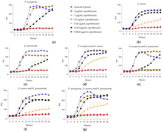
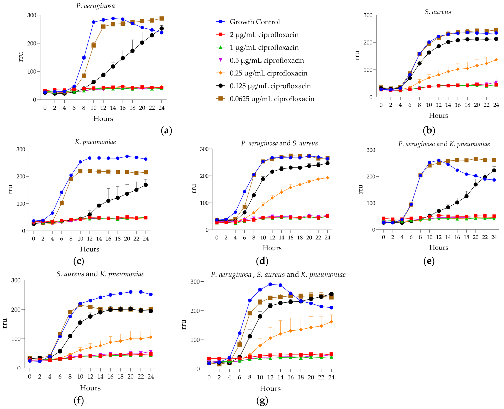
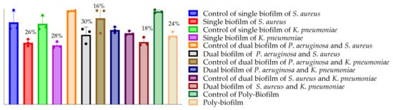
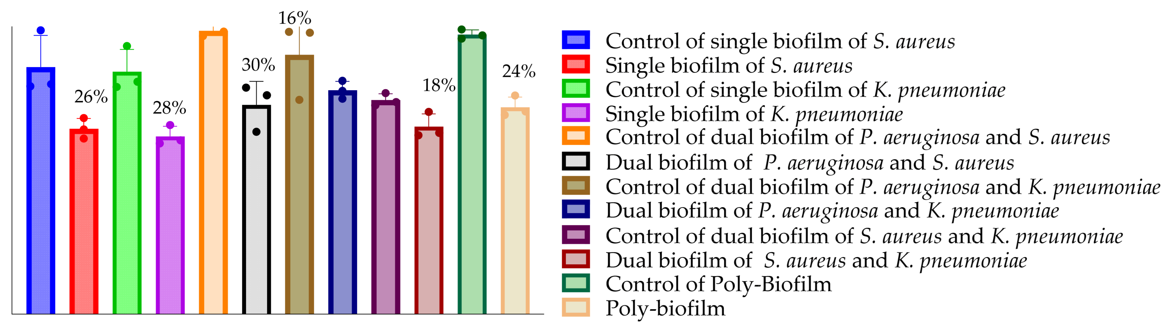
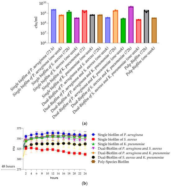
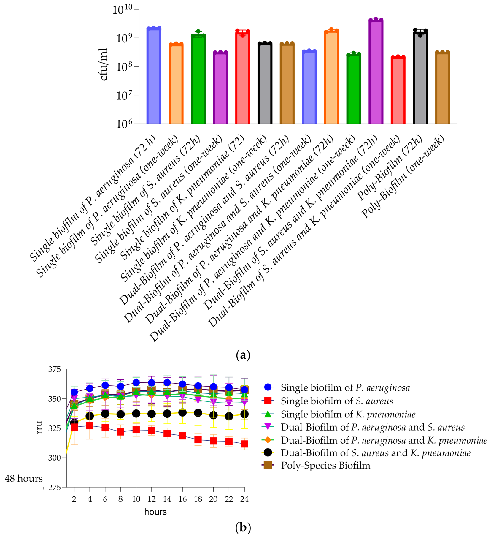
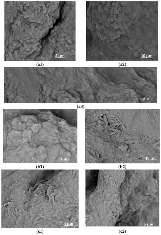
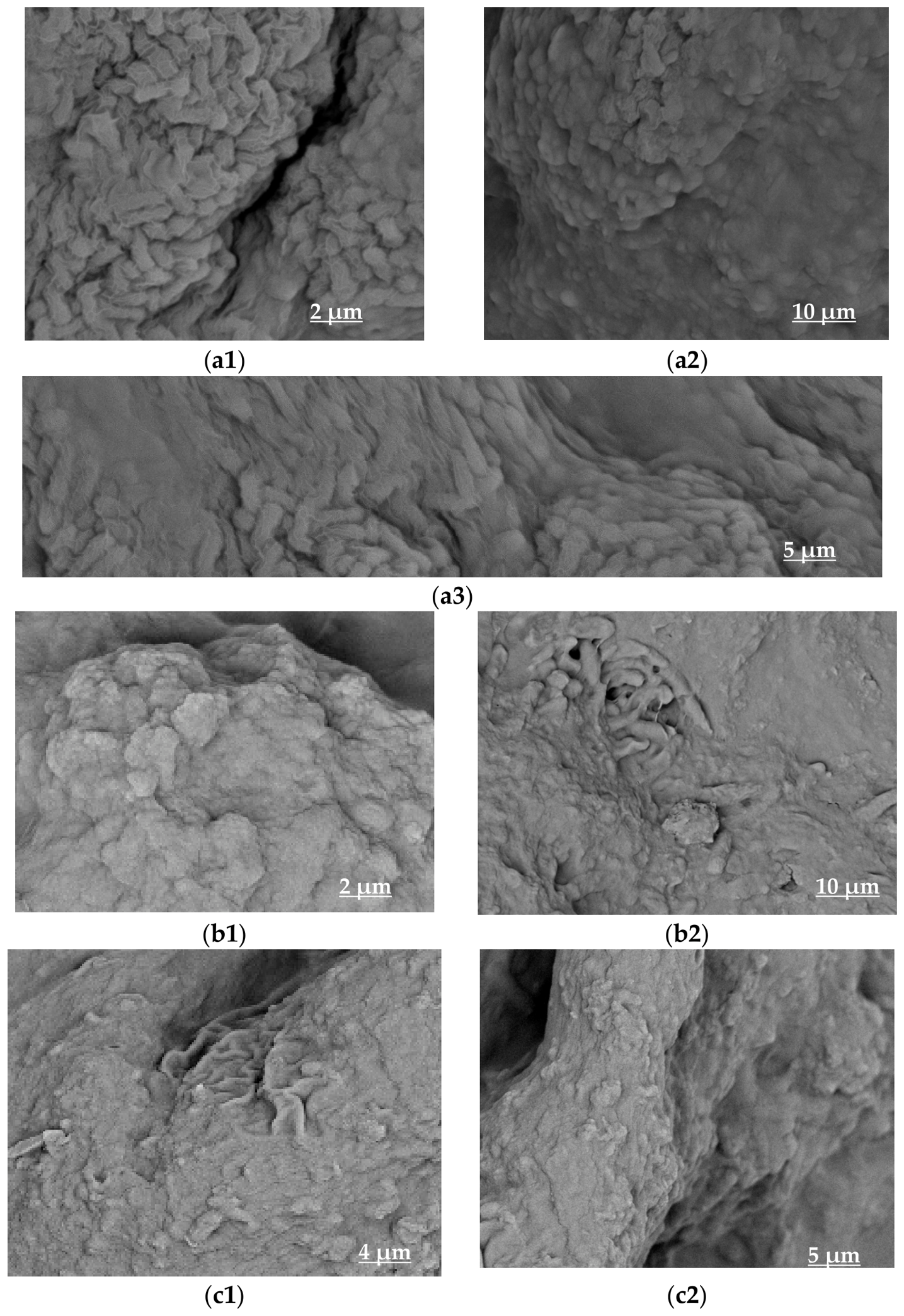


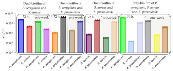
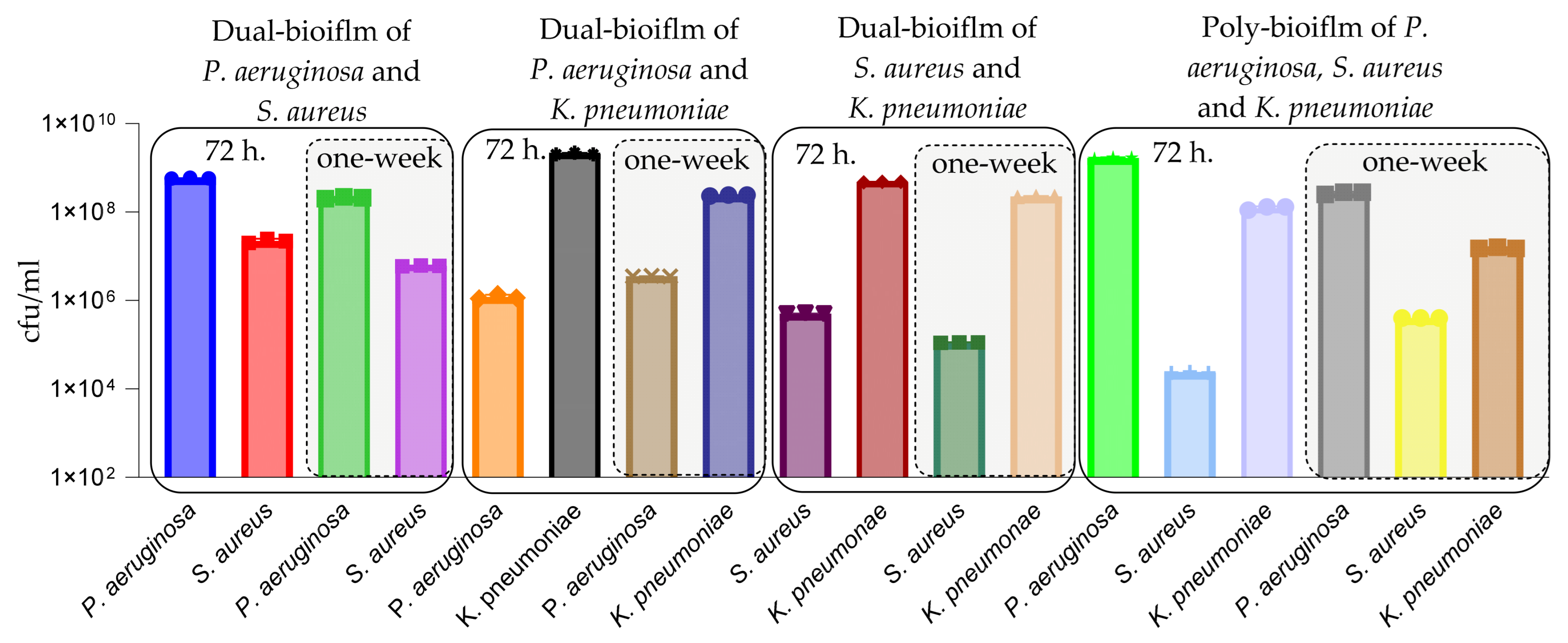
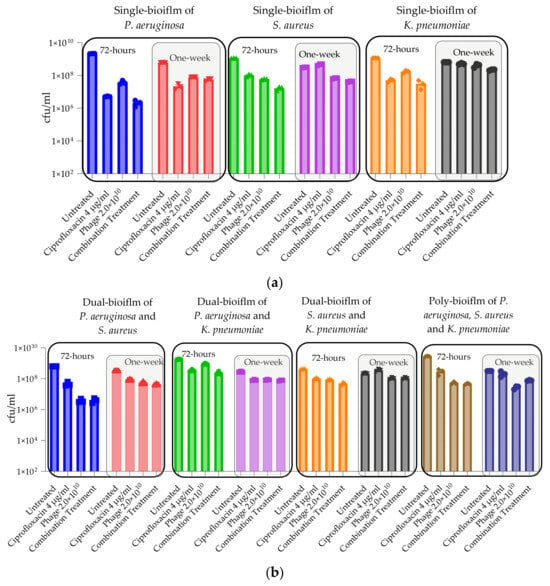
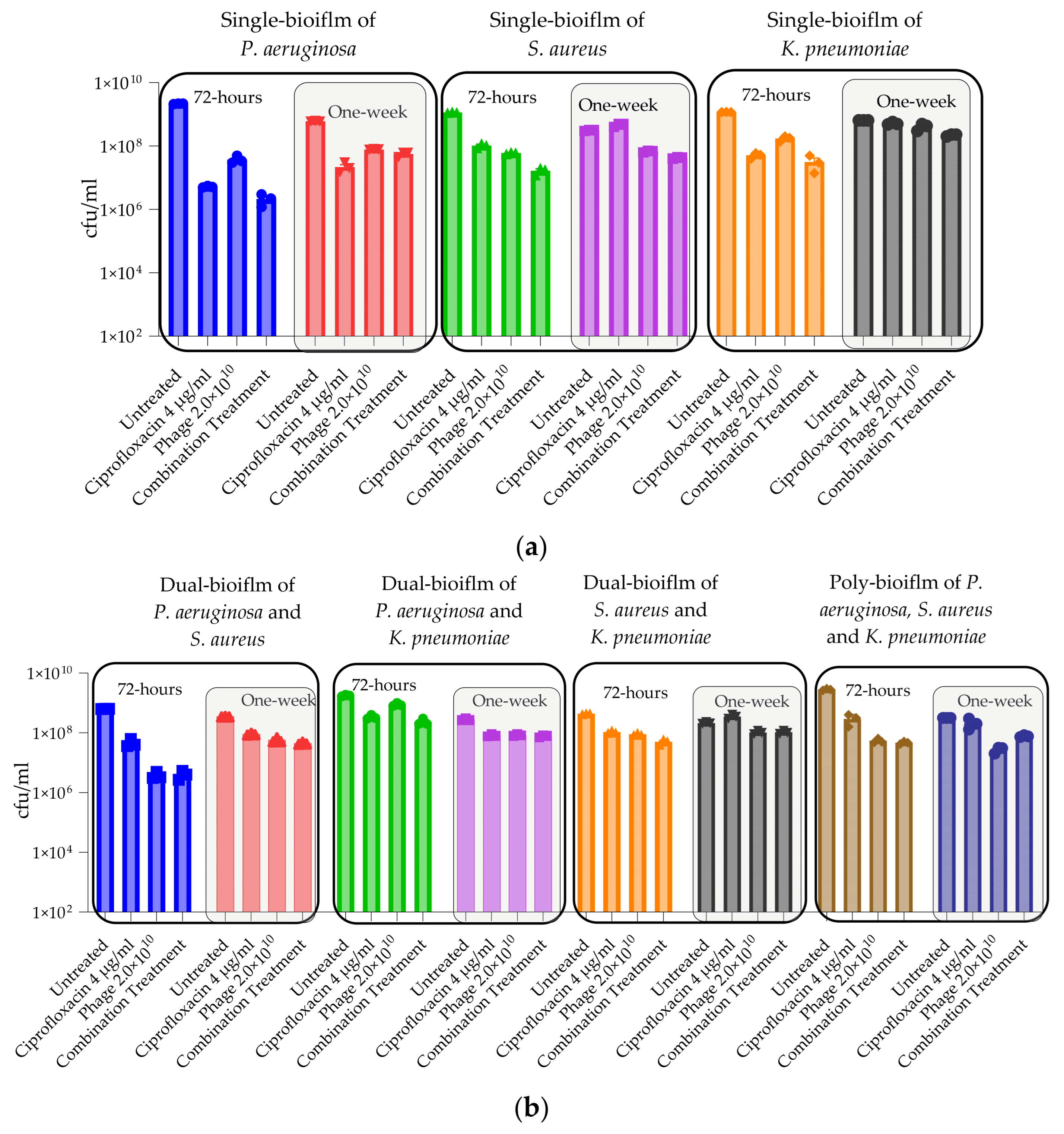
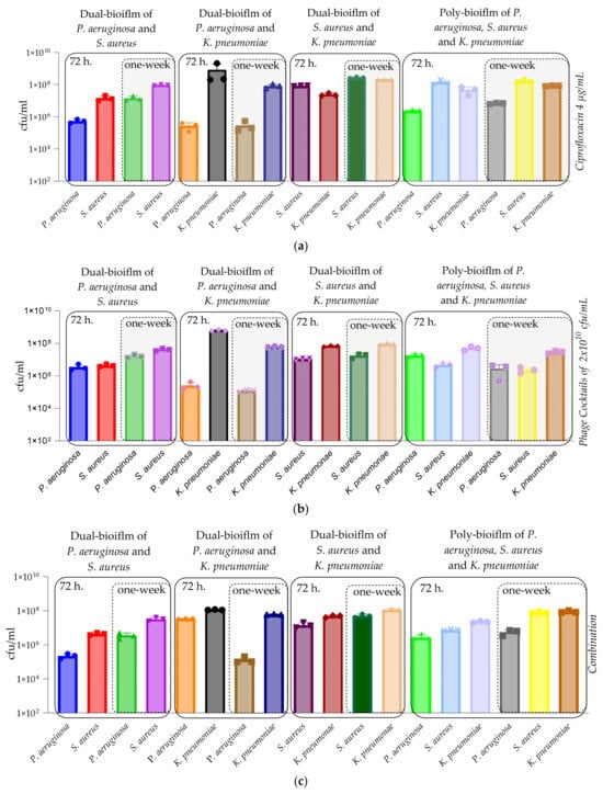
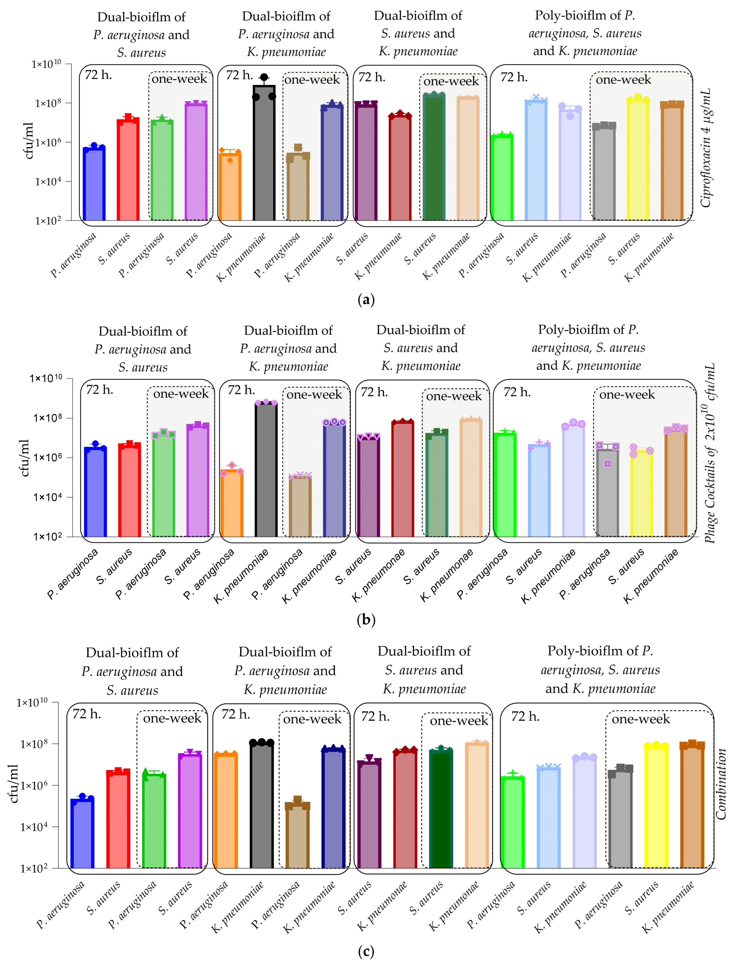
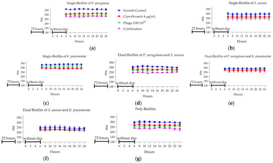
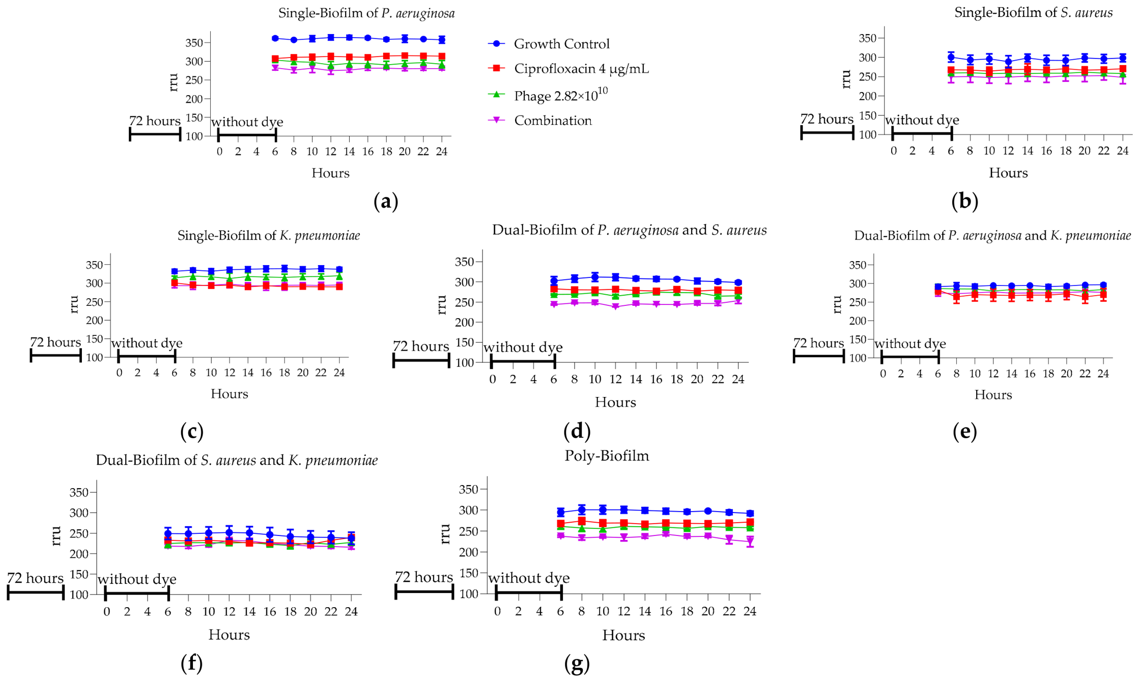
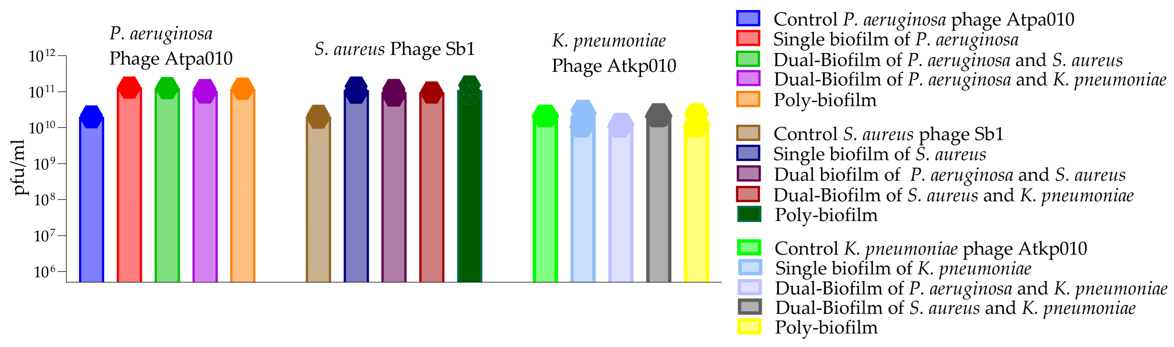
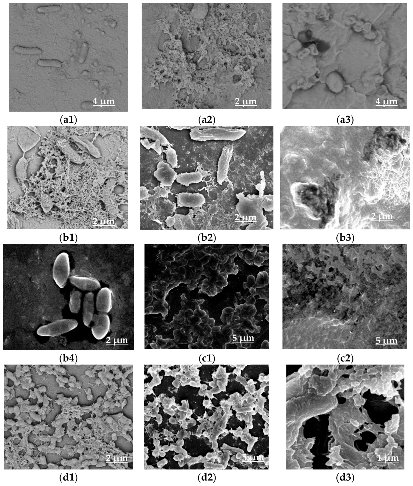
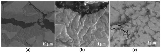
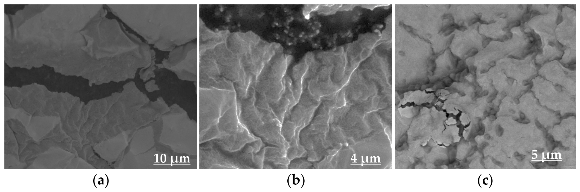
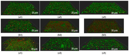
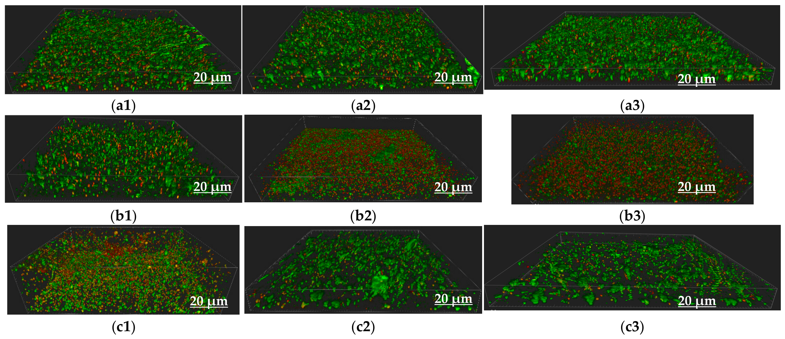
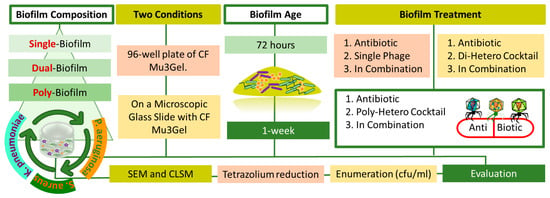
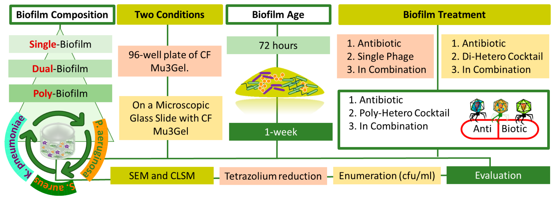

Disclaimer/Publisher’s Note: The statements, opinions and data contained in all publications are solely those of the individual author(s) and contributor(s) and not of MDPI and/or the editor(s). MDPI and/or the editor(s) disclaim responsibility for any injury to people or property resulting from any ideas, methods, instructions or products referred to in the content. |
© 2026 by the authors. Licensee MDPI, Basel, Switzerland. This article is an open access article distributed under the terms and conditions of the Creative Commons Attribution (CC BY) license.