Article

Global Antimicrobial Resistance Gene Study of Helicobacter pylori: Comparison of Detection Tools, ARG and Efflux Pump Gene Analysis, Worldwide Epidemiological Distribution, and Information Related to the Antimicrobial-Resistant Phenotype

by
Ricky Indra Alfaray
1,2,
Batsaikhan Saruuljavkhlan
1,
Kartika Afrida Fauzia
1,2,3,
Roberto C. Torres
4,
Kaisa Thorell
5,
Selva Rosyta Dewi
1,2,
Kirill A. Kryukov
6,
Takashi Matsumoto
1,
Junko Akada
1,
Ratha-korn Vilaichone
7,8,9,10,
Muhammad Miftahussurur
2,10,* and
Yoshio Yamaoka
1,10,11,12,*
1
Department of Environmental and Preventive Medicine, Faculty of Medicine, Oita University, Oita 879-5593, Japan
2
Helicobacter pylori and Microbiota Study Group, Institute of Tropical Disease, Universitas Airlangga, Surabaya 60286, Indonesia
3
Department of Public Health and Preventive Medicine, Faculty of Medicine, Universitas Airlangga, Surabaya 60132, Indonesia
4
The Center for Microbes, Development and Health, Key Laboratory of Molecular Virology and Immunology, Institute Pasteur of Shanghai, Chinese Academy of Sciences, Shanghai 200031, China
5
Department of Chemistry and Molecular Biology, Faculty of Science, University of Gothenburg, 405 30 Gothenburg, Sweden
6
Biological Networks Laboratory, Department of Informatics, National Institute of Genetics, Shizuoka 411-8540, Japan
7
Gastroenterology Unit, Department of Medicine, Faculty of Medicine, Thammasat University Hospital, Khlong Nueng 12120, Pathumthani, Thailand
8
Center of Excellence in Digestive Diseases, Thammasat University, Thailand Science Research and Innovation Fundamental Fund, Bualuang ASEAN Chair Professorship at Thammasat University, Khlong Nueng 12121, Pathumthani, Thailand
9
Department of Medicine, Chulabhorn International College of Medicine (CICM), Thammasat University, Khlong Nueng 12121, Pathumthani, Thailand
10
Division of Gastroentero-Hepatology, Department of Internal Medicine, Faculty of Medicine, Dr. Soetomo Teaching Hospital, Universitas Airlangga, Surabaya 60286, Indonesia
11
The Research Center for GLOBAL and LOCAL Infectious Diseases (RCGLID), Oita University, Oita 870-1192, Japan
12
Department of Medicine, Gastroenterology and Hepatology Section, Baylor College of Medicine, Houston, TX 77030, USA
*
Authors to whom correspondence should be addressed.
Antibiotics 2023, 12(7), 1118; https://doi.org/10.3390/antibiotics12071118
Submission received: 25 May 2023 / Revised: 15 June 2023 / Accepted: 25 June 2023 / Published: 28 June 2023
(This article belongs to the Special Issue Antibiotic Resistance and Therapy for Helicobacter pylori Infection)

Abstract

:
We conducted a global-scale study to identify H. pylori antimicrobial-resistant genes (ARG), address their global distribution, and understand their effect on the antimicrobial resistance (AMR) phenotypes of the clinical isolates. We identified ARG using several well-known tools against extensive bacterial ARG databases, then analyzed their correlation with clinical antibiogram data from dozens of patients across countries. This revealed that combining multiple tools and databases, followed by manual selection of ARG from the annotation results, produces more conclusive results than using a single tool or database alone. After curation, the results showed that H. pylori has 42 ARG against 11 different antibiotic classes (16 genes related to single antibiotic class resistance and 26 genes related to multidrug resistance). Further analysis revealed that H. pylori naturally harbors ARG in the core genome, called the ‘Set of ARG commonly found in the Core Genome of H. pylori (ARG-CORE)’, while ARG-ACC—the ARG in the accessory genome—are exclusive to particular strains. In addition, we detected 29 genes of potential efflux pump-related AMR that were mostly categorized as ARG-CORE. The ARG distribution appears to be almost similar either by geographical or H. pylori populations perspective; however, some ARG had a unique distribution since they tend to be found only in a particular region or population. Finally, we demonstrated that the presence of ARG may not directly correlate with the sensitive/resistance phenotype of clinical patient isolates but may influence the minimum inhibitory concentration phenotype.

Graphical Abstract

1. Introduction

Helicobacter pylori is a stomach Gram-negative pathogen that infects around half of the global human population, affecting more than 4 billion people [1,2,3]. Persistent infection of this bacterium can lead to various clinical implications, including gastritis, gastric atrophy, gastric peptic ulcers, gastric intestinal metaplasia, mucosa-associated lymphoid tissue lymphoma, and gastric cancer [4]. As an effective vaccine for H. pylori is yet to be discovered, curative treatment using antimicrobial drugs remains crucial to prevent further transmission and resolve disease augmentation [4,5]. However, several issues have arisen, including bad practices in the use of antibiotics leading to the antimicrobial resistance (AMR) phenomenon, which has become a leading concern for H. pylori eradication in the last decade [6,7,8].
The molecular driving mechanisms of AMR in H. pylori are already well-studied [5]. However, most studies merely focus on the mutation of antibiotic target alteration genes, despite the ability for the acquisition or loss of antimicrobial-resistant genes (ARG) by processes such as horizontal gene transfer (HGT) [9]. One reason is that mutation is believed to be the primary mechanism of AMR development in clinical conditions [5]. Nevertheless, this has resulted in less study of the current worldwide epidemiology of ARG in H. pylori. Thus, a global-scale study needs to be undertaken to understand the current landscape of H. pylori ARG epidemiology.
Several challenges need to be addressed, beginning with the preferred tools to better detect ARG in the H. pylori genome, the global H. pylori ARG distribution, and the impact of the presence of ARG on the clinical AMR phenotype. Hence, we attempted to address these challenges using a global H. pylori genome collection covering 68 countries from 5 different continents. We focused on ARG against drugs or other potential antimicrobial substances. In this study, we defined ARG as any potential acquired antimicrobial resistance-related gene listed in public ARG databases that include AMR-associated genes tested in many bacterial species (including members of the family Helicobacteraceae), but excluding most of well-known genes covering AMR through mutations, such as the mutations in pbp1A (ß-lactam-resistant), gyrA (fluoroquinolones-resistant), 23S rRNA (macrolides-resistant), 16S rRNA (tetracycline-resistant), and rpoB (ryfamycins-resistant), which mainly belong to the antibiotic target alteration gene, also known as drug target-mediated resistance or modification of the drug target [5]. Based on these criteria, we highlighted the global epidemiology of ARG in H. pylori and found that most are yet to be experimentally proven. In addition, we also analyzed the potential efflux pumps (EPs)-related AMR. These results will lead to many opportunities for future H. pylori ARG studies. Finally, we also attempted to identify new ARG candidates in H. pylori using antibiogram data retrieved from multinational clinical patient isolate strains.

2. Results

2.1. ARG Detection Tools and Comparison of Results

The ARG detection tools and methods varied between H. pylori genome studies. While some studies simply identified ARG using tools working with the BLAST-matches-based method for the nucleotide sequence [10,11], other studies have identified ARG using tools that work mainly with amino acid sequence database homology and single-nucleotide polymorphism (SNP) models [12,13]. Each tool was also used with different parameters (e.g., identity, coverage, e-value, and bit score), resulting in variable results between studies. These tools also rely on databases that are mostly not curated for H. pylori. To the best of our knowledge, there is no guideline for ARG detection purposes in the H. pylori study field. Thus, we tried to compare tools with different methods that can be used for ARG detection for H. pylori. These tools work with the BLAST-matches-based method, the homology and SNP model-based methods, the Hidden Markov model (HMM) screening method, or a combination of these (Table 1).
Initially, we performed ARG detection using the ABRICATE tool against several well-known ARG databases that use the BLAST-matches-based method [14]. Databases such as ARG-ANNOT [15], CARD [16,17], MEGARes [18], and ResFinder [19] are commonly used to identify ARG in the prokaryotic genome [20]. Therefore, we used these databases to obtain optimum results. In addition, we compared three different parameters for minimum identity and coverage: cov = 50 id = 50, cov = 70 id = 70, and cov = 90 id = 90. The results of these parameter sets were nearly the same, with the only slight difference being because of the coverage, as all results had the same identity of more than 90% (Figure S1). Therefore, using this tool’s parameter set of cov = 90 id = 90 for ARG detection could be a wise option to obtain more accurate results.
Using the parameter settings of minimum coverage and identity set to 90, we found 5 strains containing 5 genes from 3 different ARG using ARG-ANNOT, 2161 strains containing 2166 genes from 4 different ARG using CARD, 2159 strains containing 2162 genes from 3 different ARG using MEGARes, and 5 strains containing 5 genes from 3 different ARG using ResFinder (Figure 1a and Figure S1). The genes found by ARG-ANNOT were similar to those from ResFinder, and those found by CARD were the same as MEGARes, except for TEM. The CARD and MEGARes results included the results from ARG-ANNOT and ResFinder. Thus, the BLAST-matches-based method, using either the CARD or MEGARes database, yielded the best results for detecting ARG in H. pylori compared to other current databases.
Although the ARG names between CARD and MEGARes were slightly different, they referred to the same genes. For instance, hp1181 in CARD and HP in MEGARes both refer to the same Major Facilitator Superfamily (MFS). We confirmed this finding by aligning the gene sequences from both databases and the representative sequence from the H. pylori strain dataset (Figure S2a,b). Intriguingly, this gene was found in 99.35% (2156/2170) of the total strains. Each strain possessed one copy of this ARG, except for two strains (strain MS203 had three copies, and UM067 had two copies) (Spreadsheet S1).
After ABRICATE, we performed ARG detection using ResFinder [19]. This tool was mainly employed using the BLAST-matches-based method [19]. ResFinder and ABRICATE differ in terms of their databases: the ResFinder database in ABRICATE only contains ARG, while the ResFinder tool database is more varied, especially because it also contains disinfectant resistance genes and chromosomal mutations [19]. We used the ResFinder tool against the acquired and disinfectant resistance gene databases, using the default parameters, with the minimum coverage and identity set as 90%. The result for the acquired resistance genes was the same as ABRICATE using the ResFinder database (Figure 1a, Spreadsheet S2). No anti-disinfectant genes were detected.
Next, we performed ARG detection using the Resistance Gene Identifier (RGI) [17]. This tool works using the homology and SNP model-based methods [17]. In brief, RGI will algorithmically predict ARG and mutations from genomes using a mix of Prodigal open reading frame prediction [21], BLAST [22] or DIAMOND [23] sequence alignment, and curated resistance mutations included in the AMR detection model [17]. We used the default parameters and included the results based on the ‘strict’ and ‘perfect’ criteria only, except for hp1181, which was mainly detected as ‘loose’. However, after hand curation, hp1181 appeared in 99.95% (2169/2170) of the total strains. This result is accurate according to the previous version of the RGI (v5.2.1), where hp1181 was included in the ‘strict’ group. Nevertheless, in any version of RGI, the default database is the CARD database [17]. Using RGI v6.0.1 and the CARD database v3.2.5, 2022, we identified 4328 genes from seven different ARG (Figure 1a, Spreadsheet S3). The difference to the ABRICATE results was that RGI could detect vanT, including its variant vanTr. RGI could also detect more strains containing ARG and more hp1181 genes compared with ABRICATE (i.e., UM067 with two genes, N6 with two genes, MS203 with three genes, ASHA009 with two genes, and BGD53 with two genes). RGI could detect more ARG even if the genes were defined as a hypothetical protein by the Prokka annotation tool (Figure S3).
The next ARG detection tool was AMRFinderPlus, which combines BLAST-matches and the HMM screening method, with some improvements in the algorithm [24]. This tool is newly developed by the National Center for Biotechnology Information (NCBI) and combines nucleotide, protein, and HMM databases. We used the default parameters and added a ‘--plus’ flag to run AMRFinderPlus. Based on the recommendations of the software, we used the FASTA nucleotide assembly sequence (.fasta/.fna), protein sequence (.faa), and general feature format (.gff) to obtain the most accurate results [24]. We found 2176 genes from 5 different ARG based on AMRFinderPlus (Figure 1a, Spreadsheet S4). The difference between this tool and the other tools is that AMRFinderPlus detected copA, a gene encoding copper-binding metallochaperone CopP, in all strains.
Finally, the last ARG detection tool was HMMER, against the HMM ResFam Profile Database [25,26]. This tool specifically uses the HMM screening method. We used the parameter setting of e-value ≤ 1 × 10⁻¹⁰. As for the bit score, this score setting should be relative to every analysis, and we should understand the good score for the ideal query to obtain the best results while using the HMM ResFam Profile Database. Therefore, we compared results with different bit score values (≥100, ≥200, ≥250, ≥300, ≥400, and ≥500) and then evaluated the resul ts by hand curation against Prokka annotation results for several randomly selected representative strains that, relatively, had a higher number of ARG detected (Spreadsheet S5). We excluded the results if they contained antibiotic target alterations or unclear information (e.g., no clear information about the antibiotic class affected by the presence of the gene). The results implied that setting the score at ≥100 and ≥200 yielded many false-positive results (genes were detected as ARG but were actually various functional genes). In addition, many genes were detected with the same ARG name as if they were a copy number or variant, but each gene was actually different. The score ≥ 250 yielded a more accurate result, with particular caution for ACR_tran and msbA, as these genes often appear to have multiple copy numbers or variants when only one is correct and the other is actually a different gene. The score ≥ 500 yielded a more accurate result; however, many ARG could be left undetected. Thus, a bit score ≥ 250 should be used for H. pylori; however, the result should be treated carefully, for example, with further hand curation against genome annotation tool results (e.g., Prokka, RAST, PGAP, or D-FAST). Many inconsistencies of the annotation results could be found, with several genes annotated with different names compared to the Prokka annotation (e.g., MATE_efflux should be yeeO by Prokka). Therefore, careful hand curation, including comparison of the ResFam updated metadata and Prokka results (e.g., the gene’s name, location, similarity, and other information), is recommended. Finally, a total of 15,056 genes covering 7 ARG were found after hand curation, excluding inconsistent results and modification of the drug target (mutation-related ARG) (Spreadsheet S5).
An interesting finding was that one of the most predominant ARG was hp1181, or the MFS EPs family (found in 99.86%; 2167/2170 of the total strains based on HMMER against the HMM ResFam Profile Database). This gene was found by HMMER, RGI, and ABRICATE using the CARD or MEGARes databases, both of which contained a few H. pylori ARG. According to the data in CARD, this EPs is related to fluoroquinolone, nitroimidazole, and tetracycline antibiotic resistance [16,27]. The expression levels of these genes correlate with AMR development, but the protein structural analysis and functional mechanism in H. pylori remain unclear [27]. Therefore, we tried to predict the structure and function by looking for the closest protein reported in the Protein Data Bank (PDB) database. Investigation of the similarity of this protein against protein databases using Phyre2 showed that this MFS EPs structure is similar to the ‘drug efflux protein—MFS transporter’ reported in E. coli [28,29,30]. By Phyre2, as much as 99% of the amino acid template from the H. pylori MFS could be modeled with 100.0% confidence and 99% coverage against the E. coli MFS [29,30]. Next, we used a structure comparison tool via the SWISS-MODEL and MASS to confirm the similarity between H. pylori and E. coli MFS [31,32]. We built a new protein model of the H. pylori MFS using AlphaFold2 to yield a more accurate protein structure [33]. Overall, the results suggested that these structures have similarity (average root mean square deviation (RMSD) = 0.84) to E. coli (Figure S4); thus, it might have the same function in H. pylori as the E. coli MFS, that is, to help the bacteria becoming resistant to particular antibiotics.
Table 1. Tools, methods, and databases that are often used for H. pylori ARG detection.
ToolMethodDatabaseProvide Option for Mutation DetectionH. pylori ARG is IncludedDatabase Version, Year UpdatedTotal Number of Genes Found in 2170 StrainsTotal Number of Strains with ARGTotal ARG Name FoundTotal ARG Class FoundParameter Used
ABRICATE v1.0.1 [14]BLAST-matchesARG-ANNOT [15]NoNot yetv5, 20195533Minimum coverage and identity of 90
CARD [16,17]NoYes, but limitedMarch 2020 update 2166216144
MEGARes [18]NoNot yetv2.0, 20202162215933
ResFinder [19]NoYes20195533
ResFinder v4.2.3 [19]The aligners KMA and BLAST-matchesResFinder and DisinFinder [19]YesYes, but limited20225533Minimum coverage and identity of 90
The Resistance Gene Identifier (RGI) v6.0.1 [17]Homology and SNP modelsCARD [16,17,34]YesYes, but limitedv3.2.5, 20224328217074Default parameter and filter to obtain the ‘strict’ and ‘perfect’ results only
AMRFinderPlus v3.11.2 [24]Combination of BLAST-matches, HMM screening, and other improvement methodsCombination of the following:
Pathogen Detecton Reference Gene Catalog (https://www.ncbi.nlm.nih.gov/pathogens/refgene/#, accessed on 22 July 2021)
Pathogen Detecton Reference HMM Catalog (https://www.ncbi.nlm.nih.gov/pathogens/hmm/, accessed on 22 July 2021)
Bacterial Antimicrobial Resistance Reference Gene Database (https://www.ncbi.nlm.nih.gov/bioproject/PRJNA313047, accessed on 22 July 2021)
NCBIfam-AMRFinder (https://ftp.ncbi.nlm.nih.gov/hmm/NCBIfam-AMRFinder/latest/, accessed on 22 July 2021)
YesNot yetv2022-12-19.1, 20222176217054Default parameter, add’—plus’ flag, and three files typed as input (.fna, .faa, and .gff)
HMMER v3.3.2 [25]HMM screeningHMM ResFam Profile [26], which was trained using ARG protein sequence from CARD, the Lactamase Engineering Database (LacED), and Jacoby and Bush’s collection of curated beta-lactamase proteins.PossibleNot yetv1.2.2, 201815,0562170710e-value ≤ 1 × 10⁻¹⁰ and bit score ≥ 250 after hand curation

2.2. Pros and Cons of ARG Detection Tools

If we simply wished to perform an analysis targeting only complete genes and expecting only genes with high-similarity nucleotide sequences as the result, a convenient tool using BLAST-matches-based methods, such as ABRICATE, is a good choice, as the results are expected to only represent the complete genes. However, while the results are most likely accurate, these results will be limited because it will not detect as many ARG as other tool results. Another tool with the same BLAST-matches-based method, such as ResFam or ResFinder, might not provide the optimum results for ARG detection in H. pylori. The ResFam database does not contain specific H. pylori ARG compared with the CARD or MEGARes databases, while ResFinder is not as comprehensive as the CARD or MEGARes databases. The advantage of ResFinder is that it can also detect mutations related to AMR, as this tool applies KMA (k-mer alignment).
Next, for the RGI, our observations suggested that there are three advantages of RGI compared with ABRICATE and ResFinder. The first is that detection becomes more accurate and easier when we use assembly sequence as the input, because when we submit a nucleotide sequence, RGI first uses Prodigal to predict entire open reading frames (ORFs) and then evaluates the predicted protein sequences. This includes a subsequent correction by RGI if Prodigal misses the correct start codon, to anticipate entire AMR genes. Thus, it may detect more ARG even though the gene sequence might be partially different from the input and is often described as a hypothetical protein by the annotation tool (Figure S3). The second advantage is that it can also detect some mutations of antibiotic target alteration genes in H. pylori compared to other tools. This study found 611 23S rRNA genes with 3 well-known AMR mutations. Since these genes were not analogous to the focus of our study, we omitted these genes from the analysis results. The third advantage is that RGI could detect partial genes (e.g., we detected two MFS in BGD53, one complete while the other was partial). The consequence of this, that can become a limitation of this method, is that careful parameter measurement is required if we want to filter out these partial genes from our analysis.
Finally, for the HMM screening method, in this study, this method could detect more ARG variants compared with ABRICATE, ResFam, and RGI. The HMMER algorithms can predict genes that cannot be easily detected only using nucleotide BLAST-matches-based (mass screening) methods [25]. This method will detect genes with relatively high nucleotide sequence variation. For example, the outer membrane efflux protein (OEP) family often has poor sequence conservation in several species, including H. pylori [35]. The HMM ResFam profile was constructed using ResFam and other databases, including TIGRFam [26]. The developer compared HMMs from ResFam with pairwise sequence alignment (BLAST) against the CARD and ARDB databases to evaluate their ability to predict AR function and found that ResFam detected more genes compared to the others [26]. However, for H. pylori genome analysis, hand curation is strongly recommended before drawing conclusions from the results because many false positives can be detected. Although the curation step is time-consuming, it will yield more accurate and conclusive results.

2.3. Curation of ARG Detection Results Is Necessary

Detection of ARG should be followed by careful curation, as the results from the detection tools can vary depending on their built-in methods and databases. For example, this curation can be performed by matching the identified ARG results with the whole-genome annotation results. After combining the genes identified by all tools, including ‘loose’ hits by RGI, we performed curation, then confirmed whether these genes existed within the Prokka results. Next, hypothetical proteins from the Prokka results were assigned as ARG if they were identified as resistance genes by RGI in the ‘strict’ and ‘perfect’ parameters. This curation step provides high confidence to the ARG detection results. A total of 30 ARG were included after this curation step.
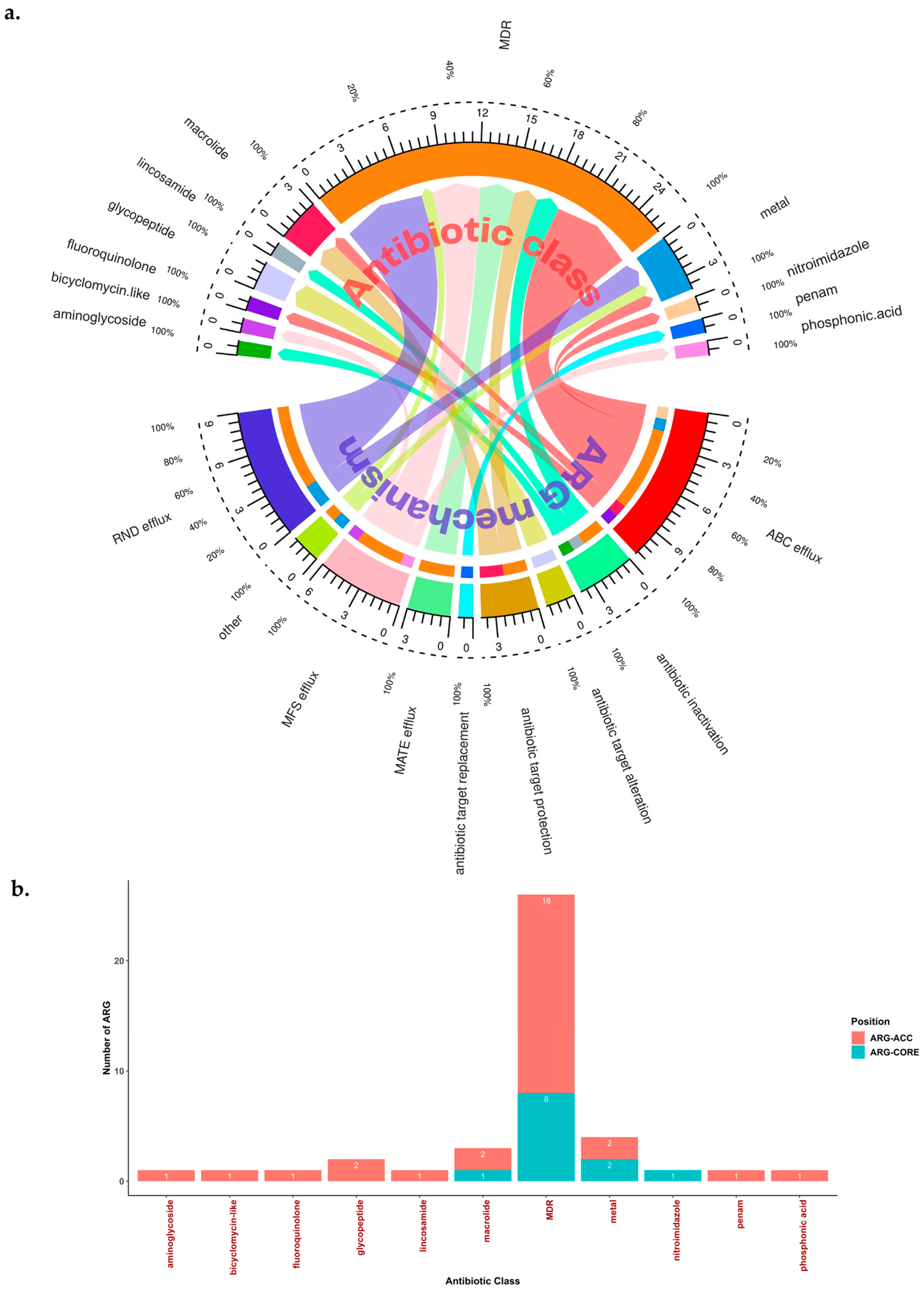
In addition, we realized that Prokka could possibly find more ARG that sometimes could not be detected by ARG detection tools. Indeed, when we extracted genes that contained any of the following keywords: ‘resistant’, ‘resistancy’, ‘resistance’, ‘drug’, ‘antibiotic’, and ‘antimicrobial’, we newly found 12 ARG that were not yet identified by any of the ARG detection tools. Finally, a total of 42 ARG were used for the subsequent analyses in this study (Figure 1b, Spreadsheet S6).

2.4. Summary of Global ARG Detection Results from All Tools and Databases

Of the 42 ARG, 16 were related to single antibiotic class resistance (from 9 different antibiotic classes), while the other 26 were related to multidrug resistance (MDR) (Table 2). These 42 ARG have several resistant mechanisms, with the most common mechanism being correlated with EPs (69.05%; 29/42) [26] (Figure 2a). We found 4 major families of EPs in H. pylori ARG: Resistance Nodulation Cell Division (RND) efflux (31.03%; 9/29), ATP Binding Cassette (ABC) efflux (37.93%; 11/29), Major Facilitator Superfamily (MFS) efflux (20.69%; 6/29), and Multidrug and Toxin Extrusion (MATE) efflux (10.34%; 3/29). These EPs were related to single drug class resistance (27.59%; 8/29) or MDR (72.41; 21/29) (Figure 2a).
Further in-depth genome observations revealed that 12 of the ARG could be found in the core genome (with the cut-off value of ≥95% of the total strains), while the other 30 could be found in the accessory genomes (Figure 2b). We called this ARG core genome the ‘Set of ARG commonly found in the Core Genome of H. pylori’ (ARG-CORE) (28.57%; 12/42), while ARG in the accessory genome were termed the ‘Set of ARG commonly found in the Accessory Genome of H. pylori’ (ARG-ACC) (71.43%; 30/42). Genes related to MDR were the most commonly found ARG in both ARG-CORE and ARG-ACC (66.67%; 8/12, and 60.00%; 18/30, respectively). Interestingly, genes such as msbA and carA were included in the ARG-CORE group, which aligns with the high prevalence of metronidazole and clarithromycin resistance in the current global H. pylori AMR status [7]. Other genes such as vanT and vanTr can be found in most of the strains (Table 2). Although these genes had a resistant mechanism as the antibiotic target alteration, we still included them in our analysis because the presence of vanT or vanTr may lead to a different phenotype which has not been thoroughly studied yet in H. pylori, thus, leading to an interesting opportunity for future study. This gene can be considered as ARG-CORE if vanT and vanTr together are considered as a same gene. However, we separated this gene following the tool and database identification results. Based on the identification results, vanT and vanTr will usually never be found in a single strain. Among other reasons, vanT can partially answer why H. pylori is naturally resistant to glycopeptides (e.g., vancomycin) [36].

2.5. Potential Efflux Pumps (EP)-Related AMR and MDR in H. pylori

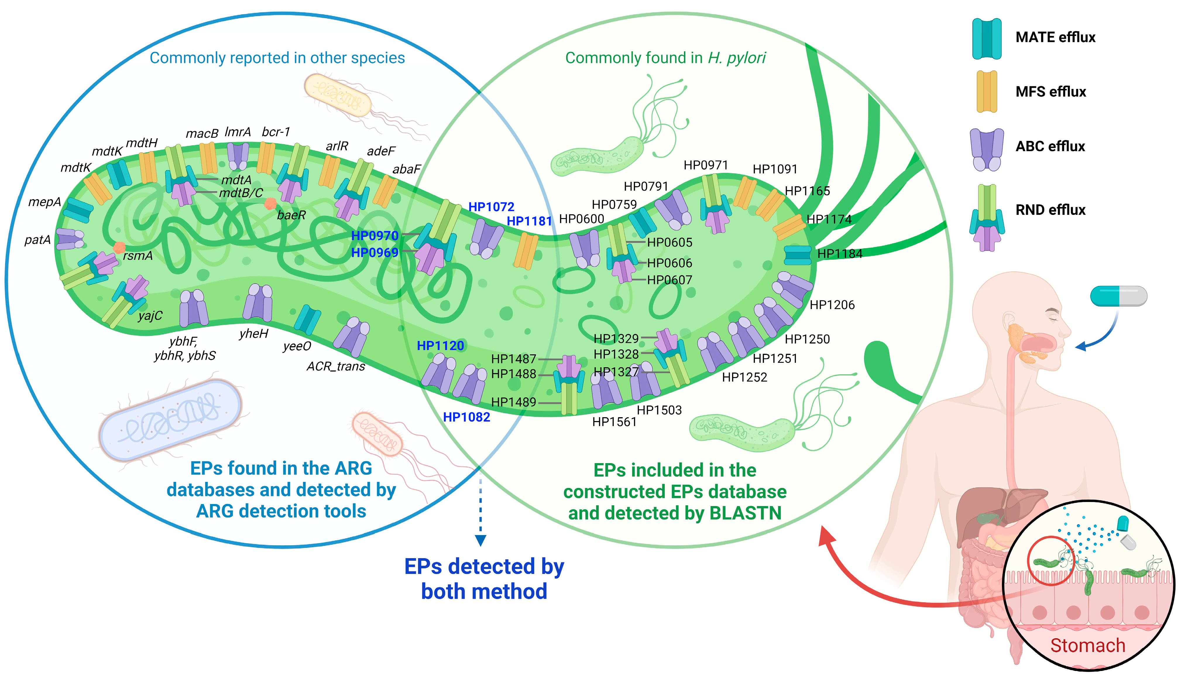
Global ARG detection results showed several ARG belonging to EPs. Thus, other EPs that may correlate with AMR in H. pylori might be left undetected. Previous studies in H. pylori showed that several EPs could be related to AMR and MDR phenotypes in H. pylori [27,37,38,39,40,41,42,43,44,45,46,47]. A few of these EP genes were already identified in the ARG detection results (e.g., HP1181 in Figure 1b), while the others remain unidentified. This is because these databases (Table 1) may not contain all the EPs related to AMR in H. pylori. Therefore, we tried to construct a special query database containing EP sequences that were previously reported to correlate with AMR in H. pylori. A total of 28 EPs related to AMR or MDR based on previous reports were included, including EP genes that were identified by the ARG detection tools (Table S1) [27,37,38,39,40,41,42,43,44,45,46,47]. We added one more EP gene which detected because the name containing the ‘keywords’ (i.e., HP1120), therefore, the total of EPs-related AMR becomes 29 genes. Next, we tried to detect these EP genes using the BLASTN method against 2170 WGS. Initially, we tried to compare different parameters of identity and coverage to obtain the optimum results. Parameter identity and coverage of 50, 70, and 80 showed comparable results in all the EP genes, while identity and coverage of 90 showed a different value for HP1250 and HP1561. The total number of HP1250 should be almost similar to HP1251 and HP1252 since they are a set of ABC EPs (Figure S5). Notably, depending on the objective, higher identity and coverage should yield fewer false-positive results for simple presence/absence analysis using BLASTN. Thus, a parameter identity and coverage of 80 might be the optimum settings to detect EP genes in H. pylori, as shown at least in this study setting. The EP detection results showed that almost all EP genes were ARG-CORE, except HP1206 and HP0600 (Table 2). Generally, each strain will have one of each EP gene.
Table 2. Summary of ARG detected by tools after curation and EPs related to AMR.
Gene Name (According to the Databases) ARG-CORE (Detected in ≥95% of Total Strains) or ARG-ACCAntibiotic Target Resistance MechanismAdditional Information (Including Gene Description by Prokka or Protein Homologous Name or Another Alternative Name a)AMR Gene Family (by CARD)Prevalence in Total Genome Dataset (n = 2170)
ARG detected by tools after curation
abaFARG-ACCphosphonic acid antiobioticMFS effluxMajor Facilitator Superfamily (MFS) antibiotic efflux pump; fosfomycin resistance protein AbaFMajor Facilitator Superfamily (MFS) antibiotic efflux pump1.01%
(22/2170)
adeFARG-ACCMDR (e.g., tetracycline, fluoroquinolone)RND efflux-resistance-nodulation-cell division (RND) antibiotic efflux pump0.05%
(1/2170)
APH(3)-IIIaARG-ACCaminoglycosideantibiotic inactivation-APH(3′)0.09%
(2/2170)
arlRARG-COREMDR (e.g., fluoroquinolone, disinfecting agents, and antiseptics)MFS effluxResponse regulator ArlRMajor Facilitator Superfamily (MFS) antibiotic efflux pump99.91%
(2168/2170)
baeRARG-ACCMDR (e.g., aminocoumarin antibiotic, aminoglycoside antibiotic)RND effluxTranscriptional regulatory protein BaeRresistance-nodulation-cell division (RND) antibiotic efflux pump0.05%
(1/2170)
bcr-1ARG-ACCBicyclomycin-like antibiotic (it is also possible as MDR)MFS effluxBicyclomycin resistance proteinMajor Facilitator Superfamily (MFS) antibiotic efflux pump0.05%
(1/2170)
TEM-116ARG-ACCMDRantibiotic inactivation-TEM beta-lactamase0.09%
(2/2170)
carAARG-COREmacrolideantibiotic target protection-Miscellaneous ABC-F subfamily ATP-binding cassette ribosomal protection proteins99.82%
(2166/2170)
cnrBARG-ACCmetalotherNickel and cobalt resistance protein CnrB4-hydroxy-tetrahydrodipicolinate synthase0.18%
(4/2170)
copAARG-COREmetalABC effluxCopper-exporting P-type ATPase-99.95%
(2169/2170)
czcAARG-COREmetalRND effluxCobalt-zinc-cadmium resistance protein CzcA-99.82%
(2166/2170)
czcBARG-ACCmetalRND effluxCobalt-zinc-cadmium resistance protein CzcB-74.65%
(1620/2170)
ebrBARG-ACCMDR (e.g., carbapenem, cephalosporin, penam)antibiotic inactivation-Multidrug resistance protein EbrB0.05%
(1/2170)
hp1181ARG-COREMDR (e.g., tetracycline, nitroimidazole, fluoroquinolone)MFS effluxin Prokka, can be detected as yfcJ; putative MFS-type transporter YfcJMajor Facilitator Superfamily (MFS) antibiotic efflux pump99.95%
(2169/2170)
lmrAARG-ACCMDR (e.g., lincosamide antibiotic)ABC effluxMultidrug resistance ABC transporter ATP-binding and permease protein-0.28%
(6/2170)
lnuAARG-ACClincosamideantibiotic inactivationlinAlincosamide nucleotidyltransferase (LNU)0.05%
(1/2170)
macBARG-ACCmacrolideABC effluxpvdTATP-binding cassette (ABC) antibiotic efflux pump0.09%
(2/2170)
mdtAARG-ACCMDR (e.g., aminocoumarin)RND effluxyegM; Multidrug resistance protein MdtAresistance-nodulation-cell division (RND) antibiotic efflux pump0.09%
(2/2170)
mdtBARG-ACCMDR (e.g., aminocoumarin)RND effluxyegN; Multidrug resistance protein MdtBresistance-nodulation-cell division (RND) antibiotic efflux pump0.28%
(6/2170)
mdtCARG-COREMDR (e.g., aminocoumarin)RND effluxyegO; Multidrug resistance protein MdtCresistance-nodulation-cell division (RND) antibiotic efflux pump99.54%
(2160/2170)
mdtHARG-ACCMDR (e.g., fluoroquinolone antibiotic)MFS effluxyceL; Multidrug resistance protein MdtHresistance-nodulation-cell division (RND) antibiotic efflux pump0.32%
(7/2170)
mdtKARG-ACCMDR (e.g., aminocoumarin)MATE efflux norE; norM; ydhE; Multidrug resistance protein MdtKresistance-nodulation-cell division (RND) antibiotic efflux pump1.15%
(25/2170)
mdtLARG-ACCMDR (e.g., aminocoumarin)MFS effluxyidY; Multidrug resistance protein MdtLresistance-nodulation-cell division (RND) antibiotic efflux pump0.09%
(2/2170)
mecAARG-ACCpenamantibiotic target replacementAdapter protein MecAMethicillin-resistant PBP20.05%
(1/2170)
mepAARG-COREMDR (e.g., tetracycline, glycylcycline)MATE effluxMultidrug export protein MepAmultidrug and toxic compound extrusion (MATE) transporter99.63%
(2162/2170)
MFS_effluxARG-COREMDRABC effluxHP1120 (COG1131); Multidrug efflux system ATP-binding protein -99.68%
(2163/2170)
msbAARG-COREnitroimidazoleABC effluxLipid A export ATP-binding/permease protein MsbAATP-binding cassette (ABC) antibiotic efflux pump99.82%
(2166/2170)
patAARG-ACCfluoroquinoloneABC effluxPeptidoglycan O-acetyltransferaseATP-binding cassette (ABC) antibiotic efflux pump13.69%
(297/2170)
ramAARG-ACCMDR (e.g., tetracycline, rifamycin, phenicol, carbapenem, penem, penam, cephalosporin, cephamycin, glycylcycline, disinfecting agents and antiseptics, monobactam, fluoroquinolone)Other (reduced permeability to antibiotic, based on CARD, it can actually be considered as an efflux pump complex or subunit conferring antibiotic resistance)RamA (resistance antibiotic multiple) is a positive regulator of AcrAB-TolC. General Bacterial Porin with reduced permeability to beta-lactams, resistance-nodulation-cell division (RND) antibiotic efflux pump0.09%
(2/2170)
rsmAARG-COREMDR (e.g., diaminopyrimidine, phenicol, fluoroquinolone)RND effluxcsrA; Ribosomal RNA small subunit methyltransferase Aresistance-nodulation-cell division (RND) antibiotic efflux pump99.91%
(2168/2170)
salBARG-ACCMDR (e.g., streptogramin, lincosamide, pleuromutilin, streptogramin A)antibiotic target protection-sal-type ABC-F protein0.05%
(1/2170)
salCARG-ACCMDR (e.g., streptogramin, lincosamide, pleuromutilin, streptogramin A)antibiotic target protection-sal-type ABC-F protein1.11%
(24/2170)
srmBARG-ACCmacrolideantibiotic target protectionATP-dependent RNA helicase SrmBMiscellaneous ABC-F subfamily ATP-binding cassette ribosomal protection proteins0.97%
(21/2170)
vanT gene in vanG clusterARG-ACCglycopeptideantibiotic target alteration-glycopeptide resistance gene cluster, vanT75.62%
(1641/2170)
vanTr gene in vanL clusterARG-ACCglycopeptideantibiotic target alteration-glycopeptide resistance gene cluster, vanT24.33%
(528/2170)
yajCARG-ACCMDR (e.g., tetracycline, disinfecting agents and antiseptics, glycylcycline, rifamycin, cephalosporin, penam, phenicol, fluoroquinolone, glycopeptide, oxazolidinone)RND effluxSec translocon accessory complex subunit YajCresistance-nodulation-cell division (RND) antibiotic efflux pump47.05%
(1021/2170)
ybhFARG-ACCMDRABC effluxputative multidrug ABC-transporter ATP-binding protein YbhF-0.05%
(1/2170)
ybhRARG-ACCMDRABC effluxputative multidrug ABC transporter permease YbhR-7.33%
(159/2170)
ybhSARG-ACCMDRABC effluxputative multidrug ABC transporter permease YbhS-57.47%
(1247/2170)
yheHARG-ACCMDRABC effluxputative multidrug resistance ABC transporter ATP-binding/permease protein YheH; bmrA-1.29%
(28/2170)
MATE_efflux_yeeOARG-COREMDRMATE effluxCan be shown as yeeO by Prokka annotation-0.05%
(1/2170)
ACR_tranARG-COREMDRABC effluxCan be shown as several different names (mdtcA, czcA, cusA, or bepA) by Prokka annotation. Should be manually curated to differ from the above genes.-99.91% (2168/2170)
EPs related to AMR a,b
(Locus tag based on H. pylori strain 26695 [NC_000915.1]. Please refer to Table S1 for more information for the reference and gene characteristics.)
HP0600ARG-ACCMDR (e.g., metronidazole, levofloxacin)ABC effluxspaB-62.53%
(1357/2170)
HP0605ARG-COREMDR (bilesalt, cefotaxime, ceragenin, clindamycine, clarithromycin, erythromycin, ethidium bromide (EtBr), novobiocin, metal ion, nickel, sodium deoxycholate, tetracycline)RND effluxhefA; efflux RND transporter outer membane subunit HefA-99.68%
(2163/2170)
HP0606ARG-COREMDR (bilesalt, cefotaxime, ceragenin, clindamycine, clarithromycin, erythromycin, EtBr, novobiocin, metal ion, nickel, sodium deoxycholate, tetracycline)RND effluxhefB (alternative name: acrA or mtrC); efflux RND transporter periplasmic adaptor subunit HefB -99.68%
(2163/2170)
HP0607ARG-COREMDR (bilesalt, cefotaxime, ceragenin, clindamycine, clarithromycin, erythromycin, EtBr, novobiocin, metal ion, nickel, sodium deoxycholate, tetracycline)RND effluxhefC (alternative name: acrB); efflux RND transporter permease subunit HefC-99.63%
(2162/2170)
HP0759ARG-COREMDRMATE effluxconserved hypothetical integral membrane protein; MATE family efflux transporter-99.77%
(2165/2170)
HP0791ARG-COREmetal (cadmium, zinc)ABC effluxcadA; heavy-metal translocating P-type ATPase-99.59%
(2161/2170)
HP0969ARG-COREMDR (e.g., cadmium, metronidazole, nickel, zinc)RND effluxhefF (alternative name: czcA1 or cznA)-99.86%
(2167/2170)
HP0970ARG-COREMDR (e.g., cadmium, metronidazole, nickel, zinc)RND effluxhefE (alternative name: czcB1 or cznB); efflux RND transporter periplasmic adaptor subunit-99.77%
(2165/2170)
HP0971ARG-COREMDR (e.g., cadmium, metronidazole, nickel, zinc)RND effluxhefD (alternative name: cznC)
Note: HefFDE is a homolog of MexA
-99.77%
(2165/2170)
HP1072ARG-COREcopperABC efflux copA-99.72%
(2164/2170)
HP1082ARG-COREMDR (erythromycin, etbr, novobiocin, rifampin, and lipopolysaccharide)ABC effluxmsbA-99.63%
(2162/2170)
HP1091ARG-CORE-MFS effluxkgtP-98.57%
(2139/2170)
HP1120ARG-COREMDRABC effluxCcmA; Multidrug efflux system ATP-binding protein (NP_208012.1); ABC-type multidrug transport system, ATPase component (COG1131)-99.68%
(2163/2170)
HP1165ARG-COREtetracyclineMFS effluxtetA-99.49%
(2159/2170)
HP1174ARG-CORED Glactose (non-drug)MFS effluxgluP-99.68%
(2163/2170)
HP1181ARG-COREMDR (e.g., tetracycline, nitroimidazole, fluoroquinolone)MFS effluxMultidrug efflux transporter-99.95%
(2169/2170)
HP1184ARG-COREnorfloxacin and ethidiumMATE effluxNorM; HP1184 family multidrug efflux MATE transporter, conserved hypothetical integral membrane protein; MatE Polysacc_synt_C-99.77%
(2165/2170)
HP1206ARG-ACCMDR (possibly related to metronidazole and levofloxacin resistance)ABC effluxhetA; multidrug resistance protein (HetA)-94.70%
(2050/2170)
HP1250ARG-CORE-ABC effluxCsd5-96.96%
(2104/2170)
HP1251ARG-CORE-ABC effluxoligopeptide ABC transporter, permease protein (OppB); microcin C transport system permease protein-99.72%
(2164/2170)
HP1252ARG-CORE-ABC effluxOppA-99.59%
(2161/2170)
HP1327ARG-COREmetal (copper, cobalt, zinc cadmium ion)RND effluxhefG (alternative name: crdB); copper resistance outer membrane protein CrdB-99.40%
(2157/2170)
HP1328ARG-COREmetal (copper, cobalt, zinc cadmium ion)RND effluxhefH (alternative name: czcB2); efflux RND transporter periplasmic adaptor subunit-99.63%
(2162/2170)
HP1329ARG-COREmetal (copper, cobalt, zinc cadmium ion)RND effluxhefI (alternative name: czcA2, cusA)-99.31%
(2155/2170)
HP1487ARG-COREMDR (e.g., novobiocin, deoxycholate, EtBr resistance)RND efflux (or ABC efflux)ABC-2 type transport system permease protein-99.59%
(2161/2170)
HP1488ARG-COREMDR (e.g., novobiocin, deoxycholate, EtBr resistance)RND efflux (or ABC efflux)Membrane-fusion protein HlyD family secretion protein-99.59%
(2161/2170)
HP1489ARG-COREMDR (e.g., novobiocin, deoxycholate, EtBr resistance)RND efflux (or ABC efflux)TolC-like outer membrane efflux protein -99.59%
(2161/2170)
HP1503ARG-COREmetalABC effluxcation-transporting ATPase, P-type (copA), P-type Cu+ transporter-99.77%
(2165/2170)
HP1561ARG-COREmetal (nickel, copper)ABC effluxIron(III) ABC transporter, periplasmic iron-binding protein (ceuE), iron complex transport system substrate-binding protein-98.02%
(2127/2170)
a The information (including the short description, homolog, and alternative name) and mechanisms of the EP genes were obtained based on previous publications and the KEGG database for Helicobacter pylori 26695 [5,35,38,44,47,48,49,50,51,52]. Some of the information on antibiotic targets and alternative names was also presented based on the CARD database [34]. The efflux pumps’ classification was assigned based on previous studies and established databases [47,53]. We have supplemented additional optional information whenever discrepancies were identified between the studies. b Some of the gene names were described based on the previous studies; therefore, some names might be different than the original annotation of the H. pylori 26695 genome in GenBank (NC_000915.1 or NC_018939.1).
Next, the comparison between ARG detection and BLASTN against the constructed 29 EPs database showed that six H. pylori EPs could be detected by both ARG detection tools and BLASTN (Figure 3). This result suggests that the ARG databases already contain some EPs that are commonly found in H. pylori (ARG-CORE), although the names might be different between databases and H. pylori annotations. The combination of these methods showed that H. pylori could have at least 50 EPs-related AMR genes. While most of the EPs in the constructed EPs gene database that had detected by BLASTN were already reported in H. pylori (Figure 3, green circle), most of the genes detected by ARG detection tools were commonly reported in other species (e.g., E. coli). Some genes detected by ARG detection tools were only identified in a few strains, suggesting that these genes might have been obtained by HGT via Mobile Genetic Elements (MGE), possibly from other species living in the gastrointestinal tract (Figure 3, blue circle).

2.6. Global Geographic and Population Distribution of the ‘Set of ARG Commonly Found in the Accessory Genome of H. pylori’ (ARG-ACC)

Understanding the distribution of ARG is essential to illuminate the general epidemiology of ARG patterns in global H. pylori. Since all H. pylori have ARG because of ARG-CORE, we focused on the distribution of ARG-ACC, which is unique for every strain. We clustered the ARG-ACC genes based on their antibiotic class and identified their distribution based on the geographic location and H. pylori population.
The geographic assessment showed that the distribution of ARG-ACC genes was almost same between countries. However, we found that some antibiotic classes are unique to a particular country or region (Figure 4a and Figure S6, Spreadsheet S7). These antibiotic classes were the aminoglycosides, bicyclomycin-like, lincosamide, and penam, which were only found in Poland, Finland, Venezuela, and Switzerland, respectively, in our dataset. In addition, genes related to nickel and cobalt resistance were only found in China, Vietnam, and Australia. Overall, while ARG-ACC genes from different antibiotic classes were generally found on all continents, each continent had unique characteristics, e.g., aminoglycoside and lincosamide were only shown in Europe and America, respectively, and the phosphonic acid proportion was lower in Africa compared to the other continents (Figure 4b).
Next, the assessment of the H. pylori population showed that several ARG-ACC in some antibiotic classes seemed evenly distributed, while some were predominantly found in a particular population. The ARG-ACC related to bicyclomycin-like and metal resistance (nickel, cobalt, zinc, and cadmium) could be found in all H. pylori populations. In addition, the ARG-ACC related to macrolide and fluoroquinolone resistance was predominantly found in HpEurope, compared to other populations (Figure 4c).

2.7. Association between ARG and Antimicrobial-Resistant Phenotype in H. pylori

Acquiring ARG via HGT may affect the resistance phenotype in bacteria; however, the development of resistance is multifactorial and unique between species of bacteria [54]. We attempted to understand the difference in the presence of ARG between the AMR phenotypes of clinical isolates using antibiogram data from clinical isolates collected from multiple nations. The result suggested no difference in the total ARG between resistant and susceptible strains. However, H. pylori resistant to metronidazole and levofloxacin tended to have a higher average total number of ARG compared with the susceptible group (Figure S7). Therefore, this result suggests that higher number of ARG maybe correlate with the H. pylori MIC phenotype in certain antibiotics.
Next, we tried to screen and predict other genes as new ARG candidates related to resistance phenotypes by analyzing whole-genome sequencing (WGS). A total of 11, 12, and 11 genes were significantly higher in the resistant group compared with the susceptible group for clarithromycin, metronidazole, and levofloxacin, respectively. Most of these genes were hypothetical proteins (Table 3, Spreadsheet S8).
These new ARG candidates were analyzed based on the gene presence or absence, which needs careful interpretation. The findings of this method could be confusing if the results showed genes that should theoretically appear in most strains or are commonly found as core or essential genes. Examples of these genes were topA and lolD. Beginning with the topA, our further validation suggested that most strains have a minimum of one topA (2168/2170; 99.91%). This result can be partially explained when we understand the input file of the analysis tool. When Prokka finds more than one hit of the same gene within a genome, the General Feature Format (.gff) file output will yield the number for each gene name, which is considered the copy number. In the strains with a single topA copy, Prokka’s .gff file will show it as ‘topA’, while in the strains with multiple copies, the .gff file will show them numbered, such as ‘topA_1′ and ‘topA_2′, among others. In this study, we found that the presence of ‘topA_2′ had a significant difference between resistant and susceptible groups. Therefore, this result suggests that strains with multiple copies of the topA gene were significantly more frequent in the resistant group, compared to the susceptible group which consisted mostly of strains with a single topA copy. The presence of multiple topA in some strains is theoretically possible since this gene has several important roles and can exist in the plasticity region of the type four secretion system (tfs) in H. pylori [55,56]. After topA, the next example is lolD. While the other lipoprotein (Lol) system might not exist in H. pylori (e.g., lolB), a study just recently reported that lolD was present in H. pylori as HP0179 [57]. In this study, we found that lolD (encoding LolD or LolD-like protein) was present in majority of the strains (2112/2170; 97.33%). In this study dataset, only 9 out of 18 metronidazole-susceptible strains had lolD, while 37 out of 43 resistant strains had lolD. These results yielded statistically significant differences based on the Scoary algorithm (Spreadsheet S8).

3. Discussion

AMR is currently the most significant handicap for the eradication of H. pylori. While there has been comprehensive research on the development of AMR related to mutations in well-known genes [5], studies on the global H. pylori ARG epidemiology are limited. Our work herein illuminated several challenges in the study of H. pylori ARG, including the preferred tools for the detection of ARG in the H. pylori genome, the global distribution of H. pylori ARG, and the effect of the presence of ARG on the clinical AMR phenotype.
The first challenge is that there is no standard or recommended tool for ARG detection in H. pylori. This challenge needs to be resolved because different tools lead to different outcomes. The discrepancies between the results of recent studies support this finding. For example, a recent study showed that HP1181 was absent from most of their strain dataset [10], while another study showed that this gene was commonly found in all strains in their dataset [12]. While different strain datasets may be another reason for discrepancies, we showed in this study that even within the same dataset, the results could differ when the analysis was performed using different tools. This is because different tools use different databases and methods for ARG detection, with none of them being specific to H. pylori [20]. Thus, avoiding the use of just a single tool or database for ARG detection might yield the best results. Currently, combining the results from RGI, AMRFinderPlus, and HMMER using the ResFam profile and the hand selection of ARG from annotation results yields more conclusive results than a single-tool result.
A curation step should then be performed on the combined ARG results. This curation step aims to determine whether the genes are present and minimize false-positive results. Herein, we proposed an easy method, comparing the ARG detection results with the annotation results. This method also provided an advantage in that it led to the discovery of undetected ARG. While the curation step is viable and favored, especially to validate the results from the BLAST-matches-based method, it may be challenging for homology modeling, especially the HMM screening method. This is because the curation method is based on the nucleotide sequence, while homology modeling is often based on the amino acid perspective, which can sometimes be relatively different. Nonetheless, the curation method is still required for more accurate results. Many tools or databases were also curating their results before publishing the final data, for example, the hand curation of the HMM profile database that was performed by the developer of ResFam [26]. In addition, it may be difficult to remove the possibility of duplication genes resulting from sequencing, assembly, and/or annotation artifacts. Therefore, future studies should be careful to identify the gene copy number of ARG, especially when the sequence quality is low or unknown.
An interesting finding in this study after hand curation was that several ARG could be found in most strains. We categorized these ARG as the core genome, called ARG-CORE. An example of this group is genes related to EPs, such as msbA. This finding is supported by previous studies stating that several ARG were also found in most of their strain datasets [10,12].
In addition, our findings regarding some EPs could be found in almost all the strains, which was in line with a recent study conducted in Shanghai [10]. While previous studies were mostly conducted in a limited region with a limited sample, this study yielded broader knowledge regarding ARG and EPs-related AMR at the global level.
The second challenge is the lack of global epidemiological data on ARG distribution. We reconciled this challenge by presenting the current ARG data based on the geographic location and the H. pylori population. We focused on the ARG-ACC rather than the ARG-CORE. For the geographic distribution, the ARG-ACC distribution seemed to be nearly similarbetween geographic locations. The differences between the geographic locations arose because certain genes can be found in a few strains within a certain region. As for the distribution based on the H. pylori population, we found that certain ARG from some antibiotic classes could be mainly found in certain populations compared to others. These findings suggest that the dissemination of certain ARG might be related not only with geographical location, but also with H. pylori population. This is possible because different populations may have different genetic characteristics, leading to different compatibilities and capabilities for HGT, for example, by the Mobile Genetic Elements [58].
Finally, the last challenge is the effect of the presence of ARG on the clinical AMR phenotype. Our analysis suggested that ARG may not significantly affect the AMR status of H. pylori. However, we found that a higher total number of ARG within the genome may lead to a higher MIC phenotype. Previous experimental studies for particular H. pylori ARG support this finding. For example, a study showed that hp1181 correlated with the active efflux phenotype of multidrug-resistant H. pylori isolates [27].
In addition, we tried to look for ARG candidates simply by comparing the presence and absence of the genes between resistant and susceptible strains. Some genes that were found significantly more frequently in the resistant strains compared to the susceptible strains might provide a starting point for future studies of H. pylori antimicrobial resistance. One example of such gene is cptA. While previous studies showed that cptA may or may not be correlated with the AMR phenotype of a particular antibiotic [59,60], a study of the effect of cptA on clarithromycin resistance in H. pylori remains necessary. This gene was found as part of the toxin–antitoxin system (TAS) in H. pylori [61], and it is well-known that the TAS has a role in maintaining and disseminating antimicrobial resistance in Gram-negative bacteria [62]. In addition, we reported several rare genes usually present in other species for the first time. For example, mecA is well-known to be correlated with penicillin-like antibiotic resistance (e.g., methicillin or penicillin) in some species, especially methicillin-resistant Staphylococcus aureus (MRSA) [63]. The presence of a highly similar sequence in H. pylori suggests that this gene may be inserted via HGT during the transfer of MGE. Many MGE are well-known to have a role in the dissemination of ARG via HGT, either intra- or inter-species [54,64,65]. Since there is no global-level study of MGE in H. pylori, their role in H. pylori remains unclear, thus, leading to new opportunities for future study.
There are a number of limitations associated with this study that should be taken into account for future research in H. pylori AMR, ARG, and EPs-related resistance studies. First, different annotation tools might yield different results, and we did not compare the results based on other tools such as RAST or PGAP. Prokka annotation itself should be performed by using several good complete genomes as the reference to obtain better results. Second, the distribution number was obtained based on the presence or absence of genes by ABRICATE, while different tools might yield slightly different total number results. We tried to compare the results by using: (1) ABRICATE, (2) BLASTN of nucleotide queries against genome assemblies, (3) BLASTP (protein) of amino acid queries against gene protein sequences, and (4) grep by using keywords (the name of the genes for the .fna, .faa, and .gff files of the Prokka output). The results showed almost similar results for the total number of positive strains (i.e., strains that harbour ARG or EPs-related AMR); however, different results for the gene copy number. Thus, any of these methods are acceptable for checking the presence or absence of the genes; however, BLASTN or BLASTP might be better to detect more accurate gene copy numbers with high similarity sequences. In this study, we focused on analyzing the gene presence or absence per strain, and we used ABRICATE for identifying the epidemiology distribution of each gene since ABRICATE yields the most convenient output result and it can use BLASTN or BLASTP. Third, although Prokka may detect multiple copies of a gene, there seemed to be some differences between those copies since our homology search could usually only find 1 or 2 copies. For example, Prokka detected five copies of topA in strain HP ISR15; however, the homology result for this strain showed only two hits (in all searches and filters). Future studies should assess this difference to yield more conclusive results for topA-related AMR studies in H. pylori. Finally, the new ARG candidate was identified simply using the presence/absence of genes with a relatively small number of strains, even though the data were collected from several countries. Thus, it might not be enough to represent the general H. pylori condition. Nevertheless, the findings in this study need to be confirmed by mutation analysis of well-known antibiotic target alteration genes, as this is the primary mechanism of AMR phenotype development, as reported by previous studies [5,66,67]. A higher number of strain datasets with antibiogram data, as well as further in-depth analysis, for example, by genome-wide association studies at a global level (with the H. pylori population constructed by fineSTRUCTURE), are necessary to provide more valuable information regarding the impact of ARG on the phenotypes of clinical isolates.
This study leads to many opportunities for future H. pylori AMR investigations. We have provided a list of the ARG that can be used to construct a new database for H. pylori ARG and EPs-related AMR detection from the genome sequence. Developing a tool that detect ARG and mutations with the database curated for H. pylori is necessary. Furthermore, most of the genes reported as ARG in this study have a lack of experimental validation in H. pylori. Further experimental studies on these genes are appealing to be conducted by future study.

4. Materials and Methods

4.1. WGS Dataset Collection

A total of 2185 WGS data points were retrieved. Except for isolates from Indonesia, Vietnam, and Bangladesh, all data in this investigation were obtained directly from public databases and downloaded consecutively up to 22 June 2021 (DDBJ/EnteroBase/EMBL/GenBank/Patric) [68,69,70,71,72,73]. Genomes were reported from more than 60 countries on 6 continents worldwide. After removing duplications, we performed quality-control filtering of results via QUAST version 5.0.2 [74]. Following the RefSeq criteria, all genomes with fragmented assemblies (L50 > 500, N50 < 5000, or consisting of more than 2000 contigs), too large/too small genome lengths (shorter than 807.5 kbp or longer than 2422.5 kbp), and format abnormalities (sequence containing non-ACGTN characters) were excluded [75]. Finally, 2170 WGS were selected for further genomic analysis (Spreadsheet S9).

4.2. WGS Annotation and H. pylori Population Construction

All WGS data were annotated with Prokka version 1.14.6 to identify WGS coding sequences [76]. We used seven H. pylori housekeeping genes to identify the populations according to PubMLST [77]. We extracted the genes using NCBI Blast+ version 2.12.0 [78], then aligned them using MAFFT version 770 [78]. We used the best method based on the model finder in IQ-TREE version 2.0.6 [79].

4.3. ARG and EP Detection

Several in silico analysis tools and well-known databases were applied to increase the sensitivity of ARG detection. The nucleotide fasta files (.ffn) from the Prokka output were used to run each process. The tools and databases used were as follows:
  • ABRICATE v1.0.1 (BLAST-matches-based method that applies BLASTN) [14], against several ARG databases:
    (a)
    ARG-ANNOT (v5, last update 2019) [15]
    (b)
    CARD (Narch 2020 update) [16,17]
    (c)
    MEGARes (v2.0, last update 2020) [18]
    (d)
    ResFinder (last update 2019) [19]
    Parameter settings: minimum coverage and identity, 90.
  • ResFinder v4.2.3 (BLAST-matches-based method) [19], against the ResFinder database (last update October 2022).
    Parameter settings: minimum coverage and identity, 90.
  • The Resistance Gene Identifier (RGI) v6.0.1 (homology and SNP models) [17], against the CARD database (v3.2.5, last update 2022) [34].
    Parameter settings: default, including ‘strict’ and ‘perfect’ only.
  • AMRFinderPlus v3.11.2 (combination of BLAST-matches, HMM screening, and other improvements) [24], against a set of databases that combine:
    (a)
    Pathogen Detection Reference Gene Catalog (https://www.ncbi.nlm.nih.gov/pathogens/refgene/#, accessed on 22 July 2021)
    (b)
    Pathogen Detection Reference HMM Catalog (https://www.ncbi.nlm.nih.gov/pathogens/hmm/, accessed on 22 July 2021)
    (c)
    Bacterial Antimicrobial Resistance Reference Gene Database (https://www.ncbi.nlm.nih.gov/bioproject/PRJNA313047, accessed on 22 July 2021)
    (d)
    NCBIfam-AMRFinder (https://ftp.ncbi.nlm.nih.gov/hmm/NCBIfam-AMRFinder/latest/, accessed on 22 July 2021)
    The aforementioned information regarding databases is derived automatically from ‘AMRFinderFinderPlus’ as the default source. Our access to these database was not manually one by one; rather, we obtained it by querying the ‘AMRFinderFinderPlus’ database as a unified resource (for more information please visit: https://github.com/ncbi/amr/wiki, accessed on 21 December 2022). Parameter settings: default parameter, add ‘—plus’ flag, and three file types as input (.fna, .faa, and .gff).
  • HMMER v3.3.2 (http://hmmer.org/ (accessed on 21 December 2022) Howard Hughes Medical Institute) (The Hidden Markov model (HMM)-based method), against the ResFam Profile Database [25,26] (v1.2.2, last update 2018).
    Parameter settings: --incE, 1 × 10⁻¹⁰; -E, 1 × 10⁻¹⁰; bit score ≥100, ≥200, ≥250, ≥300, ≥400, and ≥500. We used the Prokka (.faa) output file to run HMMER. We excluded the results if they contained ‘antibiotic target alteration’ or unclear information. After hand curation against the Prokka annotation results, we excluded genes that should not belong to ARG.
Please refer to the Supplementary Spreadsheets for the results of each detection method. In addition to the BLAST-matches-based method, the CLC Genomics Workbench 22 (QIAGEN, Aarhus, Denmark) was used to align the MFS sequences from the CARD or MEGARes databases, as well as MFS sequences from some of the representative strains.
As for Eps’ detection, we constructed a reference-based sequence database of 29 EPs related to AMR or MDR. The sequences of the EP genes were retrieved from H. pylori strain 26695 (NC_000915.1). The presence/absence detection was performed by using ABRICATE v1.0.1 with the minimum identity and coverage of 80. The results were considered as a duplication if they were found within the same contig with an overlapping region. The duplicate results were removed, retaining only the genes with the highest sequence identity and coverage.

4.4. Curation of ARG Detection Results

The curation was carried out by matching the results of the identified ARG(s) with the results of the Prokka annotation. After combining the genes identified by all the ARG detection tools (Methods 4.3), we identified whether these genes could initially be detected in Prokka by screening whether the detected ARG names were also detected in the Prokka annotation. If the genes linked to the keyword were undetected, we confirmed their absence by performing BLAST from the undetected ARG reference sequence against all the sample fasta files, with a minimum identity and coverage of 90. Finally, we excluded all the false-positive detection results and summarized them as the final results after curation.

4.5. Finding Additional ARG from Prokka

We identified the additional ARG not detected by the ARG detection tools by extracting genes containing any of the following keywords in their product: ‘resistant/resistancy/resistance/drug/antibiotic/antimicrobial’.

4.6. Protein Model Analysis

We used several tools to obtain the results. Phyre2 was used to infer the similarity between H. pylori MFS and previously reported proteins with similar structures [28]. AlphaFold2 was used to build the MFS antibiotic EP model in H. pylori [33]. We chose the best model from five optional predicted models of AlphaFold2 based on its recommendation (Figure S4). The selected model was then used as the input for the protein structure comparison against the ‘drug efflux protein—MFS Transporter’ reported from E. coli (PDB DOI: 10.2210/pdb3WDO/pdb). This PDB was chosen because it showed the highest coverage, identity, and confidence in the Phyre2 results. The protein structure comparison was performed by SWISS-MODEL (showing the structure as well as the ensemble consistency and variance) and MASS (calculating the average RMSD) [31,32].

4.7. Analysis of the ARG Presence Status with the AMR Phenotype and Detection of New ARG Candidates

A total of 164 antibiogram data points (MIC value and resistance status following the EUCAST criteria) from 61 strains were retrieved from public databases [80,81,82] and our group experimental data collection [83,84,85]. These antibiogram data had WGS and were already included in 2185 WGS datasets. Antibiogram data were available for metronidazole, clarithromycin, and levofloxacin (Spreadsheet S10). We used the total number of ARG, including their copy numbers. The average total gene number was used to compare the phenotype status (resistant/susceptible) of the H. pylori clinical isolates.
The identification of new ARG candidates was carried out using Scoary v1.6.16. Initially, we constructed the pan-genome from the strains with antibiogram data using Roary v3.12.0 (parameter settings: -e --mafft -i 50 -cd 50). The gene presence/absence file was then used together with the susceptible/resistant information for each antibiotic as the input data for Scoary (with default parameters). We only considered genes with a naive p-value < 0.05, and the genes should present a minimum of 75% (ratio 1:3) more often in the resistant group than in the susceptible group.

5. Conclusions

In this study, we addressed several challenges in the global epidemiology of ARG and EPs-related AMR in H. pylori. Beginning with tools and databases for ARG detection, our analysis showed that combining several tools and their databases (for instance, RGI, AMRFinderPlus, and the ResFam profile), followed by the manual selection of ARG from the annotation results, yielded more conclusive results than the use of a single tool or database. Combining the final results, the geographic assessment showed that the distributions of the ARG and EPs-related AMR genes were almost the similar between countries, with some genes from certain antibiotic classes being unique to a particular country or continent. The same tendency could be observed in the distribution based on H. pylori populations. Some ARG antibiotic classes tended to be found in a particular population. Finally, we showed that the presence of ARG may not directly affect with the sensitive/resistant phenotype but may correlate with the MIC phenotype, and this remains to be further investigated. In the future, the development of a tool that focuses on detecting ARG in H. pylori is needed. This tool should contain an updated ARG database based on the H. pylori studies and should be able to detect not only the presence or absence of ARG but also mutations related to the AMR phenotype in well-known essential genes.

Supplementary Materials

The following supporting information can be downloaded at: https://www.mdpi.com/article/10.3390/antibiotics12071118/s1, Figure S1: Results’ comparison of different parameter settings for minimum identity and coverage. Figure S2: Comparison of nucleotide sequences of the Major Facilitator Superfamily (MFS) efflux pump family retrieved from the CARD database, the MEGARes database, and representative strains (UM067, BGD53, and MS203). Figure S3: An example of an RGI finding that is annotated as a hypothetical protein by Prokka. Figure S4: Protein modeling for the H. pylori MFS efflux pump family and its comparison against the ‘drug efflux protein—MFS Transporter’ reported in E. coli. Figure S5: The ARG-ACC distribution based on the geographic location. Figure S6: Comparison and trends of ARG numbers between resistant and susceptible H. pylori clinical isolates and their MIC data. Table S1: EPs related to AMR reported in H. pylori. Most of these genes were already mentioned by recent original and review studies [10,47]. The EPs gene name, length, and GC content were retrieved from H. pylori strain 26695 (NC_000915.1). Spreadsheet S1: Global ARG identification results from ABRICATE using several ARG databases. Spreadsheet S2: Global ARG identification results from ResFinder. Spreadsheet S3: Global ARG identification results from RGI–CARD. Spreadsheet S4: Global ARG identification results from AMRFinderPlus. Spreadsheet S5: Hand curation of HMMER against the HMM ResFam Profile. Spreadsheet S6: Hand curation of all results against Prokka results. Spreadsheet S7: Global distribution of ARG-ACC. Spreadsheet S8: ARG candidates by Scoary analysis. Spreadsheet S9: Genome metadata. Spreadsheet S10: Clinical implication metadata.

Author Contributions

Conceptualization, R.I.A. and Y.Y.; methodology, R.I.A.; software, R.I.A.; validation, R.I.A., K.A.K. and R.C.T.; formal analysis, R.I.A., K.A.F. and K.A.K.; investigation, R.I.A.; resources, R.I.A., B.S. and S.R.D.; data curation, R.I.A. and B.S.; writing—original draft preparation, R.I.A.; writing—review and editing, R.I.A., K.T., M.M., J.A., T.M., R.C.T. and Y.Y.; visualization, R.I.A.; supervision, T.M., J.A., R.-k.V., M.M. and Y.Y.; project administration, R.I.A. and Y.Y.; funding acquisition, Y.Y. All authors have read and agreed to the published version of the manuscript.

Funding

This work was supported by the e-ASIA Joint Research Program (e-ASIA JRP) (22jm0210090h0002) and the Grants-in-Aid for Scientific Research from the Ministry of Education, Culture, Sports, Science, and Technology (MEXT) of Japan, to Y.Y. (18KK0266, 19H03473, 21H00346, and 22H02871). R.I.A., S.B. and S.R.D. are doctoral students supported by the Japanese Government (MEXT) Scholarship Program since October 2019 and 2022. This work was also supported by Penelitian Dasar Kemitraan (PDK) from Ministry of Research, Technology and Higher Education Indonesia to M.M. and Y.Y. (725/UN3.LPPM/PT.01.03/2023).

Institutional Review Board Statement

Not applicable.

Informed Consent Statement

Not applicable.

Data Availability Statement

Acknowledgments

We thank Retno Ayu Trihartati, Cyrilla Ayu Pamela, Mulyono, Suwarni, Rizki Amalia, Camilia Metadea Aji Savitri, Susi Hidayah, and all member of the Helicobacter pylori and Microbiota Study Group, Institute of Tropical Disease, Universitas Airlangga, as well as all the member of the Department of Environmental and Preventive Medicine, Faculty of Medicine, Oita University, for their support during the initiation of this study. We thank Arjun Prasad and Vyacheslav Brover for helping and providing several suggestions via Github while using AMRFinderPlus.

Conflicts of Interest

The authors declare no conflict of interest.

References

  1. Alexander, S.M.; Retnakumar, R.J.; Chouhan, D.; Devi, T.N.B.; Dharmaseelan, S.; Devadas, K.; Thapa, N.; Tamang, J.P.; Lamtha, S.C.; Chattopadhyay, S. Helicobacter pylori in Human Stomach: The Inconsistencies in Clinical Outcomes and the Probable Causes. Front. Microbiol. 2021, 12, 713955. [Google Scholar] [CrossRef] [PubMed]
  2. Hooi, J.K.Y.; Lai, W.Y.; Ng, W.K.; Suen, M.M.Y.; Underwood, F.E.; Tanyingoh, D.; Malfertheiner, P.; Graham, D.Y.; Wong, V.W.S.; Wu, J.C.Y.; et al. Global Prevalence of Helicobacter pylori Infection: Systematic Review and Meta-Analysis. Gastroenterology 2017, 153, 420–429. [Google Scholar] [CrossRef] [PubMed]
  3. Alfaray, R.I.; Saruuljavkhlan, B.; Ansari, S.; Fauzia, K.A.; Yamaoka, Y. Review: Epidemiology of Helicobacter pylori Infection. Microbiota Health Dis. 2022, 4, e733. [Google Scholar] [CrossRef]
  4. Yamaoka, Y. How to eliminate gastric cancer-related death worldwide? Nat. Rev. Clin. Oncol. 2018, 15, 407–408. [Google Scholar] [CrossRef]
  5. Tshibangu-Kabamba, E.; Yamaoka, Y. Helicobacter pylori infection and antibiotic resistance—From biology to clinical implications. Nat. Rev. Gastroenterol. Hepatol. 2021, 18, 613–629. [Google Scholar] [CrossRef] [PubMed]
  6. Malfertheiner, P.; Megraud, F.; O’Morain, C.A.; Gisbert, J.P.; Kuipers, E.J.; Axon, A.T.; Bazzoli, F.; Gasbarrini, A.; Atherton, J.; Graham, D.Y.; et al. Management of Helicobacter pylori infection-the Maastricht V/Florence Consensus Report. Gut 2017, 66, 6–30. [Google Scholar] [CrossRef]
  7. Savoldi, A.; Carrara, E.; Graham, D.Y.; Conti, M.; Tacconelli, E. Prevalence of Antibiotic Resistance in Helicobacter pylori: A Systematic Review and Meta-analysis in World Health Organization Regions. Gastroenterology 2018, 155, 1372–1382.e17. [Google Scholar] [CrossRef]
  8. Fallone, C.A.; Moss, S.F.; Malfertheiner, P. Reconciliation of Recent Helicobacter pylori Treatment Guidelines in a Time of Increasing Resistance to Antibiotics. Gastroenterology 2019, 157, 44–53. [Google Scholar] [CrossRef]
  9. Gong, Y.; Yuan, Y. Resistance mechanisms of Helicobacter pylori and its dual target precise therapy. Crit. Rev. Microbiol. 2018, 44, 371–392. [Google Scholar] [CrossRef]
  10. Liu, Y.; Wang, S.; Yang, F.; Chi, W.; Ding, L.; Liu, T.; Zhu, F.; Ji, D.; Zhou, J.; Fang, Y.; et al. Antimicrobial resistance patterns and genetic elements associated with the antibiotic resistance of Helicobacter pylori strains from Shanghai. Gut Pathog. 2022, 14, 14. [Google Scholar] [CrossRef]
  11. Tshibangu-Kabamba, E.; Ngoma-Kisoko, P.J.; Tuan, V.P.; Matsumoto, T.; Akada, J.; Kido, Y.; Tshimpi-Wola, A.; Tshiamala-Kashala, P.; Ahuka-Mundeke, S.; Ngoy, D.M.; et al. Next-Generation Sequencing of the Whole Bacterial Genome for Tracking Molecular Insight into the Broad-Spectrum Antimicrobial Resistance of Helicobacter pylori Clinical Isolates from the Democratic Republic of Congo. Microorganisms 2020, 8, 887. [Google Scholar] [CrossRef]
  12. Mehrotra, T.; Devi, T.B.; Kumar, S.; Talukdar, D.; Karmakar, S.P.; Kothidar, A.; Verma, J.; Kumari, S.; Alexander, S.M.; Retnakumar, R.J.; et al. Antimicrobial resistance and virulence in Helicobacter pylori: Genomic insights. Genomics 2021, 113, 3951–3966. [Google Scholar] [CrossRef]
  13. Sukri, A.; Hanafiah, A.; Yusoff, H.; Shamsul Nizam, N.A.; Nameyrra, Z.; Wong, Z.; Raja Ali, R.A. Multidrug-Resistant Helicobacter pylori Strains: A Five-Year Surveillance Study and Its Genome Characteristics. Antibiotics 2022, 11, 1391. [Google Scholar] [CrossRef]
  14. Seemann, T. ABRIcate. Available online: https://github.com/tseemann/abricate (accessed on 22 July 2021).
  15. Gupta, S.K.; Padmanabhan, B.R.; Diene, S.M.; Lopez-Rojas, R.; Kempf, M.; Landraud, L.; Rolain, J.M. ARG-ANNOT, a new bioinformatic tool to discover antibiotic resistance genes in bacterial genomes. Antimicrob. Agents Chemother. 2014, 58, 212–220. [Google Scholar] [CrossRef] [PubMed]
  16. Jia, B.; Raphenya, A.R.; Alcock, B.; Waglechner, N.; Guo, P.; Tsang, K.K.; Lago, B.A.; Dave, B.M.; Pereira, S.; Sharma, A.N.; et al. CARD 2017: Expansion and model-centric curation of the comprehensive antibiotic resistance database. Nucleic Acids Res. 2017, 45, D566–D573. [Google Scholar] [CrossRef] [PubMed]
  17. Alcock, B.P.; Raphenya, A.R.; Lau, T.T.Y.; Tsang, K.K.; Bouchard, M.; Edalatmand, A.; Huynh, W.; Nguyen, A.V.; Cheng, A.A.; Liu, S.; et al. CARD 2020: Antibiotic resistome surveillance with the comprehensive antibiotic resistance database. Nucleic Acids Res. 2020, 48, D517–D525. [Google Scholar] [CrossRef]
  18. Doster, E.; Lakin, S.M.; Dean, C.J.; Wolfe, C.; Young, J.G.; Boucher, C.; Belk, K.E.; Noyes, N.R.; Morley, P.S. MEGARes 2.0: A database for classification of antimicrobial drug, biocide and metal resistance determinants in metagenomic sequence data. Nucleic Acids Res. 2020, 48, D561–D569. [Google Scholar] [CrossRef] [PubMed]
  19. Bortolaia, V.; Kaas, R.S.; Ruppe, E.; Roberts, M.C.; Schwarz, S.; Cattoir, V.; Philippon, A.; Allesoe, R.L.; Rebelo, A.R.; Florensa, A.F.; et al. ResFinder 4.0 for predictions of phenotypes from genotypes. J. Antimicrob. Chemother. 2020, 75, 3491–3500. [Google Scholar] [CrossRef] [PubMed]
  20. Papp, M.; Solymosi, N. Review and Comparison of Antimicrobial Resistance Gene Databases. Antibiotics 2022, 11, 339. [Google Scholar] [CrossRef] [PubMed]
  21. Hyatt, D.; Chen, G.L.; Locascio, P.F.; Land, M.L.; Larimer, F.W.; Hauser, L.J. Prodigal: Prokaryotic gene recognition and translation initiation site identification. BMC Bioinform. 2010, 11, 119. [Google Scholar] [CrossRef] [PubMed]
  22. Camacho, C.; Coulouris, G.; Avagyan, V.; Ma, N.; Papadopoulos, J.; Bealer, K.; Madden, T.L. BLAST+: Architecture and applications. BMC Bioinform. 2009, 10, 421. [Google Scholar] [CrossRef] [PubMed]
  23. Buchfink, B.; Xie, C.; Huson, D.H. Fast and sensitive protein alignment using DIAMOND. Nat. Methods 2015, 12, 59–60. [Google Scholar] [CrossRef] [PubMed]
  24. Feldgarden, M.; Brover, V.; Gonzalez-Escalona, N.; Frye, J.G.; Haendiges, J.; Haft, D.H.; Hoffmann, M.; Pettengill, J.B.; Prasad, A.B.; Tillman, G.E.; et al. AMRFinderPlus and the Reference Gene Catalog facilitate examination of the genomic links among antimicrobial resistance, stress response, and virulence. Sci. Rep. 2021, 11, 12728. [Google Scholar] [CrossRef] [PubMed]
  25. Eddy, S.R. Accelerated Profile HMM Searches. PLoS Comput. Biol. 2011, 7, e1002195. [Google Scholar] [CrossRef]
  26. Gibson, M.K.; Forsberg, K.J.; Dantas, G. Improved annotation of antibiotic resistance determinants reveals microbial resistomes cluster by ecology. ISME J. 2015, 9, 207–216. [Google Scholar] [CrossRef]
  27. Falsafi, T.; Ehsani, A.; Attaran, B.; Niknam, V. Association of hp1181 and hp1184 Genes With the Active Efflux Phenotype in Multidrug-Resistant Isolates of Helicobacter pylori. Jundishapur. J. Microbiol. 2016, 9, e30726. [Google Scholar] [CrossRef]
  28. Kelley, L.A.; Mezulis, S.; Yates, C.M.; Wass, M.N.; Sternberg, M.J.E. The Phyre2 web portal for protein modeling, prediction and analysis. Nat. Protoc. 2015, 10, 845–858. [Google Scholar] [CrossRef]
  29. Jiang, D.; Zhao, Y.; Wang, X.; Fan, J.; Heng, J.; Liu, X.; Feng, W.; Kang, X.; Huang, B.; Liu, J.; et al. Structure of the YajR transporter suggests a transport mechanism based on the conserved motif A. Proc. Natl. Acad. Sci. USA 2013, 110, 14664–14669. [Google Scholar] [CrossRef]
  30. Nishino, K.; Yamaguchi, A. Analysis of a complete library of putative drug transporter genes in Escherichia coli. J. Bacteriol. 2001, 183, 5803–5812. [Google Scholar] [CrossRef]
  31. Waterhouse, A.; Bertoni, M.; Bienert, S.; Studer, G.; Tauriello, G.; Gumienny, R.; Heer, F.T.; de Beer, T.A.P.; Rempfer, C.; Bordoli, L.; et al. SWISS-MODEL: Homology modelling of protein structures and complexes. Nucleic Acids Res. 2018, 46, W296–W303. [Google Scholar] [CrossRef]
  32. Dror, O.; Benyamini, H.; Nussinov, R.; Wolfson, H. MASS: Multiple structural alignment by secondary structures. Bioinformatics 2003, 19 (Suppl. S1), i95–i104. [Google Scholar] [CrossRef] [PubMed]
  33. Jumper, J.; Evans, R.; Pritzel, A.; Green, T.; Figurnov, M.; Ronneberger, O.; Tunyasuvunakool, K.; Bates, R.; Zidek, A.; Potapenko, A.; et al. Highly accurate protein structure prediction with AlphaFold. Nature 2021, 596, 583–589. [Google Scholar] [CrossRef] [PubMed]
  34. Alcock, B.P.; Huynh, W.; Chalil, R.; Smith, K.W.; Raphenya, A.R.; Wlodarski, M.A.; Edalatmand, A.; Petkau, A.; Syed, S.A.; Tsang, K.K.; et al. CARD 2023: Expanded curation, support for machine learning, and resistome prediction at the Comprehensive Antibiotic Resistance Database. Nucleic Acids Res. 2022, 51, D690–D699. [Google Scholar] [CrossRef]
  35. van Amsterdam, K.; Bart, A.; van der Ende, A. A Helicobacter pylori TolC efflux pump confers resistance to metronidazole. Antimicrob. Agents Chemother. 2005, 49, 1477–1482. [Google Scholar] [CrossRef]
  36. Megraud, F.; Lehours, P. Helicobacter pylori detection and antimicrobial susceptibility testing. Clin. Microbiol. Rev. 2007, 20, 280–322. [Google Scholar] [CrossRef]
  37. Nielsen, D.; Skovsgaard, T. P-glycoprotein as multidrug transporter: A critical review of current multidrug resistant cell lines. Biochim. Biophys. Acta 1992, 1139, 169–183. [Google Scholar] [CrossRef]
  38. Bina, J.E.; Alm, R.A.; Uria-Nickelsen, M.; Thomas, S.R.; Trust, T.J.; Hancock, R.E. Helicobacter pylori uptake and efflux: Basis for intrinsic susceptibility to antibiotics in vitro. Antimicrob. Agents Chemother. 2000, 44, 248–254. [Google Scholar] [CrossRef]
  39. Miyamae, S.; Ueda, O.; Yoshimura, F.; Hwang, J.; Tanaka, Y.; Nikaido, H. A MATE family multidrug efflux transporter pumps out fluoroquinolones in Bacteroides thetaiotaomicron. Antimicrob. Agents Chemother. 2001, 45, 3341–3346. [Google Scholar] [CrossRef] [PubMed]
  40. Waidner, B.; Melchers, K.; Ivanov, I.; Loferer, H.; Bensch, K.W.; Kist, M.; Bereswill, S. Identification by RNA profiling and mutational analysis of the novel copper resistance determinants CrdA (HP1326), CrdB (HP1327), and CzcB (HP1328) in Helicobacter pylori. J. Bacteriol. 2002, 184, 6700–6708. [Google Scholar] [CrossRef]
  41. Yang, S.; Clayton, S.R.; Zechiedrich, E.L. Relative contributions of the AcrAB, MdfA and NorE efflux pumps to quinolone resistance in Escherichia coli. J. Antimicrob. Chemother. 2003, 51, 545–556. [Google Scholar] [CrossRef]
  42. Cagliero, C.; Mouline, C.; Cloeckaert, A.; Payot, S. Synergy between efflux pump CmeABC and modifications in ribosomal proteins L4 and L22 in conferring macrolide resistance in Campylobacter jejuni and Campylobacter coli. Antimicrob. Agents Chemother. 2006, 50, 3893–3896. [Google Scholar] [CrossRef] [PubMed]
  43. Li, Y.; Dannelly, H.K. Inactivation of the putative tetracycline resistance gene HP1165 in Helicobacter pylori led to loss of inducible tetracycline resistance. Arch. Microbiol. 2006, 185, 255–262. [Google Scholar] [CrossRef]
  44. Stahler, F.N.; Odenbreit, S.; Haas, R.; Wilrich, J.; Van Vliet, A.H.; Kusters, J.G.; Kist, M.; Bereswill, S. The novel Helicobacter pylori CznABC metal efflux pump is required for cadmium, zinc, and nickel resistance, urease modulation, and gastric colonization. Infect. Immun. 2006, 74, 3845–3852. [Google Scholar] [CrossRef] [PubMed]
  45. Hirata, K.; Suzuki, H.; Nishizawa, T.; Tsugawa, H.; Muraoka, H.; Saito, Y.; Matsuzaki, J.; Hibi, T. Contribution of efflux pumps to clarithromycin resistance in Helicobacter pylori. J. Gastroenterol. Hepatol. 2010, 25 (Suppl. S1), S75–S79. [Google Scholar] [CrossRef]
  46. Ge, X.; Cai, Y.; Chen, Z.; Gao, S.; Geng, X.; Li, Y.; Li, Y.; Jia, J.; Sun, Y. Bifunctional Enzyme SpoT Is Involved in Biofilm Formation of Helicobacter pylori with Multidrug Resistance by Upregulating Efflux Pump Hp1174 (gluP). Antimicrob. Agents Chemother. 2018, 62, e00957-18. [Google Scholar] [CrossRef]
  47. Raj, D.S.; Kumar Kesavan, D.; Muthusamy, N.; Umamaheswari, S. Efflux pumps potential drug targets to circumvent drug Resistance—Multi drug efflux pumps of Helicobacter pylori. Mater. Today Proc. 2021, 45, 2976–2981. [Google Scholar] [CrossRef]
  48. Trainor, E.A.; Horton, K.E.; Savage, P.B.; Testerman, T.L.; McGee, D.J. Role of the HefC efflux pump in Helicobacter pylori cholesterol-dependent resistance to ceragenins and bile salts. Infect. Immun. 2011, 79, 88–97. [Google Scholar] [CrossRef]
  49. Kim, H.S.; Kim, J.; Im, H.N.; An, D.R.; Lee, M.; Hesek, D.; Mobashery, S.; Kim, J.Y.; Cho, K.; Yoon, H.J.; et al. Structural basis for the recognition of muramyltripeptide by Helicobacter pylori Csd4, a D,L-carboxypeptidase controlling the helical cell shape. Acta Cryst. D Biol. Cryst. 2014, 70, 2800–2812. [Google Scholar] [CrossRef]
  50. Sycuro, L.K.; Wyckoff, T.J.; Biboy, J.; Born, P.; Pincus, Z.; Vollmer, W.; Salama, N.R. Multiple peptidoglycan modification networks modulate Helicobacter pylori’s cell shape, motility, and colonization potential. PLoS Pathog. 2012, 8, e1002603. [Google Scholar] [CrossRef]
  51. Tomb, J.F.; White, O.; Kerlavage, A.R.; Clayton, R.A.; Sutton, G.G.; Fleischmann, R.D.; Ketchum, K.A.; Klenk, H.P.; Gill, S.; Dougherty, B.A.; et al. The complete genome sequence of the gastric pathogen Helicobacter pylori. Nature 1997, 388, 539–547. [Google Scholar] [CrossRef]
  52. Zanotti, G.; Cendron, L. Structural and functional aspects of the Helicobacter pylori secretome. World J. Gastroenterol. 2014, 20, 1402–1423. [Google Scholar] [CrossRef] [PubMed]
  53. Keseler, I.M.; Gama-Castro, S.; Mackie, A.; Billington, R.; Bonavides-Martinez, C.; Caspi, R.; Kothari, A.; Krummenacker, M.; Midford, P.E.; Muniz-Rascado, L.; et al. The EcoCyc Database in 2021. Front. Microbiol. 2021, 12, 711077. [Google Scholar] [CrossRef] [PubMed]
  54. von Wintersdorff, C.J.; Penders, J.; van Niekerk, J.M.; Mills, N.D.; Majumder, S.; van Alphen, L.B.; Savelkoul, P.H.; Wolffs, P.F. Dissemination of Antimicrobial Resistance in Microbial Ecosystems through Horizontal Gene Transfer. Front. Microbiol. 2016, 7, 173. [Google Scholar] [CrossRef]
  55. Phuc, B.H.; Tuan, V.P.; Dung, H.D.Q.; Binh, T.T.; Tung, P.H.; Tri, T.D.; Thuan, N.P.M.; Van Khien, V.; Trang, T.T.H.; Akada, J.; et al. Helicobacter pylori type 4 secretion systems as gastroduodenal disease markers. Sci. Rep. 2021, 11, 4584. [Google Scholar] [CrossRef]
  56. Delahay, R.M.; Croxall, N.J.; Stephens, A.D. Phylogeographic diversity and mosaicism of the Helicobacter pylori tfs integrative and conjugative elements. Mob. DNA 2018, 9, 5. [Google Scholar] [CrossRef]
  57. McClain, M.S.; Bryant, K.N.; McDonald, W.H.; Algood, H.M.S.; Cover, T.L. Identification of an Essential LolD-Like Protein in Helicobacter pylori. J. Bacteriol. 2023, 205, e0005223. [Google Scholar] [CrossRef]
  58. Castledine, M.; Newbury, A.; Lewis, R.; Hacker, C.; Meaden, S.; Buckling, A. Antagonistic Mobile Genetic Elements Can Counteract Each Other’s Effects on Microbial Community Composition. mBio 2023, 14, e00460-23. [Google Scholar] [CrossRef] [PubMed]
  59. Mohapatra, S.S.; Dwibedy, S.K.; Padhy, I. Polymyxins, the last-resort antibiotics: Mode of action, resistance emergence, and potential solutions. J. Biosci. 2021, 46, 85. [Google Scholar] [CrossRef]
  60. Tamayo, R.; Choudhury, B.; Septer, A.; Merighi, M.; Carlson, R.; Gunn, J.S. Identification of cptA, a PmrA-regulated locus required for phosphoethanolamine modification of the Salmonella enterica serovar typhimurium lipopolysaccharide core. J. Bacteriol. 2005, 187, 3391–3399. [Google Scholar] [CrossRef]
  61. Gupta, N.; Behera, D.K.; Prajapati, V.K.; Verma, V.K. A comprehensive approach to discover Toxin-Antitoxin systems from human pathogen Helicobacter pylori: A poison and its antidote encapsulated in the genome. Life Sci. 2022, 288, 120149. [Google Scholar] [CrossRef]
  62. Yang, Q.E.; Walsh, T.R. Toxin-antitoxin systems and their role in disseminating and maintaining antimicrobial resistance. FEMS Microbiol. Rev. 2017, 41, 343–353. [Google Scholar] [CrossRef]
  63. Wielders, C.L.; Fluit, A.C.; Brisse, S.; Verhoef, J.; Schmitz, F.J. mecA gene is widely disseminated in Staphylococcus aureus population. J. Clin. Microbiol. 2002, 40, 3970–3975. [Google Scholar] [CrossRef]
  64. Partridge, S.R.; Kwong, S.M.; Firth, N.; Jensen, S.O. Mobile Genetic Elements Associated with Antimicrobial Resistance. Clin. Microbiol. Rev. 2018, 31, e00088-17. [Google Scholar] [CrossRef]
  65. Durrant, M.G.; Li, M.M.; Siranosian, B.A.; Montgomery, S.B.; Bhatt, A.S. A Bioinformatic Analysis of Integrative Mobile Genetic Elements Highlights Their Role in Bacterial Adaptation. Cell Host Microbe 2020, 27, 140–153.e149. [Google Scholar] [CrossRef]
  66. Fauzia, K.A.; Aftab, H.; Tshibangu-Kabamba, E.; Alfaray, R.I.; Saruuljavkhlan, B.; Cimuanga-Mukanya, A.; Matsumoto, T.; Subsomwong, P.; Akada, J.; Miftahussurur, M.; et al. Mutations Related to Antibiotics Resistance in Helicobacter pylori Clinical Isolates from Bangladesh. Antibiotics 2023, 12, 279. [Google Scholar] [CrossRef] [PubMed]
  67. Alba, C.; Blanco, A.; Alarcon, T. Antibiotic resistance in Helicobacter pylori. Curr. Opin. Infect. Dis. 2017, 30, 489–497. [Google Scholar] [CrossRef]
  68. Kodama, Y.; Shumway, M.; Leinonen, R.; International Nucleotide Sequence Database Collaboration. The Sequence Read Archive: Explosive growth of sequencing data. Nucleic Acids Res. 2012, 40, D54–D56. [Google Scholar] [CrossRef]
  69. Zhou, Z.; Alikhan, N.F.; Mohamed, K.; Fan, Y.; Agama Study, G.; Achtman, M. The EnteroBase user’s guide, with case studies on Salmonella transmissions, Yersinia pestis phylogeny, and Escherichia core genomic diversity. Genome Res. 2020, 30, 138–152. [Google Scholar] [CrossRef] [PubMed]
  70. Kanz, C.; Aldebert, P.; Althorpe, N.; Baker, W.; Baldwin, A.; Bates, K.; Browne, P.; van den Broek, A.; Castro, M.; Cochrane, G.; et al. The EMBL Nucleotide Sequence Database. Nucleic Acids Res. 2005, 33, D29–D33. [Google Scholar] [CrossRef] [PubMed]
  71. Davis, J.J.; Wattam, A.R.; Aziz, R.K.; Brettin, T.; Butler, R.; Butler, R.M.; Chlenski, P.; Conrad, N.; Dickerman, A.; Dietrich, E.M.; et al. The PATRIC Bioinformatics Resource Center: Expanding data and analysis capabilities. Nucleic Acids Res. 2020, 48, D606–D612. [Google Scholar] [CrossRef]
  72. Clark, K.; Karsch-Mizrachi, I.; Lipman, D.J.; Ostell, J.; Sayers, E.W. GenBank. Nucleic Acids Res. 2016, 44, D67–D72. [Google Scholar] [CrossRef] [PubMed]
  73. Sayers, E.W.; Bolton, E.E.; Brister, J.R.; Canese, K.; Chan, J.; Comeau, D.C.; Connor, R.; Funk, K.; Kelly, C.; Kim, S.; et al. Database resources of the national center for biotechnology information. Nucleic Acids Res. 2022, 50, D20–D26. [Google Scholar] [CrossRef] [PubMed]
  74. Gurevich, A.; Saveliev, V.; Vyahhi, N.; Tesler, G. QUAST: Quality assessment tool for genome assemblies. Bioinformatics 2013, 29, 1072–1075. [Google Scholar] [CrossRef] [PubMed]
  75. RefSeq. Assembly Anomalies and Other Reasons a Genome Assembly may be Excluded from RefSeq. Available online: https://www.ncbi.nlm.nih.gov/assembly/help/anomnotrefseq/ (accessed on 18 December 2020).
  76. Seemann, T. Prokka: Rapid prokaryotic genome annotation. Bioinformatics 2014, 30, 2068–2069. [Google Scholar] [CrossRef] [PubMed]
  77. Jolley, K.A.; Bray, J.E.; Maiden, M.C.J. Open-access bacterial population genomics: BIGSdb software, the PubMLST.org website and their applications. Wellcome Open Res. 2018, 3, 124. [Google Scholar] [CrossRef] [PubMed]
  78. Katoh, K.; Standley, D.M. MAFFT multiple sequence alignment software version 7: Improvements in performance and usability. Mol. Biol. Evol. 2013, 30, 772–780. [Google Scholar] [CrossRef]
  79. Kalyaanamoorthy, S.; Minh, B.Q.; Wong, T.K.F.; von Haeseler, A.; Jermiin, L.S. ModelFinder: Fast model selection for accurate phylogenetic estimates. Nat. Methods 2017, 14, 587–589. [Google Scholar] [CrossRef]
  80. Saranathan, R.; Levi, M.H.; Wattam, A.R.; Malek, A.; Asare, E.; Behin, D.S.; Pan, D.H.; Jacobs, W.R., Jr.; Szymczak, W.A. Helicobacter pylori Infections in the Bronx, New York: Surveying Antibiotic Susceptibility and Strain Lineage by Whole-Genome Sequencing. J. Clin. Microbiol. 2020, 58, e01591-19. [Google Scholar] [CrossRef]
  81. King, Z. Genomic Characterisation and Comparison of Antibiotic Resistant and Sensitive Helicobacter Pylori in New Zealand; Auckland University of Technology: Auckland, New Zealand, 2021. [Google Scholar]
  82. Tuan, V.P.; Narith, D.; Tshibangu-Kabamba, E.; Dung, H.D.Q.; Viet, P.T.; Sokomoth, S.; Binh, T.T.; Sokhem, S.; Tri, T.D.; Ngov, S.; et al. A Next-Generation Sequencing-Based Approach to Identify Genetic Determinants of Antibiotic Resistance in Cambodian Helicobacter pylori Clinical Isolates. J. Clin. Med. 2019, 8, 858. [Google Scholar] [CrossRef]
  83. Miftahussurur, M.; Syam, A.F.; Nusi, I.A.; Makmun, D.; Waskito, L.A.; Zein, L.H.; Akil, F.; Uwan, W.B.; Simanjuntak, D.; Wibawa, I.D.; et al. Surveillance of Helicobacter pylori Antibiotic Susceptibility in Indonesia: Different Resistance Types among Regions and with Novel Genetic Mutations. PLoS ONE 2016, 11, e0166199. [Google Scholar] [CrossRef]
  84. Miftahussurur, M.; Waskito, L.A.; Syam, A.F.; Nusi, I.A.; Siregar, G.; Richardo, M.; Bakry, A.F.; Rezkitha, Y.A.A.; Wibawa, I.D.N.; Yamaoka, Y. Alternative eradication regimens for Helicobacter pylori infection in Indonesian regions with high metronidazole and levofloxacin resistance. Infect. Drug Resist. 2019, 12, 345–358. [Google Scholar] [CrossRef] [PubMed]
  85. Aftab, H.; Miftahussurur, M.; Subsomwong, P.; Ahmed, F.; Khan, A.K.; Yamaoka, Y. Helicobacter pylori antibiotic susceptibility patterns in Bangladesh: Emerging levofloxacin resistance. J. Infect. Dev. Ctries. 2016, 10, 245–253. [Google Scholar] [CrossRef] [PubMed]
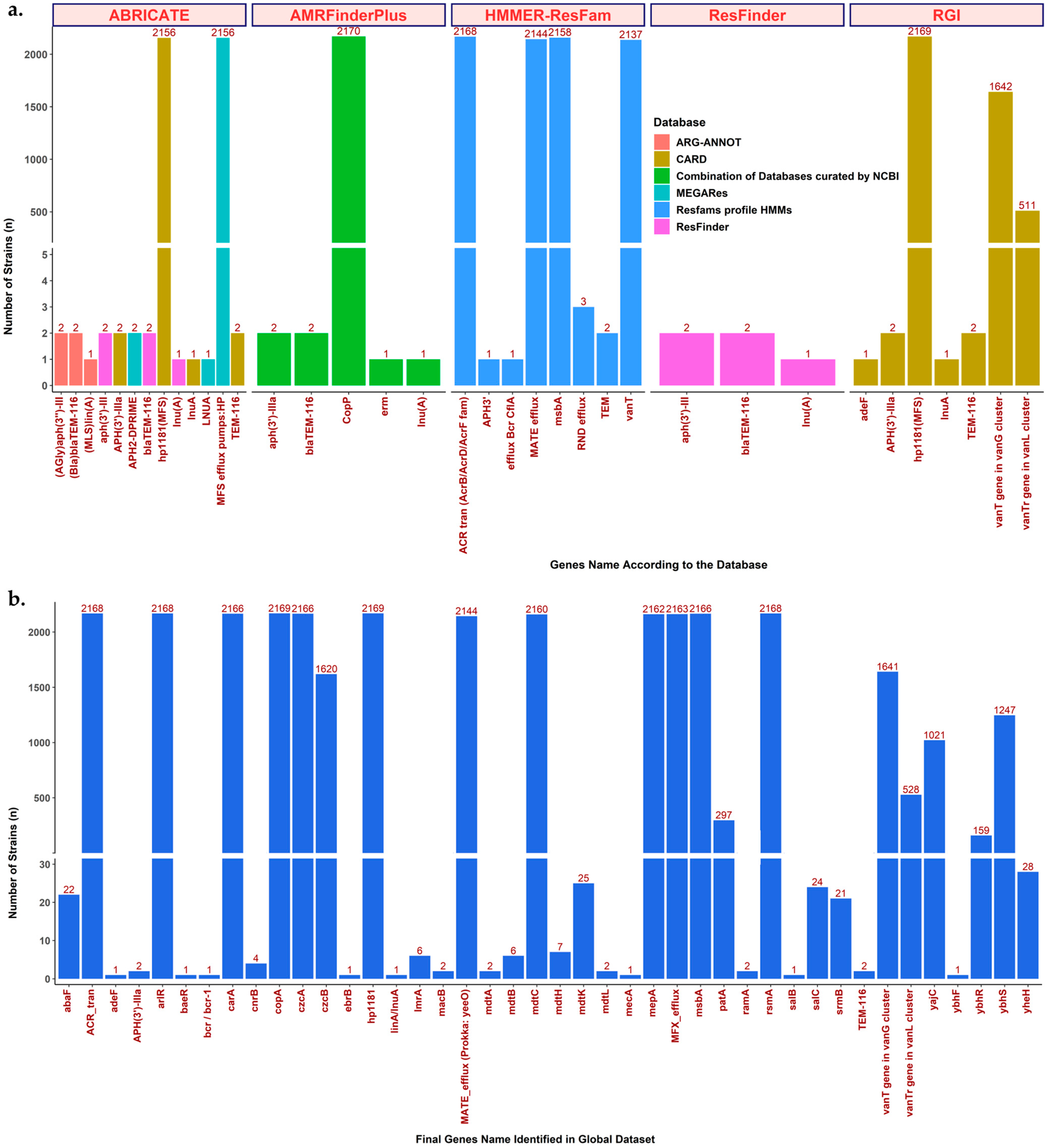
Figure 1. ARG detection by several tools, methods, and databases. (a) The ARG detection results are based on well-known ARG detection tools, showing that each tool could yield different results since they have different detection methods or databases. (b) Final ARG detection results in the global H. pylori dataset. A total of 42 ARG were included after collecting the results from all tools and databases, then performing careful curation with Prokka annotation to delete duplication, confirm the presence of the gene, and find undetected ARG.
Antibiotics 12 01118 g001
Figure 2. Antibiotic class, mechanism, and position of ARG identified within the H. pylori genome. (a) Schematic link between the ARG antibiotic class and the ARG resistance mechanism. Most of the MDR ARG were related to transporters or EPs. The inner and outer scales represent the number and proportion of ARG inside each antibiotic class. (b) Position of ARG within the H. pylori genome. The number of ARG represents the gene name that could be found within the antibiotic class. ARG-CORE: Set of ARG commonly found in the Core Genome of H. pylori. ARG-ACC: Set of ARG commonly found in the Accessory Genome of H. pylori. The core genome was defined if the ARG was found in ≥95% of the total global H. pylori dataset (n = 2170 strains).
Antibiotics 12 01118 g002
Figure 3. Summary of efflux pumps (EPs) related to AMR detected from the H. pylori genome. The green circle contains Eps reported by previous H. pylori AMR studies. The sequences of these Eps were collected from H. pylori strain 26695 then used as a query for BLASTN against the global H. pylori sequence dataset (2170 strains). The result showed that most of these EPs were ARG-CORE. The blue circle contains EPs identified by ARG detection tools and databases. Many of these EPs were found in low numbers of H. pylori and were commonly reported in other species of bacteria. This result suggests that some EP genes may have been obtained by HGT via MGE transfer.
Antibiotics 12 01118 g003
Figure 4. The ARG-ACC distribution based on the geographic location and H. pylori population. The ARG-ACC were clustered based on their antibiotic classes. (a) The ARG-ACC distribution tended to be similar between countries. Some areas are magnified to show the distribution more clearly. The magnified regions were displayed in correspondence with their respective numerical designations. (b) The ARG-ACC proportion for each continent. The proportion tended to be the same for each continent. (c) The ARG-ACC distribution is based on the H. pylori population. Each dot outside the phylogenetic tree represents the ARG-ACC distribution, with each color representing its antibiotic class.
Antibiotics 12 01118 g004
Table 3. Genes that were significantly present in the resistant strains compared to the susceptible strains.
Gene Annotation (Gene Name or Non-Unique Gene Name)Total Present in Resistant Strains,
pr (pr/r; %)
Total Present in Susceptible Strains,
ps (ps/s; %)
Naive p-Value
Clarithromycin
(n = 61, r = 35, s = 26)
  hypothetical protein10 (10/35; 28.57)0 (0/26; 0.00)0.003
  phosphoethanolamine transferase CptA (cptA_1)18 (18/35; 51.43)5 (5/26; 19.23)0.016
  hypothetical protein8 (8/35; 22.86)0 (0/26; 0.00)0.016
  hypothetical protein7 (7/35; 20.00)0 (0/26; 0.00)0.017
  hypothetical protein19 (19/35; 54.29)6 (6/26; 23.08)0.019
  phosphoethanolamine transferase CptA (cptA_2)17 (17/35; 48.57)5 (5/26; 19.23)0.030
  hypothetical protein11 (11/35; 31.43)2 (2/26; 7.69)0.030
  DNA topoisomerase 1 (topA_2)11 (11/35; 31.43)2 (2/26; 7.69)0.030
  hypothetical protein9 (9/35; 25.71)1 (1/26; 3.85)0.034
  hypothetical protein13 (13/35; 37.14)3 (3/26; 11.54)0.038
  hypothetical protein (recF_1)14 (14/35; 40.00)4 (4/26; 15.38)0.049
Metronidazole
(n = 61, r = 43, s = 18)
  lipoprotein-releasing system ATP-binding protein LolD37 (37/43; 86.05)9 (9/18; 50.00)0.007
  hypothetical protein17 (17/43; 39.53)1 (1/18; 5.56)0.012
  hypothetical protein16 (16/43; 37.21)1 (1/18; 5.56)0.013
  trifunctional nucleotide phosphoesterase protein YfkN (yfkN)41 (41/43; 95.35)13 (13/18; 72.22)0.020
  hypothetical protein (SIRT5_2)10 (10/43; 23.26)0 (0/18; 0.00)0.026
  hypothetical protein10 (10/43; 23.26)0 (0/18; 0.00)0.026
  apolipoprotein N-acyltransferase (Int)34 (34/43; 79.07)9 (9/18; 50.00)0.033
  hypothetical protein20 (20/43; 46.51)3 (3/18; 16.67)0.042
  hypothetical protein (gspA)40 (40/43; 93.02)13 (13/18; 72.22)0.042
  hypothetical protein9 (9/43; 20.93)0 (0/18; 00)0.047
  hypothetical protein9 (9/43; 20.93)0 (0/18; 00)0.047
  hypothetical protein (hsdM)9 (9/43; 20.93)0 (0/18; 00)0.047
Levofloxacin
(n = 42, r = 12, s = 30)
  chromosome partition protein Smc (smc)4 (4/12; 33.33)1 (1/30; 3.33)0.018
  hypothetical protein4 (4/12; 33.33)1 (1/30; 3.33)0.018
  hypothetical protein4 (4/12; 33.33)1 (1/30; 3.33)0.018
  hypothetical protein4 (4/12; 33.33)1 (1/30; 3.33)0.018
  hypothetical protein4 (4/12; 33.33)1 (1/30; 3.33)0.018
  hypothetical protein3 (3/12; 25.00)0 (0/30; 0.00)0.019
  transcription-repair-coupling factor (mfd)3 (3/12; 25.00)0 (0/30; 0.00)0.019
  peptide deformylase 1 (def)3 (3/12; 25.00)0 (0/30; 0.00)0.019
  competence protein ComM (comM)3 (3/12; 25.00)0 (0/30; 0.00)0.019
  hypothetical protein3 (3/12; 25.00)0 (0/30; 0.00)0.019
  flagellar basal-body rod protein FlgG (flgG_2)3 (3/12; 25.00)0 (0/30; 0.00)0.019
n, Total number of strains; r, total number of resistant strains; s, total number of susceptible strains; pr, total present in resistant strains; ps, total present in susceptible strains.
Disclaimer/Publisher’s Note: The statements, opinions and data contained in all publications are solely those of the individual author(s) and contributor(s) and not of MDPI and/or the editor(s). MDPI and/or the editor(s) disclaim responsibility for any injury to people or property resulting from any ideas, methods, instructions or products referred to in the content.