
Green Synthesis of Bioinspired Nanoparticles Mediated from Plant Extracts of Asteraceae Family for Potential Biological Applications

 ,
,  , , ,
, , , 
Abstract
1. Introduction
2. Plant-Based Green Synthesis of Nanoparticles
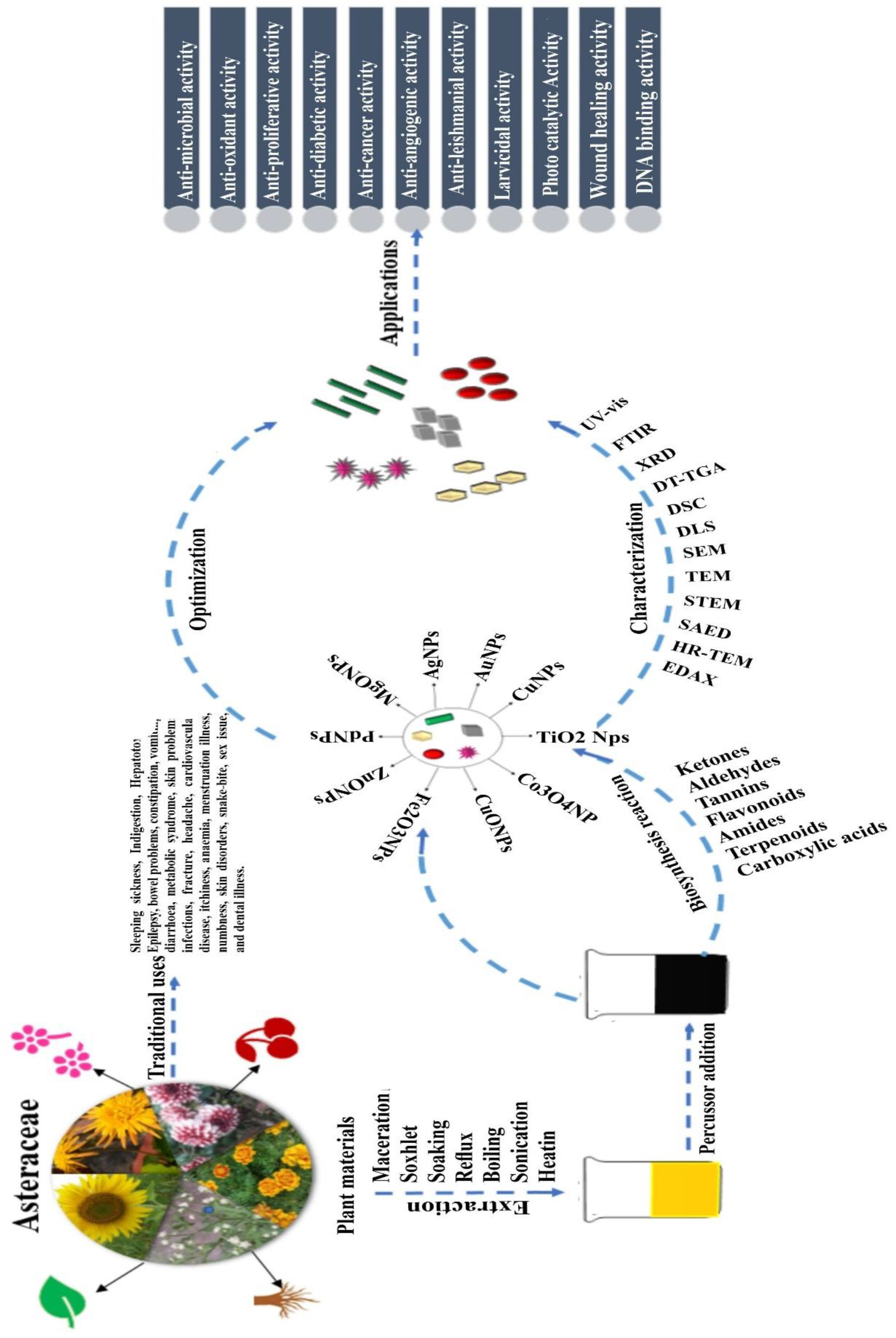
3. Asteraceae Mediated Nanoparticle Synthesis: The Pursued Routes
3.1. Plant Material Used
3.2. Extraction Methods
3.3. Solvents Used
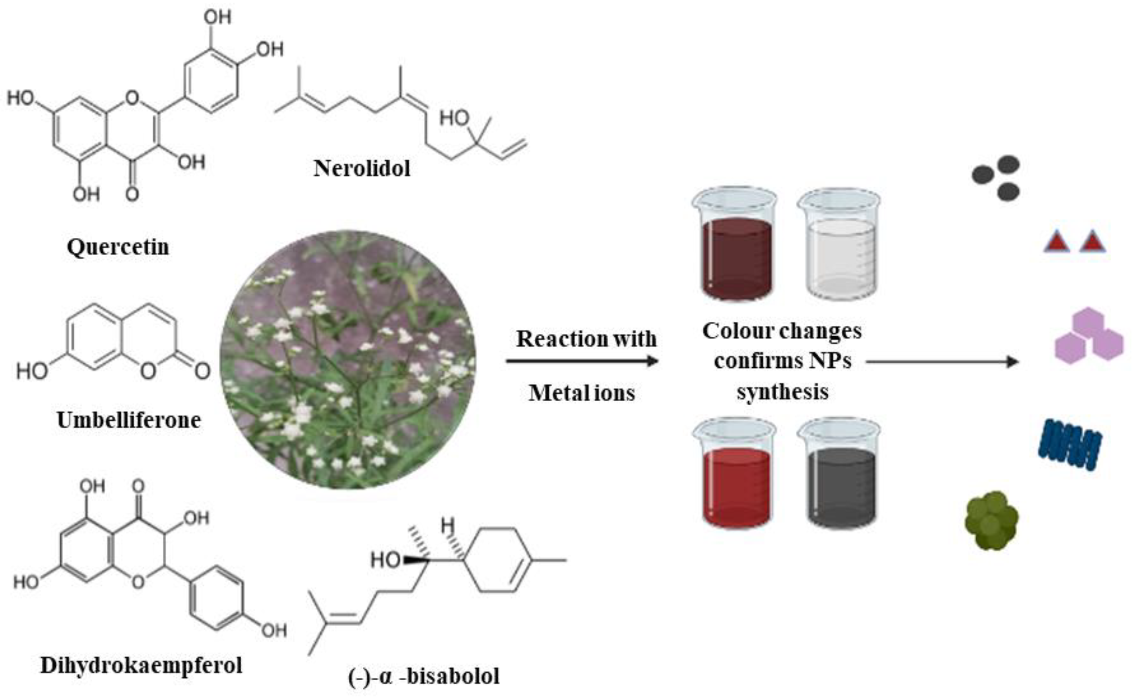
3.4. Phytochemicals Involved
3.5. Nanoparticle Synthesis from Asteraceae Species
3.5.1. Factors Affecting the Synthesis of Asteraceae NPs
Temperature
pH
Reaction Time
Metal Ion Concentration
Plant Extract Concentration
3.6. Separation of NPs
3.7. Characterization
3.7.1. UV–Visible Spectroscopy
3.7.2. Fourier Transforms Infrared Spectroscopy
3.7.3. X-ray Diffraction
3.7.4. Zeta Potential
3.7.5. Dynamic Light Scattering (DLS)
3.7.6. Differential Scanning Calorimetry (DSC)
3.7.7. Thermogravimetric Analysis (TGA)
3.7.8. Selected Area Electron Diffraction (SAED)
3.7.9. Scanning Electron Microscopy (SEM)
3.7.10. Transmission Electron Microscopy (TEM)
3.7.11. Scanning Transmission Mode (STEM)
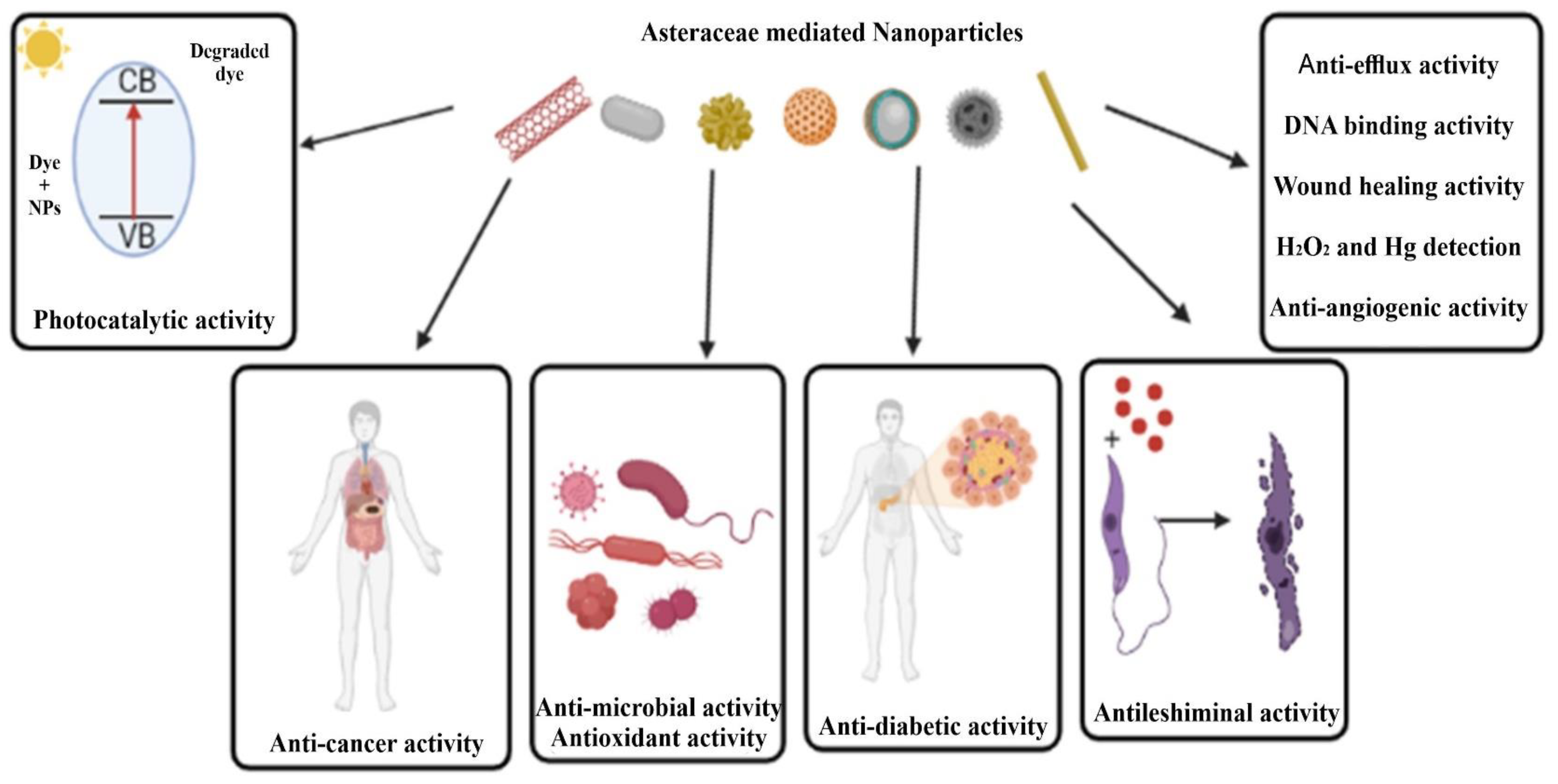
4. Application of Asteraceae-Based Nanoparticles
4.1. Antimicrobial Activity
4.2. Antioxidant Activity
4.3. Anticancer Activity
4.4. Antidiabetic Activity
4.5. Antileishmanial Activity
4.6. Anti-Angiogenic Activity
4.7. Photocatalytic Activity
4.8. Other Activities
5. Toxicity of Asteraceae Mediated Nanoparticles
6. Constraints of Asteraceae-Mediated Nanoparticle Synthesis
7. Conclusions and Prospects
Author Contributions
Funding
Institutional Review Board Statement
Informed Consent Statement
Data Availability Statement
Acknowledgments
Conflicts of Interest
References
- Rahman, H.M.M.; Parvin, M.I.A. Taxonomic Studies on the Family Fabaceae (weeds) at Rajshahi University Campus. Plant 2015, 3, 20. [Google Scholar] [CrossRef]
- Gao, T.; Yao, H.; Song, J.; Zhu, Y.; Liu, C.; Chen, S. Evaluating the Feasibility of Using Candidate DNA Barcodes in Discriminating Species of the Large Asteraceae Family. BMC Evol. Biol. 2010, 10, 324. [Google Scholar] [CrossRef] [PubMed]
- Konovalov, D.A. Polyacetylene Compounds of Plants of the Asteraceae Family (Review). Pharm. Chem. J. 2014, 48, 613–631. [Google Scholar] [CrossRef]
- Michel, J.; Abd Rani, N.Z.; Husain, K. A Review on the Potential Use of Medicinal Plants From Asteraceae and Lamiaceae Plant Family in Cardiovascular Diseases. Front. Pharmacol. 2020, 11, 852. [Google Scholar] [CrossRef]
- Arokiyaraj, S.; Saravanan, M.; Badathala, V. Green Synthesis of Silver Nanoparticles Using Aqueous Extract of Taraxacum Officinale and Its Antimicrobial Activity. South Indian J. Biol. Sci. 2015, 1, 115. [Google Scholar] [CrossRef]
- Nguyen, T.T.-N.; Vo, T.-T.; Nguyen, B.N.-H.; Nguyen, D.-T.; Dang, V.-S.; Dang, C.-H.; Nguyen, T.-D. Silver and Gold Nanoparticles Biosynthesized by Aqueous Extract of Burdock Root, Arctium Lappa as Antimicrobial Agent and Catalyst for Degradation of Pollutants. Environ. Sci. Pollut. Res. 2018, 25, 34247–34261. [Google Scholar] [CrossRef]
- Kilic, A.; Altınkaynak, C.; Ildiz, N.; Ozdemir, N.; Yilmaz, V.; Ocsoy, I. A New Approach for Green Synthesis and Characterization of Artemisia L. (Asteraceae) Genotype Extracts -Cu2 Nanocomplexes (nanoflower) and Their Effecitve Antimicrobial Activity. Med. Sci. Int. Med. J. 2020, 9, 191. [Google Scholar] [CrossRef]
- Madivoli, E.S.; Kareru, P.G.; Maina, E.G.; Nyabola, A.O.; Wanakai, S.I.; Nyang’au, J.O. Biosynthesis of Iron Nanoparticles Using Ageratum Conyzoides Extracts, Their Antimicrobial and Photocatalytic Activity. SN Appl. Sci. 2019, 1, 500. [Google Scholar] [CrossRef]
- Suresh, J.; Pradheesh, G.; Alexramani, V.; Sundrarajan, M.; Hong, S.I. Green Synthesis and Characterization of Zinc Oxide Nanoparticle Using Insulin Plant ( Costus Pictus D. Don ) and Investigation of Its Antimicrobial as Well as Anticancer Activities. Adv. Nat. Sci. Nanosci. Nanotechnol. 2018, 9, 015008. [Google Scholar] [CrossRef]
- Awwad, A.M.; Salem, N.M.; Abdeen, A.O. Green Synthesis of Silver Nanoparticles Using Carob Leaf Extract and Its Antibacterial Activity. Int. J. Ind. Chem. 2013, 4, 29. [Google Scholar] [CrossRef]
- Wangkheirakpam, S.D.; Devi, W.R.; Singh, C.B.; Laitonjam, W.S. Green Synthesis of Silver Nanoparticles Using Strobilanthes Flaccidifolius Nees. Leaf Extract and Its Antibacterial Activity. J. Adv. Chem. 2016, 8, 1523–1532. [Google Scholar] [CrossRef]
- Rao, B.; Tang, R.-C. Green Synthesis of Silver Nanoparticles with Antibacterial Activities Using Aqueous Eriobotrya Japonica Leaf Extract. Adv. Nat. Sci. Nanosci. Nanotechnol. 2017, 8, 015014. [Google Scholar] [CrossRef]
- Chandraker, S.K.; Lal, M.; Shukla, R. DNA-Binding, Antioxidant, H2O2 Sensing and Photocatalytic Properties of Biogenic Silver Nanoparticles Using Ageratum Conyzoides L. Leaf Extract. RSC Adv. 2019, 9, 23408–23417. [Google Scholar] [CrossRef] [PubMed]
- Kumar, P. Nanomaterial’s Synthesis, Types and Their Use in Bioremediation and Agriculture. Nat. Resour. Hum. Health 2022, 2, 349–365. [Google Scholar] [CrossRef] [PubMed]
- Kumar, V.; Gundampati, R.K.; Singh, D.K.; Jagannadham, M.V.; Sundar, S.; Hasan, S.H. Photo-Induced Rapid Biosynthesis of Silver Nanoparticle Using Aqueous Extract of Xanthium Strumarium and Its Antibacterial and Antileishmanial Activity. J. Ind. Eng. Chem. 2016, 37, 224–236. [Google Scholar] [CrossRef]
- Elemike, E.E.; Onwudiwe, D.C.; Fayemi, O.E.; Botha, T.L. Green Synthesis and Electrochemistry of Ag, Au, and Ag–Au Bimetallic Nanoparticles Using Golden Rod (Solidago Canadensis) Leaf Extract. Appl. Phys. A 2019, 125, 42. [Google Scholar] [CrossRef]
- Ghotekar, S.; Pansambal, S.; Pawar, S.P.; Pagar, T.; Oza, R.; Bangale, S. Biological Activities of Biogenically Synthesized Fluorescent Silver Nanoparticles Using Acanthospermum Hispidum Leaves Extract. SN Appl. Sci. 2019, 1, 1342. [Google Scholar] [CrossRef]
- Baharara, J.; Namvar, F.; Ramezani, T.; Hosseini, N.; Mohamad, R. Green Synthesis of Silver Nanoparticles Using Achillea Biebersteinii Flower Extract and Its Anti-Angiogenic Properties in the Rat Aortic Ring Model. Molecules 2014, 19, 4624–4634. [Google Scholar] [CrossRef] [PubMed]
- Behdad, R.; Mirzaie, A.; Zare Karizi, S. Green Synthesis of Silver Nanoparticle Using Acroptilon Repens Extract and Evaluation of Its Anti-Efflux Activity against Acinetobacter Bumanni Clinical Isolates. J. Microb. World 2017, 10, 210–221. [Google Scholar]
- Gautam, S.K.; Baid, Y.; Magar, P.T.; Binadi, T.R.; Regmi, B. Antimicrobial Study of Green Synthesized Silver Nanoparticles (AgNPs) by Using Ageratina Adenophora and Its Characterization. Int. J. Appl. Sci. Biotechnol. 2021, 9, 128–132. [Google Scholar] [CrossRef]
- Morejón, B.; Pilaquinga, F.; Domenech, F.; Ganchala, D.; Debut, A.; Neira, M. Larvicidal Activity of Silver Nanoparticles Synthesized Using Extracts of Ambrosia arborescens (Asteraceae) to Control Aedes aegypti L. (Diptera: Culicidae). J. Nanotechnol. 2018, 2018, 6917938. [Google Scholar] [CrossRef]
- Dehghanizade, S.; Arasteh, J.; Mirzaie, A. Green Synthesis of Silver Nanoparticles Using Anthemis Atropatana Extract: Characterization and in Vitro Biological Activities. Artif. Cells Nanomed. Biotechnol. 2018, 46, 160–168. [Google Scholar] [CrossRef] [PubMed]
- Dobrucka, R.; Długaszewska, J. Antimicrobial Activities of Silver Nanoparticles Synthesized by Using Water Extract of Arnicae Anthodium. Indian J. Microbiol. 2015, 55, 168–174. [Google Scholar] [CrossRef] [PubMed]
- Salehi, S.; Shandiz, S.A.S.; Ghanbar, F.; Darvish, M.R.; Ardestani, M.S.; Mirzaie, A.; Jafari, M. Phytosynthesis of Silver Nanoparticles Using Artemisia Marschalliana Sprengel Aerial Part Extract and Assessment of Their Antioxidant, Anticancer, and Antibacterial Properties. Int. J. Nanomed. 2016, 11, 1835–1846. [Google Scholar]
- Mousavi, B.; Tafvizi, F.; Bostanabad, S.Z. Green Synthesis of Silver Nanoparticles Using Artemisia turcomanica Leaf Extract and the Study of Anti-Cancer Effect and Apoptosis Induction on Gastric Cancer Cell Line (AGS). Artif. Cells Nanomed. Biotechnol. 2018, 46, 499–510. [Google Scholar] [CrossRef]
- Rasheed, T.; Bilal, M.; Iqbal, H.M.N.; Li, C. Green Biosynthesis of Silver Nanoparticles Using Leaves Extract of Artemisia Vulgaris and Their Potential Biomedical Applications. Colloids Surf. B Biointerfaces 2017, 158, 408–415. [Google Scholar] [CrossRef]
- Nyabola, A.O.; Kareru, P.G.; Madivoli, E.S.; Wanakai, S.I.; Maina, E.G. Formation of Silver Nanoparticles via Aspilia Pluriseta Extracts Their Antimicrobial and Catalytic Activity. J. Inorg. Organomet. Polym. Mater. 2020, 30, 3493–3501. [Google Scholar] [CrossRef]
- Abbas, Q.; Saleem, M.; Phull, A.R.; Rafiq, M.; Hassan, M.; Lee, K.-H.; Seo, S.-Y. Green Synthesis of Silver Nanoparticles Using Extract and Their Tyrosinase Activity. Iran J Pharm Res 2017, 16, 763–770. [Google Scholar]
- Mtambo, S.E.; Krishna, S.B.N.; Sershen; Govender, P. Physico-Chemical, Antimicrobial and Anticancer Properties of Silver Nanoparticles Synthesised from Organ-Specific Extracts of Bidens pilosa L. S. Afr. J. Bot. 2019, 126, 196–206. [Google Scholar] [CrossRef]
- Rohankumar, R.C.; Somnath, D.B.; Mangesh, A.B.; Dheeraj, S.R.; Ganesh, H.W.; Sachin, S.T.; Mukund, N.U. Characterization, Antioxidant, Antimicrobial and Cytotoxic Activities of Green Synthesized Silver and Iron Nanoparticles Using Alcoholic Blumea Eriantha DC Plant Extract. Mater. Today Commun. 2020, 24, 101320. [Google Scholar]
- Baghizadeh, A.; Ranjbar, S.; Gupta, V.K.; Asif, M.; Pourseyedi, S.; Karimi, M.J.; Mohammadinejad, R. Green Synthesis of Silver Nanoparticles Using Seed Extract of Calendula officinalis in Liquid Phase. J. Mol. Liq. 2015, 207, 159–163. [Google Scholar] [CrossRef]
- Ahn, E.-Y.; Jin, H.; Park, Y. Green Synthesis and Biological Activities of Silver Nanoparticles Prepared by Carpesium Cernuum Extract. Arch. Pharm. Res. 2019, 42, 926–934. [Google Scholar] [CrossRef]
- Rodríguez-Félix, F.; López-Cota, A.G.; Moreno-Vásquez, M.J.; Graciano-Verdugo, A.Z.; Quintero-Reyes, I.E.; Del-Toro-Sánchez, C.L.; Tapia-Hernández, J.A. Sustainable-Green Synthesis of Silver Nanoparticles Using Safflower (Carthamus Tinctorius L.) Waste Extract and Its Antibacterial Activity. Heliyon 2021, 7, e06923. [Google Scholar] [CrossRef] [PubMed]
- Tüzün, B.S.; Hohmann, J.; Kivcak, B. Green Bio-Inspired Synthesis, Characterization and Activity of Silver Nanoparticle Forms of Centaurea Virgata Lam. and the Isolated Flavonoid Eupatorin. Green Process. Synth. 2018, 7, 372–379. [Google Scholar] [CrossRef]
- Sadiqa, A.; Gilani, S.R.; Anwar, A.; Mehboob, A.; Saleem, A.; Rubab, S. Biogenic Fabrication, Characterization and Drug Loaded Antimicrobial Assay of Silver Nanoparticles Using Centratherum Anthalminticum (L.) Kuntze. J. Pharm. Sci. 2021, 110, 1969–1978. [Google Scholar] [CrossRef]
- Erjaee, H.; Rajaian, H.; Nazifi, S. Synthesis and Characterization of Novel Silver Nanoparticles Using Chamaemelum Nobile Extract for Antibacterial Application. Adv. Nat. Sci. Nanosci. Nanotechnol. 2017, 8, 025004. [Google Scholar] [CrossRef]
- Jayeoye, T.J.; Eze, F.N.; Olatunde, O.O.; Benjakul, S.; Rujiralai, T. Synthesis of Silver and Silver@zero Valent Iron Nanoparticles Using Chromolaena odorata Phenolic Extract for Antibacterial Activity and Hydrogen Peroxide Detection. J. Environ. Chem. Eng. 2021, 9, 105224. [Google Scholar] [CrossRef]
- Arokiyaraj, S.; Arasu, M.V.; Vincent, S.; Prakash, N.U.; Choi, S.H.; Oh, Y.-K.; Choi, K.C.; Kim, K.H. Rapid Green Synthesis of Silver Nanoparticles from Chrysanthemum indicum L. and Its Antibacterial and Cytotoxic Effects: An in Vitro Study. Int. J. Nanomed. 2014, 9, 379–388. [Google Scholar] [CrossRef]
- He, Y.; Du, Z.; Lv, H.; Jia, Q.; Tang, Z.; Zheng, X.; Zhang, K.; Zhao, F. Green Synthesis of Silver Nanoparticles by Chrysanthemum Morifolium Ramat. Extract and Their Application in Clinical Ultrasound Gel. Int. J. Nanomed. 2013, 8, 1809–1815. [Google Scholar] [CrossRef] [PubMed]
- Behboodi, S.; Baghbani-Arani, F.; Abdalan, S.; Sadat Shandiz, S.A. Green Engineered Biomolecule-Capped Silver Nanoparticles Fabricated from Cichorium intybus Extract: In Vitro Assessment on Apoptosis Properties Toward Human Breast Cancer (MCF-7) Cells. Biol. Trace Elem. Res. 2019, 187, 392–402. [Google Scholar] [CrossRef] [PubMed]
- Mohamad, R. Biosynthesis of Au, Ag and Bimetallic Au-Ag Nanoparticles Using Aqueous Leaf Extract of Cosmos Caudatus. Ph.D. Thesis, Universiti Teknologi Malaysia, Johor Bahru, Malaysia, 2013. [Google Scholar]
- Malaka, R.; Hema, J.A.; Muthukumarasamy, N.P.; Sambandam, A.; Subramanian, S.; Sevanan, M. Green Synthesis of Silver Nanoparticles Using Cosmos Sulphureus and Evaluation of Their Antimicrobial and Antioxidant Properties. Nano Biomed. Eng. 2016, 7, 160–168. [Google Scholar] [CrossRef]
- Adewale, O.B.; Egbeyemi, K.A.; Onwuelu, J.O.; Potts-Johnson, S.S.; Anadozie, S.O.; Fadaka, A.O.; Osukoya, O.A.; Aluko, B.T.; Johnson, J.; Obafemi, T.O.; et al. Biological Synthesis of Gold and Silver Nanoparticles Using Leaf Extracts of Crassocephalum Rubens and Their Comparative in Vitro Antioxidant Activities. Heliyon 2020, 6, e05501. [Google Scholar] [CrossRef] [PubMed]
- de Ruíz-Baltazar, Á.J.; de Jesús Ruíz-Baltazar, Á.; Reyes-López, S.Y.; de Lourdes Mondragón-Sánchez, M.; Estevez, M.; Hernández-Martinez, A.R.; Pérez, R. Biosynthesis of Ag Nanoparticles Using Cynara Cardunculus Leaf Extract: Evaluation of Their Antibacterial and Electrochemical Activity. Results Phys. 2018, 11, 1142–1149. [Google Scholar] [CrossRef]
- Erdogan, O.; Abbak, M.; Demirbolat, G.M.; Birtekocak, F.; Aksel, M.; Pasa, S.; Cevik, O. Green Synthesis of Silver Nanoparticles via Cynara Scolymus Leaf Extracts: The Characterization, Anticancer Potential with Photodynamic Therapy in MCF7 Cells. PLoS ONE 2019, 14, e0216496. [Google Scholar] [CrossRef] [PubMed]
- Roy, K.; Sarkar, C.K.; Ghosh, C.K. Rapid Colorimetric Detection of Hg2+ Ion by Green Silver Nanoparticles Synthesized Using Dahlia Pinnata Leaf Extract. Green Process. Synth. 2015, 4, 455–461. [Google Scholar] [CrossRef]
- Arya, G.; Malav, A.K.; Gupta, N.; Kumar, A.; Nimesh, S. Biosynthesis and in Vitro Antimicrobial Potential of Silver Nanoparticles Prepared Using Dicoma Tomentosa Plant Extract. Nanosci. Nanotechnol.-Asia 2018, 8, 240–247. [Google Scholar] [CrossRef]
- Sedira, S.; Sobti, N. Silver Nanoparticles Bioreduction by Dittrichia Viscosa Leaves Extract and Its Bactericidal Effects. Int. J. Nanoparticles 2016, 9, 19. [Google Scholar] [CrossRef]
- Gecer, E.N.; Erenler, R.; Temiz, C.; Genc, N.; Yildiz, I. Green Synthesis of Silver Nanoparticles from Echinacea Purpurea (L.) Moench with Antioxidant Profile. Part. Sci. Technol. 2022, 40, 50–57. [Google Scholar] [CrossRef]
- Murthy, H.C. Green Silver Nanoparticles Synthesised Using Medicinal Plant Echinops Sp. Root Extract for Antimicrobial Applications. Nanochemistry Res. 2020, 5, 128–140. [Google Scholar] [CrossRef]
- Premasudha, P.; Venkataramana, M.; Abirami, M.; Vanathi, P.; Krishna, K.; Rajendran, R. Biological Synthesis and Characterization of Silver Nanoparticles Using Eclipta Alba Leaf Extract and Evaluation of Its Cytotoxic and Antimicrobial Potential. Bull. Mater. Sci. 2015, 38, 965–973. [Google Scholar] [CrossRef]
- Kharat, S.N.; Mendhulkar, V.D. Synthesis, Characterization and Studies on Antioxidant Activity of Silver Nanoparticles Using Elephantopus Scaber Leaf Extract. Mater. Sci. Eng. C Mater. Biol. Appl. 2016, 62, 719–724. [Google Scholar] [CrossRef] [PubMed]
- Kumar, V.; Singh, D.K.; Mohan, S.; Hasan, S.H. Photo-Induced Biosynthesis of Silver Nanoparticles Using Aqueous Extract of Erigeron Bonariensis and Its Catalytic Activity against Acridine Orange. J. Photochem. Photobiol. B 2016, 155, 39–50. [Google Scholar] [CrossRef] [PubMed]
- Elemike, E.; Onwudiwe, D.; Ekennia, A.; Sonde, C.; Ehiri, R. Green Synthesis of Ag/Ag2O Nanoparticles Using Aqueous Leaf Extract of Eupatorium Odoratum and Its Antimicrobial and Mosquito Larvicidal Activities. Molecules 2017, 22, 674. [Google Scholar] [CrossRef]
- Mahmod, M.; Junayed, A.; Bhowmick, C.; Sompa, S.; Sultana, T.; Akter, T.; Abedin, M.; Zubair, M.; Islam, M.; Mogal, M.; et al. Antibacterial Activity of Silver Nanoparticles Synthesized from Leaf and Flower Extracts of Galinsoga Formosa. J. Adv. Biotechnol. Exp. Ther. 2021, 4, 178. [Google Scholar] [CrossRef]
- Shahzadi, T.; Kanwal, A.; Jabeen, H.; Riaz, T.; Zaib, M. Eco-Friendly synthesis of silver nanopartricles using gazania rigens and evaluation of activities. J. Environ. Eng. Landsc. Manage. 2021, 20, 43–52. [Google Scholar] [CrossRef]
- Han, S.; Ahmeda, A.; Jalalvand, A.R.; Lu, W.; Zangeneh, M.M.; Zangeneh, A. Application of Silver Nanoparticles Containing Gundelia tournefortii L. Leaf Aqueous Extract in the Treatment of Microbial Diseases and Cutaneous Wound Healing. Appl. Organomet. Chem. 2022, 36, e5491. [Google Scholar] [CrossRef]
- Nadzir, M.M.; Idris, F.N.; Hat, K. Green Synthesis of Silver Nanoparticle Using Gynura procumbens Aqueous Extracts. In Proceedings of the 6th International Conference on Environment (ICENV2018): Empowering Environment and Sustainable Engineering Nexus Through Green Technology, Penang, Malaysia, 11–13 December 2018. [Google Scholar] [CrossRef]
- Yazdi, M.E.T.; Amiri, M.S.; Hosseini, H.A.; Oskuee, R.K.; Mosawee, H.; Pakravanan, K.; Darroudi, M. Plant-Based Synthesis of Silver Nanoparticles in Handelia trichophylla and Their Biological Activities. Bull. Mater. Sci. 2019, 42, 155. [Google Scholar] [CrossRef]
- Yazdi, M.E.T.; Amiri, M.S.; Akbari, S.; Sharifalhoseini, M.; Nourbakhsh, F.; Mashreghi, M.; Yousefi, E.; Abbasi, M.R.; Modarres, M.; Es-haghi, A. Green Synthesis of Silver Nanoparticles Using Helichrysum graveolens for Biomedical Applications and Wastewater Treatment. BioNanoScience 2020, 10, 1121–1127. [Google Scholar] [CrossRef]
- Riaz, M.; Altaf, M.; Khan, M.Q.; Manzoor, S.; Shekheli, M.A.; Shah, M.A.; Ilyas, S.Z.; Hussain, Z. Green Synthesis of Silver Nanoparticles Using Jurinea Dolomiaea and Biological Activities. J. Nanosci. Nanotechnol. 2018, 18, 8386–8391. [Google Scholar] [CrossRef] [PubMed]
- Kanagamani, K.; Muthukrishnan, P.; Shankar, K.; Kathiresan, A.; Barabadi, H.; Saravanan, M. Antimicrobial, Cytotoxicity and Photocatalytic Degradation of Norfloxacin Using Kleinia Grandiflora Mediated Silver Nanoparticles. J. Clust. Sci. 2019, 30, 1415–1424. [Google Scholar] [CrossRef]
- Kanchana, A.; Agarwal, I.; Sunkar, S.; Nellore, J.; Namasivayam, K. Biogenic Silver Nanoparticles From Spinacia Oleracea And Lactuca sativa And Their Potential Antimicrobial Activity. Dig. J. Nanomater. Biostructures 2011, 6, 1741–1750. [Google Scholar]
- Essien, E.R.; Atasie, V.N.; Udobang, E.U.; Umanu, G. Preparation of Monodispersed and Cytotoxic Silver Nanoparticles Using Launaea Taraxacifolia Leaf Extract. J. Nanostructure Chem. 2019, 9, 259–268. [Google Scholar] [CrossRef]
- Uddin, I.; Ahmad, K.; Khan, A.A.; Kazmi, M.A. Synthesis of Silver Nanoparticles Using Matricaria Recutita (Babunah) Plant Extract and Its Study as Mercury Ions Sensor. Sens. Bio-Sens. Res. 2017, 16, 62–67. [Google Scholar] [CrossRef]
- Biswas, A.; Vanlalveni, C.; Adhikari, P.P.; Lalfakzuala, R.; Rokhum, L. Biosynthesis, Characterisation and Antibacterial Activity of Mikania Micrantha Leaf Extract-mediated AgNPs. Micro Nano Lett. 2019, 14, 799–803. [Google Scholar] [CrossRef]
- Okaiyeto, K.; Ojemaye, M.O.; Hoppe, H.; Mabinya, L.V.; Okoh, A.I. Phytofabrication of Silver/Silver Chloride Nanoparticles Using Aqueous Leaf Extract of Oedera Genistifolia: Characterization and Antibacterial Potential. Molecules 2019, 24, 4382. [Google Scholar] [CrossRef] [PubMed]
- Ahsan, A.; Farooq, M.A.; Ahsan Bajwa, A.; Parveen, A. Green Synthesis of Silver Nanoparticles Using Parthenium Hysterophorus: Optimization, Characterization and In Vitro Therapeutic Evaluation. Molecules 2020, 25, 3324. [Google Scholar] [CrossRef] [PubMed]
- Mofolo, M.J.; Kadhila, P.; Chinsembu, K.C.; Mashele, S.; Sekhoacha, M. Green Synthesis of Silver Nanoparticles from Extracts of Pechuel-Loeschea Leubnitziae: Their Anti-Proliferative Activity against the U87 Cell Line. Inorg. Nano-Met. Chem. 2020, 50, 949–955. [Google Scholar] [CrossRef]
- Abdelmoteleb, A.; Valdez-Salas, B.; Carrillo-Beltran, M.; Hernandez, D.D.; González-Mendoza, D. Green Synthesis of Silver Nanoparticles Using Pluchea Sericea a Native Plants from Baja California, Mexico and Their Potential Application as Antimicrobials. Iran. J. Sci. Technol. Trans. A Sci. 2018, 42, 457–463. [Google Scholar] [CrossRef]
- Khan, M.; Khan, M.; Adil, S.F.; Tahir, M.N.; Tremel, W.; Alkhathlan, H.Z.; Al-Warthan, A.; Siddiqui, M.R.H. Green Synthesis of Silver Nanoparticles Mediated by Pulicaria Glutinosa Extract. Int. J. Nanomed. 2013, 8, 1507. [Google Scholar]
- Qhtani, M.S.J.A.; Al Qhtani, M.S.J.; El-Debaiky, S.A.; Sayed, M. Antifungal and Cytotoxic Activities of Biosynthesized Silver, Zinc and Gold Nanoparticles by Flower Extract of Rhanterium Epapposum. Open J. Appl. Sci. 2020, 10, 663–674. [Google Scholar]
- Aslam, M.; Fozia, F.; Gul, A.; Ahmad, I.; Ullah, R.; Bari, A.; Mothana, R.A.; Hussain, H. Phyto-Extract-Mediated Synthesis of Silver Nanoparticles Using Aqueous Extract of Sanvitalia Procumbens, and Characterization, Optimization and Photocatalytic Degradation of Azo Dyes Orange G and Direct Blue-15. Molecules 2021, 26, 6144. [Google Scholar] [CrossRef] [PubMed]
- Abd El-Aziz, A.R.M.; Gurusamy, A.; Alothman, M.R.; Shehata, S.M.; Hisham, S.M.; Alobathani, A.A. Silver Nanoparticles Biosynthesis Using Saussurea Costus Root Aqueous Extract and Catalytic Degradation Efficacy of Safranin Dye. Saudi J. Biol. Sci. 2021, 28, 1093–1099. [Google Scholar] [CrossRef]
- Ayromlou, A.; Masoudi, S.; Mirzaie, A. Scorzonera Calyculata Aerial Part Extract Mediated Synthesis of Silver Nanoparticles: Evaluation of Their Antibacterial, Antioxidant and Anticancer Activities. J. Clust. Sci. 2019, 30, 1037–1050. [Google Scholar] [CrossRef]
- Qasim Nasar, M.; Zohra, T.; Khalil, A.T.; Saqib, S.; Ayaz, M.; Ahmad, A.; Shinwari, Z.K. Seripheidium Quettense Mediated Green Synthesis of Biogenic Silver Nanoparticles and Their Theranostic Applications. Green Chem. Lett. Rev. 2019, 12, 310–322. [Google Scholar] [CrossRef]
- Gopalakrishnan, R.; Loganathan, B.; Raghu, K. Green Synthesis of Au–Ag Bimetallic Nanocomposites Using Silybum Marianum Seed Extract and Their Application as a Catalyst. RSC Adv. 2015, 5, 31691–31699. [Google Scholar] [CrossRef]
- Kumar, V.A.; Uchida, T.; Mizuki, T.; Nakajima, Y.; Katsube, Y.; Hanajiri, T.; Maekawa, T. Synthesis of Nanoparticles Composed of Silver and Silver Chloride for a Plasmonic Photocatalyst Using an Extract from a Weed Solidago altissima (goldenrod). Adv. Nat. Sci. Nanosci. Nanotechnol. 2016, 7, 015002. [Google Scholar] [CrossRef]
- Botha, T.L.; Elemike, E.E.; Horn, S.; Onwudiwe, D.C.; Giesy, J.P.; Wepener, V. Cytotoxicity of Ag, Au and Ag-Au Bimetallic Nanoparticles Prepared Using Golden Rod (Solidago canadensis) Plant Extract. Sci. Rep. 2019, 9, 4169. [Google Scholar] [CrossRef] [PubMed]
- Rethinam, R.; Jeyachandran, R. Green Synthesis of Silver Nanoparticles Using Aqueous Leaf Extract of Spilanthes Calva Dc. World J. Pharm. Res. 2016, 5, 822–828. [Google Scholar]
- Laguta, I.; Stavinskaya, O.; Kazakova, O.; Fesenko, T.; Brychka, S. Green Synthesis of Silver Nanoparticles Using Stevia Leaves Extracts. Appl. Nanosci. 2018, 9, 755–765. [Google Scholar] [CrossRef]
- Ogunsile, B.O.; Labulo, A.H.; Fajemilehin, A.M. Green Synthesis of Silver Nanoparticles from Leaf Extracts of Parquetina Nigrescens and Synedrella Nodiflora and Their Antimicrobial Activity. Ife J. Sci. 2016, 18, 245–254. [Google Scholar]
- Katta, V.K.M.; Dubey, R.S. Green Synthesis of Silver Nanoparticles Using Tagetes Erecta Plant and Investigation of Their Structural, Optical, Chemical and Morphological Properties. Mater. Today 2021, 45, 794–798. [Google Scholar] [CrossRef]
- Dubey, S.P.; Lahtinen, M.; Sillanpää, M. Tansy Fruit Mediated Greener Synthesis of Silver and Gold Nanoparticles. Process Biochem. 2010, 45, 1065–1071. [Google Scholar] [CrossRef]
- Saratale, R.G.; Benelli, G.; Kumar, G.; Kim, D.S.; Saratale, G.D. Bio-Fabrication of Silver Nanoparticles Using the Leaf Extract of an Ancient Herbal Medicine, Dandelion (Taraxacum officinale), Evaluation of Their Antioxidant, Anticancer Potential, and Antimicrobial Activity against Phytopathogens. Environ. Sci. Pollut. Res. Int. 2018, 25, 10392–10406. [Google Scholar] [CrossRef] [PubMed]
- Tran, T.T.T.; Vu, T.T.H.; Nguyen, T.H. Biosynthesis of Silver Nanoparticles Using Tithonia Diversifolia Leaf Extract and Their Antimicrobial Activity. Mater. Lett. 2013, 105, 220–223. [Google Scholar] [CrossRef]
- Jabbari, R.; Ghasemi, N. Investigating Methylene Blue Dye Adsorption Isotherms Using Silver Nano Particles Provided by Aqueous Extract of Tragopogon Buphthalmoides. Chem. Methodol. 2020, 5, 21–29. [Google Scholar] [CrossRef]
- Seifipour, R.; Nozari, M.; Pishkar, L. Green Synthesis of Silver Nanoparticles Using Tragopogon Collinus Leaf Extract and Study of Their Antibacterial Effects. J. Inorg. Organomet. Polym. Mater. 2020, 30, 2926–2936. [Google Scholar] [CrossRef]
- Kushwaha, H.B.; Malik, C.P. Nanofabrication of Silver Nanoparticles from the Stem and Leaf Extract of Verbesina Encelioides. Natl. Acad. Sci. Lett. 2012, 35, 555–563. [Google Scholar] [CrossRef]
- Joseph, J.; Khor, K.Z.; Moses, E.J.; Lim, V.; Aziz, M.Y.; Abdul Samad, N. In Vitro Anticancer Effects of Leaf Extract and Green-Synthesised Silver Nanoparticles. Int. J. Nanomed. 2021, 16, 3599–3612. [Google Scholar] [CrossRef] [PubMed]
- Sahayaraj, K.; Roobadevi, M.; Rajesh, S.; Azizi, S. Vernonia cinerea (L.) Less. Silver Nanocomposite and Its Antibacterial Activity against a Cotton Pathogen. Res. Chem. Intermed. 2015, 41, 5495–5507. [Google Scholar] [CrossRef]
- Paul Das, M.; Rebecca Livingstone, J.; Veluswamy, P.; Das, J. Exploration of Wedelia Chinensis Leaf-Assisted Silver Nanoparticles for Antioxidant, Antibacterial and in Vitro Cytotoxic Applications. J. Food Drug Anal. 2018, 26, 917–925. [Google Scholar] [CrossRef]
- Singh, Y.; Gaur, S.; Singhal, A.; Chauhan, D.K. Phytotoxic Assessment Of Agno3 And Znso4 Vis À Vis Agnps And Znonps In Tagetes Erecta L. And Zinnia Elegans Jacq. Plant Arch. 2021, 21, 724–730. [Google Scholar] [CrossRef]
- Dobrucka, R.; Romaniuk-Drapała, A.; Kaczmarek, M. Biologically Synthesized of Au/Pt/ZnO Nanoparticles Using Arctium Lappa Extract and Cytotoxic Activity against Leukemia. Biomed. Microdevices 2020, 22, 72. [Google Scholar] [CrossRef]
- Abdoli, M.; Arkan, E.; Shekarbeygi, Z.; Khaledian, S. Green Synthesis of Gold Nanoparticles Using Centaurea Behen Leaf Aqueous Extract and Investigating Their Antioxidant and Cytotoxic Effects on Acute Leukemia Cancer Cell Line (THP-1). Inorg. Chem. Commun. 2021, 129, 108649. [Google Scholar] [CrossRef]
- Torabi, N.; Nowrouzi, A.; Ahadi, A.; Vardasbi, S.; Etesami, B. Green Synthesis of Gold Nanoclusters Using Seed Aqueous Extract of Cichorium intybus L. and Their Characterization. SN Appl. Sci. 2019, 1, 981. [Google Scholar] [CrossRef]
- Attar, A.; Yapaoz, M.A. Biomimetic Synthesis, Characterization and Antibacterial Efficacy of ZnO and Au Nanoparticles Using Echinacea Flower Extract Precursor. Mater. Res. Express 2018, 5, 055403. [Google Scholar] [CrossRef]
- Vijayakumar, S.; Vinayagam, R.; Anand, M.A.V.; Venkatachalam, K.; Saravanakumar, K.; Wang, M.-H.; Sangeetha, C.C.; Gothandam, K.M.; David, E. Green Synthesis of Gold Nanoparticle Using Eclipta Alba and Its Antidiabetic Activities through Regulation of Bcl-2 Expression in Pancreatic Cell Line. J. Drug Deliv. Sci. Technol. 2020, 58, 101786. [Google Scholar] [CrossRef]
- Mendhulkar, V.; Shinde, A. Anticancer Activity of Gold Nanobioconjugates Synthesized from Elephantopus Scaber (linn.) Leaf Extract. J. Cancer Res. Ther. 2021, 10. [Google Scholar] [CrossRef]
- Velmurugan, P.; Cho, M.; Lee, S.-M.; Park, J.-H.; Lee, K.-J.; Myung, H.; Oh, B.-T. Phyto-Crystallization of Silver and Gold by Erigeron Annuus (L.) Pers Flower Extract and Catalytic Potential of Synthesized and Commercial Nano Silver Immobilized on Sodium Alginate Hydrogel. J. Saudi Chem. Soc. 2016, 20, 313–320. [Google Scholar] [CrossRef]
- Punnoose, M.S.; Bijimol, D.; Mathew, B. Microwave Assisted Green Synthesis of Gold Nanoparticles for Catalytic Degradation of Environmental Pollutants. Environ. Nanotechnol. Monit. Manag. 2021, 16, 100525. [Google Scholar]
- Zhaleh, M.; Zangeneh, A.; Goorani, S.; Seydi, N.; Zangeneh, M.M.; Tahvilian, R.; Pirabbasi, E. In Vitro and in Vivo Evaluation of Cytotoxicity, Antioxidant, Antibacterial, Antifungal, and Cutaneous Wound Healing Properties of Gold Nanoparticles Produced via a Green Chemistry Synthesis Using Gundelia tournefortii L. as a Capping and Reducing Agent. Appl. Organomet. Chem. 2019, 33, e5015. [Google Scholar] [CrossRef]
- Mariychuk, R.; Grulova, D.; Grishchenko, L.M.; Linnik, R.P.; Lisnyak, V.V. Green Synthesis of Non-Spherical Gold Nanoparticles Using Solidago Canadensis L. Extract. Appl. Nanosci. 2020, 10, 4817–4826. [Google Scholar] [CrossRef]
- Sadeghi, B.; Mohammadzadeh, M.; Babakhani, B. Green Synthesis of Gold Nanoparticles Using Stevia Rebaudiana Leaf Extracts: Characterization and Their Stability. J. Photochem. Photobiol. B 2015, 148, 101–106. [Google Scholar] [CrossRef]
- Del Moral, A.; Borjas-Garcia, S.E.; Rosas, G. Green Synthesis of Gold Nanoparticles Using Taraxacum officinale Extract. Microsc. Microanal. 2018, 24, 1740–1741. [Google Scholar] [CrossRef]
- Vijaya Kumar, P.; Mary Jelastin Kala, S.; Prakash, K.S. Synthesis of Gold Nanoparticles Using Xanthium Strumarium Leaves Extract and Their Antimicrobial Studies: A Green Approach. Rasayan J. Chem. 2018, 11, 1544–1551. [Google Scholar] [CrossRef]
- Wang, G.; Ahmeda, A.; Malek, Z.; Mansooridara, S.; Zangeneh, A.; Zangeneh, M.M. Chemical Characterization and Therapeutic Properties of Achillea Biebersteinii Leaf Aqueous Extract Synthesized Copper Nanoparticles against Methamphetamine-induced Cell Death in PC12: A Study in the Nanotechnology and Neurology Fields. Appl. Organomet. Chem. 2020, 34, e5488. [Google Scholar] [CrossRef]
- Binawati, G.; Ilham, M.; Ida, K. Biosynthesis Copper Nanoparticles Using Blumea Balsamifera Leaf Extracts: Characterization of Its Antioxidant and Cytotoxicity Activities. Surf. Interfaces 2020, 21, 100799. [Google Scholar]
- Chung, I.-M.; Abdul Rahuman, A.; Marimuthu, S.; Kirthi, A.V.; Anbarasan, K.; Padmini, P.; Rajakumar, G. Green Synthesis of Copper Nanoparticles Using Leaves Extract and Their Antioxidant and Cytotoxic Activities. Exp. Ther. Med. 2017, 14, 18–24. [Google Scholar] [CrossRef] [PubMed]
- León-Jimenez, E.; California, B.; Valdéz-Salas, B.; González-Mendoza, D.; Tzintzun-Camacho, O.; Gutiérrez, T. Synthesis and Insecticide Activity of Cu-Nanoparticles from Prosopis Juliflora (Sw) DC and Pluchea Sericea (Nutt.) on Phenacoccus Solenopsis Tinsley (Hemiptera: Pseudococcidae). Rev. De La Soc. EntomolÓGica Argent. 2019, 78, 12–21. [Google Scholar] [CrossRef]
- Kalpana, V.N.; Chakraborthy, P.; Palanichamy, V.; Rajeswari, V.D. Synthesis and Characterization of Copper Nanoparticles Using Tridax Procumbens and Its Application in Degradation of Bismarck Brown. Analysis 2016, 10, 17. [Google Scholar]
- Khan, M.; Khan, M.; Kuniyil, M.; Adil, S.F.; Al-Warthan, A.; Alkhathlan, H.Z.; Tremel, W.; Tahir, M.N.; Siddiqui, M.R.H. Biogenic Synthesis of Palladium Nanoparticles Using Pulicaria Glutinosa Extract and Their Catalytic Activity towards the Suzuki Coupling Reaction. Dalton Trans. 2014, 43, 9026–9031. [Google Scholar] [CrossRef] [PubMed]
- Wang, D.; Cui, L.; Chang, X.; Guan, D. Biosynthesis and Characterization of Zinc Oxide Nanoparticles from Artemisia Annua and Investigate Their Effect on Proliferation, Osteogenic Differentiation and Mineralization in Human Osteoblast-like MG-63 Cells. J. Photochem. Photobiol. B 2020, 202, 111652. [Google Scholar] [CrossRef]
- Gomathi, R.; Suhana, H. Green Synthesis, Characterization and Antimicrobial Activity of Zinc Oxide Nanoparticles Using Artemisia Pallens Plant Extract. Synth. React. Inorg. Met.-Org. Nano-Met. Chem. 2021, 51, 1663–1672. [Google Scholar] [CrossRef]
- Mohammadi Shivyari, A.; Tafvizi, F.; Noorbazargan, H. Anti-Cancer Effects of Biosynthesized Zinc Oxide Nanoparticles Using Artemisia Scoparia in Huh-7 Liver Cancer Cells. Synth. React. Inorg. Met.-Org. Nano-Met. Chem. 2022, 52, 375–386. [Google Scholar]
- Rajapriya, M.; Sharmili, S.A.; Baskar, R.; Balaji, R.; Alharbi, N.S.; Kadaikunnan, S.; Khaled, J.M.; Alanzi, K.F.; Vaseeharan, B. Correction to: Synthesis and Characterization of Zinc Oxide Nanoparticles Using Cynara Scolymus Leaves: Enhanced Hemolytic, Antimicrobial, Antiproliferative, and Photocatalytic Activity. J. Clust. Sci. 2020, 31, 803. [Google Scholar] [CrossRef]
- Balogun, F.O.; Ashafa, A.O.T. Green-Synthesized Zinc Oxide Nanoparticles from Aqueous Root Extract of Dicoma Anomala (Sond.) Mitigates Free Radicals and Diabetes-Linked Enzymes. Nanosci. Nanotechnol.-Asia 2020, 10, 918–929. [Google Scholar] [CrossRef]
- Hoseinpour, V.; Souri, M.; Ghaemi, N.; Shakeri, A. Optimization of green synthesis of ZnO nanoparticles by Dittrichia graveolens (L.) aqueous extract. Health Biotechnol. Biopharma 2017, 1, 39–49. [Google Scholar]
- Xu, J.; Luo, X.; Wang, Y.; Feng, Y. Evaluation of Zinc Oxide Nanoparticles on Lettuce (Lactuca sativa L.) Growth and Soil Bacterial Community. Environ. Sci. Pollut. Res. Int. 2018, 25, 6026–6035. [Google Scholar] [CrossRef]
- Datta, A.; Patra, C.; Bharadwaj, H.; Kaur, S.; Khajuria, R. Green Synthesis of Zinc Oxide Nanoparticles Using Parthenium Hysterophorus Leaf Extract and Evaluation of Their Antibacterial Properties. J. Biotechnol. Biomater 2017, 7, 271–276. [Google Scholar] [CrossRef]
- Kolahalam, L.A.; Prasad, K.R.S.; Murali Krishna, P.; Supraja, N. Plant Rhizome Extract-Based Zinc Oxide Nanoparticles: Synthesis, Characterization and Its Antibacterial, Antifungal Activities and Cytotoxic Studies against Chinese Hamster Ovary (CHO) Cell Lines. Heliyon 2021, 7, e07265. [Google Scholar] [CrossRef] [PubMed]
- Hameed, S.; Khalil, A.T.; Ali, M.; Numan, M.; Khamlich, S.; Shinwari, Z.K.; Maaza, M. Greener Synthesis of ZnO and Ag-ZnO Nanoparticles Using Silybum Marianum for Diverse Biomedical Applications. Nanomedicine 2019, 14, 655–673. [Google Scholar] [CrossRef] [PubMed]
- Ilangovan, A.; Venkatramanan, A.; Thangarajan, P.; Saravanan, A.; Rajendran, S.; Kaveri, K. Green Synthesis of Zinc Oxide Nanoparticles (ZnO NPs) Using Aqueous Extract of Tagetes Erecta Flower and Evaluation of Its Antioxidant, Antimicrobial, and Cytotoxic Activities on HeLa Cell Line. Curr. Biotechnol. 2021, 10, 61–76. [Google Scholar] [CrossRef]
- Obayomi, K.S.; Oluwadiya, A.E.; Lau, S.Y.; Dada, A.O.; Akubuo-Casmir, D.; Adelani-Akande, T.A.; Fazle Bari, A.S.M.; Temidayo, S.O.; Rahman, M.M. Biosynthesis of Tithonia Diversifolia Leaf Mediated Zinc Oxide Nanoparticles Loaded with Flamboyant Pods (Delonix Regia) for the Treatment of Methylene Blue Wastewater. Arab. J. Chem. 2021, 14, 103363. [Google Scholar] [CrossRef]
- Seifipour, R.; Nozari, M.; Pishkar, L. Preparation of ZnO Nanoparticles Using Tragopogon Collinus Leaf Extract and Study of Its Antibacterial Effects for Therapeutic Applications. J. Plant Biochem. Biotechnol. 2021, 30, 586–595. [Google Scholar] [CrossRef]
- Ossai, A.N.; Ezike, S.C.; Dikko, A.B. Bio-Synthesis of Zinc Oxide Nanoparticles from Bitter Leaf (vernonia Amygdalina) Extract for Dye-Sensitized Solar Cell Fabrication. Available online: https://www.jmaterenvironsci.com/Document/vol11/vol11_N3/JMES-2020-11-38-Ossai.pdf (accessed on 31 July 2022).
- Kouhbanani, M.A.J.; Beheshtkhoo, N.; Amani, A.M.; Taghizadeh, S.; Beigi, V.; Bazmandeh, A.Z.; Khalaf, N. Green Synthesis of Iron Oxide Nanoparticles Using Artemisia Vulgaris Leaf Extract and Their Application as a Heterogeneous Fenton-like Catalyst for the Degradation of Methyl Orange. Mater. Res. Express 2018, 5, 115013. [Google Scholar] [CrossRef]
- Wanakai, S.I.; Kareru, P.G.; Makhanu, D.S.; Madivoli, E.S.; Maina, E.G.; Nyabola, A.O. Catalytic Degradation of Methylene Blue by Iron Nanoparticles Synthesized Using Galinsoga parviflora, Conyza bonariensis and Bidens pilosa Leaf Extracts. SN Appl. Sci. 2019, 1, 1148. [Google Scholar] [CrossRef]
- Davarnejad, R.; Azizi, A.; Mohammadi, M.; Mansoori, S. A Green Technique for Synthesising Iron Oxide Nanoparticles by Extract of Centaurea Cyanus Plant: An Optimised Adsorption Process for Methylene Blue. Int. J. Environ. Anal. Chem. 2022, 102, 2379–2393. [Google Scholar] [CrossRef]
- Biswas, A.; Vanlalveni, C.; Lalfakzuala, R.; Nath, S.; Rokhum, L. Mikania Mikrantha Leaf Extract Mediated Biogenic Synthesis of Magnetic Iron Oxide Nanoparticles: Characterization and Its Antimicrobial Activity Study. Mater. Today Proc. 2021, 42, 1366–1373. [Google Scholar] [CrossRef]
- Khatami, M.; Alijani, H.Q.; Fakheri, B.; Mobasseri, M.M.; Heydarpour, M.; Farahani, Z.K.; Khan, A.U. Super-Paramagnetic Iron Oxide Nanoparticles (SPIONs): Greener Synthesis Using Stevia Plant and Evaluation of Its Antioxidant Properties. J. Clean. Prod. 2019, 208, 1171–1177. [Google Scholar] [CrossRef]
- Habtemariam, A.B. Biosynthesis of Magnetite (Fe3O4) Nanostructures Using Vernonia Amygdalina Leaves Extract. Lett. Appl. NanoBioScience 2021, 10, 2777–2783. [Google Scholar]
- Rather, M.Y.; Sundarapandian, S. Magnetic Iron Oxide Nanorod Synthesis by Wedelia Urticifolia (Blume) DC. Leaf Extract for Methylene Blue Dye Degradation. Appl. Nanosci. 2020, 10, 2219–2227. [Google Scholar] [CrossRef]
- Pansambal, S. Phytosynthesis and Biological Activities of Fluorescent CuO Nanoparticles Using Acanthospermum Hispidum L. Extract. J. Nanostructures 2017, 7, 165–174. [Google Scholar] [CrossRef]
- Nasrollahzadeh, M.; Mohammad Sajadi, S.; Rostami-Vartooni, A. Green Synthesis of CuO Nanoparticles by Aqueous Extract of Anthemis Nobilis Flowers and Their Catalytic Activity for the A3 Coupling Reaction. J. Colloid Interface Sci. 2015, 459, 183–188. [Google Scholar] [CrossRef] [PubMed]
- Gowri, M.; Latha, N.; Rajan, M. Copper Oxide Nanoparticles Synthesized Using Eupatorium Odoratum, Acanthospermum Hispidum Leaf Extracts, and Its Antibacterial Effects Against Pathogens: A Comparative Study. Bionanoscience 2019, 9, 545–552. [Google Scholar] [CrossRef]
- Ganesan, S.; Ganesh Babu, I.; Mahendran, D.; Indra Arulselvi, P.; Elangovan, N.; Geetha, N.; Venkatachalam, P. Green Engineering of Titanium Dioxide Nanoparticles Using Ageratina Altissima (L.) King & H.E. Robines. Medicinal Plant Aqueous Leaf Extracts for Enhanced Photocatalytic Activity. Ann. Phytomedicine Int. J. 2016, 5, 69–75. [Google Scholar]
- Dobrucka, R. Synthesis of Titanium Dioxide Nanoparticles Using Herba. Iran J Pharm. Res. 2017, 16, 756–762. [Google Scholar] [PubMed]
- Babu, N.; Pathak, V.M.; Singh, A.; Navneet, A. Navneet Sonchus Asper Leaves Aqueous Extract Mediated Synthesis of Titanium Dioxide Nanoparticles. Pharma Innov. 2019, 8, 817–822. [Google Scholar]
- Wardani, M.; Yulizar, Y.; Abdullah, I.; Apriandanu, D.O.B. Synthesis of NiO Nanoparticles via Green Route Using Ageratum Conyzoides L. Leaf Extract and Their Catalytic Activity. IOP Conf. Ser. Mater. Sci. Eng. 2019, 509, 012077. [Google Scholar] [CrossRef]
- Likasari, I.D.; Astuti, R.W.; Yahya, A.; Isnaini, N.; Purwiandono, G.; Hidayat, H.; Wicaksono, W.P.; Fatimah, I. NiO Nanoparticles Synthesized by Using Tagetes Erecta L Leaf Extract and Their Activities for Photocatalysis, Electrochemical Sensing, and Antibacterial Features. Chem. Phys. Lett. 2021, 780, 138914. [Google Scholar] [CrossRef]
- Rasheed, T.; Nabeel, F.; Bilal, M.; Iqbal, H.M.N. Biogenic Synthesis and Characterization of Cobalt Oxide Nanoparticles for Catalytic Reduction of Direct Yellow-142 and Methyl Orange Dyes. Biocatal. Agric. Biotechnol. 2019, 19, 101154. [Google Scholar] [CrossRef]
- Dobrucka, R. Synthesis of MgO Nanoparticles Using Artemisia Abrotanum Herba Extract and Their Antioxidant and Photocatalytic Properties. Iran. J. Sci. Technol. Trans. A Sci. 2016, 42, 547–555. [Google Scholar] [CrossRef]
- Essien, E.R.; Atasie, V.N.; Oyebanji, T.O.; Nwude, D.O. Biomimetic Synthesis of Magnesium Oxide Nanoparticles Using Chromolaena odorata (L.) Leaf Extract. Chem. Pap. 2020, 74, 2101–2109. [Google Scholar] [CrossRef]
- Amina, M.; Al Musayeib, N.M.; Alarfaj, N.A.; El-Tohamy, M.F.; Oraby, H.F.; Al Hamoud, G.A.; Bukhari, S.I.; Moubayed, N.M.S. Biogenic Green Synthesis of MgO Nanoparticles Using Saussurea Costus Biomasses for a Comprehensive Detection of Their Antimicrobial, Cytotoxicity against MCF-7 Breast Cancer Cells and Photocatalysis Potentials. PLoS ONE 2020, 15, e0237567. [Google Scholar] [CrossRef]
- R, R.; Ranjithkumar, R.; Chandar, S.B.; Senthil Kumaran, C.K.; Sharmila, C.; Simi, V. Green Synthesis Of Silver Nanoparticles Using Graviola Leaf Aqueous Extract At Room Temperature. Kongunadu Res. J. 2015, 2, 6–10. [Google Scholar] [CrossRef]
- Shanker, U.; Jassal, V.; Rani, M.; Kaith, B.S. Towards Green Synthesis of Nanoparticles: From Bio-Assisted Sources to Benign Solvents. A Review. Int. J. Environ. Anal. Chem. 2016, 96, 801–835. [Google Scholar]
- Ahmed, R.H.; Mustafa, D.E. Green Synthesis of Silver Nanoparticles Mediated by Traditionally Used Medicinal Plants in Sudan. Int. Nano Lett. 2020, 10, 1–14. [Google Scholar] [CrossRef]
- Li, S.; Shen, Y.; Xie, A.; Yu, X.; Qiu, L.; Zhang, L.; Zhang, Q. Green Synthesis of Silver Nanoparticles Using Capsicum Annuum L. Extract. Green Chem. 2007, 9, 852–858. [Google Scholar] [CrossRef]
- Gu, J.; Aidy, A.; Goorani, S. Anti-Human Lung Adenocarcinoma, Cytotoxicity, and Antioxidant Potentials of Copper Nanoparticles Green-Synthesized by Calendula Officinalis. J. Exp. Nanosci. 2022, 17, 285–296. [Google Scholar] [CrossRef]
- Rai, A.; Lall, R. Antimicrobial, Antioxidant and Cytotoxic Activity of Green Synthesized Copper Nanoparticle of Parthenium Hysterophorus L. Int. J. Multidiscip. Res. Anal. 2021, 4, 101–116. [Google Scholar] [CrossRef]
- Khalil, M.M.H.; Ismail, E.H.; El-Baghdady, K.Z.; Mohamed, D. Green Synthesis of Silver Nanoparticles Using Olive Leaf Extract and Its Antibacterial Activity. Arab. J. Chem. 2014, 7, 1131–1139. [Google Scholar] [CrossRef]
- Balalakshmi, C.; Gopinath, K.; Govindarajan, M.; Lokesh, R.; Arumugam, A.; Alharbi, N.S.; Kadaikunnan, S.; Khaled, J.M.; Benelli, G. Green Synthesis of Gold Nanoparticles Using a Cheap Sphaeranthus Indicus Extract: Impact on Plant Cells and the Aquatic Crustacean Artemia Nauplii. J. Photochem. Photobiol. B 2017, 173, 598–605. [Google Scholar] [CrossRef] [PubMed]
- AlSalhi, M.; Devanesan, S.; Alfuraydi, A.; Vishnubalaji, R.; Munusamy, M.A.; Murugan, K.; Nicoletti, M.; Benelli, G. Green Synthesis of Silver Nanoparticles Using Pimpinella Anisum Seeds: Antimicrobial Activity and Cytotoxicity on Human Neonatal Skin Stromal Cells and Colon Cancer Cells. Int. J. Nanomed. 2016, 11, 4439–4449. [Google Scholar] [CrossRef] [PubMed]
- Baker, S.; Rakshith, D.; Kavitha, K.S.; Santosh, P.; Kavitha, H.U.; Rao, Y.; Satish, S. Plants: Emerging as Nanofactories towards Facile Route in Synthesis of Nanoparticles. Bioimpacts 2013, 3, 111–117. [Google Scholar] [PubMed]
- Bhushan, I.; Singh, V.K.; Tripathi, D.K. Nanomaterials and Environmental Biotechnology; Springer Nature: Berlin/Heidelberg, Germany, 2020; ISBN 9783030345440. [Google Scholar]
- Bell, J.E.; Ellis Bell, J.; Hall, C. UV and Visible Absorbance Spectroscopy. In Spectroscopy in Biochemistry; CRC Press: Boca Raton, FL, USA, 2018; pp. 3–62. [Google Scholar]
- Nagaraj, B.; Malakar, B.; Divya, T.K.; Krishnamurthy, N.; Liny, P.; Dinesh, R.; Iconaru, S.; Ciobanu, C. Synthesis of Plant Mediated Gold Nanoparticles Using Flower Extracts of Carthamus Tinctorius L. (safflower) and Evaluation of Their Biological Activities. Dig. J. Nanomater. Biostruct. 2012, 7, 1289–1296. [Google Scholar] [CrossRef]
- Chandraker, S.K.; Lal, M.; Ghosh, M.K.; Tiwari, V.; Ghorai, T.K.; Shukla, R. Green Synthesis of Copper Nanoparticles Using Leaf Extract of Ageratum Houstonianum Mill. and Study of Their Photocatalytic and Antibacterial Activities. Nano Express 2020, 1, 010033. [Google Scholar] [CrossRef]
- Nasrazadani, S.; Hassani, S. Modern Analytical Techniques in Failure Analysis of Aerospace, Chemical, and Oil and Gas Industries. In Handbook of Materials Failure Analysis with Case Studies from the Oil and Gas Industry; Elsevier: Amsterdam, The Netherlands, 2016; pp. 39–54. ISBN 9780081001172. [Google Scholar]
- Berg, J.M.; Romoser, A.; Banerjee, N.; Zebda, R.; Sayes, C.M. The Relationship between pH and Zeta Potential of ∼ 30 Nm Metal Oxide Nanoparticle Suspensions Relevant Toin Vitrotoxicological Evaluations. Nanotoxicology 2009, 3, 276–283. [Google Scholar] [CrossRef]
- Xu, R. Progress in Nanoparticles Characterization: Sizing and Zeta Potential Measurement. Particuology 2008, 6, 112–115. [Google Scholar] [CrossRef]
- Cho, T.J.; Hackley, V.A. Fractionation and Characterization of Gold Nanoparticles in Aqueous Solution: Asymmetric-Flow Field Flow Fractionation with MALS, DLS, and UV-Vis Detection. Anal. Bioanal. Chem. 2010, 398, 2003–2018. [Google Scholar] [CrossRef] [PubMed]
- Zou, C.; Gao, Y.; Yang, B.; Zhai, Q. Synthesis and DSC Study on Sn3.5Ag Alloy Nanoparticles Used for Lower Melting Temperature Solder. J. Mater. Sci. Mater. Electron. 2010, 21, 868–874. [Google Scholar] [CrossRef]
- Dongargaonkar, A.A.; Clogston, J.D. Quantitation of Surface Coating on Nanoparticles Using Thermogravimetric Analysis. Methods Mol. Biol. 2018, 1682, 57–63. [Google Scholar] [PubMed]
- Bajpai, O.P.; Panja, S.; Chattopadhyay, S.; Setua, D.K. Process–structure–property Relationships in Nanocomposites Based on Piezoelectric-Polymer Matrix and Magnetic Nanoparticles. In Manufacturing of Nanocomposites with Engineering Plastics; Elsevier: Amsterdam, The Netherlands, 2015; pp. 255–278. ISBN 9781782423089. [Google Scholar]
- Polshettiwar, V.; Asefa, T. Nanocatalysis: Synthesis and Applications; John Wiley & Sons: Hoboken, NJ, USA, 2013; ISBN 9781118609804. [Google Scholar]
- Pogrebnjak, A.D.; Novosad, V. Advances in Thin Films, Nanostructured Materials, and Coatings: Selected Papers from the 2018 International Conference on “Nanomaterials: Applications & Properties”; Springer: Berlin/Heidelberg, Germany, 2019; ISBN 9789811361333. [Google Scholar]
- Nezamabadi, V.; Akhgar, M.R.; Tahamipour, B.; Rajaei, P. Biosynthesis and Antibacterial Activity of ZnO Nanoparticles by Extract. Iran. J. Biotechnol. 2020, 18, e2426. [Google Scholar] [PubMed]
- José-Yacamán, M.; Marín-Almazo, M.; Ascencio, J.A. High Resolution TEM Studies on Palladium Nanoparticles. J. Mol. Catal. A Chem. 2001, 173, 61–74. [Google Scholar] [CrossRef]
- Mayoral, A.; Mejía-Rosales, S.; Mariscal, M.M.; Pérez-Tijerina, E.; José-Yacamán, M. The Co-Au Interface in Bimetallic Nanoparticles: A High Resolution STEM Study. Nanoscale 2010, 2, 2647–2651. [Google Scholar] [CrossRef] [PubMed]
- Karthik, L.; Vishnu Kirthi, A.; Ranjan, S.; Mohana Srinivasan, V. Biological Synthesis of Nanoparticles and Their Applications; CRC Press: Boca Raton, FL, USA, 2019; ISBN 9780429555787. [Google Scholar]
- Maji, A.; Beg, M.; Das, S.; Aktara, M.N.; Nayim, S.; Patra, A.; Islam, M.M.; Hossain, M. Study on the Antibacterial Activity and Interaction with Human Serum Albumin of Tagetes Erecta Inspired Biogenic Silver Nanoparticles. Process Biochem. 2020, 97, 191–200. [Google Scholar] [CrossRef]
- Rajiv, P.; Rajeshwari, S.; Venckatesh, R. Bio-Fabrication of Zinc Oxide Nanoparticles Using Leaf Extract of Parthenium Hysterophorus L. and Its Size-Dependent Antifungal Activity against Plant Fungal Pathogens. Spectrochim. Acta A Mol. Biomol. Spectrosc. 2013, 112, 384–387. [Google Scholar] [CrossRef]
- Bedlovičová, Z.; Strapáč, I.; Baláž, M.; Salayová, A. A Brief Overview on Antioxidant Activity Determination of Silver Nanoparticles. Molecules 2020, 25, 3191. [Google Scholar] [CrossRef] [PubMed]
- Khshan, K.T.; Alkafaje, H.A. Biosynthesis of Silver Nanoparticles Using Calendula officinalis (L.) Extract and Evaluating Their Antioxidant Activity. IOP Conf. Ser. Earth Environ. Sci. 2021, 735, 012073. [Google Scholar] [CrossRef]
- ACAR, Ç.A. Green Synthesis of Zinc Oxide Nanoparticles Using Aqueous Extract of Achiella Millefolium L.: In Vitro Anti-Cancer Potential On Lung And Colon Cancer Cells. Turk. J. Health Sci. Life 2021, 4, 40–45. [Google Scholar]
- Ul Haq, M.N.; Shah, G.M.; Gul, A.; Foudah, A.I.; Alqarni, M.H.; Yusufoglu, H.S.; Hussain, M.; Alkreathy, H.M.; Ullah, I.; Khan, A.M.; et al. Biogenic Synthesis of Silver Nanoparticles Using and Its In Vivo Anti-Diabetic Effect against Alloxan-Induced Diabetic Wistar Rats. Nanomaterials 2022, 12, 830. [Google Scholar] [CrossRef] [PubMed]
- Iqbal, J.; Andleeb, A.; Ashraf, H.; Meer, B.; Mehmood, A.; Jan, H.; Zaman, G.; Nadeem, M.; Drouet, S.; Fazal, H.; et al. Potential Antimicrobial, Antidiabetic, Catalytic, Antioxidant and ROS/RNS Inhibitory Activities of Mediated Biosynthesized Copper Oxide Nanoparticles. RSC Adv. 2022, 12, 14069–14083. [Google Scholar] [CrossRef]
- De Queiroz, A.C.; de Dias, T.L.M.F.; Da Matta, C.B.B.; Cavalcante Silva, L.H.A.; de Araújo-Júnior, J.X.; de Araújo, G.B.; de Moura, F.B.P.; Alexandre-Moreira, M.S. Antileishmanial Activity of Medicinal Plants Used in Endemic Areas in Northeastern Brazil. Evid. Based. Complement. Alternat. Med. 2014, 2014, 478290. [Google Scholar] [CrossRef] [PubMed]
- Gangwar, J.; Sebastian, J.K. Unlocking the Potential of Biosynthesized Zinc Oxide Nanoparticles for Degradation of Synthetic Organic Dyes as Wastewater Pollutants. Water Sci. Technol. 2021, 84, 3286–3310. [Google Scholar] [CrossRef] [PubMed]
- Bahadar, H.; Maqbool, F.; Niaz, K.; Abdollahi, M. Toxicity of Nanoparticles and an Overview of Current Experimental Models. Iran. Biomed. J. 2016, 20, 1–11. [Google Scholar] [PubMed]
- Yashveer, S.; Redhu, N.; Singh, V.; Sangwan, S.; Laxman, H.; Tokas, J.; Malhotra, S.; Khurana, S.; Sindhu, A. Nanoparticles in Agriculture: Characterization, Uptake and Role in Mitigating Heat Stress. Nat. Resour. Hum. Health 2022, 2, 160–181. [Google Scholar] [CrossRef]
- Arokiyaraj, S.; Dinesh Kumar, V.; Elakya, V.; Kamala, T.; Park, S.K.; Ragam, M.; Saravanan, M.; Bououdina, M.; Arasu, M.V.; Kovendan, K.; et al. Biosynthesized Silver Nanoparticles Using Floral Extract of Chrysanthemum indicum L.—Potential for Malaria Vector Control. Environ. Sci. Pollut. Res. Int. 2015, 22, 9759–9765. [Google Scholar] [CrossRef] [PubMed]
- Hajra, A.; Dutta, S.; Mondal, N.K. Mosquito Larvicidal Activity of Cadmium Nanoparticles Synthesized from Petal Extracts of Marigold (Tagetes sp.) and Rose (Rosa sp.) Flower. J. Parasit. Dis. 2016, 40, 1519–1527. [Google Scholar] [CrossRef] [PubMed]


| Plant | Part Used | Solvent Used | Extraction Method | Phytochemicals | Characterization Techniques | SPR Peak (nm) | Nanoparticle Size (nm) | Activity | References |
|---|---|---|---|---|---|---|---|---|---|
| Silver NPs | |||||||||
| Acanthospermum hispidum | Leaf | DiW | Reflux | Saponins, coumarins, phenols, flavonoids, volatile oils, tannins, and sterols | UV–Vis, FE-SEM, EDX, TEM, FTIR, Particle size, and zeta potential | 417 | 20–60 | Antibacterial, antifungal, antimalarial, and antimycobacterial activity | [17] |
| Achillea biebersteinii | Flower | DDW | Boiling | Polysaccharides, polyphenols, and proteins | UV–Vis, TEM, zeta potential, and EDX | 460 | 12 ± 2 | Anti-angiogenesis activity | [18] |
| Acroptilon repens | Whole plant | DDW | Reflux | Caryophyllene oxide, α-copaene, β-caryophylene, and β-copaene-4-α-ol | UV–Vis, SEM, and TEM | 420 | 38.89 | Anti-efflux activity | [19] |
| Ageratina adenophora | Leaf | - | - | Carbohydrates, alkaloids, phenols, flavonoids, xanthoprotein, glycosides, tannins, steroids, and terpenoids | XRD, and FTIR | - | 25 | Antimicrobial activity | [20] |
| Ageratum conyzoides | Leaf | DDW | Boiling | Alkaloids, flavonoids, chromenes, benzofurans, and terpenoids | UV–Vis, FTIR, SEM, TEM, XRD, and EDX | 443 | 14–48 | DNA-binding, antioxidant, H2O2 sensing, and photocatalytic properties | [13] |
| Ambrosia arborescens | Leaf | DW | Stirring | Sesquiterpenic lactones, monoterpenes, terpenoids, and polyacetylenic resins | UV–Vis, FTIR, STEM, and SEM-EDX, | 414 | 14 ± 6 | Larvicidal activity | [21] |
| Anthemis atropatana | Aerial parts | Methanol | Boiling | Flavonoids, and phenolic compounds | UV–Vis, XRD, TEM, SEM, and FTIR | 430 | 38.89 | Antibacterial and cytotoxic activity | [21,22] |
| Arctium lappa | Whole plant | DW | Boiling | Phenolic acids, flavonoids, alkaloids, and terpenoids | UV–Vis, XRD, TEM, HRTEM, FTIR, EDX, TG, and DTA | 435 | 21.3 | Antimicrobial activity and catalyst for degradation of pollutants | [6] |
| Arnicae anthodium | Whole plant | DW | Boiling | Flavonoids. Triterpenes, sesquiterpene lactones and essential oils. | UV–Vis, FTIR, TXRF, and SEM-EDS | 458 | 90–118 | Antimicrobial activity | [23] |
| Artemisia marschalliana | Aerial parts | 50% ethanol | Boiling | Phenolic acids and flavonoids | UV–Vis, XRD, FTIR, TEM, SEM, zeta potential, and EDS | 430 | 5–50 | Antioxidant, anticancer, and antibacterial activity | [24] |
| Artemisia turcomanica | Leaf | 50% ethanol | Boiling | Phenolic acids, flavonoids, alkaloids and terpenoids | UV–Vis, TEM, SEM, XRD, and FTIR | 430 | 22 | Cytotoxic and anti-cancer activity | [25] |
| Artemisia vulgaris | Leaf | Methanol | Maceration | Phenolic acids, flavonoids, and alkaloids | UV–Vis, SEM, EDX, TEM, AFM, and FTIR | 420 | 25 | Antimicrobial, antioxidant, and antiproliferative activities | [26] |
| Aspilia pluriseta | Leaf | DW | Boiling | Flavonoids, phenols, alkaloids, and amino acids | UV–Vis, FTIR, SEM, DLS, TEM, and XRD | 427 | 6 | Antimicrobial and catalytic activity | [27] |
| Bidens frondosa | Whole plant | DW | Boiling | Terpenoids, phenolics and proteins | UV–Vis, FTIR, FESEM, and EDS | 443 | 20–70 | Tyrosinase activity | [28] |
| Bidens pilosa | Leaf, stem, and root | DW | Stirring | Terpenes, essential oils, tannins, polysaccharides, phenols, amino acids, ascorbic acid and organic acids | UV–Vis, SEM, TEM, EDX, and FTIR | 410 | 17 | Antimicrobial and anticancer activity | [29] |
| Blumea eriantha | Whole plant | Ethanol | Soxhlet | Phenols and flavonoids | UV–Vis, FTIR, SEM, XRD, and TEM | 445 | 10 | Antioxidant, antimicrobial, and cytotoxic activities | [30] |
| Calendula officinalis | Seed | DW | Boiling | Triterpenoids, flavonoids, coumarines, quinones, volatile oil, carotenoids, and amino acids | UV–Vis, TEM, XRD, and FTIR | 440 | 05–10 | - | [31] |
| Carpesium cernuum | Whole plant | Methanol | Reflux | Polyphenols | UV–Vis, and HR-TEM | 430 | 13.0 ± 0.2 | Antioxidant and anticancer activity | [32] |
| Carthamus tinctorius | Stem and Leaf | DW | Boiling | Flavonoids, polyphenols, proteins, sugars and saponins | HR-TEM, FTIR, and SEM | - | 10 | Antibacterial activity | [33] |
| Centaurea virgata | Aerial parts | N-hexane, chloroform, and methanol: water | Soxhlet | Flavonoids, phenolic acids, and terpenes | UV–Vis, FTIR, TEM, SEM, EDX, TGA XRD, and zeta potential | 420 | 25–50 | Antioxidant activity | [34] |
| Centratherum anthalminticum | Whole plant | DW | Heating | Phenolics and flavones | UV–Vis, XRD, SEM, FTIR, Particle size, DLS, and zeta potential | 436 | <50 | Antimicrobial activity | [35] |
| Chamaemelum nobile | Whole plant | DW | Heating | Phenolics and flavones | UV–Vis, DLS, FTIR, XRD, and TEM | 422 | 24.2 ± 3.1 | Antibacterial activity | [36] |
| Chromoleana odorata | Leaf | - | - | - | UV–Vis, FTIR, XRD, SEM, FE-SEM, and EDX | 428 | 20–25 | Antibacterial activity and hydrogen peroxide detection | [37] |
| Chrysanthemum indicum | Flower | DW | Boiling | Flavonoids, terpenoids, and glycosides | UV–Vis, XRD, TEM, and EDX | 435 | 37.71–71.99 | Antibacterial and cytotoxic activity | [38] |
| Chrysanthemum morifolium | Flower | DW | Boiling | Flavonoids, caffeoylquinic acids, chlorogenic acid, phenolic acids | UV–Vis, FTIR, XRD, and TEM | 430 | 20–50 | Antibacterial activity | [39] |
| Cichorium intybus | Leaf | DDW | Boiling | Phenolic acids, triterpenoids, sterols, and hydroxycinnamic acid derivatives | XRD, FTIR, zeta potential, TEM, SEM, and EDS | - | 17.17 | Anticancer activity | [40] |
| Cosmos caudatus | Leaf | DW | Boiling | Phenolic acids, triterpenoids, and sterols | UV–Vis, XRD, FTIR, FESEM-EDX, and TEM | 439 | 21.49 ± 7.43 | - | [41] |
| Cosmos sulphureus | Leaf | DW | Boiling | Phenols, polyphenolic, and flavonoids | UV–Vis, Particle size, zeta potential, DLS, and SEM | 430–440 | 55–80 | Antimicrobial and antioxidant properties | [42] |
| Crassocephalum rubens | Leaf | DW | Boiling | Flavonoids, and polyphenols | UV–Vis, EDX, TEM, SEM, and FTIR | 470 | 15–25 | Antioxidant activity | [43] |
| Cynara cardunculus | Leaf | DW | Boiling | Polyphenols, flavonoids, and terpenoids | TEM, EDS, FTIR, and XPS | 435 | 45 | Antibacterial and electrochemical activity | [44] |
| Cynara scolymus | Leaf | DW | Heating | Alkaloids, polyphenols, flavonoid, and amino acid | UV–Vis, FTIR, SEM, EDX, and zeta sizer | 434 | 98.47 ± 2.04 | Anticancer activity | [45] |
| Dahlia pinnata | Leaf | DW | Boiling | Flavonoids, and phenolics | UV–Vis, XRD, TEM, and FTIR | 460 | 15 | Detection of Hg2+ ion | [46] |
| Dicoma tomentosa | Bark | DW | Boiling | Flavonoids, phenolic acids, and terpenes | UV–Vis | 430–480 | - | Antimicrobial activity | [47] |
| Dittrichia viscosa | Leaf | DW | Boiling | Flavonoids and polyphenols | UV–Vis, XRD, FTIR, and TEM | 406 | 5–25 | Bactericidal effects | [48] |
| Echinacea purpurea | Whole plant | DW | Heating | Caffeic acid derivatives, polysaccharides, alkaloids, alkylamides, and polyphenols | UV–Vis, XRD, SEM, and FTIR | 481 | 68.24 | Antioxidant activity | [49] |
| Echinops sp. | Root | DW | Heating | Carbohydrates, alkaloids, phenols, flavonoids, xanthoprotein, glycosides, tannins, steroids, and terpenoid | UV–Vis, UV-DRS, FTIR, XRD, SEM, EDXA, TEM, HRTEM, and SAED | 454 | 33.86 | Antimicrobial activity | [50] |
| Eclipta alba | Leaf | DW | Boiling | Phenols, flavonoids, and aldehydes | UV–Vis, DLS, FTIR, XRD, and SEM | 433 | 310–400 | Antimicrobial and cytotoxic activity | [51] |
| Elephantopus scaber | Leaf | DW | Boiling | Phenolics, amino acids, aliphatic, and aromatic hydroxyl groups | UV–Vis, NTA, TEM, XRD, and FTIR | 435 | 50 | Antioxidant activity | [52] |
| Erigeron bonariensis | Leaf | DW | Boiling | Terpenoids, flavonoids, and phenol derivatives | UV–Vis, SEM, EDX, TEM, XRD, AFM, and FTIR | 422 | 13 | Catalytic activity | [53] |
| Eupatorium odoratum | Leaf | DW | Boiling | Tannins, saponins, phytates, flavonoids, betacyanins, and alkaloids, steroids, terpenoids, phenols, quinones, and glycosides | UV–Vis, particle size, TEM, and PXRD | 424 | 23.6 | Antimicrobial and mosquito larvicidal activity | [54] |
| Galinsoga formosa | Leaf and Flower | DW | Boiling | Phenolics, amino acids, aliphatic, and aromatic hydroxyl groups | UV–Vis | 350–400 | - | Photocatalytic degradation activity | [55] |
| Gazania rigens | Whole plant | DW | Boiling | Flavonoids, polyphenols, proteins, sugars, and saponins | UV–Vis, XRD, EDX, and SEM | 425–460 | 31.35 | Antioxidant and photocatalytic degradation activity | [56] |
| Gundelia tournefortii | Leaves | DW | Stirring | Scopoletin, chlorogenic acids, terpinen-4-ol, linalool, zingiberene, caffeic acid, cymene, p-cymene, limonene, gallic acid, stigmasterol, aesculin, quercetin, and β-sitosterol. | UV–Vis, FE-SEM, TEM, XRD, and FTIR | 419 | 16.5 | Fungicidal, bactericidal, and cutaneous wound healing effects | [57] |
| Gynura procumbens | Leaves | DiW | Heating | Flavonoid and glycosides | UV–Vis, FTIR, TEM, and zeta potential | 449–471 | 100 | - | [58] |
| Handelia trichophylla | Flower | DiW | Stirring | - | UV–Vis, FESEM, EDX, TEM, FTIR, and XRD | 448 | 20–50 | Cytotoxic and antibacterial activity | [59] |
| Helichrysum graveolens | Shoot | DW | Flavonoid and other secondary metabolites | UV–Vis, FTIR, and TEM | 439 | 11 | Antimicrobial, anticancer, and photocatalytic degradation activity | [60] | |
| Jurinea dolomiaea | Root | DW and methanol | Soaking | Phenols and flavonoids | UV–Vis, XRD, SEM, and FTIR | 444 | 24.58 | Antimicrobial activity | [61] |
| Kleinia grandiflora | Leaf | DiW | Boiling | - | UV–Vis, FTIR, XRD, SEM, TEM, and EDX | 436–448 | 20–50 | Antimicrobial, cytotoxicity, and photocatalytic degradation activity | [62] |
| Lactuca sativa | Leaf | Ultrapure water | Boiling | Polyphenols, flavonoids, sterols, triterpenes, triterpenoid saponins, beta-phenylethylamines, tetrahydroisoquinolines, reducing sugars such as glucose and fructose, amino acids, and proteins | UV–Vis, TEM, SEM, and FTIR | 450 | 40–70 | Antimicrobial activity | [63] |
| Launaea taraxacifolia | Leaf | DW | Heating | Alcohols, amides, and carbohydrates | UV–Vis, SEM, EDX, and TEM | 440 | 9–15.5 | Antibacterial activity | [64] |
| Matricaria recutita | Stem | DW and absolute ethanol | Boiling | Terpenoids, flavonoids, and coumarins | UV–Vis, SAED, HRTEM, and FTIR | 445 | 11 | Mercury ions sensor | [65] |
| Mikania micrantha | Leaf | DW | Boiling | Polyphenols, polyamides, and flavonoids | UV–Vis, FTIR, XRD, EDX, and TEM | 425 | 5–20 | Antibacterial activity | [66] |
| Oedera genistifolia | Leaf | DW | Heating | Phenolic, flavonoids, carbohydrates, terpenoids, and proteins | UV–Vis, FTIR, SEM, EDX, TEM, XRD, and TGA | 400–500 | 34.2 | Cytotoxic and antibacterial activity | [67] |
| Parthenium hysterophorus | Leaf | DW | Boiling | Alkaloids, glycoside, proteins, terpenoids, flavonoids, saponins, and tannins | UV–Vis, DLS, zeta potential, SEM, TEM, and FTIR | 432 | 20–25 | Anti-bacterial and antioxidant activity | [68] |
| Pechuelloeschea leubnitziae | Root | Hexane, dichloromethane, and methanol | Rotary evaporator | Saponins, anthraquinones, flavonoids, and polyphenols | UV–Vis, FTIR, XRD, EDX, and TEM | 400 | 100 | Anti-proliferative activity | [69] |
| Pluchea sericea | Leaf | DW | Heating | Flavonoids and phenolic compounds | UV–Vis, EDS, zeta potential, DLS, and EDS | 487 | 59.2 | Antibacterial activity | [70] |
| Pulicaria glutinosa | Whole plant | DiW | Reflux | Flavonoids and polyphenols | UV–Vis, XRD, TEM, EDX, and FTIR | 422–459 | 40–60 | - | [71] |
| Rhanterium epapposum | Flower | 70% Methanol | Heating | - | UV–Vis, XRD, TEM, and FTIR | 423 | 16.3 | Antifungal and cytotoxic activities | [72] |
| Sanvitalia procumbens | Whole plant | DW | Heating | Flavonoids, phenolic groups, organic acids, and proteins | UV–Vis, FTIR, XRD, EDX, and SEM | 438 | 46 | Photocatalytic degradation activity | [73] |
| Saussurea costus | Root | - | - | - | UV–Vis, SEM, TEM, EDX, and FTIR | 420 | 5–15 | Photocatalytic degradation activity | [74] |
| Scorzonera calyculata | Aerial part | Ethanol and water | Stirring | Phenolic acid, flavonoids, alkaloids, and terpenoids | UV–Vis, TEM, SEM, FTIR, and XRD | 420 | 25.28 | Antibacterial, anticancer, and antioxidant activity | [75] |
| Seripheidium quettense | Aerial part | DW | Boiling | Phenols and flavonoids | UV–Vis, FTIR, XRD, SEM, TEM, and EDX | 428 | 48.40–55.35 | Antibacterial, antifungal, and cytotoxic activity | [76] |
| Silybum marianum | Seed | DW | Boiling | Proteins, polysaccharides, and flavonoids | UV–Vis, XRD, and TEM | 425 | 1–25 | - | [77] |
| Solidago altissima | Leaf | Millipore water | Boiling | - | UV–Vis, FTIR, EDS, SEM, TEM, and XRD | 462 | 111 | Antibacterial and photocatalytic activity | [78] |
| Solidago canadensis | Leaf | DW | Boiling | - | UV–Vis, and TEM | - | 180.6 | Cytotoxic activity | [79] |
| Spilanthes calva | Leaf | DW | Boiling | - | UV–Vis, SEM, EDAX, and FTIR | 448.5 | 5–50 | - | [80] |
| Stevia rebaudiana | Leaf | 70% Ethanol | Heating | Flavonoids, phenolic acids, fatty acids, proteins, and vitamins | UV–Vis, and SEM | 450 | 16–25 | - | [81] |
| Synedrella nodiflora | Leaf | - | - | - | UV–Vis, FTIR, and XRD | 460 | - | Antimicrobial activity | [82] |
| Tagetes erecta | Flower | DiW | Boiling | - | UV–Vis, FTIR, XRD, SEM, and EDAX | 420 | 24–49 | Photocatalytic degradation activity | [83] |
| Tanacetum vulgare | Fruit | Ultrapure water | Boiling | - | UV–Vis, TEM, XRD, EDX, and FTIR | 452 | 10–40 | - | [84] |
| Taraxacum officinale | Leaf | Milli-Q water | Boiling | Flavonoid and phenolics acids (caffeic acid, and chlorogenic acid) | UV–Vis, XRD, FTIR, and HR-TEM | 435 | 15 | Antimicrobial, antioxidant, and anticancer activity | [85] |
| Tithonia diversifolia | Leaf | DW | Boiling | Proteins, polysaccharides, and terpenoids | UV–Vis, TEM, EDX, TG-DTA, and FT-IR | 435 | 25 | Antimicrobial activity | [86] |
| Tragopogon buphthalmoides | Whole plant | DW | Boiling | - | UV–Vis, XRD, FESEM, TEM and FTIR | 420 | - | Photocatalytic degradation activity | [87] |
| Tragopogon collinus | Leaf | Ethanol and methanol | Soaking and boiling | - | UV–Vis, TEM, XRD, and FT-IR | 400 | 7 | Antibacterial activity | [88] |
| Verbesina encelioides | Leaf and stem | DiW | Boiling | Sesquiterpenes, flavonoids, galegine, triterpenoids friedelin, epifriedelin, lupeol, a-, b-amyrin, stigmasterol, botulin, and bsitosterol | UV–Vis, FTIR, SEM, and XRD | 430 | 54.6 | Antimicrobial activity | [89] |
| Vernonia amygdalina | Leaf | Ethanol, 50% ethanol, DiW | Sonication | - | SEM, TEM, EDX, and FTIR | - | 41.555 ± 2.488 | Anticancer activity | [90] |
| Vernonia cinerea | Leaf | DDW | Boiling | - | UV–Vis, TEM, XRD, and FTIR | 430 | 5–50 | Antibacterial activity | [91] |
| Wedelia chinensis | Leaf | Milli-Q water | Boiling | Flavonoids and polyphenols | UV–Vis, TEM, EDX, XRD, XPS, and FTIR | 408 | 31.68 | Antioxidant, antibacterial and cytotoxic activity | [92] |
| Xanthium strumarium | Leaf | DiW | Boiling | Alkaloids, flavonoids, triterpenoids, terpenoids, tannin, saponin, quinone, protein, and sugars | HRTEM, SAED, FESEM, EDX, XRD, AFM, and FTIR | 436 | - | Antibacterial and antileishmanial activity | [15] |
| Zinnia elegans | Seed | - | - | - | UV–Vis, and DLS | 439 | 79.5 | Antioxidant activity | [93] |
| Gold NPs | |||||||||
| Arctium lappa | Whole plant | DDW | Heating | - | UV, SEM, TEM, FTIR, and AFM | 580 | 10–40 | Cytotoxic activity | [94] |
| Centaurea behen | Leaf | DiW | Boiling | Flavonoids, alkaloids, sesquiterpene lactones, lignans, chlorogenic, caffeic, ferulic, p-coumaric acids, isoquercitrin, and coumarin | UV–Vis, FTIR, XRD, EDX, and TEM | 538 | 50 | Antioxidant and anticancer activity | [95] |
| Cichorium intybus | Seed | DDW | Reflux | Alkaloids, inulin, sesquiterpene lactones, coumarins, vitamins, chlorophyll pigments, unsaturated sterols, flavonoids, saponins, tannins, and polyphenols. | UV–Vis, DLS, TEM, zeta potential, XRD, and FTIR | 540 | 10–30 | Antiproliferative, antioxidant, and photocatalytic activities | [96] |
| Crassocephalum rubens | Leaf | DW | Boiling | Flavonoids and polyphenols | UV–Vis, TEM, SEM, and FTIR | 540 | 15–25 | Antioxidant activity | [43] |
| Echinacea angustifolia | Flower | DW | Heating and stirring | Flavonoids, phenolics, flavones, and terpenoid | UV–Vis, FTIR and SEM | 560 | 80–120 | Antibacterial activity | [97] |
| Eclipta alba | Whole plant | Methanol | Soxhlet | - | UV–Vis, XRD, FTIR, DLS, TEM, SEM, and AFM | 536 | 26 | Antibacterial, antidiabetic, and anti-apoptotic activity | [98] |
| Elephantopus scaber | Leaf | - | - | - | UV–Vis, FTIR, SEM, and TEM | 540 | 20–40 | Anticancer activity | [99] |
| Erigeron annuus | Flower | - | - | - | UV–Vis, HR-TEM, XRD, EDS, FTIR and zeta potential | 537 | 20–100 | Catalytic activity | [100] |
| Eupatorium odoratum | Leaf | DiW | Heating | - | UV–Vis, DLS, FTIR, and TEM | 528 | 10–20 | Catalytic activity | [101] |
| Gundelia tournefortii | Leaf | DW | Soxhlet | - | UV–Vis, FTIR, FESEM, and EDS | 528 | 40–45 | Cytotoxicity, antioxidant, antibacterial, antifungal, and cutaneous wound healing activity | [102] |
| Rhanterium epapposum | Flower | Methanol | Heating | - | UV–Vis, XRD, TEM, and FTIR | 525 | 17.9 | Antifungal and cytotoxic activities | [72] |
| Solidago canadensis | Leaf | DDW | Maceration | Flavonoids, phenolic acids, glucosides, polysaccharides, diterpenes, triterpenoid saponosides, saponins, tannins, and essential oils | UV–Vis, ATR-FTIR, XRD, TEM, EDX, SAED, and SEM | 530 | 8–200 | - | [103] |
| Stevia rebaudiana | Leaf | Methanol | Soxhlet | - | UV–Vis, FTIR, XRD, SEM, and TEM | 500–550 | 17 | - | [104] |
| Taraxacum officinale | Whole plant | DW | Heating | - | UV–Vis, SEM, TEM, and XRD | 500–600 | 15 | - | [105] |
| Xanthium strumarium | Leaf | DiW | Heating | - | UV–Vis, FTIR, XRD, SEM, and TEM | - | 9.60–11.70 | Antibacterial and antifungal activity | [106] |
| Copper NPs | |||||||||
| Achillea biebersteinii | Leaf | DW | Stirring | Phenolics, anthraquinone, alkaloids, steroids, flavonoids, saponin, and tannin | UV–Vis, FTIR, EDS, TEM, and FESEM | 577 | 16.8 | Cytotoxic activity | [107] |
| Ageratum houstonianum | Leaf | DDW | Heating | Flavonoids, alkaloids, tannins, terpenes, steroid, and saponins, | UV–Vis, XRD, SEM, FTIR, TEM, and particle size analyzer | - | ~80 | Photocatalytic and antibacterial activity | [13] |
| Blumea balsamifera | Leaf | Ethyl acetate, n-hexane, and acetate | Rotary evaporator | Flavonoids and terpenoids | UV–Vis, SEM, and EDX | 540 | 30–55 | Antioxidant and cytotoxicity activity | [108] |
| Eclipta prostrata | Leaf | DW | Boiling | Thiophene-derivatives, steroids, triterpenes, flavonoids, polyacetylenes, polypeptides, and coumestans | UV–Vis, XRD, SEM, FTIR, EDX and HRTEM | 695 | 31 ± 1.2 | Antioxidant and cytotoxicity activity | [109] |
| Pluchea sericea | Leaf | DDW | Boiling | Phenols, flavonoids, and proteins | FTIR, EDS, and SEM | - | 68.1 | Insecticide activity | [110] |
| Tridax procumbens | Leaf | DW | Boiling | Alkaloid, carbohydrates, phenols, flavonoids, protein, amino acids, and phytosterol | UV–Vis, FTIR, SEM and XRD | 320 | 71 | Antioxidant, antibacterial, photocatalytic degradation activity | [111] |
| Palladium NPs | |||||||||
| Pulicaria glutinosa | Whole plant | DiW | Reflux | Polyphenolic and flavonoidic groups | UV–Vis, XRD, TEM, EDX, and FTIR | 415 | 20–25 | Catalytic activity | [112] |
| Plant | Part Used | Solvent Used | Extraction Method | Phytochemicals | Characterization Techniques | SPR Peak (nm) | Nanoparticle Size (nm) | Activity | References |
|---|---|---|---|---|---|---|---|---|---|
| Zinc oxide NPs | |||||||||
| Arctium lappa | Whole plant | DDW | Heating and stirring | Polyacetylenes, arctinol, arctinal, arctinon, guaiane lactones, lignans, flavonoids, phenolic acids, inulin phytosterols, essential oil potassium, magnesium, and calcium salts, sesquiterpene bitter | UV, SEM, TEM, FTIR, and AFM | 350 | 10 to 40 | Cytotoxic activity | [94] |
| Artemisia annua | Whole plant | - | Heating and stirring | - | UV, FTIR, XRD, and TEM | 330 | 20 | Cytotoxic activity | [113] |
| Artemisia pallens | Whole plant | DDW | Distillation | - | UV, FTIR, XRD, SEM, and TEM | 370 | 50–100 | Antimicrobial activity | [114] |
| Artemisia scoparia | Whole plant | - | - | - | UV, FT-IR, XRD, TEM, FESEM, EDX, DLS, and zeta potential | 370 | 9.00 ± 4.00 | Anticancer activity | [115] |
| Cynara scolymus | Leaf | DW | Boiling | Phenolics acids, bitter sesquiterpenes lactones, and flavonoids | UV, FTIR, SEM, TEM, EDXA, and XRD | 371 | 65 | Antimicrobial, antiproliferative, and photocatalytic activity | [116] |
| Dicoma anomala | - | - | - | Alkaloids, flavonoids, tannins, and saponins | UV–Vis, TEM, FTIR, EDS, and XRD | 386 | - | Antidiabetic activity | [117] |
| Dittrichia graveolens | Whole plant | - | - | - | UV–Vis, FTIR, and FESEM | 285–320 | 100 | - | [118] |
| Echinacea angustifolia | Flower | DW | Heating and stirring | Flavonoids, phenolics, flavones, and terpenoids | UV–Vis, FTIR, and SEM | 368 | 90–170 | Antibacterial activity | [97] |
| Lactuca sativa | Whole plant | - | - | - | SEM, zeta potential, and DLS | - | 90 | - | [119] |
| Parthenium hysterophorus | Leaf | DDW | Heating | - | UV–Vis, SEM, TEM, and SEM-EDX, | 400 | 16–45 | Antibacterial activity | [120] |
| Saussurea lappa | Root | Methanol | Soaking | - | UV–Vis, FTIR, XRD, FESEM, and EDX | 430 | 26 ± 1 | Cytotoxic, antibacterial, and antifungal activities | [121] |
| Silybum marianum | Whole plant | DW | Heating and stirring | Polyphenols and flavonoids | UV–Vis, FTIR, XRD, HRSEM, and HRTEM | 374 | 25 | Antibacterial, antifungal, cytotoxicity, antileishmanial, antioxidant, and enzyme inhibition activity. | [122] |
| Tagetes erecta | Flower | - | - | Alkaloids, flavonoids, carbohydrates, amino acids, tannins, and proteins | UV, XRD, and SEM | 364.15 | 30–50 | Antioxidant, antimicrobial, and cytotoxic activities | [123] |
| Tithonia diversifolia | Leaf | DDW | Heating and stirring | Flavonoid, tannin, glycoside, alkaloids, saponin, steroids, and phenol. | UV–Vis, FTIR, XRD, SEM, EDX, and TEM | 385 | 9.83–28.85 | Dye degradation activity | [124] |
| Tragopogon collinus | Leaf | Ethanol | Boiling | Phenols | UV–Vis, TEM, XRD, and FT-IR | 369 | 21 | Antibacterial activity | [125] |
| Vernonia amygdalina | Leaf | Ethanol | Heating and stirring | - | UV–Vis, SEM, FTIR, XRD, and EDX | 347 | 9.5 | - | [126] |
| Zinnia elegans | Seed | - | - | - | UV–Vis, and DLS | 350 | 82.6 | Antioxidant activity | [93] |
| Iron Oxide NPs | |||||||||
| Ageratum conyzoides | Whole | DW | Boiling | Phenols and flavonoids | UV–Vis, FTIR, XRD, SEM, and SEM-EDX | 390 | 85.9 | Antimicrobial and photocatalytic activity | [8] |
| Artemisia vulgaris | Leaf | DiW | Heating | - | TEM, PSA, XRD, FTIR, VSM, and TGA | - | 30 | Photocatalytic degradation activity | [127] |
| Bidens pilosa | Leaf | DW | Heating | Phenols and flavonoids | UV–Vis, FTIR, EDXRF, XRD, and SEM | 288 | - | Photocatalytic degradation activity | [128] |
| Centaurea cyanus | Whole | DDW | Heating | Polyphenols, phenols, and flavonoids | XRD, BET, FTIR, and FE-SEM | - | 24 | Photocatalytic degradation activity | [129] |
| Galinsoga parviflora | Leaf | DW | Heating | Phenols and flavonoids | UV–Vis, FTIR, EDXRF, XRD, and SEM | 267 | - | Photocatalytic degradation activity | [128] |
| Mikania mikrantha | Leaf | DDW | Boiling | - | UV–Vis, XRD, SEM, TEM, and FTIR | - | 20.27 | Antimicrobial activity | [130] |
| Stevia | Whole | DiW | - | - | XRD, FESEM, HRTEM, TGA, XPS, VSM, and zeta potential | - | 20 | Antioxidant activity | [131] |
| Vernonia amygdalina | Leaf | DiW | Boiling | - | UV, FTIR, XRD, and SEM | 396 | - | - | [132] |
| Wedelia urticifolia | Leaf | DDW | Heating | - | UV, FTIR, XRD, TEM, and PPMS. | 320 | 70 | Photocatalytic degradation activity | [133] |
| Copper Oxide NPs | |||||||||
| Acanthospermum hispidum | Leaf | DiW | Reflux | Coumarins, tannins, saponins, phenols, flavonoids, sterols, and volatile oils | FESEM, EDX, TEM, XRD, and FTIR | - | 9–21 | Antimicrobial, antimalarial and antimycobacterial activity | [134] |
| Anthemis nobilis | Flower | DDW | Reflux | Luteolin-7-O-glucoside, apigenin-7-O-apioglucoside, and apigenin-7-O-glucoside. | UV–Vis, SEM, EDS, XRD, and FTIR | 250 | - | Catalytic activity | [135] |
| Eupatorium odoratum | Leaf | DW | Boiling | Flavonoids, phenolic compounds, and triterpenoids | UV–Vis, FTIR, XRD, SEM, TEM, and EDAX | 211 and 305 | - | Antibacterial activity | [136] |
| Titanium oxide NPs | |||||||||
| Ageratina altissima | Leaf | DDW | Boiling | - | UV–Vis, FTIR, XRD, and FESEM | 332 | 60–100 | Photocatalytic degradation activity | [137] |
| Echinacea purpurea | Whole plant | DDW | Boiling | Alkamides, cichroic acid, and polysaccharides | UV–Vis, SEM, TXRF, and FTIR | 280 | 120 | - | [138] |
| Sonchus asper | Leaf | DW | Soxhlet | - | XRD, FTIR, and FESEM | - | 9–15 | Antimicrobial activity | [139] |
| Nickel oxide NPs | |||||||||
| Ageratum conyzoides | Leaf | Methanol | Maceration | Alkaloids, tannins, phenols, saponin, and flavonoids | UV–Vis, FTIR, particle size, XRD, and TEM | 324 | 8–15 | Photocatalytic activity | [140] |
| Tagetes erecta | Leaf | DDW | Boiling | Flavonoids and phenols | XRD, SEM-EDX, TEM, and XPS | 266–285 | 18.2 | Photocatalytic, electrochemical sensing, and antibacterial activity | [141] |
| Cobalt Oxide NPs | |||||||||
| Taraxacum Officinale | Leaf | DiW | Soaking | Flavonoids and phenols | UV–Vis, FTIR, SEM, and TEM | 319 | 50–100 | Catalytic activity | [142] |
| Magnesium oxide NPs | |||||||||
| Artemisia abrotanum | Whole plant | DW | Magnetic stirring | Polyphenols, flavonoids (aglycones and glycosylates), and hydroxycinnamic derivatives | UV–Vis, FTIR, XRD, SEM, and TEM | 300 | 10 | Antioxidant and photocatalytic activity | [143] |
| Chromolaena odorata | Leaf | DiW | Water bath | Alkaloids, flavonoids, tannins, and other phenolic compounds | UV–Vis, FTIR, SEM, EDX, TEM, XRD, TGA, and DTA | 270 | 12.3 | Antimicrobial and catalytic activity | [144] |
| Saussurea costus | Root | Methanol | Reflux | Sesquiterpenes, alkaloid, triterpenes, lignans, and tannins | UV–Vis, FTIR, XRD, SEM, zeta potential, and DLS | 250 and 320 | 34 | Antimicrobial, anticancer, and photocatalytic activity | [145] |
| Plant Name | FTIR Absorption Bands (cm−1) | Possible Functional Group | References | |
|---|---|---|---|---|
| Plant Extract | NPs | |||
| Silver NPs | ||||
| Acanthospermum hispidum | 3786 | - | -OH | [17] |
| 2964 | - | C-H | ||
| 1706 | - | C=O | ||
| 1601 | - | C=C | ||
| 1016 | - | C-O | ||
| Ageratum conyzoides | - | 3440.29 | N-H stretching | [13] |
| - | 2358.95 | C-H | ||
| 1383.98 | - | Alcohol, ethers, esters, carboxylic acids, and amino acids | ||
| 1613.99 | - | C=O | ||
| 1074.83 | - | C-OH | ||
| Ambrosia arborescens | - | 1570 | C=C | [21] |
| - | 1050 | CO | ||
| 1337.47 | - | O-H | ||
| 3280 | - | OH | ||
| - | - | |||
| Anthemis atropatana | - | 1014 | C-O | [22] |
| - | 1048 | C-O stretching | ||
| 3344 | 1595 | N-H bending | ||
| - | 2368 | Cyanide | ||
| Arctium lappa | 596 | 632 | C-Cl stretching | [6] |
| 1033 | 1036 | C-N | ||
| 1336 | 1384 | N-H | ||
| 2870 | 2853 | C-H stretching | ||
| 3375 | 3375 | O-H stretching | ||
| Arnicae anthodium | 3284 | - | -OH stretching | [23] |
| 2853 | - | -C-H stretching | ||
| 1735 | - | C=C | ||
| 1622 | - | C=O | ||
| 1370 | - | -C-O | ||
| 1027 | - | -C-O-C | ||
| - | 430, 395 | -OH | ||
| Artemisia marschalliana | 3463 | - | O-H | [24] |
| 3510 | - | Protein binding | ||
| 2962, 2823 | - | C-H | ||
| 1624 | - | C-O | ||
| - | 1398 | C-N | ||
| 1049 | 1038 | C-O-C | ||
| Artemisia turcomanica | 13,429 | 3429–3473 | O-H | [25] |
| 3029 | - | C-H | ||
| 2929 | - | Aliphatic group | ||
| 1635 | - | C=O | ||
| 1459 | - | CH2 | ||
| 1273 | - | C-O-C phenolic stretching | ||
| 1064, 1119, 1168 | - | C-O-C | ||
| 1201 | - | C-O-C stretching | ||
| 1000 | - | C=C-H | ||
| - | 1635–1624 | Carbonyl amide group | ||
| - | 1382 | N=O | ||
| Artemisia vulgaris | 3419, 3151 | - | O=H | [26] |
| 1619 | - | -C=O | ||
| 1400 | - | -C-N | ||
| 1069 | - | -C-O | ||
| Carthamus tinctorius | 3293 | - | -OH | [33] |
| 2932 | - | C-H | ||
| 1725 | - | C=O | ||
| 1599 | 1533 | C=C | ||
| 1414 | - | C=C aromatic | ||
| 1053 | - | C-O | ||
| 860 | - | C-H | ||
| 818 | - | #ERROR! | ||
| 776 | 323 | N-H | ||
| Chrysanthemum indicum | 3293 | - | –OH | [38] |
| 2932 | - | C–H | ||
| 1725 | - | C=O | ||
| 1053 | - | C=C,C–O–H | ||
| 1599 | - | C=C | ||
| - | 1288 to 1299 | Ag | ||
| Chrysanthemum morifolium | 1406 | - | C=C group | [39] |
| 1078 | - | C–O stretch | ||
| 2921 | - | C–H | ||
| 3384 | - | O–H | ||
| Cichorium intybus | 3413.05 | - | O–H alcoholic group | [40] |
| 2922.98 | - | Aliphatic C–H group | ||
| 1619.08 | - | C=C | ||
| 1384.6 | - | C–H | ||
| 1114.28 | - | C–O–C | ||
| - | 874.47 | N–H | ||
| Cosmos caudatus | 3364.81 | - | O–H | [41] |
| 2925.49 | - | C–H | ||
| 1650.59 | - | C=O | ||
| 1384.67 | - | C-N | ||
| 1067.62 | - | O–H secondary alcohols | ||
| Cosmos Sulphureus | - | 1643.35 | ––C==C–– | [42] |
| - | 2980.02 | C––H | ||
| - | 3421.72 | O––H | ||
| 1637.56 | - | ––C==C–– | ||
| 2981.95, 3748.2 | - | C––H | ||
| Cynara scolymus | - | 538 | Ag+ to Ag | [45] |
| Dahlia pinnata | 1064 and 3265 | - | Aromatic compounds | [46] |
| 2916 | - | C-H stretching of aldehydes | ||
| 673 and 1595 | - | Halo-alkanes and bending of C-H bonds | ||
| Echinacea purpurea | 3,203 | - | OH stretching | [49] |
| 2929 and 2829 | - | C–H bonds | ||
| Echinops sp. | 3395 | - | OH stretching frequencies | [50] |
| 1718 | - | C=O vibration of ketonic groups | ||
| 2925 | - | C−H stretching mode | ||
| 601 | - | Ag–O bond | ||
| Eclipta alba | 3603 and 3471 | - | O–H stretch | [51] |
| 3379 and 3278 | - | Primary and secondary amines and amides | ||
| 2931 | - | C–H stretch | ||
| 1064 | - | C–N stretch represents aliphatic amines | ||
| Elephantopus scaber | 1611 to 1400 | - | Presence of aromatic rings in the leaf extract. | [52] |
| 1109 | - | Presence of OH groups | ||
| Erigeron bonariensis | 3376 | - | -OH groups of phenolic compounds and -NH stretching of the proteins | [53] |
| - | 3434 | Intensity of Ag | ||
| Helichrysum graveolens | 2927 | - | C–O stretching, free | [60] |
| 1608 | - | C=O stretching | ||
| 1035 | - | C–N stretching | ||
| 1417 | - | O–H bend | ||
| - | 820 | C–O stretching | ||
| - | 606 | C–X stretching vibration | ||
| 2358 | - | C–H asymmetric stretching | ||
| Oedera genistifolia | 1117 | - | Plant extract | [67] |
| 1118 | - | NP synthesized | ||
| Spilanthes calva | 3919.31 | - | O-H-stretch | [80] |
| 3435 | - | O-H-stretch | ||
| 1412.79 | - | C-F stretch | ||
| 1257.83 | - | C-F stretch | ||
| Tagetes erecta | 3401 | - | O–H group | [83] |
| 2940 | - | Aromatic compounds | ||
| 1673 | - | –C=C– bond | ||
| 1104 | - | C–N bond | ||
| Taraxacum officinale | 3360 to 3400 | - | -NH2 in primary aromatic amines and -OH groups | [85] |
| 2300 to 2990 | - | C-H | ||
| 1421 | - | C=C | ||
| 1610 | - | C=O | ||
| 1063 | - | C-OH | ||
| Tithonia diversifolia | 3398 | - | O–H stretching vibrations of polyols | [86] |
| 1641 | - | Stretching vibration of (NH) C O group | ||
| - | 672 | N–H | ||
| Tragopogon Collinus | 3385 | - | OH | [88] |
| 2921 | - | NH | ||
| - | 1640 | C–O in amide I | ||
| - | 1413 | NH2 group in amide II | ||
| Vernonia cinerea | 1633 | - | Amide I, C=O groups | [91] |
| 3431 | - | O–H stretching | ||
| 1515 and 1540 | - | –C=C (aromatic ring) | ||
| 1380 | - | O–H in-plane bend of phenol | ||
| Wedelia chinensis | 1022 | - | C–O | [92] |
| 1326 | - | C-O-C stretching | ||
| 1696 | - | C=O | ||
| Gold NPs | ||||
| Arctium lappa | 3307 | - | -OH stretching and the aliphatic methylene group -C-H stretching | [94] |
| 2151 | - | Alkynes group | ||
| 1634 | - | Carboxyl stretching | ||
| - | 415, 406, 394 and 383 | Metal biomolecules found in the extract | ||
| Erigeron annuus | 3100, 2850, 2620, 1300, 1100, and 620 | - | Extract | [100] |
| 2900 | - | C-H stretching vibration in methylene group | ||
| 1405 | - | Hydrocarbons of methylene group | ||
| Rhanterium epapposum | 1622 to 1630 | - | C=O stretching of carbonyl groups | [72] |
| - | 925 to 553 | Stretching of haloalkanes | ||
| Stevia rebaudiana | 1078 | - | Nitrogen–carbon C-N bond stretching of aliphatic amine groups | [104] |
| 240 and 1634 | - | Amides III and II bands of proteins | ||
| - | 1629 | Amide I | ||
| Copper NPs | ||||
| Ageratum houstonianum | 3264.96 | - | O–H stretch | [159] |
| 2916.19 | - | N+–H stretch | ||
| 2359.9 | - | C–H stretching | ||
| - | 1074.64 | O-C stretching | ||
| - | 667.81 | Aromatic H bending | ||
| - | 597.86 | |||
| Blumea balsamifera | 3378 | - | OH bond of phenolic compound such as flavonoids, tannins, and glycoside | [108] |
| 1100 and 1700 | C-O and C=O | |||
| 610 | Cu NPs | |||
| Eclipta prostrata | 3333 | - | Hydroxy group | [109] |
| 2917 | - | Methylene C-H asym./sym. stretch | ||
| 1615 | - | Aromatic ring stretch | ||
| - | 1610 | NH C=O to metals CuNPs | ||
| Pluchea sericea | 3341 | - | O-H stretching | [110] |
| 2935 | - | C-H and N-H bonds | ||
| 1623–1410 | - | C=N stretching vibrations | ||
| 1046 | - | C=O | ||
| - | 622 | Cu NPs | ||
| Titanium oxide NPs | ||||
| Ageratina altissima | 3287 | - | Alcohol, phenols with O-H stretches | [137] |
| 2922 | - | Ammonium ions with N-H stretching | ||
| 1645 | - | Acyclic compound with C-C stretching | ||
| 1537 | - | Aliphatic of the nitro compound with stretching of N-O | ||
| 1238 | - | C-O stretching | ||
| 1150 | - | Alcohol compound with C-O stretching | ||
| Echinacea purpurea | 1024 | - | C-O stretching alcohols | [138] |
| 1385 | - | C-H rock alkenes | ||
| 1590 | - | C=C characteristic of saturated hydrocarbons | ||
| 3320 | - | O-H | ||
| Sonchus asper | 3937 | - | OH stretching vibrations | [139] |
| 3190 | - | N-H stretching | ||
| 2851 | - | Symmetric CH2 stretching bands | ||
| 2600 | - | H bonded NH vibrations | ||
| - | 1000 and 500 | Ti-O-Ti linkage in TiO2 | ||
| Copper oxide NPs | ||||
| Eupatorium odoratum | 3976 | - | Adsorbed water molecules | [136] |
| 3406 | - | -OH stretching vibrations of phenolic group | ||
| 1520 | - | C‚ C stretch in aromatic rings | ||
| 1420 | - | O-H bend of polyphenol | ||
| - | 1121 | Cu-OH vibrations | ||
| - | 815 and 613 | -CH bending vibrations | ||
| - | 653 and 610 | Cu-O signals | ||
Disclaimer/Publisher’s Note: The statements, opinions and data contained in all publications are solely those of the individual author(s) and contributor(s) and not of MDPI and/or the editor(s). MDPI and/or the editor(s) disclaim responsibility for any injury to people or property resulting from any ideas, methods, instructions or products referred to in the content. |
© 2023 by the authors. Licensee MDPI, Basel, Switzerland. This article is an open access article distributed under the terms and conditions of the Creative Commons Attribution (CC BY) license (https://creativecommons.org/licenses/by/4.0/).
Share and Cite
Jaison, J.P.; Balasubramanian, B.; Gangwar, J.; James, N.; Pappuswamy, M.; Anand, A.V.; Al-Dhabi, N.A.; Valan Arasu, M.; Liu, W.-C.; Sebastian, J.K. Green Synthesis of Bioinspired Nanoparticles Mediated from Plant Extracts of Asteraceae Family for Potential Biological Applications. Antibiotics 2023, 12, 543. https://doi.org/10.3390/antibiotics12030543
Jaison JP, Balasubramanian B, Gangwar J, James N, Pappuswamy M, Anand AV, Al-Dhabi NA, Valan Arasu M, Liu W-C, Sebastian JK. Green Synthesis of Bioinspired Nanoparticles Mediated from Plant Extracts of Asteraceae Family for Potential Biological Applications. Antibiotics. 2023; 12(3):543. https://doi.org/10.3390/antibiotics12030543
Chicago/Turabian StyleJaison, Juhi Puthukulangara, Balamuralikrishnan Balasubramanian, Jaya Gangwar, Nilina James, Manikantan Pappuswamy, Arumugam Vijaya Anand, Naif Abdullah Al-Dhabi, Mariadhas Valan Arasu, Wen-Chao Liu, and Joseph Kadanthottu Sebastian. 2023. "Green Synthesis of Bioinspired Nanoparticles Mediated from Plant Extracts of Asteraceae Family for Potential Biological Applications" Antibiotics 12, no. 3: 543. https://doi.org/10.3390/antibiotics12030543
APA StyleJaison, J. P., Balasubramanian, B., Gangwar, J., James, N., Pappuswamy, M., Anand, A. V., Al-Dhabi, N. A., Valan Arasu, M., Liu, W.-C., & Sebastian, J. K. (2023). Green Synthesis of Bioinspired Nanoparticles Mediated from Plant Extracts of Asteraceae Family for Potential Biological Applications. Antibiotics, 12(3), 543. https://doi.org/10.3390/antibiotics12030543