Article

Genomic Analysis Reveals New Integrative Conjugal Elements and Transposons in GBS Conferring Antimicrobial Resistance

by
Uzma Basit Khan
1,2,
Edward A. R. Portal
1,3,4,
Kirsty Sands
1,4,
Stephanie Lo
2,
Victoria J. Chalker
3,
Elita Jauneikaite
5,6,*,† and
Owen B. Spiller
1,3,*,†
1
Department of Medical Microbiology, Division of Infection and Immunity, Cardiff University, 6th Floor University Hospital of Wales, Cardiff CF14 4XN, UK
2
Parasites and Microbes Programme, The Wellcome Sanger Institute, Wellcome Genome Campus, Hinxton, Cambridge CB10 1SA, UK
3
Bacterial Reference Department, UK Health Security Agency, London NW9 5DF, UK
4
Department of Biology, Ineos Oxford Institute, University of Oxford, Oxford OX1 3RE, UK
5
NIHR Health Protection Research Unit in Healthcare Associated Infections and Antimicrobial Resistance, Department of Infectious Disease, Imperial College London, London W12 0NN, UK
6
Department of Infectious Disease Epidemiology, School of Public Health, Imperial College London, London W2 1PG, UK
*
Authors to whom correspondence should be addressed.
These authors contributed equally to this work.
Antibiotics 2023, 12(3), 544; https://doi.org/10.3390/antibiotics12030544
Submission received: 2 February 2023 / Revised: 2 March 2023 / Accepted: 3 March 2023 / Published: 9 March 2023
(This article belongs to the Section Mechanism and Evolution of Antibiotic Resistance)

Abstract

:
Streptococcus agalactiae or group B streptococcus (GBS) is a leading cause of neonatal sepsis and increasingly found as an invasive pathogen in older patient populations. Beta-lactam antibiotics remain the most effective therapeutic with resistance rarely reported, while the majority of GBS isolates carry the tetracycline resistance gene tet(M) in fixed genomic positions amongst five predominant clonal clades. In the UK, GBS resistance to clindamycin and erythromycin has increased from 3% in 1991 to 11.9% (clindamycin) and 20.2% (erythromycin), as reported in this study. Here, a systematic investigation of antimicrobial resistance genomic content sought to fully characterise the associated mobile genetic elements within phenotypically resistant GBS isolates from 193 invasive and non-invasive infections of UK adult patients collected during 2014 and 2015. Resistance to erythromycin and clindamycin was mediated by erm(A) (16/193, 8.2%), erm(B) (16/193, 8.2%), mef(A)/msr(D) (10/193, 5.1%), lsa(C) (3/193, 1.5%), lnu(C) (1/193, 0.5%), and erm(T) (1/193, 0.5%) genes. The integrative conjugative elements (ICEs) carrying these genes were occasionally found in combination with high gentamicin resistance mediating genes aac(6′)-aph(2″), aminoglycoside resistance genes (ant(6-Ia), aph(3′-III), and/or aad(E)), alternative tetracycline resistance genes (tet(O) and tet(S)), and/or chloramphenicol resistance gene cat(Q), mediating resistance to multiple classes of antibiotics. This study provides evidence of the retention of previously reported ICESag37 (n = 4), ICESag236 (n = 2), and ICESpy009 (n = 3), as well as the definition of sixteen novel ICEs and three novel transposons within the GBS lineage, with no evidence of horizontal transfer.

1. Introduction

Streptococcus agalactiae, the group B streptococcus (GBS), is a frequent cause of neonatal sepsis. In non-pregnant adults, cases of GBS infections (e.g., bacteraemia) are common in older adults (>75 years), with underlying conditions relative to cases reported in 44–74 year olds [1,2]. Guidelines for first-line therapeutics recommend ampicillin for the treatment of invasive GBS infections in all age groups, with the addition of gentamicin for infants and neonates [3]. Otherwise, penicillin remains the drug of choice for treating GBS infections [4], since GBS is usually susceptible to penicillin. Resistance breakpoints are not available for penicillin in the relevant Clinical Laboratory Standards Institute (CLSI) document [5], while the EUCAST epidemiologic cut-off value for benzylpenicillin has been set at 0.125 mg/L. Multiple emerging reports of GBS with reduced penicillin susceptibility (PRGBS) have been documented globally [6,7,8,9,10,11,12,13,14,15]. Clindamycin and erythromycin were previously uniformly active against GBS; however, an increase in erythromycin-resistant GBS from 3% to 15% and clindamycin-resistant GBS from 3% to 9% was noted in the UK between 1991 and 2010 [16]. Similar increasing trends in macrolide-resistant GBS have been reported in other countries, including Ireland, Italy, China, Portugal, Taiwan, and the USA [4]. In GBS, resistance to macrolides, lincosamides (such as clindamycin), and streptogramin B (MLSB) is primarily associated with the acquisition of target modification enzymes encoded by erm genes, mediating resistance to all three classes or macrolide-efflux pump encoding mef genes [17]. The lsa and lnu genes responsible for targeted lincosamide resistance are observed in GBS intermittently [18]. All resistance genes are associated with mobile genetic elements (MGEs), which are often combined with resistance genes for other antimicrobial classes, such as tetracyclines and aminoglycosides. In addition, they are consolidated as integrative conjugal elements (ICEs) that can move horizontally between bacterial cells, as well as being inherited vertically [17,19,20].
In the present study, we sought to examine the concordance between genotypic and phenotypic antimicrobial resistance profiles of 193 GBS isolates, recovered from adults in the UK between 2014 and 2015. We further analysed whole genome sequences of these GBS to validate the genes conferring resistance and to characterise the MGEs carrying these antibiotic resistance genes.

2. Results

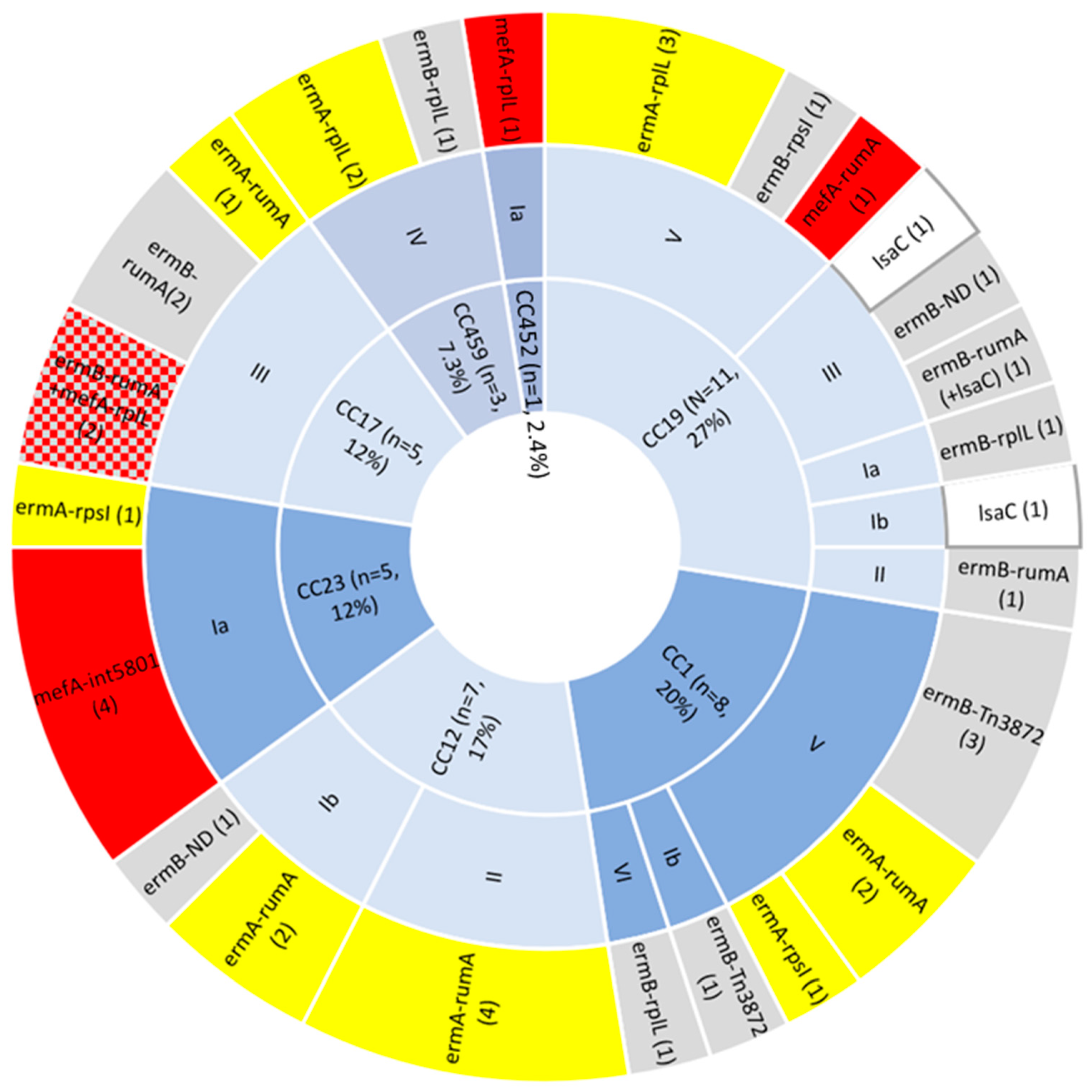
2.1. Phenotypic and Genotypic AMR Profiling

Seventeen (17/193, 8.8%) GBS isolates carried non-characterised antibiotic resistance genes and were phenotypically susceptible to all eight antimicrobials tested, including tetracycline. Within the 193 GBS isolates from different patients examined, 17 different antibiotic resistance determinants were identified. Of which, 70.5% (136/193) carried resistance genes to only one class of antibiotic and 20.7% (40/193) carried resistance genes to more than one class of antibiotic, i.e., aminoglycosides, macrolides, chloramphenicol, and/or tetracycline. All GBS isolates (n = 193) were found to be phenotypically susceptible to ampicillin and vancomycin, consistent with the absence of resistance determinants for these antimicrobials found by in silico genome analysis. All 193 GBS isolates carried penicillin-binding protein (PBP) types (1a, 1b, 2a, 2b, and 2x); therefore, any protein sequence variations identified (Supplementary Materials, Table S1) were presumed to represent non-resistance polymorphisms concordant with ampicillin susceptible phenotypes. Susceptibility for penicillin was assumed in proxy based on the universal susceptibility to ampicillin.
Established aminoglycoside resistance genes were detected in (8/193, 4.1%) isolates—aph(3′-III) and ant(6-Ia) (7/8, 87.5%), aad(E) (4/8, 50%), and aac(6′)-aph(2″) (1/8, 12.5%) (Table 1). Phenotypic testing for high gentamicin resistance identified a single isolate (PHEGBS0139) resistant to gentamicin (1/193, 0.5%), consistent with the carriage of aac(6′)-aph(2″). Resistance to chloramphenicol was found in three isolates (3/193, 1.5%), corresponding to the presence of cat(Q) (2/3, 66.6%; PHEGBS0595, PHEGBS0608) (Table 1) or catpC194 (1/3, 33.3%; PHEGBS0738) genes. MIC gradient strips confirmed that all cat(Q) (n = 2) and catpC194 (n = 1) positive isolates had a chloramphenicol MIC of 32 or 48 mg/L. Tetracycline resistance determinants were carried by most GBS isolates (174/193, 90.1%). The most common tetracycline resistance gene was tet(M) (154/174, 88.5%), followed by tet(O) (23/174, 13.2%), with single isolates carrying tet(W), tet(S), or tet(L) (1/174, 0.5%) genes, although tet(M) was additionally present for the tet(L) and three tet(O)-carrying isolates. One of the tet(M) positive isolates (PHEGBS0156) remained tetracycline susceptible on phenotypic and MIC testing, with MIC value of 0.06 µg/mL for tetracycline. Genomic analysis for this isolate showed a premature stop codon in the tet(M) gene with a 213bp deletion at the 5′ end.
Forty-one (21.2%) GBS isolates showed resistance to macrolides, mediated by at least one macrolide resistance gene, including six isolates (PHEGBS0139, PHEGBS0288, PHEGBS0577, PHEGBS0595, PHEGBS0608, and PHEGBS0071) that harboured multiple macrolide genes (Table 1). The most common macrolide resistance determinant was erm(A) or erm(B), distributed equally in (16/41 isolates each, 39%), followed by the identification of mef(A) and msr(D) in (10/41, 24.3%) isolates, lsa(C) in (3/41, 7.3%) isolates, and a single isolate carrying erm(T) and lnu(C) (1/41, 2.4% each) (Table 1), although five isolates carried combinations of these. Of these genes, erm(T) was uniquely carried on plasmid pGB2001 (NZ_JF308630.1), while all other macrolide resistance genes were carried on genomic ICEs. Upon phenotypic testing, resistance rates to erythromycin and clindamycin were 20.2% (39/193) and 11.9% (23/193), respectively, with 10.8% (21/193) resistant to both. Inducible clindamycin resistance was observed in 31.7% (13/41) of macrolide-resistant GBS isolates, representing 12 isolates carrying erm(A) and the single isolate carrying erm(T).
Four GBS isolates (PHEGBS0139, PHEGBS0559, PHEGBS0595, and PHEGBS0608) were also resistant to levofloxacin (MIC > 256 mg/L), mediated by double topoisomerase/gyrase point mutations (Ser81Leu for gyrA, Ser79Tyr for parC), with two isolates (PHEGBS0595 and PHEGBS0608) carrying the H221Y non-resistance polymorphism in parE. Twenty-eight additional levofloxacin-susceptible isolates carried the H221Y parE substitution, in the absence of gyrA and/or parC mutations, indicating that this altered amino acid did not contribute to the resistance phenotype. Three isolates (PHEGBS0139, PHEGBS0595, and PHEGBS0608) resistant to levofloxacin were serotype V and ST19, while the other isolate (PHEGBS0559) was composed of serotype III and ST17. Importantly, all four levofloxacin resistant isolates were already multi-drug resistant due to the coincident carriage of ARGs for macrolides (n = 4), high gentamicin (n = 1), and/or chloramphenicol (n = 2) resistance.

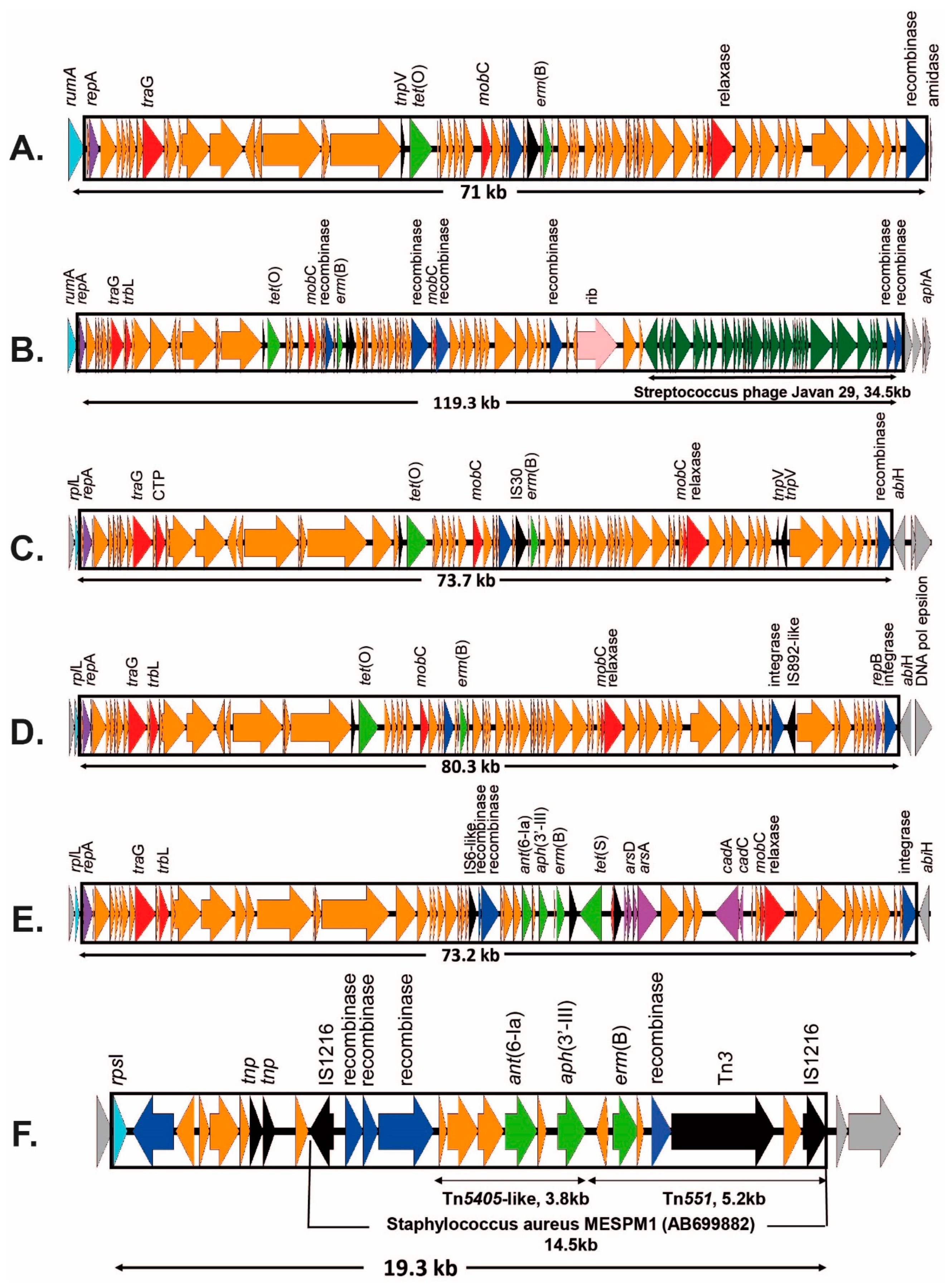
2.2. Tetracycline Resistance and Mobile Genetic Elements

Tetracycline resistance was most commonly mediated by the tet(M) gene (154/174, 88.5%), which was predominantly carried by the transposon Tn916 (accession no. U09422.1) or Tn916-like MGE (87/154, 56.4%), with a lesser number associated with the Tn5801 MGE (67/154, 43.5%) (accession no. HF930766.1). Among the five common CCs, Tn916 or Tn916-like elements were most common in CC19 (81.2%, 26/32), CC1 (71.8%, 23/32), and CC12 (41.9%, 13/31), whereas Tn5801 was found in CC23 (89.1%, 41/46), CC17 (58.6%, 17/29), CC452 (100%, 7/7), and CC327 (100%, 3/3). This is consistent with findings of Da Cunha et al. [21]. A novel Tn916-like transposon (Tn7539; accession no. OP715847) was identified and found to carry the tet(M) gene, with the addition of 9 putative additional hypothetical genes, in CC1 (PHEGBS0533) and CC459 (PHEGBS0463) isolates.
Tetracycline resistance was not solely mediated by the tet(M) gene: a single novel Tn916-like transposon (Tn7518; accession no. OP715846) in PHEGBS0586 (CC12, serotype II) was identified, which was found to carry tet(L) in combination with tet(M). A novel ICE (ICESag100) was identified to carry the tetracycline resistance gene tet(W) alone (isolate PHEGBS0100; ICESag100 accession no. OP715836; Figure 1) inserted at the rumA site.
Twenty-three tetracycline-resistant isolates carried tet(O) (23/174, 13.2%) and one isolate carried tet(S) (PHEGBS0662). Among the 23 tet(O) positive isolates, 11 (47.8%) harboured tet(O) as a single ARG on an ICE, of which 10/11 (90.9%) carried tet(O) inserted into the genome at the 23S rRNA methylase gene (rumA), a common target for ICEs carrying serine recombinases [23]. Phylogenetic comparison of tet(O)-carrying ICEs indicated two ICE clusters (Figure S1). Cluster 1 was comprised of six closely related ICEs (median SNPs difference of eight SNPs; range 7–70 SNPs) identified in CC12 isolates (PHEGBS0068, PHEGBS0092, PHEGBS0145, PHEGBS0171, PHEGBS0390, and PHEGBS0393), where three elements were identified in serotype II and three in serotype Ib. Cluster 2 was comprised of three moderately related ICEs (median SNPs difference of 312 SNPs; range 207–417 SNPs) (Figure S1), identified in three isolates (PHEGBS0097, PHEGBS0222, and PHEGBS0618). These ICEs were identified in diverse isolates of CC12 serotype Ib, CC19 serotype III, and CC23 serotype Ia. A single ICE carrying tet(O) was inserted into a different genomic location into the 50S ribosomal accessory protein L7/12 (rplL) for isolate PHEGBS0132 (serotype III, CC19). Despite the different genomic locations, the rplL tet(O) ICE was most similar (82%) to the ICEs identified in isolates PHEGBS0097 and PHEGBS0618. A comparison of the ICE-carrying tet genes identified in this study, which were inserted into rumA, revealed that the novel tet(W) ICE was distinct and did not cluster with any of the tet(O) ICEs, with the closest homology of 59.4% to ICESag061 from isolate PHEGBS0061 (Figure S1).
Tetracycline is not used as a therapy for invasive GBS infection. When excluding ICEs that carry only tetracycline resistance, 41/193 (21.2%) carried resistance genes for macrolides and/or lincosamides. One of these detected genes (erm(T)) in PHEGBS0539 was carried on a GBS plasmid pGB2001 (NZ_JF308630.1). Figure 2 shows the diversity of CCs and serotypes, which were found to carry macrolide/lincosamide ARGs on ICEs.
In Table 1, it can be observed that eight CC1 isolates and eleven CC19 isolates predominantly carried erm(A) or erm(B) methylase genes, and to a lesser extent lnu(C), lsa(C), or efflux pump mef(A)/msr(D) genes. The genomic insertion sites of ICEs carrying these genes widely varied to include Tn3872, with no specific insertion site (n = 4), rumA (n = 5), rplL (n = 5), rpsI (n = 2), rumA + lsa(C) (unidentified) (n = 1), and two more lsa(C)-containing ICEs (undefinable due to the short contig length) (Figure 2 and Table 1). In contrast, the remaining clonal clade CC12 (n = 7), CC23 (n = 6), CC17 (n = 5), CC459 (n = 3), and CC452 (n = 1) isolates appeared to be less complex and consisted of only 1–2 serotypes and 1–2 ICE insertion sites. CC12, CC23, and CC452 carried single ARG ICEs at a single insertion site. CC17 was comprised of all serotype III and included four isolates carrying the multiple ARG-containing ICESag37 [20] at rumA; however, two of these isolates had an additional ARG-containing ICE (ICESpy009-like) inserted at rplL. The final CC17 isolate carried the novel ICESag066 carrying erm(A) at the rumA insertion site (Figure 2 and Table 1).

2.3. Macrolide Resistance and Mobile Genetic Elements

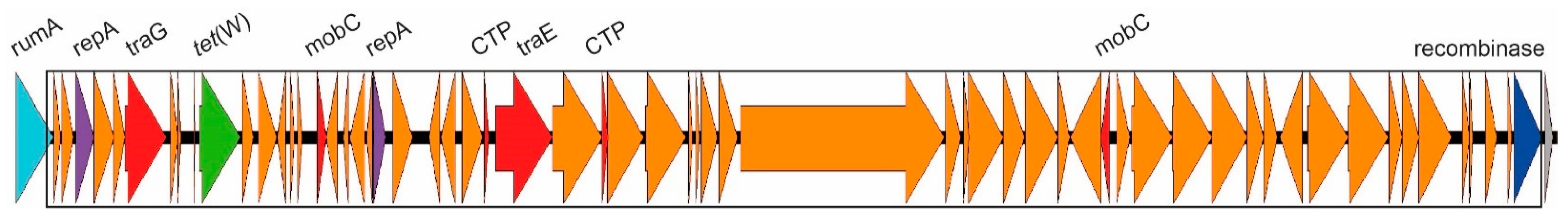
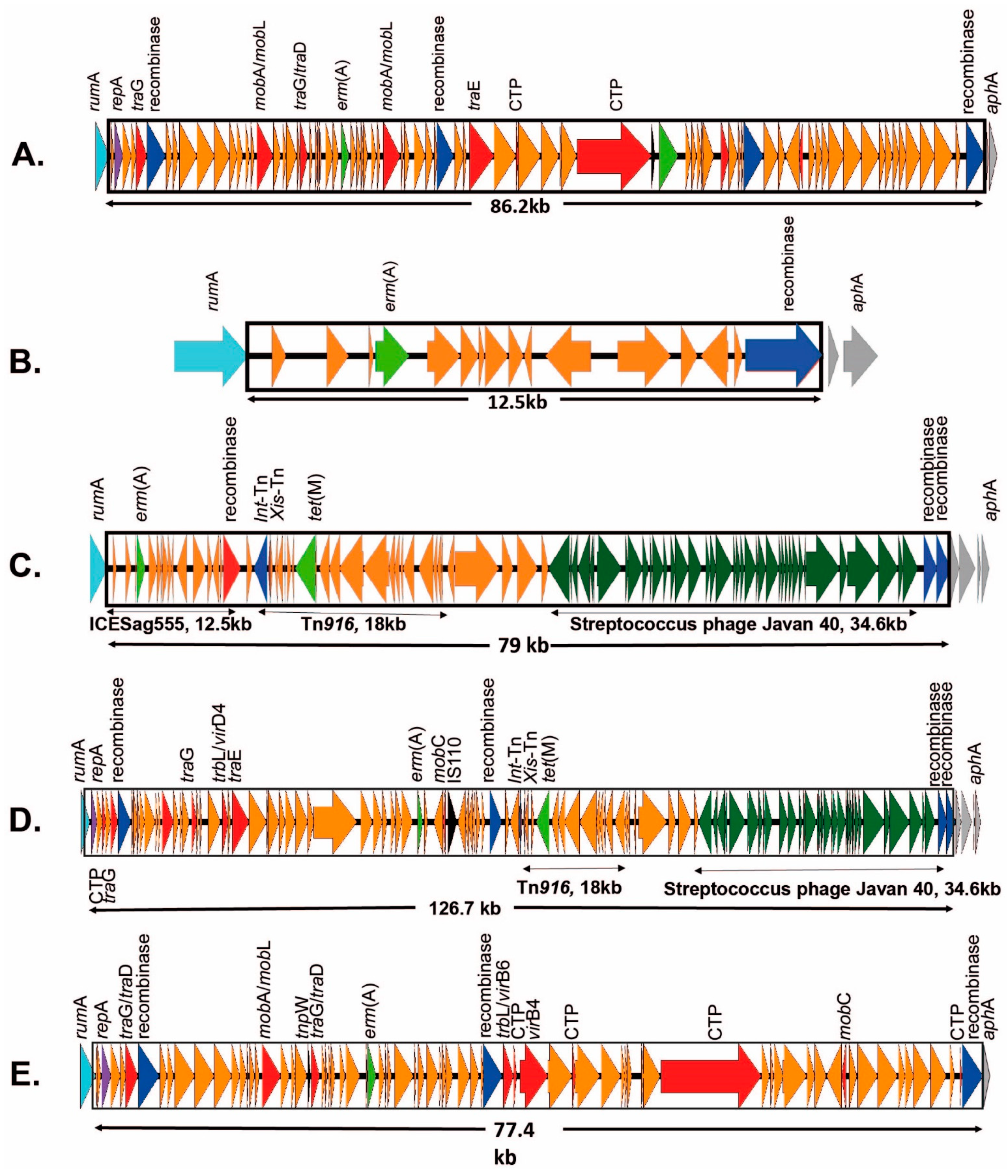
2.3.1. ICEs Carrying the erm(A) Gene

Sixteen of the isolates carried the macrolide resistance gene erm(A) contained in 12 separate ICEs, of which eight ICEs are novel to this study. The erm(A) gene was inserted at rumA (n = 9), rplL (n = 4), rpsI (n = 2), or at an indeterminate site (n = 1) (Table 1). Four of these isolates carried a nearly identical ICE, containing both erm(A) and tet(O), inserted at rumA (representative ICE = ICESag509; accession no. OP715843; Figure 3A). They were initially identified as similar to Sp2905 in S. pyogenes (group A streptococcus; accession no. FR691055); however, homology was low (68.2%) and only 26/63 genes (excluding the site-specific serine recombinase) were shared between the ICEs of four GBS isolates and Sp2905. Five additional erm(A)-containing novel ICEs were inserted at the rumA gene, but lacked the tet(O) gene of the previous four isolates: ICESag066 (accession no. OP715837; Figure 3B); ICESag082 (accession no. OP715838; Figure 3C); ICESag098 (accession no. OP715840; Figure 3D); ICESag428 (accession no. OP715841; Figure 3E); and ICESag555 (which is present within ICESag082, highlighted in Figure 3C). ICE size ranged from 12,518 to 126,707 bp and was found in serotype III (CC17, n = 1), serotype V (CC1, n = 2), and serotype Ib (CC12, n = 2). ICESag066 and ICESag555 were nearly identical in size and only differed by 72 SNP, despite being isolated from different serotypes (PHEGBS0066; CC17, serotype III; Figure 3B and PHEGBS0555; CC12, serotype Ib; shown as part of Figure 3C). ICESag082 was found in CC1, serotype V isolate PHEGBS0082 (total length of 79,052 bp; Figure 3C). However, comparing the first 14,150 bp (to the end of the recombinase) shows only 22 SNP relative to ICESag555, despite being from two completely different CC and serotypes. It was observed that the latter was picked up as part of the complex ICE that forms ICESag082.
Four additional erm(A)-carrying ICEs were found to be inserted adjacent to the somatic gene rplL, including the previously identified ICESag236 [24] (accession no. OP508058; Figure 4A) in CC19, serotype V isolates, PHEGBS0595 and PHEGBS0608, and those also carried cat(Q) and mef(A)/msr(D) ARGs. Moreover, novel ICESag139 (accession no. OP508059; Figure 4B) was isolated from CC19, serotype V isolate, PHEGBS0139, and was the only ICE to carry the high gentamicin level resistance aac(6′)-aph(2″) genes in this dataset. Novel ICESag084 (accession no. OP715839; Figure 4C) identified in the CC459, serotype IV isolate, PHEGBS0084, carried erm(A) as an isolated ARG. Moreover, a second CC459, serotype IV isolate (PHEGBS0266), carried a single erm(A) gene on a sequence contig very short to determine the insertion location (Table 1). The homology between this short contig and ICESag082 was only 68.6%, suggesting that they did not come from a common ancestor ICE. Furthermore, ICE-carrying erm(A) was found to be inserted adjacent to the rpsI (30S ribosomal protein S9) gene (Table 1), a considerably less common somatic insertion site [23]. Isolate 100414 (serotype V, CC1) carried a novel 76-kb ICE- carrying erm(A) (ICESag100414; accession no. OP715842; Figure 4D), and a spectinomycin phosphotransferase gene and isolate PHEGBS0044 (serotype Ia, CC23) carried these genes at the same insertion site, with ICE truncated post 55.6 kb, preventing a full ICE comparison. However, the homology between ICESag100414 and the partial ICE for ICESag044 was 27.8%, indicating no common ancestry.

2.3.2. ICEs Carrying the erm(B) Gene

Sixteen isolates were found to carry the macrolide resistance gene erm(B), four of which have been discussed above as part of transposon Tn3875. For two isolates, sequencing was unable to identify the genomic location due to the short contig length. The remaining 10 erm(B)-carrying ICEs were inserted at rumA (n = 6), rplL (n = 3), or rpsI (n = 1) (Table 1). The previously characterised ICESag37 [20], carrying erm(B), tet(O), and three aminoglycoside resistance genes (ant(6-Ia), aph(3′-III), and aad(E)), was found in four isolates (PHEGBS0207, PHEGBS0288, PHEGBS0559, and PHEGBS0577), which were all closely related to serotype III, CC17 with identical ICEs. This suggests retention within lineage for these ICEs, with no evidence of horizontal transfer. The remaining two rumA-associated erm(B) ICEs, carrying both tet(O) and erm(B), lacked aminoglycoside ARGs, and were identified as two novel ICEs: ICESag624 (accession no. OP715844; Figure 5A) identified in PHEGBS0624 (serotype II, CC19) and ICESag071 (accession no. OQ054582; Figure 5B) identified in PHEGBS0071 (serotype III, CC19) (Table 1). These two ICEs were distinctly different from each other and ICESag37, suggesting different evolutionary origins. Moreover, isolates PHEGBS0195 (serotype Ia, CC19) and PHEGBS0206 (serotype IV, CC459) had a combination of erm(B) and tet(O), inserted at rplL with two further unique ICEs: ICESag195 (accession no. OP508060; Figure 5C) and ICESag206 (accession no. OP508061; Figure 5D). Furthermore, ICESag662 (accession no. OP508062; Figure 5E) carried erm(B), and was found to be inserted at rplL with tet(S), ant(6-Ia), and aph(3′-III). The only erm(B)-carrying ICE (ICESag738; accession no. OP508063; Figure 5F) inserted at rpsI was found in isolate PHEGBS0738 (serotype V, CC19), which carried a 19.3-kb ICE in combination with aminoglycoside ARGs ant(6-Ia), aph(3′-III), and heavy metal resistance genes. This ICE appears to be similar to the Staphylococcus aureus composite MGE structure (MESPM1) (accession no. AB699882; Figure S2). A second isolate (PHEGBS0156; serotype III, CC19) also had a MESPM1 homologous ICE; however, it lacked the IS1618 transposase. Moreover, genomic analysis indicated that the rpsI region was not disrupted, making it unclear where this last ICE (on a contig devoid of any GBS somatic genes) was inserted (Figure S2). A further difference between PHEGBS0156 and PHEGBS0738 was the latter isolate, which was the only isolate to carry the ARG cat(pC-194) inserted near one of the 16S rRNA genes, starting with a site-specific integrase and ending at an IS1216E transposase. The final erm(B)-carrying isolate (PHEGBS0622; serotype Ib, CC12) was on a contig very short to clearly define the potential ICE insertion site (Table 1). However, the rumA, rpsI, and rplL sites showed no insertion, and Tn3872 was not present.

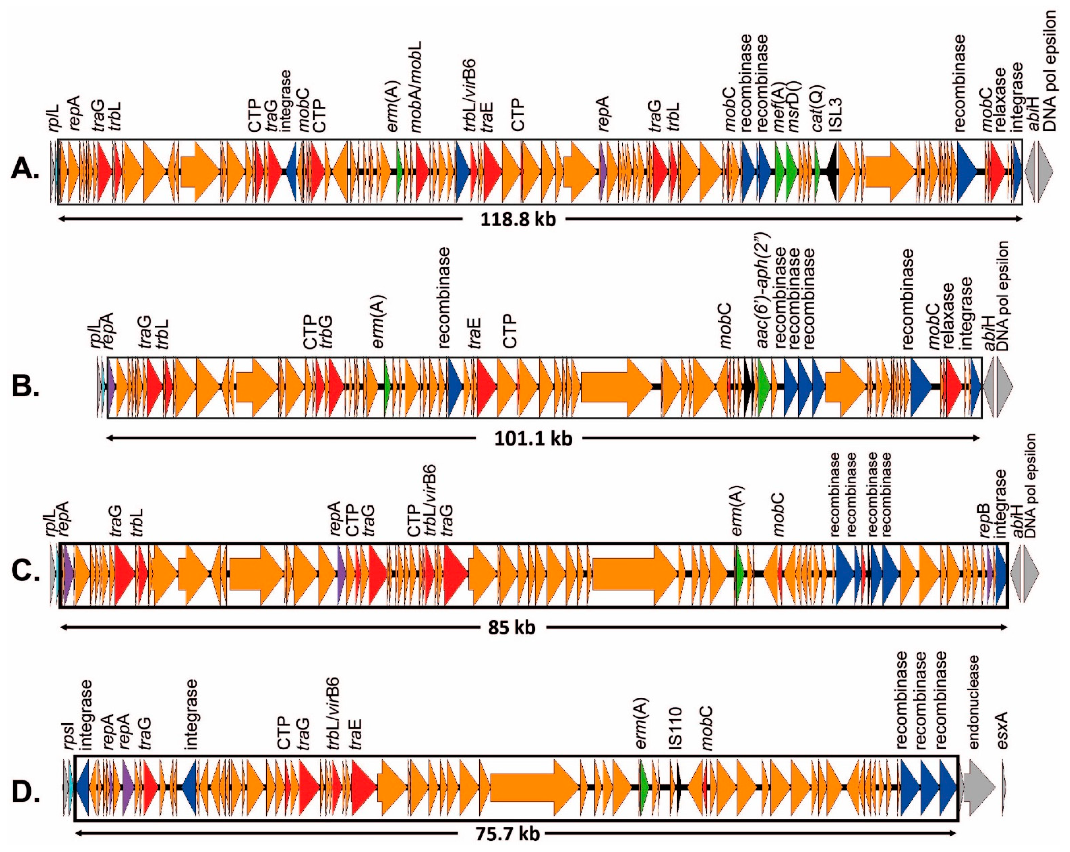
2.3.3. ICEs Carrying mef(A)/msr(D) Genes

Ten isolates were found to carry the macrolide resistance gene dyad mef(A)/msr(D) (Table 1). Only a single isolate (PHEGBS0639, serotype V, CC19) was found to carry the macrolide ARG mef(A)/msr(D) adjacent to the rumA gene (Table 1). It was adjacent to the complete genome for Streptococcus phage Javan69 (accession no. MK448828.1) and recombinase genes, but the contig was very short to define the genomic location or putative ICE. Five mef(A)/msr(D)-carrying ICEs were associated in ICEs located at the rplL gene insertion site: Two of these were in combination with erm(A) and cat(Q), similar to the ICESag236 shown above (Figure 4A), while the other three mef(A)/msr(D)-carrying ICEs inserted at rplL had homology to the MGE ICESpy009 (accession no. KU056701) [25]. Two of these isolates (PHEGBS0288 and PHEGBS0577) were serotype III, CC17, and carried the erm(B) genes in an ICESag37 inserted at rumA (as detailed above), while the other isolate PHEGBS0518 was serotype Ia, CC452 (Table 1). Four of the remaining mef(A)/msr(D)-carrying isolates were serotype Ia and CC23 (PHEGBS0067, PHEGBS0070, PHEGBS0248, and PHEGBS0625) and the mef(A)/msr(D) genes were located adjacent to a tet(M)/Tn5801 cassette with no clear MGE. The final mef(A)/msr(D)-carrying isolate near to, but more distant from, a Tn5801 cassette was of a different serotype (PHEGBS0625; ST23, serotype Ia), with no clear MGE (Table 1).

2.3.4. ICEs Carrying the lsa(C) Gene

Lincosamide-resistance genes were also identified. A single isolate (PHEGBS0139) carried the lnu(C) gene on a 1724-bp region, including an IS1 transposase homologue and bounded by 25-bp imperfect inverted repeats flanked by 8-bp direct repeats, as reported previously for another GBS isolate [26]. However, this isolate also carried a unique erm(B)-carrying ICE (ICESag139) inserted at the rplL site, making the lnu(C) gene redundant, as discussed above (Figure 4B). Three isolates were found to carry lsa(C) genes: these were CC19 isolates and despite being different serotypes (PHEGBS0222; serotype Ib and PHEGBS0372; serotype III), the lsa(C) insertion site has been previously described as ICE2603 tRNAlys element [27]. In the third CC19 isolate (PHEGBS0071; serotype III), the lsa(C) gene appears to have been incorporated into a tet(M)-positive tn916-like MGE (novel Tn7589; accession no. OQ269476), which has been inserted at the stop codon of an TVP38/TMEM64 family protein gene. All these isolates yielded intermediate resistance to clindamycin, except for PHEGBS0071, where the high level clindamycin resistance is likely mediated by erm(B) found on ICESag071 (Figure 5B), obscuring the activity of lsa(C) for this isolate.

3. Discussion

Overall, a high concordance rate (99%) between phenotypic and in silico predicted genotypic antibiotic resistance profiles were found. No phenotypic or genotypic resistance to the current first line antibiotic (penicillin) was detected in this study; therefore, all variations in the penicillin binding protein (1a, 1b, 2a, 2b, and 2x) are deemed to be non-resistance mediating polymorphisms, consistent with the theory that resistance to penicillin is rare in GBS [4,5]. However, there are growing reports of isolates with PRGBS in some geographical regions, including two isolates identified to date in Hong Kong [6], seventy-five in Japan [7,8,28], fifty-two in the USA [10,11,12], two in Canada [13,14], seven in Mozambique [15], and two in South Africa [29] and Korea [30]. However, the resistance rates for second-line antibiotics, such as erythromycin and clindamycin, continue to increase internationally, to the point where current recommendations for second-line therapeutics for adults have changed to cephalosporins or vancomycin, in response to the detailed BMJ best practice guidelines [3].
To treat severe invasive GBS infections, such as infective endocarditis, periprosthetic joint infections, or even early-onset disease (EOD) in babies [3,4], combination therapy that includes penicillin and gentamicin has been suggested. The first case of GBS showing high-level resistance to gentamicin was observed in a strain isolated in 1987 from a French patient [31]. The mechanism of acquisition of high-level gentamicin resistance determinants in GBS is largely unknown. We identified a single isolate in this study with high level gentamicin resistance. Taking into account the emerging resistance trends, there is a need to consider other classes of antibiotics with therapeutic potential for GBS infections.
Tetracycline is no longer a therapeutic option for GBS, since the extensive use after its first discovery in 1948 led to the high resistance profile in many bacteria, including GBS. Da Cunha et al. speculated that the extensive use of tetracycline in the 1960s exerted selective pressure on GBS, leading to genetic diversity loss and emergence of few tetracycline resistant pathogenic clones, which also carried mobile genetic elements with a mixture of ARGs, including tetracycline resistance [21]. As expected, in our study, the rates of resistance to tetracycline were high (174/193, 90.1%), largely mediated by tet(M) (154/174, 88.5%) and predominantly carried by two related ICEs: Tn916/Tn916-like elements (87/154, 56.4%) and Tn5801 [32] (67/154, 43.5%), which were found to be prevalent in CC1, CC12, CC19, CC17, and CC23, respectively. Similar observations were seen in previous studies [21,33], suggesting that these ICE-harbouring tet(M) are clonally related and acquired through limited and rare insertion events, followed by expansion within lineages. While these MGEs are generally accepted as promiscuous, Da Cunha et al. [21] previously demonstrated that five of the major clonal complex clades have fixed genomic insertion sites, resulting from the high level of tetracycline use in the 1960s, followed by expansion within lineages. This includes the erm(B)-carrying variant referred to as Tn3872 (CZ-NI-013 strain ENA, accession no. ERR048605), which Da Cunha et al. found clustered in CC1. Moreover, this is consistent with our finding that only CC1 isolates carried erm(B) in Tn3872 (accession no. OP715845).
We also found four isolates resistant to levofloxacin (fluoroquinolone) due to double point mutations in gyrA and parC. Interestingly, all four isolates containing quinolone resistance determinants belonged to CC19, and were resistant to antibiotics from at least two other classes, as seen previously [34].
Previous studies have examined the correlation between serotypes and clonal complexes, relative to antimicrobial resistance or the presence of ARG classes. Some studies suggest that erythromycin resistance is more associated with serotype V [35,36] or CC12 and CC17 [37], which is not supported in our data. However, all studies examining resistance trends agree that increases in macrolide and lincosamide resistance are fairly universal [38]. In the UK, the reported increase for erythromycin (3% to 15%) and clindamycin resistance (3% to 9%) noted from 1991 to 2010 [16] has continued, with recent reports indicating 31% clindamycin and 36% erythromycin resistance in 2020 (Laboratory surveillance of pyogenic and non-pyogenic streptococcal bacteremia in England: 2020 update (publishing.service.gov.uk). In this study, the data from 2014 to 2015 are consistent with this trend for both clindamycin (11.9%) and erythromycin (20.2%). Further in-depth investigations of larger GBS datasets are needed, since this limited study indicates that no single ARG drives the increase in resistance. While 32 of 193 isolates carried erm(A) or erm(B) genes, they were carried on 16 unique ICE elements (Table 1); five isolates had contigs very short to clearly define potential ICEs (Table 1). A maximum of four isolates carried the same ICE element (Tn3872, ICESag37, and ICESp2905-like) and in each case, they were all found in the same CC (Table 1), as observed previously for Tn3872 in CC1 [21] and ICESag37 in CC17 [20], suggesting their association, spread, and maintenance.
We also identified three hotspot integration sites for ICE-carrying macrolide resistance determinants, which includes the most frequent rumA found for sixteen ICEs, rplL for ten ICEs, and less commonly for rpsI ICEs (n = 3) (Figure 1 and Table 1). These integration sites have previously been classified as well-known integration sites for ICEs in streptococci [23]. Additionally, sixteen novel ICEs carrying macrolide alone or with other antibiotic resistance determinants (ICESag066, ICESag071, ICESag082, ICESag084, ICESag098, ICESag139, ICESag195, ICESag206, ICESag236, ICESag428, ICESag509, ICESag555 (separate as well as part of ICESag082), ICESag624, ICESag662, ICESag738, and ICESag100414), a single novel ICE carrying the tetracycline resistance gene tet(W) alone (ICESag100), and three new transposons (i.e., Tn7518 carrying a combination of tet(M) and tet(L) genes, Tn7589 carrying tet(M) and lsa(C) genes, and Tn7539 carrying typical Tn916 with an additional nine ORFs) were identified.
The number of novel ICEs identified in this small dataset (only 193 GBS isolates from the UK) is surprising, and confirms that the GBS population is highly diverse. This indicates that specific antimicrobial resistance patterns conferred via MGEs could potentially remain within specific lineages and expand or emerge, rather than spread via the horizontal transfer. Therefore, the proposed GBS vaccines would not only potentially eliminate the most common serotypes/genotypes of disease-causing GBS, but also could be used to limit the emergence and spread of antimicrobial-resistant and multi-drug-resistant GBS lineages. To better understand how vaccines and changes in antimicrobial prescriptions for the treatment of GBS infections would impact antimicrobial resistance, global longitudinal molecular epidemiology studies of GBS carriage and disease are required. Moreover, this will provide details on the molecular basis of antibiotic resistance.

4. Materials and Methods

4.1. Clinical GBS Strains and Their Whole Genome Sequences

The 193 invasive and non-invasive GBS isolates collected from adults in the UK between 2014 and 2015, as analysed in our previous study [39], were used in this study.

4.2. Phenotypic Antibiotic Susceptibility Testing (AST)

All susceptibility testing was performed on pure archived GBS strains, recovered from freezing by overnight culture on blood agar supplemented with 5% sheep blood (Oxoid, Basingstoke, UK). Then, overnight bacteria inocula were resuspended in sterile saline to 0.5 McFarland units, containing 105 cfu/mL. Disk diffusion and gradient strip AST were performed on Mueller Hinton with 5% sheep blood (MH-F plates; Oxoid, Basingstoke, UK). In addition, uniform lawns of freshly-made isolate suspensions were inoculated prior to overlaying with antimicrobial impregnated disks or gradient strips (Oxoid), after visible liquid had dried from the plate surface. A disk dispenser (Thermo Fisher Ltd., Loughborough, UK) was used to ensure uniform spacing of 1 cm between disks on the 90 mm plates. Then, the plates were incubated in an aerobic incubator at 35 °C for 24 h.
All 193 isolates were tested for antimicrobial susceptibility by disk diffusion using the following concentrations: Ampicillin (10 µg), vancomycin (30 µg), tetracycline (30 µg), erythromycin (15 µg), clindamycin (2 µg), chloramphenicol (30 µg), high level gentamicin (120 µg), and levofloxacin (5 µg) (Oxoid, Basinstoke, UK). Briefly, the inhibition zone sizes were recorded for each antimicrobial agent and interpreted according to the Clinical Laboratory Standards Institute (CLSI) 2022 guidelines [5]. For this method, erythromycin and clindamycin disks were adjacent and 1 cm apart to identify clindamycin resistance induction via the D-zone test, as previously described [5,40]. Briefly, blunting (or D appearance) or bacterial growth within the clindamycin zone of inhibition proximal to the erythromycin disk was documented as inducible resistance, commonly mediated by MLSB-inducible methylation. In the case where the zone of inhibition did not clearly define the sensitivity of an isolate, or where genotypic evaluation of an isolate did not match the expected phenotypic profile (except for tetracycline), gradient strips (MIC test strip; Liofilchem, Italy) were employed to determine a more accurate MIC on MH-F plates, as prepared above for disk diffusion. The MIC value, where the growth of the GBS touched the edge of the strip, was interpreted according to the CLSI 2017 guidelines [5].

4.3. Long-Read Whole Genome Sequencing

Ten isolates (study accession no. PRJEB59039) were selected for additional long read sequencing, guided by the initial short read bioinformatics analysis. The gDNA was prepared as previously described [41] using the Qiagen QIAamp DNA kit and extracted on the QIAcube platform (Qiagen, Manchester, UK). Moreover, the gDNA was quantified using the dsDNA HS assay kit and the Qubit 4.0 fluorometer (Thermo Fisher Scientific Inc., Glasgow, UK). Then, it was purified and concentrated using SPRI beads (Mag-Bind TotalPure, Omega Bio-tek, Inc., Norcross, GA, USA) at a 1:1 ratio with a final elution volume of 15 µL, in order to achieve an optimal range between 40 and 60 ng/µL. Moreover, the gDNA was re-quantified with the dsDNA BR assay kit. Genomic libraries were prepared using the Rapid Barcoding Kit (SQK-RBK004; Oxford Nanopore; Oxford, UK), sequenced on a R9.4 flow cell using a MinION (Oxford Nanopore Technologies; Oxford, UK), and base-calling was performed using Guppy v4.0.9 within MinKNOW v20.06.4. Long reads were demultiplexed using porechop (v0.2.4) [28], and assembled against corresponding short reads generated from the Illumina MiSeq using Unicycler (v0.4.7) [42] with default parameters. The hybrid assembly was assessed using Quast (v5.0.2), and ABRicate (v0.9.7) (>98% coverage and identity) was used to detect antimicrobial resistance genes (ARG). The contig containing the ARG of interest was exported to a separate FASTA file for comparative analysis using Bandage (v0.8.1), in order to accurately determine the boundaries of the ICE carrying the ARGs, or to accurately determine the full novel ICE composition where ARGs were not bounded by somatic genes on both sides in short-read contigs [43]. Novel transposons were assigned Tn numbers using the Transposon registry [44].

4.4. Resistome Profile Associated with the Mobile Genetic Elements

Clonal complexes (CCs) were assigned by PubMLST using S. agalactiae MLST database (https://pubmlst.org/sagalactiae/, accessed on 15 December 2022). AMR genes were identified by ResFinder 2.1 database/webserver [45]. Assembled GBS genomes were screened for MGEs using Geneious Prime® (Biomatters, Inc., Auckland, New Zealand) by annotation (threshold 97% identity) with genes identified by ResFinder. Genomic regions containing MGE became apparent when assemblies were annotated using the prototype genome FDAARGOS_520 (accession no. CP033810), which was selected as it did not have any evidence of ICEs. When a second round of annotation was performed using a database of existing ResFinder resistance genes, the presence of ARG annotations within these gaps identified the MGEs. These MGE insertions usually corresponded to previously reported Streptococci insertion “hotspots” [32], i.e., bounded on one end by rumA (23S rRNA (uracil-5) methyltransferase; protein_id: AQY23883.1, rplL (50S ribosomal accessory protein L7/12; protein_id: AWZ30204.1), or rpsI (30S ribosomal protein S9; protein_id: AKI94631.1), which were confirmed by the alignment of these references to query assembled genomes in Geneious Prime. BLASTn searches against NCBI databases in Geneious Prime® were carried out as the final step in identifying and annotating the MGE-associated genes present between DRs of the putative ICEs in each genome. Artemis [46] was used to colour the detected signature proteins of an ICE, such as tyrosine/serine integrases/recombinases, relaxases, plasmid mobilisation protein genes (mobA/mobL), conjugal transfer protein genes (virB4, virD4, traL, and trbL), and insertion sequences. Bakta v1.5 was used to annotate putative MGEs (https://github.com/oschwengers/bakta, accessed on 15 December 2022). Pairwise BLASTn alignment of identified MGEs was performed and visualised using Easyfig v2.2.2.

4.5. Phylogenetic Analysis

Neighbour joining method (using Jukes-Cantor genetic distance model, global alignment with free end gaps, and cost matrix of 65% similarity) was used to construct the phylogenetic tree for ICEs carrying the tet(O) gene alone with an ICE-carrying tet(W), which was used as an outgroup in Geneious Prime® (Biomatters Ltd., Auckland, New Zealand) tree builder tool.

5. Conclusions

This study defines sixteen novel ICEs and three novel transposons, with no evidence of horizontal transfer. In addition, it provides evidence of retention within the GBS lineage of previously reported ICEs. The discovery of numerous novel MGE-carrying ARGs in a small number of isolates reflects the importance of ARG study in human pathogens, in which resistance is of increasing concern. It was surprising to identify numerous novel ICEs present in a small cohort. The GenBank reference genomes for the mobile elements discovered here will be of benefit to future studies in determining whether increasing antimicrobial resistance is a result of global spread of the ICE defined here, or whether they are being caused by the sporadic capture of other novel ICEs. The ICE sequences and genomic dataset provided by this and future studies enable the development of tests to detect genes encoding resistance, as well as the improvement of the surveillance and monitoring of resistance potential in the population. Moreover, it could aid in an increasingly rapid therapeutic intervention than sensitivity testing alone.

Supplementary Materials

The following supporting information can be downloaded at: https://www.mdpi.com/article/10.3390/antibiotics12030544/s1. Figure S1: Neighbour joining phylogenetic tree for the ICEs carrying carrying tetO gene alone; Figure S2: Comparison of Staphylococcus aureus composite mobile element structure MESPM1 to CC19 GBS isolates; Table S1: Substitutions in five penicillin binding proteins (PBPs) identified in 193 GBS isolates relative to the reference PBPs.

Author Contributions

Conceptualization, O.B.S., E.J. and V.J.C.; methodology, O.B.S., E.J., E.A.R.P. and K.S.; software, K.S.; formal analysis, investigation, resources, data curation, U.B.K., S.L., E.J., V.J.C. and O.B.S.; writing and revising, U.B.K., S.L., E.J., V.J.C. and O.B.S.; supervision, E.J. and O.B.S. All authors have read and agreed to the published version of the manuscript.

Funding

This research received no external funding.

Institutional Review Board Statement

Not applicable.

Informed Consent Statement

Not applicable.

Data Availability Statement

Genomic data are available according to the accession numbers provided in the text. Other data are available from the authors upon reasonable request.

Acknowledgments

The authors would like to thank UKHSA reference laboratory colleagues, led by Juliana Coelho, for provision of isolates, sequences, and metadata. This publication made use of the PubMLST website (https://pubmlst.org/, accessed on 15 December 2022) developed by Keith Jolley (Jolley and Maiden 2010, BMC Bioinformatics, 11:595) and cited at the University of Oxford. The development of the website was funded by the Wellcome Trust. E.J. is a Rosetrees/Stoneygate 2017 Imperial College Research Fellow, funded by Rosetrees Trust and the Stoneygate Trust (Fellowship no. M683). E.J. is affiliated with the National Institute for Health Research Health Protection Research Unit (NIHR HPRU) in Healthcare Associated Infections and Antimicrobial Resistance at Imperial College London in partnership with the UK Health Security Agency (previously PHE), in collaboration with Imperial Healthcare Partners, University of Cambridge, and University of Warwick.

Conflicts of Interest

The authors declare no conflict of interest.

References

  1. Navarro-Torné, A.; Curcio, D.; Moïsi, J.C.; Jodar, L. Burden of invasive group B Streptococcus disease in non-pregnant adults: A systematic review and meta-analysis. PLoS ONE 2021, 16, e0258030. [Google Scholar] [CrossRef]
  2. Rudman, J.; Carter, E.; Coehlo, J.; Hopkins, K.; Chalker, V.J.; Guy, R.; Lamagni, T.L. Laboratory surveillance of pyogenic and non-pyogenic streptococcal bacteraemia in England (2020). Health Prot. Rep. 2020, 15, 1–23. [Google Scholar]
  3. Healy, B. Public H.W. Group B Streptococcal Infection. Available online: https://bestpractice.bmj.com/topics/en-gb/924 (accessed on 3 December 2022).
  4. Hayes, K.; O’Halloran, F.; Cotter, L. A review of antibiotic resistance in Group B Streptococcus: The story so far. Crit. Rev. Microbiol. 2020, 46, 253–269. [Google Scholar] [CrossRef]
  5. CLSI. M100 Performance Standards for Antimicrobial Susceptibility Testing, 32nd ed.; Clinical and Laboratory Standards Institute: Wayne, PA, USA, 2022; ISBN 978-1-68440-135-2. [Google Scholar]
  6. Chu, Y.W.; Tse, C.; Tsang, G.K.L.; So, D.K.S.; Fung, J.T.L.; Lo, J.Y.C. Invasive group B Streptococcus isolates showing reduced susceptibility to penicillin in Hong Kong. J. Antimicrob. Chemother. 2007, 60, 1407–1409. [Google Scholar] [CrossRef] [Green Version]
  7. Kimura, K.; Suzuki, S.; Wachino, J.I.; Kurokawa, H.; Yamane, K.; Shibata, N.; Nagano, N.; Kato, H.; Shibayama, K.; Arakawa, Y. First molecular characterization of group B streptococci with reduced penicillin susceptibility. Antimicrob. Agents Chemother. 2008, 52, 2890–2897. [Google Scholar] [CrossRef] [Green Version]
  8. Nagano, N.; Nagano, Y.; Toyama, M.; Kimura, K.; Tamura, T.; Shibayama, K.; Arakawa, Y. Nosocomial spread of multidrug-resistant group B streptococci with reduced penicillin susceptibility belonging to clonal complex 1. J. Antimicrob. Chemother. 2012, 67, 849–856. [Google Scholar] [CrossRef]
  9. Seki, T.; Kimura, K.; Reid, M.E.; Miyazaki, A.; Banno, H.; Jin, W.; Wachino, J.I.; Yamada, K.; Arakawa, Y. High isolation rate of MDR group B streptococci with reduced penicillin susceptibility in Japan. J. Antimicrob. Chemother. 2015, 70, 2725–2728. [Google Scholar] [CrossRef] [Green Version]
  10. Dahesh, S.; Hensler, M.E.; Van Sorge, N.M.; Gertz, R.E.; Schrag, S.; Nizet, V.; Beall, B.W. Point mutation in the group B streptococcal pbp2x gene conferring decreased susceptibility to β-lactam antibiotics. Antimicrob. Agents Chemother. 2008, 52, 2915–2918. [Google Scholar] [CrossRef] [Green Version]
  11. Metcalf, B.J.; Chochua, S.; Gertz, R.E.; Hawkins, P.A.; Ricaldi, J.; Li, Z.; Walker, H.; Tran, T.; Rivers, J.; Mathis, S.; et al. Short-read whole genome sequencing for determination of antimicrobial resistance mechanisms and capsular serotypes of current invasive Streptococcus agalactiae recovered in the USA. Clin. Microbiol. Infect. 2017, 23, 574.e7–574.e14. [Google Scholar] [CrossRef] [Green Version]
  12. Francois Watkins, L.K.; McGee, L.; Schrag, S.J.; Beall, B.; Jain, J.H.; Pondo, T.; Farley, M.M.; Harrison, L.H.; Zansky, S.M.; Baumbach, J.; et al. Epidemiology of Invasive Group B Streptococcal Infections among Nonpregnant Adults in the United States, 2008–2016. JAMA Intern. Med. 2019, 179, 479–488. [Google Scholar] [CrossRef] [PubMed]
  13. Gaudreau, C.; Lecours, R.; Ismaïl, J.; Gagnon, S.; Jetté, L.; Roger, M. Prosthetic hip joint infection with a Streptococcus agalactiae isolate not susceptible to penicillin G and ceftriaxone. J. Antimicrob. Chemother. 2009, 65, 594–595. [Google Scholar] [CrossRef]
  14. Longtin, J.; Vermeiren, C.; Shahinas, D.; Tamber, G.S.; McGeer, A.; Low, D.E.; Katz, K.; Pillai, D.R. Novel mutations in a patient isolate of Streptococcus agalactiae with reduced penicillin susceptibility emerging after long-term oral suppressive therapy. Antimicrob. Agents Chemother. 2011, 55, 2983–2985. [Google Scholar] [CrossRef] [Green Version]
  15. Sigaúque, B.; Kobayashi, M.; Vubil, D.; Nhacolo, A.; Chaúque, A.; Moaine, B.; Massora, S.; Mandomando, I.; Nhampossa, T.; Bassat, Q.; et al. Invasive bacterial disease trends and characterization of group B streptococcal isolates among young infants in southern Mozambique, 2001–2015. PLoS ONE 2018, 13, e0191193. [Google Scholar] [CrossRef] [PubMed] [Green Version]
  16. Lamagni, T.L.; Keshishian, C.; Efstratiou, A.; Guy, R.; Henderson, K.L.; Broughton, K.; Sheridan, E. Emerging trends in the epidemiology of invasive group B streptococcal disease in England and Wales, 1991–2010. Clin. Infect. Dis. 2013, 57, 682–688. [Google Scholar] [CrossRef] [PubMed] [Green Version]
  17. Varaldo, P.E.; Montanari, M.P.; Giovanetti, E. Genetic elements responsible for erythromycin resistance in streptococci. Antimicrob. Agents Chemother. 2009, 53, 343–353. [Google Scholar] [CrossRef] [PubMed] [Green Version]
  18. Malbruny, B.; Werno, A.M.; Murdoch, D.R.; Leclercq, R.; Cattoir, V. Cross-resistance to lincosamides, streptogramins A, and pleuromutilins due to the lsa(C) Gene in Streptococcus agalactiae UCN70. Antimicrob. Agents Chemother. 2011, 55, 1470–1474. [Google Scholar] [CrossRef] [PubMed] [Green Version]
  19. Hawkins, P.A.; Law, C.S.; Metcalf, B.J.; Chochua, S.; Jackson, D.M.; Westblade, L.F.; Jerris, R.; Beall, B.W.; McGee, L. Cross-resistance to lincosamides, streptogramins A and pleuromutilins in Streptococcus agalactiae isolates from the USA. J. Antimicrob. Chemother. 2017, 72, 1886–1892. [Google Scholar] [CrossRef] [Green Version]
  20. Zhou, K.; Xie, L.; Han, L.; Guo, X.; Wang, Y.; Sun, J. ICESag37, a novel integrative and conjugative element carrying antimicrobial resistance genes and potential virulence factors in Streptococcus agalactiae. Front. Microbiol. 2017, 8, 1921. [Google Scholar] [CrossRef]
  21. Cunha, V.D.; Davies, M.R.; Douarre, P.; Rosinski-, I. Streptococcus agalactiae clones infecting humans were selected and fixed through the extensive use of tetracycline. Nat. Commun. 2015, 5, 4544. [Google Scholar] [CrossRef] [Green Version]
  22. Brochet, M.; Couvé, E.; Glaser, P.; Guédon, G.; Payot, S. Integrative conjugative elements and related elements are major contributors to the genome diversity of Streptococcus agalactiae. J. Bacteriol. 2008, 190, 6913–6917. [Google Scholar] [CrossRef] [Green Version]
  23. Ambroset, C.; Coluzzi, C.; Guédon, G.; Devignes, M.-D.; Loux, V.; Lacroix, T.; Payot, S.; Leblond-Bourget, N. New Insights into the Classification and Integration Specificity of Streptococcus Integrative Conjugative Elements through Extensive Genome Exploration. Front. Microbiol. 2015, 6, 1483. [Google Scholar] [CrossRef] [Green Version]
  24. Morici, E.; Simoni, S.; Brenciani, A.; Giovanetti, E.; Varaldo, P.E.; Mingoia, M. A new mosaic integrative and conjugative element from Streptococcus agalactiae carrying resistance genes for chloramphenicol (cat(Q)) and macrolides [mef(I) and erm(TR)]. J. Antimicrob. Chemother. 2017, 72, 64–67. [Google Scholar] [CrossRef] [Green Version]
  25. Grosso, M.D.; Camilli, R.; Rizzi, E.; Pietrelli, A.; Bellis, G. De ICE Spy 009, a Conjugative Genetic Element Carrying mef (E) in Streptococcus pyogenes. Antimicrob. Agents Chemother 2012, 60, 3906–3912. [Google Scholar] [CrossRef] [Green Version]
  26. Achard, A.; Villers, C.; Pichereau, V.; Leclercq, R. New lnu (C) gene conferring resistance to lincomycin by nucleotidylation in Streptococcus agalactiae UCN36. Antimicrob. Agents Chemother. 2005, 49, 2716–2719. [Google Scholar] [CrossRef] [Green Version]
  27. Douarre, P.E.; Sauvage, E.; Poyart, C.; Glaser, P. Host specificity in the diversity and transfer of lsa resistance genes in group B Streptococcus. J. Antimicrob. Chemother. 2015, 70, 3205–3213. [Google Scholar] [CrossRef] [Green Version]
  28. Nagano, N.; Nagano, Y.; Kimura, K.; Tamai, K.; Yanagisawa, H.; Arakawa, Y. Genetic heterogeneity in pbp genes among clinically isolated group B streptococci with reduced penicillin susceptibility. Antimicrob. Agents Chemother. 2008, 52, 4258–4267. [Google Scholar] [CrossRef] [Green Version]
  29. Africa, C.W.J.; Kaambo, E. Group B Streptococcus serotypes in pregnant women from the Western Cape region of South Africa. Front. Public Health 2018, 6, 356. [Google Scholar] [CrossRef] [PubMed]
  30. Yi, A.; Kim, C.K.; Kimura, K.; Arakawa, Y.; Hur, M.; Yun, Y.M.; Moon, H.W. First case in Korea of group B Streptococcus with reduced penicillin susceptibility harboring amino acid substitutions in penicillin-binding protein 2x. Ann. Lab. Med. 2019, 39, 414–416. [Google Scholar] [CrossRef] [Green Version]
  31. Buu-Hoi, A.; Le Bouguenec, C.; Horaud, T. High-level chromosomal gentamicin resistance in Streptococcus agalactiae (Group B). Antimicrob. Agents Chemother. 1990, 34, 985–988. [Google Scholar] [CrossRef] [PubMed] [Green Version]
  32. Mingoia, M.; Morici, E.; Tili, E.; Giovanetti, E.; Montanari, M.P.; Varaldo, P.E. Characterization of Tn5801.Sag, a variant of Staphylococcus aureus Tn916 family transposon Tn5801 that is widespread in clinical isolates of Streptococcus agalactiae. Antimicrob. Agents Chemother. 2013, 57, 4570–4574. [Google Scholar] [CrossRef] [PubMed] [Green Version]
  33. Teatero, S.; Ramoutar, E.; McGeer, A.; Li, A.; Melano, R.G.; Wasserscheid, J.; Dewar, K.; Fittipaldi, N. Clonal Complex 17 Group B Streptococcus strains causing invasive disease in neonates and adults originate from the same genetic pool. Sci. Rep. 2016, 6, 20047. [Google Scholar] [CrossRef] [PubMed] [Green Version]
  34. Gao, K.; Gao, C.; Huang, L.; Guan, X.; Ji, W.; Chang, C.Y.; McIver, D.J.; Deng, Q.; Zhong, H.; Xie, Y.; et al. Predominance of III/ST19 and Ib/ST10 Lineages with High Multidrug Resistance in Fluoroquinolone-Resistant Group B Streptococci Isolates in Which a New Integrative and Conjugative Element Was Identified. Front. Microbiol. 2021, 11, 609526. [Google Scholar] [CrossRef] [PubMed]
  35. Diekema, D.J.; Andrews, J.I.; Huynh, H.; Rhomberg, P.R.; Doktor, S.R.; Beyer, J.; Shortridge, V.D.; Flamm, R.K.; Jones, R.N.; Pfaller, M.A. Molecular epidemiology of macrolide resistance in neonatal bloodstream isolates of group B streptococci. J. Clin. Microbiol. 2003, 41, 2659–2661. [Google Scholar] [CrossRef] [Green Version]
  36. Von Both, U.; Ruess, M.; Mueller, U.; Fluegge, K.; Sander, A.; Berner, R. A serotype V clone is predominant among erythromycin-resistant Streptococcus agalactiae isolates in a southwestern region of Germ(A)ny. J. Clin. Microbiol. 2003, 41, 2166–2169. [Google Scholar] [CrossRef] [Green Version]
  37. Tsai, M.H.; Hsu, J.F.; Lai, M.Y.; Lin, L.C.; Chu, S.M.; Huang, H.R.; Chiang, M.C.; Fu, R.H.; Lu, J.J. Molecular characteristics and antimicrobial resistance of group B Streptococcus strains causing invasive disease in neonates and adults. Front. Microbiol. 2019, 10, 264. [Google Scholar] [CrossRef] [PubMed]
  38. Lopes, E.; Fernandes, T.; Machado, M.P.; Carriço, J.A.; Melo-Cristino, J.; Ramirez, M.; Martins, E.R.; Oliveira, H.; Vaz, T.; Gião, M.; et al. Increasing macrolide resistance among Streptococcus agalactiae causing invasive disease in non-pregnant adults was driven by a single capsular-transformed lineage, Portugal, 2009 to 2015. Eurosurveillance 2018, 23, 1700473. [Google Scholar] [CrossRef] [Green Version]
  39. Khan, U.B.; Jauneikaite, E.; Andrews, R.; Chalker, V.J.; Spiller, O.B. Identifying large-scale recombination and capsular switching events in Streptococcus agalactiae strains causing disease in adults in the UK between 2014 and 2015. Microb. Genom. 2022, 8, 000783. [Google Scholar] [CrossRef] [PubMed]
  40. Mousavi, S.M.; Nasaj, M.; Hosseini, S.M.; Arabestani, M.R. Survey of strain distribution and antibiotic resistance pattern of group B streptococci (Streptococcus agalactiae) isolated from clinical specimens. GMS Hyg. Infect. Control 2016, 11, Doc18. [Google Scholar] [CrossRef]
  41. Sands, K.; Carvalho, M.J.; Portal, E.; Thomson, K.; Dyer, C.; Akpulu, C.; Andrews, R.; Ferreira, A.; Gillespie, D.; Hender, T.; et al. Characterization of antimicrobial-resistant Gram-negative bacteria that cause neonatal sepsis in seven low- and middle-income countries. Nat. Microbiol. 2021, 6, 512–523. [Google Scholar] [CrossRef]
  42. Wick, R.R.; Judd, L.M.; Gorrie, C.L.; Holt, K.E. Unicycler: Resolving bacterial genome assemblies from short and long sequencing reads. PLoS Comput. Biol. 2017, 13, e1005595. [Google Scholar] [CrossRef] [Green Version]
  43. Wick, R.R.; Schultz, M.B.; Zobel, J.; Holt, K.E. Bandage: Interactive visualization of de novo genome assemblies. Bioinformatics 2015, 31, 3350–3352. [Google Scholar] [CrossRef] [Green Version]
  44. Tansirichaiya, S.; Rahman, M.A.; Roberts, A.P. The Transposon Registry. Mob. DNA 2019, 10, 40. [Google Scholar] [CrossRef]
  45. Zankari, E.; Hasman, H.; Kaas, R.S.; Seyfarth, A.M.; Agersø, Y.; Lund, O.; Larsen, M.V.; Aarestrup, F.M. Genotyping using whole-genome sequencing is a realistic alternative to surveillance based on phenotypic antimicrobial susceptibility testing. J. Antimicrob. Chemother. 2013, 68, 771–777. [Google Scholar] [CrossRef] [PubMed] [Green Version]
  46. Carver, T.; Harris, S.R.; Berriman, M.; Parkhill, J.; McQuillan, J.A. Artemis: An integrated platform for visualization and analysis of high-throughput sequence-based experimental data. Bioinformatics 2012, 28, 464–469. [Google Scholar] [CrossRef] [Green Version]
Figure 1. A novel 63.9-kb ICESag100 carrying the tet(W) gene (green) inserted at the rumA site (turquoise). Arrows indicate mobility genes and conjugal transfer proteins (red), repA initiator genes (purple), critical site-specific recombinase (blue), and other ICE genes (orange) relative to somatic post-insertion genes (grey). Gene orientation is depicted by the arrow direction.
Antibiotics 12 00544 g001
Figure 2. Distribution of macrolide and lincosamide resistance genes found in 41 of the 193 GBS isolates. Inner ring clonal clade frequency (number, percent of resistant isolates), middle ring serotype breakdown for clonal clade, outer ring macrolide/lincosamide resistance genes (colour coded: lsa(C) white, erm(A) yellow, erm(B) grey, mef(A) red, and erm(B)/mef(A) red and grey), noting associated mobile elements or site of genomic insertion; ND: Not determined. The lsa(C) element does not have associated MGE that was identified in this study.
Antibiotics 12 00544 g002
Figure 3. Novel ICE-carrying erm(A) genes inserted into the GBS chromosome at rumA gene (light blue). Arrows indicate gene orientation, antimicrobial resistance genes (light green), phage CDS (dark green), recombinase/integrase genes (dark blue), conjugal transfer proteins and other mobilising elements (red), replication initiator genes (purple), and other ICE genes (orange) relative to somatic post-insertion genes (grey); kb indicates the ICE size. (A) ICE-carrying tet(M) tn916 elements for novel ICESag509, accession no. OP715843, found in four isolates. (B) ICESag066, accession no. OP715837, one isolate. (C) ICESag082, accession no. OP715838, which includes ICESag555 (found independently as a shorter ICE in a separate isolate). (D) ICESag098, accession no. OP715840, one isolate. (E) ICESag428, accession no. OP715841, one isolate.
Antibiotics 12 00544 g003
Figure 4. Novel ICE-carrying erm(A) genes inserted at rplL (AC) or rpsI (D) genes. Orientation is shown by arrows for antimicrobial resistance genes (light green), recombinase/integrase genes (dark blue), conjugal transfer/mobilising elements (red), replication initiator genes (purple), and other ICE genes (orange) relative to conserved GBS genes (grey). (A) ICESag236 (accession no. OP508058, two isolates). (B) ICESag139 (accession no. OP508059, one isolate). (C) ICESag084 (accession no. OP715839, one isolate). (D) ICESag100414 (accession no. OP715842, one isolate).
Antibiotics 12 00544 g004
Figure 5. Novel ICE-carrying erm(B) genes inserted at rumA (A,B), rplL (CE), or rpsI (F) genes. Orientation is shown by arrows for antimicrobial resistance genes (light green), recombinase/integrase genes (dark blue), conjugal transfer/mobilising elements (red), heavy metal resistance (purple), transposases (black), and other ICE genes (orange) relative to conserved GBS genes (grey). (A) ICESag624 (accession no. OP715844). (B) ICESag071 (accession no. OQ054582) (C). ICESag195 (accession no. OP508060) (D). ICESag206 (accession no. OP508061) (E). ICESag662 (accession no. OP508062). (F) ICESag738 (accession no. OP508063).
Antibiotics 12 00544 g005
Table 1. Summary of GBS macrolide and lincosamide resistance genes by clonal complex and mobile genetic element.
Clonal ComplexSerotypeInsertion SiteNumber of
Isolates
ARGsMobile Genetic ElementNovelAccession Number
CC1Ibrandom A1erm(B) + tet(M)Tn3872NOP715845
Vrandom3erm(B) + tet(M)Tn3872NOP715845
VrpsI1erm(A)ICESag100414YOP715842
VrumA2erm(A) + tet(M)
erm(A) + tet(M)
ICESag082
ICESag098
Y
Y
OP715838
OP715840
VIrplL1erm(B) + ant(6-Ia) + aph(3′-III) + tet(S)ICESag662YOP508062
CC12IbrumA2erm(A)
erm(A)
ICESag428
ICESag555
Y
in ICESag082
OP715841
OP715841
Ibunknown B1erm(B) + tet(M)undefinedNDnone
IIrumA4erm(A) + tet(O)ICESag509YOP715843
CC17IIIrumA1erm(A)ICESag066YOP715837
IIIrumA2erm(B) + ant(6-Ia) + aph(3′-III) + aad(E) + tet(O)ICESag37NOP508056
IIIrumA + rplL2erm(B) + ant(6-Ia) + aph(3′-III) + aad(E) + tet(O) + mef(A)/msr(D) CICESag37 + ICESpy009NOP508056 + OP508057
CC19IIrumA1erm(B) + tet(O)ICESag624YOP715844
IIIrumA
(random)
1erm(B) + tet(M)
(+ lsa(C))
ICESag071
(+Tn7589)
Y
Y
OQ054582
OQ269476
IarplL1erm(B) + tet(O)ICESag195YOP508060
Ibunknown B1lsa(C) DICE2603tRNAlysNnone
IIIunknown B1lsa(C) DICE2603tRNAlysNnone
IIIunknown B1erm(B) + ant(6-Ia) + aph(3′-III)undefinedNDnone
VrplL1erm(A) + aac(6′)-aph(2″)ICESag139YOP508059
VrplL2erm(A) + cat(Q) + mef(A) + msr(D)ICESag236NOP508058
VrpsI1erm(B) + ant(6-Ia) + aph(3′-III)ICESag738YOP508063
VrumA1mef(A)/msr(D)not apparent ENDnone
CC23IarpsI1erm(A)incomplete FNDnone
IaTn58014mef(A)/msr(D) adjacent to tet(M)not apparentNDnone
Iaplasmid1erm(T)plasmidNNZ_JF308630
CC459IVunknown1erm(A) + tet(M)undefinedNDnone
IVrplL2erm(A)
erm(B) + tet(O)
ICESag084
ICESag206
Y
Y
OP715839
OP508061
CC452IarplL1mef(A)/msr(D)ICESpy009NOP508061
Legend: A Tn3872 elements are not directed by site-specific transposases, but are located in the same genomic location [21]. B Isolates, where the assembly contigs were quite short to identify the insertion sites, are listed as unknown. C Four isolates were identified with an identical insertion of ICESag37, two of which had an identical additional insertion of ICESpy009 at a different location [21]. D The two isolates with lsa(C) genes are located in ICE_2603_tRNAlys elements, as described elsewhere [22]. E The mef(A)/msr(D) genes were located between the rumA gene and full Javan69 phage sequence (accession no. MK448828), but no mobile elements carrying these genes could be definitively defined. F The erm(A) gene was found to be inserted at the rpsI gene located on the same assembly contig; however, the contig ended prior to identifying the 3′ end of the insertion; ND: Not determined.
Disclaimer/Publisher’s Note: The statements, opinions and data contained in all publications are solely those of the individual author(s) and contributor(s) and not of MDPI and/or the editor(s). MDPI and/or the editor(s) disclaim responsibility for any injury to people or property resulting from any ideas, methods, instructions or products referred to in the content.

