Article

Preparation and Evaluation of Nanoemulsion of Citronella Essential Oil with Improved Antimicrobial and Anti-Cancer Properties

1
Department of Pharmacology, College of Medicine, Imam Mohammad Ibn Saud Islamic University (IMSIU), Riyadh 13317, Saudi Arabia
2
Department of Basic Medical Science, College of Medicine, Dar Al Uloom University, Riyadh 13314, Saudi Arabia
3
Department of Physiology, Santosh Deemed to be University, Ghaziabad 201009, India
4
Department of Pharmaceutical Chemistry, College of Pharmacy, Prince Sattam Bin Abdulaziz University, Al-Kharj 11942, Saudi Arabia
5
Department of Pathology, College of Medicine, Imam Mohammad Ibn Saud Islamic University (IMSIU), Riyadh 13317, Saudi Arabia
6
Department of Physiology, College of Medicine, Imam Mohammad Ibn Saud Islamic University (IMSIU), Riyadh 13317, Saudi Arabia
7
Department of Pharmacognosy, College of Pharmacy, Prince Sattam Bin Abdulaziz University, Al-Kharj 11942, Saudi Arabia
*
Author to whom correspondence should be addressed.
Antibiotics 2023, 12(3), 478; https://doi.org/10.3390/antibiotics12030478
Submission received: 6 February 2023 / Revised: 22 February 2023 / Accepted: 24 February 2023 / Published: 27 February 2023

Abstract

:
The development of new pharmaceutical solutions for treating various diseases results from a growing understanding of the benefits of using essential oils. One of the most often used volatile materials among essential oils is the oil of the citronella plant, termed citronella essential oil (CITEO), which has potential for use in food and medicine. Its wide use is limited due to lipophilicity, high volatility and poor physicochemical stability. With this background, the present study aims to evaluate the properties of CITEO-nanoemulsion (CITEO-NE) by analyzing its antimicrobial activities against Staphylococcus aureus (S. aureus) and Candida albicans (C. albicans) and its anticancer activity against, human skin adenocarcinoma cell line (A431). The CITEO-NE was prepared and evaluated for the size range of 130 ± 5 nm, polydispersity index (PDI) of 0.127 and zeta potential −12.6 mV. The percentage % of entrapment efficiency (%EE) of nanoemulsions loaded with CIT was very high at the beginning of the study, at 95.5 ± 4.775%. The MIC was observed to be 500 µg/mL for CITEO and 250 µg/mL for CITEO-NE against S. aureus and 250 µg/mL for CITEO and 125 µg/mL for CITEO-NE against C. albicans. The time-kill assay also suggests the effectiveness of CITEO-NE against the test pathogens as a novel alternative therapy. The IC50 values of CITEO and CITEO-NE exhibited significant cytotoxic properties against the A431 cell line, with 41.20 μg/mL and 37.71 μg/mL, respectively. Hence, our findings revealed that encapsulation of CITEO increased the pharmacological properties.

1. Introduction

Essential oils (EOs) are also known as ethereal oils or volatile oils [1]. They are derived from natural plants, including leaves, flowers, grains, and even flower buds, and belong to a distinct class of heterogeneous secondary metabolites [2]. Natural volatile extracts of plant substances known as EOs have great export potential due to their popularity in the food and pharmaceutical industries. In the Middle and Far East, plant essences and extracts, from which our current EOs have evolved, have been widely used in Rome, Greece, Egypt and other ancient civilizations for many centuries [3]. On the global market, there are at least 150 different kinds of essential oils that are marketed [2].
In modern aromatherapy, the essential oils of a variety of herbs and spices are used because these oils have been shown to possess a wide range of biological activities, especially the great potential to act as antimicrobial, antifungal, antiviral, analgesic, anticarcinogenic, antiparasitic, anti-inflammatory, and antioxidant agents [4]. For this reason, the essential oil chosen was that contained in the leaves of the test plant for this study, citronella, known scientifically as Cymbopogon winterianus Jowitt [5]. Citronella essential oil (CITEO) has been recommended for a variety of purposes, since the FDA, FEMA, and independent studies have all agreed that citronella oil and its main ingredient geraniol are safe [6]. Citronella oil and its constituents are used extensively in the manufacture of soaps, detergents, and fragrances; it is also used as a flavoring in foods [7]. It is useful in medicine as an analgesic, anticonvulsant, and anxiolytic, among other things, and it is also an agent for fighting off infections caused by fungi, bacteria, parasites, and worms [8].
Essential oils are chemical substances that are easily destroyed by light or temperature fluctuations. Consequently, encapsulating essential oils is a strategy used to prevent deterioration and decrease unwanted interactions with other components of the formulation. Encapsulation not only preserves the chemical properties and biological activity but also allows them to be released into the medium in a controlled manner. This prevents the rapid loss of substances that would otherwise occur due to the volatility of essential oils [8,9,10]. Although a significant amount of work has been published on the citronella plant, one of the plant’s limiting issues is its high volatility, which makes it difficult to release CITEO in a controlled way [11]. Recently, there has been a substantial amount of effort devoted to the development of nanoemulsions as a delivery mechanism for bioactive compounds derived from plants. Therefore, it has been proposed to use nanoscale CITEO emulsions to improve their industrial use by lowering the required doses [12]. Despite the growing literature on nanoemulsions, there are relatively few papers in which plant oils have been used; this is probably because these oils are insoluble in water. This restriction can be overcome by encapsulating these oils in water–oil emulsions or nanoemulsions [13].
In response to this, throughout the course of the last several decades, growing research efforts have concentrated on the investigation of nanoemulsions as well as the formulation processes involved in nano-emulsification [14,15,16]. A nanoemulsion is a colloidal dispersion of two immiscible liquids that is both isotropic and kinetically stable due to the presence of a surfactant. Its droplet size range of 20–200 nm indicates promising industrial applications and excellent stability for months [17]. The low complexity of these systems as well as their easy formulation procedures, potential for industrial scaling up, lack of toxicity, and promising biomedical or nanomedical applications are the primary reasons for this. Nanoemulsions are made of very tiny oil droplets that are maintained in aqueous environments by surfactants [18].
Agrawal et al. [11] successfully used cavitation-assisted techniques to synthesize nanoemulsion formulations of citronella. However, owing to their limited water solubility, the efficacy of CITEO nanoemulsion in antibacterial and anticancer applications has not yet been evaluated. Therefore, the aim of the present study was threefold: first, the development and evaluation of a CITEO-incorporated nano-sized emulsion suitable for application; second, comparative antimicrobial activities against Staphylococcus aureus (S. aureus) and Candida albicans (C. albicans); third, the evaluation of anti-cancer activity against human skin adenocarcinoma cell line (A431).

2. Results and Discussion

2.1. Morphological Study

The morphology and size of emulsion droplets are known to be affected by the emulsifier used, the type of oil, and the manufacturing method [19]. The SEM images obtained from the surface of the CITEO-loaded nanoemulsions in this study are depicted in Figure 1. The dried CITEO-loaded nanoemulsions had a non-uniform structure and aggregation among the particles. This shape is likely attributed to the drying process of the sample prior to SEM imaging. Aggregates of droplets were also observed, which might be caused by droplet flocculation or insufficient sublimation of water during sample preparation.

2.2. Size Distribution, Particle Size, and Zeta Potential

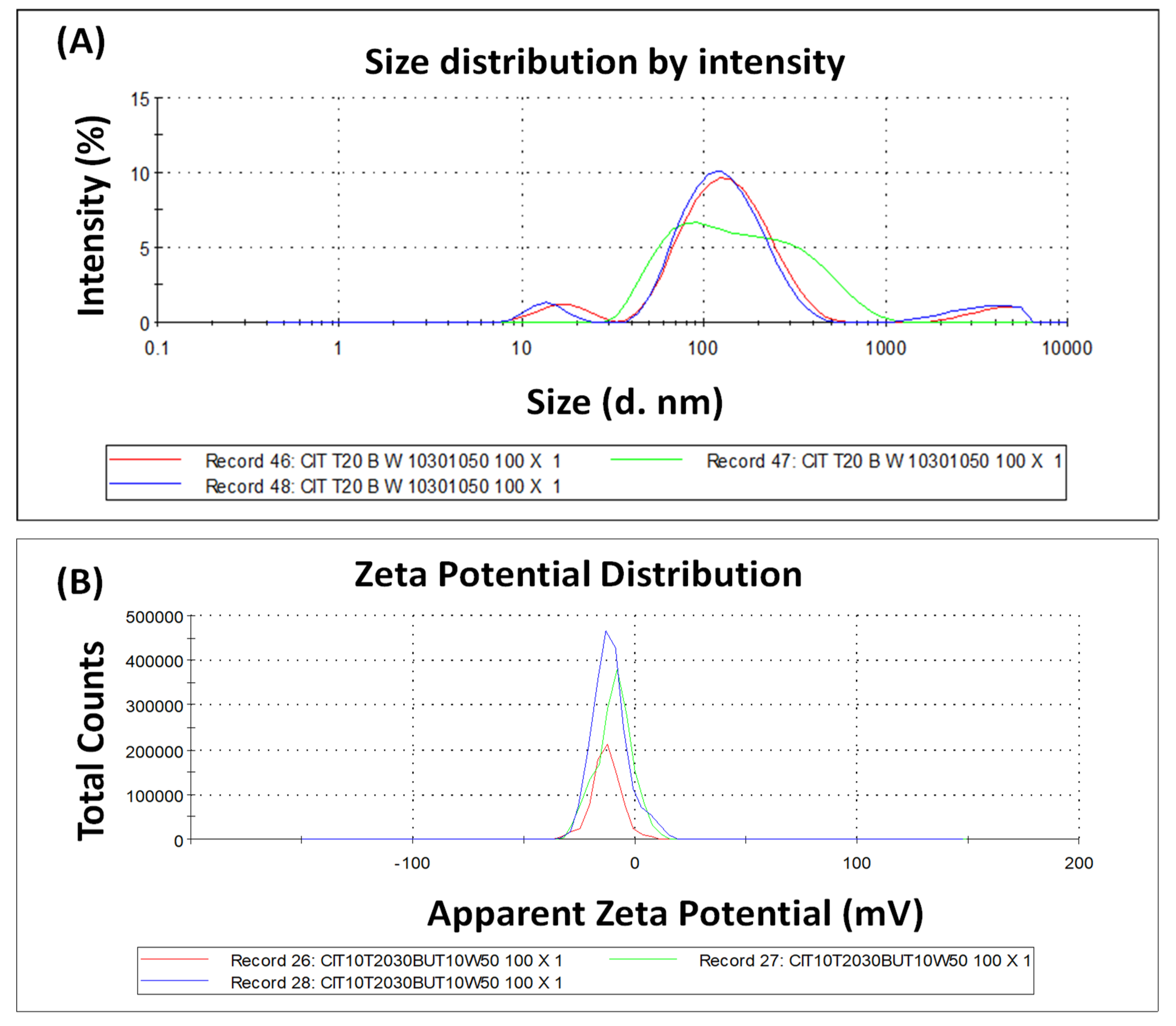
The DLS technique was used to accomplish measurements of the size and polydispersity index of CITEO-loaded nanoemulsions; the results of these measurements are shown in Figure 2A. The mean hydrodynamic diameter of CITEO-loaded nanoemulsions were found to be less than 200 nm. The CITEO-loaded nanoemulsion displayed small oil droplet size, with the particle size of 130 ± 5 nm and a polydispersity index (PDI) of 0.127. The PdI indicates that bacterial samples are generally quite polydispersed, with a broad size distribution. The globule size of CITEO-loaded nanoemulsions observed in dynamic light scattering (DLS) studies are in agreement with what we observed in the scanning electron microscopic evaluations. The Zetasizer nano ZS (Malvern, UK) was used to experimentally determine the Zeta potential of loaded CITEO nanoemulsions by evaluating the electrophoretic mobility of the oil globules or the rate at which they move towards opposing electrodes when subjected to electric fields. As shown in Equation (1), a higher zeta potential means a higher electrophoretic mobility or speed.
μ e = 2 z ε f ( k a )   3 η  
The zeta potential of CITEO-loaded nanoemulsions was found to be −12.6 mV, as shown in Figure 2B. The presence of a high surface charge is thought to be the cause of the nanoemulsions’ excellent stability at high zeta potential values, since particles with potentials greater than +30 mV and lower than 30 mV are regarded to be stable. This minimizes the likelihood of coagulation owing to electrostatic repulsion between particles with identical electric charges, prolongs the system’s stability, and facilitates re-dispersion [20].

2.3. Percentage Entrapment Efficiency (%EE)

The percentage EE of nanoemulsions loaded with CITEO was very high at the beginning of the study, at 95.5 ± 4.775%. After 30 days, we again determined the %EE of nanoemulsions loaded with CITEO and observed 94.1 ± 4.705%. This means that after 30 days of storage, no significant change (p > 0.05) in the %EE was observed. These results clearly show that the preparation of a nanoemulsion of CITEO helps to maintain EO despite its volatility.

2.4. Antimicrobial Activity

The comparative antibacterial assay of CITEO and CITEO-NE was performed in S. aureus bacteria using the well diffusion technique by taking different concentrations of 0.1%, 0.15%, 0.2%, and 0.25%, as shown in Table 1. The results clearly indicate that citronella nanoemulsion showed best zones of inhibition against S. aureus, with 19.3 ± 0.7 mm diameters at 0.2% concentration as compared to Citronella oil. Citronella oil showed the best zones of inhibition of against S. aureus, 12.5 ± 2.1 mm in diameter at 0.2% concentration. The MIC was observed to be 0.05% for CITEO and 0.025% for CITEO-NE against S. aureus. The case of a standard drug, i.e., amoxycillin, showed 34 mm of ZOI against S. aureus. After this, we determined the antifungal activity of the prepared formulation, i.e., CITEO-NE and bare CITEO, against Candida albicans (C. albicans) in concentrations of 0.1%, 0.15%, 0.2%, and 0.25%. The results clearly indicate that CITEO-NE showed the best anti-fungal activity against C. albicans with 25.6 ± 1.4 mm of ZOI, whereas in the case of bare CITEO, ZOI was observed to be 22 ± 1.4 mm at a concentration of 0.2%. The MIC was observed to be 0.025% for CITEO and 0.0125% for CITEO-NE against C. albicans.
From the reports in Figure 3, it is clear that nanoemulsions loaded with CITEO exhibited a larger bacterial and fungal inhibitory zone compared to bare CITEO. The data (Table 1) also show that the formulations with the highest concentrations of nanoparticles had the widest growth-free halo, indicating the antibacterial capabilities of CITEO-NE. In addition, we found that a higher concentration of CITEO and CITEO-NE resulted in a larger zone of inhibition. These results indicate that CITEO-NE interacts with the bacterial cell. The results showed that CITEO-NE was more effective than CITEO against S. aureus and C. albicans.

2.5. Time-Kill Analysis

The effect of CITEO and CITEO-NE on the growth curve of bacteria and fungi was evaluated separately in the culture medium of S. aureus and C. albicans. This was done to determine the effect of CITEO and CITEO-NE on the growth curve as shown in Figure 4. The findings for the control group, which received no therapy, were 6 to 8.85 log10 CFU/mL growth. In contrast, S. aureus growth was significantly reduced in the treatment given CITEO and CITEO-NE during the first 2 to 12 h. The results of treating S. aureus with CITEO remained the same at about 3.5 to 3.9 log10 CFU/mL and 2.3 to 2.9 log10 CFU/mL with 1× MIC (0.05%) and 2× MIC (0.1%), respectively, as shown in Figure 3A. The growth of S. aureus in the untreated group was 6 to 8.69 log10 CFU/mL after 4 h of treatment with CITEO-NE. The growth of S. aureus in the 1× MIC treatment group was 3.33 log10 CFU/mL, while in the 2× MIC treatment groups was, it was 1.69 to 2.3 log10 CFU/mL, as shown in Figure 3B.
In a similar manner, we carried out the comparative, time-kill assay of CITEO and CITEO-NE against C. albicans. The time-kill assay was observed to be 6 to 8.8 log10 CFU/mL in the case of the vehicle treatment. After receiving CITEO, the development of C. albicans was significantly inhibited in the first 4 to 8 h after treatment. It remained the same at approximately 2.6 × log10 CFU/mL to 2.95 × log10 CFU/mL when treated with 1× MIC and 1.6 × log10 CFU/mL to 2 × log10 CFU/mL when treated with 2× MIC, as shown in Figure 3C. Treatment with CITEO-NE against C. albicans, remained constant at approximately 2 × log10 CFU/mL to 2.5 × log10 CFU/mL and 1.3 × log10 CFU/mL to 1.78 × log10 CFU/mL for 1× MIC and 2× MIC treatment, respectively. The results showed that CITEO-NE is more effective than pure CITEO, as shown in Figure 3D. Hence, the summary of the time-kill assay suggests that the effectiveness of CITEO-NE against the test pathogens as a novel alternative therapy.

2.6. Anticancer Activity

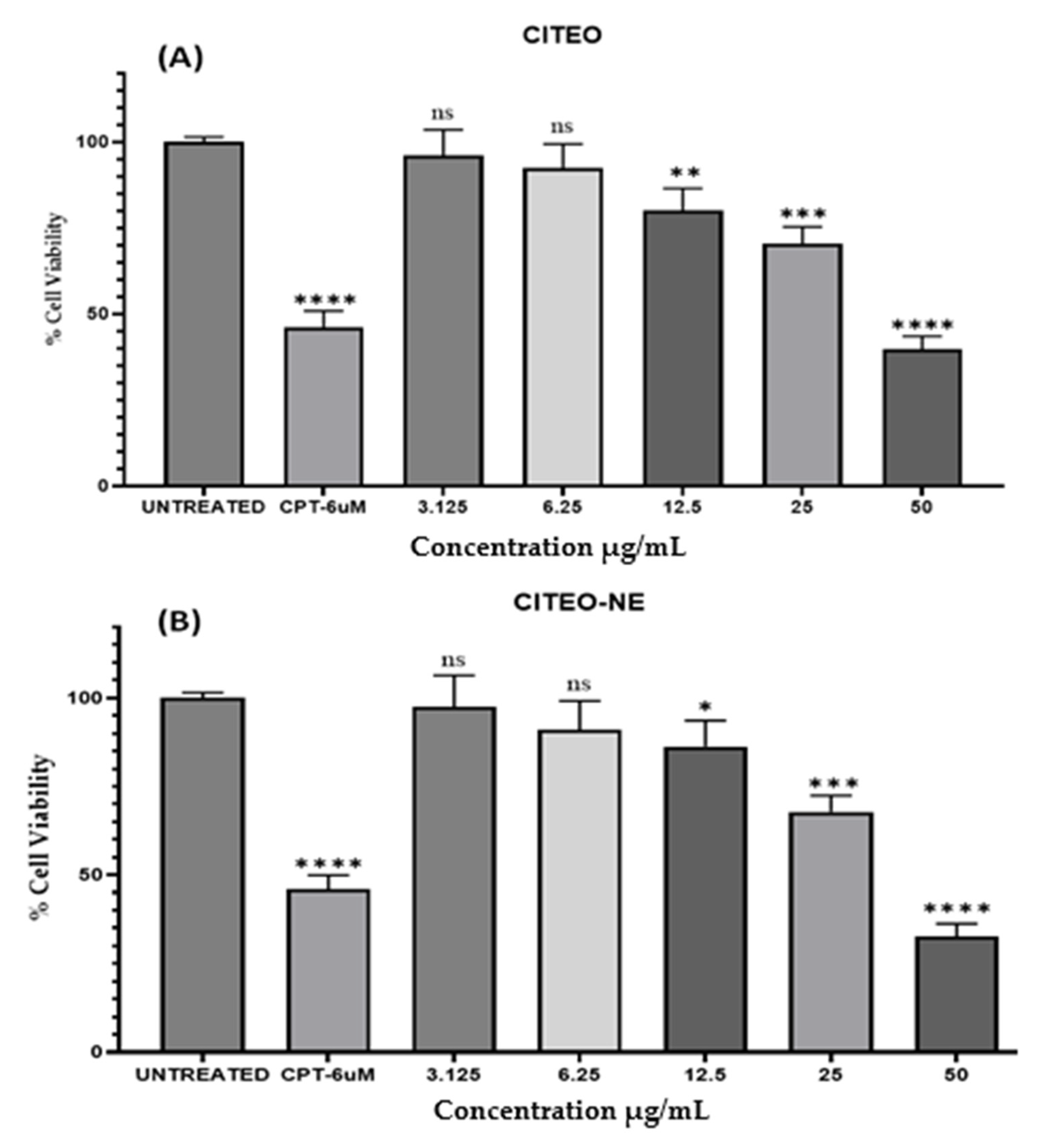
To investigate the anticancer effect of CITEO and CITEO-NE in vitro, cell viability and cytotoxicity were tested in A431 (human skin adenocarcinoma cell line), as shown in Figure 5. The statistical data of cell cytotoxicity by MTT indicate that the test compounds, i.e., CITEO and CITEO-NE, exhibited significant cytotoxic properties against the A431 cell line, with IC50 values of 41.20 ± 3.8 µg/mL and 37.71 ± 4.2 µg/mL, respectively. Camptothecin (IC50 value of 28.6 ± 3.2 µg/mL) was used as the standard for the study. Among the two test compounds, CITEO-NE was considered to be effective against skin cancer due to its lower IC50 value. The results clearly show that the CITEO-NE–based drug delivery system can be delivered to targeted tumor cells with higher efficiency compared to CITEO.

3. Materials and Methods

Citronella essential oil (CITEO) was procured from Indian essential oil (New Delhi, India), while nutrient media and Sabouraud dextrose agar media was purchased from HiMedia (Mumbai, India). Tween 80, MTT reagent, Dimethyl sulfoxide (DMSO), and Camptothecin (CPT) were purchased from Sigma Merck (St Louis, MO, USA). The other reagents or chemicals used in the study were analytically graded. Staphylococcus aureus (ATCC 25923) and Candida albicans (ATCC 10231) were collected from the Department of Pharmaceutics, College of Pharmacy, Prince Sattam Bin Abdulaziz University (PSAU), Al-Kharj. Solvents and other chemicals were also purchased from Sigma Merck. The human skin adenocarcinoma cell line A431 was obtained as gift from Dr. Mohammad Raish, Department of Pharmaceutics, King Saud University (Riyadh, Saudi Arabia).

3.1. Nanoemulsion Preparation of Essential Oils

We prepared the CITEO-loaded nanoemulsions (CITEO-NE) using a probe sonicator (VCX 750, SONICS, Wallingford, CT, USA). For this, we took CITEO as an oil phase, Tween 20 as an emulsifier, and Butanaol as a co-emulsifier. We took distilled water as an aqueous phase in different proportions: 10%, 30%, 10%, and 50% v/v. We then left the combination in a cold bath. It was then sonicated with a 6 mm probe at an amplitude of 60% for 3 min with on and off cycles of 10 s. Nanoemulsions were kept at room temperature to make sure they were stable and there was no formation of phase separation or creaming.

3.2. Study of Physical Appearance and Morphology

For the characterization of morphology, we carried out scanning electron microscopy (SEM; Hitachi High Technologies, Hillsboro, OR, USA). To observe the morphology of the outer surface of CITEO-NE, we kept the samples on a polycarbonate substrate. They were left at room temperature to dry and then were kept in a critical dryer. After drying, the samples were coated with gold and further examined under SEM [21].

3.3. Size Distribution, Particle Size, and Zeta Potential

To observe the particle size and size distribution, the dynamic light diffusion (DLS) instrument (Malvern Instruments, Malvern Worcestershire, UK) technique was utilized in conjunction with the Zetasizer Nano ZS, model ZEN3500. To analyze the sample, we took 10 µL of the CITEO-NE and 990 microliters of deionized water. These were mixed properly to create a dilution of 100 times. After this, the sample was placed in a cuvette and further analyzed at 25 °C. We then determined the zeta potential of the CITEO-NE using a Zetasizer Nano ZS. Zeta sizing was carried out by the use of a one-of-a-kind mixed-mode measurement or phase analysis light scattering technique. This measurement enables high-precision determination for both the average zeta potential and the distribution (M3-PALS) [22].

3.4. Percentage Entrapment Efficiency (%EE)

To determine the %EE of CITEO in CITEO-NE, the desired concentration of the mixture was ensured by combining 100 mg of CITEO with 10 mL of NE solution and then agitating the mixture at 1000 rpm for 60 min. After this, to load the CITEO into the nanoemulsion, we kept the prepared solution mixture at room temperature for 24 h. After 24 h, the mixture was centrifuged at 5000 rpm for 30 min. After this, the supernatant solution and CITEO-NE were separated. The recovered sample was further dispersed in Milli-Q water before using [23]. Using a UV-vis spectrophotometer (Shimadzu, Kyoto, Japan) at the λmax value at 360 nm, the concentration of the CITEO supernatator was calculated. The following formula was used to calculate the entrapment effectiveness of CITEO:
%   EE = ( Total   amount   of   CITEO   added Amount   of   free   CITEO   in   supernatent ) Total   amount   of   CITEO   added   × 100

3.5. Determination of Minimum Inhibition Concentration (MIC) and Minimum Bactericidal Concentration (MBC)

3.5.1. Preparation of Nutrient Media

To prepare the nutrient media, we used 1 liter of distilled water to dissolve 28 g of the nutrient media. Before the sterilization process began, the pH of the media was measured. After, this the nutrient media was sterilized in an autoclave at 121 ℃ for 15 min. After completion of the sterilization process, the nutrient media was cooled. After cooling, the nutrient media was poured into the plates. It was then placed in the laminated air flow until the agar became film.

3.5.2. Antibacterial Activity

We determined out the antibacterial activity of the CITEO and CITEO-NE in bacteria S. aureus by the well diffusion technique. In this, we first prepared 10 mg of the standard stock solution of amoxicillin in 100 mL of distilled water to obtain 0.01% of the standard solution. After that, 25 mg of test samples of CITEO and CITEO-NE were prepared in 10 mL of solvent (5% dimethyl sulfoxide (DMSO)) to obtain a 0.25% test solution. After this, the inoculum was prepared; the test organism S. aureus was inoculated with 10 mL of nutrient broth. Bacterial suspensions were standardized at 108 CFU/mL bacteria and were kept in shakers. Afterward, 50 µL of the broth inoculum (108 CFU/mL) was removed with a micropipette and transferred to fresh sterile solidified agar media plates [24]. On a sterilized agar plate, the inoculation was uniformly spread using a sterile spatula. Using a clean cork cleaner, four holes measuring 6 millimeters were drilled into the medium that was to be inoculated. Samples with different concentrations, from 0.1% to 0.25%, were placed in a well plate. On a separate plate, 50 µL of the reference drug was added. The samples were then incubated at 37 °C for 18–24 h. It was then allowed to diffuse at room temperature for 30 min. Following this, we incubated the sample and observed the formation of a discrete zone around the wells corresponding to the antibacterial activity of the antibiotic tested. We observed the zone of inhibition (ZOI) in millimeters (mm) according to the method described in [25].

3.5.3. In Vitro Antifungal Activity

Preparation of Sabouraud Dextrose Agar (SDA) Media

Sabouraud dextrose agar media (65.0 g) was dissolved in 1 L of distilled water to prepare the SDA medium. Before sterilization of the prepared media, we measured the pH. After measuring the pH, media was autoclaved at 121 °C at 15 lb pressure for 15 min for sterilization. After completion of sterilization, media was kept for cooling without solidification. The SDA medium was then poured onto the plates and then placed in a laminar air flow until the agar became firm. This procedure was repeated until the agar was completely set.

Antifungal Activity

We tested the antifungal activity of the prepared formulation (CITEO and CITEO-NE sample) in C. albicans. For this purpose, a culture of C. albicans was first spread on the prepared SDA medium. To prepare a standard stock solution of fluconazole with a concentration of 0.01% and the test solution with a concentration of 0.1% to 0.25%, CITEO and CITEO-NE were dissolved in 5% DMSO. After this, we followed all the steps discussed in this manuscript in Section 3.5.2 using the method given by Mohammadi-Sichani et al., 2012 [24].

3.6. Minimum Inhibitory Concentration (MIC)

The MIC of prepared formulations (CITEO and CITEO-NE) was evaluated using the agar well diffusion test [26]. In this technique, an inoculum of 50 μL of selected bacteria S. aureus and fungus C. albicans was seeded onto sterile Mueller Hinton (MH) agar plates. Then, a well of 6 mm in diameter was drilled into the agar with the use of a sterile cork borer. The well was filled with 50 μL of different concentrations of CITEO and CITEO-NE samples prepared in 5% DMSO. After depositing the different sample concentrations into the wells, they were kept at room temperature for 30 min and then at 37 °C for 24 h. Measurements were taken in mm to determine the circular inhibition zones. The MIC was calculated by microdilution, and the test was performed in MH broth. We prepared different dilutions of the test samples; 100 μL aliquot was diluted in MH broth containing 106 CFU/mL. Different plates were incubated at different temperatures, i.e., the plates of S. aureus containing bacteria were kept at 30 °C ± 5 °C/24 h and 25 °C ± 5 °C for C. albicans. After 24 h, the lowest CITEO and CITEO-NE concentration that completely inhibited microbial growth was considered the MIC.

3.7. Time-Kill Analysis

Time-kill kinetics of the chosen formulations (CITEO and CITEO-NE) were determined by adapting a method from a published paper [27]. We took two different concentrations equivalent to 1× MIC (0.05% and 0.025%) and 2× MIC (0.1% and 0.05%) for the kill kinetic assay against S. aureus. Similarly, two different concentrations equivalent to 1× MIC (0.025% and 0.0125%) and 2× MIC (0.05% and 0.025%) were selected for the kill kinetic assay against C. albicans. An inoculum of size 1 × 10⁶ CFU/mL was added to it and further incubated at 37 °C. At intervals of 0, 2, 4, 8, 12, 16, and 20 h, a total of 1 mm of inoculum was taken from the medium. The colony-forming units (CFUs) of bacterial cells were determined. A negative control with organisms and DMSO (without essential oils) was also evaluated. Assays were performed in triplicate, and time-kill plots were generated by calculating log10 CFU/mL of mean colony count versus time.

3.8. Anti-Cancer Activity

We carried out the anti-cancer activity by MTT assay against the Human Skin adenocarcinoma cell line (A431) using the test sample CITEO and CITEO-NE in the concentrations ranging from 3.125 µg/mL to 50 µg/mL. For this purpose, 200 µL of cell suspension was added to a 96-well plate at the required cell density (20,000 cells per well) without the assay medium. The cells allowed to grow for approximately 24 h. After this, the selected concentration of the test agents was added for incubation at 37 °C for 24 h in 5% CO2. The plates were removed from the incubator after completion of the incubation period. MTT reagents were added to a final concentration of 0.5 mg/mL of total volume after the spent medium was removed. In order to prevent the plates from being exposed to light, aluminum foil was used to cover them. After three hours in the incubator, the plates were removed. After removing the MTT reagents, 100 µL of a solution containing DMSO was added. Dissolution may be improved by using a gyro shaker to provide a gentle agitation. The absorption is measured using a spectrophotometer or an ELISA reader with a wavelength of 570 nm. The percent cell viability is calculated using the following formula:
% cell viability = Abs of treated cells/Abs of Untreated cells × 100
The IC50 value was determined using a linear regression equation, i.e., Y = Mx + C, where Y = 50, and M and C values are derived from the viability graph.

4. Conclusions

Essential oils derived from plants could provide a natural, more effective alternative to antibiotics made from synthetic chemicals. Due to the exceptional pharmacological effects, citronella essential oil (CITEO) has attracted the most interest among these numerous compounds. Essential oils such as CITEO can be challenging to use in crop protection due to their poor water solubility, high volatility, and instability. These are the three main physicochemical properties that contribute to this difficulty. Nanoformulation provides an answer to a number of the major problems associated with EOs. These include the low water solubility of EOs, their volatility, and their chemical instability. By preparing and analyzing nanoemulsion of CITEO, the current study aimed to achieve its primary goal of finding solutions to the previously identified problems. According to the data described above, encapsulation of CITEO in nanoemulsions is an effective method to achieve this goal. Compared with uncoated CITEO, encapsulation of CITEO in nanoemulsion is one of the most promising strategies to enhance pharmacological activities such as antibacterial and anticancer effects. On the other hand, further studies are needed to evaluate the potential impact of CITEO-based nanoformulations for different diseases.

Author Contributions

Conceptualization, T.J. and A.A.; Methodology, M.M.K. and B.M.; Software, A.M.A. and B.M.; Validation, T.J., R.A. and S.A.; Formal analysis, M.M.K. and A.A.; Investigation, B.M., M.K. and R.A.; Resources, T.J., B.M. and R.A.; Data curation, A.M.A. and M.K.; Writing—original draft, A.M.A., M.K. and S.A.; Writing—review & editing, R.A., S.A. and A.A.; Visualization, A.M.A., R.A. and A.A.; Supervision, M.M.K. and A.A.; Project administration, T.J. and S.A.; Funding acquisition, T.J. and M.M.K. All authors have read and agreed to the published version of the manuscript.

Funding

This study is supported via funding from Prince sattam bin Abdulaziz University project number (PSAU/2023/R/1444). The authors would like to thank Kedar Gupta and Rick Schwerdtfeger for their generous support and encouragement during the preparation this manuscript.

Institutional Review Board Statement

Not applicable.

Informed Consent Statement

Not applicable.

Data Availability Statement

The data presented in this study are available on request from the corresponding author.

Acknowledgments

Authors would like to thanks Mohammad Jawed Ansari, Department of Pharmaceutics, Prince Sattam Bin Abdulaziz Unversity, for their support during gel formulation and evaluation.

Conflicts of Interest

The authors declare no conflict of interest.

References

  1. Balasubramani, S.; Sabapathi, G.; Moola, A.K.; Solomon, R.V.; Venuvanalingam, P.; Bollipo Diana, R.K. Evaluation of the Leaf Essential Oil from Artemisia vulgaris and Its Larvicidal and Repellent Activity against Dengue Fever Vector Aedes aegypti—An Experimental and Molecular Docking Investigation. ACS Omega 2018, 3, 15657–15665. [Google Scholar] [CrossRef] [Green Version]
  2. Kusuma, H.S.; Mahfud, M. The extraction of essential oils from patchouli leaves (Pogostemon cablin benth) using a microwave air-hydrodistillation method as a new green technique. RSC Adv. 2017, 7, 1336–1347. [Google Scholar] [CrossRef] [Green Version]
  3. Mohammed, H.H.; Laftah, W.A.; Noel Ibrahim, A.; Che Yunus, M.A. Extraction of essential oil from Zingiber officinale and statistical optimization of process parameters. RSC Adv. 2022, 12, 4843–4851. [Google Scholar] [CrossRef]
  4. Youssef, F.S.; Labib, R.M.; Gad, H.A.; Eid, S.; Ashour, M.L.; Eid, H.H. Pimenta dioica and Pimenta racemosa: GC-based metabolomics for the assessment of seasonal and organ variation in their volatile components, in silico and in vitro cytotoxic activity estimation. Food Funct. 2021, 12, 5247–5259. [Google Scholar] [CrossRef]
  5. Ahmed, K.B.M.; Khan, M.M.A.; Jahan, A.; Siddiqui, H.; Uddin, M. Gamma rays induced acquisition of structural modification in chitosan boosts photosynthetic machinery, enzymatic activities and essential oil production in citronella grass (Cymbopogon winterianus Jowitt). Int. J. Biol. Macromol. 2020, 145, 372–389. [Google Scholar] [CrossRef] [PubMed]
  6. Guedes, A.R.; de Souza, A.R.C.; Zanoelo, E.F.; Corazza, M.L. Extraction of citronella grass solutes with supercritical CO2, compressed propane and ethanol as cosolvent: Kinetics modeling and total phenolic assessment. J. Supercrit. Fluids 2018, 137, 16–22. [Google Scholar] [CrossRef]
  7. Silva, M.R.; Ximenes, R.M.; Da Costa, J.G.M.; Leal, L.K.A.M.; De Lopes, A.A.; De Barros Viana, G.S. Comparative anticonvulsant activities of the essential oils (EOs) from Cymbopogon winterianus Jowitt and Cymbopogon citratus (DC) Stapf. in mice. Naunyn. Schmiedebergs Arch. Pharmacol. 2010, 381, 415–426. [Google Scholar] [CrossRef] [PubMed]
  8. Timung, R.; Barik, C.R.; Purohit, S.; Goud, V.V. Composition and anti-bacterial activity analysis of citronella oil obtained by hydrodistillation: Process optimization study. Ind. Crops Prod. 2016, 94, 178–188. [Google Scholar] [CrossRef]
  9. Neves, J.S.; Lopes-Da-Silva, Z.; De Sousa Brito Neta, M.; Chaves, S.B.; Karla De Medeiros Nóbrega, Y.; Henrique De Lira Machado, A.; Machado, F. Preparation of terpolymer capsules containing rosmarinus officinalis essential oil and evaluation of its antifungal activity. RSC Adv. 2019, 9, 22586–22596. [Google Scholar] [CrossRef] [Green Version]
  10. Alam, A.; Foudah, A.I.; Salkini, M.A.; Raish, M. Herbal Fennel Essential Oil Nanogel: Formulation, Characterization and Antibacterial Activity against Staphylococcus aureus. Gels 2022, 8, 736. [Google Scholar] [CrossRef]
  11. Agrawal, N.; Maddikeri, G.L.; Pandit, A.B. Sustained release formulations of citronella oil nanoemulsion using cavitational techniques. Ultrason. Sonochem. 2017, 36, 367–374. [Google Scholar] [CrossRef] [PubMed]
  12. Osman Mohamed Ali, E.; Shakil, N.A.; Rana, V.S.; Sarkar, D.J.; Majumder, S.; Kaushik, P.; Singh, B.B.; Kumar, J. Antifungal activity of nano emulsions of neem and citronella oils against phytopathogenic fungi, Rhizoctonia solani and Sclerotium rolfsii. Ind. Crops Prod. 2017, 108, 379–387. [Google Scholar] [CrossRef]
  13. Chang, Y.; McLandsborough, L.; McClements, D.J. Physical properties and antimicrobial efficacy of thyme oil nanoemulsions: Influence of ripening inhibitors. J. Agric. Food Chem. 2012, 60, 12056–12063. [Google Scholar] [CrossRef]
  14. Hassan, K.A.M.; Ali Mujtaba, M.D. Antibacterial efficacy of garlic oil nano-emulsion. AIMS Agric. Food 2019, 4, 194–205. [Google Scholar] [CrossRef]
  15. Akram, S.; Anton, N.; Omran, Z.; Vandamme, T. Water-in-oil nano-emulsions prepared by spontaneous emulsification: New insights on the formulation process. Pharmaceutics 2021, 13, 1030. [Google Scholar] [CrossRef]
  16. Rodrigues, A.B.L.; Martins, R.L.; Rabelo, É.D.M.; Tomazi, R.; Santos, L.L.; Brandão, L.B.; Faustino, C.G.; Farias, A.L.F.; Dos Santos, C.B.R.; Cantuária, P.d.C.; et al. Development of nano-emulsions based on Ayapana triplinervis essential oil for the control of Aedes aegypti larvae. PLoS ONE 2021, 16, e0254225. [Google Scholar] [CrossRef]
  17. Barradas, T.N.; de Holanda e Silva, K.G. Nanoemulsions of essential oils to improve solubility, stability and permeability: A review. Environ. Chem. Lett. 2021, 19, 1171. [Google Scholar] [CrossRef]
  18. Anton, N.; Saulnier, P. Adhesive water-in-oil nano-emulsions generated by the phase inversion temperature method. Soft Matter 2013, 9, 6465–6474. [Google Scholar] [CrossRef] [Green Version]
  19. Lee, M.H.; Lee, I.Y.; Chun, Y.G.; Kim, B.K. Formulation and characterization of β-caryophellene-loaded lipid nanocarriers with different carrier lipids for food processing applications. LWT 2021, 149, 111805. [Google Scholar] [CrossRef]
  20. Shahavi, M.H.; Hosseini, M.; Jahanshahi, M.; Meyer, R.L.; Darzi, G.N. Clove oil nanoemulsion as an effective antibacterial agent: Taguchi optimization method. Desalin. Water Treat. 2016, 57, 18379–18390. [Google Scholar] [CrossRef]
  21. Sundararajan, B.; Sathishkumar, G.; Seetharaman, P.k.; Moola, A.K.; Duraisamy, S.M.; Mutayran, A.A.S.B.; Seshadri, V.D.; Thomas, A.; Ranjitha Kumari, B.D.; Sivaramakrishnan, S.; et al. Biosynthesized Gold Nanoparticles Integrated Ointment Base for Repellent Activity Against Aedes aegypti L. Neotrop. Entomol. 2022, 51, 151–159. [Google Scholar] [CrossRef] [PubMed]
  22. Azam, F.; Alqarni, M.H.; Alnasser, S.M.; Alam, P.; Jawaid, T.; Kamal, M.; Khan, S.; Alam, A. Formulation, In Vitro and In Silico Evaluations of Anise (Pimpinella anisum L.) Essential Oil Emulgel with Improved Antimicrobial Effects. Gels 2023, 9, 111. [Google Scholar] [CrossRef] [PubMed]
  23. Alam, A.; Jawaid, T.; Alsanad, S.M.; Kamal, M.; Rawat, P.; Singh, V.; Alam, P.; Alam, P. Solubility Enhancement, Formulation Development, and Antibacterial Activity of Xanthan-Gum-Stabilized Colloidal Gold Nanogel of Hesperidin against Proteus vulgaris. Gels 2022, 8, 655. [Google Scholar] [CrossRef]
  24. Mohammadi-Sichani, M. Effect of different extracts of Stevia rebaudiana leaves on Streptococcus mutans growth. J. Med. Plants Res. 2012, 6, 4731–4734. [Google Scholar] [CrossRef]
  25. Manandhar, S.; Luitel, S.; Dahal, R.K. In Vitro Antimicrobial Activity of Some Medicinal Plants against Human Pathogenic Bacteria. J. Trop. Med. 2019, 2019, 1–5. [Google Scholar] [CrossRef] [Green Version]
  26. Alam, A.; Jawaid, T.; Alsanad, S.M.; Kamal, M. Essential Oil Extracted from Psidium guajava (L) Leaves. Plants 2023, 12, 246. [Google Scholar] [CrossRef] [PubMed]
  27. Moo, C.L.; Osman, M.A.; Yang, S.K.; Yap, W.S.; Ismail, S.; Lim, S.H.E.; Chong, C.M.; Lai, K.S. Antimicrobial activity and mode of action of 1,8-cineol against carbapenemase-producing Klebsiella pneumoniae. Sci. Rep. 2021, 11, 20824. [Google Scholar] [CrossRef]
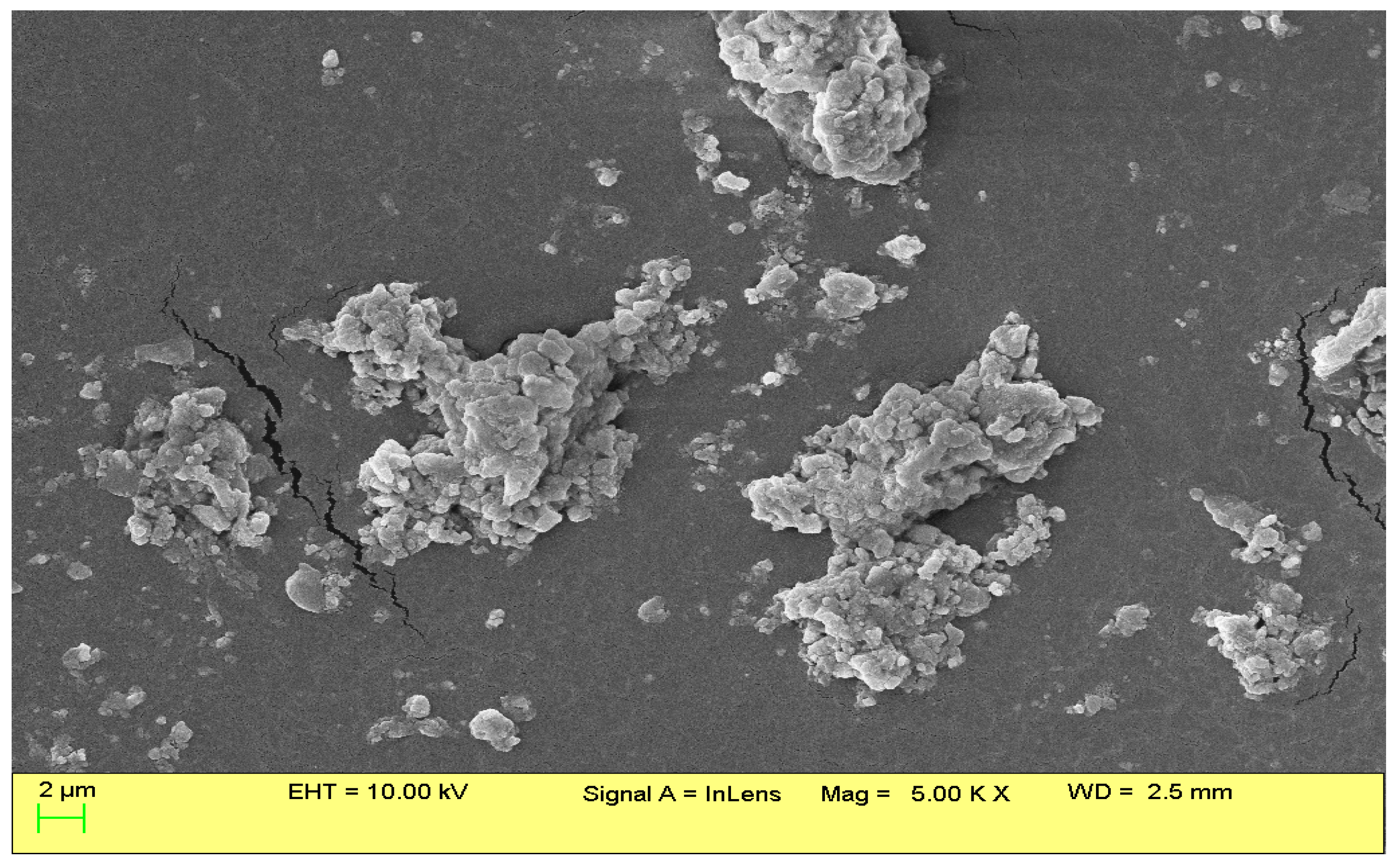
Figure 1. The SEM image of CITEO-loaded nanoemulsions.
Antibiotics 12 00478 g001
Figure 2. (A) Particle size and size distribution; (B) Zeta size of CITEO-loaded nanoemulsion.
Antibiotics 12 00478 g002
Figure 3. Antimicrobial activity of citronella essential oil (CITEO) and its nanoemulsion (CITEO-NE) against S. aureus. and C. albicans.
Antibiotics 12 00478 g003
Figure 4. Time-killing assays of (A) CITEO against S. aureus, (B) CITEO-NE against S. aureus, (C) CITEO against C. albicans and (D) CITEO-NE against C. albicans.
Antibiotics 12 00478 g004
Figure 5. Cytotoxicity assay of CITEO (A) and CITEO-NE (B) with different concentrations against A431. p-value (* p < 0.05, ** p < 0.01, *** p < 0.001 and **** p < 0.0001; ns: not significant) when compared to the untreated group using two-way ANOVA of GraphPad prism 9.5.0.
Antibiotics 12 00478 g005
Table 1. Comparative zone of inhibition (means ± SD) and minimum inhibitory concentration (MIC) study of CITEO and CITEO-NE against S. aureus and C. albicans.
Concentration (%)CITEO CITEO-NE
S. aureusC. albicansS. aureusC. albicans
0.15 ± 0.88.3 ± 2.113 ± 0.710.6 ± 0.7
0.157.5 ± 0.211.6 ± 1.415 ± 0.714 ± 1.4
0.210 ± 1.415.6 ± 1.416.3 ± 0.720.3 ± 1.4
0.2512.5 ± 2.122 ± 1.419.3 ± 0.725.6 ± 1.4
MIC500 µg/mL250 µg/mL250 µg/mL125 µg/mL
Disclaimer/Publisher’s Note: The statements, opinions and data contained in all publications are solely those of the individual author(s) and contributor(s) and not of MDPI and/or the editor(s). MDPI and/or the editor(s) disclaim responsibility for any injury to people or property resulting from any ideas, methods, instructions or products referred to in the content.