Review

Biosynthesis and Chemical Synthesis of Albomycin Nucleoside Antibiotics

1
Biotechnology Research Center, Southwest University, Chongqing 400715, China
2
Chongqing Key Laboratory of Plant Resource Conservation and Germplasm Innovation, Southwest University, Chongqing 400715, China
3
State Cultivation Base of Crop Stress Biology for Southern Mountainous Land, Academy of Agricultural Sciences, Southwest University, Chongqing 400715, China
*
Author to whom correspondence should be addressed.
Antibiotics 2022, 11(4), 438; https://doi.org/10.3390/antibiotics11040438
Submission received: 1 March 2022 / Revised: 21 March 2022 / Accepted: 21 March 2022 / Published: 24 March 2022

Abstract

:
The widespread emergence of antibiotic-resistant bacteria highlights the urgent need for new antimicrobial agents. Albomycins are a group of naturally occurring sideromycins with a thionucleoside antibiotic conjugated to a ferrichrome-type siderophore. The siderophore moiety serves as a vehicle to deliver albomycins into bacterial cells via a “Trojan horse” strategy. Albomycins function as specific inhibitors of seryl-tRNA synthetases and exhibit potent antimicrobial activities against both Gram-negative and Gram-positive bacteria, including many clinical pathogens. These distinctive features make albomycins promising drug candidates for the treatment of various bacterial infections, especially those caused by multidrug-resistant pathogens. We herein summarize findings on the discovery and structure elucidation, mechanism of action, biosynthesis and immunity, and chemical synthesis of albomcyins, with special focus on recent advances in the biosynthesis and chemical synthesis over the past decade (2012–2022). A thorough understanding of the biosynthetic pathway provides the basis for pathway engineering and combinatorial biosynthesis to create new albomycin analogues. Chemical synthesis of natural congeners and their synthetic analogues will be useful for systematic structure–activity relationship (SAR) studies, and thereby assist the design of novel albomycin-derived antimicrobial agents.

1. Introduction

The emergence and rapid spread of antibiotic resistance among pathogens has become a major healthcare problem worldwide. This problem becomes even worse with a coincident decline in the supply of novel antimicrobial agents [1,2]. Therefore, there is an urgent need for the discovery and development of novel antimicrobial drugs that either act on new cellular targets or bypass the development of resistance. Natural products offer a great variety of chemical diversity with distinctive biological activities. It is estimated that more than half of approved drugs, from the period of 1981 to 2019, are either natural products or derivatives thereof [3]. Thus, natural products remain the most promising sources for drug discovery and development [4,5]. In recent years, aminoacyl-tRNA synthetases (aaRSs) have emerged as attractive targets for the development of novel antimicrobial drugs [6]. To ensure the fidelity of protein synthesis, these enzymes catalyze the charging of tRNA with cognate amino acids in a two-step process [7]. The first step involves formation of an aminoacyl–adenylate (aa-AMP) intermediate with concomitant release of pyrophosphate (PPi). In the second step, the aminoacyl moiety is transferred to the 3′-terminal adenosine of the cognate tRNA (Scheme 1). The 20 aaRSs have been classified into two classes: Class Ⅰ and Class Ⅱ, mainly based on the structures of their active sites [7]. Considering the vital role of aaRSs in protein synthesis, it is reasonable to expect that inhibition of these enzymes is detrimental to cell survival.
Sideromycins are a unique subset of siderophores that are comprised of an antibacterial moiety covalently linked to a siderophore. They are actively transported into bacterial cells via a “Trojan horse” strategy by hijacking the siderophore uptake pathway, that is commonly used by bacteria to scavenge environmental iron [8]. These siderophore-antibiotic conjugates are promising drug candidates for the treatment of various bacterial infections, including those caused by multidrug-resistant pathogens. Over the past few decades, only a few naturally occurring sideromycins, such as albomycins, salmycins, and ferrimycins, have been identified [8,9]. Of particular interest are albomycins that function as inhibitors of bacterial seryl-tRNA synthetase (SerRS). Examples of other aaRSs inhibitors include microcin C and agrocin 84 (Figure 1), targeting aspartyl tRNA synthetase (AspRS) and leucyl-tRNA synthetase (LeuRS), respectively [10]. The biosynthesis, mode of action and self-resistance of microcin C have been reviewed comprehensively [10,11]. At present, our understanding on synthesis, mode of action and self-resistance of agrocin 84 is still limited [12,13]. In this review, we summarize findings on the discovery and structure elucidation, mode of action, biosynthesis, self-resistance, and chemical synthesis of albomycins.

2. Discovery and Structure Elucidation

Albomycin, originally designated as grisein, was first isolated from the soil-dwelling Streptomyces griseus in the 1940s [14,15]. It was also identified in Streptomyces subtropicus (previously known as Actinomyces subtropicus) [16,17]. It is noteworthy that albomycin was also known as alveomycin, antibiotics A 1787, LA 5352 and LA 5937, and Ro 5-2667 in the literature [18,19]. Thirty-five years after the initial isolation, chemical structures of three albomycin congeners (δ1, δ2, and ε) were fully elucidated by Benz and coworkers in 1982 [20,21]. The albomycins are composed of a ferrichrome-type siderophore and a 6′-amino-4′-thioheptose nucleoside that are linked via amide linkages to a serine residue (Figure 1). The siderophore moiety consists of three tandem N5-acetyl-N5-hydroxy-L-ornithine residues, mimicking the siderophore ferrichrome from fungi [22,23]. The most striking feature of the nucleoside moiety is the substitution of a sulfur atom for an oxygen atom on the pentose ring, that is most commonly found in other nucleoside antibiotics, such as liposidomycin, caprazamycin, muraymycin, muraminomicin, and A-90289 [24,25]. It is worth noting that the three albomycin congeners differ mainly in the C4 substituent of the pyrimidine nucleoside (Figure 1).

3. Mechanism of Action

Albomycins have attracted significant attention due to their potent antibacterial activities against both Gram-negative and Gram-positive bacteria [15,16,26]. For example, the major congener δ2 exhibited minimum inhibitory concentrations (MICs) as low as 5 ng/mL against Escherichia coli and 10 ng/mL against Streptococcus pneumoniae [27]. The excellent antibacterial activities of albomycins were attributed to their ability to hijack the ferric hydroxamate transport system and gain access to bacterial cells. For illustration purposes, we herein only briefly highlight current understanding on active uptake of albomycins in E. coli and S. pneumoniae. Readers interested in other bacteria species are referred to two recent reviews [10,28]. In E. coli, transport across the outer membrane is facilitated by a ferrichrome receptor FhuA [29], while transport across the cytoplasmic membrane is mediated by a ABC transporter FhuBCD [30]. FhuA consists of an N-terminal cork domain and a C-terminal transmembrane β-barrel domain. Binding of a ligand promotes the FhuA interaction with TonB, which in turn stimulates a rearrangement of the cork domain and the release of the ligand into the periplasmic space [31,32]. Once translocated into the periplasm, albomycin is bound by the periplasmic binding protein FhuD [33]. The antibiotic is then shuttled to the inner membrane ABC transporter FhuBC, and actively transported into the cytoplasm [34]. In E. coli, all genes encoding the transporter system are organized within the fhuABCD operon. Bacterial strains with mutations in any of these genes lost the ability to ingest albomycins and thus are resistant to their antimicrobial effects [35]. A similar transporter system was also characterized in S. pneumoniae [27]. The transporter system was encoded by the fhu locus consisting of fhuD, fhuB, fhuG, and fhuC. The fhuD encodes a binding lipoprotein, that is thought to be responsible for substrate recognition. The fhuB and fhuG encode transmembrane transport proteins, while the fhuC encodes an ATPase that presumably provides energy for transmembrane uptake [27]. Upon entry into the cell, albomycins will be hydrolyzed by host peptidases to release the thionucleoside warhead, referred to as SB-217452, from the iron-chelating siderophore moiety [36]. The SB-217452 resembles seryl adenylate, and selectively inhibits seryl-tRNA synthetases (SerRSs), and thereby interfering with host protein synthesis [27]. Thus, the potent antimicrobial activities of albomycins are attributed to the ferrichrome-mediated active uptake and subsequent liberation of the toxic thionucleoside SB-217452.

4. Biosynthetic Pathway

Albomycins are endowed with unique structural features, suggesting the occurrence of unusual enzymatic reactions during the biosynthetic process. To gain insight into this biosynthetic pathway, the gene cluster for albomycin biosynthesis has been identified by screening a cosmid library of S. griseus ATCC 700974 [37]. The gene cluster consists of 25 complete open reading frames (ORFs) including ORFs 1–7 and 18 genes from abmA to abmR (Figure 2A). It was proposed that the 18 genes from abmA to abmR are required for albomycin production, while ORFs 1–7 are not involved in albomycin biosynthesis [37]. A few recent studies have been directed to elucidate biosynthetic process of the thionucleoside SB-217452 [37,38,39]. These studies established that the formation of SB-217452 proceeds through complex enzymatic reactions with the participation of AbmH, AbmD, AbmF, AbmK, and AbmJ (Figure 2B). AbmH is a pyridoxal 5′-phosphate (PLP)-dependent transaldolase responsible for catalyzing a threo-selective aldol-type reaction to generate the thioheptose core with a D-ribofuranose ring and an L-amino acid moiety. Subsequently, the conversion of L- to D-amino acid configuration is achieved through the action of a PLP-dependent epimerase (AbmD) [38]. Then, an aminoacyl-tRNA synthetase (AbmF) catalyzes condensation between the 6′-amino-4′-thionucleoside and the D-ribo configuration and seryl-adenylate. Of note is that the seryl-adenylate appears to be supplied by the seryl-tRNA synthetase (AbmK). The D-ribo to D-xylo conversion of the thiofuranose ring is thought to be catalyzed by a radical S-adenosyl-L-methionine (SAM) enzyme (AbmJ) [38,39]. It is interesting to note that AbmK directly participates in the formation of SB-217452 by providing a substrate for albomycin biosynthesis. Similarly, a gene (vlmL) encoding a class II seryl-tRNA synthetase was also identified within the gene cluster for valanimycin biosynthesis in Streptomyces viridifaciens. Studies revealed that this seryl-tRNA synthetase is responsible for catalyzing seryl transfer in the valanimycin biosynthetic pathway [40,41]. Furthermore, two enzymes, a N-methyltransferase (AbmI) and a carbamoyltransferase (AbmE), have been identified to be responsible for the tailoring modifications of N3-methylation and N4-carbamoylation of cytidine [37]. Although significant progress has been made in uncovering biosynthetic machinery of SB-217452, the order of known steps needs further clarification. Moreover, many details of its biosynthesis remain elusive. For example, it remains a mystery how the characteristic sulfur atom is incorporated in 6. Thus, more genetic and biochemical studies are required to reveal the complex process.
As mentioned above, the presence of the ferrichrome-type siderophore allows active transport of albomycin into bacterial cells. It was hypothesized that three genes, abmA, abmB, and abmQ, are responsible for the formation of the ferrichrome siderophore [37]. The pathway is initiated by the N5 hydroxylation of L-ornithine to N5-hydroxy-L-ornithine, catalyzed by a flavin-dependent ornithine monooxygenase (AbmB). Subsequently, N5-hydroxy-L-ornithine is converted to N5-acetyl-N5-hydroxy-L-ornithine through the action of an N-acyltransferase (AbmA). Three molecules of N5-acetyl-N5-hydroxy-L-ornithine are then used by, AbmQ, a nonribosomal peptide synthetase (NRPS), for iterative condensation to generate the tripeptide (Figure 2B). Sequence analysis suggested that AbmQ contains one adenylating (A) domain, two condensation (C) domains, and three thiolation (T) domains (Figure 2B). The presence of a single adenylating domain is a unique feature of AbmQ, implying that this domain supplies substrates for both condensation domains [37,39]. Furthermore, a typical NRPS contains a C-terminal thioesterase (TE) domain to catalyze hydrolysis of the thioester intermediate to release the peptide product from the phosphopantetheine group linked to the peptidyl carrier protein (PCP) domain [42]. However, AbmQ lacks a C-terminal TE domain. This atypical domain organization implies that no free siderophore is released from AbmQ and that the thioester intermediate may directly serve as the electrophile in the subsequent amide bond formation with either SB-217452 or L-serine. Genetic studies suggest that AbmC may participate in this amide bond formation [39]. However, biochemical evidence is needed to clarify its function. It is noteworthy that the biosynthetic pathway for the ferrichrome-type siderophore in albomycin has been proposed based on putative functions of associated genes. Further experimental evidence is definitely needed to support this assumption.

5. Self-Resistance

Typically, self-resistance is a prerequisite for antibiotic-producing bacteria. They have evolved a variety of mechanisms to ensure protection from self-made cytotoxic compounds. This is particularly true for the bacterial genus Streptomyces, prolific producers of antibiotics and many other bioactive secondary metabolites. The thionucleoside SB-217452 exhibits its antimicrobial activity by targeting SerRS. SerRS belongs to the class-II family of aminoacyl-tRNA synthetases (aaRSs) [43]. These enzymes catalyze the attachment of amino acids to their corresponding tRNAs [7]. Due to the pivotal role of aaRSs in the protein synthesis, inhibition of a member of this family is detrimental to cell viability. It is conceivable that the compound is also toxic to the natural producer. One early study identified two SerRS encoding genes (serS1 and serS2) in the genome of albomycin-producing strain S. griseus ATCC 700974 [44]. Sequence analysis suggested that serS1 encodes a housekeeping SerRS, while serS2 encodes a SerRS that is significantly divergent from SerRS1. Of note is that serS2 is located within the gene cluster for albomycin biosynthesis, thus it is designated as abmK [37]. However, serS1 is situated elsewhere in the chromosome. When heterologously overexpressed in E. coli JM109, serS2 (abmK) confers immunity to albomycin in vivo, while serS1 fails to do so. Furthermore, SerRS2 was specifically resistant to SB-217452 in vitro [44]. A similar phenomenon has also been reported for other natural aaRSs inhibitors, such as mupirocin (isoleucyl-tRNA synthetases inhibitor) [45], borrelidin (threonyl-tRNA synthetase inhibitor) [46], and agrocin 84 (leucyl-tRNA synthetase inhibitor) [13].
It is not uncommon that antibiotic-producing bacteria duplicate genes encoding target proteins to confer resistance [45,47,48,49]. It is noteworthy that some resistance genes may be situated within or adjacent to the gene clusters for antibiotic biosynthesis. Based on these observations, resistance-guided genome mining strategy has been devised and used successfully for the discovery of novel antimicrobial agents [1,2,49,50]. Previous studies suggest that AbmK plays dual role in the biosynthesis and immunity of albomycins. A recent study indicated that AbmK participates directly in the formation of SB-217452 during biosynthesis of albomycins [39]. In the meantime, AbmK confers resistance to albomycins to avoid suicide [44]. Thus, abmK is a good candidate to be used as a query to search for homologues that will be helpful for the discovery of novel albomycin analogues or related compounds.

6. Chemical Synthesis

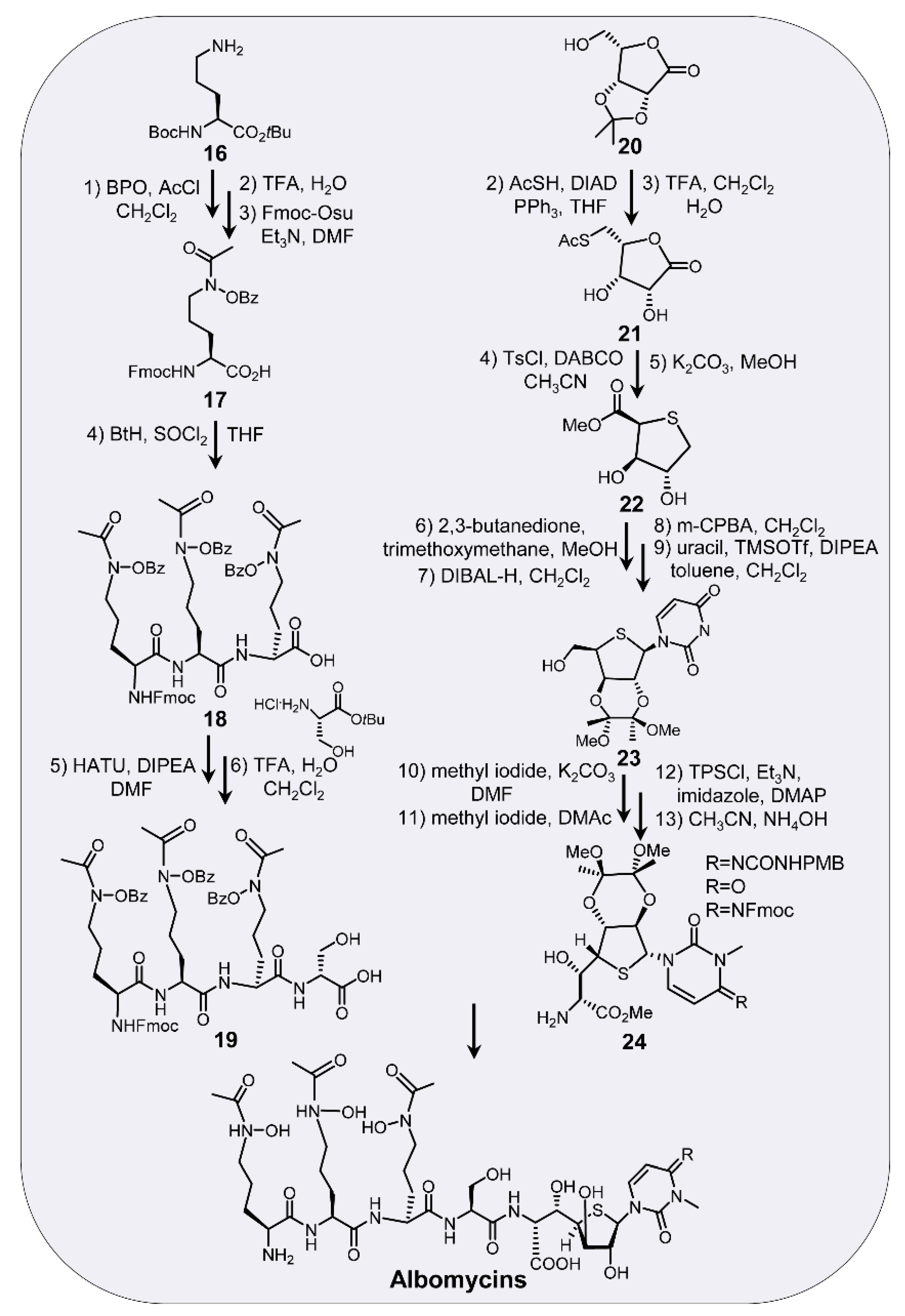
Chemical synthesis of the tri-δ-N-hydroxy-L-ornithine peptide siderophore was initially described by Benz and coworkers in 1984 [51,52]. Later, different synthetic strategies were described by Miller and coworkers in 1990s [53,54,55]. The synthesis of the thionucleoside moiety of albomycin δ1 (13) was also reported by Holzapfel and coworkers [56]. In one recent study, 13 and its isoleucyl and aspartyl analogues (13a and 13b) were synthesized by Van Aerschot and coworkers [57]. Of note is that an oxygen analog of albomycin δ1 (1d) was synthesized by Benz and coworkers [58]. Surprisingly, the oxygen analog lost its antibacterial activity, suggesting that the sulfur atom is indispensable for the bioactivity of albomycins [59]. Furthermore, two aryl-tetrazole containing albomycin analogues (15a and 15b) were obtained by Van Aerschot and coworkers (Figure 3). The two analogues were synthesized by coupling of aryl-tetrazole-containing compounds 14a (CB168) and 14b (CB432) to the tri-δ-N-hydroxy-L-ornithine peptide siderophore [60]. CB168 and CB432 were synthesized by Cubist Pharmaceuticals as potent aaRS inhibitors. Both CB168 and CB432 contain an aryl-tetrazole in place of the adenine moiety, and connected through a two-carbon linker to ribose [61]. The tri-δ-N-hydroxy-L-ornithine peptide siderophore were prepared as described by Miller and coworkers [53]. Antimicrobial activities were then tested against Staphylococcus aureus (ATCC 6538), Staphylococcus epidermidis RP62A (ATCC 35984), Pseudomonas aeruginosa PAO1, Sarcina lutea (ATCC 9341), and Candida albicans CO11. Only one of the compounds showed weak activity against S. aureus and C. albicans, though both analogues showed good activity against IleRS in vitro [60]. Total synthesis of the three natural albomycin congeners has been accomplished recently by He and coworkers [59]. For illustration purposes, we here only show a simplified scheme of albomycins synthesis (Scheme 2). The efficient synthesis of albomycins enables biological evaluations of these potent inhibitors against three Gram-positive bacteria (S. pneumoniae ATCC 49619, S. aureus USA 300 NRS 384, and Bacillus subtilis ATCC 6633) and three Gram-negative bacteria (E. coli BJ 5183, Neisseria gonorrhoeae ATCC 49226, and Salmonella typhi), as well as 27 clinical S. pneumoniae and S. aureus isolates (three of them are methicillin-resistant strains). The results revealed that C4 substituent on the nucleobase in albomycin plays an essential role in their antibacterial activity. Importantly, the systematic SAR study suggested that albomycin δ2 is the most promising candidate for further clinical drug development. Of particular interest is that the potency of albomycin δ2 exceeds that of well-established antibiotics ciprofloxacin, vancomycin, and penicillin [59]. Inspired by these studies, more previously inaccessible analogues can be synthesized and subjected to systematic SAR studies. These studies will substantially expand the repertoire of novel albomycins that will ultimately expedite the design and development of effective antimicrobial agents.

7. Concluding Remarks

Aminoacyl-tRNA synthetases (aaRSs) catalyze the charging of tRNA with a cognate amino acid. They are indispensable for protein synthesis in all three kingdoms of life. Due to their important role in cellular viability, aaRSs have been recognized as suitable targets for the development of novel antimicrobial agents [6]. At present, there are three aaRS inhibitors in clinical practice. Mupirocin is an inhibitor of bacterial isoleucyl-tRNA synthetase (IleRS). It is mainly used as an antibacterial agent for the treatment of infection caused by Gram-positive bacteria, including methicillin-resistant Staphylococcus aureus (MRSA) [62,63]. Tavaborole is an inhibitor of fungal leucyl-tRNA synthetases (LeuRSs). It was approved by the US Food and Drug Administration (FDA) for the treatment of onychomycosis caused by Trichophyton species [64,65]. Halofuginone is an inhibitor of proline-tRNA synthetases (ProRSs). This drug was FDA approved for the treatment of apicomplexan parasite infections in chickens and scleroderma in humans [66].
Albomycins utilize the “Trojan horse” strategy to enter target bacterial cells. Once inside the cell, albomycins will be hydrolyzed by host peptidases to release the thionucleoside SB-217452 warhead that targets bacterial SerRSs. Thus, albomycins exhibit potent antibacterial activities against many model bacteria and a number of clinical pathogens [59]. Over the past several decades, a significant progress has been made on the mechanism of action, biosynthesis and immunity, and chemical synthesis of albomycins. Previous studies suggested that the biosynthetic pathway proceeds through multiple steps of unusual enzymatic reactions [38,39]. However, there are still many missing gaps in our understanding of the biosynthetic process. Therefore, further genetic and biochemical studies are needed to bridge these gaps. For example, further studies are needed to clarify the functions of AbmQ and AbmC in the amide bond formation. Moreover, further experimental evidence is also required for the formation of ferrichrome-type siderophore moiety. These studies will provide the basis for pathway engineering and combinatorial biosynthesis to create new albomycin analogues.
Natural products still hold out the best options for finding novel antimicrobial agents [3]. Advances in omics and bioinformatics have accelerated the identification of gene clusters for antibiotics biosynthesis. In recent years, resistance-guided genome mining has been used successfully for the discovery of novel antimicrobial agents [1,2,49,50]. As mentioned above, abmK can serve as a good candidate to search for homologous genes in public databases. Analysis of abmK homologues and the neighboring genes will be helpful for high-throughput screening of gene clusters encoding novel albomycin analogues. This large-scale genome mining open up new opportunities for the discovery and development of antimicrobial drugs to address the growing global threat of antimicrobial resistance.

Author Contributions

M.W.: Writing, reviewing, and editing; Y.Z.: Writing, reviewing, and editing; L.L.: Reviewing, and editing; D.K.: Reviewing, and editing; G.N.: Conceptualization, writing original draft, reviewing, and editing. All authors have read and agreed to the published version of the manuscript.

Funding

This work was supported in part by grants from the National Natural Science Foundation of China (No. 31870061), Chongqing Science and Technology Commission (No. cstccxljrc201904), and Southwest University (No. SWU117015).

Conflicts of Interest

The authors declare no conflict of interest.

References

  1. Niu, G. Genomics-driven natural product discovery in actinomycetes. Trends Biotechnol. 2018, 36, 238–241. [Google Scholar] [CrossRef] [PubMed]
  2. Niu, G.; Li, W. Next-generation drug discovery to combat antimicrobial resistance. Trends Biochem. Sci. 2019, 44, 961–972. [Google Scholar] [CrossRef] [PubMed]
  3. Newman, D.J.; Cragg, G.M. Natural products as sources of new drugs over the nearly four decades from 01/1981 to 09/2019. J. Nat. Prod. 2020, 83, 770–803. [Google Scholar] [CrossRef] [PubMed]
  4. Shen, B. A new golden age of natural products drug discovery. Cell 2015, 163, 1297–1300. [Google Scholar] [CrossRef] [Green Version]
  5. Harvey, A.L.; Edrada-Ebel, R.; Quinn, R.J. The re-emergence of natural products for drug discovery in the genomics era. Nat. Rev. Drug Discov. 2015, 14, 111–129. [Google Scholar] [CrossRef] [Green Version]
  6. Pang, L.; Weeks, S.D.; Van Aerschot, A. Aminoacyl-tRNA synthetases as valuable targets for antimicrobial drug discovery. Int. J. Mol. Sci. 2021, 22, 1750. [Google Scholar] [CrossRef]
  7. Ibba, M.; Soll, D. Aminoacyl-tRNA synthesis. Annu. Rev. Biochem. 2000, 69, 617–650. [Google Scholar] [CrossRef]
  8. Gorska, A.; Sloderbach, A.; Marszall, M.P. Siderophore-drug complexes: Potential medicinal applications of the ‘Trojan horse’ strategy. Trends Pharmacol. Sci. 2014, 35, 442–449. [Google Scholar] [CrossRef]
  9. Braun, V.; Pramanik, A.; Gwinner, T.; Koberle, M.; Bohn, E. Sideromycins: Tools and antibiotics. Biometals 2009, 22, 3–13. [Google Scholar] [CrossRef] [Green Version]
  10. Travin, D.Y.; Severinov, K.; Dubiley, S. Natural Trojan horse inhibitors of aminoacyl-tRNA synthetases. RSC Chem. Biol. 2021, 2, 468–485. [Google Scholar] [CrossRef]
  11. Vondenhoff, G.H.; Van Aerschot, A. Microcin C: Biosynthesis, mode of action, and potential as a lead in antibiotics development. Nucleosides Nucleotides Nucleic. Acids 2011, 30, 465–474. [Google Scholar] [CrossRef] [PubMed]
  12. Kim, J.G.; Park, B.K.; Kim, S.U.; Choi, D.; Nahm, B.H.; Moon, J.S.; Reader, J.S.; Farrand, S.K.; Hwang, I. Bases of biocontrol: Sequence predicts synthesis and mode of action of agrocin 84, the Trojan horse antibiotic that controls crown gall. Proc. Natl. Acad. Sci. USA 2006, 103, 8846–8851. [Google Scholar] [CrossRef] [PubMed] [Green Version]
  13. Chopra, S.; Palencia, A.; Virus, C.; Schulwitz, S.; Temple, B.R.; Cusack, S.; Reader, J. Structural characterization of antibiotic self-immunity tRNA synthetase in plant tumour biocontrol agent. Nat. Commun. 2016, 7, 12928. [Google Scholar] [CrossRef] [PubMed] [Green Version]
  14. Reynolds, D.M.; Schatz, A.; Waksman, S.A. Grisein, a new antibiotic produced by a strain of Streptomyces griseus. Proc. Soc. Exp. Biol. Med. 1947, 64, 50–54. [Google Scholar] [CrossRef]
  15. Reynolds, D.M.; Waksman, S.A. Grisein, an antibiotic produced by certain strains of Streptomyces griseus. J. Bacteriol. 1948, 55, 739–752. [Google Scholar] [CrossRef] [PubMed] [Green Version]
  16. Gause, G.F. Recent studies on albomycin, a new antibiotic. Br. Med. J. 1955, 2, 1177–1179. [Google Scholar] [CrossRef] [Green Version]
  17. Stapley, E.O.; Ormond, R.E. Similarity of albomycin and grisein. Science 1957, 125, 587–589. [Google Scholar] [CrossRef]
  18. Maehr, H.; Pitcher, R.G. Identity of albomycin δ2 and antibiotic Ro 5-2667. J. Antibiot. 1971, 24, 830–834. [Google Scholar] [CrossRef]
  19. Maehr, H. Antibiotics and other naturally occurring hydroxamic acids and hydroxamates. Pure Appl. Chem. 1971, 28, 603–636. [Google Scholar] [CrossRef]
  20. Benz, G.; Schröder, T.; Kurz, J.; Wünsche, C.; Karl, W.; Steffens, G.; Pfitzner, J.; Schmidt, D. Constitution of the deferriform of the albomycins δ1, δ2 and ε. Angew. Chem. Int. Ed. Engl. 1982, 21, 527–528. [Google Scholar] [CrossRef]
  21. Benz, G.; Schröder, T.; Kurz, J.; Wünsche, C.; Karl, W.; Steffens, G.; Pfitzner, J.; Schmidt, D. Konstitution der Desferriform der Albomycine δ1, δ2, ε. Angew. Chem. Int. Ed. Engl. 1982, 21, 1322–1335. [Google Scholar] [CrossRef]
  22. Haas, H. Fungal siderophore metabolism with a focus on Aspergillus fumigatus. Nat. Prod. Rep. 2014, 31, 1266–1276. [Google Scholar] [CrossRef] [PubMed] [Green Version]
  23. Asai, Y.; Hiratsuka, T.; Ueda, M.; Kawamura, Y.; Asamizu, S.; Onaka, H.; Arioka, M.; Nishimura, S.; Yoshida, M. Differential biosynthesis and roles of two ferrichrome-type siderophores, ASP2397/AS2488053 and ferricrocin, in Acremonium persicinum. ACS Chem. Biol. 2022, 17, 207–216. [Google Scholar] [CrossRef] [PubMed]
  24. Niu, G.; Tan, H. Nucleoside antibiotics: Biosynthesis, regulation, and biotechnology. Trends Microbiol. 2015, 23, 110–119. [Google Scholar] [CrossRef]
  25. Shiraishi, T.; Kuzuyama, T. Recent advances in the biosynthesis of nucleoside antibiotics. J. Antibiot. 2019, 72, 913–923. [Google Scholar] [CrossRef]
  26. Pramanik, A.; Stroeher, U.H.; Krejci, J.; Standish, A.J.; Bohn, E.; Paton, J.C.; Autenrieth, I.B.; Braun, V. Albomycin is an effective antibiotic, as exemplified with Yersinia enterocolitica and Streptococcus pneumoniae. Int. J. Med. Microbiol. 2007, 297, 459–469. [Google Scholar] [CrossRef]
  27. Pramanik, A.; Braun, V. Albomycin uptake via a ferric hydroxamate transport system of Streptococcus pneumoniae R6. J. Bacteriol. 2006, 188, 3878–3886. [Google Scholar] [CrossRef] [Green Version]
  28. Al Shaer, D.; Al Musaimi, O.; de la Torre, B.G.; Albericio, F. Hydroxamate siderophores: Natural occurrence, chemical synthesis, iron binding affinity and use as Trojan horses against pathogens. Eur. J. Med. Chem. 2020, 208, 112791. [Google Scholar] [CrossRef]
  29. Neilands, J.B. Microbial envelope proteins related to iron. Annu. Rev. Microbiol. 1982, 36, 285–309. [Google Scholar] [CrossRef]
  30. Braun, V. Active transport of siderophore-mimicking antibacterials across the outer membrane. Drug Resist. Update 1999, 2, 363–369. [Google Scholar] [CrossRef]
  31. Ferguson, A.D.; Braun, V.; Fiedler, H.P.; Coulton, J.W.; Diederichs, K.; Welte, W. Crystal structure of the antibiotic albomycin in complex with the outer membrane transporter FhuA. Protein Sci. 2000, 9, 956–963. [Google Scholar] [CrossRef] [PubMed]
  32. Killmann, H.; Herrmann, C.; Torun, A.; Jung, G.; Braun, V. TonB of Escherichia coli activates FhuA through interaction with the β-barrel. Microbiology 2002, 148, 3497–3509. [Google Scholar] [CrossRef] [PubMed] [Green Version]
  33. Rohrbach, M.R.; Braun, V.; Koster, W. Ferrichrome transport in Escherichia coli K-12: Altered substrate specificity of mutated periplasmic FhuD and interaction of FhuD with the integral membrane protein FhuB. J. Bacteriol. 1995, 177, 7186–7193. [Google Scholar] [CrossRef] [PubMed] [Green Version]
  34. Schultz-Hauser, G.; Koster, W.; Schwarz, H.; Braun, V. Iron (III) hydroxamate transport in Escherichia coli K-12: FhuB-mediated membrane association of the FhuC protein and negative complementation of fhuC mutants. J. Bacteriol. 1992, 174, 2305–2311. [Google Scholar] [CrossRef] [Green Version]
  35. Kadner, R.J.; Heller, K.; Coulton, J.W.; Braun, V. Genetic control of hydroxamate-mediated iron uptake in Escherichia coli. J. Bacteriol. 1980, 143, 256–264. [Google Scholar] [CrossRef] [Green Version]
  36. Braun, V.; Gunthner, K.; Hantke, K.; Zimmermann, L. Intracellular activation of albomycin in Escherichia coli and Salmonella typhimurium. J. Bacteriol. 1983, 156, 308–315. [Google Scholar] [CrossRef] [Green Version]
  37. Zeng, Y.; Kulkarni, A.; Yang, Z.; Patil, P.B.; Zhou, W.; Chi, X.; Van Lanen, S.; Chen, S. Biosynthesis of albomycin δ2 provides a template for assembling siderophore and aminoacyl-tRNA synthetase inhibitor conjugates. ACS Chem. Biol. 2012, 7, 1565–1575. [Google Scholar] [CrossRef] [Green Version]
  38. Ushimaru, R.; Liu, H.W. Biosynthetic origin of the atypical stereochemistry in the thioheptose core of albomycin nucleoside antibiotics. J. Am. Chem. Soc. 2019, 141, 2211–2214. [Google Scholar] [CrossRef]
  39. Ushimaru, R.; Chen, Z.; Zhao, H.; Fan, P.H.; Liu, H.W. Identification of the enzymes mediating the maturation of the Seryl-tRNA synthetase inhibitor SB-217452 during the biosynthesis of albomycins. Angew. Chem. Int. Ed. Engl. 2020, 59, 3558–3562. [Google Scholar] [CrossRef]
  40. Garg, R.P.; Gonzalez, J.M.; Parry, R.J. Biochemical characterization of VlmL, a Seryl-tRNA synthetase encoded by the valanimycin biosynthetic gene cluster. J. Biol. Chem. 2006, 281, 26785–26791. [Google Scholar] [CrossRef] [Green Version]
  41. Garg, R.P.; Qian, X.L.; Alemany, L.B.; Moran, S.; Parry, R.J. Investigations of valanimycin biosynthesis: Elucidation of the role of seryl-tRNA. Proc. Natl. Acad. Sci. USA 2008, 105, 6543–6547. [Google Scholar] [CrossRef] [PubMed] [Green Version]
  42. Mullowney, M.W.; McClure, R.A.; Robey, M.T.; Kelleher, N.L.; Thomson, R.J. Natural products from thioester reductase containing biosynthetic pathways. Nat. Prod. Rep. 2018, 35, 847–878. [Google Scholar] [CrossRef] [PubMed]
  43. Močibob, M.; Rokov-Plavec, J.; Godinic-Mikulcic, V.; Gruic-Sovulj, I. Seryl-tRNA synthetases in translation and beyond. Croat. Chem. Acta 2016, 89, 261–276. [Google Scholar] [CrossRef]
  44. Zeng, Y.; Roy, H.; Patil, P.B.; Ibba, M.; Chen, S. Characterization of two seryl-tRNA synthetases in albomycin-producing Streptomyces sp. strain ATCC 700974. Antimicrob. Agents Chemother. 2009, 53, 4619–4627. [Google Scholar] [CrossRef] [Green Version]
  45. Yanagisawa, T.; Kawakami, M. How does Pseudomonas fluorescens avoid suicide from its antibiotic pseudomonic acid?: Evidence for two evolutionarily distinct isoleucyl-tRNA synthetases conferring self-defense. J. Biol. Chem. 2003, 278, 25887–25894. [Google Scholar] [CrossRef] [Green Version]
  46. Olano, C.; Wilkinson, B.; Sanchez, C.; Moss, S.J.; Sheridan, R.; Math, V.; Weston, A.J.; Brana, A.F.; Martin, C.J.; Oliynyk, M.; et al. Biosynthesis of the angiogenesis inhibitor borrelidin by Streptomyces parvulus Tu4055: Cluster analysis and assignment of functions. Chem. Biol. 2004, 11, 87–97. [Google Scholar]
  47. Vecchione, J.J.; Sello, J.K. Characterization of an inducible, antibiotic-resistant aminoacyl-tRNA synthetase gene in Streptomyces coelicolor. J. Bacteriol. 2008, 190, 6253–6257. [Google Scholar] [CrossRef] [Green Version]
  48. Kitabatake, M.; Ali, K.; Demain, A.; Sakamoto, K.; Yokoyama, S.; Soll, D. Indolmycin resistance of Streptomyces coelicolor A3(2) by induced expression of one of its two tryptophanyl-tRNA synthetases. J. Biol. Chem. 2002, 277, 23882–23887. [Google Scholar] [CrossRef] [Green Version]
  49. Yan, Y.; Liu, N.; Tang, Y. Recent developments in self-resistance gene directed natural product discovery. Nat. Prod. Rep. 2020, 37, 879–892. [Google Scholar] [CrossRef]
  50. Flemming, A. Antibacterials: Resistance-guided discovery of new antibiotics. Nat. Rev. Drug Discov. 2013, 12, 826. [Google Scholar] [CrossRef]
  51. Benz, G.; Schmidt, D. Albomycine, IV. Isolierung und totalsynthese von (N5-acetyl-N5-hydroxy-L-ornithyl)-(N5-acetyl-N5-hydroxy-L-ornithyl)-N5-acetyl-N5-hydroxy-L-ornithin. Liebigs Ann. Chem. 1984, 1984, 1434–1440. [Google Scholar] [CrossRef]
  52. Benz, G. Albomycins, III. Synthesis of N5-acetyl-N5-hydroxy-L-ornithine from L-glutamic acid. Liebigs Ann. Chem. 1984, 8, 1424–1433. [Google Scholar] [CrossRef]
  53. Lin, Y.-M.; Miller, M.J. Practical synthesis of hydroxamate-derived siderophore components by an indirect oxidation method and syntheses of a DIG−siderophore conjugate and a biotin−siderophore conjugate. J. Org. Chem. 1999, 64, 7451–7458. [Google Scholar] [CrossRef]
  54. Dolence, E.K.; Lin, C.E.; Miller, M.J.; Payne, S.M. Synthesis and siderophore activity of albomycin-like peptides derived from N5-acetyl-N5-hydroxy-L-ornithine. J. Med. Chem. 1991, 34, 956–968. [Google Scholar] [CrossRef]
  55. Dolence, E.K.; Minnick, A.A.; Miller, M.J. N5-acetyl-N5-hydroxy-L-ornithine-derived siderophore-carbacephalosporin beta-lactam conjugates: Iron transport mediated drug delivery. J. Med. Chem. 1990, 33, 461–464. [Google Scholar] [CrossRef]
  56. Bredenkamp, A.D.; Martin, W.; Holzapfel, C.W.; Swanepoel, A.D. Synthesis of the thionucleoside moiety of albomycin δ1. S. Afr. Chem. 1991, 44, 31–33. [Google Scholar]
  57. Gadakh, B.; Vondenhoff, G.; Pang, L.; Nautiyal, M.; De Graef, S.; Strelkov, S.V.; Weeks, S.D.; Van Aerschot, A. Synthesis and structural insights into the binding mode of the albomycin δ1 core and its analogues in complex with their target aminoacyl-tRNA synthetase. Bioorg. Med. Chem. 2020, 28, 115645. [Google Scholar] [CrossRef]
  58. Paulsen, H.; Brieden, M.; Benz, G. Verzweigte und kettenverlängerte Zucker, XXXI. Synthese des Sauerstoffanalogons der Desferriform von δ1-Albomycin. Liebigs Ann. Chem. 1987, 1987, 565–575. [Google Scholar] [CrossRef]
  59. Lin, Z.; Xu, X.; Zhao, S.; Yang, X.; Guo, J.; Zhang, Q.; Jing, C.; Chen, S.; He, Y. Total synthesis and antimicrobial evaluation of natural albomycins against clinical pathogens. Nat. Commun. 2018, 9, 3445. [Google Scholar] [CrossRef] [Green Version]
  60. Vondenhoff, G.H.; Gadakh, B.; Severinov, K.; Van Aerschot, A. Microcin C and albomycin analogues with aryl-tetrazole substituents as nucleobase isosters are selective inhibitors of bacterial aminoacyl tRNA synthetases but lack efficient uptake. Chembiochem 2012, 13, 1959–1969. [Google Scholar] [CrossRef]
  61. Hill, J.M.; Yu, G.; Shue, Y.K.; Zydowsky, T.M.; Rebek, J. Aminoacyl Adenylate Mimics as Novel Antimicrobial and Antiparasitic Agents. U.S. Patent 5,726,195, 10 March 1998. [Google Scholar]
  62. Khoshnood, S.; Heidary, M.; Asadi, A.; Soleimani, S.; Motahar, M.; Savari, M.; Saki, M.; Abdi, M. A review on mechanism of action, resistance, synergism, and clinical implications of mupirocin against Staphylococcus aureus. Biomed. Pharmacother. 2019, 109, 1809–1818. [Google Scholar] [CrossRef] [PubMed]
  63. Tucaliuc, A.; Blaga, A.C.; Galaction, A.I.; Cascaval, D. Mupirocin: Applications and production. Biotechnol. Lett. 2019, 41, 495–502. [Google Scholar] [CrossRef] [PubMed]
  64. Barak, O.; Loo, D.S. AN-2690, a novel antifungal for the topical treatment of onychomycosis. Curr. Opin. Investig. Drugs 2007, 8, 662–668. [Google Scholar] [PubMed]
  65. Coghi, P.S.; Zhu, Y.; Xie, H.; Hosmane, N.S.; Zhang, Y. Organoboron compounds: Effective antibacterial and antiparasitic agents. Molecules 2021, 26, 3309. [Google Scholar] [CrossRef] [PubMed]
  66. Pines, M.; Snyder, D.; Yarkoni, S.; Nagler, A. Halofuginone to treat fibrosis in chronic graft-versus-host disease and scleroderma. Biol. Blood Marrow Transplant. 2003, 9, 417–425. [Google Scholar] [CrossRef] [Green Version]
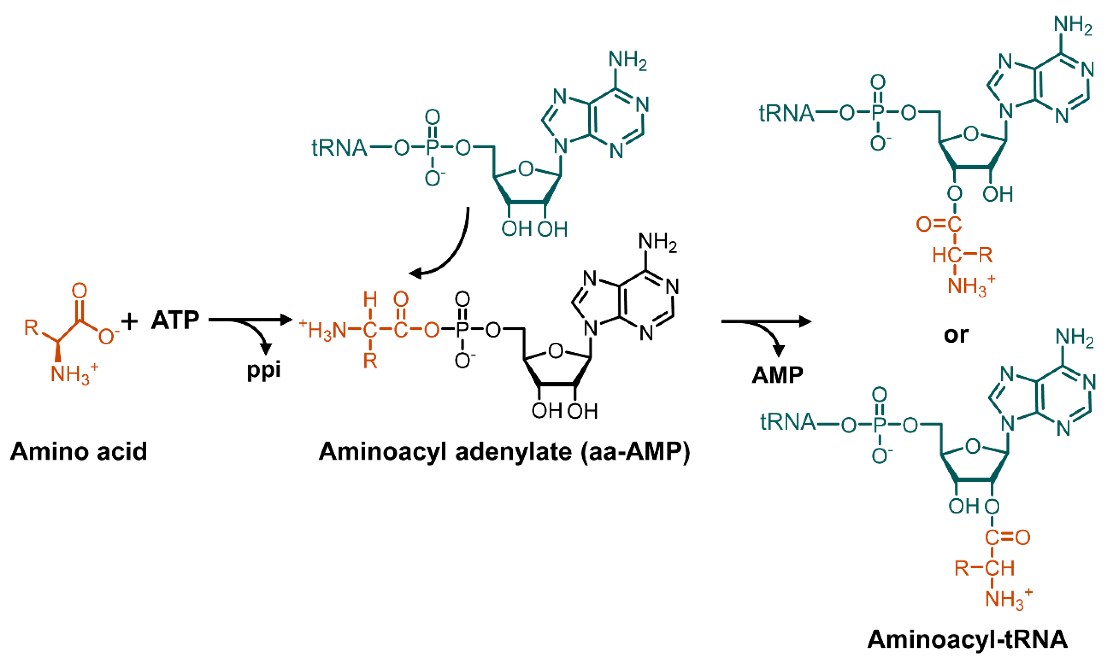
Scheme 1. The two-step aminoacylation of tRNA catalyzed by aminoacyl-tRNA synthetases.
Antibiotics 11 00438 sch001
Figure 1. Structures of representative sideromycins and aaRSs inhibitors. The three naturally occurring albomycin congeners differ mainly in C4 substituent of the pyrimidine nucleoside as indicated. Parts shaded in navy blue indicate the thionucleoside scaffold of albomycins.
Antibiotics 11 00438 g001
Figure 2. Pathway for the biosynthesis of albomycins. (A). Genetic organization of gene cluster for the biosynthesis of albomycins in S. griseus ATCC 700974. Genes from abmA to abmR were thought to be required for albomycin biosynthesis. (B). Proposed biosynthetic pathway of albomycins. AbmQ was highlighted to show the unusual domain organization. AbmG, a homologue of deoxycytidine kinase, was proposed to be responsible for the formation of 7. Biosynthetic process from 7 to 12 was believed to be catalyzed sequentially by AbmH, AbmD, AbmF, AbmK, and AbmJ. AbmE and AbmI were responsible for the tailoring modifications of thionucleoside moiety.
Antibiotics 11 00438 g002
Scheme 2. Chemical synthesis of natural albomycin congeners. A simplified scheme was shown here.
Antibiotics 11 00438 sch002
Figure 3. Structures of synthetic albomycin analogues.
Antibiotics 11 00438 g003
Publisher’s Note: MDPI stays neutral with regard to jurisdictional claims in published maps and institutional affiliations.