Advancements in Nanomaterial-Based Biosensors for Neuropsychiatric and Neurodegenerative Diagnostics: From Biomarker Discovery to Clinical Translation


Abstract
1. Introduction
2. Nanomaterial Systems and Integrated Sensing Modes
2.1. The Functional Role of Nanomaterials in Biosensors
2.2. Classification of Nanobiosensors: Sensing Modalities and Material Systems
2.2.1. Electrochemical Sensing: Carbon-Based Nanomaterials
2.2.2. Optical/SERS Sensing: Noble Metal Nanomaterials
2.2.3. Fluorescence/Photoelectrochemical Sensing: Quantum Dots and Semiconductor Materials
2.2.4. Magnetic Nanomaterials: Separation, Enrichment, and Multimodal Detection
2.2.5. Electrocatalytic and Synergistic Sensing: Metal Oxides and Hybrid Nanomaterials
2.3. Design and Integration of Molecular Recognition Elements
2.3.1. Antibodies: Advantages and Limitations of Classic Recognition Elements
2.3.2. Nucleic Acid Aptamers: Programmable Chemical Antibodies
2.3.3. Molecularly Imprinted Polymers: Synthetic Antibodies and Interface Engineering
2.3.4. CRISPR-Cas System: A Paradigm Shift in Nucleic Acid Recognition
2.3.5. Immobilization and Functionalization Strategies
- Covalent coupling: This remains the gold standard for achieving high long-term stability. Common strategies include EDC/NHS crosslinking chemistry (which forms stable amide bonds between carboxyl and amine groups of proteins and functionalized carbon/metal oxide nanomaterials) and thiol-metal chemistry (e.g., forming robust Au-S bonds between thiolated DNA/aptamers and noble metal nanoparticles) [150]. Covalent bonding prevents sensor degradation and allows for targeted orientation.
- Affinity-based immobilization: Interactions such as the biotin-streptavidin system are widely favored due to their exceptionally high affinity and structural stability under varying pH and temperature conditions. This approach allows for the highly directional orientation of bulky recognition elements like antibodies, maximizing their capture efficiency.
- Physical adsorption: While it offers the advantage of simplicity without requiring chemical modification, adsorption via electrostatic or Van der Waals interactions is prone to desorption during washing steps or in complex biological matrices, and is therefore typically used in conjunction with protective membranes.
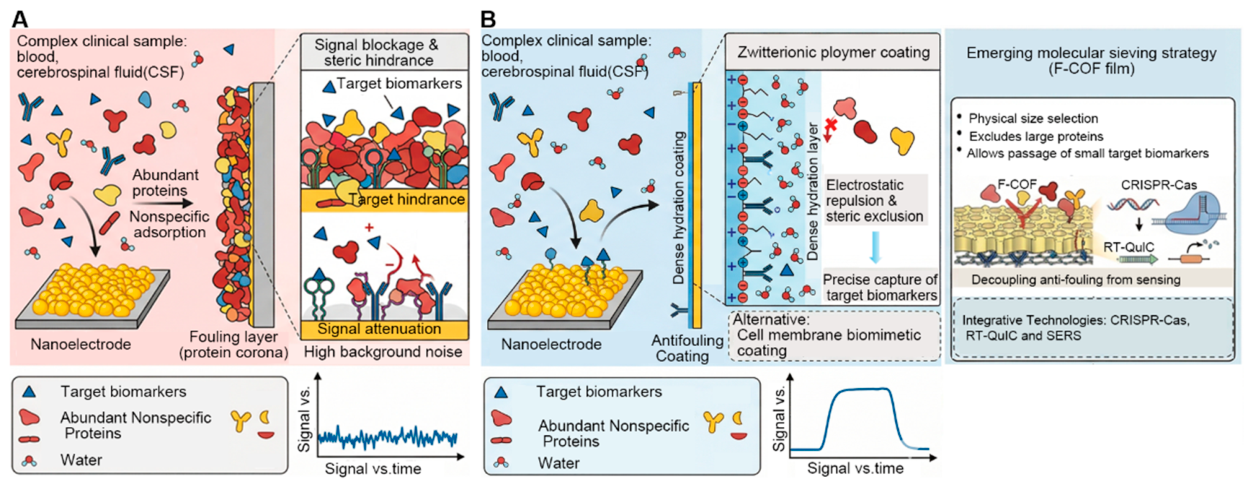
- Surface passivation and spacer engineering: To prevent steric hindrance and nonspecific fouling, nanomaterial surfaces are frequently pre-functionalized with self-assembled monolayers (SAMs) or polymer brushes (such as PEG or zwitterionic layers). These functional interfaces serve as precisely controlled scaffolds, ensuring optimal spacing between adjacent bioreceptors to maintain their native three-dimensional conformations [151,152].
2.3.6. Synergistic Integration of Multiple Recognition Elements and Future Prospects
2.3.7. Biofouling Mitigation and Interfacial Shielding Strategies
3. Application of Nanobiosensors in the Diagnosis of Neurological Diseases
3.1. For Early Screening: High-Sensitivity Quantitative Detection of Established Protein Biomarkers
3.2. For Disease Progression Monitoring: Real-Time Monitoring of Dynamic Neurochemical Substances
3.3. Ultrasensitive Detection of Established and Emerging Biomarkers: Exosomes and Circulating microRNAs
4. Conclusions and Future Perspectives
4.1. Summary and Outlook
4.1.1. Balancing Sensitivity and Selectivity
4.1.2. Multiplex Detection in Complex Matrices
4.2. Next-Generation Diagnostic Paradigm: Multimodal Fusion and Chemical Brain–Computer Interfaces (cBCIs)
4.2.1. Deep Integration of Multimodal and Multiplexing Platforms
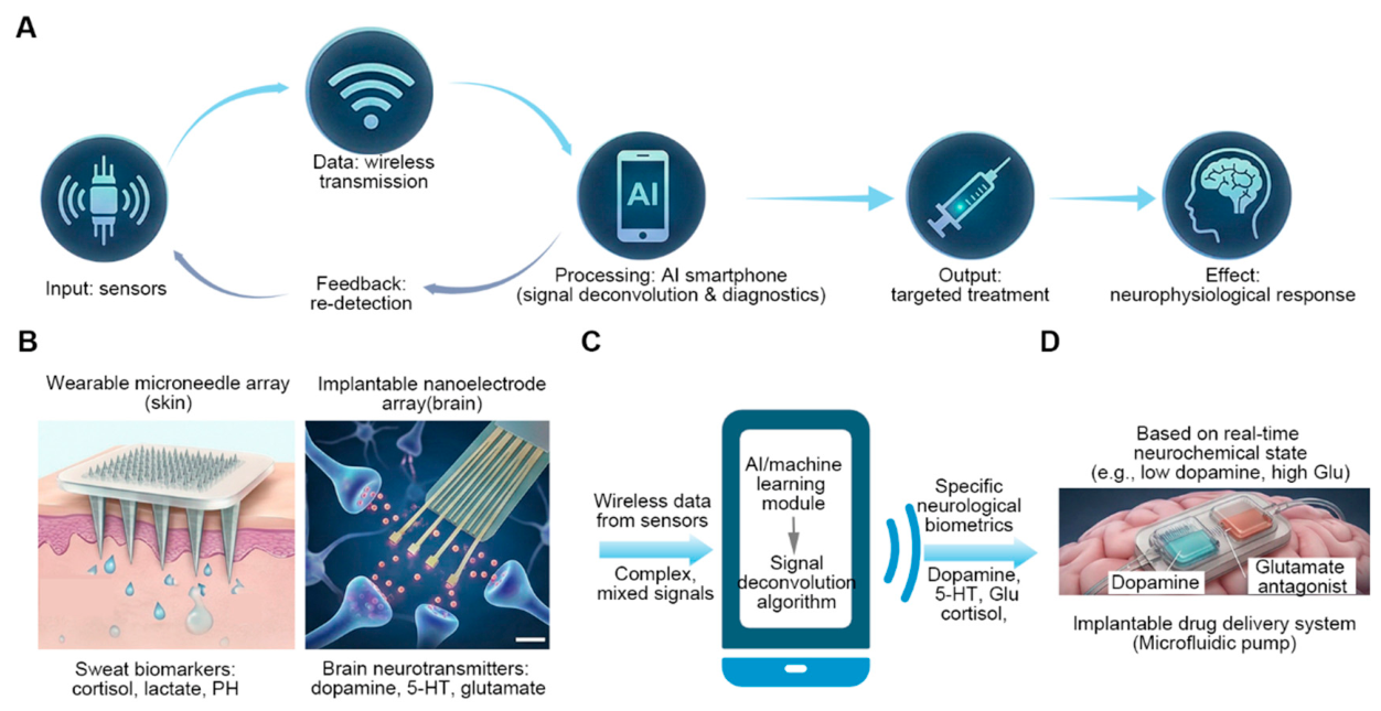
4.2.2. Closed-Loop Chemical Brain–Computer Interfaces (cBCIs)
4.2.3. AI-Driven Predictive Diagnosis
Author Contributions
Funding
Institutional Review Board Statement
Informed Consent Statement
Data Availability Statement
Acknowledgments
Conflicts of Interest
Abbreviations
| cBCIs | closed-loop chemical brain–computer interfaces |
| AD | Alzheimer’s disease |
| PD | Parkinson’s disease |
| ELISA | Enzyme-linked immunosorbent assay |
| MRI | Magnetic Resonance Imaging |
| PET | Positron Emission Tomography |
| CSF | Cerebrospinal fluid |
| fM | Femtomolar |
| pM | Picomolar |
| aM | Attomolar |
| POCT | Point-of-Care Testing |
| MICP | Molecularly imprinted composite polymers |
| LSPR | Local surface plasmon resonance |
| SERS | Surface-enhanced Raman scattering |
| DA | Dopamine |
| NE | Norepinephrine |
| 5-HT | Serotonin |
| NfL | Neurofilament light chain |
| miRNA | MicroRNA |
| α-syn | Alpha-synuclein |
| CCMs | Carbon-coated microelectrodes |
| GFETs | Graphene field-effect transistors |
| F-COFs | Fluorinated covalent organic framework |
| PEC | Photoelectrochemical |
| FRET | Fluorescence resonance energy transfer |
| MIPs | Molecularly imprinted polymers |
| SELEX | Systematic evolution of ligands by exponential enrichment |
| AI | Artificial intelligence |
| OECT | Organic electrochemical transistor |
| GABA | γ-aminobutyric acid |
| ECL | Electrochemiluminescence |
| Aβ | Amyloid-beta |
| p-tau | Phosphorylated tau protein |
| AUC | Area under the curve |
| MCI | Mild cognitive impairment |
| FET | Field-effect transistor |
| LOD | The limit of detection |
References
- Cao, Y.; Xu, Y.; Cao, M.; Chen, N.; Zeng, Q.; Lai, M.K.; Fan, D.; Sethi, G.; Cao, Y. Fluid-based biomarkers for neurodegenerative diseases. Ageing Res. Rev. 2025, 108, 102739. [Google Scholar] [CrossRef] [PubMed]
- Houfkova, A.; Schmidt, M. Amyloid-β and Mitochondrial Membranes: A Missing Link in Alzheimer’s Pathogenesis. Mol. Neurobiol. 2026, 63, 493. [Google Scholar] [CrossRef]
- Balagurusamy, B.; Ganesan, V.; Gopi, V.; Ilayaperumal, P. Liposomal and Nanomaterial-Based Strategies for Targeted Alzheimer’s Disease Therapy. ACS Omega 2025, 10, 60004–60019. [Google Scholar] [CrossRef]
- Kotarba, S.; Kozłowska, M.; Scios, M.; Saramowicz, K.; Barczuk, J.; Granek, Z.; Siwecka, N.; Wiese, W.; Golberg, M.; Galita, G.; et al. Potential Mechanisms of Tunneling Nanotube Formation and Their Role in Pathology Spread in Alzheimer’s Disease and Other Proteinopathies. Int. J. Mol. Sci. 2024, 25, 10797. [Google Scholar] [CrossRef]
- Tutubala, T.E.; Egunlusi, A.O.; Fisher, D.; Dube, A.; Joubert, J. Nanomedicine Solutions for Alzheimer’s Disease: A Critical Review of Therapeutic Nanoparticle Strategies. ACS Omega 2025, 10, 53633–53657. [Google Scholar] [CrossRef]
- Putnin, T.; Chanarsa, S.; Yaiwong, P.; Ngamaroonchote, A.; Aroonyadet, N.; Jakmunee, J.; Bamrungsap, S.; Laocharoensuk, R.; Ounnunkad, K. Unraveling the Impact of Polyethylenimine-Coated Gold Nanoparticle Size on the Efficiency of Sandwich-Style Electrochemical Immunosensors. ACS Meas. Sci. Au 2025, 5, 96–108. [Google Scholar] [CrossRef]
- Zhu, W.; Wang, Y.; Qin, M.; Zhao, Q.; Feng, L.; Li, M. Insights into Biomarkers of Alzheimer’s Disease: From Core Markers to Emerging Directions. Aging Dis. 2025. [Google Scholar] [CrossRef]
- Kumar, K.; Singh, K.; Rathour, R.; Singh, J.; Bhattacharya, S.; Pandey, S. Fabrication of Nanobioengineered Interfaces Utilizing Quaternary Nanocomposite for Highly Efficient and Selective Electrochemical Biosensing of Urea. Langmuir 2024, 40, 21052–21066. [Google Scholar] [CrossRef]
- Tao, D.; Xie, C.; Fu, S.; Rong, S.; Song, S.; Ye, H.; Jaffrezic-Renault, N.; Guo, Z. Thionine-functionalized three-dimensional carbon nanomaterial-based aptasensor for analysis of Aβ oligomers in serum. Anal. Chim. Acta 2021, 1183, 338990. [Google Scholar] [CrossRef]
- Yadav, S.; Bukke, S.P.N.; Prajapati, S.; Singh, A.P.; Chettupalli, A.K.; Nicholas, B. Nanobiosensors in neurodegenerative disease diagnosis: A promising pathway for early detection. Digit. Health 2025, 11, 20552076251342457. [Google Scholar] [CrossRef]
- Liu, Q.; Xu, X.; Zheng, T. Recent advances in nanomaterial-based biosensors for Alzheimer’s disease diagnosis: A review. Chem. Commun. 2025, 61, 13503–13522. [Google Scholar] [CrossRef]
- Rissin, D.M.; Kan, C.W.; Campbell, T.G.; Howes, S.C.; Fournier, D.R.; Song, L.; Piech, T.; Patel, P.P.; Chang, L.; Rivnak, A.J.; et al. Single-molecule enzyme-linked immunosorbent assay detects serum proteins at subfemtomolar concentrations. Nat. Biotechnol. 2010, 28, 595–599. [Google Scholar] [CrossRef]
- Hansson, O.; Lehmann, S.; Otto, M.; Zetterberg, H.; Lewczuk, P. Advantages and disadvantages of the use of the CSF Amyloid β (Aβ) 42/40 ratio in the diagnosis of Alzheimer’s Disease. Alzheimers Res. Ther. 2019, 11, 34. [Google Scholar] [CrossRef]
- Engelborghs, S.; Niemantsverdriet, E.; Struyfs, H.; Blennow, K.; Brouns, R.; Comabella, M.; Dujmovic, I.; Van Der Flier, W.; Frölich, L.; Galimberti, D.; et al. Consensus guidelines for lumbar puncture in patients with neurological diseases. Alzheimers Dement. Diagn. Assess. Dis. Monit. 2017, 8, 111–126. [Google Scholar] [CrossRef]
- Karikari, T.K.; Pascoal, T.A.; Ashton, N.J.; Janelidze, S.; Benedet, A.L.; Rodriguez, J.L.; Chamoun, M.; Savard, M.; Kang, M.S.; Therriault, J.; et al. Blood phosphorylated tau 181 as a biomarker for Alzheimer’s disease: A diagnostic performance and prediction modelling study using data from four prospective cohorts. Lancet Neurol. 2020, 19, 422–433. [Google Scholar] [CrossRef]
- Mattsson, N.; Andreasson, U.; Persson, S.; Arai, H.; Batish, S.D.; Bernardini, S.; Bocchio-Chiavetto, L.; Blankenstein, M.A.; Carrillo, M.C.; Chalbot, S.; et al. The Alzheimer’s Association external quality control program for cerebrospinal fluid biomarkers. Alzheimers Dement. 2011, 7, 386–395.e386. [Google Scholar] [CrossRef]
- Cohen, L.; Walt, D.R. Single-Molecule Arrays for Protein and Nucleic Acid Analysis. Annu. Rev. Anal. Chem. 2017, 10, 345–363. [Google Scholar] [CrossRef]
- Mosconi, L. Brain glucose metabolism in the early and specific diagnosis of Alzheimer’s disease. Eur. J. Nucl. Med. Mol. Imaging 2005, 32, 486–510. [Google Scholar] [CrossRef]
- Ossenkoppele, R.; Rabinovici, G.D.; Smith, R.; Cho, H.; Schöll, M.; Strandberg, O.; Palmqvist, S.; Mattsson, N.; Janelidze, S.; Santillo, A.; et al. Discriminative Accuracy of [18F]flortaucipir Positron Emission Tomography for Alzheimer Disease vs Other Neurodegenerative Disorders. JAMA 2018, 320, 1151–1162. [Google Scholar] [CrossRef]
- Schöll, M.; Lockhart, S.N.; Schonhaut, D.R.; O’neil, J.P.; Janabi, M.; Ossenkoppele, R.; Baker, S.L.; Vogel, J.W.; Faria, J.; Schwimmer, H.D.; et al. PET Imaging of Tau Deposition in the Aging Human Brain. Neuron 2016, 89, 971–982. [Google Scholar] [CrossRef]
- Becker, J.A.; Hedden, T.; Carmasin, J.; Maye, J.; Rentz, D.M.; Putcha, D.; Fischl, B.; Greve, D.N.; Marshall, G.A.; Salloway, S.; et al. Amyloid-β associated cortical thinning in clinically normal elderly. Ann. Neurol. 2011, 69, 1032–1042. [Google Scholar] [CrossRef]
- Frisoni, G.B.; Fox, N.C.; Jack, C.R.; Scheltens, P.; Thompson, P.M. The clinical use of structural MRI in Alzheimer disease. Nat. Rev. Neurol. 2010, 6, 67–77. [Google Scholar] [CrossRef]
- Nakamura, A.; Kaneko, N.; Villemagne, V.L.; Kato, T.; Doecke, J.; Doré, V.; Fowler, C.; Li, Q.-X.; Martins, P.; Rowe, C.; et al. High performance plasma amyloid-β biomarkers for Alzheimer’s disease. Nature 2018, 554, 249–254. [Google Scholar] [CrossRef]
- Watson, C.M.; Dammer, E.B.; Ping, L.; Duong, D.M.; Modeste, E.; Carter, E.K.; Johnson, E.C.B.; Levey, A.I.; Lah, J.J.; Roberts, B.R.; et al. Quantitative Mass Spectrometry Analysis of Cerebrospinal Fluid Protein Biomarkers in Alzheimer’s Disease. Sci. Data 2023, 10, 261. [Google Scholar] [CrossRef]
- Anandan, S.; Maciak, K.; Breinbauer, R.; Otero-Ortega, L.; Feliciello, G.; Gužvić, N.S.; Torkildsen, O.; Myhr, K.-M. In-Depth Characterization of L1CAM+ Extracellular Vesicles as Potential Biomarkers for Anti-CD20 Therapy Response in Relapsing–Remitting Multiple Sclerosis. Int. J. Mol. Sci. 2025, 26, 7213. [Google Scholar] [CrossRef]
- Wu, F.; Li, W.; Lu, H.; Li, L. Recent Advances in Mass Spectrometry-Based Studies of Post-Translational Modifications in Alzheimer’s Disease. Mol. Cell. Proteom. 2025, 24, 101003. [Google Scholar] [CrossRef]
- Qi, Y.; Jang, D.; Ryu, J.; Bai, T.; Shin, Y.; Gu, W.; Iyer, A.; Li, G.; Ma, H.; Liou, J.-Y.; et al. Stabilized carbon coating on microelectrodes for scalable and interoperable neurotransmitter sensing. Nat. Commun. 2025, 16, 3300. [Google Scholar] [CrossRef]
- Gu, S.; Xu, D.; Huang, J.; Zhou, X.; Liu, Y.; Zhang, Z. Photoelectrochemical biosensor with single atom sites for norepinephrine sensing and brain region synergy in epilepsy. Nat. Commun. 2025, 16, 4765. [Google Scholar] [CrossRef]
- Khan, A.; Shen, J.; Zahid, A.; Hu, C.; Xu, X.; Cheng, X. Label-Free SERS Sensing of Native Aβ with Aptamer-Modified Mesoporous Gold and Gold-Coated Magnetic Nanoparticles Using a Clinical Cohort. ACS Appl. Mater. Interfaces 2025, 18, 4855–4864. [Google Scholar] [CrossRef]
- Huang, J.; Gu, S.; Zhou, X.; Liu, Y.; Zhang, Z. Aptamer single-molecule dispersion on single-atom anchoring sites for high-selectivity in vivo detection. Chem. Sci. 2025, 16, 9720–9729. [Google Scholar] [CrossRef]
- Bodily, T.A.; Ramanathan, A.; Wei, S.; Karkisaval, A.; Bhatt, N.; Jerez, C.; Haque, A.; Ramil, A.; Heda, P.; Wang, Y.; et al. In pursuit of degenerative brain disease diagnosis: Dementia biomarkers detected by DNA aptamer-attached portable graphene biosensor. Proc. Natl. Acad. Sci. USA 2023, 120, e2311565120. [Google Scholar] [CrossRef]
- Xiang, Q.; Guo, F.; Li, D.; Xu, F.; Zhou, W. Low-background CRISPR/Cas12a sensing system with circular CRISPR RNA for amplified fluorescent detection of antibody in human serum. Talanta 2025, 288, 127730. [Google Scholar] [CrossRef]
- Xu, X.; Xu, D.; Lu, S.; Zhou, X.; Yang, S.; Zhang, Z. Atomically dispersed recognition unit for selective in vivo photoelectrochemical medicine detection. Nat. Commun. 2024, 15, 8827. [Google Scholar] [CrossRef]
- Zambrano, A.C.; Loiola, L.M.D.; Bukhamsin, A.; Gorecki, R.; Harrison, G.; Mani, V.; Fatayer, S.; Nunes, S.P.; Salama, K.N. Porous Laser-Scribed Graphene Electrodes Modified with Zwitterionic Moieties: A Strategy for Antibiofouling and Low-Impedance Interfaces. ACS Appl. Mater. Interfaces 2024, 16, 4408–4419. [Google Scholar] [CrossRef]
- Song, Z.; Han, R.; Yu, K.; Luo, X. Designing Zwitterionic Peptide with Superhydrophilicity through Carboxyl–Amino Spacing Adjustment for Robust Antifouling Electrochemical Biosensing in Complex Biofluids. Anal. Chem. 2025, 97, 17849–17856. [Google Scholar] [CrossRef]
- Rossato, B.G.; Moresco, R.N. Oxidative interference in immunoturbidimetric assays for the quantification of immunoglobulins and complement proteins. Anal. Biochem. 2026, 711, 116050. [Google Scholar] [CrossRef]
- Xie, Q.; Chen, G.-Y.; Chu, C.-F.; Lin, C.-T. An improved electrochemical biosensing device architecture with ultra-thin and surface-modified dielectric stacking structure. Sens. Actuators B Chem. 2025, 441, 137993. [Google Scholar] [CrossRef]
- Herrero, E.J.; Bühlmann, P. Challenges and opportunities in the development of planar potentiometric sensors for point-of-care analysis. TrAC Trends Anal. Chem. 2024, 181, 118002. [Google Scholar] [CrossRef]
- Chen, H.; Chu, T.; Ge, S.; Zhang, B.; Xuan, F.-Z.; Yin, R. Depositing Pt nanoparticles on crumpled Ti3C2Tx for enhanced electrochemical sensing. Carbon Future 2025, 2, 9200047. [Google Scholar] [CrossRef]
- Cuniberto, E.; Alharbi, A.; Huang, Z.; Wu, T.; Kiani, R.; Shahrjerdi, D. Anomalous sensitivity enhancement of nano-graphitic electrochemical micro-sensors with reducing the operating voltage. Biosens. Bioelectron. 2021, 177, 112966. [Google Scholar] [CrossRef]
- Zhang, Y.; Wei, Q. The role of nanomaterials in electroanalytical biosensors: A mini review. J. Electroanal. Chem. 2016, 781, 401–409. [Google Scholar] [CrossRef]
- Arduini, F.; Micheli, L.; Moscone, D.; Palleschi, G.; Piermarini, S.; Ricci, F.; Volpe, G. Electrochemical biosensors based on nanomodified screen-printed electrodes: Recent applications in clinical analysis. TrAC Trends Anal. Chem. 2016, 79, 114–126. [Google Scholar] [CrossRef]
- Gallo-Orive, Á.; Moreno-Guzmán, M.; Sanchez-Paniagua, M.; Montero-Calle, A.; Barderas, R.; Escarpa, A. Gold Nanoparticle-Decorated Catalytic Micromotor-Based Aptassay for Rapid Electrochemical Label-Free Amyloid-β42 Oligomer Determination in Clinical Samples from Alzheimer’s Patients. Anal. Chem. 2024, 96, 5509–5518. [Google Scholar] [CrossRef]
- Zhao, P.; Wang, J.; Mao, H.; Zhou, L.; Wu, Z.; Lu, Y.; Sun, T.; Hui, J.; Ma, G. AuNP/Magnetic Bead-Enhanced Electrochemical Sensor Toward Dual Saliva Alzheimer’s Biomarkers Detection. Sensors 2025, 25, 4088. [Google Scholar] [CrossRef]
- Zhang, Y.; Si, G.; Wang, Z.; Wang, Y.; Cui, X.; Yang, H.; Si, F.; Liu, Y. Sensitive electrochemical detection of DR1 based on gold nanoparticle-modified MoS2 and hyaluronic acid-based thionine. Sens. Diagn. 2025, 4, 529–537. [Google Scholar] [CrossRef]
- Huang, H.; Wang, C.; Liu, G. Tailoring the interface structures of micro-nano materials for electrochemical and biomedical applications: A review. J. Alloys Compd. 2025, 1035, 181532. [Google Scholar] [CrossRef]
- Solanki, P.R.; Kaushik, A.; Agrawal, V.V.; Malhotra, B.D. Nanostructured metal oxide-based biosensors. NPG Asia Mater. 2011, 3, 17–24. [Google Scholar] [CrossRef]
- Lee, D.H.; Lee, W.-Y.; Kim, J. Introducing Nanoscale Electrochemistry in Small-Molecule Detection for Tackling Existing Limitations of Affinity-Based Label-Free Biosensing Applications. J. Am. Chem. Soc. 2023, 145, 17767–17778. [Google Scholar] [CrossRef]
- Faysal, A.A.; Cetinkaya, A.; Erdoğan, T.; Ozkan, S.A.; Gölcü, A. Creation of an ultra-sensitive and specific molecularly imprinted polymer sensor with ZnO nanoparticle assistance for valganciclovir detection. Microchim. Acta 2025, 193, 30. [Google Scholar] [CrossRef]
- Şak, B.; Sousa, H.B.A.; Prior, J.A.V. Carbon Nanomaterial-Based Electrochemical Biosensors for Alzheimer’s Disease Biomarkers: Progress, Challenges, and Future Perspectives. Biosensors 2025, 15, 684. [Google Scholar] [CrossRef]
- Yu, L.; Zhu, Y.; Zheng, X.; Huang, R.; Song, S.; Liu, Y.; Liu, Z.; Chen, B.; Zhu, R. Material-based neuroimaging and biomarker detection for central nervous system disorder. Mater. Today Bio 2025, 35, 102615. [Google Scholar] [CrossRef] [PubMed]
- Mahmoudpour, M.; Jouyban, A.; Soleymani, J.; Rahimi, M. Rational design of smart nano-platforms based on antifouling-nanomaterials toward multifunctional bioanalysis. Adv. Colloid Interface Sci. 2022, 302, 102637. [Google Scholar] [CrossRef] [PubMed]
- Zheng, D.; Vashist, S.K.; Dykas, M.M.; Saha, S.; Al-Rubeaan, K.; Lam, E.; Luong, J.H.; Sheu, F.-S. Graphene versus Multi-Walled Carbon Nanotubes for Electrochemical Glucose Biosensing. Materials 2013, 6, 1011–1027. [Google Scholar] [CrossRef]
- Ge, L.; Liu, N.; Yang, P.; Cheng, X. Composite TiO2 Film with Quantum Dots Fabricated Through a Sol-Gel Process. J. Nanosci. Nanotechnol. 2013, 13, 2943–2947. [Google Scholar] [CrossRef]
- Yang, Z.; Guo, J.; Wang, L.; Zhang, J.; Ding, L.; Liu, H.; Yu, X. Nanozyme-Enhanced Electrochemical Biosensors: Mechanisms and Applications. Small 2024, 20, 2307815. [Google Scholar] [CrossRef]
- Lupoi, T.; Baccara, S.; Leroux, Y.R.; Feier, B.; Pasturel, M.; Cercus, J.-L.; Cristea, C.; Geneste, F. Optimizing self-assembled monolayer construction for aptasensors and preventing false signals. The case of erythromycin detection. Electrochim. Acta 2025, 525, 146097. [Google Scholar] [CrossRef]
- Bakestani, R.; Wu, Y.; Glahn-Martínez, B.; Kippin, T.; Plaxco, K.; Kolkman, R. Carboxylate-Terminated Electrode Surfaces Improve the Performance of Electrochemical Aptamer-Based Sensors. ACS Appl. Mater. Interfaces 2025, 17, 8706–8714. [Google Scholar] [CrossRef]
- Guo, K.; Grünberg, R.; Ren, Y.; Chang, T.; Wustoni, S.; Strnad, O.; Koklu, A.; Díaz-Galicia, E.; Agudelo, J.P.; Druet, V.; et al. SpyDirect: A Novel Biofunctionalization Method for High Stability and Longevity of Electronic Biosensors. Adv. Sci. 2024, 11, 2306716. [Google Scholar] [CrossRef] [PubMed]
- Mor, D.C.; Plšek, A.B.; Asokan, P.; Vu, V.; Aktug, G.; Giordani, E.; Ricciardi, E.; Perutka, J.; Önder, K.; Fekete, L.; et al. Facile Fabrication of Large-Area Biofunctionalized Metallic Nanostructures for High Performance Affinity Plasmonic Biosensors. Adv. Mater. Technol. 2025, 10, e00356. [Google Scholar] [CrossRef]
- Cao, Q.; Shao, Z.; Hensley, D.; Venton, B. Carbon nanospike coated nanoelectrodes for measurements of neurotransmitters. Faraday Discuss. 2021, 233, 303–314. [Google Scholar] [CrossRef]
- Liu, K.; Liu, R.; Wang, D.; Pan, R.; Chen, H.-Y.; Jiang, D. Real-Time and Spatial Electroanalysis of Biomolecules in One Living Cell Using Liquid-Phase Modified Nanopipette. CCS Chem. 2023, 5, 1285–1292. [Google Scholar] [CrossRef]
- Sun, B.; Shu, Q.; Wang, L.; Liu, K.; Gao, T. Fluorinated Covalent Organic Framework Films Modified Graphene Field-Effect Transistor Biosensors with Size-Dependent Sieving and Antibiofouling Effect. Nano Lett. 2025, 25, 15150–15157. [Google Scholar] [CrossRef] [PubMed]
- Guo, S.; Hou, P.-X.; Zhang, F.; Liu, C.; Cheng, H.-M. Gas Sensors Based on Single-Wall Carbon Nanotubes. Molecules 2022, 27, 5381. [Google Scholar] [CrossRef]
- Dhas, N.; Parekh, K.; Pandey, A.; Kudarha, R.; Mutalik, S.; Mehta, T. Two dimensional carbon based nanocomposites as multimodal therapeutic and diagnostic platform: A biomedical and toxicological perspective. J. Control. Release 2019, 308, 130–161. [Google Scholar] [CrossRef]
- Wannassi, J.; Essousi, H.; Kahri, H.; Barhoumi, H. Recent progress in nanostructured materials for electrochemical dopamine detection. Microchem. J. 2025, 219, 115941. [Google Scholar] [CrossRef]
- Wareham-Mathiassen, S.; Jolly, P.; Shanmugam, N.R.; Jagannath, B.; Prabhala, P.; Zhai, Y.; Ozkan, A.; Naziripour, A.; Singh, R.; Bengtsson, H.; et al. An Antimicrobial and Antifibrotic Coating for Implantable Biosensors. Biosensors 2025, 15, 171. [Google Scholar] [CrossRef]
- Zhu, X.; Wang, Z.; Tang, Q.; Shi, L.; Li, B.; Jin, Y. Self-assembled DNA micelle-GNP for simultaneous detecting miRNAs of Alzheimer’s disease via lateral flow assays. Biosens. Bioelectron. 2026, 295, 118300. [Google Scholar] [CrossRef]
- Hassan, Y.M.; Wanas, A.; Ali, A.A.; El-Sayed, W.M. Integrating artificial intelligence with nanodiagnostics for early detection and precision management of neurodegenerative diseases. J. Nanobiotechnol. 2025, 23, 668. [Google Scholar] [CrossRef]
- Dang, H.; Joung, Y.; Jeong, C.; Jeon, C.S.; Pyun, S.H.; Park, S.; Choo, J. Nanoplasmonic assay platforms for reproducible SERS detection of Alzheimer’s disease biomarker. Bull. Korean Chem. Soc. 2023, 44, 441–448. [Google Scholar] [CrossRef]
- Sarathkumar, E.; Menon, R.N.; Jayasree, R.S. Ultrasensitive SERS-LFA for the detection of neurofilament light chain and machine learning-assisted Alzheimer’s disease classification. Nanoscale 2025, 17, 25616–25630. [Google Scholar] [CrossRef]
- Vasilescu, A.; Gáspár, S.; Gheorghiu, M.; Polonschii, C.; Banciu, R.M.; David, S.; Gheorghiu, E.; Marty, J.-L. Promising Solutions to Address the Non-Specific Adsorption in Biosensors Based on Coupled Electrochemical-Surface Plasmon Resonance Detection. Chemosensors 2025, 13, 92. [Google Scholar] [CrossRef]
- Mondal, S.; Sahoo, P. Quantum dot-enabled optical codes for neurotransmitter sensing at the bio-nano interface with translational relevance. Chem. Commun. 2026, 62, 5384–5404. [Google Scholar] [CrossRef]
- Yin, L.; Du, Y.; Ge, M.; Zhang, X.; Du, X.; Wu, X. Rod-Shaped NanoZnTPyP Paper-Based Sensor for Visual Detection of Dopamine in Human Plasma. J. Anal. Methods Chem. 2025, 2025, 9981628. [Google Scholar] [CrossRef]
- Yadav, A.; Dogra, P.; Sagar, P.; Srivastava, M.; Srivastava, A.; Kumar, R.; Srivastava, S. A contemporary overview on quantum dots-based fluorescent biosensors: Exploring synthesis techniques, sensing mechanism and applications. Spectrochim. Acta Part A Mol. Biomol. Spectrosc. 2025, 335, 126002. [Google Scholar] [CrossRef]
- Lian, Q.; Huang, Z.; Liu, Y.; Lin, X.; Zheng, L.; Zhan, S.; Wang, Y.; Weng, S. Effectively Determining Dopamine Based on Dopamine Self-polymerization through Fluorescent Method Using Silicon Quantum Dots as Probe. Chem. Pharm. Bull. 2025, 73, 724–731. [Google Scholar] [CrossRef]
- Flores, L.C.F.; Younas, N.; Goebel, S.; Dittmar, K.; Saleem, T.; Younas, A.; Budde, H.; Legler, T.J.; Möbius, W.; Hermann, P.; et al. Multiple Antibody-Coated Gold Nanoparticle-Based ExoAssay for Rapid Isolation of CNS-Specific Exosomes from Blood. J. Neurochem. 2025, 169, e70263. [Google Scholar] [CrossRef]
- Jang, Y.O.; Roh, Y.; Shin, W.; Jo, S.; Koo, B.; Liu, H.; Kim, M.G.; Lee, H.J.; Qiao, Z.; Lee, E.Y.; et al. Transferrin-conjugated magnetic nanoparticles for the isolation of brain-derived blood exosomal MicroRNAs: A novel approach for Parkinson’s disease diagnosis. Anal. Chim. Acta 2024, 1306, 342623. [Google Scholar] [CrossRef]
- Solovicová, V.; Ďatková, A.; Bertók, T.; Kasák, P.; Vikartovská, A.; Lorencová, L.; Tkac, J. Advances in magnetic affinity-based isolation/detection of exosomes for robust diagnostics. Mikrochim. Acta 2025, 192, 206. [Google Scholar] [CrossRef]
- Lu, Q.; Zhang, Z.; Ding, X. Isolation Techniques of Micro/Nano-Scaled Species for Biomedical Applications. Adv. Sci. 2025, 12, e2414109. [Google Scholar] [CrossRef]
- Arivuthilagam, I.S.; Shahid, R.; Rahman, M.M.; Lee, J.-J. Advancements in Single-Atom Catalysts: Synthesis, Characterization, and Applications in Sensing Technologies. Small Sci. 2025, 5, e202500449. [Google Scholar] [CrossRef]
- Hamed, E.M.; Elsaady, M.M.; Li, S.F.Y. Single-Atom Nanozymes in Analytical Chemistry: Opportunities and Challenges. Anal. Chem. 2025, 97, 26313–26325. [Google Scholar] [CrossRef]
- Wei, D.; Bailey, M.J.A.; Andrew, P.; Ryhänen, T. Electrochemical biosensors at the nanoscale. Lab A Chip 2009, 9, 2123–2131. [Google Scholar] [CrossRef]
- Sadik, O.A.; Aluoch, A.O.; Zhou, A. Status of biomolecular recognition using electrochemical techniques. Biosens. Bioelectron. 2009, 24, 2749–2765. [Google Scholar] [CrossRef]
- Dequaire, M.; Degrand, C.; Limoges, B. An electrochemical metalloimmunoassay based on a colloidal gold label. Anal. Chem. 2000, 72, 5521–5528. [Google Scholar] [CrossRef]
- Ma, X.; Ma, X.; Ma, Y.; Sun, X.; Cheng, T.; Jia, Z.; Li, H.; Zhang, J.; Zhang, X.; Li, W. Multidimensional signal amplification architectures in electrochemical immunosensing integrate porous nanomaterials, biocatalysis, and nucleic acid circuits to achieve attomolar detection. RSC Adv. 2025, 15, 33229–33251. [Google Scholar] [CrossRef]
- Mao, X.; Gurung, A.; Xu, H.; Baloda, M.; He, Y.; Liu, G. Simultaneous detection of nucleic acid and protein using gold nanoparticles and lateral flow device. Anal. Sci. Int. J. Jpn. Soc. Anal. Chem. 2014, 30, 637–642. [Google Scholar] [CrossRef]
- Hao, C.; Meng, D.; Shi, W.; Xu, C.; Wang, Q.; Kuang, H. Chiral Gold Nanostructure Monolayers as SERS Substrates for Ultrasensitive Detection of Enantiomer Biomarkers of Alzheimer’s Disease. Angew. Chem. 2025, 64, e202502115. [Google Scholar] [CrossRef]
- Cecchini, M.P.; Turek, V.A.; Paget, J.; Kornyshev, A.A.; Edel, J.B. Self-assembled nanoparticle arrays for multiphase trace analyte detection. Nat. Mater. 2013, 12, 165–171. [Google Scholar] [CrossRef]
- Kamal Eddin, F.B.; Wing Fen, Y. Recent Advances in Electrochemical and Optical Sensing of Dopamine. Sensors 2020, 20, 1039. [Google Scholar] [CrossRef]
- Li, L.-L.; Luo, D.-Y.; Hou, Y.-S.; Wang, C.-B.; Nsanzamahoro, S.; Meng, X.-H.; Zhang, Y.; Yang, A.-M.; Yang, J.-L. Benzyl Chloroformate-Functionalized Fluorescent Silica Nanoparticles for Detection of Norepinephrine. ACS Appl. Nano Mater. 2025, 8, 22927–22937. [Google Scholar] [CrossRef]
- Yu, J.; Zhou, R.; Liu, S.; Zheng, J.; Yan, H.; Su, S.; Chai, N.; Segal, E.; Jiang, C.; Guo, K.; et al. Electrochemical Biosensors for the Detection of Exosomal microRNA Biomarkers for Early Diagnosis of Neurodegenerative Diseases. Anal. Chem. 2025, 97, 5355–5371. [Google Scholar] [CrossRef]
- Li, T.; Wang, K.; Zheng, C.; Zheng, W.; Cheng, Y.; Ning, Q.; Xu, H.; Cui, D. Magnetic frequency mixing technological advances for the practical improvement of point-of-care testing. Biotechnol. Bioeng. 2022, 119, 347–360. [Google Scholar] [CrossRef]
- Wang, M.; Jin, L.; Leung, P.H.-M.; Chow, F.W.-N.; Zhao, X.; Chen, H.; Pan, W.; Liu, H.; Li, S. Advancements in magnetic nanoparticle-based biosensors for point-of-care testing. Front. Bioeng. Biotechnol. 2024, 12, 1393789. [Google Scholar] [CrossRef]
- Tiwari, J.N.; Vij, V.; Kemp, K.C.; Kim, K.S. Engineered Carbon-Nanomaterial-Based Electrochemical Sensors for Biomolecules. ACS Nano 2016, 10, 46–80. [Google Scholar] [CrossRef]
- Yang, C.; Denno, M.E.; Pyakurel, P.; Venton, B.J. Recent trends in carbon nanomaterial-based electrochemical sensors for biomolecules: A review. Anal. Chim. Acta 2015, 887, 17–37. [Google Scholar] [CrossRef]
- Gritsok, D.; Hedström, M.; Montenegro, M.C.B.S.M.; Amorim, C.G. Electrochemical molecularly imprinted polymer sensors in viral diagnostics: Innovations, challenges and case studies. Biosens. Bioelectron. 2025, 287, 117678. [Google Scholar] [CrossRef]
- Huang, Z.; Li, M.; Zhang, L.; Liu, Y. Electrochemical immunosensor based on superwettable microdroplet array for detecting multiple Alzheimer’s disease biomarkers. Front. Bioeng. Biotechnol. 2022, 10, 1029428. [Google Scholar] [CrossRef]
- Gordón Pidal, J.M.; Moreno-Guzmán, M.; Montero-Calle, A.; Barderas, R.; López, M.Á.; Escarpa, A. Dual on-the-move electrochemical immunoassays for the simultaneous determination of amyloid-β (1−42) and Tau in Alzheimer’s patient samples. Sens. Actuators B Chem. 2025, 423, 136785. [Google Scholar] [CrossRef]
- Cunha, D.R.; Segundo, M.A.; Quinaz, M.B. Electrochemical methods for evaluation of therapeutic monoclonal antibodies: A review. Biosens. Bioelectron. 2025, 271, 116988. [Google Scholar] [CrossRef]
- Song, G.; Han, H.; Ma, Z. Anti-Fouling Strategies of Electrochemical Sensors for Tumor Markers. Sensors 2023, 23, 5202. [Google Scholar] [CrossRef]
- Kaur, B.P.; Sahoo, S.; Chaurasia, R.; Bhattacharyya, A.; Sharma, A.K.; Mukherjee, M.; Chakraborty, G. Advances in design and application of molecularly imprinted polymers for selective brain protein recognition in neurology. Biomed. Mater. 2026, 21, 022004. [Google Scholar] [CrossRef]
- Russo, M.J.; Han, M.; Desroches, P.E.; Manasa, C.S.; Dennaoui, J.; Quigley, A.F.; Kapsa, R.M.I.; Moulton, S.E.; Guijt, R.M.; Greene, G.W.; et al. Antifouling Strategies for Electrochemical Biosensing: Mechanisms and Performance toward Point of Care Based Diagnostic Applications. ACS Sens. 2021, 6, 1482–1507. [Google Scholar] [CrossRef]
- Bae, M.; Kim, N.; Cho, E.; Lee, T.; Lee, J.-H. Recent Advances in Electrochemical Biosensors for Neurodegenerative Disease Biomarkers. Biosensors 2025, 15, 151. [Google Scholar] [CrossRef]
- Ji, C.; Wei, J.; Zhang, L.; Hou, X.; Tan, J.; Yuan, Q.; Tan, W. Aptamer-Protein Interactions: From Regulation to Biomolecular Detection. Chem. Rev. 2023, 123, 12471–12506. [Google Scholar] [CrossRef]
- Didarian, R.; Ozbek, H.K.; Ozalp, V.C.; Erel, O.; Yildirim-Tirgil, N. Enhanced SELEX Platforms for Aptamer Selection with Improved Characteristics: A Review. Mol. Biotechnol. 2025, 67, 2962–2977. [Google Scholar] [CrossRef]
- Stephens, C.; Goodey, N.M.; Gubler, U. A beginners guide to SELEX and DNA aptamers. Anal. Biochem. 2025, 703, 115890. [Google Scholar] [CrossRef]
- Eilers, A.; Witt, S.; Walter, J. Aptamer-Modified Nanoparticles in Medical Applications. In Aptamers in Biotechnology. Advances in Biochemical Engineering/Biotechnology; Springer: Cham, Switzerland, 2020; Volume 174, pp. 161–193. [Google Scholar] [CrossRef]
- Bayat, P.; Nosrati, R.; Alibolandi, M.; Rafatpanah, H.; Abnous, K.; Khedri, M.; Ramezani, M. SELEX methods on the road to protein targeting with nucleic acid aptamers. Biochimie 2018, 154, 132–155. [Google Scholar] [CrossRef]
- Wang, T.; Hoy, J.A.; Lamm, M.H.; Nilsen-Hamilton, M. Computational and Experimental Analyses Converge to Reveal a Coherent Yet Malleable Aptamer Structure That Controls Chemical Reactivity. J. Am. Chem. Soc. 2009, 131, 14747–14755. [Google Scholar] [CrossRef][Green Version]
- Cheng, T.; Afshan, N.; Jiao, J.; Jiao, J. Current progress in aptamer-based sensors for the detection of protein biomarkers in neurodegenerative diseases. Biosens. Bioelectron. X 2024, 20, 100528. [Google Scholar] [CrossRef]
- Zheng, Y.; Zhang, L.; Zhao, J.; Li, L.; Wang, M.; Gao, P.; Wang, Q.; Zhang, X.; Wang, W. Advances in aptamers against Aβ and applications in Aβ detection and regulation for Alzheimer’s disease. Theranostics 2022, 12, 2095–2114. [Google Scholar] [CrossRef]
- Yang, X.; Chan, C.H.; Yao, S.; Chu, H.Y.; Lyu, M.; Chen, Z.; Xiao, H.; Ma, Y.; Yu, S.; Li, F.; et al. DeepAptamer: Advancing high-affinity aptamer discovery with a hybrid deep learning model. Mol. Ther. Nucleic Acids 2025, 36, 102436. [Google Scholar] [CrossRef]
- Slalmi, A.; Rabbah, N.; Battas, I.; Debbarh, I.; Medromi, H.; Abourriche, A. Artificial Intelligence-Driven SELEX Design of Aptamer Panels for Urinary Multi-Biomarker Detection in Prostate Cancer: A Systematic and Bibliometric Review. Biomedicines 2025, 13, 2877. [Google Scholar] [CrossRef] [PubMed]
- Jayawardena, T.; Karunarathna, T.; Naranwala, H.; Abeysinghe, S.; Kasturiarachchi, J. Computational approaches for aptamer research. Discov. Appl. Sci. 2025, 7, 1444. [Google Scholar] [CrossRef]
- Amu, G.; Yang, X.; Luo, H.; Yu, S.; Zhang, H.; Tian, Y.; Yu, Y.; Ding, S.; Pan, Y.; Chen, Z.; et al. Machine learning-powered, high-affinity modification strategies for aptamers. Acta Mater. Medica 2025, 4, 122–136. [Google Scholar] [CrossRef]
- Barros, M.; Kasirajan, G.; Jones, A.; Schlichting, A.; Ruiz-Ciancio, D.; Lin, L.-H.; Narayan, C.; Veeramani, S.; Thiel, K.W.; Kennedy, G.C.; et al. ASSET: A Framework for Decoding Aptamer Specificity of an Enriched Library by Next-Generation Sequencing of Experimental Samples. bioRxiv 2025. [Google Scholar] [CrossRef] [PubMed]
- Ishida, R.; Adachi, T.; Yokota, A.; Yoshihara, H.; Aoki, K.; Nakamura, Y.; Hamada, M. RaptRanker: In silico RNA aptamer selection from HT-SELEX experiment based on local sequence and structure information. Nucleic Acids Res. 2020, 48, e82. [Google Scholar] [CrossRef] [PubMed]
- Wu, Y.; Belmonte, I.; Sykes, K.S.; Xiao, Y.; White, R.J. Perspective on the Future Role of Aptamers in Analytical Chemistry. Anal. Chem. 2019, 91, 15335–15344. [Google Scholar] [CrossRef]
- Zhao, Y.; Yavari, K.; Liu, J. Critical evaluation of aptamer binding for biosensor designs. TrAC Trends Anal. Chem. 2022, 146, 116480. [Google Scholar] [CrossRef]
- Toma, L.; Mattarozzi, M.; Ronda, L.; Marassi, V.; Zattoni, A.; Fortunati, S.; Giannetto, M.; Careri, M. Are Aptamers Really Promising as Receptors for Analytical Purposes? Insights into Anti-Lysozyme DNA Aptamers through a Multitechnique Study. Anal. Chem. 2024, 96, 2719–2726. [Google Scholar] [CrossRef]
- Muhammad, T.; Zhao, D.; Guerreiro, A.; Muhammad, I.; Aimaitiniyazi, M.; Ding, B.; Zheng, Y.; Yimamumaimaiti, T.; Chen, L.; Piletsky, S.A. Solid phase synthesis of molecularly imprinted polymers for analytical and life science applications. TrAC Trends Anal. Chem. 2025, 184, 118134. [Google Scholar] [CrossRef]
- Mishra, A.; Mathur, A.; Dadial, A.S. An Overview of MIPs as Artificial Antibodies: Fundamentals and Various Applications. In Molecularly Imprinted Polymers: Path to Artificial Antibodies; Springer: Singapore, 2024; pp. 1–28. [Google Scholar]
- Ahmad, O.S.; Bedwell, T.S.; Esen, C.; Garcia-Cruz, A.; Piletsky, S.A. Molecularly Imprinted Polymers in Electrochemical and Optical Sensors. Trends Biotechnol. 2019, 37, 294–309. [Google Scholar] [CrossRef] [PubMed]
- Xu, J.; Miao, H.; Wang, J.; Pan, G. Molecularly Imprinted Synthetic Antibodies: From Chemical Design to Biomedical Applications. Small 2020, 16, e1906644. [Google Scholar] [CrossRef]
- Yuphintharakun, N.; Nurerk, P.; Chullasat, K.; Kanatharana, P.; Davis, F.; Sooksawat, D.; Bunkoed, O. A nanocomposite optosensor containing carboxylic functionalized multiwall carbon nanotubes and quantum dots incorporated into a molecularly imprinted polymer for highly selective and sensitive detection of ciprofloxacin. Spectrochim. Acta. Part A Mol. Biomol. Spectrosc. 2018, 201, 382–391. [Google Scholar] [CrossRef]
- Murugesan, K.; Kumar, M.D.; Kaniraja, G.; Ananthappan, P.; Vasantha, V.S.; Karunakaran, C. Theoretical screening and electrochemical sensor for determination of norepinephrine using a molecularly imprinted poly (3-amiophenylboronic acid). Anal. Biochem. 2025, 696, 115676. [Google Scholar] [CrossRef]
- Gorla, F.A.; Do Prado Ferreira, M.; Dos Santos, C.S.; De Matos, R.; Segatelli, M.G.; Tarley, C.R.T. Highly sensitive and selective dopamine determination using a photoelectrochemical sensor based on the BiVO4 and surface molecularly imprinted poly(acrylic acid-co-TRIM) on vinyl functionalized carbon nanotubes. J. Electroanal. Chem. 2024, 953, 117988. [Google Scholar] [CrossRef]
- Ali, G.K.; Omer, K.M. Molecular imprinted polymer combined with aptamer (MIP-aptamer) as a hybrid dual recognition element for bio(chemical) sensing applications. Review. Talanta 2022, 236, 122878. [Google Scholar] [CrossRef]
- Balasubramanian, P.; Shen, R.; Wang, Z.; Han, S.-T.; Zhang, M. Boronate affinity-based molecularly imprinted organic electrochemical transistor sensor for the selective detection of dopamine. Microchem. J. 2025, 209, 112634. [Google Scholar] [CrossRef]
- Tang, K.; Turner, C.; Case, L.; Mehrehjedy, A.; He, X.; Miao, W.; Guo, S. Organic Electrochemical Transistor with Molecularly Imprinted Polymer-Modified Gate for the Real-Time Selective Detection of Dopamine. ACS Appl. Polym. Mater. 2022, 4, 2337–2345. [Google Scholar] [CrossRef]
- Li, Y. Fabrication of ultra-sensitive and selective dopamine electrochemical sensor based on molecularly imprinted polymer modified graphene@carbon nanotube foam. Electrochem. Commun. 2016, 64, 42–45. [Google Scholar] [CrossRef]
- Wang, Y.; Lei, S.; Zeng, Y.; Ni, W.; Zhou, J.; Zhang, G.-J. CRISPR-mediated 3D nanostructured surface plasmon resonance imaging biosensor for highly sensitive microRNA detection. Sens. Actuators B Chem. 2026, 447, 138826. [Google Scholar] [CrossRef]
- Yang, J.; Hu, Y.; Li, Y. Molecularly imprinted polymer-decorated signal on-off ratiometric electrochemical sensor for selective and robust dopamine detection. Biosens. Bioelectron. 2019, 135, 224–230. [Google Scholar] [CrossRef] [PubMed]
- Ng’andu, C.; Nsibande, S.A. Analytical application of carbon dot-based molecularly imprinted polymers in the fabrication of selective fluorescence and electrochemical sensors—A review. Talanta 2026, 297, 128706. [Google Scholar] [CrossRef]
- Wang, J.; Zhou, H.; Liang, R.; Qin, W. Chronopotentiometric Nanopore Sensor Based on a Stimulus-Responsive Molecularly Imprinted Polymer for Label-Free Dual-Biomarker Detection. Anal. Chem. 2024, 96, 9370–9378. [Google Scholar] [CrossRef]
- Kong, X.; Yu, Z.; Sun, Q.; Liu, Y.; Yin, Z.-Z. An integrative and efficient microbiosensor for β-amyloid42 based on a molecularly imprinted layer coordinating built-in hemin on the acupuncture needle. Bioelectrochemistry 2026, 167, 109095. [Google Scholar] [CrossRef]
- Yashooa, R.K.; Nabi, A.Q.; Smail, S.W.; Azeez, S.S.; Nooh, W.A.; Mustafa, S.A.; Al-Farha, A.A.-B.; Capitanio, N.; Shekha, M.S. CRISPR-Cas technologies in neurodegenerative disorders: Mechanistic insights, therapeutic potential, and translational challenges. Front. Neurol. 2025, 16, 1737468. [Google Scholar] [CrossRef]
- Feng, L.; Yu, P.; He, N.; Zhang, Q.; Tang, Y.; Geng, J.; Lu, Q.; Song, C.; Chen, F. Label-Free MicroRNA Diagnostics: From CRISPR Nucleases to Nanomaterial-Enhanced Transducers. ACS Synth. Biol. 2026, 15, 915–954. [Google Scholar] [CrossRef] [PubMed]
- Ramos, J.H.; Matsuoka, Y.; Sewid, A.H.; Rice, J.H.; Dalrymple, D.Z.; Eda, S. Proof-of-concept for an alternative prion detection method utilizing nanoparticle-enhanced, aptamer-template loop-mediated isothermal amplification. Sci. Rep. 2025, 15, 33223. [Google Scholar] [CrossRef]
- Choi, M.Y.; Li, C.; Choi, J.-H.; Choi, J.-W. Intracellular biosensors by functional nanomaterial-integrated CRISPR technologies for real-time molecular sensing. Chem. Commun. 2026, 62, 58–70. [Google Scholar] [CrossRef]
- Zhang, L.; Du, X.; Yang, C.; Gong, H.; Zhang, M.; Han, Y.; Luo, H. Ultrasensitive CRISPR-based biosensing platform for early diagnosis of Alzheimer’s disease by simultaneous detection of plasma Aβ and hyperphosphorylated tau. Sens. Actuators B Chem. 2025, 443, 138239. [Google Scholar] [CrossRef]
- Guo, W.; Cheng, Y.; Yin, H.; Li, S.; Wan, Y.; Li, C.; Jiang, C.; Zhou, J.; Yuan, X.; Wang, J. CRISPR-AD: Combinational Detection of Blood Protein and miRNA with Digital CRISPR-Based Assay Enable to Improve the Diagnostic Performance of Alzheimer’s Disease. Anal. Chem. 2026, 98, 3849–3859. [Google Scholar] [CrossRef]
- Wang, B.; Zhou, S.; Zhang, X.; Wang, R.; Huang, S.; Li, A.; He, H.; Zhang, Y.; Wei, Y.; Yang, Z.; et al. PRICE: Direct and robust detection of microRNAs at single-nucleotide resolution. Nat. Commun. 2026, 17, 2647. [Google Scholar] [CrossRef]
- Wang, H.; Niu, Y.; Liu, H.; Zhu, Y. A modularized universal strategy by integrating CRISPR/Cas with nanoporous materials for ultrasensitive determination of nucleic acids. Chem. Eng. J. 2025, 506, 160065. [Google Scholar] [CrossRef]
- Dai, Y.; Qin, Y.; Lu, A.; Huang, L.; He, J.; Chen, Q.; Lan, T.; Cheng, N.; Zhao, L.; Wang, R. Dual-mode detection of AβO using CRISPR/Cas12a gene editing technology combined with Nanopipettes. J. Electroanal. Chem. 2025, 999, 119597. [Google Scholar] [CrossRef]
- Jahani, R.; Munusamy, S.; Zheng, H.; Kong, J.; Chen, J.; Zhou, S.; Guan, X. Ultrasensitive Detection of Attomolar Neurofilament Light Chain Using Gold Nanoparticle-Assisted CRISPR-Cas12a Enhanced Fluorescent Assay. ACS Chem. Neurosci. 2025, 16, 2844–2853. [Google Scholar] [CrossRef]
- Yu, T.; Zou, S.; Long, Y.; Ou, Y.; Liu, S.; Kang, T.; Song, L.; Sun, C.; Liu, G. Glass fiber-interfaced CRISPR/Cas biosensing adaptable for diverse biomarker detection. Trends Biotechnol. 2025, 43, 2003–2028. [Google Scholar] [CrossRef]
- Li, Y.; Li, S.; Wang, J.; Liu, G. CRISPR/Cas Systems towards Next-Generation Biosensing. Trends Biotechnol. 2019, 37, 730–743. [Google Scholar] [CrossRef] [PubMed]
- Wan, Z.; Lu, J.; Lu, L.; Zhao, W.; Jiang, W. A binding-triggered hybridization chain reaction cascade multi-site activated CRISPR/Cas12a signal amplification strategy for sensitive detection of α-synuclein. Analyst 2024, 149, 3725–3731. [Google Scholar] [CrossRef] [PubMed]
- Wang, J.; Li, K.; Li, F.; Li, X.; Zhou, J.; Yang, M.; Zhang, X.; Wang, M.; Li, L. Interaction of Nanomaterials with Nucleic Acids and Their Applications in Nucleic Acid Analysis. Int. J. Biol. Sci. 2025, 21, 4051–4068. [Google Scholar] [CrossRef]
- Pavón, C.; Benetti, E.M.; Lorandi, F. Polymer Brushes on Nanoparticles for Controlling the Interaction with Protein-Rich Physiological Media. Langmuir 2024, 40, 11843–11857. [Google Scholar] [CrossRef] [PubMed]
- Raza, M.A.; Sharma, M.K.; Nagori, K.; Jain, P.; Ghosh, V.; Gupta, U.; Ajazuddin. Recent trends on polycaprolactone as sustainable polymer-based drug delivery system in the treatment of cancer: Biomedical applications and nanomedicine. Int. J. Pharm. 2024, 666, 124734. [Google Scholar] [CrossRef] [PubMed]
- Mariello, M. Microfabricated Neural Biosensors for Detection of Neurotransmitters, Biomarkers, and Small Molecules: Emerging Trends on Self-Sustained Systems and Energy Harvesting. Adv. Sens. Res. 2026, 5, e00111. [Google Scholar] [CrossRef]
- Negahdary, M.; Angnes, L. Electrochemical aptamer-based nanobiosensors for diagnosing Alzheimer’s disease: A review. Mater. Sci. Eng. C Mater. Biol. Appl. 2022, 135, 112689. [Google Scholar] [CrossRef] [PubMed]
- Yang, S.; Qiao, X.; Ma, J.; Yang, Z.; Luo, X.; Du, Z. Recent Advances in Flexible Sensors for Neural Interfaces: Multimodal Sensing, Signal Integration, and Closed-Loop Feedback. Biosensors 2025, 15, 424. [Google Scholar] [CrossRef]
- Madhusudhan, K.; Padmanaban, A.; Parvathi, V.D. Early detection of Parkinson’s disease via aptamer-CRISPR platform. Neuroscience 2025, 586, 163–195. [Google Scholar] [CrossRef]
- Salim, I.; Richards, E.F.; Ashley, J. Molecular imprinting of aptamer/carbamazepine complexes for the development of an optical nanosensor. Analyst 2026, 151, 2544–2551. [Google Scholar] [CrossRef]
- Yuan, X.; Xu, Y.; Zhang, G.; Wang, Y.; Jin, X. CRISPR/Cas12a-Mediated Electrochemical Aptasensor for Simultaneous Determination of Alzheimer’s Disease Biomarkers in Human Blood. Anal. Chem. 2025, 97, 17715–17724. [Google Scholar] [CrossRef]
- Yang, X.; Shi, X.; Lv, C.; Liu, W.; Zhang, F.; Liu, B. Integrating CRISPR-Cas12a with Aptamer as a Logic Gate Biosensing Platform for the Detection of CD33 and CD123. ACS Omega 2025, 10, 13634–13644. [Google Scholar] [CrossRef]
- Zhan, X.; Jiang, Y.; Li, Z.; Hu, X.; Lan, F.; Ying, B.; Wu, Y. Split DNA Tetrahedron-Mediated Spatiotemporal-Hierarchy CRISPR Cascade Integrated with Au@Pt Nanolabels and Artificial Intelligence for a Cervical Cancer MicroRNA Bioassay. ACS Nano 2025, 19, 22849–22869. [Google Scholar] [CrossRef]
- Zhang, J.; Tang, S.; Liu, C.; Cai, Y.; Zeng, H.; Meng, X.; Liu, B.; Zhang, Y.; Wang, Y. Single-round evolution of RNA aptamers with GRAPE-LM. Nat. Biotechnol. 2026. [Google Scholar] [CrossRef] [PubMed]
- Somandi, K.; Mwale, T.S.; Sobiech, M.; Klejn, D.; Mahumane, G.D.; Giebułtowicz, J.; van Vuuren, S.; Choonara, Y.E.; Luliński, P. A Dual-Template Molecularly Imprinted Polymer to Inhibit Quorum Sensing Molecules: Theoretical Design, Optimized Synthesis, Physicochemical Characterization and Preliminary Microbiological Analysis. Int. J. Mol. Sci. 2025, 26, 8015. [Google Scholar] [CrossRef]
- Tseng, L.-Y.; Huang, P.-H.; Ciou, J.-Y.; Chen, Y.-W.; Shi, Y.-C.; Wang, C.-L.; Hou, C.-Y.; Shih, M.-K. Enhancing the quality indicators and shelf-life of cantonese mooncakes by employing different flour complex ratios formulating crusts to minimize oil penetration. Appl. Food Res. 2025, 5, 100722. [Google Scholar] [CrossRef]
- de Souza, H.C.A.; Panzenhagen, P.; Portes, A.B.; dos Santos, A.M.P.; Fidelis, J.; Junior, C.A.C. CRISPR-Cas systems in combating antimicrobial resistance: Which system to choose? A systematic review. World J. Microbiol. Biotechnol. 2025, 41, 381. [Google Scholar] [CrossRef]
- Gupta, T.; Sharma, P.; Malik, S.; Pant, P. AIoptamer: Artificial Intelligence-Driven Aptamer Optimization Pipeline for Targeted Therapeutics in Healthcare. Mol. Pharm. 2025, 22, 4076–4090. [Google Scholar] [CrossRef]
- Stevens, E.; Curtolo, F.; Derby, B.; Wallace, D.; Zhao, Y.; Dong, S.S. Modeling Molecularly Imprinted Nanoparticles with LNKD: A Resource Efficient Algorithm for Polymer Cross-Linking. J. Chem. Inf. Model. 2025, 65, 10408–10417. [Google Scholar] [CrossRef]
- Shaizadinova, A.; Amanzholova, M.; Rukavitsina, I.; Abeldenov, S.; Zhumakayev, A.R. CRISPR/Cas12a-based method coupled with isothermal amplification to identify Alternaria spp. isolated from wheat grain samples. Front. Microbiol. 2024, 15, 1468336. [Google Scholar] [CrossRef]
- Cheeseman, S.; Moazzam, P.; Mahmoudi, N.; Mahmoudi, M.; Caruso, F.; Tricoli, A.; Nisbet, D.R. The Challenges and Opportunities of Protein Coronas for Nanoscale Biomolecular Sensing. Small 2025, 21, e03820. [Google Scholar] [CrossRef]
- Dar, A.I.; Randhawa, S.; Verma, M.; Saini, T.C.; Acharya, A. Debugging the dynamics of protein corona: Formation, composition, challenges, and applications at the nano-bio interface. Adv. Colloid Interface Sci. 2025, 342, 103535. [Google Scholar] [CrossRef]
- Lee, S.; Cha, J.; Shin, S.; Yoon, M.; Sánchez-Velázquez, G.; Yang, S.; Strano, M.S.; Cho, S.-Y. Corona Dynamics of Nanoparticles and Their Functional Design Space in Molecular Sensing. ACS Nano 2025, 19, 24653–24668. [Google Scholar] [CrossRef]
- Xie, H.; Zhang, H.; Wang, X.; Wei, G.; Chen, S.; Quan, X. Conductive and stable polyphenylene/CNT composite membrane for electrically enhanced membrane fouling mitigation. Front. Environ. Sci. Eng. 2024, 18, 3. [Google Scholar] [CrossRef]
- García, K.P.; Zarschler, K.; Barbaro, L.; A Barreto, J.; O’Malley, W.; Spiccia, L.; Stephan, H.; Graham, B. Zwitterionic-Coated “Stealth” Nanoparticles for Biomedical Applications: Recent Advances in Countering Biomolecular Corona Formation and Uptake by the Mononuclear Phagocyte System. Small 2014, 10, 2516–2529. [Google Scholar] [CrossRef]
- Mayordomo, N.M.; Zatarain-Beraza, A.; Valerio, F.; Álvarez-Méndez, V.; Turegano, P.; Herranz-García, L.; de Aguileta, A.L.; Cattani, N.; Álvarez-Alonso, A.; Fanarraga, M.L. The Protein Corona Paradox: Challenges in Achieving True Biomimetics in Nanomedicines. Biomimetics 2025, 10, 276. [Google Scholar] [CrossRef]
- Bruch, R.; Baaske, J.; Chatelle, C.; Meirich, M.; Madlener, S.; Weber, W.; Dincer, C.; Urban, G.A. CRISPR/Cas13a-Powered Electrochemical Microfluidic Biosensor for Nucleic Acid Amplification-Free miRNA Diagnostics. Adv. Mater. 2019, 31, e1905311. [Google Scholar] [CrossRef] [PubMed]
- Chen, J.; Hu, X.; Huang, Y.; Jin, C.; Hong, X.; Ruan, Z.; Gan, N.; Huang, S. Zwitterionic Thiophene-Functionalized Sensing Interface Enables Robust Antifouling Electrochemical Analysis. Anal. Chem. 2025, 97, 14781–14791. [Google Scholar] [CrossRef] [PubMed]
- Chen, Z. Surface Hydration and Antifouling Activity of Zwitterionic Polymers. Langmuir ACS J. Surf. Colloids 2022, 38, 4483–4489. [Google Scholar] [CrossRef]
- Baigonakova, G.; Marchenko, E.; Chekalkin, T.; Kang, J.-H.; Weiss, S.; Obrosov, A. Influence of Silver Addition on Structure, Martensite Transformations and Mechanical Properties of TiNi–Ag Alloy Wires for Biomedical Application. Materials 2020, 13, 4721. [Google Scholar] [CrossRef]
- Li, M.; Zhuang, B.; Yu, J. Functional Zwitterionic Polymers on Surface: Structures and Applications. Chem. Asian J. 2020, 15, 2060–2075. [Google Scholar] [CrossRef]
- Qiao, X.; Han, R.; Li, L.; Hou, Y.; Luo, X. Design of Dendritic Zwitterionic Oligopeptides with Superior Antifouling Capability for Electrochemical Detection in Diverse Complex Biological Fluids. Anal. Chem. 2025, 97, 22960–22967. [Google Scholar] [CrossRef]
- Zhao, S.; Zhang, Y.; Xu, Z.; Wang, H.; Xu, L.; Wu, Y.; Zeng, X.; Luo, X. A low-fouling electrochemical biosensor for biomarker detection in serum based on designed α/β-peptides with anti-enzymolysis and antifouling capabilities. Anal. Chim. Acta 2023, 1263, 341244. [Google Scholar] [CrossRef]
- Song, Z.; Han, R.; Yu, K.; Li, R.; Luo, X. Antifouling strategies for electrochemical sensing in complex biological media. Mikrochim. Acta 2024, 191, 138. [Google Scholar] [CrossRef]
- Anthi, J.; Vaněčková, E.; Spasovová, M.; Houska, M.; Vrabcová, M.; Vogelová, E.; Holubová, B.; Vaisocherová-Lísalová, H.; Kolivoška, V. Probing charge transfer through antifouling polymer brushes by electrochemical methods: The impact of supporting self-assembled monolayer chain length. Anal. Chim. Acta 2023, 1276, 341640. [Google Scholar] [CrossRef] [PubMed]
- McGowan, M.P.; Trowbridge, A.J.; Reitemeier, J.; Jordan, K.M.; Fu, K.X. Surface charge effects of monovalent and zwitterionic monolayers to differentiate structurally similar aminoglycosides with electrochemical aptamer biosensors. Biosens. Bioelectron. 2025, 276, 117229. [Google Scholar] [CrossRef]
- Lin, C.-H.; Luo, S.-C. Zwitterionic Conducting Polymers: From Molecular Design, Surface Modification, and Interfacial Phenomenon to Biomedical Applications. Langmuir ACS J. Surf. Colloids 2022, 38, 7383–7399. [Google Scholar] [CrossRef]
- Xu, Y.; Luan, X.; He, P.; Zhu, D.; Mu, R.; Wang, Y.; Wei, G. Fabrication and Functional Regulation of Biomimetic Interfaces and Their Antifouling and Antibacterial Applications: A Review. Small 2024, 20, e2308091. [Google Scholar] [CrossRef]
- Fukuda, S.; Xu, Y. A biomimetic anti-biofouling coating in nanofluidic channels. J. Mater. Chem. B 2022, 10, 2481–2489. [Google Scholar] [CrossRef]
- Wu, Q.; Geng, F.; Liu, C.; Wang, J.; Song, X.; Ding, C. Ratiometric electrochemiluminescence biosensor based on red blood cell membrane as an efficient antifouling interface for ultrasensitive analysis of circulating tumor cells in human serum. Biosens. Bioelectron. 2025, 278, 117358. [Google Scholar] [CrossRef]
- Wei, H.; Wu, F.; Li, L.; Yang, X.; Xu, C.; Yu, P.; Ma, F.; Mao, L. Natural Leukocyte Membrane-Masked Microelectrodes with an Enhanced Antifouling Ability and Biocompatibility for In Vivo Electrochemical Sensing. Anal. Chem. 2020, 92, 11374–11379. [Google Scholar] [CrossRef]
- Hasan, M.L.; Kim, G.E.; Elnaggar, M.A.; Yang, D.H.; Joung, Y.K. Neural interfacing biomaterials coated with the firmly tethered neuro-specific lipid bilayer. Appl. Surf. Sci. 2023, 624, 156424. [Google Scholar] [CrossRef]
- Wei, H.; Wu, F.; Li, L.; Yang, X.; Xu, C.; Yu, P.; Ma, F.; Mao, L. Zwitterionic materials for antifouling membrane surface construction. Acta Biomater. 2016, 40, 142–152. [Google Scholar] [CrossRef]
- Huang, Y.; Wu, H.; Xie, N.; Zhang, X.; Zou, Z.; Deng, M.; Cheng, W.; Guo, X.; Ding, S.; Guo, B. Conductive Antifouling Sensing Coating: A Bionic Design Inspired by Natural Cell Membrane. Adv. Healthc. Mater. 2023, 12, e2202790. [Google Scholar] [CrossRef]
- Wu, H.; Meng, Z.; Wang, J.; Yao, G.; Yang, L.; Zeng, Z.; She, K.; Zhao, S.; Wang, G.; Zhang, Y.; et al. Aptamer functionalized cell membrane for brain and nerve cell sensing with high sensitivity and stability. Biosens. Bioelectron. 2023, 227, 115149. [Google Scholar] [CrossRef]
- Zhao, Y.; Liu, X.; Yang, S.; Wang, J.; Wu, D.; Bu, Y.; Xie, X. Electrochemical biosensors with right-side-out-oriented cell membrane coating for the evaluation of AChE inhibitors as potential anti-Alzheimer’s disease agents. Acta Pharm. Sin. B 2025, 15, 5988–6000. [Google Scholar] [CrossRef]
- Yin, C.; Tang, X.; Zeng, J.; Wang, Z.; Mi, J.; Liang, Y.; Qin, D.; Feng, Q.; Wu, A. Next-generation biosensor technologies for Alzheimer’s disease: Innovations in diagnosis, monitoring, and treatment. Ageing Res. Rev. 2026, 114, 102954. [Google Scholar] [CrossRef]
- Li, B.; Jain, P.; Ma, J.; Smith, J.K.; Yuan, Z.; Hung, H.-C.; He, Y.; Lin, X.; Wu, K.; Pfaendtner, J.; et al. Trimethylamine N-oxide–derived zwitterionic polymers: A new class of ultralow fouling bioinspired materials. Sci. Adv. 2019, 5, eaaw9562. [Google Scholar] [CrossRef]
- Jarosińska, E.; Zambrowska, Z.; Witkowska Nery, E. Methods of Protection of Electrochemical Sensors against Biofouling in Cell Culture Applications. ACS Omega 2024, 9, 4572–4580. [Google Scholar] [CrossRef]
- Greene, G.W.; Ortiz, V.; Pozo-Gonzalo, C.; Moulton, S.E.; Wang, X.; Martin, L.L.; Michalczky, A.; Howlett, P.C. Lubricin Antiadhesive Coatings Exhibit Size-Selective Transport Properties that Inhibit Biofouling of Electrode Surfaces with Minimal Loss in Electrochemical Activity. Adv. Mater. Interfaces 2018, 5, 1701296. [Google Scholar] [CrossRef]
- Jiang, S.; Cao, Z. Ultralow-fouling, functionalizable, and hydrolyzable zwitterionic materials and their derivatives for biological applications. Adv. Mater. 2010, 22, 920–932. [Google Scholar] [CrossRef]
- Dhaffouli, A. Next-generation antifouling electrochemical sensors: From surface design to practical applications. Microchem. J. 2026, 220, 116637. [Google Scholar] [CrossRef]
- Trowbridge, A.J.; Huldin, G.F.; Pyo, M.; Jordan, K.M.; Rincon, M.A.; Kim, T.; Webber, M.J.; Gao, H.; Park, C.H.; Fu, K.X. Zwitterionic Polymer Brushes Inside Nanoporous Gold Electrodes Enable Fouling-Resistant Electrochemical Biosensing. ACS Appl. Polym. Mater. 2025, 7, 15241–15254. [Google Scholar] [CrossRef]
- Rao, L.; Bu, L.; Xu, J.; Cai, B.; Yu, G.; Yu, X.; He, Z.; Huang, Q.; Li, A.; Guo, S.; et al. Red Blood Cell Membrane as a Biomimetic Nanocoating for Prolonged Circulation Time and Reduced Accelerated Blood Clearance. Small 2015, 11, 6225–6236. [Google Scholar] [CrossRef]
- Alimohammadvand, S.; Kaveh Zenjanab, M.; Mashinchian, M.; Shayegh, J.; Jahanban-Esfahlan, R. Recent advances in biomimetic cell membrane–camouflaged nanoparticles for cancer therapy. Biomed. Pharmacother. 2024, 177, 116951. [Google Scholar] [CrossRef]
- Zhao, W.; Zhang, W.; Chen, J.; Li, H.; Han, L.; Li, X.; Wang, J.; Song, W.; Xu, C.; Cai, X.; et al. Sensitivity-Enhancing Strategies of Graphene Field-Effect Transistor Biosensors for Biomarker Detection. ACS Sens. 2024, 9, 2705–2727. [Google Scholar] [CrossRef] [PubMed]
- Sun, Q.; Yan, F.; Yao, L.; Su, B. Anti-Biofouling Isoporous Silica-Micelle Membrane Enabling Drug Detection in Human Whole Blood. Anal. Chem. 2016, 88, 8364–8368. [Google Scholar] [CrossRef]
- Udayakumar, S.; Girigoswami, A.; Girigoswami, K. Liquid biopsy in neurodegenerative disease: A minimally invasive approach for early diagnosis. Clin. Chim. Acta Int. J. Clin. Chem. 2026, 578, 120517. [Google Scholar] [CrossRef]
- Hansson, O. Biomarkers for neurodegenerative diseases. Nat. Med. 2021, 27, 954–963. [Google Scholar] [CrossRef]
- Montine, T.J. Parkinson disease: PD biomarkers-use of α-synuclein reaches new levels. Nat. Rev. Neurol. 2011, 7, 308–309. [Google Scholar] [CrossRef][Green Version]
- Hazan, J.; Abel, E.; Grilo, M.R.; Alawode, D.; Laranjinha, I.; Heslegrave, A.J.; Liu, K.Y.; Schott, J.M.; Howard, R.; Zetterberg, H.; et al. How well do plasma Alzheimer’s disease biomarkers reflect the CSF amyloid status? J. Neurol. Neurosurg. Psychiatry 2025, 96, 566. [Google Scholar] [CrossRef]
- Zhang, Y.; Zhao, Y.; Zhang, J.; Gao, Y.; Gao, X.; Li, S.; Chang, C.; Yang, G. Proteomics of plasma-derived extracellular vesicles reveals S100A8 as a novel biomarker for Alzheimer’s disease: A preliminary study. J. Proteom. 2024, 308, 105279. [Google Scholar] [CrossRef]
- Nikam, T.; Rana, A.; Saraf, S.A.; Awasthi, S. Micro- and nanoscale biosensing technologies for early diagnosis of Parkinson’s disease. Microchim. Acta 2025, 192, 691. [Google Scholar] [CrossRef]
- Yin, Y.; Zhou, B.; Zhang, W.; Feng, Y.; Hu, F.; Liu, H.J.A.C. Decoding Early Neurochemical Dynamics in Circuit Dysfunction of Parkinson’s Disease via Synergetic SERS and Electrophysiological Probe Suite. Anal. Chem. 2026, 98, 15174–15187. [Google Scholar] [CrossRef] [PubMed]
- Al Ja’FArawy, M.S.; Yang, J.; Mun, C.; Lee, S.; Cho, C.B.; Um, Y.H.; Lee, M.; Park, S.; Kim, D.; Yang, S.H.; et al. Label-Free SERS Fingerprinting of Neuroprotein Conformational Dynamics in Human Saliva. Adv. Mater. 2026, 38, e13500. [Google Scholar] [CrossRef]
- Werner, F.-M.; Coveñas, R. Classical neurotransmitters and neuropeptides involved in major depression: A review. Int. J. Neurosci. 2010, 120, 455–470. [Google Scholar] [CrossRef] [PubMed]
- Cuniberto, E.; Alharbi, A.; Wu, T.; Huang, Z.; Sardashti, K.; You, K.-D.; Kisslinger, K.; Taniguchi, T.; Watanabe, K.; Kiani, R.; et al. Nano-engineering the material structure of preferentially oriented nano-graphitic carbon for making high-performance electrochemical micro-sensors. Sci. Rep. 2020, 10, 9444. [Google Scholar] [CrossRef] [PubMed]
- Sheng, Z.-H.; Zheng, X.-Q.; Xu, J.-Y.; Bao, W.-J.; Wang, F.-B.; Xia, X.-H. Electrochemical sensor based on nitrogen doped graphene: Simultaneous determination of ascorbic acid, dopamine and uric acid. Biosens. Bioelectron. 2012, 34, 125–131. [Google Scholar] [CrossRef] [PubMed]
- Laochai, T.; Yukird, J.; Promphet, N.; Qin, J.; Chailapakul, O.; Rodthongkum, N. Non-invasive electrochemical immunosensor for sweat cortisol based on L-cys/AuNPs/MXene modified thread electrode. Biosens. Bioelectron. 2022, 203, 114039. [Google Scholar] [CrossRef]
- Wang, B.; Zhao, C.; Wang, Z.; Yang, K.-A.; Cheng, X.; Liu, W.; Yu, W.; Lin, S.; Zhao, Y.; Cheung, K.M.; et al. Wearable aptamer-field-effect transistor sensing system for noninvasive cortisol monitoring. Sci. Adv. 2022, 8, eabk0967. [Google Scholar] [CrossRef]
- Nicolaides, N.C.; Charmandari, E.; Chrousos, G.P.; Kino, T. Circadian endocrine rhythms: The hypothalamic–pituitary–adrenal axis and its actions. Ann. N. Y. Acad. Sci. 2014, 1318, 71–80. [Google Scholar] [CrossRef]
- Steckl, A.J.; Ray, P. Stress Biomarkers in Biological Fluids and Their Point-of-Use Detection. ACS Sens. 2018, 3, 2025–2044. [Google Scholar] [CrossRef]
- Grothe, M.J.; Moscoso, A.; Ashton, N.J.; Karikari, T.K.; Lantero-Rodriguez, J.; Snellman, A.; Zetterberg, H.; Blennow, K.; Schöll, M.; Initiative, F.T.A.D.N. Associations of fully automated CSF and novel plasma biomarkers with Alzheimer disease neuropathology at Autopsy. Neurology 2021, 17, e1229–e1242. [Google Scholar] [CrossRef]
- Cho, Y.; Choi, Y.; Jang, Y.; Seong, H. Nanomaterial-Enhanced Biosensing: Mechanisms and Emerging Applications. Adv. Healthc. Mater. 2025, 14, 2500189. [Google Scholar] [CrossRef]
- Li, C.; Yang, J.; Xu, R.; Wang, H.; Zhang, Y.; Wei, Q. Progress and Prospects of Electrochemiluminescence Biosensors Based on Porous Nanomaterials. Biosensors 2022, 12, 508. [Google Scholar] [CrossRef]
- Yang, M.; Xie, C.; Lu, H. Advances in MXene-Based Electrochemical Sensors for Multiplexed Detection in Biofluids. Int. J. Mol. Sci. 2025, 26, 5368. [Google Scholar] [CrossRef]
- Kim, P.; Choi, M.Y.; Lee, Y.; Lee, K.-B.; Choi, J.-H. Multiplexed Optical Nanobiosensing Technologies for Disease Biomarker Detection. Biosensors 2025, 15, 682. [Google Scholar] [CrossRef]
- Bocan, A.; Moakhar, R.S.; Mata, C.d.R.; Petkun, M.; De Iure-Grimmel, T.; Yedire, S.G.; Shieh, H.; Jahromi, A.K.; Mahshid, S.S.; Mahshid, S. Machine-Learning-Aided Advanced Electrochemical Biosensors. Adv. Mater. 2025, 37, e2417520. [Google Scholar] [CrossRef]
- Zaini Ambia, N.F.A.; Hisham, S.; Abu Bakar, N.; Majid, S.R.; Nordin, N. Smart microfluidic biosensor with polyamine saccharide-GluOx-modified LIG electrodes and real-time ML signal deconvolution for glucose monitoring. Biosens. Bioelectron. 2025, 289, 117914. [Google Scholar] [CrossRef]
- Hu, C.; Guan, M.; Mi, F.; Zhang, S.; Geng, P. Multimodal SERS Biosensing Platforms: Emerging Opportunities for Ultrasensitive Biomarker Detection and Intelligent Diagnostics. ACS Sens. 2026, 11, 1794–1830. [Google Scholar] [CrossRef]
- Shin, H.; Scheitler, K.M.; Cabrera, J.M.R.; Goyal, A.; Boesche, J.B.; Rusheen, A.E.; Yuen, J.; Hanna, B.; Karanovic, U.; Vettleson-Trutza, S.; et al. A multimodal platform for real-time neurochemical and electrophysiologic monitoring for intraoperative neurosurgical applications. Biosens. Bioelectron. 2026, 293, 118151. [Google Scholar] [CrossRef] [PubMed]
- Chou, J.-A.; Chung, C.-L.; Ho, P.-C.; Luo, C.-H.; Tsai, Y.-H.; Wu, C.-K.; Kuo, C.-W.; Hsiao, Y.-S.; Yu, H.-H.; Chen, P. Organic Electrochemical Transistors/SERS-Active Hybrid Biosensors Featuring Gold Nanoparticles Immobilized on Thiol-Functionalized PEDOT Films. Front. Chem. 2019, 7, 281. [Google Scholar] [CrossRef] [PubMed]
- Zulkarnine, N.H.N.; Faramarzi, V.; Huang, S.; Chandrasekar, N.; Perala, R.S.; Park, J.S.; Koprowski, K.; Chen, S.; Zhang, Y.; Darsi, S.; et al. Multi-modal biosensing enabled by on-chip nano-corrugated graphene. Device 2025, 3, 100572. [Google Scholar] [CrossRef]
- Wu, B.; Thompson, C.; Deakin, T.; Xu, Y.; McClung, C.; Cui, X.T. Zwitterionic polymer coating enabled chronic dopamine sensing and electrophysiology recording in free-moving mice. bioRxiv 2026. [Google Scholar] [CrossRef]
- Wang, Y.; Gao, R.; Wei, T.; Johnston, L.; Yuan, X.; Zhang, Y.; Yu, Z. Predicting long-term progression of Alzheimer’s disease using a multimodal deep learning model incorporating interaction effects. J. Transl. Med. 2024, 22, 265. [Google Scholar] [CrossRef]





| Comparison Dimension | ELISA | MRI/PET | Mass Spectrometry Analysis | Nano-Electrochemical Sensor |
|---|---|---|---|---|
| Principle | ELISA based on chromogenic reaction | Magnetic resonance imaging/Positron emission tomography | Ion separation and detection based on mass-to-charge ratio | Nano-interface electrochemical signal transduction (current/impedance) |
| Recognition Mechanism | Antigen–antibody specific recognition | Molecular probe/tracer targeting | m/z discrimination + fragmentation pattern matching | Biorecognition elements + nanomaterial signal amplification |
| Invasiveness | Moderate-High (blood, CSF, or tissue sample required) | Non-invasive (MRI)/Low-Moderate (PET requires tracer injection) | Moderate-High (biopsy, CSF or blood sample required) | Low to non-invasive (adaptable to sweat/saliva/urine) |
| Limit of Detection (LOD) | Chemiluminescence method: pM-fM (~pg/mL) | mM-μM (MRI)/pM (PET) | fM~aM | fM~aM |
| Temporal Resolution | Static/hour-level (~1–4 h) | Static/minute-level (~15–60 min) | Static/minute- to hour-level | Millisecond To second-level (real-time/dynamic) |
| Sample Preparation | Complex (blocking, washing, incubation) | Simple (MRI)/Required (PET tracer labeling) | Complex (extraction, derivatization) | Simple (direct detection) |
| Multiplex Detection Capability | Low (predominantly single-analyte) | Low (MRI mainly anatomical; PET multiplexing possible but complex) | High (simultaneous multi-omics detection) | Moderate-High (array/multi-channel design) |
| Key Advantages | Mature, standardized, widely available instrumentation | High spatial resolution (MRI); high sensitivity (PET) | High sensitivity, high specificity, multi-omics capability | Real-time, miniaturized, low-cost, POCT-friendly |
| Key Limitations | Time-consuming, labeling required, poor dynamic detection | Expensive, non-portable, no real-time molecular dynamics | Expensive, complex, not suitable for point-of-care testing | Stability, consistency, interference from complex matrices |
| Ref. | [12,13,14,15,16,17] | [18,19,20,21,22] | [23,24,25,26] | [27,28,29,30,31,32,33,34,35] |
| Nanomaterial Type | Carbon-Based Materials (Graphene, Carbon Nanotubes) | Precious Metal Nanoparticles (Gold, Silver) | Semiconductor Materials (TiO2, Quantum Dots) | Magnetic Nanoparticles (Fe3O4) | Single-Atom Material (Cu/TiO2) |
|---|---|---|---|---|---|
| Key properties | High conductivity, wide potential window, and suitable for screen printing | Local surface plasmon resonance (LSPR), surface-enhanced Raman scattering (SERS) activity | Photo-generated electron-hole pairs and fluorescence characteristics | Superparamagnetic, easily separable and enriched | Atomic-level dispersion sites and maximum atomic utilization rate |
| Electrical conductivity | Excellent | Good | Tunable | Poor | Tunable |
| Optical activity | None | Strong | Strong | None | None |
| Main applicable sensing principles | Electrochemical sensing | SERS, SPR/LSPR | Photoelectrochemistry, Fluorescence Sensing | Electrochemistry (as a carrier) | Photoelectrochemistry, Electrochemistry |
| Target neural markers | Dopamine (DA), norepinephrine (NE), serotonin (5-HT) | Aβ oligomers, neurofilament light chain (NfL), microRNA (miRNA) | Neurotransmitters (NE, DA), tetracycline (model drug) | Brain-derived exosomes, Aβ42, alpha-synuclein (α-syn) | Tetracycline (in vivo drug monitoring), DA |
| Potential clinical application (research prototype) | Real-time electrochemical monitoring of neurotransmitters | SERS/SPR detection of AD biomarkers | Fluorescence sensing of neurotransmitters, bedside testing and point-of-care diagnostics | Liquid biopsy of neurodegenerative diseases (AD, PD), isolation of brain-derived exosomes | In vivo dynamic monitoring of neurotransmitters |
| Key advantage | Scalable fabrication, integration with neural electrodes | Clinical cohort validation, low-cost POCT | Wearable and implantable platform compatibility Liquid biopsy | Multiplexed biomarker detection | No biological components, long-term stability |
| Key Limitations | Electrode fouling and biofouling, Long-term in vivo stability, Non-specific adsorption | Matrix interference, non-specific adsorption, chiral molecule detection, fabrication reproducibility | Fluorescence blinking effect, surface chemical complexity, toxicity, long-term photostability | Exosome isolation efficiency and specificity, functionalization stability, biocompatibility | Atomic agglomeration, fabrication complexity, limited enzyme activity, cost and scalability |
| Ref. | [27,40,62,63,64,65,66] | [29,59,67,68,69,70,71] | [72,73,74,75] | [29,76,77,78,79] | [28,30,33,80,81] |
Disclaimer/Publisher’s Note: The statements, opinions and data contained in all publications are solely those of the individual author(s) and contributor(s) and not of MDPI and/or the editor(s). MDPI and/or the editor(s) disclaim responsibility for any injury to people or property resulting from any ideas, methods, instructions or products referred to in the content. |
© 2026 by the authors. Licensee MDPI, Basel, Switzerland. This article is an open access article distributed under the terms and conditions of the Creative Commons Attribution (CC BY) license.
Share and Cite
Li, X.; Han, X.; Han, Q.; He, X.; Huang, Y.; Liu, A. Advancements in Nanomaterial-Based Biosensors for Neuropsychiatric and Neurodegenerative Diagnostics: From Biomarker Discovery to Clinical Translation. Biosensors 2026, 16, 327. https://doi.org/10.3390/bios16060327
Li X, Han X, Han Q, He X, Huang Y, Liu A. Advancements in Nanomaterial-Based Biosensors for Neuropsychiatric and Neurodegenerative Diagnostics: From Biomarker Discovery to Clinical Translation. Biosensors. 2026; 16(6):327. https://doi.org/10.3390/bios16060327
Chicago/Turabian StyleLi, Xinyue, Xiaopeng Han, Qing Han, Xuan He, Yixin Huang, and Aimei Liu. 2026. "Advancements in Nanomaterial-Based Biosensors for Neuropsychiatric and Neurodegenerative Diagnostics: From Biomarker Discovery to Clinical Translation" Biosensors 16, no. 6: 327. https://doi.org/10.3390/bios16060327
APA StyleLi, X., Han, X., Han, Q., He, X., Huang, Y., & Liu, A. (2026). Advancements in Nanomaterial-Based Biosensors for Neuropsychiatric and Neurodegenerative Diagnostics: From Biomarker Discovery to Clinical Translation. Biosensors, 16(6), 327. https://doi.org/10.3390/bios16060327