Article

Fluorescent–Electrochemical–Colorimetric Triple-Model Immunoassays with Multifunctional Metal–Organic Frameworks for Signal Amplification

Henan Province Key Laboratory of New Opto-Electronic Functional Materials, College of Chemistry and Chemical Engineering, Anyang Normal University, Anyang 455000, China
*
Author to whom correspondence should be addressed.
Biosensors 2025, 15(6), 376; https://doi.org/10.3390/bios15060376
Submission received: 19 May 2025 / Revised: 6 June 2025 / Accepted: 10 June 2025 / Published: 11 June 2025
(This article belongs to the Special Issue Signal Amplification in Biosensing)

Abstract

:
Multimode immunoassays based on multiple response mechanisms have received great attention due to their capacity to effectively improve the accuracy and reliability of biosensing platforms. However, few strategies have been reported for triple-mode immunoassays due to the shortage of multifunctional sensing materials and the incompatibility of signal transduction methods in different detection modes. In this work, a fluorescent–electrochemical–colorimetric triple-mode immunoassay platform was proposed with Cu-based metal–organic frameworks (MOFs) as the signal labels. The captured Cu-MOFs were successfully decomposed under an acidic condition, leading to the release of numerous Cu2+ ions and 2-aminobenzene-1,4-dicarboxylic acid (NH2-BDC) ligands. The released NH2-BDC were determined by fluorescence titration. Meanwhile, the released Cu2+ were readily quantified by differential pulse voltammetry (DPV) and simply detected through the catalytic oxidation of chromogenic substrate 3,3′,5,5′-tetramethylbenzidine (TMB). Taking alpha-fetoprotein (AFP) as a model analyte, the designed triple-mode immunoassays showed good performances with the linear range of 10–200 pg/mL, 10–200 pg/mL, and 1–100 pg/mL for the fluorescent, electrochemical, and colorimetric modes, respectively. The proposed triple-mode biosensing platforms show great potential for the applications in disease diagnosis, since they can be easily extended to other bioassays by changing the targets and recognition elements.

1. Introduction

Biomarkers are a type of markers associated with cell growth and proliferation. In recent years, with the development of immunology and molecular biology technologies, more and more biomarkers have been discovered. They mainly exist in blood, urine, feces, tissues, cells, and hair [1,2]. The accurate determination of biomarkers can enable an exploration of pathogenesis at the molecular level, providing early warnings and an auxiliary basis for clinical diagnosis and helping to evaluate the damage in the early low-level stages of diseases. Developing advanced technologies for the accurate and sensitive detection of biomarkers is of great significance for disease identification, early diagnosis and prevention, and treatment evaluation. The current methods for monitoring the levels of biomarkers mainly include chemiluminescence, mass spectrometry, electrophoresis, and immunoassays [3,4]. Among them, immunoassays based on specific antigen–antibody interactions have been widely developed and applied for the analysis and detection of biomarkers in various biological samples due to their high sensitivity and specificity [5]. However, these techniques typically refer to a single-mode detection format, which is susceptible to various factors such as unstable experimental environments, non-standard experimental procedures, and unskilled operators, resulting in abnormal fluctuations in signal strength [6]. From this viewpoint, there is still a need to improve the accuracy and reliability of immunoassays.
Dual-mode adaptive immunoassays, such as colorimetric/fluorescent, colorimetric/electrochemical, and photothermal/colorimetric methods, have developed into attractive and promising technologies that can ensure reliable detection by combining different sensing mechanisms and cross-validation capabilities [6,7,8,9]. The dual-mode immunoassay is a type of analytical technique that can produce two detectable signals when an antibody binds to a target in a single reaction system. The results of dual-mode immunoassays allow for signal readout using two unique biosensing technologies, given their intelligent design. Although dual-mode immunoassays require more experimental steps compared to those with a single-mode detection format, their advantages are very attractive, such as an expanded detection range, improved detection diversity, and good cross validation by combining the advantages of various signal output modes. Generally speaking, the basic design of dual-mode immunoassays involves dual-functional nanomaterials, two different signal probes, and sample concentration methods such as magnetic separation or centrifugation [7]. This allows for the flexible use of two object-detection options in various analytical applications.
In order to further improve the accuracy and reliability and expand the linear range of sensing platforms, triple-mode biosensors have received great attention. For example, Zheng et al. prepared a multifunctional hybrid material named ethyl violet (EV)@NH2-MIL-88B(Fe) for the photoelectrochemical–fluorescent–colorimetric triple-mode detection of human papillomavirus 16 DNA with the assistance of CRISPR-Cas12a [10]. Gao et al. proposed a colorimetric/surface-enhanced Raman scattering/fluorescence triple-mode method for the rapid and selective detection of viral RNA by using AuNPs to adsorb DNA [11]. Meng et al. reported a photoelectrochemical–colorimetric–photothermal triple-mode immunoassay platform for the detection of prostate-specific antigen (PSA) by using multifunctional TiO2/ZIF-8/Cu(II) hybrids and alkaline phosphatase-labeled magnetic probes [12]. Very recently, Guo et al. developed a colorimetric–colorimetric–fluorescent triple-readout immunoassay platform for the detection of ochratoxin A with alkaline phosphatase as the signal label and Cu2+ as the trigger [13]. These triple-mode biosensing platforms can improve analytical accuracy and reliability due to the mutual validation in multiple detection modes, thus showing great potential in bioanalysis for the early diagnosis of diseases. Nevertheless, the reported triple-mode biosensors are far less than the proposed dual-mode platforms due to the lack of multifunctional sensing materials. Especially, few methods were proposed for triple-mode immunoassays.
Metal–organic frameworks (MOFs) are a class of porous crystalline materials which are formed by the assembly of metal ions/clusters and organic ligands. They have been widely employed in the field of adsorption and separation, heterogeneous catalysis, drug delivery, light harvesting, and chemical/biological sensing [14,15,16,17]. Both pristine MOFs and MOF-based composites have been successfully used as the signal labels of various immunoassays [18,19,20]. However, few works have addressed MOF-based triple-mode biosensing platforms. An elaborate selection of metal ions and organic ligands can endow MOFs with unique functionalities such as optical/electrochemical properties and catalytic activities. Redox-active metal ions or organic ligands serving as the precursors of MOFs can be directly determined by optical or electrochemical techniques. For example, taking advantage of the well-defined voltametric signal and catalytic activity of Cu2+, Cu-based MOFs have been used as redox probes and nanozymes for the development of sensing platforms [21,22]. In addition, based on the optical properties of organic ligands, MOFs can serve as the signal labels of colorimetric and fluorescence immunoassays. For instance, MOFs with 2-aminobenzene-1,4-dicarboxylic acid (NH2-BDC) as the fluorescent organic ligand have been applied for the design of colorimetric–fluorescence dual-mode immunoassays in combination with other nanomaterials for signal readout and amplification [23]. MOFs or metal nanoparticles modified on MOFs showing peroxidase-like activity can catalyze chromogenic reactions to produce colorimetric signals. Metal ions and small organic molecules serving as signaling reporters exhibit intrinsic advantages such as simple preparation, high scalability, good stability, and signal multiplicity. The structures of MOFs can be dismantled under an acidic or alkaline condition, leading to the release of fluorescence ligands and metal ions [23,24,25,26]. After the immunoreactions, a large number of metal ions or signaling molecules can be released and determined by different techniques, allowing for multiple-mode and signal-amplified detection. Therefore, the methods based on the assembly of metal ions and signaling molecules will be very sensitive and feasible for multiple-mode immunoassays. To the best of our knowledge, there are few reports on the design of triple-mode immunoassays with MOFs as signal labels.
Copper, a fundamental biological metal element and essential cofactor of many metalloenzymes, shows well-defined redox waves and high catalytic activities. Cu2+ and its complexes can be electrochemically reduced and can promote the oxidation of some reducing reagents or the reduction in H2O2 and O2 via redox cycling or catalytic reactions. The past few decades have witnessed a large number of reports on Cu2+ detection and copper-based biosensors with improved simplicity and sensitivity [21,27]. In this work, we proposed a triple-mode immunoassay platform by using Cu-BDC MOFs as the signal labels. The dissolution of Cu-BDC MOFs under an acidic condition resulted in the release of numerous NH2-BDC molecules and Cu2+ ions. The released NH2-BDC molecules emitted a strong fluorescence signal. Meanwhile, the released Cu2+ could be readily quantified by differential pulse voltammetry (DPV) and simply determined based on the catalytic oxidation of chromogenic substrate 3,3′,5,5′-tetramethylbenzidine (TMB) by H2O2. Therefore, a fluorescent–electrochemical–colorimetric triple-mode immunoassay platform was developed by determining the amounts of NH2-BDC and Cu2+ released from the multifunctional signal labels.

2. Materials and Methods

2.1. Chemical Reagents

Alpha-fetoprotein (AFP), anti-AFP, and biotinylated detection antibody (bio-Ab) were obtained from Linc-Bio Science Co., Ltd. (Shanghai, China). Prostate-specific antigen (PSA) and carcinoembryonic antigen (CEA) were ordered from Sangon Biotech Co., Ltd. (Shanghai, China). Recombinant streptavidin (rSA) was obtained from Prospec-Tany Technogene Ltd. (Ness-Ziona, Israel). Bovine serum albumin (BSA), thrombin, and NH2-BDC were provided by Sigma-Aldrich Company (Shanghai, China). Gibco fetal bovine serum was ordered from Thermo Scientific, Inc. (Shanghai, China). Carboxylated magnetic bead (MB) was ordered from Nanjing XFNANO Materials Tech Co., Ltd. (Nanjing, China). Polyvinyl pyrrolidone (PVP), N,N-dimethylformamide (DMF), Cu(NO3)2⋅3H2O, N-(3-(dimethylamino)propyl)-N′-ethylcarbodiimide (EDC), N-hydroxysulfosuccinimide (sulfo-NHS), and other reagents were ordered from Aladdin Reagent Company (Shanghai, China). To decompose Cu-BDC MOFs, 10 mM hydrochloric acid solution containing 10% DMF was used. All chemical reagents were of analytical grade and used without further purification. Ultrapure water (18.2 MΩ cm) was prepared using a purification system for the preparation of aqueous solutions.

2.2. Apparatus

Scanning electron microscopy (SEM) (Hitachi, Tokyo, Japan) equipped with an energy dispersive X-ray spectroscopy (EDS) was used to characterize the morphology and elemental composition of Cu-BDC MOFs. The crystal structure of MOFs was characterized by an Ultima III X-ray powder diffractometer (XRD) (Rigaku, Osaka, Japan). The Fourier transform infrared (FT-IR) spectra were collected on a NEXUS-470 spectrometer (Thermo Scientific, Inc., Waltham, MA, USA). Dynamic light scattering (DLS) and Zeta potential were measured on a Malvern Zetasizer Nano ZS model ZEN 3600 (Worcestershire, UK). Differential pulse voltammetry (DPV) measurement was conducted on a CHI660E electrochemical workstation (CH Instruments Inc., Shanghai, China) with a three-electrode system. A glass carbon electrode, a platinum wire, and a Ag/AgCl electrode were used as the working, auxiliary, and reference electrode, respectively. UV–vis absorption spectra were collected on a Cary 60 spectrophotometer (Santa Clara, CA, USA). Fluorescence data were collected on a Hitachi F-4600 fluorescence spectrometer (Hitachi High-Tech, Tokyo, Japan) with 5 nm excitation and emission slit width.

2.3. Preparation and Modification of Cu-BDC MOFs

For the preparation of Cu-BDC MOFs, NH2-BDC and Cu(NO3)2⋅3H2O were used as the organic ligand and metal material, respectively, and PVP was used as the stabilizer. First, 0.2 g of PVP was dissolved in 8 mL of DMF/ethanol mixed solvent (V/V = 1:1). This was followed by the addition of Cu(NO3)2⋅3H2O (24.2 mg) and NH2-BDC (5.4 mg) dissolved in 4 mL DMF. The mixture was sonicated for 30 min and then transferred to a Teflon-lined stainless-steel autoclave and reacted at 100 °C for 8 h. After cooling to room temperature, the Cu-BDC MOFs were centrifuged several times with DMF and ethanol to eliminate the residual surfactant or excess reagent. The obtained solid was finally dried under vacuum at 60 °C overnight.
The avidin–biotin system has been commonly used in immunoassays for the immobilization of biotinylated antibody. In this work, His6-tagged rSA was attached onto the surface of Cu-BDC MOFs via the metal coordination interactions, which can allow for the immobilization of biotinylated antibody with high avidity, specificity, and stability [28]. In brief, 0.5 mg Cu-BDC MOFs were dispersed in 1 mL of 20 mM phosphate-buffer solution (pH 7.4), followed by the addition of 50 μL of 1 μg/mL rSA. After being shaken at room temperature for 60 min, the rSA-modified Cu-BDC MOFs (Cu-BDC@rSA) were collected by centrifugation and washing and then dispersed in 1 mL of phosphate buffer. Then, 100 μL of bio-Ab (0.1 mg/mL) was added to the Cu-BDC@rSA suspension. After being shaken for 30 min, the detection antibodies were attached on the MOF surface through the avidin–biotin interactions. The resulting Cu-BDC@rSA-Ab conjugates were centrifuged, thoroughly washed with water, and then re-dispersed in 5 mL of phosphate buffer for use.

2.4. Modification of MB with Anti-AFP

The surface of carboxylated MB has abundant carboxyl active sites. Anti-AFP was attached onto the MB surface via the standard amino coupling reaction. In brief, 200 μL of carboxylated MB was diluted to 800 μL with 50 mM phosphate buffer (pH 6), followed by the addition of 0.4 M EDC and 0.1 M sulfo-NHS dissolved in 500 μL of phosphate buffer. After reacting for 15 min, the activated MB was separated by magnet and then incubated with 0.5 mL of 1 mg/mL anti-AFP for 2 h, followed by the addition of 100 μL of 10 μM BSA to seal the unreacted activated carboxyl groups. After incubation for 1 h, the resulting anti-AFP modified MB (denoted as AbMB) was washed three times and then re-suspended in 1 mL of phosphate buffer (20 mM, pH 7.4) for use.

2.5. Procedure for Triple-Mode Immunoassays

AFP was determined with the following procedures:
(i)
The capture of AFP: A total of 20 μL of the prepared AbMB was mixed with a given concentration of AFP in 500 μL phosphate buffer (50 mM, pH 7.4) for 30 min.
(ii)
The capture of Cu-BDC@rSA-Ab: After magnetic separation and washing three times with water, the AFP@AbMB was supplemented with 50 μL Cu-BDC@rSA-Ab conjugates to incubate for 30 min.
(iii)
The dissolution of Cu-MOFs: The resulting Cu-BDC@rSA-Ab@AFP@AbMB conjugates were separated by magnet and washed three times with water. This was followed by the addition of 10 mM hydrochloric acid solution containing 10% DMF (25 μL) to release NH2-BDC and Cu2+ from the conjugates.
(iv)
Fluorescence and electrochemical measurement: A total of 100 μL of 50 mM acetic acid buffer solution containing 0.1 NaCl was added to dilute the supernatant for the fluorescence assay and DPV measurement. The fluorescence spectra were collected with an excitation wavelength of 340 nm. The DPV responses were recorded from 0.25 to –0.05 V.
(v)
Colorimetric assay: A total of 10 μL of 2.5 mM TMB and 10 μL of 1 M H2O2 were successively added to 80 μL of the released supernatant. After reacting for 15 min, color change was monitored by the naked eye and the UV–vis absorption spectrum was determined using the spectrophotometer.

3. Results and Discussion

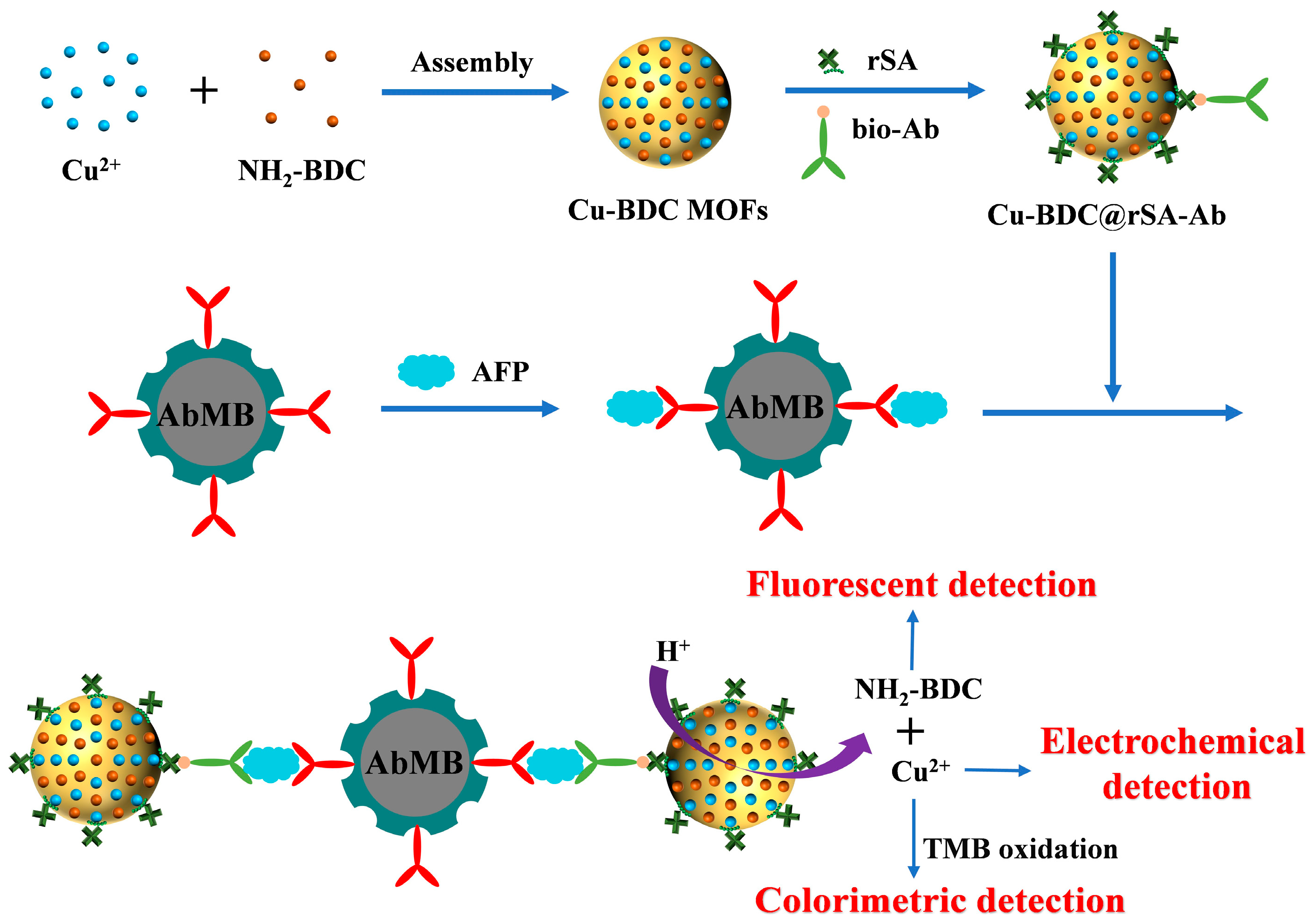
3.1. Principle of Triple-Mode Immunoassays

In this work, the recognized cancer biomarker AFP was determined as the model [29,30]. Sandwich-like immunoreactions were performed on capture antibody-covered magnetic beads (AbMB) to achieve signal-on triple-mode immunoassays of AFP with low background signals. The detection principle is depicted in Scheme 1. Cu-BDC MOFs prepared by the assembly of Cu2+ and NH2-BDC were used as multifunctional sensing materials, which have the following advantages: (1) the simple preparation and modification processes of MOFs, (2) the strong fluorescence signal of the NH2-BDC ligand, and (3) well-defined electrochemical signal and high catalytic activity of Cu2+. Site-specific and oriented immobilization of recognition elements on the solid surface can facilitate the intermolecular interactions. Peptides or proteins with multiple imidazole groups in the histidine amino acid residues can be anchored on the metal surface through the coordination interactions of unsaturated metal sites. For this consideration, many commercial proteins are prepared by recombinant techniques to endow them with hexahistidine (His6) tags for convenience of separation and purification. The non-covalent and strong interactions between the unsaturated metal sites and the His6 tags have been used for the site-specific immobilization or labeling of proteins, thereby promoting the development of various biosensors [31,32]. It has been suggested that the His6-tagged proteins can be immobilized on the surface of MOFs by metal coordination interactions [28]. Such interactions have facilitated the modification of MOFs with biorecognition elements for the design of various biosensors. Therefore, recombinant protein streptavidin (rSA) was modified on the surface of MOFs through the metal coordination interactions, which can attach biotinylated antibody (bio-Ab) and eliminate non-specific adsorption (Scheme 1). In the presence of AFP, Cu-BDC@rSA-Ab conjugates were captured by AbMB through the antigen–antibody interactions and then decomposed by an acidic solution to release NH2-BDC and free Cu2+. Thus, the target was readily determined with triple-mode signals based on the fluorescent titration of NH2-BDC, the electrochemical reduction in Cu2+, and the Cu2+-catalytic oxidation of TMB. The proposed fluorescent–electrochemical–colorimetric triple-mode biosensing platform shows great potential for different applications, since it can be easily extended to other bioassays by changing the targets and receptors.

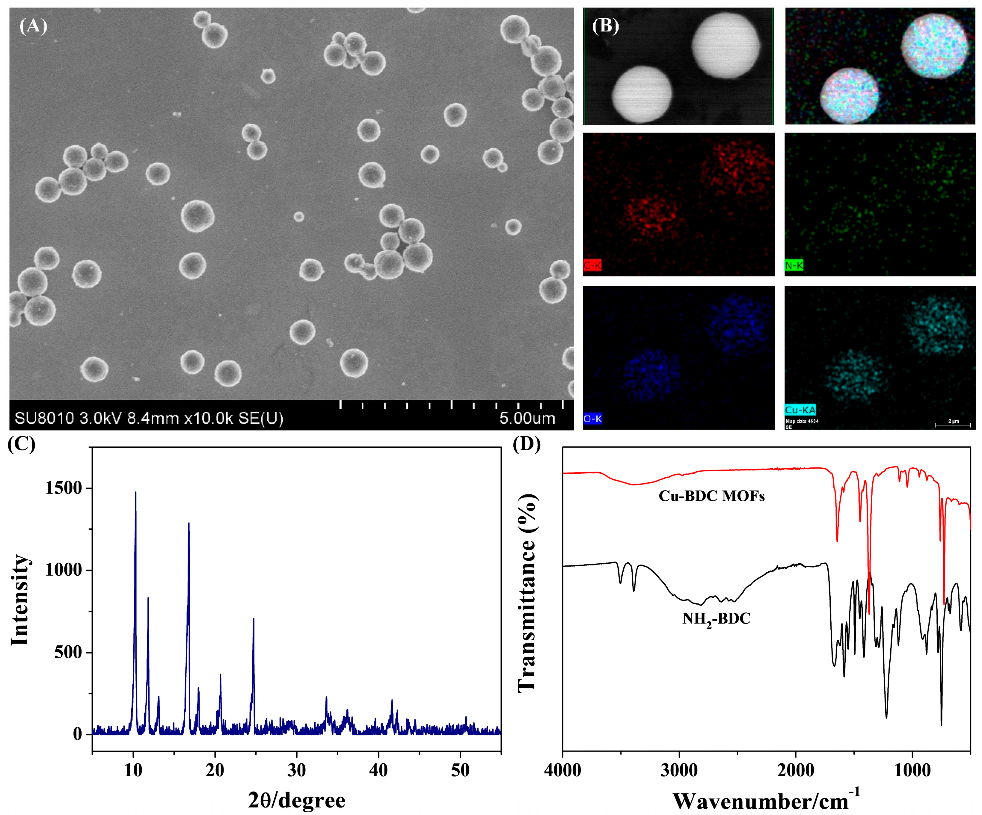
3.2. Characterization of Cu-BDC MOFs

As presented in Figure 1A, the prepared Cu-BDC showed a sphere-shaped morphology. Their elemental composition was analyzed by EDS. As shown in Figure 1B, the elemental mappings of Cu-BDC displayed the distribution of C, N, O, and Cu elements, demonstrating that the Cu-BDC MOFs were successfully prepared. The structure of Cu-BDC MOFs was characterized by XRD. As shown in Figure 1C, the diffraction peaks were in good agreement with those reported in the literature [33], indicating that the prepared Cu-BDC MOFs exhibit a high degree of crystallinity. In addition, the FT-IR spectra of Cu-BDC and pure NH2-BDC samples were collected. As shown in Figure 1D, the stretching vibration peaks of NH2-BDC from the O–H and H bonding in the band of 2500~3000 cm−1 disappeared after the coordination with Cu2+, indicating Cu2+ was coordinated by the carboxyl groups in NH2-BDC. In this work, the Cu-BDC@rSA hybrids were used as the signal labels of immunoassays. To prove that rSA was modified on the MOF surface, the size and ζ-potential changes in Cu-BDC were monitored. It was noticed that the size of Cu-BDC MOFs measured by DLS (Figure 2A) was larger than that observed by SEM. This is understandable, since the DLS size is derived from the hydration radius but not the real diameter of particles. It was found that the average size of Cu-BDC increased from 842 to 863 nm after the modification of rSA, which was accompanied by the change in ζ-potential from −21.1 to −15.6 mV (Figure 2B). The results confirmed that rSA was successfully modified on the surface of Cu-BDC MOFs. In this work, the detection antibody of bio-Ab was conjugated onto the MOF surface through the classical avidin–biotin interaction. We found that the modification of bio-Ab did not induce a significant change in the size of Cu-BDC@rSA, but the ζ-potential changed from −15.6 into −18.2 mV.

3.3. Feasibility of Triple-Mode Immunoassays

To investigate the feasible applicability of the multifunctional MOFs in fluorescent–electrochemical–colorimetric triple-mode biosensing platforms, three key issues should be elucidated: (1) whether the Cu-BDC MOFs can be decomposed under an acidic condition to release Cu2+ and NH2-BDC, (2) whether the released Cu2+ and NH2-BDC can be determined by electrochemical, colorimetric, and fluorescent assays, and (3) whether Cu2+ and NH2-BDC interfere with each other in their respective assays. As shown in Figure 3A, the morphology of Cu-BDC MOFs did not alter after the modification of rSA. However, no sphere-shaped MOFs were found when they were incubated with 10 mM HCl, suggesting that the Cu-BDC MOFs could be decomposed under an acidic condition. The HCl-treated Cu-BDC showed a fluorescence signal at 428 nm and a reduction peak at 0.08 V (black curve in Figure 3B,C), which was ascribed to NH2-BDC and free Cu2+, respectively. In addition, we found that the fluorescence intensity and peak current of the released NH2-BDC and Cu2+ in an acidic acetic acid buffer solution were higher than the respective values in a neutral phosphate-buffer solution (red curve). This result is understandable, since the fluorescence of NH2-BDC could be quenched by Cu2+ at the neutral pH due to the ligand–metal charge transfer (LMCT) and the electrochemical signal of Cu2+ can be reduced due to the Cu2+-phosphate coordination interaction. It is noted that NH2-BDC and Cu2+ did not interfere with each other in their respective fluorescent and electrochemical signals, and the TMB-based colorimetric assays are usually conducted under acidic conditions. The released NH2-BDC and Cu2+ from the decomposition of MOFs in the following triple-mode assays were determined in an acetic buffer. The well-defined reduction peak of Cu2+ and the excellent fluorescent signal of NH2-BDC implied that the Cu-BDC MOFs can be used as the signal labels of multimode immunoassays. In addition, we found that the HCl-treated Cu-BDC could catalyze the oxidation of TMB by H2O2, which can endow the triple-mode assays with more information and visual self-validation.

3.4. Sensitivity of Triple-Mode Immunoassays

To evaluate the sensitivity of this strategy, AFP was determined based on the proposed fluorescent–electrochemical–colorimetric triple-mode immunoassays. As shown in Figure 4A–C, the triple signals are proportional to AFP concentrations, indicating more AFP can facilitate the capture of more Cu-BDC MOFs and lead to the release of more NH2-BDC and Cu2+. For the fluorescent immunoassay, the linear equation between the fluorescence signal of NH2-BDC and AFP concentration within the range of 10–200 pg/mL is FL = 1.3[AFP] (pg/mL) − 2.3 (R2 = 0.988) (Figure 4D). For the electrochemical immunoassay, the peak current was linearly related to AFP concentration, ranging from 10 to 200 pg/mL with a linear equation of Ipc = 0.003[AFP] (pg/mL) + 0.04 (R2 = 0.979) (Figure 4E). For the colorimetric immunoassay, the linear relationship between the absorbance intensity and AFP concentration ranging from 1 to 100 pg/mL is Abs = 0.008[AFP] (pg/mL) + 0.065 (R2 = 0.997) (Figure 4F). The detection limits of the three methods were estimated to be 10, 10, and 1 pg/mL by determining the minimum target concentration that can be apparently distinguished with that of the background control. Compared with the fluorescence assay, the blank control for the electrochemical and colorimetric mode showed a significant background signal, which should be attributed to the free Cu2+ resulting from the buffer or atmospheric dust, since the DPV and TMB-based methods are highly sensitive for the detection of Cu2+. The fluorescent–electrochemical–colorimetric triple-mode immunoassays exhibited lower detection limits and wider linear ranges through the release-based signal amplification strategy. The sensitivity is comparable to other immunoassay platforms based on the stimuli-responsive release of signaling molecules or ions, including coumarin 153-loaded polystyrene particles for C-reactive protein detection (4.9 ng/mL) [34], thymolphthalein-loaded mesoporous silica nanoparticles or metal-polydopamine frameworks for colorimetric immunoassays of PSA (0.36 pg/mL) or AFP (2.3 pg/mL) [35,36], self-assembled TMB nanoparticles for colorimetric immunoassays of interleukin-6 (1.92 pg/mL) [37], self-assembled tetra(4-carboxyphenyl)porphyrin (TCPP) nanoparticles for fluorescent immunoassays of IgG (2.05 pM) [38], and fluorescein-loaded gold nanoflower for fluorescent–colorimetric dual-mode immunoassays of AFP (29 fg/mL) [39]. In particular, the colorimetric assay exhibited high sensitivity and simplicity, and the detection results of the three methods could be self-validated with each other. The analytical performances for AFP detection were comparable to or even better than others achieved by a single-mode immunosensing platform (Table 1). In addition, the methods are sensitive, in contrast to the previously reported dual-mode immunoassays of protein biomarkers (Table 2). The proposed triple-mode sensing platform shows great potential for different applications, since it can be easily extended to other bioassays by changing the targets and receptors. Although the proposed triple-mode biosensors show high throughput, provide multilevel detection data and reliable results, and obtain a greater amount of available information, they require three different devices to collect the detection signals. This will increase the difficulty in clinical trials when taking into account assay time, detection cost, and operation steps. Thus, portable devices with on-site detection are required to meet point of-care test detection by directly converting the target concentrations into readable multiple signals within the same reaction system. In addition, although MOFs are believed to be the next-generation sensing materials, the large-scale environment-friendly production of MOFs with multi-functionality and high stability in an aqueous solution should be considered for biosensing applications [40,41].

3.5. Selectivity and Recovery

Selectivity is a key factor in evaluating the application of biosensors in complex biological matrixes. As shown in Figure 5, serum protein BSA, protease thrombin, and different protein biomarkers (CEA, PSA, and AFP) were selected as the possible interferents to evaluate the selectivity of the proposed triple-mode immunoassays. It was found that only AFP caused large fluorescent, electrochemical, and colorimetric signals, although the interferents were tested at the concentration of 10-fold higher than that of the target. In addition, the presence of the interferences did not affect the detection of AFP. The results suggest that the proposed triple-mode immunoassays possess satisfactory selectivity and good anti-interference ability.
To further investigate the feasibility of the triple-mode sensing platforms for target detection in complex biological samples, a commercial bovine serum was analyzed with negligible effect of original AFP. Three concentrations of AFP standard samples were spiked in the serum, and then the fluorescent, electrochemical, and colorimetric signals of the triple-mode immunoassays were recorded. The final concentrations were calculated based on the corresponding linear equations. As shown in Table 3, the contents found were close to the added values, and the recoveries varied from 94.4% to 107.2% with relative standard deviations (RSDs) lower than 11%. This indicated that the recoveries obtained from the triple-mode immunoassays were acceptable. In addition, the quantification results were convincing, since the multimode detection methods can be mutually validated.

4. Conclusions

In summary, a triple-mode immunoassay platform was developed by using multifunctional Cu-BDC MOFs as the signal labels. Based on the strong fluorescent signal of NH2-BDC, the well-defined electrochemical signal of Cu2+, and the high catalytic activity of Cu2+ toward TMB oxidation, the developed fluorescent–electrochemical–colorimetric triple-mode biosensing platform exhibited excellent performances for the detection of AFP with wide linear ranges and low detection limits. In addition, the proposed triple-mode immunoassays possess improved detection accuracy and reliability through mutual validation in three detection modes, showing great potential in disease diagnosis and bioanalysis. Different detection modes can compensate for each other’s limitations. However, there still remains room for improvement in the development of triple-mode immunoassays to meet the demand of practical sensing applications. For example, multimode portable devices should be developed to enable efficient multiple-signal detection for practical applications, and advanced nanomaterials with multi-functionality, uniform morphology, and low toxicity should be explored to increase the detection accuracy and environmental friendliness of biosensors. Efforts are being made in our group to explore multiple-mode signal labels with high stability, low production cost, and excellent environmental friendliness.

Author Contributions

Conceptualization, N.X.; methodology, N.X.; investigation, N.X., C.Z. and G.L.; data curation, C.Z. and G.L.; writing—original draft preparation, N.X.; writing—review and editing, N.X.; funding acquisition, N.X. All authors have read and agreed to the published version of the manuscript.

Funding

This research was funded by the Program for Innovative Research Team of Science and Technology in Anyang Normal University (2023ASCXTD03).

Institutional Review Board Statement

Not applicable.

Informed Consent Statement

Not applicable.

Data Availability Statement

Data are contained within the article.

Conflicts of Interest

The authors declare no conflicts of interest.

References

  1. Yusa, V.; Millet, M.; Coscolla, C.; Roca, M. Analytical methods for human biomonitoring of pesticides. A review. Anal. Chim. Acta 2015, 891, 15–31. [Google Scholar] [CrossRef]
  2. Yeo, W.-H.; Kim, J.-H. Recent advances in portable biosensors for biomarker detection in body fluids. Biosensors 2020, 10, 127. [Google Scholar] [CrossRef]
  3. Sun, H.; Liu, J.; Zhang, Q.; Yang, L.; Zhou, M.; Song, D. Advances in carcinoembryonic antigen detection: A review of clinical applications and standardization. Anal. Bioanal. Chem. 2025, 417, 1751–1764. [Google Scholar] [CrossRef] [PubMed]
  4. Farka, Z.; Juřík, T.; Kovář, D.; Trnkova, L.; Skládal, P. Nanoparticle-based immunochemical biosensors and assays: Recent advances and challenges. Chem. Rev. 2017, 117, 9973–10042. [Google Scholar] [CrossRef] [PubMed]
  5. Rizzo, F. Optical immunoassays methods in protein analysis: An overview. Chemosensors 2022, 10, 326. [Google Scholar] [CrossRef]
  6. Chen, M.; Qileng, A.; Liang, H.; Lei, H.; Liu, W.; Liu, Y. Advances in immunoassay-based strategies for mycotoxin detection in food: From single-mode immunosensors to dual-mode immunosensors. Compr. Rev. Food Sci. Food Saf. 2023, 22, 1285–1311. [Google Scholar] [CrossRef]
  7. Ma, X.; Ge, Y.; Xia, N. Overview of the design and application of dual-signal immunoassays. Molecules 2024, 29, 4551. [Google Scholar] [CrossRef]
  8. Saleh, R.O.; Almajidi, Y.Q.; Mansouri, S.; Hammoud, A.; Rodrigues, P.; Mezan, S.O.; Maabreh, H.G.; Deorari, M.; Shakir, M.N.; Alasheqi, M.Q. Dual-mode colorimetric and fluorescence biosensors for the detection of foodborne bacteria. Clin. Chim. Acta 2024, 553, 117741. [Google Scholar] [CrossRef] [PubMed]
  9. Han, Q.; Wang, H.; Wang, J. Multi-mode/signal biosensors: Electrochemical integrated sensing techniques. Adv. Funct. Mater. 2024, 34, 2403122. [Google Scholar] [CrossRef]
  10. Zheng, H.; Wang, H.; Du, C.; Zhang, X.; Chen, J. Multifunctional ethyl violet@NH2-MIL-88B(Fe) hybrids: CRISPR Cas12a-assisted PEC-FL-CL triple-mode sensitive detection of HPV-16. Anal. Chem. 2024, 96, 15657–15664. [Google Scholar] [CrossRef]
  11. Gao, Y.; Han, Y.; Wang, C.; Qiang, L.; Gao, J.; Wang, Y.; Liu, H.; Han, L.; Zhang, Y. Rapid and sensitive triple-mode detection of causative SARS-CoV-2 virus specific genes through interaction between genes and nanoparticles. Anal. Chim. Acta 2021, 1154, 338330. [Google Scholar] [CrossRef] [PubMed]
  12. Meng, X.X.; Wang, J.; Diao, L.L.; Li, C.P. Construction of multi-mode photoelectrochemical immunoassays for accurate detection of cancer markers: Assisted with MOF-confined plasmonic nanozyme. Anal. Chem. 2024, 96, 1336–1344. [Google Scholar] [CrossRef]
  13. Guo, Y.; Zhang, X.; Yang, H.; Zhou, Y. Triple-readout immunoassay based on copper ion trigger for the detection of ochratoxin A. Anal. Chim. Acta 2025, 1345, 343750. [Google Scholar] [CrossRef]
  14. Ghosh, A.; Kallungal, S.F.T.; Ramaprabhu, S. 2D Metal-organic frameworks: Properties, synthesis, and applications in electrochemical and optical biosensors. Biosensors 2023, 13, 123. [Google Scholar] [CrossRef]
  15. Chang, J.; Hu, R.; Zhang, J.; Hou, T.; Li, F. Two-dimensional metal-organic framework nanozyme-mediated portable paper-based analytical device for dichlorophen assay. Biosens. Bioelectron. 2024, 255, 116271. [Google Scholar] [CrossRef] [PubMed]
  16. Leoi, M.W.N.; Zheng, X.T.; Yu, Y.; Gao, J.; Ong, D.H.S.; Koh, C.Z.H.; Chen, P.; Yang, L. Redefining metal organic frameworks in biosensors: Where are we now? ACS Appl. Mater. Interfaces 2025, 17, 13246–13278. [Google Scholar] [CrossRef]
  17. Lin, Y.; Huang, Y.; Chen, X. Recent advances in metal-organic frameworks for biomacromolecule sensing. Chemosensors 2022, 10, 412. [Google Scholar] [CrossRef]
  18. Chang, Y.; Lou, J.; Yang, L.; Liu, M.; Xia, N.; Liu, L. Design and application of electrochemical sensors with metal-organic frameworks as the electrode materials or signal tags. Nanomaterials 2022, 12, 3248. [Google Scholar] [CrossRef]
  19. Mohanty, B.; Kumari, S.; Yadav, P.; Kanoo, P.; Chakraborty, A. Metal-organic frameworks (MOFs) and MOF composites based biosensors. Coord. Chem. Rev. 2024, 519, 216102. [Google Scholar] [CrossRef]
  20. Dourandish, Z.; Tajik, S.; Beitollahi, H.; Jahani, P.M.; Nejad, F.G.; Sheikhshoaie, I.; Bartolomeo, A.D. A comprehensive review of metal–organic framework: Synthesis, characterization, and investigation of their application in electrochemical biosensors for biomedical analysis. Sensors 2022, 22, 2238. [Google Scholar] [CrossRef]
  21. Yu, X.; Wang, Y.; Zhang, J.; Liu, J.; Wang, A.; Ding, L. Recent development of copper-based nanozymes for biomedical applications. Adv. Healthc. Mater. 2024, 13, 2302023. [Google Scholar] [CrossRef] [PubMed]
  22. Xia, N.; Liu, G.; Zhang, S.; Shang, Z.; Yang, Y.; Li, Y.; Liu, L. Oxidase-mimicking peptide-copper complexes and their applications in sandwich affinity biosensors. Anal. Chim. Acta 2022, 1214, 339965. [Google Scholar] [CrossRef]
  23. Wei, J.; Zhang, D.; Zhang, L.; Ouyang, H.; Fu, Z. Alkaline hydrolysis behavior of metal-organic frameworks NH2-MIL-53(Al) employed for sensitive immunoassay via releasing fluorescent molecules. ACS Appl. Mater. Interfaces 2019, 11, 35597–35603. [Google Scholar] [CrossRef] [PubMed]
  24. Hu, X.; Wei, Z.; Sun, C.; Long, Y.; Zheng, H. Bifunctional antibody and copper-based metal-organic framework nanocomposites for colorimetric α-fetoprotein sensing. Microchim. Acta 2020, 187, 465. [Google Scholar] [CrossRef]
  25. Bunzen, H. Chemical stability of metal-organic frameworks for applications in drug delivery. ChemNanoMat 2021, 7, 998. [Google Scholar] [CrossRef]
  26. Wang, L.; Li, X.; Yang, B.; Xiao, K.; Duan, H.; Zhao, H. The chemical stability of metal-organic frameworks in water treatments: Fundamentals, effect of water matrix and judging methods. Chem. Eng. J. 2022, 450, 138215. [Google Scholar] [CrossRef]
  27. Liu, X.; Liu, S.; Jin, X.; Liu, H.; Sun, K.; Wang, X.; Li, M.; Wang, P.; Chang, Y.; Wang, T.; et al. An encounter between metal ions and natural products: Natural products-coordinated metal ions for the diagnosis and treatment of tumors. J. Nanobiotech. 2024, 22, 726. [Google Scholar] [CrossRef]
  28. Röder, R.; Preiß, T.; Hirschle, P.; Steinborn, B.; Zimpel, A.; Höhn, M.; Rädler, J.O.; Bein, T.; Wagner, E.; Wuttke, S.; et al. Multifunctional nanoparticles by coordinative self-assembly of His tagged units with metal−organic frameworks. J. Am. Chem. Soc. 2017, 139, 2359–2368. [Google Scholar] [CrossRef] [PubMed]
  29. Ramachandran, L.; Rub, F.A.; Hajja, A.; Alodhaibi, I.; Arai, M.; Alfuwais, M.; Makhzoum, T.; Yaqinuddin, A.; Al-Kattan, K.; Broering, D.C.; et al. Biosensing of alpha-fetoprotein: A key direction toward the early detection and management of hepatocellular carcinoma. Biosensors 2024, 14, 235. [Google Scholar] [CrossRef]
  30. Shan, C.-W.; Chen, Z.; Han, G.-C.; Feng, X.-Z.; Kraatz, H.-B. Electrochemical immuno-biosensors for the detection of the tumor marker alpha-fetoprotein: A review. Talanta 2024, 271, 125638. [Google Scholar] [CrossRef]
  31. Zhu, L.; Chang, Y.; Li, Y.; Qiao, M.; Liu, L. Biosensors based on the binding events of nitrilotriacetic acid–metal complexes. Biosensors 2023, 13, 507. [Google Scholar] [CrossRef] [PubMed]
  32. Chang, Y.; Liu, G.; Li, S.; Liu, L.; Song, Q. Biorecognition element-free electrochemical detection of recombinant glycoproteins using metal-organic frameworks as signal tags. Anal. Chim. Acta 2023, 1273, 341540. [Google Scholar] [CrossRef]
  33. Wang, S.; Deng, W.; Yang, L.; Tan, Y.; Xie, Q.; Yao, S. Copper-based metal–organic framework nanoparticles with peroxidase-like activity for sensitive colorimetric detection of staphylococcus aureus. ACS Appl. Mater. Interfaces 2017, 9, 24440–24445. [Google Scholar] [CrossRef]
  34. Geißler, D.; Wegner, K.D.; Fischer, C.; Resch-Genger, U. Exploring simple particle-based signal amplification strategies in a heterogeneous sandwich immunoassay with optical detection. Anal. Chem. 2024, 96, 5078–5085. [Google Scholar] [CrossRef]
  35. Shao, F.; Zhang, L.; Jiao, L.; Wang, X.; Miao, L.; Li, H.; Zhou, F. Enzyme-free immunosorbent assay of prostate specific antigen amplified by releasing pH indicator molecules entrapped in mesoporous silica nanoparticles. Anal. Chem. 2018, 90, 8673–8679. [Google Scholar] [CrossRef]
  36. Ren, R.; Cai, G.; Yu, Z.; Zeng, Y.; Tang, D. Metal-polydopamine framework: An innovative signal-generation tag for colorimetric immunoassay. Anal. Chem. 2018, 90, 11099–11105. [Google Scholar] [CrossRef] [PubMed]
  37. Jiao, L.; Yan, H.; Xu, W.; Wu, Y.; Gu, W.; Li, H.; Du, D.; Lin, Y.; Zhu, C. Self-assembly of all-inclusive allochroic nanoparticles for the improved ELISA. Anal. Chem. 2019, 91, 8461–8465. [Google Scholar] [CrossRef] [PubMed]
  38. Gibson, L.E.; Wright, D.W. Sensitive method for biomolecule detection utilizing signal amplification with porphyrin nanoparticles. Anal. Chem. 2016, 88, 5928–5933. [Google Scholar] [CrossRef]
  39. Zhou, Y.; Huang, X.; Xiong, S.; Li, X.; Zhan, S.; Zeng, L.; Xiong, Y. Dual-mode fluorescent and colorimetric immunoassay for the ultrasensitive detection of alpha-fetoprotein in serum samples. Anal. Chim. Acta 2018, 1038, 112–119. [Google Scholar] [CrossRef]
  40. Xie, Z.; Hou, J. Industrialising metal−organic frameworks bridging laboratory innovation and future applications. Ind. Eng. Chem. Res. 2025, 64, 7941–7955. [Google Scholar] [CrossRef]
  41. Salehipour, M.; Rezaei, S.; Rezaei, M.; Yazdani, M.; Mogharabi-Manzari, M. Opportunities and challenges in biomedical applications of metal–organic frameworks. J. Inorg. Organomet. Polym. Mater. 2021, 31, 4443–4462. [Google Scholar] [CrossRef]
  42. Sun, J.; Hu, T.; Chen, C.; Zhao, D.; Yang, F.; Yang, X. Fluorescence immunoassay system via enzyme-enabled in situ synthesis of fluorescent silicon nanoparticles. Anal. Chem. 2016, 88, 9789–9795. [Google Scholar] [CrossRef] [PubMed]
  43. Lin, Z.; Li, M.; Lv, S.; Zhang, K.; Lu, M.; Tang, D. In situ synthesis of fluorescent polydopamine nanoparticles coupled with enzyme-controlled dissolution of MnO2 nanoflakes for a sensitive immunoassay of cancer biomarkers. J. Mater. Chem. B 2017, 5, 8506–8513. [Google Scholar] [CrossRef] [PubMed]
  44. Chen, C.; Zhao, J.; Lu, Y.; Sun, J.; Yang, X. Fluorescence immunoassay based on the phosphate-triggered fluorescence turn-on detection of alkaline phosphatase. Anal. Chem. 2018, 90, 3505–3511. [Google Scholar] [CrossRef]
  45. Sun, Z.H.; Zhang, X.X.; Xu, D.; Liu, J.; Yu, R.J.; Jing, C.; Han, H.X.; Ma, W. Silver-amplified fluorescence immunoassay via aggregation-induced emission for detection of disease biomarker. Talanta 2021, 225, 121963–121968. [Google Scholar] [CrossRef]
  46. Wang, Y.-W.; Chen, L.; Liang, M.; Xu, H.; Tang, S.; Yang, H.-H.; Song, H. Sensitive fluorescence immunoassay of alpha-fetoprotein through copper ions modulated growth of quantum dots in-situ. Sens. Actuat. B Chem. 2017, 247, 408–413. [Google Scholar] [CrossRef]
  47. Yang, T.; Li, C.M.; He, J.H.; Chen, B.; Li, Y.F.; Huang, C.Z. Ratiometrically fluorescent electrospun nanofibrous film as a Cu2+-mediated solid-phase immunoassay platform for biomarkers. Anal. Chem. 2018, 90, 9966–9974. [Google Scholar] [CrossRef]
  48. Gao, F.; Chang, Y.; Xia, N.; Li, Y.; Liu, L. Multifunctional self-assembled nanoparticles serving as the signal labels of fluorescent immunoassays. Microchem. J. 2024, 207, 111921. [Google Scholar] [CrossRef]
  49. Yu, L.; Cui, X.; Li, H.; Lu, J.; Kang, Q.; Shen, D. A ratiometric electrochemical sensor for multiplex detection of cancer biomarkers using bismuth as an internal reference and metal sulfide nanoparticles as signal tags. Analyst 2019, 144, 4073–4080. [Google Scholar] [CrossRef]
  50. Meng, W.; Li, M.; Zhang, Y. Adriamycin coated silica microspheres as labels for cancer biomarker alpha-fetoprotein detection. Anal. Methods 2021, 13, 2665–2670. [Google Scholar] [CrossRef]
  51. Huang, Y.; Liang, R.; Yu, H.; An, S.; Han, J.; Xie, G.; Chen, S. Thi-Au-Fe3O4 confined in ZIF-8 nanoreactor as signal-amplifying tag for constructing high-efficiency electrochemical platform. Sensor. Actuator. B Chem. 2020, 305, 127496. [Google Scholar] [CrossRef]
  52. Li, W.; Yang, Y.; Ma, C.; Song, Y.; Qiao, X.; Hong, C. A sandwich-type electrochemical immunosensor for ultrasensitive detection of multiple tumor markers using an electrical signal difference strategy. Talanta 2020, 219, 121322. [Google Scholar] [CrossRef]
  53. Li, L.; Wei, Y.; Zhang, S.; Chen, X.; Shao, T.; Feng, D. Electrochemical immunosensor based on metal ions functionalized CNSs@Au NPs nanocomposites as signal amplifier for simultaneous detection of triple tumor markers. J. Electroanal. Chem. 2021, 880, 114882. [Google Scholar] [CrossRef]
  54. Lv, S.; Zhang, K.; Zhu, L.; Tang, D.; Niessner, R.; Knopp, D. H2-Based Electrochemical Biosensor with Pd Nanowires@ZIF-67 Molecular Sieve Bilayered Sensing Interface for Immunoassay. Anal. Chem. 2019, 91, 12055–12062. [Google Scholar] [CrossRef]
  55. Li, Y.; Wu, J.; Zhang, C.; Chen, Y.; Wang, Y.; Xie, M. Manganese dioxide nanoparticle-based colorimetric immunoassay for the detection of alpha-fetoprotein. Microchim. Acta 2017, 184, 2767–2774. [Google Scholar] [CrossRef]
  56. Deng, X.; Fang, Y.; Lin, S.; Cheng, Q.; Liu, Q.; Zhang, X. Porphyrin-based porous organic frameworks as a biomimetic catalyst for highly efficient colorimetric immunoassay. ACS Appl. Mater. Interfaces 2017, 9, 3514–3523. [Google Scholar] [CrossRef]
  57. Peng, M.-P.; Ma, W.; Long, Y.-T. Alcohol dehydrogenase-catalyzed gold nanoparticle seed-mediated growth allows reliable detection of disease biomarkers with the naked eye. Anal. Chem. 2015, 87, 5891–5896. [Google Scholar] [CrossRef]
  58. Su, B.; Xu, H.; Xie, G.; Chen, Q.; Sun, Z.; Cao, H.; Liu, X. Generation of ananobody-alkaline phosphatase fusion and its application in an enzyme cascade-amplified immunoassay for colorimetric detection of alpha fetoprotein in human serum. Spectrochim. Acta Part A Mol. Biomol. Spectrosc. 2021, 262, 120088. [Google Scholar] [CrossRef]
  59. Sun, C.; Zhang, X.; Tang, M.; Liu, L.; Shi, Y.; Gao, C.; Liao, B.; Zheng, H. New optical method for the determination of β-galactosidase and α-fetoprotein based on oxidase-like activity of fluorescein. Talanta 2019, 194, 164–170. [Google Scholar] [CrossRef]
  60. Kim, J.U.; Kim, J.M.; Thamilselvan, A.; Nam, K.H.; Kim, M.I. Colorimetric and electrochemical dual-mode detection of thioredoxin 1 based on the efficient peroxidase-mimicking and electrocatalytic property of Prussian Blue nanoparticles. Biosensors 2024, 14, 185. [Google Scholar] [CrossRef]
  61. Seddaoui, N.; Attaallah, R.; Amine, A. Development of an optical immunoassay based on peroxidase-mimicking Prussian blue nanoparticles and a label-free electrochemical immunosensor for accurate and sensitive quantification of milk species adulteration. Microchim. Acta 2022, 189, 209–222. [Google Scholar] [CrossRef] [PubMed]
  62. Al-Ogaidi, I.; Aguilar, Z.P.; Suri, S.; Gou, H.; Wu, N. Dual detection of cancer biomarker CA125 using absorbance and electrochemical methods. Analyst 2013, 138, 5647–5653. [Google Scholar] [CrossRef]
  63. Hong, W.; Lee, S.; Cho, Y. Dual-responsive immunosensor that combines colorimetric recognition and electrochemical response for ultrasensitive detection of cancer biomarkers. Biosens. Bioelectron. 2016, 86, 920–926. [Google Scholar] [CrossRef] [PubMed]
  64. Choi, H.; Son, S.E.; Hur, W.; Tran, V.K.; Lee, H.B.; Park, Y.; Han, D.K.; Seong, G.H. Electrochemical immunoassay for determination of glycated albumin using nanozymes. Sci. Rep. 2020, 10, 9513–9524. [Google Scholar] [CrossRef]
  65. Xu, X.H.; Xia, J.; Song, S.; Liu, Y.; Chen, J.; Zhong, S.; Chen, H.; Zhang, Z.H. Dual-signal immuno-competitive determination of brain natriuretic peptide based on magnetic nanozyme. Electroanalysis 2023, 35, e202200500–e202200508. [Google Scholar] [CrossRef]
  66. Xie, W.; Tian, M.; Luo, X.; Jiang, Y.; He, N.; Liao, X.; Liu, Y. A dual-mode fluorescent and colorimetric immunoassay based on in situ ascorbic acid-induced signal generation from metal-organic frameworks. Sens. Actuat. B Chem. 2020, 302, 127180–127186. [Google Scholar] [CrossRef]
  67. Li, M.; Wang, Y.; Hu, H.; Feng, Y.; Zhu, S.; Li, C.; Feng, N. A dual-readout sandwich immunoassay based on biocatalytic perovskite nanocrystals for detection of prostate specific antigen. Biosens. Bioelectron. 2022, 203, 113979. [Google Scholar] [CrossRef]
  68. Zhuge, W.; Tan, X.; Zhang, R.; Li, H.; Zheng, G. Fluorescent and colorimetric immunoassay of nuclear matrix protein 22 enhanced by porous Pd nanoparticles. Chin. Chem. Lett. 2019, 30, 1307–1309. [Google Scholar] [CrossRef]
  69. Miao, L.; Jiao, L.; Tang, Q.; Li, H.; Zhang, L.; Wei, Q. A nanozyme-linked immunosorbent assay for dual-modal colorimetric and ratiometric fluorescent detection of cardiac troponin I. Sens. Actuat. B Chem. 2019, 288, 60–64. [Google Scholar] [CrossRef]
  70. Zhao, J.; Wang, S.; Lu, S.; Bao, X.; Sun, J.; Yang, X. An enzyme cascade-triggered fluorogenic and chromogenic reaction applied in enzyme activity assay and immunoassay. Anal. Chem. 2018, 90, 7754–7760. [Google Scholar] [CrossRef]
  71. Chen, C.; Zhao, D.; Wang, B.; Ni, P.; Jiang, Y.; Zhang, C.; Yang, F.; Lu, Y.; Sun, J. Alkaline phosphatase-triggered in situ formation of silicon-containing nanoparticles for a fluorometric and colorimetric dual-channel immunoassay. Anal. Chem. 2020, 92, 4639–4646. [Google Scholar] [CrossRef] [PubMed]
  72. Priyadarshini, E.; Rawat, K.; Bohidar, H.B.; Rajamani, P. Dual-probe (colorimetric and fluorometric) detection of ferritin using antibody-modified gold@carbon dot nanoconjugates. Microchim. Acta 2019, 186, 687–695. [Google Scholar] [CrossRef] [PubMed]
  73. Gao, Y.; Wu, Y.; Huang, P.; Wu, F.-Y. Carbon dot-encapsulated plasmonic core-satellite nanoprobes for sensitive detection of cancer biomarkers via dual-mode colorimetric and fluorometric immunoassay. ACS Appl. Nano Mater. 2022, 5, 11539–11548. [Google Scholar] [CrossRef]
  74. Zhu, D.; Hu, Y.; Zhang, X.J.; Yang, X.T.; Tang, Y.Y. Colorimetric and fluorometric dual-channel detection of α-fetoprotein based on the use of ZnS-CdTe hierarchical porous nanospheres. Microchim. Acta 2019, 186, 124–132. [Google Scholar] [CrossRef]
Scheme 1. A schematic representation of the fluorescent–electrochemical–colorimetric triple-mode immunoassays.
Biosensors 15 00376 sch001
Figure 1. SEM image (A), EDS elemental mappings (B), XRD pattern (C), and FT-IR spectrum (D) of Cu-BDC MOFs.
Biosensors 15 00376 g001
Figure 2. DLS (A) and ζ-potential (B) of Cu-BDC MOFs.
Biosensors 15 00376 g002
Figure 3. (A) SEM images of Cu-BDC MOFs before (left) and after (right) decomposition by 10 mM HCl. Fluorescence spectra (B) and DPV curves (C) of HCl-treated Cu-BDC in pH 4.0 acetic acid and pH 7.4 phosphate buffer, respectively.
Biosensors 15 00376 g003
Figure 4. Fluorescence spectra (A), DPV curves (B), and UV-vis spectra (C) for the detection of different concentrations of AFP. The inset in (C) depicts the solution color change in the colorimetric mode. The calibration plots of DPV current (D), fluorescence intensity (E), and absorbance intensity (F) versus AFP concentration. The inserts show the linear portions of the calibration curves.
Biosensors 15 00376 g004
Figure 5. Selectivity of the fluorescent–electrochemical–colorimetric triple-mode immunoassays, shown in Panel (AC), respectively. Samples 1~7: buffer blank, BSA, thrombin, CEA, PSA, AFP, and the mixture of 1~6. The concentration of AFP was 100 pg/mL and that of other proteins was 1 ng/mL.
Biosensors 15 00376 g005
Table 1. Analytical performances of different sandwich-like immunoassay methods for the detection of AFP.
MethodSignal LabelSignal OutputLinear Range
(ng/mL)
Detection Limit
(pg/mL)
Ref.
FLALP/Si NPsEnzyme catalysis1~601000[42]
FLAOX–AuNPs/Mn2+Enzyme catalysis0.05~2017.3[43]
FLALP/calcein-Ce3+Enzyme catalysis1~4041[44]
FLAgNPs/TPE-4TASignal probe0.01~5000 42[45]
FLCuO NPs/CdS QDsSignal probe1~80450[46]
FLCuO NPs/CdS QDs/ENFFsSignal probe0.01~2008.3 [47]
FLbio-PyFNPsSignal probe0.001~2.50.5 [48]
ECCdS QDsElectrooxidation0.001~10.11[49]
ECADM@AuNPs@SiO2Electrooxidation0.0005~750.17[50]
ECThi-Au-Fe3O4@ZIF-8 ElectroreductionNot reported0.003[51]
ECMnO2 nanosheetsElectrocatalysis0.005~1001[52]
ECCNSs@AuNPs@Zn2+Electrocatalysis0.010~802.6[53]
ECDPNsElectrocatalysis0.1~5040[54]
ColorMnO2 NPsNanocatalysis6.25~40022[55]
ColorFePor-TFPA-COPNanocatalysis0.005~1001[56]
ColorADHEnzyme catalysisNot reported1[57]
ColorALPEnzyme catalysis1250~10,000148[58]
Colorβ-galEnzyme catalysis0.1~10080[59]
ColorMPDA@TPColor indicator0.01~12.3[36]
Abbreviations: FL, fluorescence. EC, electrochemical. ALP, alkaline phosphatase. AOX, ascorbate oxidase. TPE-4TA, tetrazole-tagged luminogen. ENFFs, electrospun nanofibrous films. bio-PyFNPs, biotinylated pyrene–phenylalanine nanoparticles. QDs, quantum dots. CNSs, carbon nanospheres. AuNPs, gold nanoparticles. DPNs, dendritic Pt nanoparticles. ADM, adriamycin. Thi, thionine. ZIF-8, zeolitic imidazolate framework-8. MnO2 NPs, manganese dioxide nanoparticles. FePor-TFPA-COP, iron porphyrin-based covalent organic polymer. ADH, halcoholde hydrogenase. MPDA@TP, thymolphthalein-modified metal–polydopamine framework. β-gal, β-galactosidase.
Table 2. The analytical performances of colorimetric–electrochemical and colorimetric–fluorescent dual-mode immunoassays for the detection of protein biomarkers.
MethodSignal LabelTargetLinear RangeDetection LimitRef.
Colorimetric–ElectrochemicalPBNPsTRX110~50 ng/mL9 and 6.5 ng/mL[60]
PBNPsIgG0.5~10 µg/mL34 ng/mL[61]
ALPCA1255~1000 and 50~1000 U/mL1.3 and 40 U/mL[62]
HRP-Ppy NPsPSA0.001~40 ng/mL0.8 and 0.7 pg/mL[63]
uPtNZsGA0.01~5 and 0.005~10 mg/mL9.2 and 3.8 µg/mL[64]
H-AuNPsBNP0.001~0.2 and 5~25 ng/mL 0.03 and 80.3 pg/mL[65]
Colorimetric–FluorescentFe-MOFsPSA1~20 and 0~15 ng/mL180 pg/mL[66]
CsPbBr3 NCsPSA0.1~15 and 0.01~80 ng/mL290 and 81 pg/mL[67]
porous Pd NPsNMP220.001~0.5 ng/mL0.35 and 0.31 pg/mL[68]
nanoceriacTnI0.001~10 ng/mL227 and 413 fg/mL[69]
ALPcTnI0.2~80 and 0.05~4 ng/mL60 and 15 ng/mL[70]
ALPPSA0.02~28 and 0.02~20 ng/mL4.1 and 9.6 pg/mL[71]
Au@CDferritin1~160 and 1~120 ng/mL20 and 64 ng/mL[72]
GOxPSA0.005~20 ng/mL2.3 and 0.84 ng/mL[73]
QDs/ZnS NSsAFP0.05~12 ng/mL7 and 10 pg/mL[74]
AuNF@FluoresceinAFP5~5000 and 0.01~10 pg/mL17.7 and 0.029 fg/mL[39]
Abbreviations: PBNPs, Prussian blue nanoparticles. TRX1, thioredoxin 1. IgG, immunoglobulin G. ALP, alkaline phosphatase. CA125, carbohydrate antigen 125. HRP, horseradish peroxidase. Ppy NPs, polypyrrole nanoparticles. PSA, prostate-specific antigen. uPtNZs, urchin-like Pt nanozymes; GA, glycated albumin. H-AuNPs, hemin-modified gold nanoparticles; BNP, brain natriuretic peptide. Fe-MOFs, Fe(III)-containing metal–organic frameworks. CsPbBr3 NCs, CsPbBr3 perovskite nanocrystals. NMP22, nuclear matrix protein 22. cTnI, cardiac troponin I. Au@CD, gold@carbon dot. GOx, glucose oxidase. QDs/ZnS NSs, ZnS nanospheres modified with CdTe quantum dots; AFP, alpha-fetoprotein. AuNF, gold nanoflower.
Table 3. The results for the assays of AFP in serum samples.
Added
(pg/mL)
Found by Fluorescence
(pg/mL)
Found by DPV
(pg/mL)
Found by UV-vis
(pg/mL)
25.025.6 ± 2.524.5 ± 2.626.8 ± 2.8
50.048.8 ± 4.647.2 ± 4.850.2 ± 5.2
100.0103.5 ± 9.8101.5 ± 10.2104.5 ± 10.8
Disclaimer/Publisher’s Note: The statements, opinions and data contained in all publications are solely those of the individual author(s) and contributor(s) and not of MDPI and/or the editor(s). MDPI and/or the editor(s) disclaim responsibility for any injury to people or property resulting from any ideas, methods, instructions or products referred to in the content.

