Luminescent Hydroxyapatite Doped with Rare Earth Elements for Biomedical Applications


Abstract
:1. Introduction
2. Hydroxyapatite Doped with Photoluminescent Elements
2.1. Hydroxyapatite Doped with Terbium
2.2. Hydroxyapatite Doped with Erbium
2.3. Hydroxyapatite Doped with Europium
2.4. Hydroxyapatite Doped with Lanthanum
2.5. Hydroxyapatite Doped with Dysprosium
3. Conclusions
Author Contributions
Funding
Conflicts of Interest
References
- Qi, S.; Huang, Y.; Li, Y.; Cai, P.; Kim, S.I.; Seo, H.J. Probe spectrum measurements of Eu3+ ions as a relevant tool for monitoring in vitro hydroxyapatite formation in a new borate biomaterial. J. Mater. Chem. B 2014, 2, 6387–6396. [Google Scholar] [CrossRef]
- Zhang, C.; Li, C.; Huang, S.; Hou, Z.; Cheng, Z.; Yang, P.; Peng, C.; Lin, J. Self-activated luminescent and mesoporous strontium hydroxyapatite nanorods for drug delivery. Biomaterials 2010, 31, 3374–3383. [Google Scholar] [CrossRef] [PubMed]
- Bach, L.G.; Cao, X.T.; Islam, M.; Kim, H.G.; Lim, K.T. Combination of surface initiated reversible addition fragmentation chain transfer polymerization, thiol-ene click chemistry and coordination chemistry for the fabrication of a novel photoluminescent hydroxyapatite nanohybrids. J. Nanosci. Nanotechnol. 2015, 15, 5897–5900. [Google Scholar] [CrossRef] [PubMed]
- Liu, H.; Chen, F.; Xi, P.; Chen, B.; Huang, L.; Cheng, J.; Shao, C.; Wang, J.; Bai, D.; Zeng, Z. Biocompatible Fluorescent Hydroxyapatite: Synthesis and Live Cell Imaging Applications. J. Phys. Chem. C 2011, 115, 18538–18544. [Google Scholar] [CrossRef]
- Huang, K.; Martí, A.A. Optimizing the Sensitivity of Photoluminescent Probes Using Time-Resolved Spectroscopy: A Molecular Beacon Case Study. Anal. Chem. 2012, 84, 8075–8082. [Google Scholar] [CrossRef] [PubMed]
- Sánchez Lafarga, A.K.; Pacheco Moisés, F.P.; Gurinov, A.; Ortiz, G.G.; Carbajal Arízaga, G.G. Dual responsive dysprosium-doped hydroxyapatite particles and toxicity reduction after functionalization with folic and glucuronic acids. Mater. Sci. Eng. C 2015, 48, 541–547. [Google Scholar] [CrossRef] [PubMed]
- Fu, D.-L.; Jiang, Q.-H.; He, F.-M.; Yang, G.-L.; Liu, L. Fluorescence microscopic analysis of bone osseointegration of strontium-substituted hydroxyapatite implants. J. Zhejiang Univ. Sci. B 2012, 13, 364–371. [Google Scholar] [CrossRef] [PubMed]
- Tsai, S.-W.; Huang, S.-S.; Yu, W.-X.; Hsu, Y.-W.; Hsu, F.-Y. Fabrication and characteristics of porous hydroxyapatite-CaO composite nanofibers for biomedical applications. Nanomaterials 2018, 8, 570. [Google Scholar] [CrossRef] [PubMed]
- Alshemary, A.Z.; Akram, M.; Goh, Y.-F.; Abdul Kadir, M.R.; Abdolahi, A.; Hussain, R. Structural characterization, optical properties and in vitro bioactivity of mesoporous erbium-doped hydroxyapatite. J. Alloys Compd. 2015, 645, 478–486. [Google Scholar] [CrossRef]
- Boanini, E.; Cassani, M.; Rubini, K.; Boga, C.; Bigi, A. (9R)-9-Hydroxystearate-Functionalized Anticancer Ceramics Promote Loading of Silver Nanoparticles. Nanomaterials 2018, 8, 390. [Google Scholar] [CrossRef]
- Dorozhkin, S.V. Calcium Orthophosphates in Nature, Biology and Medicine. Materials 2009, 2, 399. [Google Scholar] [CrossRef]
- Liu, C.; Ji, X.; Cheng, G. Template synthesis and characterization of highly ordered lamellar hydroxyapatite. Appl. Surf. Sci. 2007, 253, 6840–6843. [Google Scholar] [CrossRef]
- Yang, Z.; Huang, Y.; Chen, S.-T.; Zhao, Y.-Q.; Li, H.-L.; Hu, Z.-A. Template synthesis of highly ordered hydroxyapatite nanowire arrays. J. Mater. Sci. 2005, 40, 1121–1125. [Google Scholar] [CrossRef]
- Wang, P.; Li, C.; Gong, H.; Jiang, X.; Wang, H.; Li, K. Effects of synthesis conditions on the morphology of hydroxyapatite nanoparticles produced by wet chemical process. Powder Technol. 2010, 203, 315–321. [Google Scholar] [CrossRef]
- Liu, Y.; Hou, D.; Wang, G. A simple wet chemical synthesis and characterization of hydroxyapatite nanorods. Mater. Chem. Phys. 2004, 86, 69–73. [Google Scholar] [CrossRef]
- Madhumathi, K.; Shalumon, K.; Rani, V.D.; Tamura, H.; Furuike, T.; Selvamurugan, N.; Nair, S.; Jayakumar, R. Wet chemical synthesis of chitosan hydrogel–hydroxyapatite composite membranes for tissue engineering applications. Int. J. Biol. Macromol. 2009, 45, 12–15. [Google Scholar] [CrossRef] [PubMed]
- Murugan, R.; Ramakrishna, S. Crystallographic study of hydroxyapatite bioceramics derived from various sources. Cryst. Growth Des. 2005, 5, 111–112. [Google Scholar] [CrossRef]
- Ripamonti, U. The morphogenesis of bone in replicas of porous hydroxyapatite obtained from conversion of calcium carbonate exoskeletons of coral. J. Bone Jt. Surg. Am. 1991, 73, 692–703. [Google Scholar] [CrossRef] [PubMed]
- Ripamonti, U.; Ma, S.-S.; Reddi, A. Osteogenin, a bone morphogenetic protein, adsorbed on porous hydroxyapatite substrata, induces rapid bone differentiation in calvarial defects of adult primates. Plast. Reconstr. Surg. 1992, 90, 382–393. [Google Scholar] [CrossRef] [PubMed]
- Barbeck, M.; Udeabor, S.; Lorenz, J.; Schlee, M.; Holthaus, M.G.; Raetscho, N.; Choukroun, J.; Sader, R.; Kirkpatrick, C.J.; Ghanaati, S. High-temperature sintering of xenogeneic bone substitutes leads to increased multinucleated giant cell formation: In vivo and preliminary clinical results. J. Oral Implantol. 2015, 41, e212–e222. [Google Scholar] [CrossRef]
- Wei, J.-Q.; Liu, Y.; Zhang, X.-H.; Liang, W.-W.; Zhou, T.-F.; Zhang, H.; Deng, X.-L. Enhanced critical-sized bone defect repair efficiency by combining deproteinized antler cancellous bone and autologous BMSCs. Chin. Chem. Lett. 2017, 28, 845–850. [Google Scholar] [CrossRef]
- Arita, I.; Castano, V.; Wilkinson, D. Synthesis and processing of hydroxyapatite ceramic tapes with controlled porosity. J. Mater. Sci. 1995, 6, 19–23. [Google Scholar] [CrossRef]
- Bhattacharjee, B.N.; Mishra, V.K.; Rai, S.B.; Parkash, O.; Kumar, D. Study of Morphological Behavior of Hydroxyapatite, EDTA Hydroxyapatite and Metal Doped EDTA Hydroxyapatite Synthesized by Chemical Co-Precipitation Method via Hydrothermal Route. In Proceedings of the Key Engineering Materials, Košice, Slovakia, 6–8 November 2017; pp. 210–214. [Google Scholar]
- Pham, V.-H.; Van, H.N.; Tam, P.D.; Ha, H.N.T. A novel 1540 nm light emission from erbium doped hydroxyapatite/β-tricalcium phosphate through co-precipitation method. Mater. Lett. 2016, 167, 145–147. [Google Scholar] [CrossRef]
- Pandi, K.; Viswanathan, N. In situ precipitation of nano-hydroxyapatite in gelatin polymatrix towards specific fluoride sorption. Int. J. Biol. Macromol. 2015, 74, 351–359. [Google Scholar] [CrossRef] [PubMed]
- Ben-Arfa, B.A.; Salvado, I.M.M.; Ferreira, J.M.; Pullar, R.C. Novel route for rapid sol-gel synthesis of hydroxyapatite, avoiding ageing and using fast drying with a 50-fold to 200-fold reduction in process time. Mater. Sci. Eng. 2017, 70, 796–804. [Google Scholar] [CrossRef] [PubMed]
- Asri, R.; Harun, W.; Hassan, M.-A.; Ghani, S.; Buyong, Z. A review of hydroxyapatite-based coating techniques: Sol–gel and electrochemical depositions on biocompatible metals. J. Mech. Behav. Biomed. Mater. 2016, 57, 95–108. [Google Scholar] [CrossRef] [PubMed]
- Tilkin, R.; Regibeau, N.; Grandfils, C.; Lambert, S. Optimization of hydroxyapatite synthesis via sol-gel process for bone reconstruction application. In Proceedings of the 19th international Sol-Gel Conference, Liège, Belgium, 3–8 September 2017. [Google Scholar]
- Türk, S.; Altınsoy, I.; ÇelebiEfe, G.; Ipek, M.; Özacar, M.; Bindal, C. Microwave–assisted biomimetic synthesis of hydroxyapatite using different sources of calcium. Mater. Sci. Eng. 2017, 76, 528–535. [Google Scholar] [CrossRef]
- Pokhrel, S. Hydroxyapatite: Preparation, Properties and Its Biomedical Applications. Adv. Chem. Eng. Sci. 2018, 8, 225. [Google Scholar] [CrossRef]
- Hu, C.; Aindow, M.; Wei, M. Focused ion beam sectioning studies of biomimetic hydroxyapatite coatings on Ti-6Al-4V substrates. Surf. Coat. Technol. 2017, 313, 255–262. [Google Scholar] [CrossRef]
- Cho, J.S.; Lee, J.-C.; Chung, S.H.; Seo, J.K.; Rhee, S.-H. Effect of grain size and density of spray-pyrolyzed hydroxyapatite particles on the sinterability of hydroxyapatite disk. Ceram. Int. 2014, 40, 6691–6697. [Google Scholar] [CrossRef]
- Cho, J.S.; Lee, J.C.; Rhee, S.H. Effect of precursor concentration and spray pyrolysis temperature upon hydroxyapatite particle size and density. J. Biomed. Mater. Res. 2016, 104, 422–430. [Google Scholar] [CrossRef]
- Zhang, M.; Liu, J.-K.; Miao, R.; Li, G.-M.; Du, Y.-J. Preparation and characterization of fluorescence probe from assembly hydroxyapatite nanocomposite. Nanoscale Res. Lett. 2010, 5, 675. [Google Scholar] [CrossRef]
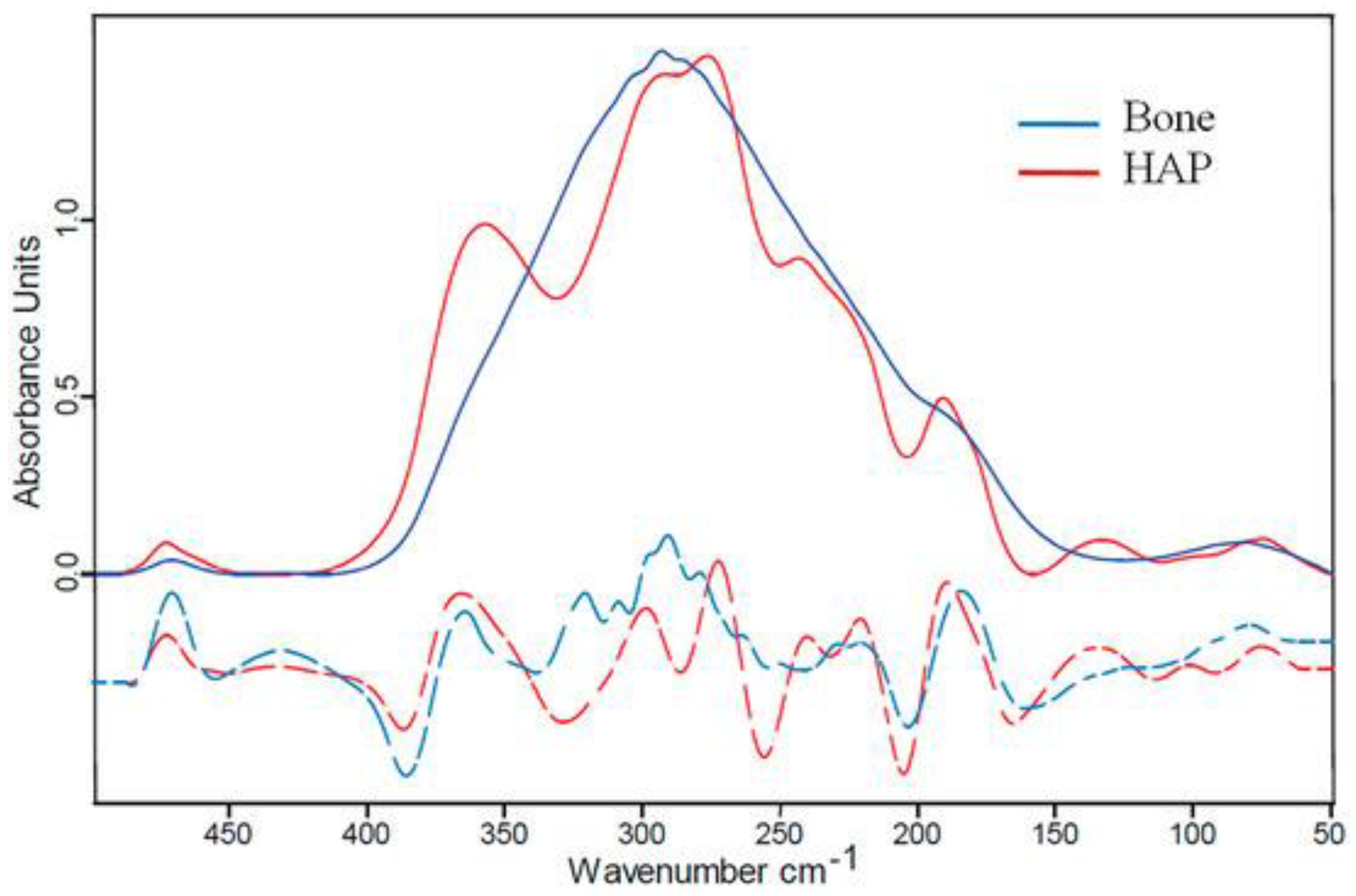
- Schuetz, R.; Fix, D.; Schade, U.; Aziz, E.F.; Timofeeva, N.; Weinkamer, R.; Masic, A. Anisotropy in Bone Demineralization Revealed by Polarized Far-IR Spectroscopy. Molecules 2015, 20, 5835. [Google Scholar] [CrossRef] [PubMed]
- Tite, T.; Popa, A.-C.; Balescu, L.M.; Bogdan, I.M.; Pasuk, I.; Ferreira, J.M.F.; Stan, G.E. Cationic Substitutions in Hydroxyapatite: Current Status of the Derived Biofunctional Effects and Their In Vitro Interrogation Methods. Materials 2018, 11, 2081. [Google Scholar] [CrossRef] [PubMed]
- Li, L.; Liu, Y.; Tao, J.; Zhang, M.; Pan, H.; Xu, X.; Tang, R. Surface modification of hydroxyapatite nanocrystallite by a small amount of terbium provides a biocompatible fluorescent probe. J. Phys. Chem. C 2008, 112, 12219–12224. [Google Scholar] [CrossRef]
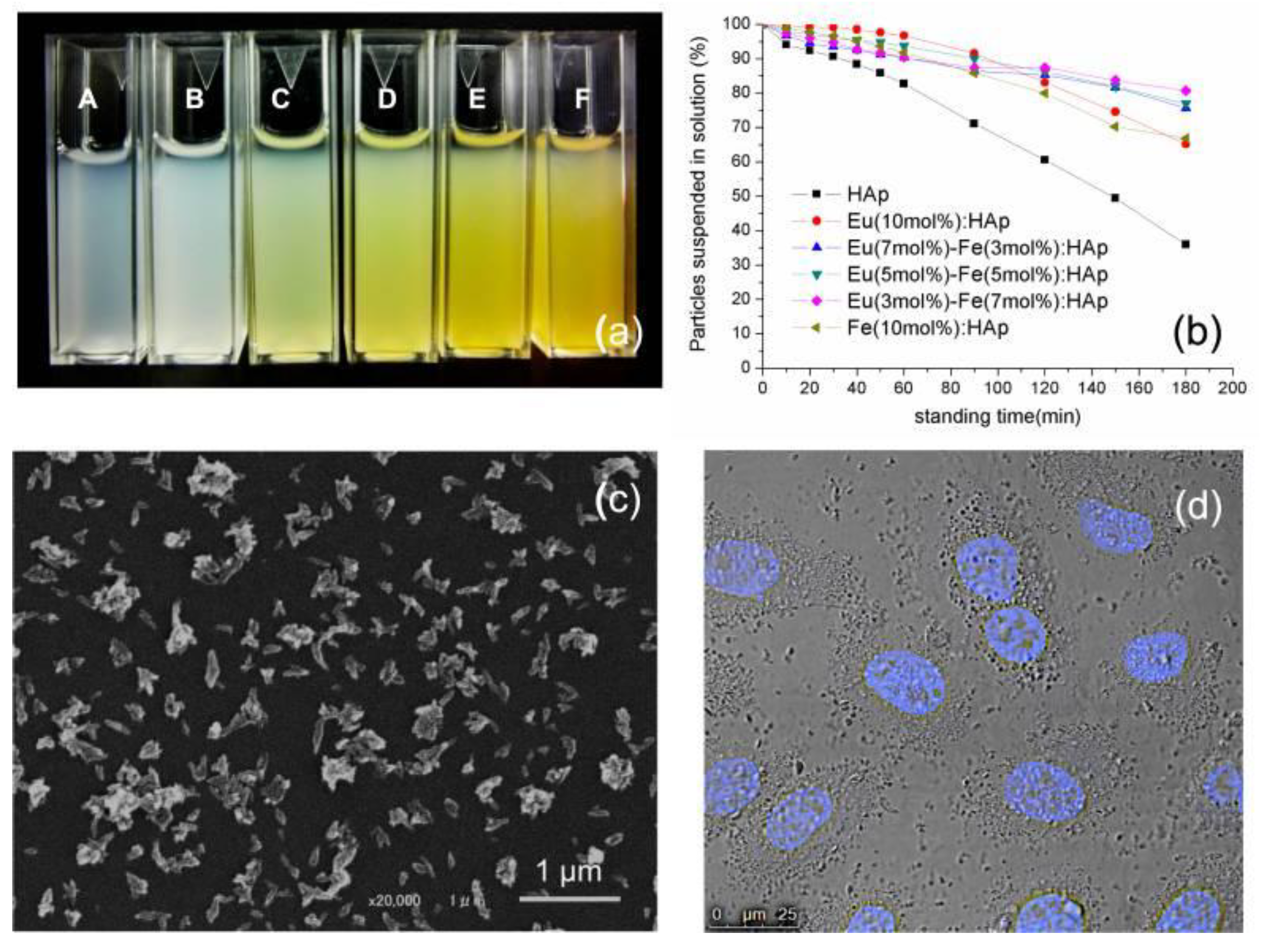
- Chen, M.-H.; Yoshioka, T.; Ikoma, T.; Hanagata, N.; Lin, F.-H.; Tanaka, J. Photoluminescence and doping mechanism of theranostic Eu3+/Fe3+ dual-doped hydroxyapatite nanoparticles. Sci. Technol. Adv. Mater. 2014, 15, 055005. [Google Scholar] [CrossRef] [PubMed]
- Chung, R.-J. Study of hydroxyapatite nano composites with photoluminescence properties. Biomed. Eng. Appl. Basis Commun. 2011, 23, 107–112. [Google Scholar] [CrossRef]
- Silva, C.; Sombra, A.; Rosa, I.; Leite, E.; Longo, E.; Varela, J.A. Study of Structural and Photoluminescent Properties of Ca8Eu2(PO4)6O2. J. Fluoresc. 2008, 18, 253–259. [Google Scholar] [CrossRef]
- Consani, S.; Balić-Žunić, T.; Cardinale, A.M.; Sgroi, W.; Giuli, G.; Carbone, C. A Novel Synthesis Routine for Woodwardite and Its Affinity towards Light (La, Ce, Nd) and Heavy (Gd and Y) Rare Earth Elements. Materials 2018, 11, 130. [Google Scholar] [CrossRef] [PubMed]
- Boesche, N.K.; Rogass, C.; Lubitz, C.; Brell, M.; Herrmann, S.; Mielke, C.; Tonn, S.; Appelt, O.; Altenberger, U.; Kaufmann, H. Hyperspectral REE (rare earth element) mapping of outcrops—Applications for neodymium detection. Remote Sens. 2015, 7, 5160–5186. [Google Scholar] [CrossRef]
- Iwanaga, H. Emission Properties, Solubility, Thermodynamic Analysis and NMR Studies of Rare-Earth Complexes with Two Different Phosphine Oxides. Materials 2010, 3, 4080. [Google Scholar] [CrossRef] [PubMed]
- Rodriguez-Ubis, J.C.; Brunet, E.; Juanes, O. Lanthanide Ions as Luminescent Probes. In Encyclopedia of Metalloproteins; Springer: Cham, Switzerland, 2013; pp. 1077–1087. [Google Scholar]
- Hein, J.R.; Koschinsky, A.; Mikesell, M.; Mizell, K.; Glenn, C.R.; Wood, R. Marine phosphorites as potential resources for heavy rare earth elements and yttrium. Minerals 2016, 6, 88. [Google Scholar] [CrossRef]
- Quarta, A.; Piccirillo, C.; Mandriota, G.; Di Corato, R. Nanoheterostructures (NHS) and Their Applications in Nanomedicine: Focusing on In Vivo Studies. Materials 2019, 12, 139. [Google Scholar] [CrossRef] [PubMed]
- Escudero, A.; Calvo, M.E.; Rivera-Fernández, S.; De la Fuente, J.M.; Ocaña, M. Microwave-assisted synthesis of biocompatible europium-doped calcium hydroxyapatite and fluoroapatite luminescent nanospindles functionalized with poly (acrylic acid). Langmuir 2013, 29, 1985–1994. [Google Scholar] [CrossRef] [PubMed]
- Deopa, N.; Rao, A. Spectroscopic studies of single near ultraviolet pumped Tb3+ doped Lithium Lead Alumino Borate glasses for green lasers and tricolour w-LEDs. J. Lumin. 2018, 194, 56–63. [Google Scholar] [CrossRef]
- Fu, Z.; Xu, P.; Yang, Y.; Li, C.; Lin, H.; Chen, Q.; Yao, G.; Zhou, Y.; Zeng, F. Study on luminescent properties of Ce3+ sensitized Tb3+ doped gadolinium borosilicate scintillating glass. J. Lumin. 2018, 196, 368–372. [Google Scholar] [CrossRef]
- Chen, Q.; Zuo, J.; He, X.; Mo, X.; Tong, P.; Zhang, L. Enhanced fluorescence of terbium with thiabendazole and application in determining trace amounts of terbium and thiabendazole. Talanta 2017, 162, 540–546. [Google Scholar] [CrossRef] [PubMed]
- Yang, Y.; Song, X.; Xu, C.; Wang, Y.; Zhang, G.; Liu, W. A multifunctional and recyclable terbium (iii) coordination polymer: Displaying highly selective and sensitive detection of Fe3+ and Cr VI anions, and picric acid in aqueous media. Dalton Trans. 2018, 47, 11077–11083. [Google Scholar] [CrossRef] [PubMed]
- De la Cruz, J.; Merino, R.P.; Trejo-García, P.; Espinosa, J.; Torres, R.A.; Moreno-Barbosa, E.; Gervacio-Arciniega, J.; Soto, E. Luminescent properties of a hybrid SiO2-PMMA matrix doped with terbium. Opt. Mater. 2018, 87, 42–47. [Google Scholar] [CrossRef]
- Chen, B.B.; Liu, M.L.; Zhan, L.; Li, C.M.; Huang, C.Z. Terbium (III) Modified Fluorescent Carbon Dots for Highly Selective and Sensitive Ratiometry of Stringent. Anal. Chem. 2018, 90, 4003–4009. [Google Scholar] [CrossRef]
- Xue, S.-F.; Chen, Z.-H.; Han, X.-Y.; Lin, Z.-Y.; Wang, Q.-X.; Zhang, M.; Shi, G. DNA Encountering Terbium (III): A Smart “Chemical Nose/Tongue” for Large-Scale Time-Gated Luminescent and Lifetime-Based Sensing. Anal. Chem. 2018, 90, 3443–3451. [Google Scholar] [CrossRef] [PubMed]
- Wang, Y.; Lin, S.; Luo, J.; Huang, R.; Cai, H.; Yan, W.; Yang, H. A Novel Tb@ Sr-MOF as Self-Calibrating Luminescent Sensor for Nutritional Antioxidant. Nanomaterials 2018, 8, 796. [Google Scholar] [CrossRef] [PubMed]
- Richards, B. Luminescent layers for enhanced silicon solar cell performance: Down-conversion. Sol. Energy Mater. Sol. Cells 2006, 90, 1189–1207. [Google Scholar] [CrossRef]
- Richards, B. Enhancing the performance of silicon solar cells via the application of passive luminescence conversion layers. Sol. Energy Mater. Sol. Cells 2006, 90, 2329–2337. [Google Scholar] [CrossRef]
- Enrichi, F.; Armellini, C.; Belmokhtar, S.; Bouajaj, A.; Chiappini, A.; Ferrari, M.; Quandt, A.; Righini, G.C.; Vomiero, A.; Zur, L. Visible to NIR downconversion process in Tb3+-Yb3+ codoped silica-hafnia glass and glass-ceramic sol-gel waveguides for solar cells. J. Lumin. 2018, 193, 44–50. [Google Scholar] [CrossRef]
- Wei, Y.; He, Y.; Li, X.; Chen, H.; Deng, X. Cellular Uptake and Delivery-Dependent Effects of Tb3+-Doped Hydroxyapatite Nanorods. Molecules 2017, 22, 1043. [Google Scholar] [CrossRef] [PubMed]
- Wang, Z.; Fang, C.; Sun, Y.; YANG, H. Synthesis and characterization of Tb-doped hydroxyapatite fluorescent nanoparticles. Mod. Chem. Ind. 2010, 30, 114–116. [Google Scholar]
- Yin, H.; Li, Y.; Bai, J.; Ma, M.; Liu, J. Effect of calcinations temperature on the luminescence intensity and fluorescent lifetime of Tb3+-doped hydroxyapatite (Tb-HA) nanocrystallines. J. Materiomics 2017, 3, 144–149. [Google Scholar] [CrossRef]
- Qiao, Y.; LI, Y.-X.; YIN, H.-R.; LIU, P.; LI, S.-Y.; ZHANG, P. Preparation and Luminescent Properties of Terbium-doped Hydroxyapatite. Chin. J. Lumin. 2015, 1, 012. [Google Scholar]
- Yang, P.; Quan, Z.; Li, C.; Kang, X.; Lian, H.; Lin, J. Bioactive, luminescent and mesoporous europium-doped hydroxyapatite as a drug carrier. Biomaterials 2008, 29, 4341–4347. [Google Scholar] [CrossRef]
- Han, Y.; Wang, X.; Li, S. Biocompatible Europium Doped Hydroxyapatite Nanoparticles as a Biological Fluorescent Probe. Curr. Nanosci. 2010, 6, 178–183. [Google Scholar] [CrossRef]
- Yang, C.; Yang, P.; Wang, W.; Wang, J.; Zhang, M.; Lin, J. Solvothermal synthesis and characterization of Ln (Eu3+, Tb3+) doped hydroxyapatite. J. Colloid Interface Sci. 2008, 328, 203–210. [Google Scholar] [CrossRef]
- Doat, A.; Fanjul, M.; Pellé, F.; Hollande, E.; Lebugle, A. Europium-doped bioapatite: A new photostable biological probe, internalizable by human cells. Biomaterials 2003, 24, 3365–3371. [Google Scholar] [CrossRef]
- Tesch, A.; Wenisch, C.; Herrmann, K.-H.; Reichenbach, J.R.; Warncke, P.; Fischer, D.; Müller, F.A. Luminomagnetic Eu3+- and Dy3+-doped hydroxyapatite for multimodal imaging. Mater. Sci. Eng. C 2017, 81, 422–431. [Google Scholar] [CrossRef]
- Mayer, I.; Layani, J.D.; Givan, A.; Gaft, M.; Blanc, P. La ions in precipitated hydroxyapatites. J. Inorg. Biochem. 1999, 73, 221–226. [Google Scholar] [CrossRef]
- Ghosh, R.; Sarkar, R.; Paul, S. Development of machinable hydroxyapatite-lanthanum phosphate composite for biomedical applications. Mater. Des. 2016, 106, 161–169. [Google Scholar] [CrossRef]
- Wang, X.; Huang, J.; Zhang, T.; Wang, K. Cytoskeleton reorganization and FAK phosphorylation are involved in lanthanum (III)-promoted proliferation and differentiation in rat osteoblasts. Prog. Nat. Sci. 2009, 19, 331–335. [Google Scholar] [CrossRef]
- Jadalannagari, S.; Deshmukh, K.; Verma, A.K.; Kowshik, R.V.; Meenal Ramanan, S.R. Lanthanum-Doped Hydroxyapatite Nanoparticles as Biocompatible Fluorescent Probes for Cellular Internalization and Biolabeling. Sci. Adv. Mater. 2014, 6, 312–319. [Google Scholar] [CrossRef]
- Ahymah Joshy, M.I.; Elayaraja, K.; Suganthi, R.V.; Chandra Veerla, S.; Kalkura, S.N. In vitro sustained release of amoxicillin from lanthanum hydroxyapatite nano rods. Curr. Appl. Phy. 2011, 11, 1100–1106. [Google Scholar] [CrossRef]


| Cation (M) | Sample Form | Doping Range {M/(M + Ca)}100 (at %) | Bio-Functionality/Effect of the Dopant |
|---|---|---|---|
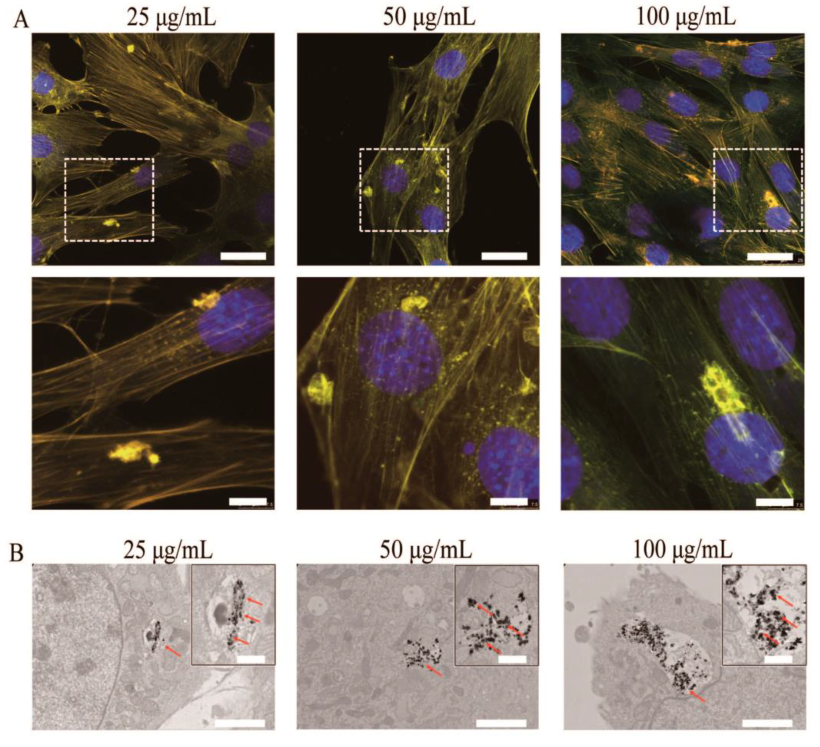
| Tb | Powder | 2–17 | In vitro cytocompatibility with MC3T3-E1 (doses of 25–100 µg mL−1 Tb-HA-NPs) and A549 (doses of 20–320 µg mL−1 Tb-HA-NPs) cell lines. |
| Er | Powder | 2–10 | Induces the formation of biomimetic apatite in-growths in simulated body fluid (SBF). |
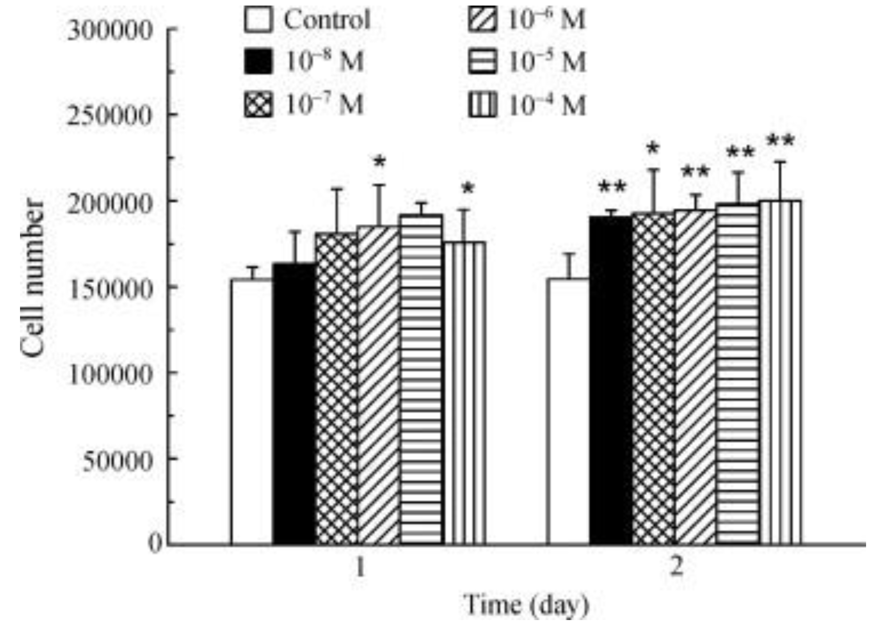
| Eu | Powder | 0.1–20 | Induces the in vitro formation of bone-like apatite in SBF; In vitro cytocompatibility with MG-63 (cell proliferation up to 4 days), HeLa, human embryonic kidney HEK 293, L929 (viability >80% for Eu-HA doses of 25–500 µg mL−1); Low cytotoxicity for human gingival fibroblast (HGF-1) cells after 24 h (500–2000 µg mL−1); Cytotoxicity for transformed human umbilical vein endothelial cells (T-HUVEC) after treatment with 0.3–30 µg mL−1 of 5 at % doped HA; Ability to kill cervical HeLa cells after 24 h when combined with 5 fluorouracil (5FU); Negligible toxicity by hen’s egg test on the chick area vasculosa (HET-CAV); Antibacterial effect against E. faecalis * (ATCC 29212), S. aureus * (0364), and P. aeruginosa * (1397); No antibacterial activity against E. coli * even at high doping; Antifungal effect against C. albicans * (ATCC 10231) with a doping content of 20 at %. |
| La | Powder Coating | 2–30 | In vitro cytocompatibility with MC3T3-E1 and L929 cell lines; No cytotoxicity for adenocarcinoma (MCF-7) and human embryonic kidney HEK cells at a doping level of 2 at %; Antibacterial effect against S. aureus (e.g., ATCC 25175), E. coli, P. aeruginosa, and Bacillus; Improvement of mechanical properties: bonding strength and Vickers hardness. |
| Dy | Powder | 0.5–10 | In vitro cytocompatibility with L929 cell line; Negligible toxicity by hen’s egg test on the chick area vasculosa (HET-CAV); Increase of oxidative stress lipoperoxides and nitric oxide indicators in the kidney, lungs, and liver of rats; lower activity of anti-oxidant glutathione peroxidase enzyme. |
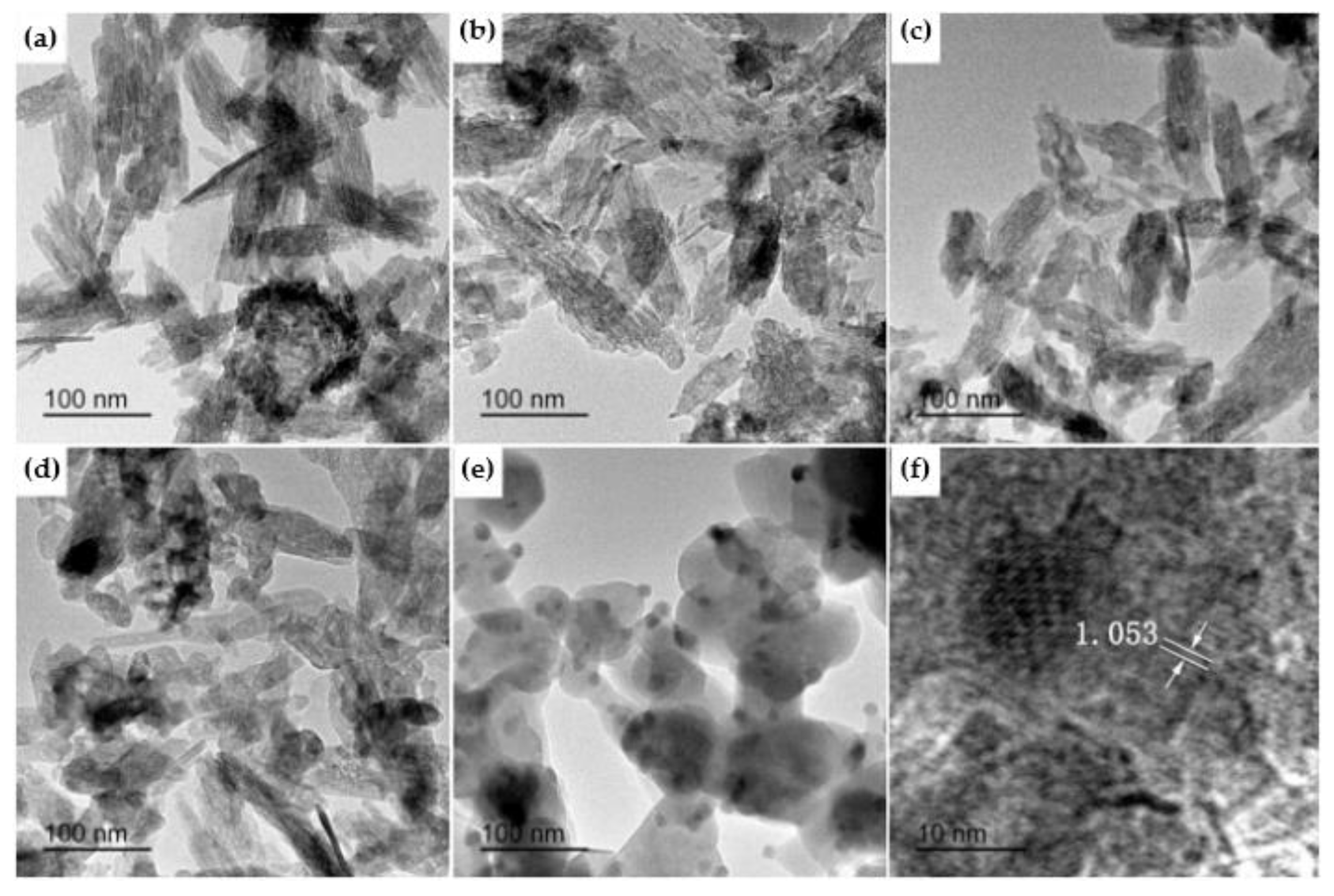
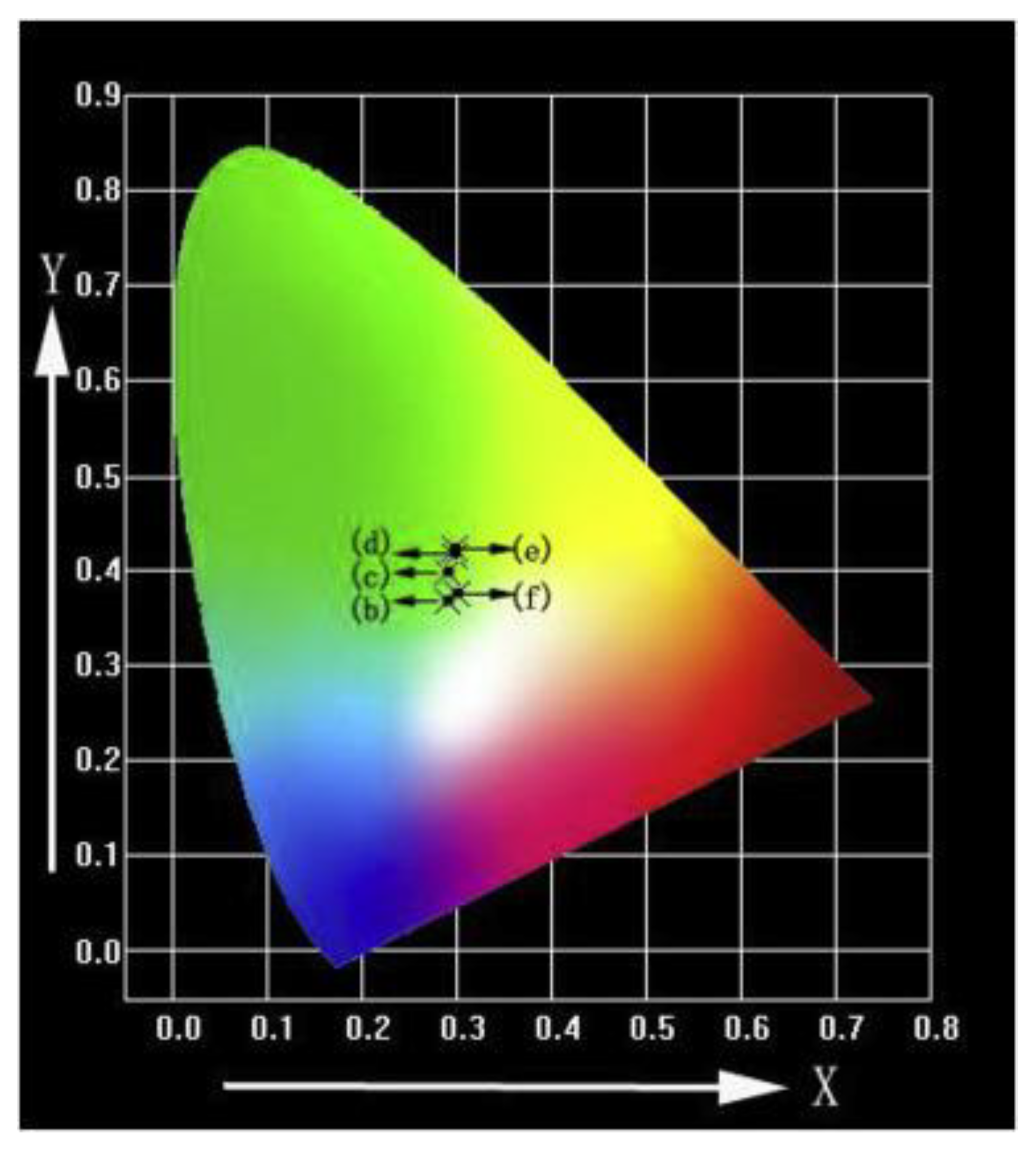
| Samples | (b) 300 °C | (c) 400 °C | (d) 500 °C | (e) 600 °C | (f) 700 °C |
|---|---|---|---|---|---|
| X | 0.2893 | 0.2910 | 0.2981 | 0.2985 | 0.3000 |
| Y | 0.3682 | 0.3976 | 0.4161 | 0.4234 | 0.3743 |
| Doping Element | Synthesis Method | Improvements of Photoluminescent Properties | Biomedical Application | References |
|---|---|---|---|---|
| Terbium | microemulsion-mediated solvothermal process | the particles could be excited by a visible light beam at 400 nm | fluorescent bio-probe | Wang et al., 2010 [60] |
| chemical deposition | excitation light is 378 nm when the wavelength of the monitoring light is 545 nm | fluorescent probe | Qiao et al., 2015 [62] | |
| Erbium | microwave-assisted precipitation method | red and green emission in the spectra | sensing material | Alshemary et al., 2015 [9] |
| Microwave-assisted wet precipitation | photoluminescence spectra—green and red emissions | bone healing process | Alshemary et al., 2015 [9] | |
| co-precipitation | near-infrared emission peaks ~1540 nm | biomedicine | Pham et al., 2016 [24] | |
| Europium | microwave-assisted synthesis | red luminescence; negligible toxicity for Vero cells | potential tools for biomedical applications | Escudero, 2013 [47] |
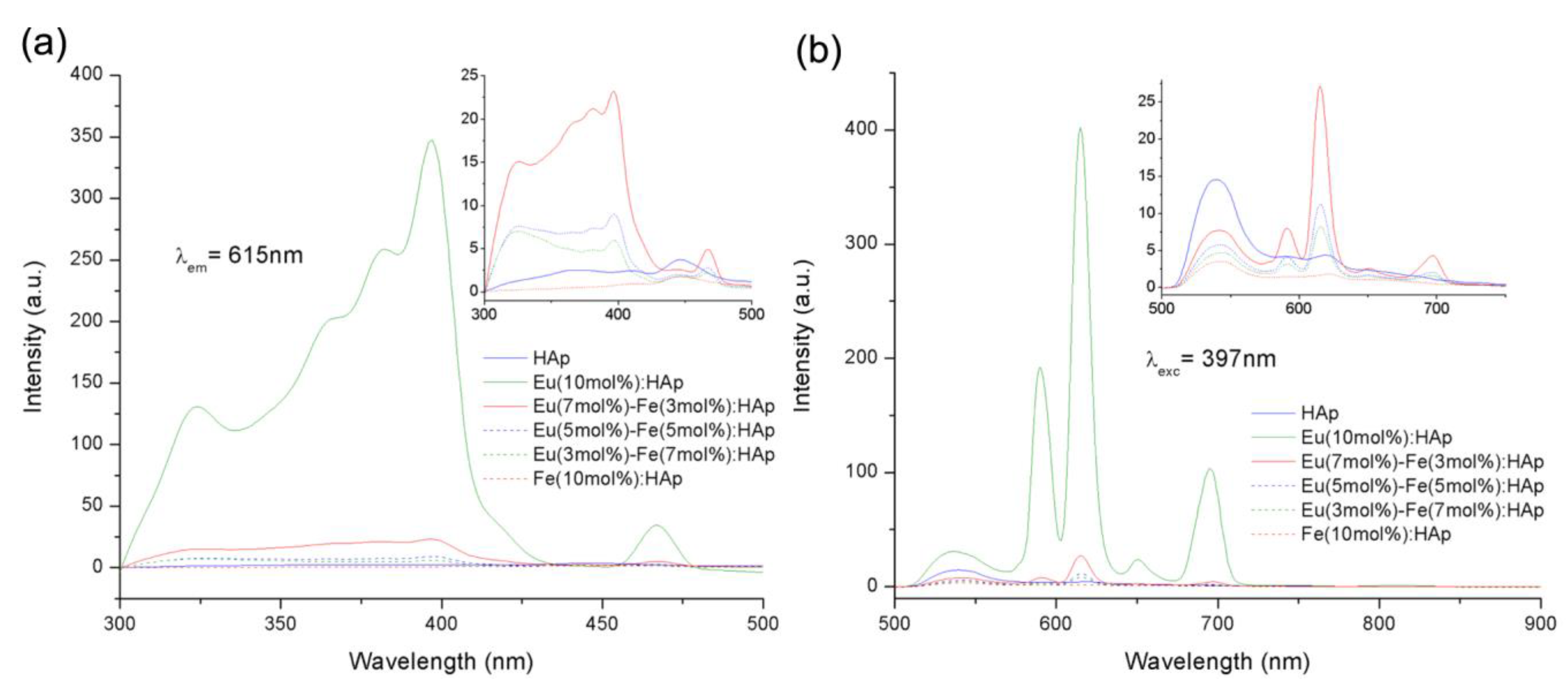
| wet chemical precipitation in water without the addition of any surfactant | luminescence at peaks at 536, 590, 615, 650, and 695 nm under 397 nm excitation | fluorescent probe for in vivo imaging | Chen et al., 2014 [38] | |
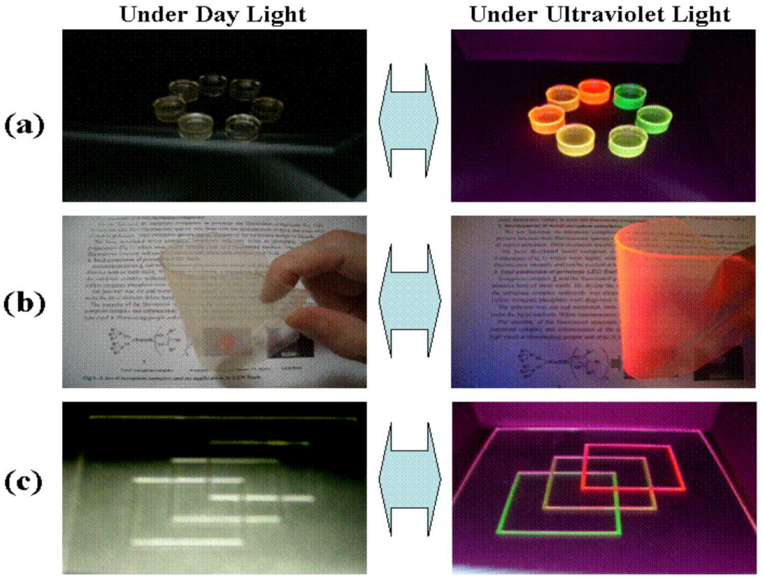
| simple one-step method using cationic surfactant as a template | red luminescence of Eu3+ (5D0–7F1,2) under UV irradiation | drug delivery disease therapy | Yang et al., 2008 [63] | |
| precipitation | strong green and red fluorescence by irradiation of blue and green light | biocompatible fluorescent labeling material in biological studies | Han et al., 2010 [64] | |
| synthetized at low temperatures (37 °C) | red luminescence is photostable; luminescence could be obtained under visible irradiation | bio-probe | Doat et al., 2003 [66] | |
| Europium and Terbium | microemulsion process under hydrothermal treatment | typical emission lines of Eu3+ and Tb3+ | carriers for drug release and targeting | Yang et al., 2008 [65] |
| Lanthanum | wet chemical synthesis method | in vitro bioactivity and biocompatibility | bioimaging phosphor/luminescent labeling materials for bioimaging | Ghosh et al., 2016 [69] |
| modified sol–gel method at a low temperature of 100 °C | fluorescence detected under TRITC (Tetramethylrhodamine) and FITC (Fluorescein isothiocyanate) filters using epifluorescence microscopy | fluorescent probes for cellular internalization and biolabeling | Jadalannagari et al., 2014 [71] | |
| sol–gel route | decrease in the dissolution of the samples as the dopant concentration increases | implant in biomedical field | Ahymah, 2011 [72] | |
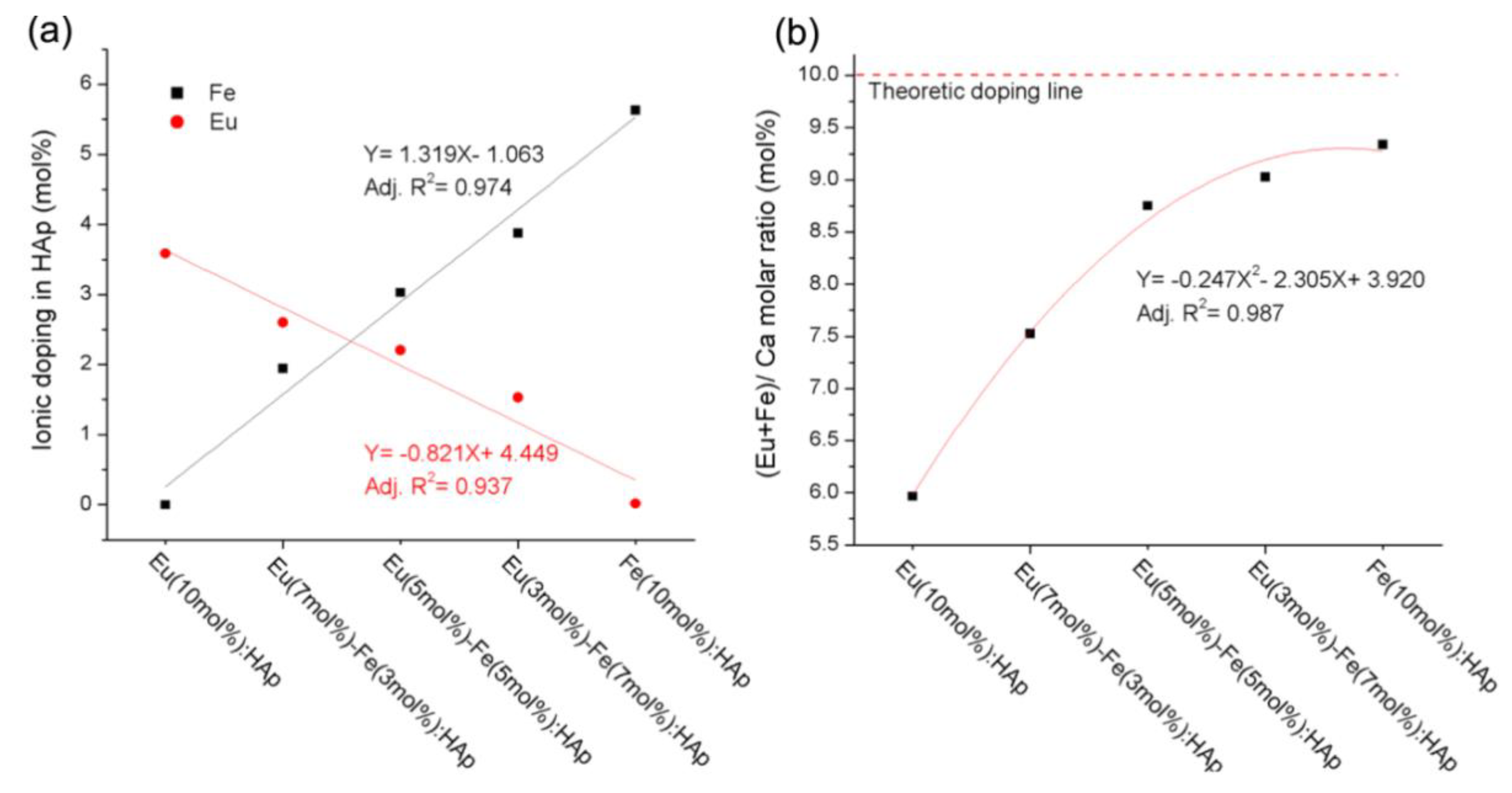
| Dysprosium and Europium | co-doping | increased photoluminescent properties; strong transverse relaxation effects | contrast agent for MRI in implantology or functional coatings | Tesch et al., 2017 [67] |
| Dysprosium | co-precipitation | fluorescent character—stimulated at 344 or 360 nm | bimodal probes with low toxicity | Sánchez et al., 2015 [6] |
© 2019 by the authors. Licensee MDPI, Basel, Switzerland. This article is an open access article distributed under the terms and conditions of the Creative Commons Attribution (CC BY) license (http://creativecommons.org/licenses/by/4.0/).
Share and Cite
Neacsu, I.A.; Stoica, A.E.; Vasile, B.S.; Andronescu, E. Luminescent Hydroxyapatite Doped with Rare Earth Elements for Biomedical Applications. Nanomaterials 2019, 9, 239. https://doi.org/10.3390/nano9020239
Neacsu IA, Stoica AE, Vasile BS, Andronescu E. Luminescent Hydroxyapatite Doped with Rare Earth Elements for Biomedical Applications. Nanomaterials. 2019; 9(2):239. https://doi.org/10.3390/nano9020239
Chicago/Turabian StyleNeacsu, Ionela Andreea, Alexandra Elena Stoica, Bogdan Stefan Vasile, and Ecaterina Andronescu. 2019. "Luminescent Hydroxyapatite Doped with Rare Earth Elements for Biomedical Applications" Nanomaterials 9, no. 2: 239. https://doi.org/10.3390/nano9020239
APA StyleNeacsu, I. A., Stoica, A. E., Vasile, B. S., & Andronescu, E. (2019). Luminescent Hydroxyapatite Doped with Rare Earth Elements for Biomedical Applications. Nanomaterials, 9(2), 239. https://doi.org/10.3390/nano9020239