Hydrothermal Fabrication of Spindle-Shaped ZnO/Palygorskite Nanocomposites Using Nonionic Surfactant for Enhancement of Antibacterial Activity


Abstract
1. Introduction
2. Materials and Methods
2.1. Materials
2.2. Preparation of ZnO/PAL Nanocomposites
2.3. Characterization
2.4. Antibacterial Assay
3. Results and Discussion
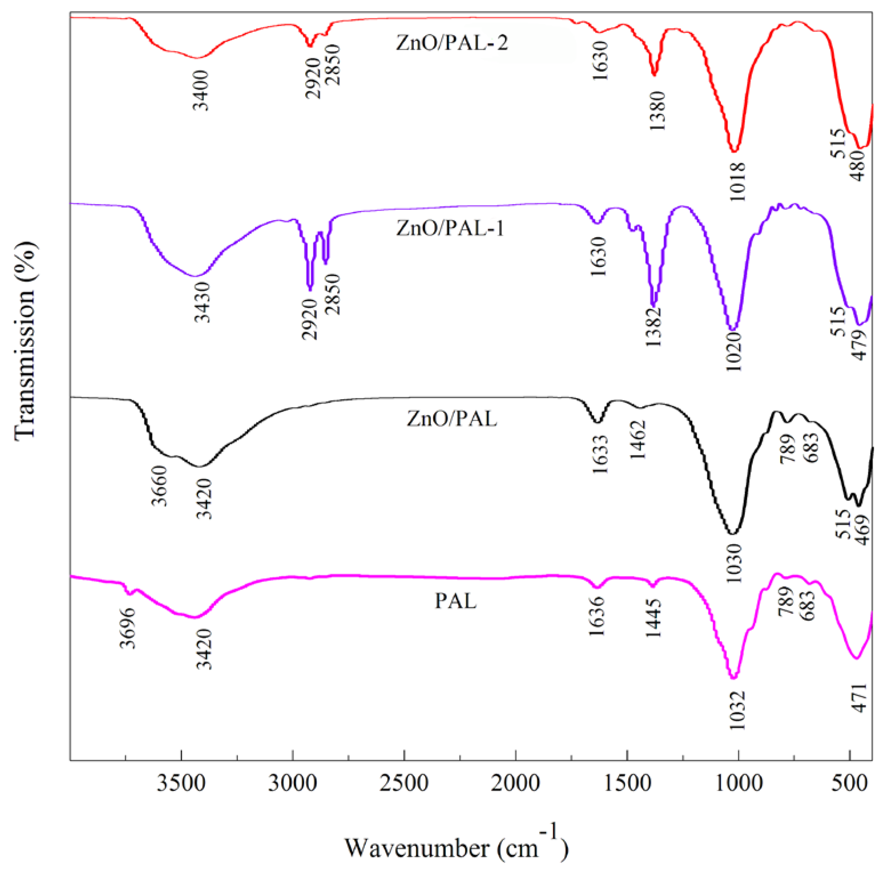
3.1. FTIR
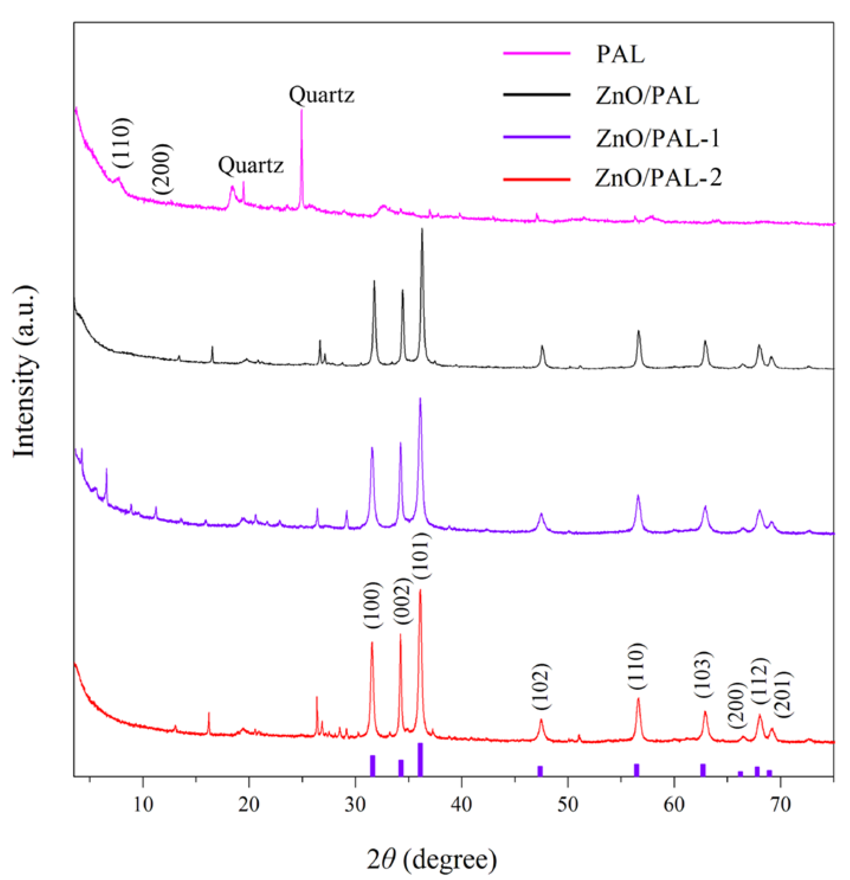
3.2. XRD Patterns
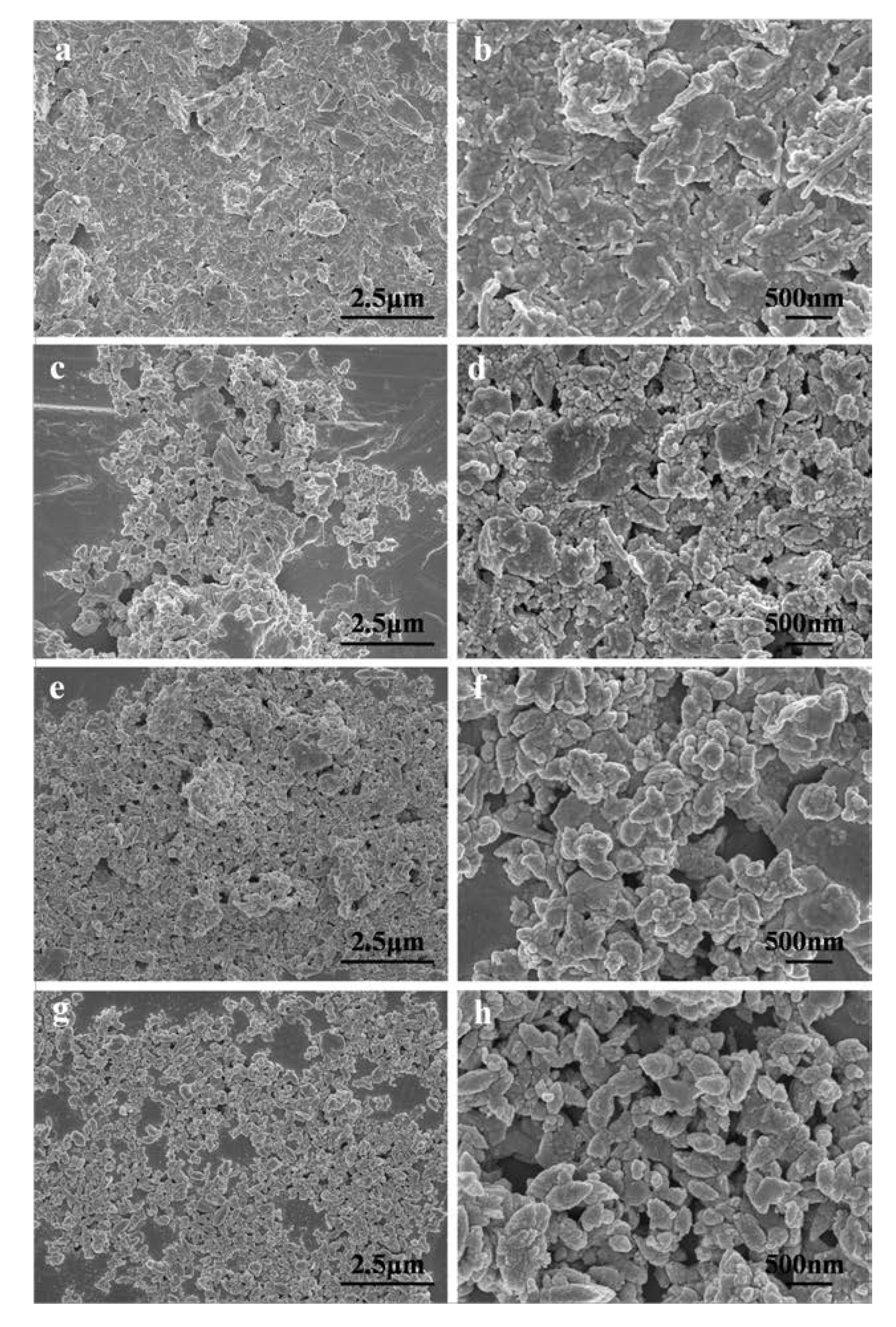
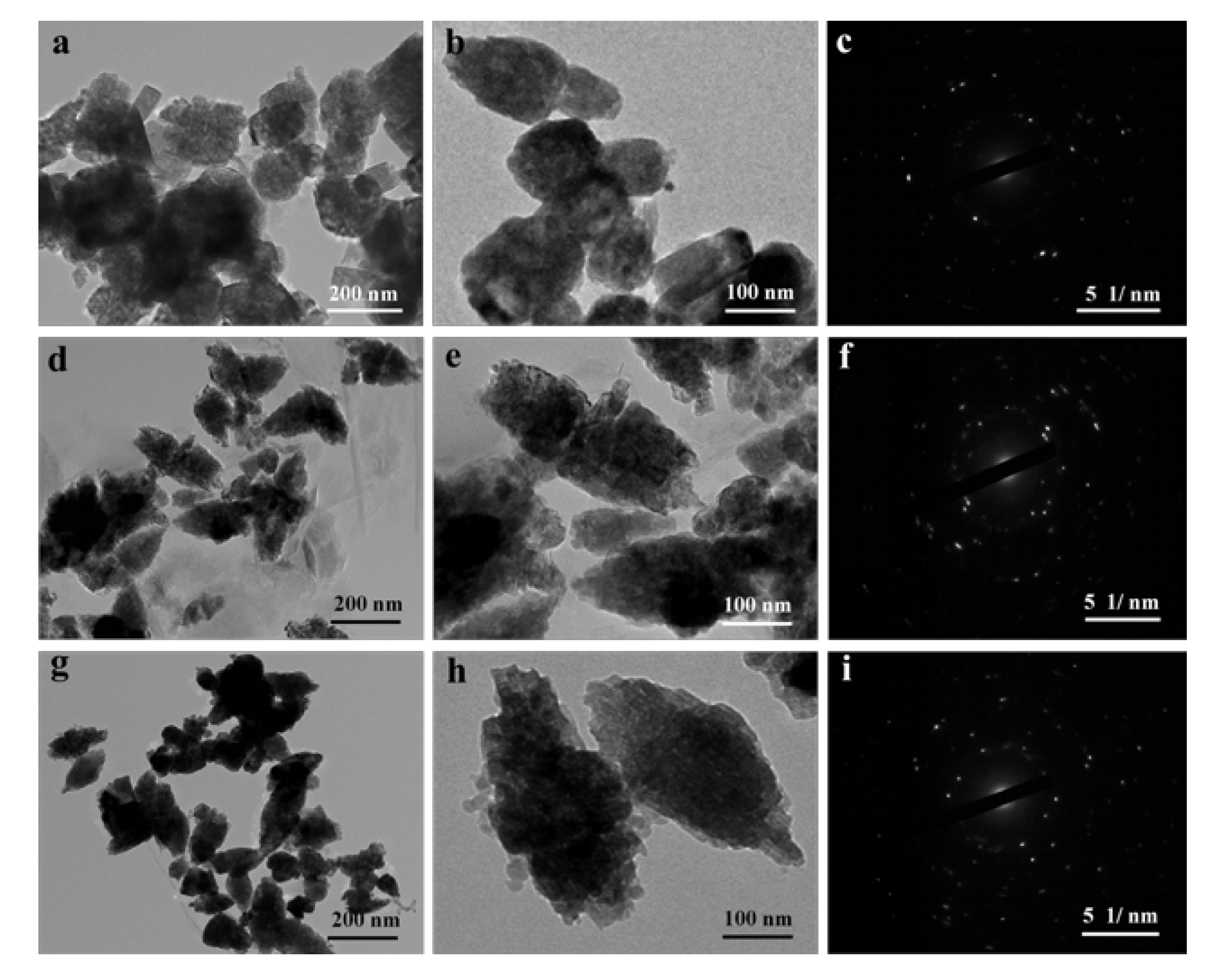
3.3. SEM and TEM
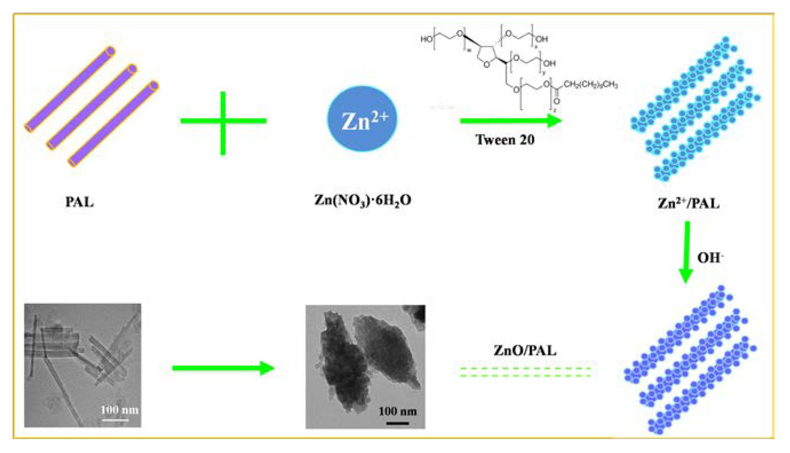
3.4. Possible Formation Mechanism of ZnO/PAL Nanocomposites
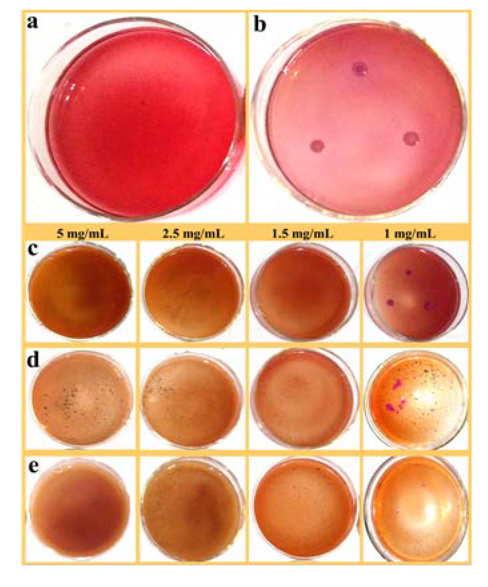
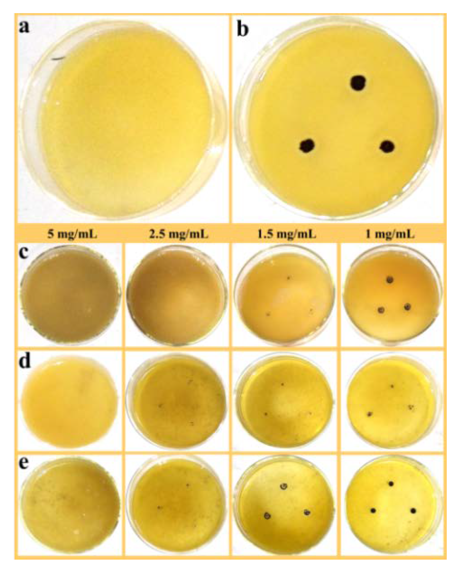
3.5. Antibacterial Evaluation
4. Conclusions
Supplementary Materials
Author Contributions
Funding
Acknowledgments
Conflicts of Interest
References
- Ventola, C.L. The antibiotic resistance crisis part 1: Causes and threats. Pharm. Ther. 2015, 40, 277–283. [Google Scholar]
- Huggins, W.M.; Minrovic, B.M.; Corey, B.W.; Jacobs, A.C.; Melander, R.J.; Sommer, R.D.; Zurawski, D.V.; Melander, C. 1,2,4-Triazolidine-3-thiones as narrow spectrum antibiotics against multidrug-resistant Acinetobacter baumannii. ACS Med. Chem. Lett. 2017, 8, 27–31. [Google Scholar] [CrossRef] [PubMed]
- Vargasreus, M.A.; Memarzadeh, K.; Huang, J.; Ren, G.G.; Allaker, R.P. Antimicrobial activity of nanoparticulate metal oxides against peri-implantitis pathogens. Int. J. Antimicrob. Ag. 2012, 40, 135–139. [Google Scholar] [CrossRef] [PubMed]
- Mckenna, M. Antibiotic resistance: The last resort. Nature 2013, 499, 394–396. [Google Scholar] [CrossRef] [PubMed]
- Klevens, R.M.; Edwards, J.R.; Tenover, F.C.; McDonald, L.C.; Horan, T.; Gaynes, R. Changes in the epidemiology of methicillin-resistant Staphylococcu saureus in intensive care units in US hospitals, 1992-2003. Clin. Infect. Dis. 2006, 42, 389–391. [Google Scholar] [CrossRef]
- Li, X.; Robinson, S.M.; Gupta, A.; Saha, K.; Jiang, Z.; Moyano, D.F.; Sahar, A.; Riley, M.A.; Rotello, V.M. Functional gold nanoparticlesas potent antimicrobial agents against multi-drug-resistant bacteria. ACS Nano 2014, 8, 10682–10686. [Google Scholar] [CrossRef] [PubMed]
- Morrison, K.D.; Underwood, J.C.; Metge, D.W.; Eberl, D.D.; Williams, L.B. Mineralogical variables that control the antibacterial effectiveness of a natural clay deposit. Environ. Geochem. Health 2014, 36, 613–631. [Google Scholar] [CrossRef]
- Rasool, K.; Helal, M.; Ali, A.; Ren, C.E.; Gogotsi, Y.; Mahmoud, A. Antibacterial activity of Ti3C2Tx MXene. ACS Nano 2016, 10, 3674–3684. [Google Scholar] [CrossRef]
- Pandit, S.; Karunakaran, S.; Boda, S.; Basu, B.; De, M. High antibacterial activity of functionalized chemically exfoliated MoS2. ACS Appl. Mater. Inter. 2016, 8, 31567–31573. [Google Scholar] [CrossRef]
- Xiu, Z.M.; Zhang, Q.B.; Puppala, H.L.; Colvin, V.L.; Alvarez, P.J. Negligible particle-specific antibacterial activity of silver nanoparticles. Nano Lett. 2012, 12, 4271–4275. [Google Scholar] [CrossRef]
- Hu, W.B.; Peng, C.; Luo, W.J.; Lv, M.; Li, X.M.; Li, D.; Huang, Q.; Fan, C.H. Graphene-based antibacterial paper. ACS Nano 2010, 4, 4317–4323. [Google Scholar] [CrossRef] [PubMed]
- Williams, L.B.; Haydel, S.E. Evaluation of the medicinal use of clay minerals as antibacterial agents. Int. Geol. Rev. 2010, 52, 745–770. [Google Scholar] [CrossRef] [PubMed]
- Haydel, S.E.; Remenih, C.M.; Williams, L.B. Broad-spectrum in vitro antibacterial activities of clay minerals against antibiotic-susceptible and antibiotic-resistant bacterial pathogens. J. Antimicrob. Chemother. 2008, 61, 353–361. [Google Scholar] [CrossRef] [PubMed]
- Williams, L.B.; Haydel, S.E.; Giese, R.F.; Eberl, D.D. Chemical and mineralogical characteristics of French green clays used healing. Clays Clay Miner. 2008, 56, 437–452. [Google Scholar] [CrossRef]
- Wang, W.B.; Wang, A.Q. Recent progress in dispersion of palygorskite crystal bundles for nanocomposites. Appl. Clay Sci. 2016, 119, 18–30. [Google Scholar] [CrossRef]
- Lu, Y.S.; Dong, W.K.; Wang, W.B.; Ding, J.J.; Wang, Q.; Hui, A.P.; Wang, A.Q. Optimal synthesis of environment-friendly iron red pigment from natural nanostructured clay minerals. Nanomaterials 2018, 8, 925. [Google Scholar] [CrossRef]
- Wang, W.B.; Wang, F.F.; Kang, Y.R.; Wang, A.Q. Nanoscale dispersion crystal bundles of palygorskite by associated modification with phytic acid and high-pressure homogenization for enhanced colloidal properties. Powder Technol. 2015, 269, 85–92. [Google Scholar] [CrossRef]
- Cai, X.; Zhang, J.L.; Ouyang, Y.; Ma, D.; Tan, S.Z.; Peng, Y.L. Bacteria-adsorbed palygorskite stabilizes the quaternary phosphonium salt with specific-targeting capability, long-term antibacterial activity, and lower cytotoxicity. Langmuir 2013, 29, 5279–5285. [Google Scholar] [CrossRef]
- Lei, H.; Wei, Q.N.; Wang, Q.; Su, A.X.; Xue, M.; Liu, Q.; Hu, Q.H. Characterization of ginger essential oil/palygorskite composite (GEO-PGS) and its antibacterial activity. Mat. Sci. Eng. C-Mater. 2017, 73, 381–387. [Google Scholar] [CrossRef]
- Zhao, D.F.; Zhou, J.; Liu, N. Preparation and characterization of Mingguang palygorskite supported with silver and copper for antibacterial behavior. Appl. Clay Sci. 2006, 33, 161–170. [Google Scholar] [CrossRef]
- Yan, R.; Zhang, L.; Yang, X.; Wen, C.; Zhou, Y.M. Bioavailability evaluation of zinc-bearing palygorskite as a zinc source for broiler chickens. Appl. Clay Sci. 2016, 119, 155–160. [Google Scholar] [CrossRef]
- Chen, Y.; Lu, W.P.; Guo, Y.C.; Zhu, Y.; Song, Y.P. Electrospun gelatin fibers surface loaded ZnO particles as a potential biodegradable antibacterial wound dressing. Nanomaterials 2019, 9, 525. [Google Scholar] [CrossRef] [PubMed]
- Jalal, R.; Goharshadi, E.K.; Abareshi, M.; Moosavi, M.; Yousefi, A.; Nancarrow, P. ZnO nanofluids: Green synthesis, characterization, and antibacterial activity. Mater. Chem. Phys. 2010, 121, 198–201. [Google Scholar] [CrossRef]
- Premanathan, M.; Karthikeyan, K.; Jeyasubramanian, K.; Manivannan, G. Selective toxicity of ZnO nanoparticles toward Gram-positive bacteria and cancer cells by apoptosis through lipid peroxidation. Nanomed. Nanotechnol. 2011, 7, 184–192. [Google Scholar] [CrossRef] [PubMed]
- Youssef, A.M.; El-Nahrawy, A.M.; Hammad, A.B.A. Sol-gel synthesis and characterizations of hybrid chitosan-PEG/calcium silicate nanocomposite modified with ZnO-NPs and (E102) for optical and antibacterial applications. Int. J. Biol. Macromol. 2017, 97, 561–567. [Google Scholar] [CrossRef] [PubMed]
- Catalano, M.; Belluso, E.; Miriello, D.; Barrese, E.; Bloise, A. Synthesis of Zn-doped talc in hydrothermal atmosphere. Cryst. Res. Technol. 2014, 49, 283–289. [Google Scholar] [CrossRef]
- Rokbani, H.; Daigle, F.; Ajji, A. Combined effect of ultrasound stimulations and autoclaving on the enhancement of antibacterial activity of ZnO and SiO2/ZnO nanoparticles. Nanomaterials 2018, 8, 129. [Google Scholar] [CrossRef]
- Dizaj, S.M.; Lotfipour, F.; Barzegar-Jalali, M.; Zarrintan, M.H.; Adibkia, K. Antimicrobial activity of the metals and metal oxide nanoparticles. Mat. Sci. Eng. C-Mater. 2014, 44, 278–284. [Google Scholar] [CrossRef]
- Leone, F.; Cataldo, R.; Mohamed, S.S.Y.; Manna, L.; Banchero, M.; Ronchetti, S.; Mandras, N.; Tullio, V.; Cavalli, R.; Onida, B. Nanostructured ZnO as multifunctional carrier for a green antibacterial drug delivery system-a feasibility study. Nanomaterials 2019, 9, 407. [Google Scholar] [CrossRef]
- Raghunath, A.; Perumal, E. Metal oxide nanoparticles as antimicrobial agents: A promise for the future. Int. J. Antimicrob. Ag. 2017, 49, 137–152. [Google Scholar] [CrossRef]
- Agnihotri, S.; Bajaj, G.; Mukherji, S.; Mukherji, S. Arginine-assisted immobilization of silver nanoparticles on ZnO nanorods: An enhanced and reusable antibacterial substrate without human cell cytotoxicity. Nanoscale 2015, 7, 7415–7429. [Google Scholar] [CrossRef] [PubMed]
- Hui, A.P.; Ma, J.Z.; Liu, J.L.; Bao, Y.; Zhang, J. Morphological evolution of Fe doped sea urchin-shaped ZnO nanoparticles with enhanced photocatalytic activity. J. Alloy Compd. 2017, 696, 639–647. [Google Scholar] [CrossRef]
- Rajabi, H.R.; Naghiha, A.; Kheirizdeh, M.; Sadatfaraji, H.; Mirzaei, A.; Alvand, Z.M. Microwave assisted extraction as an efficient approach for biosynthesis of zinc oxide nanoparticles: Synthesis, characterization, and biological properties. Mat. Sci. Eng. C-Mater. 2017, 78, 1109–1118. [Google Scholar] [CrossRef] [PubMed]
- Huo, C.L.; Yang, H.M. Synthesis and characterization of ZnO/palygorskite. Appl. Clay Sci. 2010, 50, 362–366. [Google Scholar] [CrossRef]
- Li, M.; Zhu, L.Z.; Lin, D.H. Toxicity of ZnO nanoparticles to Escherichia coli: Mechanism and the influence of medium components. Enviro. Sci. Techol. 2011, 45, 1977–1983. [Google Scholar] [CrossRef] [PubMed]
- Li, L.X.; Li, B.C.; Fan, L.; Mu, B.; Wang, A.Q.; Zhang, J.P. Palygorskite@Fe3O4@polyperfluoroalkylsilane nanocomposites for superoleophobic coatings and magnetic liquid marbles. J. Mater. Chem. A 2016, 4, 5859–5868. [Google Scholar] [CrossRef]
- Mu, B.; Wang, A.Q. One-pot fabrication of multifunctional superparamagnetic attapulgite/Fe3O4/polyaniline nanocomposites served as adsorbent and catalyst support. J. Mater. Chem. A 2015, 3, 281–289. [Google Scholar] [CrossRef]
- Tai, Y.F.; Shi, C.J.; Wang, C.H. Preparation and characterization of anioncation organopalygorskite for 2-naphthol removal from aqueous solution. J. Mol. Liq. 2014, 195, 116–124. [Google Scholar] [CrossRef]
- Wang, W.B.; Zhang, Z.F.; Tian, G.Y.; Wang, A.Q. From nanorods of palygorskite to nanosheets of smectite via a one-step hydrothermal process. Rsc Adv. 2015, 5, 58107–58115. [Google Scholar] [CrossRef]
- Geuli, O.; Lewinstein, I.; Mandler, D. Composition-tailoring of ZnO-hydroxyapatite nanocomposite as bioactive and antibacterial coating. ACS Appl. Nano Mater. 2019, 2, 2946–2957. [Google Scholar] [CrossRef]
- He, W.W.; Kim, H.K.; Wayne, G.W.; David, M.; John, H.C.; Yin, J.J. Photogenerated charge carriers and reactive oxygen species in ZnO/Au hybrid nanostructures with enhanced photocatalytic and antibacterial activity. J. Am. Chem. Soc. 2014, 136, 750–757. [Google Scholar] [CrossRef] [PubMed]
- Prasanna, V.L.; Vijayaraghavan, R. Insight into the mechanism of antibacterial activity of ZnO: Surface defects mediated reactive oxygen species even in the dark. Langmuir 2015, 31, 9155–9162. [Google Scholar] [CrossRef] [PubMed]
- Liu, J.L.; Wang, Y.H.; Ma, J.Z.; Peng, Y.; Wang, A.Q. A review on bidirectional analogies between the photocatalysis and antibacterial properties of ZnO. J. Alloy Compd. 2019, 783, 898–918. [Google Scholar] [CrossRef]
- Ramani, M.; Ponnusamy, S.; Muthamizhchelvan, C.; Marsili, E. Amino acid-mediated synthesis of zinc oxide nanostructures and evaluation of their facet-dependent antimicrobial activity. Colloids Surf. B 2014, 117, 233–239. [Google Scholar] [CrossRef] [PubMed]


| Samples | SBET (m2/g) | Smicro (m2/g) | Sext (m2/g) | Vtotal (cm3/g) |
|---|---|---|---|---|
| PAL | 173.5 | 39.3 | 134.2 | 0.197 |
| ZnO/PAL | 35.4 | 2.4 | 32.3 | 0.067 |
| ZnO/PAL-1 | 8.9 | − | 10.7 | 0.025 |
| ZnO/PAL-2 | 14.2 | − | 22.0 | 0.036 |
| Samples | MIC (mg/mL) | |||||||
|---|---|---|---|---|---|---|---|---|
| E. coli | S. aureus | |||||||
| 5 | 2.5 | 1.5 | 1 | 5 | 2.5 | 1.5 | 1 | |
| PAL | ✕ | ✕ | ✕ | ✕ | ✕ | ✕ | ✕ | ✕ |
| ZnO/PAL | ✓ | ✕ | ✕ | ✕ | ✓ | ✓ | ✕ | ✕ |
| ZnO/PAL-1 | ✓ | ✓ | ✕ | ✕ | ✓ | ✕ | ✕ | ✕ |
| ZnO/PAL-2 | ✓ | ✓ | ✓ | ✕ | ✓ | ✕ | ✕ | ✕ |
© 2019 by the authors. Licensee MDPI, Basel, Switzerland. This article is an open access article distributed under the terms and conditions of the Creative Commons Attribution (CC BY) license (http://creativecommons.org/licenses/by/4.0/).
Share and Cite
Hui, A.; Dong, S.; Kang, Y.; Zhou, Y.; Wang, A. Hydrothermal Fabrication of Spindle-Shaped ZnO/Palygorskite Nanocomposites Using Nonionic Surfactant for Enhancement of Antibacterial Activity. Nanomaterials 2019, 9, 1453. https://doi.org/10.3390/nano9101453
Hui A, Dong S, Kang Y, Zhou Y, Wang A. Hydrothermal Fabrication of Spindle-Shaped ZnO/Palygorskite Nanocomposites Using Nonionic Surfactant for Enhancement of Antibacterial Activity. Nanomaterials. 2019; 9(10):1453. https://doi.org/10.3390/nano9101453
Chicago/Turabian StyleHui, Aiping, Shuqing Dong, Yuru Kang, Yanmin Zhou, and Aiqin Wang. 2019. "Hydrothermal Fabrication of Spindle-Shaped ZnO/Palygorskite Nanocomposites Using Nonionic Surfactant for Enhancement of Antibacterial Activity" Nanomaterials 9, no. 10: 1453. https://doi.org/10.3390/nano9101453
APA StyleHui, A., Dong, S., Kang, Y., Zhou, Y., & Wang, A. (2019). Hydrothermal Fabrication of Spindle-Shaped ZnO/Palygorskite Nanocomposites Using Nonionic Surfactant for Enhancement of Antibacterial Activity. Nanomaterials, 9(10), 1453. https://doi.org/10.3390/nano9101453