Core–Shell Plasmonic Nanocomposites with Synergistic Photothermal and Photochemical Activity for Biomedical Applications

 , , , ,
  and
and 
Abstract
1. Introduction
2. Fundamentals of Core–Shell Plasmonic Nanocomposites
2.1. Structure and Synthesis
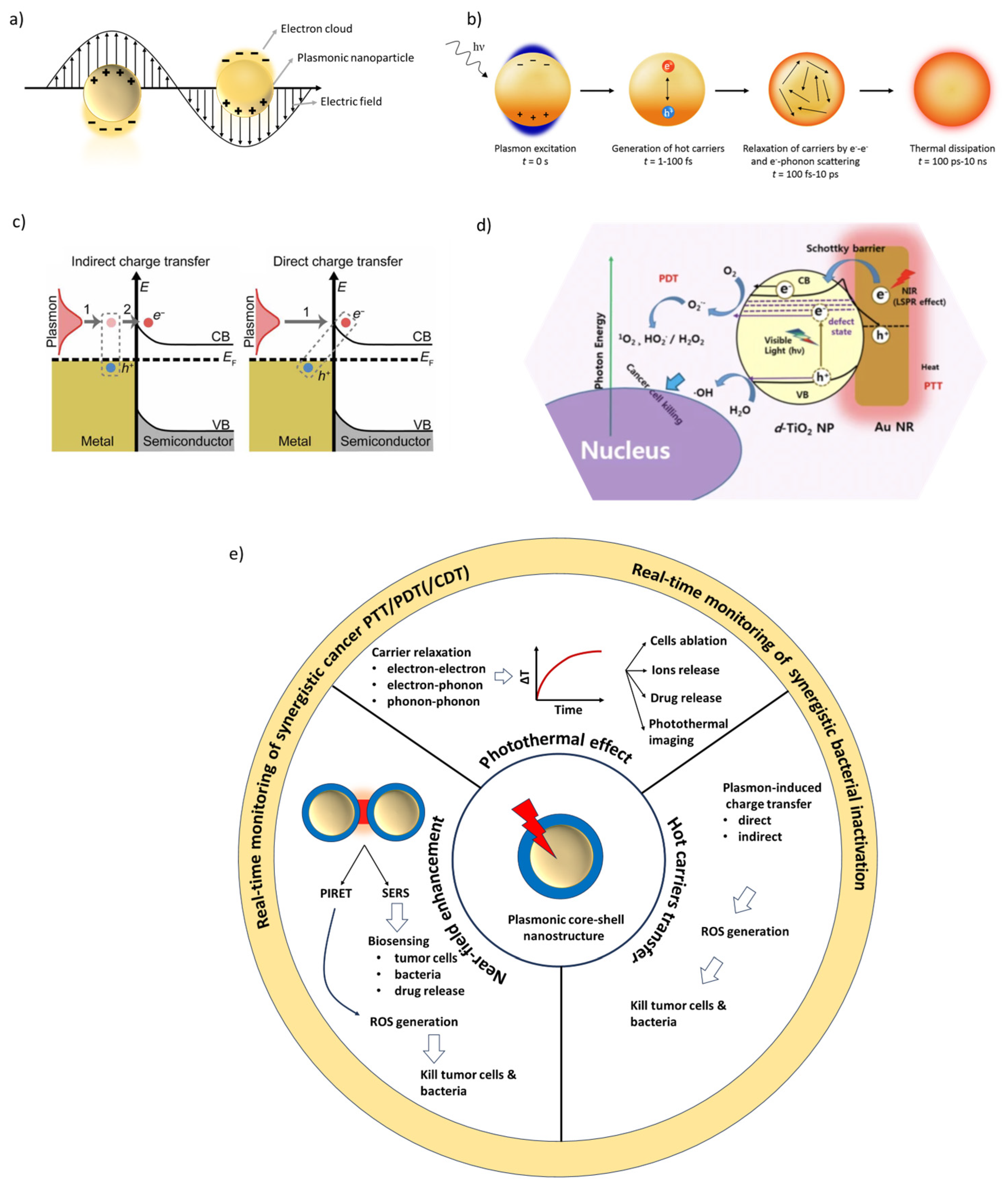
2.2. Mechanisms of LSPR-Induced Photothermal and Photochemical Effects
- Radical scavengers such as sodium azide (NaN3) to investigate the formation of ROS [47].
- Selective reactions such as the photooxidation of ABDA, a singlet oxygen-specific probe [50].
- Carefully designed in vitro and in vivo control experiments, where formulations are adjusted so that only photothermal or photochemical pathways are activated [53].

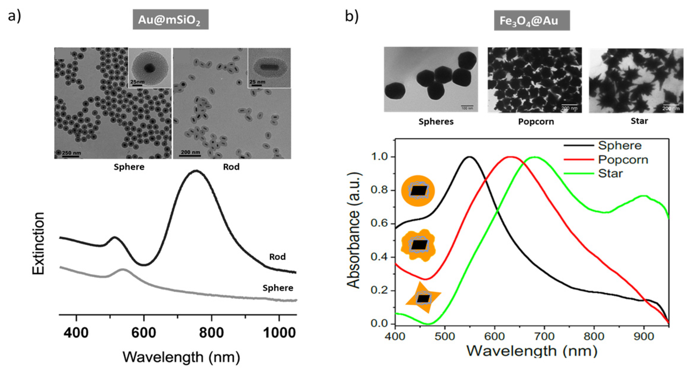
2.3. Tuning LSPR in Core–Shell Nanostructures
3. Core–Shell Architectures in Biomedical Applications
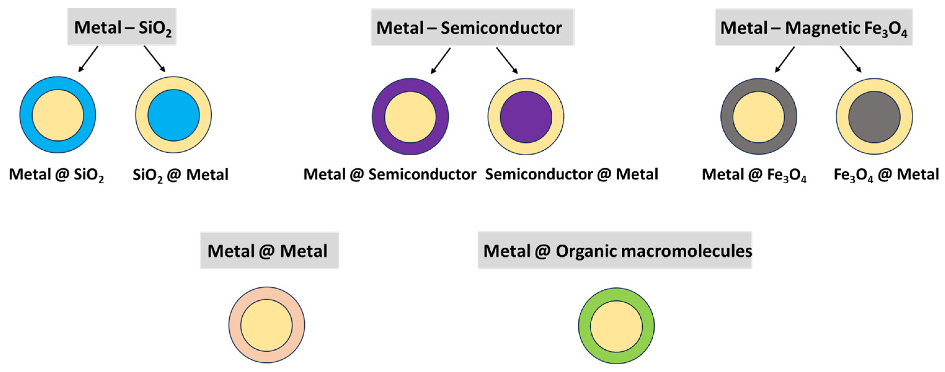
3.1. Overview
3.2. Metal-SiO2
3.3. Metal-Semiconductor
3.4. Metal-Magnetic Fe3O4
3.5. Metal @ Metal
3.6. Metal@Organic Macromolecules
3.7. Plasmonic Core–Shell Architecture Comparison
- Silica is preferred as a core component along with Au as a shell. This configuration can be tuned to absorb in the NIR domain when working with spherical particles (more stable and simpler to synthesize).
- CuS is one of the most frequently used semiconductors in plasmonic core–shell structures due to its ability to perform multiple functions in a multifunctional platform.
- Metal@metal composites show strong potential, but their absorption falls mostly in the visible range, making them more suitable for surface applications. To use this architecture in cancer therapies that require NIR absorption, stabilized anisotropic shapes such as nanorods are required.
- MOFs are an attractive component in core–shell nanostructures because they can act as a drug and photosensitizer carrier, with activation triggered by light or by chemical and physical stimuli.
- Polymers have become essential in multifunctional core–shell composites, serving either as a core or as stabilizing shells that reduce toxicity and improve photothermal performance.
- Monitoring the treatment response by SERS, photoacoustic, and magnetic imaging is increasingly important. In this context, Fe2O3 has been frequently reported as a core material in magnetic imaging-guided therapies.
4. Biomedical Applications
4.1. Antimicrobial Therapy
4.1.1. Mechanisms of Microbial Inactivation
4.1.2. Representative Core–Shell and Hybrid Systems
4.1.3. Biofilm Disruption and Therapeutic Advantages
4.1.4. Clinical Relevance
4.2. Cancer Therapy
4.2.1. Photothermal Ablation Combined with ROS-Induced Apoptosis
4.2.2. ROS-Based Therapy
4.2.3. Targeted Delivery via Tumor-Specific Ligands
4.3. Wound Healing and Tissue Regeneration
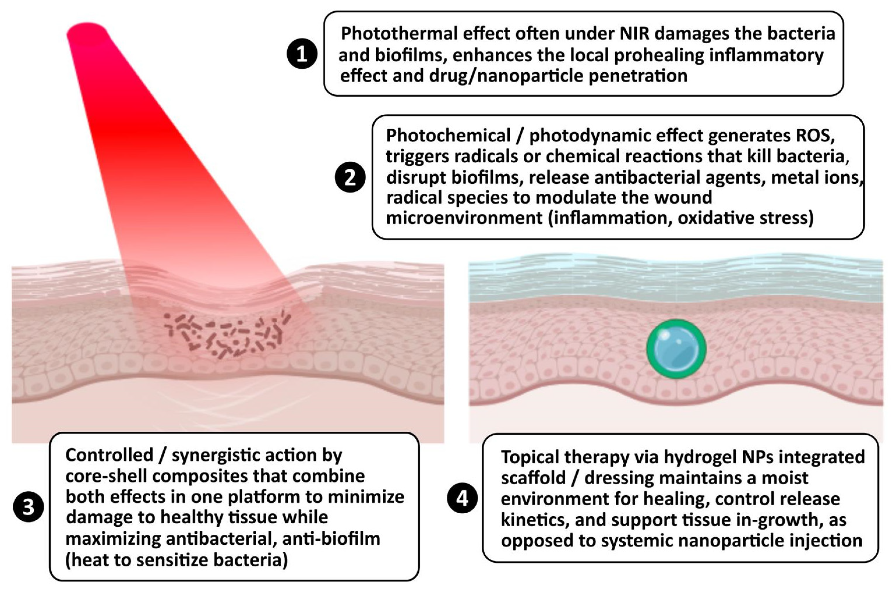
- Plasmonic cores (Au, Cu2−xS) generate heat under NIR irradiation to eliminate bacteria and biofilms,
- Semiconductor or catalytic shells (MnO2, CaO2, hydroxyapatite) regulate ROS and oxygen levels, promoting angiogenesis,
- Cu2+, Ca2+, and NO release stimulates fibroblast proliferation and collagen deposition,
- Hydrogel carrier matrices provide moisture retention, adhesion, and programmable therapeutic release [143].
- more efficient and controllable heating under light irradiation [146].
- the ability to combine multiple functionalities—e.g., a metal core for heat + a shell that carries or facilitates ROS generation, drug release, or catalytic activity [155].
- the potential for improved stability, biocompatibility, and controlled interactions with the biological environment—all of which are critical for wound healing applications [145].
4.4. Biosensing and Bioimaging
4.4.1. Surface-Enhanced Raman Scattering (SERS)
4.4.2. Fluorescence-Based Imaging
4.4.3. Photoacoustic Imaging
4.4.4. ROS and Thermal Feedback-Based Detection Systems

| Material | Applications | Wavelength | Observations | Ref. |
|---|---|---|---|---|
| SPION-PEI@AuNPs | SERS for ultrasensitive detection of circulating tumor cells, cancer biomarkers | LSPR-tuned (visible–NIR) | Enhances electromagnetic field; shell provides stability and functionalization with aptamers/antibodies (e.g., FA-conjugated rBSA); detection limits to 1 cell/mL | [154,158] |
| Ag@TiO2 | SERS for detecting proteins, DNA, small metabolites, pathogens | LSPR-tuned (visible–NIR) | Strong field enhancement; improves signal intensity and reproducibility; protects core from aggregation (supported by multiple studies on Ag/TiO2 hybrids for biomolecule SERS) | [164] |
| Au@Ag@SiO2 | Fluorescence-based imaging of cellular structures, tumor tissues | Tunable via shell thickness (visible–NIR) | PEF; optimizes emission intensity and contrast; supports multiple fluorophores for multi-biomarker imaging | [165] |
| Au/ZnO | Fluorescence imaging with radiosensitization for triple-negative breast cancer | Tunable (visible–NIR) | High brightness and photostability; modulates tumor hypoxia and ROS generation | [159] |
| Au@carbon/Au@TiO2 | Photoacoustic imaging of tumors | NIR (700–1100 nm) | High photothermal conversion efficiency; deep tissue penetration; theranostic potential combining diagnostics and therapy (supported by carbon-plasmonic photoacoustic agents; for similar Au@SiO2) | [166] |
| Au@SiO2 (functionalized) | Fluorescence imaging with targeting | NIR (700–1100 nm) | Silica shell enhances biocompatibility; allows ligand attachment for specificity | [160] |
| Au@TiO2 | ROS-based glucose sensing, oxidative stress detection | Light irradiation (UV–visible) | Photocatalytic shell generates ROS; modulates sensor responses via photothermal effects | [161] |
| SiO2/ZnO shells | Thermal feedback in dynamic biosensing, drug release monitoring | Light irradiation (UV–visible–NIR) | Integrates ROS/heat for real-time cellular microenvironment monitoring | [162,163] |
| Magnetic–plasmonic hybrids | MRI-guided cancer detection/treatment, magnetic hyperthermia | NIR-II (1000–1700 nm) | Enables deeper penetration, reduced photodamage; synergy with targeting and therapy | [167] |
| All-organic/biodegradable core–shell | Stimuli-responsive theranostics (pH/enzyme/light-triggered ROS/heat release) | NIR-II (1000–1700 nm) | Addresses toxicity concerns; integrates with immunotherapy/gene editing for precision medicine | [168] |
5. Design Considerations and Performance Optimization
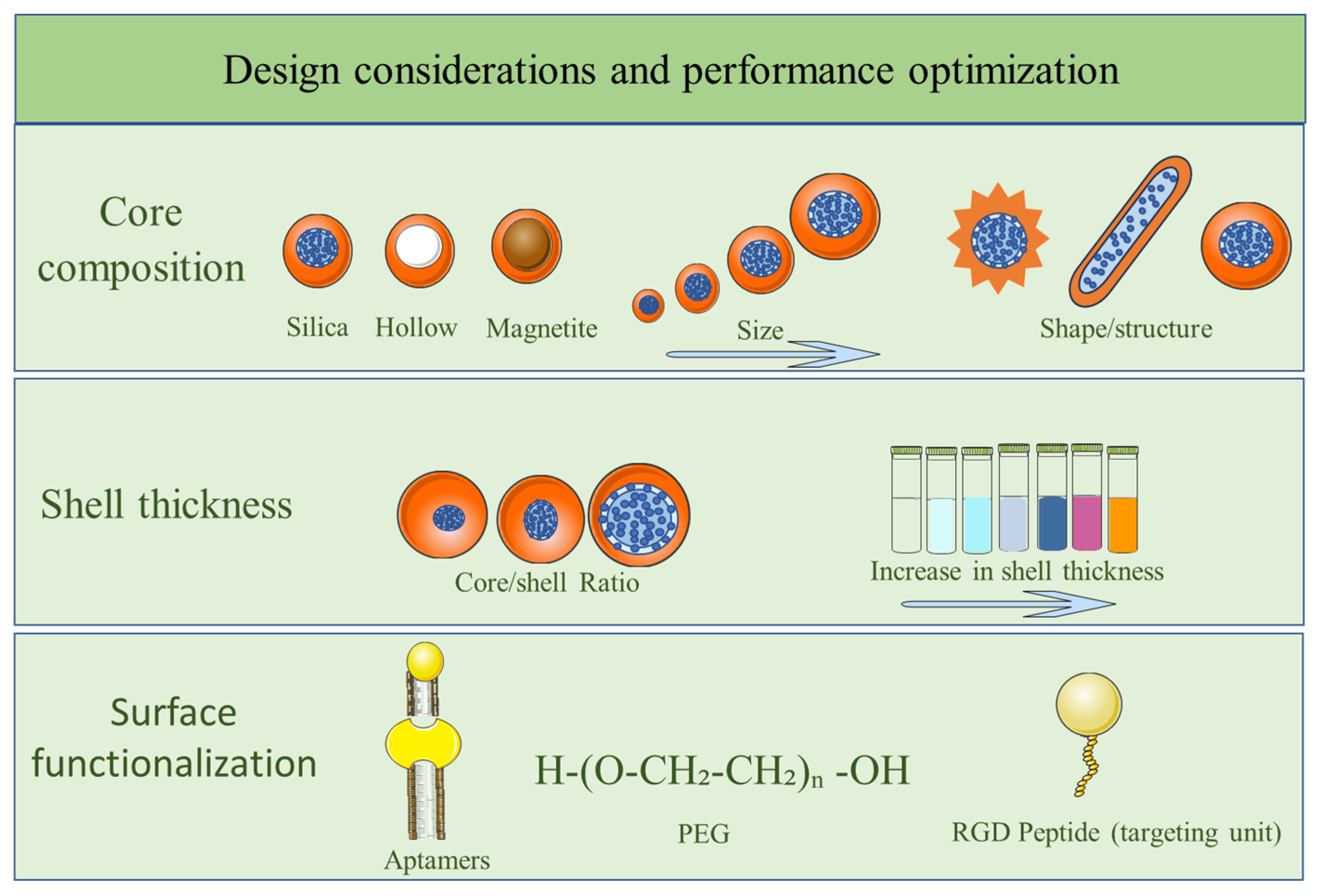
5.1. Core Material Selection Based on LSPR Range and Biocompatibility
5.2. Shell Thickness Optimization for Efficient Energy Transfer and Permeability
5.3. Stability Under Physiological Conditions
5.4. Surface Functionalization with PEG, Peptides, or Aptamers for Targeting and Stealth
6. In Vitro and In Vivo Toxicity
6.1. General Aspects
- their thickness and porosity are controlled, and
- toxic templating surfactants are removed or exchanged.
6.2. Clearance Pathways: Renal, Hepatic, RES-Mediated
- acute safety is often fine,
- the real risk is long-term RES burden plus low-grade immunotoxic and oxidative effects.
6.3. Long-Term Effects on Immunogenicity
- Innate immunity (Complement activation—C3b opsonization, Uptake by macrophages/dendritic cells, Inflammatory cytokine release -TNF-α, IL-6, IL-1β, etc) and
- Adaptive immunity (Antigen presentation, T-cell activation, Antibody production—even anti-PEG antibodies if PEGylated).
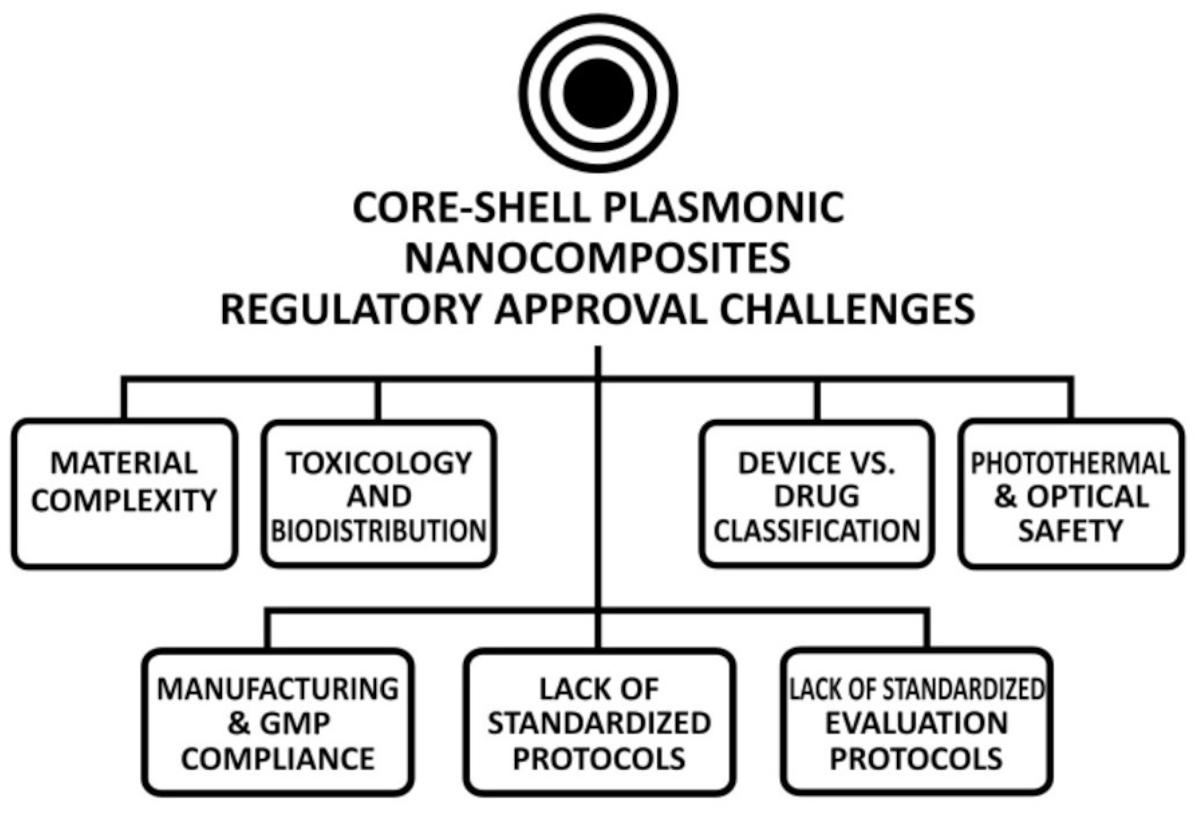
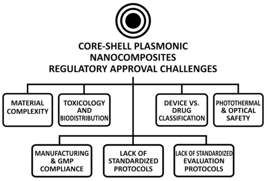
6.4. Core–Shell Plasmonic Nanocomposites: Status of Clinical Translation and Regulatory Approval Challenges
7. Challenges and Perspectives
- Strong Preclinical Efforts (more long-term toxicity/biodistribution/clearance studies and more human-tumor-mimicking in vivo models),
- Optimized Design for Translation (engineering for effective NIR windows, scaling up synthesis under Good Manufacturing Practice (GMP)-like conditions, surface adjustments for biocompatibility and targeting),
- Regulatory Strategy (early involvement with Food and Drug Administration (FDA) or European Medicines Agency (EMA), regulatory support programs),
- Clinical Trial Planning (design for safety, dose escalation, light dose, patient selection, and delivery systems),
- Business/Funding Models (collaboration among academia, industry, and regulatory bodies).
8. Conclusions
Author Contributions
Funding
Data Availability Statement
Conflicts of Interest
Abbreviations
| ALT | Alanine Aminotransferase |
| AM | Acrylamide |
| AST | Aspartate Aminotransferase |
| BSA | Bovine Serum Albumin |
| BUN | Blood Urea Nitrogen |
| CARPA | Complement Activation-Related Pseudoallergy |
| CBC | Complete Blood Count |
| CDT | Chemodynamic Therapy |
| CHX | Chlorhexidine |
| cRGD | Cyclic Arginine–Glycine–Aspartic acid |
| CTAB | Cetyltrimethylammonium Bromide |
| DC | Dendritic Cell |
| DNA | Deoxyribonucleic Acid |
| DOX | Doxorubicin |
| EMA | European Medicines Agency |
| EPR | Enhanced Permeability and Retention |
| EPS | Extracellular Polymeric Substances |
| FA | Folic Acid |
| FDA | Food and Drug Administration |
| GA | Gambogic Acid |
| GMP | Good Manufacturing Practice |
| GT | Gene Therapy |
| FHMP | Fluorescent Hexametaphosphate |
| GSH | Glutathione |
| HD | Hydrodynamic Diameter |
| IL-1β | Interleukin-1 beta |
| IL-6 | Interleukin-6 |
| IL-8 | Interleukin-8 |
| IOC | Iron Oxide Cluster |
| LSPR | Localized Surface Plasmon Resonance |
| mAbs | Monoclonal Antibodies |
| MOA | Mechanisms of action |
| MOF | Metal–Organic Frameworks |
| mPEG | Methoxy Polyethene Glycol |
| MRI | Magnetic Resonance Imaging |
| MIC | Minimum Inhibitory Concentration |
| MIP | Molecularly Imprinted Polymer |
| MUA | 11-Mercaptoundecanoic Acid |
| NIPAM | N-isopropylacrylamide |
| NIR | Near-Infrared |
| NR | Nanorods |
| NIR-II | Second Near-Infrared |
| NPs | Nanoparticles |
| NS | Nanosphere |
| NF-κB | Nuclear Factor-kappa B |
| PANI | Polyaniline |
| PBS | Phosphate-Buffered Saline |
| PCE | Photothermal Conversion Efficiency |
| PDA | Polydopamine |
| PDA-HA | Polydopamine–Hyaluronic Acid |
| PDT | Photodynamic Therapy |
| PEF | Plasmon-Enhanced Fluorescence |
| PEG | Polyethylene Glycol |
| PEI | Polyethyleneimine |
| PIRET | Plasmon-Induced Resonance Energy Transfer |
| PLA | Polylactic Acid |
| PLAL | Pulse Laser Ablation in Liquid |
| PLGA | Poly(lactic-co-glycolic acid) |
| POD | Peroxidase |
| PTT | Photothermal Therapy |
| PVP | Polyvinylpyrrolidone |
| RB | Rose Bengal |
| rBSA | Reduced Albumin from Bovine Serum |
| RCS | Reactive Chlorine Species |
| RES | Reticuloendothelial System |
| rhTNF | Recombinant Human Tumor Necrosis Factor |
| ROS | Reactive Oxygen Species |
| SERS | Surface-Enhanced Raman Scattering |
| SPION | Superparamagnetic Iron Oxide Nanoparticle |
| SPR | Surface Plasmon Resonance |
| TEOS | Tetraethyl Orthosilicate |
| TNF-α | Tumor Necrosis Factor |
| UCNP | Upconverting Nanoparticles |
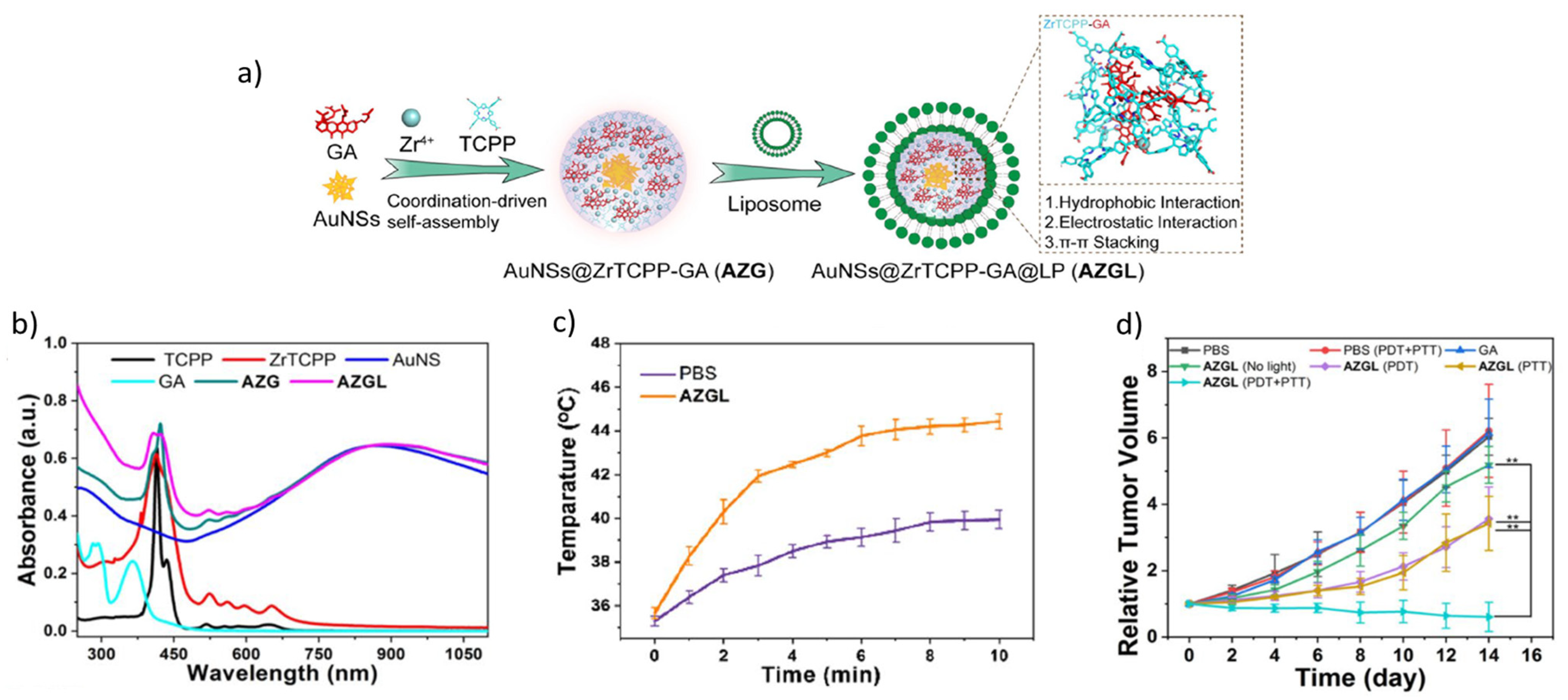
| ZrTCPP | Zirconium Tetrakis(4-carboxyphenyl)porphyrin |
References
- Durgam, L.K.; Oroszi, T.L. Revolutionizing healthcare: The transformative potential of nanotechnology in medicine. Front. Drug Deliv. 2025, 5, 1556426. [Google Scholar] [CrossRef]
- He, Z.; Zhang, C.Y.; Lei, Y.; Song, G.; Yao, Y. Plasmonic nanomaterials: A versatile phototheranostic platform of cancers. Mater. Today 2023, 62, 168–189. [Google Scholar] [CrossRef]
- Sotiriou, G.A. Biomedical applications of multifunctional plasmonic nanoparticles. Wiley Interdiscip. Rev. Nanomed. Nanobiotechnol. 2013, 5, 19–30. [Google Scholar] [CrossRef] [PubMed]
- Mcoyi, M.P.; Mpofu, K.T.; Sekhwama, M.; Mthunzi-Kufa, P. Developments in localized surface plasmon resonance. Plasmonics 2025, 20, 5481–5520. [Google Scholar] [CrossRef]
- Dediu, V.; Ghitman, J.; Gradisteanu Pircalabioru, G.; Chan, K.H.; Iliescu, F.S.; Iliescu, C. Trends in photothermal nanostructures for antimicrobial applications. Int. J. Mol. Sci. 2023, 24, 9375. [Google Scholar] [CrossRef]
- Liang, J.; Qin, Y.; Yang, Y.; Song, Z.; Li, Y.; Liu, G.L.; Hu, W. Nanoplasmonic sensor optimization via digital imaging analysis and antibody evolution. Sens. Actuators B Chem. 2024, 418, 136287. [Google Scholar] [CrossRef]
- Xu, T.; Geng, Z. Strategies to improve performances of LSPR biosensing: Structure, materials, and interface modification. Biosens. Bioelectron. 2021, 174, 112850. [Google Scholar] [CrossRef]
- Torres Fredes, I.P.; Cortés-Adasme, E.N.; Barrientos, B.A.; Real, J.P.; Gomez, C.G.; Palma, S.D.; Kogan, M.J.; Real, D.A. 3D-Printed Plasmonic Nanocomposites: VAT Photopolymerization for Photothermal-Controlled Drug Release. Pharmaceuticals 2024, 17, 1453. [Google Scholar] [CrossRef]
- Goodeve, C.; Kitchener, J. Photosensitisation by titanium dioxide. Trans. Faraday Soc. 1938, 34, 570–579. [Google Scholar] [CrossRef]
- Fujishima, A.; Honda, K. Electrochemical photolysis of water at a semiconductor electrode. Nature 1972, 238, 37–38. [Google Scholar] [CrossRef]
- Kretschmann, E.; Raether, H. Radiative decay of non radiative surface plasmons excited by light. Z. Für Naturforschung A 1968, 23, 2135–2136. [Google Scholar] [CrossRef]
- Hirsch, L.R.; Stafford, R.J.; Bankson, J.; Sershen, S.R.; Rivera, B.; Price, R.; Hazle, J.D.; Halas, N.J.; West, J.L. Nanoshell-mediated near-infrared thermal therapy of tumors under magnetic resonance guidance. Proc. Natl. Acad. Sci. USA 2003, 100, 13549–13554. [Google Scholar] [CrossRef] [PubMed]
- He, L.; Mao, C.; Brasino, M.; Harguindey, A.; Park, W.; Goodwin, A.P.; Cha, J.N. TiO2-capped gold nanorods for plasmon-enhanced production of reactive oxygen species and photothermal delivery of chemotherapeutic agents. ACS Appl. Mater. Interfaces 2018, 10, 27965–27971. [Google Scholar] [CrossRef] [PubMed]
- Lee, J.; Lee, Y.H.; Jeong, C.B.; Choi, J.S.; Chang, K.S.; Yoon, M. Gold nanorods-conjugated TiO2 nanoclusters for the synergistic combination of phototherapeutic treatments of cancer cells. J. Nanobiotechnol. 2018, 16, 104. [Google Scholar] [CrossRef] [PubMed]
- Zheng, T.; Wang, W.; Wu, F.; Zhang, M.; Shen, J.; Sun, Y. Zwitterionic polymer-gated Au@TiO2 core–shell nanoparticles for imaging-guided combined cancer therapy. Theranostics 2019, 9, 5035. [Google Scholar] [CrossRef]
- Chu, Z.; Tian, T.; Tao, Z.; Yang, J.; Chen, B.; Chen, H.; Wang, W.; Yin, P.; Xia, X.; Wang, H. Upconversion nanoparticles@AgBiS2 core–shell nanoparticles with cancer-cell-specific cytotoxicity for combined photothermal and photodynamic therapy of cancers. Bioact. Mater. 2022, 17, 71–80. [Google Scholar] [CrossRef]
- Wang, J.; Sui, L.; Huang, J.; Miao, L.; Nie, Y.; Wang, K.; Yang, Z.; Huang, Q.; Gong, X.; Nan, Y. MoS2-based nanocomposites for cancer diagnosis and therapy. Bioact. Mater. 2021, 6, 4209–4242. [Google Scholar] [CrossRef]
- Zou, Y.; Sun, Z.; Wang, Q.; Ju, Y.; Sun, N.; Yue, Q.; Deng, Y.; Liu, S.; Yang, S.; Wang, Z. Core–Shell Magnetic Particles: Tailored Synthesis and Applications. Chem. Rev. 2024, 125, 972–1048. [Google Scholar] [CrossRef]
- Fakhri, A.; Tahami, S.; Naji, M. Synthesis and characterization of core–shell bimetallic nanoparticles for synergistic antimicrobial effect studies in combination with doxycycline on burn specific pathogens. J. Photochem. Photobiol. B Biol. 2017, 169, 21–26. [Google Scholar] [CrossRef]
- Jiang, B.; Mu, M.; Zhou, Y.; Zhang, J.; Li, W. Nanoparticle-Empowered Core–Shell Microcapsules: From Architecture Design to Fabrication and Functions. Small 2024, 20, 2311897. [Google Scholar] [CrossRef]
- He, M.; Yang, J.; Li, K.; Wang, Q.; Gu, J. NIR-triggered Tandem Photothermal and Photochemical Cancer Therapy Based on Nanoscale MOFs under Hypoxic Conditions. ChemNanoMat 2022, 8, e202200205. [Google Scholar] [CrossRef]
- Park, J.H.; Choe, H.-S.; Kim, S.-W.; Im, G.-B.; Um, S.H.; Kim, J.-H.; Bhang, S.H. Silica-capped and gold-decorated silica nanoparticles for enhancing effect of gold nanoparticle-based photothermal therapy. Tissue Eng. Regen. Med. 2022, 19, 1161–1168. [Google Scholar] [CrossRef]
- Kwizera, E.A.; Chaffin, E.; Shen, X.; Chen, J.; Zou, Q.; Wu, Z.; Gai, Z.; Bhana, S.; O’Connor, R.; Wang, L. Size-and shape-controlled synthesis and properties of magnetic–plasmonic core–shell nanoparticles. J. Phys. Chem. C 2016, 120, 10530–10546. [Google Scholar] [CrossRef]
- Huang, J.; Xu, Z.; Jiang, Y.; Law, W.-c.; Dong, B.; Zeng, X.; Ma, M.; Xu, G.; Zou, J.; Yang, C. Metal organic framework-coated gold nanorod as an on-demand drug delivery platform for chemo-photothermal cancer therapy. J. Nanobiotechnol. 2021, 19, 219. [Google Scholar] [CrossRef] [PubMed]
- Nikolopoulou, S.G.; Kalska, B.; Basa, A.; Papadopoulou, A.; Efthimiadou, E.K. Novel hybrid silver–silica nanoparticles synthesized by modifications of the sol–gel method and their theranostic potential in cancer. ACS Appl. Bio Mater. 2023, 6, 5235–5251. [Google Scholar] [CrossRef]
- Li, J.; Hu, Y.; Yang, J.; Wei, P.; Sun, W.; Shen, M.; Zhang, G.; Shi, X. Hyaluronic acid-modified Fe3O4@Au core/shell nanostars for multimodal imaging and photothermal therapy of tumors. Biomaterials 2015, 38, 10–21. [Google Scholar] [CrossRef] [PubMed]
- Riedel, R.; Mahr, N.; Yao, C.; Wu, A.; Yang, F.; Hampp, N. Synthesis of gold–silica core–shell nanoparticles by pulsed laser ablation in liquid and their physico-chemical properties towards photothermal cancer therapy. Nanoscale 2020, 12, 3007–3018. [Google Scholar] [CrossRef]
- Shanon, Z.S.; Al-Helaly, M.T. Investigation of the Optical, Structural, and Morphological Properties of Triple Au@Ag@Pd Core@Shell Nanoparticles Prepared by the PLAL Technique for Antibacterial Agent Applications. Plasmonics 2025, 20, 9963–9975. [Google Scholar] [CrossRef]
- Khantamat, O.; Li, C.-H.; Yu, F.; Jamison, A.C.; Shih, W.-C.; Cai, C.; Lee, T.R. Gold nanoshell-decorated silicone surfaces for the near-infrared (NIR) photothermal destruction of the pathogenic bacterium E. faecalis. ACS Appl. Mater. Interfaces 2015, 7, 3981–3993. [Google Scholar] [CrossRef]
- Turkevich, J.; Stevenson, P.C.; Hillier, J. A study of the nucleation and growth processes in the synthesis of colloidal gold. Discuss. Faraday Soc. 1951, 11, 55–75. [Google Scholar] [CrossRef]
- Stöber, W.; Fink, A.; Bohn, E. Controlled growth of monodisperse silica spheres in the micron size range. J. Colloid Interface Sci. 1968, 26, 62–69. [Google Scholar] [CrossRef]
- Ghosh Chaudhuri, R.; Paria, S. Core/shell nanoparticles: Classes, properties, synthesis mechanisms, characterization, and applications. Chem. Rev. 2012, 112, 2373–2433. [Google Scholar] [CrossRef] [PubMed]
- Topete, A.; Varela, A.; Navarro-Real, M.; Rial, R.; Pardo, A.; Taboada, P. Revisiting gold nanoshells as multifunctional biomedical nanotools. Coord. Chem. Rev. 2025, 523, 216250. [Google Scholar] [CrossRef]
- Kim, M.; Lee, J.H.; Nam, J.M. Plasmonic Photothermal Nanoparticles for Biomedical Applications. Adv. Sci. 2019, 6, 1900471. [Google Scholar] [CrossRef]
- Ahlawat, M.; Mittal, D.; Govind Rao, V. Plasmon-induced hot-hole generation and extraction at nano-heterointerfaces for photocatalysis. Commun. Mater. 2021, 2, 114. [Google Scholar] [CrossRef]
- Chen, S.; Lei, Q.; Qiu, W.-X.; Liu, L.-H.; Zheng, D.-W.; Fan, J.-X.; Rong, L.; Sun, Y.-X.; Zhang, X.-Z. Mitochondria-targeting “Nanoheater” for enhanced photothermal/chemo-therapy. Biomaterials 2017, 117, 92–104. [Google Scholar] [CrossRef] [PubMed]
- Xu, J.; Huang, X.; Peng, J.; Li, S.; Li, J.-F. Insights into plasmon-assisted chemical reactions: From fabrication to characterization. EScience 2025, 5, 100312. [Google Scholar] [CrossRef]
- Cushing, S.K.; Li, J.; Bright, J.; Yost, B.T.; Zheng, P.; Bristow, A.D.; Wu, N. Controlling plasmon-induced resonance energy transfer and hot electron injection processes in Metal@TiO2 core–shell nanoparticles. J. Phys. Chem. C 2015, 119, 16239–16244. [Google Scholar] [CrossRef]
- Zheng, T.; Zhou, T.; Feng, X.; Shen, J.; Zhang, M.; Sun, Y. Enhanced plasmon-induced resonance energy transfer (PIRET)-mediated photothermal and photodynamic therapy guided by photoacoustic and magnetic resonance imaging. ACS Appl. Mater. Interfaces 2019, 11, 31615–31626. [Google Scholar] [CrossRef]
- Cialla-May, D.; Bonifacio, A.; Markin, A.; Markina, N.; Fornasaro, S.; Dwivedi, A.; Dib, T.; Farnesi, E.; Liu, C.; Ghosh, A. Recent advances of surface enhanced Raman spectroscopy (SERS) in optical biosensing. TrAC Trends Anal. Chem. 2024, 181, 117990. [Google Scholar] [CrossRef]
- Alhujaily, M.; Jabir, M.S.; Nayef, U.M.; Rashid, T.M.; Sulaiman, G.M.; Khalil, K.A.; Rahmah, M.I.; Najm, M.A.; Jabbar, R.; Jawad, S.F. Au/ZnO nanocomposites prepared by laser ablation for enhancement of antibacterial activity and cytotoxic properties against cancer cells. Metals 2023, 13, 735. [Google Scholar] [CrossRef]
- Cai, B.; Liu, J.; Fang, Q.; Zhou, Y.; Zahoor, S.; Guo, X.; Wen, C. Au@CuS@CuO2 nanoconstructs self-generate H2O2 and GSH-consuming enhanced CDT combined with PTT for cancer therapy. J. Photochem. Photobiol. A Chem. 2025, 472, 116758. [Google Scholar] [CrossRef]
- Lin, X.-C.; Liu, J.; Hu, M.; Song, L.; Li, M.; Kou, Q.; Huang, R.; Sun, L.; Wen, C. An Au@CuS@CuO2 nanoplatform with peroxidase mimetic activity and self-supply H2O2 properties for SERS detection of GSH. Spectrochim. Acta Part A Mol. Biomol. Spectrosc. 2025, 340, 126376. [Google Scholar] [CrossRef]
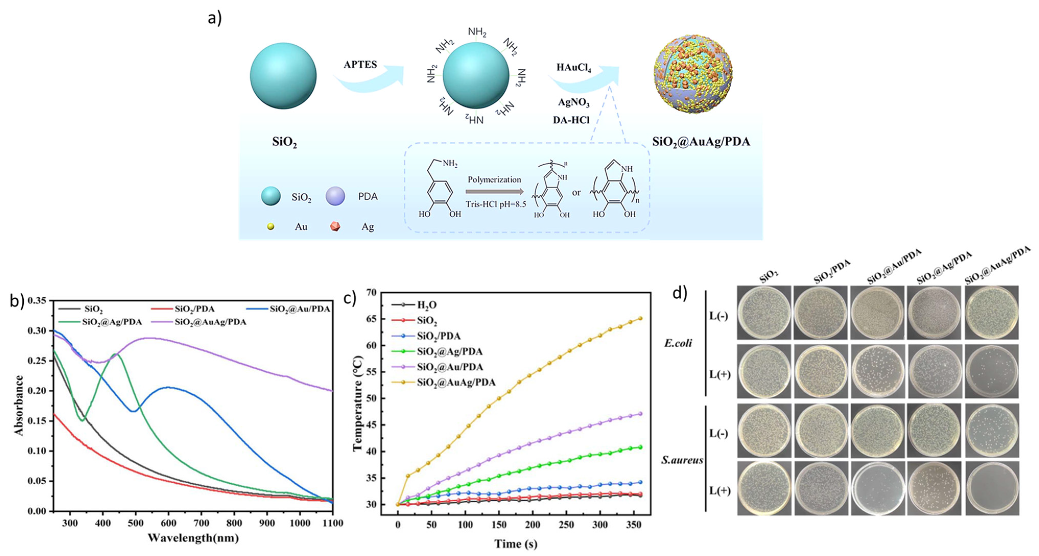
- Ci, D.; Wang, N.; Xu, Y.; Wu, S.; Wang, J.; Li, H.; Xuan, S.; Fang, Q. SiO2@AuAg/PDA hybrid nanospheres with photo-thermally enhanced synergistic antibacterial and catalytic activity. RSC Adv. 2024, 14, 4518–4532. [Google Scholar] [CrossRef]
- Zhang, S.; Yu, S.; Sun, J.; Huang, T.; Lin, H.; Li, Z.; Xiao, Z.; Lu, W. Au@CuS Nanoshells for Surface-Enhanced Raman Scattering Image-Guided Tumor Photothermal Therapy with Accelerated Hepatobiliary Excretion. Pharmaceutics 2024, 16, 1089. [Google Scholar] [CrossRef] [PubMed]
- Husain, S.; Mutalik, C.; Yougbaré, S.; Chen, C.-Y.; Kuo, T.-R. Plasmonic Au@Ag Core–Shell nanoisland Film for Photothermal Inactivation and Surface-Enhanced Raman Scattering Detection of Bacteria. Nanomaterials 2024, 14, 695. [Google Scholar] [CrossRef]
- Dhanalekshmi, K.; Magesan, P.; Sangeetha, K.; Zhang, X.; Jayamoorthy, K.; Srinivasan, N. Preparation and characterization of core–shell type Ag@SiO2 nanoparticles for photodynamic cancer therapy. Photodiagnosis Photodyn. Ther. 2019, 28, 324–329. [Google Scholar] [CrossRef] [PubMed]
- Chu, Z.; Yin, C.; Zhang, S.; Lin, G.; Li, Q. Surface plasmon enhanced drug efficacy using core–shell Au@SiO2 nanoparticle carrier. Nanoscale 2013, 5, 3406–3411. [Google Scholar] [CrossRef] [PubMed]
- Zhang, S.; Lv, H.; Zhao, J.; Cheng, M.; Sun, S. Synthesis of porphyrin-conjugated silica-coated Au nanorods for synergistic photothermal therapy and photodynamic therapy of tumor. Nanotechnology 2019, 30, 265102. [Google Scholar] [CrossRef]
- Macia, N.; Bresoli-Obach, R.; Nonell, S.; Heyne, B. Hybrid silver nanocubes for improved plasmon-enhanced singlet oxygen production and inactivation of bacteria. J. Am. Chem. Soc. 2018, 141, 684–692. [Google Scholar] [CrossRef]
- Lu, M.; Kang, N.; Chen, C.; Yang, L.; Li, Y.; Hong, M.; Luo, X.; Ren, L.; Wang, X. Plasmonic enhancement of cyanine dyes for near-infrared light-triggered photodynamic/photothermal therapy and fluorescent imaging. Nanotechnology 2017, 28, 445710. [Google Scholar] [CrossRef] [PubMed]
- Sujai, P.T.; Shamjith, S.; Joseph, M.M.; Maiti, K.K. Elucidating Gold–MnO2 core–shell nanoenvelope for real time SERS-guided photothermal therapy on pancreatic cancer cells. ACS Appl. Bio Mater. 2021, 4, 4962–4972. [Google Scholar] [CrossRef]
- Zhou, T.; Huang, J.; Zhao, W.; Guo, R.; Cui, S.; Li, Y.; Kadasala, N.R.; Jiang, Y.; Liu, Y.; Zhang, Q. A multifunctional nanoplatform based on Fe3O4@Au NCs with magnetic targeting ability for single NIR light-triggered PTT/PDT synergistic therapy of cancer. J. Alloys Compd. 2023, 944, 169206. [Google Scholar] [CrossRef]
- Ostovar, B.; Lee, S.A.; Mehmood, A.; Farrell, K.; Searles, E.K.; Bourgeois, B.; Chiang, W.-Y.; Misiura, A.; Gross, N.; Al-Zubeidi, A. The role of the plasmon in interfacial charge transfer. Sci. Adv. 2024, 10, eadp3353. [Google Scholar] [CrossRef]
- Nguyen, Q.; Zilli, A.; Celebrano, M.; Baldi, A. Tailoring core size, shell thickness, and aluminium doping of Au@ZnO core@shell nanoparticles. J. Mater. Chem. C 2025, 13, 8302–8309. [Google Scholar] [CrossRef]
- Dias, D.R.; Moreira, A.F.; Correia, I.J. The effect of the shape of gold core–mesoporous silica shell nanoparticles on the cellular behavior and tumor spheroid penetration. J. Mater. Chem. B 2016, 4, 7630–7640. [Google Scholar] [CrossRef]
- Yang, L.; Yan, Z.; Yang, L.; Yang, J.; Jin, M.; Xing, X.; Zhou, G.; Shui, L. Photothermal conversion of SiO2@Au nanoparticles mediated by surface morphology of gold cluster layer. RSC Adv. 2020, 10, 33119–33128. [Google Scholar] [CrossRef]
- Hazra, A.; Hossain, S.M.; Pramanick, A.K.; Ray, M. Spontaneous Interfacial Alloying in Au@Ag Core@Shell Nanoparticles: Unraveling the Impact on Plasmonic Absorption. J. Phys. Chem. C 2025, 129, 11003. [Google Scholar] [CrossRef]
- Li, M.; Yu, X.F.; Liang, S.; Peng, X.N.; Yang, Z.J.; Wang, Y.L.; Wang, Q.Q. Synthesis of Au–CdS core–shell hetero-nanorods with efficient exciton–plasmon interactions. Adv. Funct. Mater. 2011, 21, 1788–1794. [Google Scholar] [CrossRef]
- Wu, W.-C.; Tracy, J.B. Large-scale silica overcoating of gold nanorods with tunable shell thicknesses. Chem. Mater. 2015, 27, 2888–2894. [Google Scholar] [CrossRef]
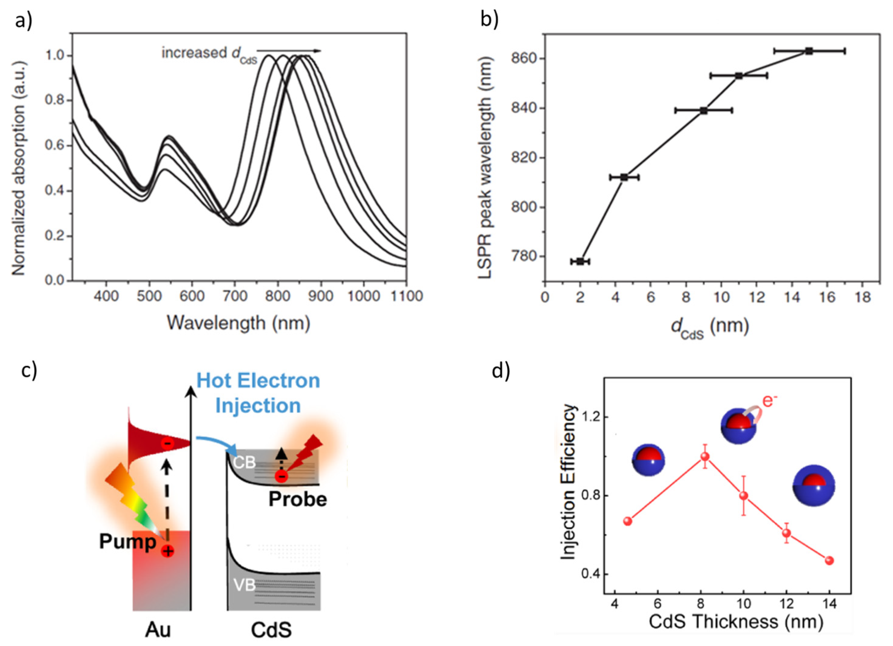
- Dong, H.; Feng, J.; Liu, J.; Wan, X.; Zhang, J.; Wang, Z.; Chen, H.; Weng, Y.-X. Shell thickness dependence of the plasmon-induced hot-electron injection process in Au@CdS core–shell nanocrystals. J. Phys. Chem. C 2021, 125, 19906–19913. [Google Scholar] [CrossRef]
- Ninakanti, R.; Borah, R.; Craig, T.; Ciocarlan, R.-G.; Cool, P.; Bals, S.; Verbruggen, S.W. Au@TiO2 Core–Shell Nanoparticles with Nanometer-Controlled Shell Thickness for Balancing Stability and Field Enhancement in Plasmon-Enhanced Photocatalysis. ACS Nano 2024, 18, 33430–33440. [Google Scholar] [CrossRef]
- Oldenburg, S.; Averitt, R.; Westcott, S.; Halas, N. Nanoengineering of optical resonances. Chem. Phys. Lett. 1998, 288, 243–247. [Google Scholar] [CrossRef]
- Li, S.; Shen, X.; Xu, Q.-H.; Cao, Y. Gold nanorod enhanced conjugated polymer/photosensitizer composite nanoparticles for simultaneous two-photon excitation fluorescence imaging and photodynamic therapy. Nanoscale 2019, 11, 19551–19560. [Google Scholar] [CrossRef]
- Wang, R.; Du, N.; Jin, L.; Chen, W.; Ma, Z.; Zhang, T.; Xu, J.; Zhang, W.; Wang, X.; Li, M. Hyaluronic acid modified Au@SiO2@Au nanoparticles for photothermal therapy of genitourinary tumors. Polymers 2022, 14, 4772. [Google Scholar] [CrossRef] [PubMed]
- Peng, F.; Ni, J.; Xu, H.; Jin, Z.; Liang, D.; Ma, W.; Zhao, Y. Au-Ag@MnO2 NPs with label-free SERS activity and plasmon-enhanced electroreduction activity for dual-mode sensing of uric acid. Sens. Actuators B Chem. 2024, 407, 135459. [Google Scholar] [CrossRef]
- Zheng, D.; Zhao, X.; Wang, Y.; Dai, X.; Guo, K.; Xu, C.; Zhao, N.; Xu, F.-J. Facile construction of polycation/Au@CuS nanohybrids for synergistic gene/photothermal therapy. Chem. Eng. J. 2024, 486, 150271. [Google Scholar] [CrossRef]
- Liu, B.; Zhou, J.; Zhang, B.; Qu, J. Synthesis of Ag@Fe3O4 nanoparticles for photothermal treatment of ovarian cancer. J. Nanomater. 2019, 2019, 6457968. [Google Scholar] [CrossRef]
- Hu, Y.; Wang, R.; Wang, S.; Ding, L.; Li, J.; Luo, Y.; Wang, X.; Shen, M.; Shi, X. Multifunctional Fe3O4@Au core/shell nanostars: A unique platform for multimode imaging and photothermal therapy of tumors. Sci. Rep. 2016, 6, 28325. [Google Scholar] [CrossRef] [PubMed]
- Zhao, W.; Zhang, D.; Zhou, T.; Huang, J.; Wang, Y.; Li, B.; Chen, L.; Yang, J.; Liu, Y. Aptamer-conjugated magnetic Fe3O4@Au core–shell multifunctional nanoprobe: A three-in-one aptasensor for selective capture, sensitive SERS detection and efficient near-infrared light triggered photothermal therapy of Staphylococcus aureus. Sens. Actuators B Chem. 2022, 350, 130879. [Google Scholar] [CrossRef]
- Wang, Y.; Li, Q.; Zhang, R.; Tang, K.; Ding, C.; Yu, S. SERS-based immunocapture and detection of pathogenic bacteria using a boronic acid-functionalized polydopamine-coated Au@Ag nanoprobe. Microchim. Acta 2020, 187, 290. [Google Scholar] [CrossRef]
- Tang, Z.; Ali, I.; Hou, Y.; Akakuru, O.U.; Zhang, Q.; Mushtaq, A.; Zhang, H.; Lu, Y.; Ma, X.; Ge, J. pH-Responsive Au@Pd bimetallic core–shell nanorods for enhanced synergistic targeted photothermal-augmented nanocatalytic therapy in the second near-infrared window. J. Mater. Chem. B 2022, 10, 6532–6545. [Google Scholar] [CrossRef] [PubMed]
- Ghosal, M.; Mondal, S.; Ghosh, T.; Prusty, D.; Senapati, D. Core-to-Shell Thickness-Regulated Ag@Au Nanocatalyst for LSPR-Improved In Situ Detection of Extracellular Peroxide: Response in a Cancer Cell. Anal. Chem. 2025, 97, 7651–7661. [Google Scholar] [CrossRef]
- Chen, H.; Liu, Z.; Li, S.; Su, C.; Qiu, X.; Zhong, H.; Guo, Z. Fabrication of graphene and AuNP core polyaniline shell nanocomposites as multifunctional theranostic platforms for SERS real-time monitoring and chemo-photothermal therapy. Theranostics 2016, 6, 1096. [Google Scholar] [CrossRef]
- Wang, J.; Gao, Y.; Liu, P.; Xu, S.; Luo, X. Core–shell multifunctional nanomaterial-based all-in-one nanoplatform for simultaneous multilayer imaging of dual types of tumor biomarkers and photothermal therapy. Anal. Chem. 2020, 92, 15169–15178. [Google Scholar] [CrossRef] [PubMed]
- He, J.; Dong, J.; Hu, Y.; Li, G.; Hu, Y. Design of Raman tag-bridged core–shell Au@Cu3(BTC)2 nanoparticles for Raman imaging and synergistic chemo-photothermal therapy. Nanoscale 2019, 11, 6089–6100. [Google Scholar] [CrossRef] [PubMed]
- Li, R.-T.; Zhu, Y.-D.; Li, W.-Y.; Hou, Y.-K.; Zou, Y.-M.; Zhao, Y.-H.; Zou, Q.; Zhang, W.-H.; Chen, J.-X. Synergistic photothermal-photodynamic-chemotherapy toward breast cancer based on a liposome-coated core–shell AuNS@NMOFs nanocomposite encapsulated with gambogic acid. J. Nanobiotechnol. 2022, 20, 212. [Google Scholar] [CrossRef]
- Albrecht, W.; van de Glind, A.; Yoshida, H.; Isozaki, Y.; Imhof, A.; van Blaaderen, A.; de Jongh, P.E.; de Jong, K.P.; Zečević, J.; Takeda, S. Impact of the electron beam on the thermal stability of gold nanorods studied by environmental transmission electron microscopy. Ultramicroscopy 2018, 193, 97–103. [Google Scholar] [CrossRef]
- Xie, X.; Albrecht, W.; van Huis, M.A.; van Blaaderen, A. Unexpectedly high thermal stability of Au nanotriangle@mSiO2 yolk–shell nanoparticles. Nanoscale 2024, 16, 4787–4795. [Google Scholar] [CrossRef]
- Planas, O.; Macia, N.; Agut, M.; Nonell, S.; Heyne, B. Distance-dependent plasmon-enhanced singlet oxygen production and emission for bacterial inactivation. J. Am. Chem. Soc. 2016, 138, 2762–2768. [Google Scholar] [CrossRef]
- Zhou, Y.; Liu, Y.; Wang, Y.; Hu, X.; Koh, K.; Chen, H. Tunable Au@SiO2/Au film metasurface as surface plasmon resonance enhancer for direct and ultrasensitive detection of exosomes. Anal. Chem. 2023, 95, 9663–9671. [Google Scholar] [CrossRef]
- Kah, J.C.Y.; Wan, R.C.Y.; Wong, K.Y.; Mhaisalkar, S.; Sheppard, C.J.R.; Olivo, M. Combinatorial treatment of photothermal therapy using gold nanoshells with conventional photodynamic therapy to improve treatment efficacy: An in vitro study. Lasers Surg. Med. Off. J. Am. Soc. Laser Med. Surg. 2008, 40, 584–589. [Google Scholar] [CrossRef]
- Prodan, E.; Radloff, C.; Halas, N.J.; Nordlander, P. A hybridization model for the plasmon response of complex nanostructures. Science 2003, 302, 419–422. [Google Scholar] [CrossRef]
- Gad, S.C.; Sharp, K.L.; Montgomery, C.; Payne, J.D.; Goodrich, G.P. Evaluation of the toxicity of intravenous delivery of auroshell particles (gold–silica nanoshells). Int. J. Toxicol. 2012, 31, 584–594. [Google Scholar] [CrossRef] [PubMed]
- Rastinehad, A.R.; Anastos, H.; Wajswol, E.; Winoker, J.S.; Sfakianos, J.P.; Doppalapudi, S.K.; Carrick, M.R.; Knauer, C.J.; Taouli, B.; Lewis, S.C. Gold nanoshell-localized photothermal ablation of prostate tumors in a clinical pilot device study. Proc. Natl. Acad. Sci. USA 2019, 116, 18590–18596. [Google Scholar] [CrossRef] [PubMed]
- Wang, H.-J.; Lin, J.-S.; Zhang, H.; Zhang, Y.-J.; Li, J.-F. Plasmonic core–shell materials: Synthesis, spectroscopic characterization, and photocatalytic applications. Acc. Mater. Res. 2022, 3, 187–198. [Google Scholar] [CrossRef]
- Fang, C.; Jia, H.; Chang, S.; Ruan, Q.; Wang, P.; Chen, T.; Wang, J. (Gold core)/(titania shell) nanostructures for plasmon-enhanced photon harvesting and generation of reactive oxygen species. Energy Environ. Sci. 2014, 7, 3431–3438. [Google Scholar] [CrossRef]
- Oh, J.T.; Chowdhury, S.R.; ll Lee, T.; Misra, M. Synergetic influence of Au/Cu2O core–shells nanoparticle on optical, photo-electrochemical, and catalytic activities of Au/Cu2O/TiO2 nanocomposite. Dye. Pigment. 2019, 160, 936–943. [Google Scholar] [CrossRef]
- Feng, L.; He, F.; Dai, Y.; Liu, B.; Yang, G.; Gai, S.; Niu, N.; Lv, R.; Li, C.; Yang, P. A versatile near infrared light triggered dual-photosensitizer for synchronous bioimaging and photodynamic therapy. ACS Appl. Mater. Interfaces 2017, 9, 12993–13008. [Google Scholar] [CrossRef]
- Misra, M.; Kapur, P.; Singla, M.L. Surface plasmon quenched of near band edge emission and enhanced visible photocatalytic activity of Au@ZnO core–shell nanostructure. Appl. Catal. B Environ. 2014, 150, 605–611. [Google Scholar] [CrossRef]
- Kuo, M.-Y.; Hsiao, C.-F.; Chiu, Y.-H.; Lai, T.-H.; Fang, M.-J.; Wu, J.-Y.; Chen, J.-W.; Wu, C.-L.; Wei, K.-H.; Lin, H.-C. Au@Cu2O core@shell nanocrystals as dual-functional catalysts for sustainable environmental applications. Appl. Catal. B Environ. 2019, 242, 499–506. [Google Scholar] [CrossRef]
- Yang, J.-L.; He, Y.-L.; Ren, H.; Zhong, H.-L.; Lin, J.-S.; Yang, W.-M.; Li, M.-D.; Yang, Z.-L.; Zhang, H.; Tian, Z.-Q. Boosting photocatalytic hydrogen evolution reaction using dual plasmonic antennas. ACS Catal. 2021, 11, 5047–5053. [Google Scholar] [CrossRef]
- Dediu, V.; Busila, M.; Tucureanu, V.; Bucur, F.I.; Iliescu, F.S.; Brincoveanu, O.; Iliescu, C. Synthesis of ZnO/Au nanocomposite for antibacterial applications. Nanomaterials 2022, 12, 3832. [Google Scholar] [CrossRef]
- Dutta, G.; Sugumaran, A. Bioengineered zinc oxide nanoparticles: Chemical, green, biological fabrication methods and its potential biomedical applications. J. Drug Deliv. Sci. Technol. 2021, 66, 102853. [Google Scholar] [CrossRef]
- Wang, M.; Hou, Z.; Al Kheraif, A.A.; Xing, B.; Lin, J. Mini review of TiO2-based multifunctional nanocomposites for near-infrared light–responsive phototherapy. Adv. Healthc. Mater. 2018, 7, 1800351. [Google Scholar] [CrossRef]
- Ning, X.-F.; Zhu, Y.-Q.; Sun, H.; Yang, Y.; Liu, M.-X. The Latest Applications of Carbon-Nitride-Based Materials for Combination Treatment of Cancer. ACS Appl. Mater. Interfaces 2024, 16, 64410–64423. [Google Scholar] [CrossRef]
- Yimeng, S.; Huilun, X.; Ziming, L.; Kejun, L.; Chaima, M.; Xiangyu, Z.; Yinchun, H.; Yan, W.; Di, H. Copper-based nanoparticles as antibacterial agents. Eur. J. Inorg. Chem. 2023, 26, e202200614. [Google Scholar] [CrossRef]
- Fang, W.; Zhang, H.; Wang, X.; Wei, W.; Shen, Y.; Yu, J.; Liang, J.; Zheng, J.; Shen, Y. Facile synthesis of tunable plasmonic silver core/magnetic Fe3O4 shell nanoparticles for rapid capture and effective photothermal ablation of bacterial pathogens. New J. Chem. 2017, 41, 10155–10164. [Google Scholar] [CrossRef]
- Xu, K.; Fang, Q.; Wang, J.; Hui, A.; Xuan, S. Magnetic-field-induced improvement of photothermal sterilization performance by Fe3O4@SiO2@Au/PDA nanochains. Materials 2022, 16, 387. [Google Scholar] [CrossRef] [PubMed]
- Huang, X.; Tang, S.; Liu, B.; Ren, B.; Zheng, N. Enhancing the photothermal stability of plasmonic metal Nanoplates by a core-shell architecture. Adv. Mater. 2011, 23, 3420–3425. [Google Scholar] [CrossRef]
- Awiaz, G.; Lin, J.; Wu, A. Recent advances of Au@Ag core–shell SERS-based biosensors. Exploration 2023, 3, 20220072. [Google Scholar] [CrossRef]
- Zhang, W.; Zhang, Q.; Zhao, H.; Yu, Y.; Liu, Y.; Gu, N.; Cheng, W. Adaptive nanoplasmonics controlled by soft polymers. ACS Appl. Mater. Interfaces 2025, 17, 46463–46489. [Google Scholar] [CrossRef]
- Hu, B.; Wang, N.; Han, L.; Chen, M.-L.; Wang, J.-H. Core–shell–shell nanorods for controlled release of silver that can serve as a nanoheater for photothermal treatment on bacteria. Acta Biomater. 2015, 11, 511–519. [Google Scholar] [CrossRef]
- Hayakawa, Y.; Furuya, M.; Tahara, H.; Kosuge, Y.; Kimura, T.; Sugawa, K.; Otsuki, J. Modulation technique of localized surface plasmon resonance of palladium nanospheres by coating with titanium dioxide shell for application to photothermal therapy agent. Nanoscale Res. Lett. 2022, 17, 60. [Google Scholar] [CrossRef] [PubMed]
- Gao, F.; He, G.; Yin, H.; Chen, J.; Liu, Y.; Lan, C.; Zhang, S.; Yang, B. Titania-coated 2D gold nanoplates as nanoagents for synergistic photothermal/sonodynamic therapy in the second near-infrared window. Nanoscale 2019, 11, 2374–2384. [Google Scholar] [CrossRef] [PubMed]
- Rizzi, F.; Fanizza, E.; Giancaspro, M.; Depalo, N.; Curri, M.L.; Gonzalez, B.; Colilla, M.; Izquierdo-Barba, I.; Vallet-Regi, M. Mesoporous silica nanostructures embedding NIR active plasmonic nanoparticles: Harnessing antimicrobial agents delivery system for photo-assisted eradicating Gram-positive bacteria. Microporous Mesoporous Mater. 2025, 383, 113414. [Google Scholar] [CrossRef]
- Zhang, H.; Lu, Y.; Zhang, R.; Tang, Z.; Lam, S.H.; Zhu, J.; Fu, R.; Hu, Y.; Iqbal, M.Z.; Kong, X. Synthesis of Multifunctional Plasmonic Nanodarts through One-End Deposition on Gold Nanobipyramids for Tumor Organoid Ablation and Antimicrobial Applications. Adv. Funct. Mater. 2024, 34, 2405588. [Google Scholar] [CrossRef]
- Chen, J.; Ye, Z.; Yang, F.; Yin, Y. Plasmonic nanostructures for photothermal conversion. Small Sci. 2021, 1, 2000055. [Google Scholar] [CrossRef]
- Majhi, R.K.; Mohanty, S.; Khan, M.I.; Mishra, A.; Brauner, A. Ag@ZnO nanoparticles induce antimicrobial peptides and promote migration and antibacterial activity of keratinocytes. ACS Infect. Dis. 2021, 7, 2068–2072. [Google Scholar] [CrossRef]
- Al-Kattan, A.; Tselikov, G.; Metwally, K.; Popov, A.A.; Mensah, S.; Kabashin, A.V. Laser ablation-assisted synthesis of plasmonic Si@Au core-satellite nanocomposites for biomedical applications. Nanomaterials 2021, 11, 592. [Google Scholar] [CrossRef]
- Savchuk, V.; Wang, R.; Small, L.; Pinchuk, A. Synergistic Effect in Hybrid Plasmonic Conjugates for Photothermal Applications. ACS Omega 2024, 9, 47436–47441. [Google Scholar] [CrossRef]
- Wang, H.; Yang, X.; Zhu, T.; Su, X.; Xiao, L.; Yu, Q.; Liu, P.; Wang, L. Plasmonic MXene/Au@Ag Core–Shell Heterostructures: A Unified Platform for Electromagnetic Stealth, Antimicrobial Action, and Environmental Remediation. Nanoscale 2025, 17, 16398–16410. [Google Scholar] [CrossRef] [PubMed]
- Ding, X.; Yuan, P.; Gao, N.; Zhu, H.; Yang, Y.Y.; Xu, Q.-H. Au-Ag core–shell nanoparticles for simultaneous bacterial imaging and synergistic antibacterial activity. Nanomed. Nanotechnol. Biol. Med. 2017, 13, 297–305. [Google Scholar] [CrossRef]
- Feng, Y.; Sun, Q.; Liu, P.; Fan, W.; Fan, B. Antibacterial property and mechanisms of Au@Ag Core–Shell nanoparticles with Near-Infrared absorption against E. faecalis infection of dentin. Int. J. Nanomed. 2024, 19, 6981–6997. [Google Scholar] [CrossRef]
- Rani, P.; Varma, R.S.; Singh, K.; Acevedo, R.; Singh, J. Catalytic and antimicrobial potential of green synthesized Au and Au@Ag core–shell nanoparticles. Chemosphere 2023, 317, 137841. [Google Scholar] [CrossRef]
- Shilpa, M.; Shetty, V.; Surabhi, S.; Jeong, J.-R.; Morales, D.; Ballal, M.; Eshwarappa, K.; Murari, M.; Nayak, R.; Gurumurthy, S. Decentralized core–shell Au/Ag bimetallic nanostructures prepared via green approach for catalytic and antimicrobial applications. Mater. Sci. Eng. B 2023, 298, 116893. [Google Scholar] [CrossRef]
- Yang, L.; Yan, W.; Wang, H.; Zhuang, H.; Zhang, J. Shell thickness-dependent antibacterial activity and biocompatibility of gold@silver core–shell nanoparticles. RSC Adv. 2017, 7, 11355–11361. [Google Scholar] [CrossRef]
- He, J.; Qiao, Y.; Zhang, H.; Zhao, J.; Li, W.; Xie, T.; Zhong, D.; Wei, Q.; Hua, S.; Yu, Y. Gold–silver nanoshells promote wound healing from drug-resistant bacteria infection and enable monitoring via surface-enhanced Raman scattering imaging. Biomaterials 2020, 234, 119763. [Google Scholar] [CrossRef]
- Mu, W.; Fang, W.; Yao, Y. Synthesis of Ag@Au core–shell NPs loaded with ciprofloxacin as enhanced antimicrobial properties for the treatment and nursing care of Escherichia coli infection. Microb. Pathog. 2021, 150, 104619. [Google Scholar] [CrossRef] [PubMed]
- Hu, X.; Zhao, Y.; Hu, Z.; Saran, A.; Hou, S.; Wen, T.; Liu, W.; Ji, Y.; Jiang, X.; Wu, X. Gold nanorods core/AgPt alloy nanodots shell: A novel potent antibacterial nanostructure. Nano Res. 2013, 6, 822–835. [Google Scholar] [CrossRef]
- Thangudu, S.; Kulkarni, S.S.; Vankayala, R.; Chiang, C.-S.; Hwang, K.C. Photosensitized reactive chlorine species-mediated therapeutic destruction of drug-resistant bacteria using plasmonic core–shell Ag@AgCl nanocubes as an external nanomedicine. Nanoscale 2020, 12, 12970–12984. [Google Scholar] [CrossRef]
- Rauf, S.; Safdar, N. Sustainable fabrication of biogenic Fe–Zn nano core–shell and their photocatalytic efficiency against third generation antibiotics. Mater. Chem. Phys. 2024, 313, 128784. [Google Scholar] [CrossRef]
- Merkl, P.; Zhou, S.; Zaganiaris, A.; Shahata, M.; Eleftheraki, A.; Thersleff, T.; Sotiriou, G.A. Plasmonic coupling in silver nanoparticle aggregates and their polymer composite films for near-infrared photothermal biofilm eradication. ACS Appl. Nano Mater. 2021, 4, 5330–5339. [Google Scholar] [CrossRef] [PubMed]
- Melancon, M.P.; Zhou, M.; Li, C. Cancer theranostics with near-infrared light-activatable multimodal nanoparticles. Acc. Chem. Res. 2011, 44, 947–956. [Google Scholar] [CrossRef] [PubMed]
- O’Neal, D.P.; Hirsch, L.R.; Halas, N.J.; Payne, J.D.; West, J.L. Photo-thermal tumor ablation in mice using near infrared-absorbing nanoparticles. Cancer Lett. 2004, 209, 171–176. [Google Scholar] [CrossRef]
- Bui, H.-T.D.; Park, Y.; Jung, Y.M.; Chew, S.Y.; Yoo, H.S. Reactive oxygen species-responsive clicked assembly of gold nanoparticles to enhance photothermal therapy. J. Mater. Chem. B 2023, 11, 6961–6974. [Google Scholar] [CrossRef]
- Yang, L.; Tseng, Y.-T.; Suo, G.; Chen, L.; Yu, J.; Chiu, W.-J.; Huang, C.-C.; Lin, C.-H. Photothermal therapeutic response of cancer cells to aptamer–gold nanoparticle-hybridized graphene oxide under NIR illumination. ACS Appl. Mater. Interfaces 2015, 7, 5097–5106. [Google Scholar] [CrossRef]
- Wei, Y.; Wang, Z.; Yang, J.; Xu, R.; Deng, H.; Ma, S.; Fang, T.; Zhang, J.; Shen, Q. Reactive oxygen species/photothermal therapy dual-triggered biomimetic gold nanocages nanoplatform for combination cancer therapy via ferroptosis and tumor-associated macrophage repolarization mechanism. J. Colloid Interface Sci. 2022, 606, 1950–1965. [Google Scholar] [CrossRef]
- Taheri-Ledari, R.; Ganjali, F.; Zarei-Shokat, S.; Dinmohammadi, R.; Asl, F.R.; Emami, A.; Mojtabapour, Z.S.; Rashvandi, Z.; Kashtiaray, A.; Jalali, F. Plasmonic porous micro-and nano-materials based on Au/Ag nanostructures developed for photothermal cancer therapy: Challenges in clinicalization. Nanoscale Adv. 2023, 5, 6768–6786. [Google Scholar] [CrossRef]
- Fan, Z.; Shelton, M.; Singh, A.K.; Senapati, D.; Khan, S.A.; Ray, P.C. Multifunctional plasmonic shell–magnetic core nanoparticles for targeted diagnostics, isolation, and photothermal destruction of tumor cells. ACS Nano 2012, 6, 1065–1073. [Google Scholar] [CrossRef]
- Chen, N.-T.; Tang, K.-C.; Chung, M.-F.; Cheng, S.-H.; Huang, C.-M.; Chu, C.-H.; Chou, P.-T.; Souris, J.S.; Chen, C.-T.; Mou, C.-Y. Enhanced plasmonic resonance energy transfer in mesoporous silica-encased gold nanorod for two-photon-activated photodynamic therapy. Theranostics 2014, 4, 798. [Google Scholar] [CrossRef]
- Trachootham, D.; Alexandre, J.; Huang, P. Targeting cancer cells by ROS-mediated mechanisms: A radical therapeutic approach? Nat. Rev. Drug Discov. 2009, 8, 579–591. [Google Scholar] [CrossRef]
- Khazaei, Z.; Sohrabivafa, M.; Momenabadi, V.; Moayed, L.; Goodarzi, E. Global cancer statistics 2018: Globocan estimates of incidence and mortality worldwide prostate cancers and their relationship with the human development index. Adv. Hum. Biol. 2019, 9, 245–250. [Google Scholar] [CrossRef]
- Siegel, R.L.; Miller, K.D.; Jemal, A. Cancer statistics, 2019. CA A Cancer J. Clin. 2019, 69, 7–34. [Google Scholar] [CrossRef]
- Cairns, R.A.; Harris, I.S.; Mak, T.W. Regulation of cancer cell metabolism. Nat. Rev. Cancer 2011, 11, 85–95. [Google Scholar] [CrossRef]
- Ercal, N.; Gurer-Orhan, H.; Aykin-Burns, N. Toxic metals and oxidative stress part I: Mechanisms involved in metal-induced oxidative damage. Curr. Top. Med. Chem. 2001, 1, 529–539. [Google Scholar] [CrossRef] [PubMed]
- Deng, Y.; Song, P.; Chen, X.; Huang, Y.; Hong, L.; Jin, Q.; Ji, J. 3-Bromopyruvate-conjugated nanoplatform-induced pro-death autophagy for enhanced photodynamic therapy against hypoxic tumor. ACS Nano 2020, 14, 9711–9727. [Google Scholar] [CrossRef] [PubMed]
- Sung, H.; Ferlay, J.; Siegel, R.L.; Laversanne, M.; Soerjomataram, I.; Jemal, A.; Bray, F. Global cancer statistics 2020: GLOBOCAN estimates of incidence and mortality worldwide for 36 cancers in 185 countries. CA A Cancer J. Clin. 2021, 71, 209–249. [Google Scholar] [CrossRef]
- Bradbury, P.; Traini, D.; Ammit, A.J.; Young, P.M.; Ong, H.X. Repurposing of statins via inhalation to treat lung inflammatory conditions. Adv. Drug Deliv. Rev. 2018, 133, 93–106. [Google Scholar] [CrossRef]
- Van Giau, V.; An, S.S.A.; Hulme, J. Recent advances in the treatment of pathogenic infections using antibiotics and nano-drug delivery vehicles. Drug Des. Dev. Ther. 2019, 13, 327–343. [Google Scholar] [CrossRef] [PubMed]
- Lindahl, J.F.; Grace, D. The consequences of human actions on risks for infectious diseases: A review. Infect. Ecol. Epidemiol. 2015, 5, 30048. [Google Scholar] [CrossRef]
- Maleki, A.; He, J.; Bochani, S.; Nosrati, V.; Shahbazi, M.-A.; Guo, B. Multifunctional photoactive hydrogels for wound healing acceleration. ACS Nano 2021, 15, 18895–18930. [Google Scholar] [CrossRef]
- Nqoro, X.; Taziwa, R. Polymer-based functional materials loaded with metal-based nanoparticles as potential scaffolds for the management of infected wounds. Pharmaceutics 2024, 16, 155. [Google Scholar] [CrossRef]
- Salehi, N.; Mohammadi, A.; Alinezhad, V.; Bochani, S.; Kalantari-Hesari, A.; Haghi, F.; Valdez, F.J.S.; Buenfil-Chi, T.J.; Maleki, A.; Beigi-Boroujeni, S. Synergistic photothermal and photodynamic therapy to promote bacteria-infected wound healing using ZnO@PDA/Ag-integrated waterborne polyurethane films. J. Mater. Chem. B 2025, 13, 6177–6198. [Google Scholar] [CrossRef]
- Rajpoot, K. Photothermal nanomaterials-based scaffolds for tissue regeneration and cancer therapy. Med. Nov. Technol. Devices 2025, 28, 100395. [Google Scholar] [CrossRef]
- Li, Q.; Zhang, W.; Zhao, D.; Qiu, M. Photothermal enhancement in core–shell structured plasmonic nanoparticles. Plasmonics 2014, 9, 623–630. [Google Scholar] [CrossRef]
- Naskar, A.; Kim, K.-s. Friends against the foe: Synergistic photothermal and photodynamic therapy against bacterial infections. Pharmaceutics 2023, 15, 1116. [Google Scholar] [CrossRef] [PubMed]
- Zhuang, Z.-M.; Wang, Y.; Xu, F.-W.; Guo, K.; Cao, L.-l.; Feng, Z.-X.; Zhong, X.-C.; Chen, C.-Y.; Chen, J.; Du, Y.-Z. Programmed nanozyme hydrogel enabling spatiotemporal modulation of wound healing achieves skin regeneration after biofilm infection. J. Nanobiotechnol. 2025, 23, 694. [Google Scholar] [CrossRef] [PubMed]
- Xu, X.; Liu, X.; Tan, L.; Cui, Z.; Yang, X.; Zhu, S.; Li, Z.; Yuan, X.; Zheng, Y.; Yeung, K.W.K. Controlled-temperature photothermal and oxidative bacteria killing and acceleration of wound healing by polydopamine-assisted Au-hydroxyapatite nanorods. Acta Biomater. 2018, 77, 352–364. [Google Scholar] [CrossRef] [PubMed]
- Xu, X.; Chao, Y.; Ma, X.; Zhang, H.; Chen, J.; Zhu, J. A photothermally antibacterial Au@Halloysite nanotubes/lignin composite hydrogel for promoting wound healing. Int. J. Biol. Macromol. 2024, 258, 128704. [Google Scholar] [CrossRef]
- Huang, X.; Xu, L.; Yu, X.; Li, Y.; Huang, Z.; Xu, R.; Zeng, W.; Zhang, Z.; Li, W.; Deng, F. Near-infrared light-responsive multifunctional hydrogel releasing peptide-functionalized gold nanorods sequentially for diabetic wound healing. J. Colloid Interface Sci. 2023, 639, 369–384. [Google Scholar] [CrossRef] [PubMed]
- Li, C.; He, X.; Li, Q.; Lv, M.; Shen, J.; Jin, L.; He, D. A photothermal-response oxygen release platform based on a hydrogel for accelerating wound healing. NPG Asia Mater. 2023, 15, 3. [Google Scholar] [CrossRef]
- Jia, P.; Zou, Y.; Jiang, J. CuS hybrid hydrogel for near-infrared-enhanced infected wound healing: A gelatin-assisted synthesis and direct incorporation strategy. ACS Appl. Mater. Interfaces 2023, 15, 22929–22943. [Google Scholar] [CrossRef] [PubMed]
- Jia, P.; Zou, Y.; Jiang, J. S-Nitrosylated CuS Hybrid Hydrogel Patches with Robust Antibacterial and Repair-Promoting Activity for Infected Wound Healing. Small 2024, 20, 2307629. [Google Scholar] [CrossRef]
- Koç, M.M.; Paksu, U.; Kurnaz Yetim, N.; Coşkun, B.; Hasanoğlu Özkan, E.; Erkovan, M. Nanoparticles in photothermal therapy-based medical and theranostic applications: An extensive review. Eur. Phys. J. Plus 2025, 140, 514. [Google Scholar] [CrossRef]
- Shaktawat, S.; Singh, K.R.; Thapa, S.; Verma, R.; Singh, J.; Singh, R.P. Optical characteristics and biosensing application of core@shell nanomaterials. Mater. Lett. X 2023, 17, 100187. [Google Scholar] [CrossRef]
- Zhou, Y.; Lu, Y.; Liu, Y.; Hu, X.; Chen, H. Current strategies of plasmonic nanoparticles assisted surface-enhanced Raman scattering toward biosensor studies. Biosens. Bioelectron. 2023, 228, 115231. [Google Scholar] [CrossRef]
- Xue, T.; Wang, S.; Ou, G.; Li, Y.; Ruan, H.; Li, Z.; Ma, Y.; Zou, R.; Qiu, J.; Shen, Z. Detection of circulating tumor cells based on improved SERS-active magnetic nanoparticles. Anal. Methods 2019, 11, 2918–2928. [Google Scholar] [CrossRef]
- Li, J.; You, Z.; Zheng, B.; Zhai, S.; Lu, K. Au–ZnO Nanomatches as Radiosensitizers with Improved Reactive Oxygen Species Generation and Tumor Hypoxia Modulation for Enhanced Radiotherapy on Triple-Negative Breast Cancer. ACS Appl. Nano Mater. 2023, 6, 16362–16372. [Google Scholar] [CrossRef]
- Chen, T.; Li, T.; Wang, J. Nanoscale Au@SiO2-drug/VEGF as an in vivo probe for osteosarcoma diagnosis and therapy. Oncol. Lett. 2021, 22, 766. [Google Scholar] [CrossRef]
- Peng, X.; Wan, G.; Wu, L.; Zeng, M.; Lin, S.; Wang, G. Peroxidase-like activity of Au@TiO2 yolk-shell nanostructure and its application for colorimetric detection of H2O2 and glucose. Sens. Actuators B Chem. 2018, 257, 166–177. [Google Scholar] [CrossRef]
- Butt, M.A. Surface plasmon resonance-based biodetection systems: Principles, progress and applications—A comprehensive review. Biosensors 2025, 15, 35. [Google Scholar] [CrossRef] [PubMed]
- Yosri, N.; Gao, S.; Zhou, R.; Wang, C.; Zou, X.; El-Seedi, H.R.; Guo, Z. Innovative quantum dots-based SERS for ultrasensitive reporting of contaminants in food: Fundamental concepts and practical implementations. Food Chem. 2025, 467, 142395. [Google Scholar] [CrossRef]
- Mi, S.; Hu, X.; Yuan, S.; Yu, H.; Guo, Y.; Cheng, Y.; Yao, W. Unveiling the correlation between protein, protein corona, and target signal loss in SERS detection. Anal. Chem. 2024, 96, 19768–19777. [Google Scholar] [CrossRef]
- Dong, L.; Liu, B.; Maenosono, S.; Yang, J. Multifunctional Au@Ag@SiO2 Core–Shell–Shell Nanoparticles for Metal-Enhanced Fluorescence, Surface-Enhanced Raman Scattering, and Photocatalysis Applications. Langmuir 2023, 39, 1593–1599. [Google Scholar] [CrossRef]
- Mallidi, S.; Luke, G.P.; Emelianov, S. Photoacoustic imaging in cancer detection, diagnosis, and treatment guidance. Trends Biotechnol. 2011, 29, 213–221. [Google Scholar] [CrossRef]
- Rodriguez-Nieves, A.L.; Shah, S.; Taylor, M.L.; Alle, M.; Huang, X. Magnetic–Plasmonic Core–Shell Nanoparticles: Properties, Synthesis and Applications for Cancer Detection and Treatment. Nanomaterials 2025, 15, 264. [Google Scholar] [CrossRef] [PubMed]
- Li, Z.; Du, L.; Du, B.; Ullah, Z.; Zhang, Y.; Tu, Y.; Zhou, Y.; Guo, B. Inorganic and hybrid nanomaterials for NIR-II fluorescence imaging-guided therapy of Glioblastoma and perspectives. Theranostics 2025, 15, 5616. [Google Scholar] [CrossRef]
- Broadbent, M.; Chadwick, S.J.; Brust, M.; Volk, M. Gold nanoparticles for photothermal and photodynamic therapy. ACS Omega 2024, 9, 44846–44859. [Google Scholar] [CrossRef] [PubMed]
- Badir, A.; Refki, S.; Sekkat, Z. Utilizing gold nanoparticles in plasmonic photothermal therapy for cancer treatment. Heliyon 2025, 11, e42738. [Google Scholar] [CrossRef]
- Zhang, L.; Jiang, C.; Li, B.; Liu, Z.; Gu, B.; He, S.; Li, P.; Sun, Y.; Song, S. A core–shell Au@Cu2−xSe heterogeneous metal nanocomposite for photoacoustic and computed tomography dual-imaging-guided photothermal boosted chemodynamic therapy. J. Nanobiotechnol. 2021, 19, 410. [Google Scholar] [CrossRef]
- Chang, Y.; Cheng, Y.; Feng, Y.; Jian, H.; Wang, L.; Ma, X.; Li, X.; Zhang, H. Resonance energy transfer-promoted photothermal and photodynamic performance of gold–copper sulfide yolk–shell nanoparticles for chemophototherapy of cancer. Nano Lett. 2018, 18, 886–897. [Google Scholar] [CrossRef]
- Yang, W.; Xia, B.; Wang, L.; Ma, S.; Liang, H.; Wang, D.; Huang, J. Shape effects of gold nanoparticles in photothermal cancer therapy. Mater. Today Sustain. 2021, 13, 100078. [Google Scholar] [CrossRef]
- Jain, P.K.; Lee, K.S.; El-Sayed, I.H.; El-Sayed, M.A. Calculated absorption and scattering properties of gold nanoparticles of different size, shape, and composition: Applications in biological imaging and biomedicine. J. Phys. Chem. B 2006, 110, 7238–7248. [Google Scholar] [CrossRef]
- Baffou, G.; Quidant, R.; Girard, C. Heat generation in plasmonic nanostructures: Influence of morphology. Appl. Phys. Lett. 2009, 94, 153109. [Google Scholar] [CrossRef]
- Riley, R.S.; Day, E.S. Gold nanoparticle-mediated photothermal therapy: Applications and opportunities for multimodal cancer treatment. Wiley Interdiscip. Rev. Nanomed. Nanobiotechnol. 2017, 9, e1449. [Google Scholar] [CrossRef]
- Vankayala, R.; Lin, C.-C.; Kalluru, P.; Chiang, C.-S.; Hwang, K.C. Gold nanoshells-mediated bimodal photodynamic and photothermal cancer treatment using ultra-low doses of near infra-red light. Biomaterials 2014, 35, 5527–5538. [Google Scholar] [CrossRef] [PubMed]
- Jiang, K.; Smith, D.A.; Pinchuk, A. Size-dependent photothermal conversion efficiencies of plasmonically heated gold nanoparticles. J. Phys. Chem. C 2013, 117, 27073–27080. [Google Scholar] [CrossRef]
- Taylor, M.L.; Wilson, R.E., Jr.; Amrhein, K.D.; Huang, X. Gold nanorod-assisted photothermal therapy and improvement strategies. Bioengineering 2022, 9, 200. [Google Scholar] [CrossRef]
- Yang, Y.; Zhang, B.; Sun, X.; Tao, Y.; Yang, G.; Liu, C.; Wang, Z.; Sun, L.; Zhang, Q. Tuning the modal coupling in three-dimensional Au@Cu2O@Au core–shell–satellite nanostructures for enhanced plasmonic photocatalysis. Chem. Sci. 2025, 16, 8069–8081. [Google Scholar] [CrossRef]
- Obeng, E.; Feng, J.; Wang, D.; Zheng, D.; Xiang, B.; Shen, J. Multifunctional phototheranostic agent ZnO@Ag for anti-infection through photothermal/photodynamic therapy. Front. Chem. 2022, 10, 1054739. [Google Scholar] [CrossRef] [PubMed]
- Lin, S.; Chen, H.; Wang, R.; Jiang, T.; Yu, F. Hollow silver–gold alloy nanoparticles for enhanced photothermal/photodynamic synergetic therapy against bacterial infection and acceleration of wound healing. Biomater. Sci. 2023, 11, 4874–4889. [Google Scholar] [CrossRef] [PubMed]
- Song, Y.; Tan, K.B.; Zhou, S.-F.; Zhan, G. Biocompatible copper-based nanocomposites for combined cancer therapy. ACS Biomater. Sci. Eng. 2024, 10, 3673–3692. [Google Scholar] [CrossRef] [PubMed]
- dos Santos Batista, J.G.; Vigilato Rodrigues, M.Á.; de Freitas, L.F.; Moreira Fonseca, A.C.; de Souza Rodrigues, A.; Cunha da Cruz, C.P.; Thipe, V.C.; Lugão, A.B. Copper-Based Nanomaterials for Biologically Relevant Compounds. In Copper-Based Nanomaterials for Biologically Relevant Compounds; CRC Press: Boca Raton, FL, USA, 2024; pp. 305–338. [Google Scholar]
- Wang, S.; Riedinger, A.; Li, H.; Fu, C.; Liu, H.; Li, L.; Liu, T.; Tan, L.; Barthel, M.J.; Pugliese, G. Plasmonic copper sulfide nanocrystals exhibiting near-infrared photothermal and photodynamic therapeutic effects. ACS Nano 2015, 9, 1788–1800. [Google Scholar] [CrossRef]
- Chaffin, E.A.; Bhana, S.; O’Connor, R.T.; Huang, X.; Wang, Y. Impact of core dielectric properties on the localized surface plasmonic spectra of gold-coated magnetic core–shell nanoparticles. J. Phys. Chem. B 2014, 118, 14076–14084. [Google Scholar] [CrossRef]
- Sikdar, D.; Rukhlenko, I.D.; Cheng, W.; Premaratne, M. Effect of number density on optimal design of gold nanoshells for plasmonic photothermal therapy. Biomed. Opt. Express 2012, 4, 15–31. [Google Scholar] [CrossRef]
- Bhana, S.; Lin, G.; Wang, L.; Starring, H.; Mishra, S.R.; Liu, G.; Huang, X. Near-infrared-absorbing gold nanopopcorns with iron oxide cluster core for magnetically amplified photothermal and photodynamic cancer therapy. ACS Appl. Mater. Interfaces 2015, 7, 11637–11647. [Google Scholar] [CrossRef]
- Yang, G.; Tao, Y.; Cheng, Q.; Liu, C.; Zhang, B.; Sun, X.; Yang, Y.; Lu, D.; Yang, J.; Deng, L.-L. Shell Dependence of Highly Tunable Circular Dichroism in Chiral Hybrid Plasmonic Nanomaterials for Chiroptical Applications. ACS Nano 2025, 19, 2961–2974. [Google Scholar] [CrossRef]
- Liu, D.-Y.; Ding, S.-Y.; Lin, H.-X.; Liu, B.-J.; Ye, Z.-Z.; Fan, F.-R.; Ren, B.; Tian, Z.-Q. Distinctive enhanced and tunable plasmon resonant absorption from controllable Au@Cu2O nanoparticles: Experimental and theoretical modeling. J. Phys. Chem. C 2012, 116, 4477–4483. [Google Scholar] [CrossRef]
- Wang, Y.; Quinsaat, J.E.Q.; Ono, T.; Maeki, M.; Tokeshi, M.; Isono, T.; Tajima, K.; Satoh, T.; Sato, S.-i.; Miura, Y. Enhanced dispersion stability of gold nanoparticles by the physisorption of cyclic poly (ethylene glycol). Nat. Commun. 2020, 11, 6089. [Google Scholar] [CrossRef]
- Pellas, V.; Hu, D.; Mazouzi, Y.; Mimoun, Y.; Blanchard, J.; Guibert, C.; Salmain, M.; Boujday, S. Gold nanorods for LSPR biosensing: Synthesis, coating by silica, and bioanalytical applications. Biosensors 2020, 10, 146. [Google Scholar] [CrossRef] [PubMed]
- Iqbal, S.; Fakhar-e-Alam, M.; Alimgeer, K.; Atif, M.; Hanif, A.; Yaqub, N.; Farooq, W.; Ahmad, S.; Chu, Y.-M.; Rana, M.S. Mathematical modeling and experimental analysis of the efficacy of photodynamic therapy in conjunction with photo thermal therapy and PEG-coated Au-doped TiO2 nanostructures to target MCF-7 cancerous cells. Saudi J. Biol. Sci. 2021, 28, 1226–1232. [Google Scholar] [CrossRef]
- Moczko, E.; Guerreiro, A.; Piletska, E.; Piletsky, S. PEG-stabilized core–shell surface-imprinted nanoparticles. Langmuir 2013, 29, 9891–9896. [Google Scholar] [CrossRef] [PubMed]
- Yoo, J.; Park, C.; Yi, G.; Lee, D.; Koo, H. Active targeting strategies using biological ligands for nanoparticle drug delivery systems. Cancers 2019, 11, 640. [Google Scholar] [CrossRef]
- Gu, W.; Hua, Z.; Li, Z.; Cai, Z.; Wang, W.; Guo, K.; Yuan, F.; Gao, F.; Chen, H. Palladium cubes with Pt shell deposition for localized surface plasmon resonance enhanced photodynamic and photothermal therapy of hypoxic tumors. Biomater. Sci. 2022, 10, 216–226. [Google Scholar] [CrossRef]
- Wang, J.; Zhu, G.; You, M.; Song, E.; Shukoor, M.I.; Zhang, K.; Altman, M.B.; Chen, Y.; Zhu, Z.; Huang, C.Z. Assembly of aptamer switch probes and photosensitizer on gold nanorods for targeted photothermal and photodynamic cancer therapy. ACS Nano 2012, 6, 5070–5077. [Google Scholar] [CrossRef]
- Yuan, H.; Fales, A.M.; Vo-Dinh, T. TAT peptide-functionalized gold nanostars: Enhanced intracellular delivery and efficient NIR photothermal therapy using ultralow irradiance. J. Am. Chem. Soc. 2012, 134, 11358–11361. [Google Scholar] [CrossRef]
- Shah, B.P.; Pasquale, N.; De, G.; Tan, T.; Ma, J.; Lee, K.-B. Core–shell nanoparticle-based peptide therapeutics and combined hyperthermia for enhanced cancer cell apoptosis. Acs Nano 2014, 8, 9379–9387. [Google Scholar] [CrossRef]
- Yang, Q.; Peng, J.; Xiao, Y.; Li, W.; Tan, L.; Xu, X.; Qian, Z. Porous Au@Pt nanoparticles: Therapeutic platform for tumor chemo-photothermal co-therapy and alleviating doxorubicin-induced oxidative damage. ACS Appl. Mater. Interfaces 2018, 10, 150–164. [Google Scholar] [CrossRef]
- Niżnik, Ł.; Noga, M.; Kobylarz, D.; Frydrych, A.; Krośniak, A.; Kapka-Skrzypczak, L.; Jurowski, K. Gold nanoparticles (AuNPs)—Toxicity, safety and green synthesis: A critical review. Int. J. Mol. Sci. 2024, 25, 4057. [Google Scholar] [CrossRef] [PubMed]
- Han, H.-W.; Joe, A.; Jang, E.-S. Reduced cytotoxicity of CTAB-templated silica layer on gold nanorod using fluorescence dyes and its application in cancer theranostics. J. Ind. Eng. Chem. 2021, 96, 202–212. [Google Scholar] [CrossRef]
- Liu, G.; Li, Q.; Ni, W.; Zhang, N.; Zheng, X.; Wang, Y.; Shao, D.; Tai, G. Cytotoxicity of various types of gold-mesoporous silica nanoparticles in human breast cancer cells. Int. J. Nanomed. 2015, 10, 6075–6087. [Google Scholar] [CrossRef] [PubMed]
- Janjua, T.I.; Cao, Y.; Kleitz, F.; Linden, M.; Yu, C.; Popat, A. Silica nanoparticles: A review of their safety and current strategies to overcome biological barriers. Adv. Drug Deliv. Rev. 2023, 203, 115115. [Google Scholar] [CrossRef]
- De Matteis, V.; Cascione, M.; Toma, C.C.; Leporatti, S. Silver nanoparticles: Synthetic routes, in vitro toxicity and theranostic applications for cancer disease. Nanomaterials 2018, 8, 319. [Google Scholar] [CrossRef]
- Singh, C.; Mehata, A.K.; Priya, V.; Malik, A.K.; Setia, A.; Suseela, M.N.L.; Vikas; Gokul, P.; Samridhi; Singh, S.K. Bimetallic Au–Ag nanoparticles: Advanced nanotechnology for tackling antimicrobial resistance. Molecules 2022, 27, 7059. [Google Scholar] [CrossRef] [PubMed]
- Alwhibi, M.S.; Ortashi, K.M.; Hendi, A.A.; Awad, M.; Soliman, D.A.; El-Zaidy, M. Green synthesis, characterization and biomedical potential of Ag@Au core–shell noble metal nanoparticles. J. King Saud Univ.-Sci. 2022, 34, 102000. [Google Scholar] [CrossRef]
- Nieto-Argüello, A.; Medina-Cruz, D.; Perez-Ramirez, Y.S.; Perez-Garcia, S.A.; Velasco-Soto, M.A.; Jafari, Z.; De Leon, I.; Gonzalez, M.U.; Huttel, Y.; Martinez, L. Composition-dependent cytotoxic and antibacterial activity of biopolymer-capped ag/au bimetallic nanoparticles against melanoma and multidrug-resistant pathogens. Nanomaterials 2022, 12, 779. [Google Scholar] [CrossRef] [PubMed]
- Kovács, D.; Igaz, N.; Marton, A.; Rónavári, A.; Bélteky, P.; Bodai, L.; Spengler, G.; Tiszlavicz, L.; Rázga, Z.; Hegyi, P. Core–shell nanoparticles suppress metastasis and modify the tumour-supportive activity of cancer-associated fibroblasts. J. Nanobiotechnol. 2020, 18, 18. [Google Scholar] [CrossRef]
- Nazari, M.; Shabani, R.; Ajdary, M.; Ashjari, M.; Shirazi, R.; Govahi, A.; Kermanian, F.; Mehdizadeh, M. Effects of Au@Ag core–shell nanostructure with alginate coating on male reproductive system in mice. Toxicol. Rep. 2023, 10, 104–116. [Google Scholar] [CrossRef]
- Khan, G.A.; Anjum, F.; Ghanem, M.A.; Ahmed, W. Enhanced SERS Detection of Toxic Dyes Using Gold–Silver Core-Shell Nanoparticles. ChemistrySelect 2025, 10, e05303. [Google Scholar] [CrossRef]
- Abed, M.A.; Mutlak, F.A.; Jabir, M.S.; Ahmed, A.F. Toxicity analysis of Ag/Au core/shell nanoparticles synthesizes via seed-growth on blood human components. AIP Conf. Proc. 2021, 2372, 100004. [Google Scholar]
- Seo, S.-H.; Joe, A.; Han, H.-W.; Manivasagan, P.; Jang, E.-S. Mesoporous Silica-layered gold nanorod Core@Silver shell nanostructures for intracellular SERS imaging and phototherapy. Pharmaceutics 2024, 16, 137. [Google Scholar] [CrossRef] [PubMed]
- Abbasi, R.; Shineh, G.; Mobaraki, M.; Doughty, S.; Tayebi, L. Structural parameters of nanoparticles affecting their toxicity for biomedical applications: A review. J. Nanoparticle Res. 2023, 25, 43. [Google Scholar] [CrossRef]
- Li, X.; Li, J.; Jiang, L. A silica-gold core–shell structure to mimic the large size of gold particles for promoting cell growth: A comparative study of the silica core size and the nanogold amount in the shell. Colloids Surf. A Physicochem. Eng. Asp. 2014, 459, 211–216. [Google Scholar] [CrossRef]
- Keerthana, L.; Ahmad Dar, M.; Dharmalingam, G. Plasmonic Au-Metal Oxide Nanocomposites for High-Temperature and Harsh Environment Sensing Applications. Chem.–Asian J. 2021, 16, 3558–3584. [Google Scholar] [CrossRef]
- Hang, Y.; Wang, A.; Wu, N. Plasmonic silver and gold nanoparticles: Shape-and structure-modulated plasmonic functionality for point-of-caring sensing, bio-imaging and medical therapy. Chem. Soc. Rev. 2024, 53, 2932–2971. [Google Scholar] [CrossRef]
- Nguyen, V.H.; Lee, B.-J. Protein corona: A new approach for nanomedicine design. Int. J. Nanomed. 2017, 12, 3137–3151. [Google Scholar] [CrossRef]
- Havelikar, U.; Ghorpade, K.B.; Kumar, A.; Patel, A.; Singh, M.; Banjare, N.; Gupta, P.N. Comprehensive insights into mechanism of nanotoxicity, assessment methods and regulatory challenges of nanomedicines. Discov. Nano 2024, 19, 165. [Google Scholar] [CrossRef]
- Kumar, R.; Kumar, A.; Bhardwaj, S.; Sikarwar, M.; Sriwastaw, S.; Sharma, G.; Gupta, M. Nanotoxicity Unveiled: Evaluating Exposure Risks and Assessing the Impact of Nanoparticles on Human Health. J. Trace Elem. Miner. 2025, 13, 100252. [Google Scholar] [CrossRef]
- Queiroz, L.G.; Faustino, L.A.; de Oliveira, P.F.; Pompêo, M.; de Torresi, S.I.C. Transformative nanobioplasmonic effects: Toxicological implications of plasmonic silver nanoparticles in aquatic biological models. Sci. Total Environ. 2024, 954, 176592. [Google Scholar] [CrossRef] [PubMed]
- Bastus, N.G.; Puntes, V. Nanosafety: Towards safer nanoparticles by design. Curr. Med. Chem. 2018, 25, 4587–4601. [Google Scholar] [CrossRef]
- Jia, Y.-P.; Ma, B.-Y.; Wei, X.-W.; Qian, Z.-Y. The in vitro and in vivo toxicity of gold nanoparticles. Chin. Chem. Lett. 2017, 28, 691–702. [Google Scholar] [CrossRef]
- Yedgar, S.; Barshtein, G.; Gural, A. Hemolytic activity of nanoparticles as a marker of their hemocompatibility. Micromachines 2022, 13, 2091. [Google Scholar] [CrossRef]
- Emam, A.; Loutfy, S.A.; Mostafa, A.A.; Awad, H.; Mohamed, M.B. Cyto-toxicity, biocompatibility and cellular response of carbon dots–plasmonic based nano-hybrids for bioimaging. RSC Adv. 2017, 7, 23502–23514. [Google Scholar] [CrossRef]
- Sarimov, R.M.; Molkova, E.A.; Simakin, A.V.; Dorokhov, A.S.; Gudkov, S.V. Protein Corona as a Result of Interaction of Protein Molecules with Inorganic Nanoparticles. Int. J. Mol. Sci. 2025, 26, 9771. [Google Scholar] [CrossRef] [PubMed]
- Pem, B.; Pongrac, I.M.; Ulm, L.; Pavičić, I.; Vrček, V.; Jurašin, D.D.; Ljubojević, M.; Krivohlavek, A.; Vrček, I.V. Toxicity and safety study of silver and gold nanoparticles functionalized with cysteine and glutathione. Beilstein J. Nanotechnol. 2019, 10, 1802–1817. [Google Scholar] [CrossRef]
- Liu, J.; Yu, M.; Zhou, C.; Zheng, J. Renal clearable inorganic nanoparticles: A new frontier of bionanotechnology. Mater. Today 2013, 16, 477–486. [Google Scholar] [CrossRef]
- Zhang, X.-D.; Yang, J.; Song, S.-S.; Long, W.; Chen, J.; Shen, X.; Wang, H.; Sun, Y.-M.; Liu, P.-X.; Fan, S. Passing through the renal clearance barrier: Toward ultrasmall sizes with stable ligands for potential clinical applications. Int. J. Nanomed. 2014, 9, 2069–2072. [Google Scholar] [CrossRef]
- Jakic, K.; Selc, M.; Razga, F.; Nemethova, V.; Mazancova, P.; Havel, F.; Sramek, M.; Zarska, M.; Proska, J.; Masanova, V. Long-term accumulation, biological effects and toxicity of BSA-coated gold nanoparticles in the mouse liver, spleen, and kidneys. Int. J. Nanomed. 2024, 19, 4103–4120. [Google Scholar] [CrossRef] [PubMed]
- Thakor, A.S.; Luong, R.; Paulmurugan, R.; Lin, F.I.; Kempen, P.; Zavaleta, C.; Chu, P.; Massoud, T.F.; Sinclair, R.; Gambhir, S.S. The fate and toxicity of Raman-active silica-gold nanoparticles in mice. Sci. Transl. Med. 2011, 3, 79ra33. [Google Scholar] [CrossRef]
- Brodin, B.A.; Saladino, G.M.; Hertz, H.M.; Arsenian-Henriksson, M.; Toprak, M.S. Innate immuno-response to nanoparticle uptake in liver and spleen mimics pathogen infection. Nanomedicine 2025, 20, 2721–2731. [Google Scholar] [CrossRef]
- Popov, A.A.; Swiatkowska-Warkocka, Z.; Marszalek, M.; Tselikov, G.; Zelepukin, I.V.; Al-Kattan, A.; Deyev, S.M.; Klimentov, S.M.; Itina, T.E.; Kabashin, A.V. Laser-ablative synthesis of ultrapure magneto-plasmonic core-satellite nanocomposites for biomedical applications. Nanomaterials 2022, 12, 649. [Google Scholar] [CrossRef]
- Lipka, J.; Semmler-Behnke, M.; Sperling, R.A.; Wenk, A.; Takenaka, S.; Schleh, C.; Kissel, T.; Parak, W.J.; Kreyling, W.G. Biodistribution of PEG-modified gold nanoparticles following intratracheal instillation and intravenous injection. Biomaterials 2010, 31, 6574–6581. [Google Scholar] [CrossRef] [PubMed]
- Kozics, K.; Sramkova, M.; Kopecka, K.; Begerova, P.; Manova, A.; Krivosikova, Z.; Sevcikova, Z.; Liskova, A.; Rollerova, E.; Dubaj, T. Pharmacokinetics, biodistribution, and biosafety of PEGylated gold nanoparticles in vivo. Nanomaterials 2021, 11, 1702. [Google Scholar] [CrossRef]
- Koushki, K.; Biswal, P.; Vijay, G.V.; Sadeghi, M.; Dehnavi, S.; Tra, N.T.; Samala, S.K.; Yousefi Taba, M.; Vasan, A.B.; Han, E. Immunomodulatory Effects of Gold Nanoparticles: Impacts on Immune Cells and Mechanisms of Action. Nanomaterials 2025, 15, 1201. [Google Scholar] [CrossRef] [PubMed]
- Chen, T.-Y.; Chen, M.-R.; Liu, S.-W.; Lin, J.-Y.; Yang, Y.-T.; Huang, H.-Y.; Chen, J.-K.; Yang, C.-S.; Lin, K.M.-C. Assessment of polyethylene glycol-coated gold nanoparticle toxicity and inflammation in vivo using NF-κB reporter mice. Int. J. Mol. Sci. 2020, 21, 8158. [Google Scholar] [CrossRef]
- Bi, J.; Mo, C.; Li, S.; Huang, M.; Lin, Y.; Yuan, P.; Liu, Z.; Jia, B.; Xu, S. Immunotoxicity of metal and metal oxide nanoparticles: From toxic mechanisms to metabolism and outcomes. Biomater. Sci. 2023, 11, 4151–4183. [Google Scholar] [CrossRef]
- Elsabahy, M.; Wooley, K.L. Cytokines as biomarkers of nanoparticle immunotoxicity. Chem. Soc. Rev. 2013, 42, 5552–5576. [Google Scholar] [CrossRef] [PubMed]
- Fu, S.; Zhu, X.; Huang, F.; Chen, X. Anti-PEG antibodies and their biological impact on PEGylated drugs: Challenges and strategies for optimization. Pharmaceutics 2025, 17, 1074. [Google Scholar] [CrossRef]
- Dykman, L.A.; Khlebtsov, N.G. Immunological properties of gold nanoparticles. Chem. Sci. 2017, 8, 1719–1735. [Google Scholar] [CrossRef]
- Alexander, E.; Leong, K.W. Toxicity and biodistribution comparison of functionalized nanodiamonds, quantum dot nanocarbons and gold nanoparticles. Front. Nanotechnol. 2025, 7, 1512622. [Google Scholar] [CrossRef]
- Kowalska, A.; Adamska, E.; Grobelna, B. Medical Applications of Silver and Gold Nanoparticles and Core-Shell Nanostructures Based on Silver or Gold Core: Recent Progress and Innovations. ChemMedChem 2024, 19, e202300672. [Google Scholar] [CrossRef]
- Halamoda-Kenzaoui, B.; Bremer-Hoffmann, S. Main trends of immune effects triggered by nanomedicines in preclinical studies. Int. J. Nanomed. 2018, 13, 5419–5431. [Google Scholar] [CrossRef]
- Monem, A.S.; Elbialy, N.; Mohamed, N. Mesoporous silica coated gold nanorods loaded doxorubicin for combined chemo–photothermal therapy. Int. J. Pharm. 2014, 470, 1–7. [Google Scholar] [CrossRef] [PubMed]
- Zhang, R.; Kiessling, F.; Lammers, T.; Pallares, R.M. Clinical translation of gold nanoparticles. Drug Deliv. Transl. Res. 2023, 13, 378–385. [Google Scholar] [CrossRef]
- Falagan-Lotsch, P.; Grzincic, E.M.; Murphy, C.J. One low-dose exposure of gold nanoparticles induces long-term changes in human cells. Proc. Natl. Acad. Sci. USA 2016, 113, 13318–13323. [Google Scholar] [CrossRef]
- Debnath, M.; Debnath, S.K.; Talpade, M.V.; Bhatt, S.; Gupta, P.P.; Srivastava, R. Surface engineered nanohybrids in plasmonic photothermal therapy for cancer: Regulatory and translational challenges. Nanotheranostics 2024, 8, 202. [Google Scholar] [CrossRef]
- Holca, A.; Cucuiet, V.; Astilean, S.; de la Chapelle, M.L.; Focsan, M. Recent advances in gold nanoparticle-graphene hybrid nanoplatforms with visible to near-infrared response for photodynamic and photothermal therapy and bioimaging. RSC Adv. 2025, 15, 11902–11922. [Google Scholar] [CrossRef] [PubMed]
- Pedrosa, P.; Vinhas, R.; Fernandes, A.; Baptista, P.V. Gold nanotheranostics: Proof-of-concept or clinical tool? Nanomaterials 2015, 5, 1853–1879. [Google Scholar] [CrossRef] [PubMed]
- Abed, Z.; Beik, J.; Laurent, S.; Eslahi, N.; Khani, T.; Davani, E.S.; Ghaznavi, H.; Shakeri-Zadeh, A. Iron oxide–gold core–shell nano-theranostic for magnetically targeted photothermal therapy under magnetic resonance imaging guidance. J. Cancer Res. Clin. Oncol. 2019, 145, 1213–1219. [Google Scholar] [CrossRef]
- Hheidari, A.; Mohammadi, J.; Ghodousi, M.; Mahmoodi, M.; Ebrahimi, S.; Pishbin, E.; Rahdar, A. Metal-based nanoparticle in cancer treatment: Lessons learned and challenges. Front. Bioeng. Biotechnol. 2024, 12, 1436297. [Google Scholar] [CrossRef]
- Prasad, R.; Selvaraj, K. Choice of nanoparticles for theranostics engineering: Surface coating to nanovalves approach. Nanotheranostics 2024, 8, 12. [Google Scholar] [CrossRef]
- Espinosa, A.; Curcio, A.; Cabana, S.; Radtke, G.; Bugnet, M.; Kolosnjaj-Tabi, J.; Péchoux, C.; Alvarez-Lorenzo, C.; Botton, G.A.; Silva, A.K. Intracellular biodegradation of Ag nanoparticles, storage in ferritin, and protection by a Au shell for enhanced photothermal therapy. ACS Nano 2018, 12, 6523–6535. [Google Scholar] [CrossRef]
- Stafford, S.; Serrano Garcia, R.; Gun’ko, Y.K. Multimodal magnetic-plasmonic nanoparticles for biomedical applications. Appl. Sci. 2018, 8, 97. [Google Scholar] [CrossRef]
- Kant, K.; Beeram, R.; Cao, Y.; Dos Santos, P.S.; González-Cabaleiro, L.; García-Lojo, D.; Guo, H.; Joung, Y.; Kothadiya, S.; Lafuente, M. Plasmonic nanoparticle sensors: Current progress, challenges, and future prospects. Nanoscale Horiz. 2024, 9, 2085–2166. [Google Scholar] [CrossRef]
- Zhang, Q.; Chen, Y.; Zhou, A.; Ma, M.; Zhou, L.; Shi, H.; Liu, M.; Iliescu, C.; Zhang, X.; Zhang, Y. An end-to-end AI framework for data-driven evaluation and optimisation of direct ink writing-based 3D printing. Virtual Phys. Prototyp. 2025, e2584948. [Google Scholar] [CrossRef]
- Liang, S.; Liu, Y.; Zhu, H.; Liao, G.; Zhu, W.; Zhang, L. Emerging nitric oxide gas-assisted cancer photothermal treatment. Exploration 2024, 4, 20230163. [Google Scholar] [CrossRef] [PubMed]
- Zhang, L.; Liu, J.; Lv, Y. Recent advances in reductive gas-augmented cancer photothermal therapy. Chem. Eng. J. 2025, 526, 170871. [Google Scholar] [CrossRef]
- Zhang, L.; Fan, Y.; Yang, Z.; Wong, C.-Y.; Yang, M. A novel reactive oxygen species nano-amplifier for tumor-targeted photoacoustic imaging and synergistic therapy. J. Colloid Interface Sci. 2025, 681, 331–343. [Google Scholar] [CrossRef]
- Magnozzi, M.; Brasse, Y.; König, T.A.; Bisio, F.; Bittrich, E.; Fery, A.; Canepa, M. Plasmonics of Au/polymer core/shell nanocomposites for thermoresponsive hybrid metasurfaces. ACS Appl. Nano Mater. 2020, 3, 1674–1682. [Google Scholar] [CrossRef]
- Duan, H.; Wang, T.; Su, Z.; Pang, H.; Chen, C. Recent progress and challenges in plasmonic nanomaterials. Nanotechnol. Rev. 2022, 11, 846–873. [Google Scholar] [CrossRef]



| Year | Material/Concept | What Changed | Biomedical Impact | Ref. |
|---|---|---|---|---|
| 1938 | TiO2 dye photobleaching (Goodeve–Kitchener) | Early photocatalysis on TiO2 | Established oxide-surface photo-redox baseline | [9] |
| 1972 | Honda–Fujishima water splitting | Semiconductor band-edge redox | Anchored TiO2 as safe photocatalyst candidate | [10] |
| 1968–1974 | Kretschmann/SPR; SERS | Excitable surface plasmons; field enhancement | Biosensing | [11] |
| 2003–2008 | Au nanoshells/AuNR PTT | Tunable NIR absorption; strong PCE | First robust in-tissue photothermal ablations; clinical pilot | [12] |
| 2018 | TiO2-capped AuNRs | Plasmon-boosted 1O2 and heat generation (synergy) | Improved performance at lower dose/temperature | [13] |
| 2018 | AuNR–TiO2 nanoclusters | Broad spectrum 500–1000 nm ROS and PTT (synergy) | Clear in vitro PDT and PTT synergy | [14] |
| 2019 | Au@TiO2 (zwitterionic-gated) | Single-wavelength PTT + PDT (synergy) | In vitro/in vivo combined therapy; drug-loading | [15] |
| 2022 | UCNP@AgBiS2 | UC-assisted PDT + strong PTT (synergy) | In vivo ablation under NIR windows | [16] |
| 2021–2024 | Au/MoS2, MOF shells | NIR catalysis, PS confinement, O2 management | Higher ROS with matched heating; toward single-beam treatment | [17] |
| Core–Shell | Material | Size | Irradiation | Application | Reference |
|---|---|---|---|---|---|
| Metal-SiO2 | Ag@SiO2 | NP: 20–40 nm shell: 2–3 nm | LED, 410 nm, 14–86 J cm−2, 16 h | Cancer therapy | [47] |
| Ag nanocubes @SiO2-RB | core: 44 nm shell: 10 nm | LED, 520 nm, 20 W, 2–4 h | Antibacterial | [50] | |
| Au@SiO2-MB | core: 40 nm shell: 40 nm | LED, 590 nm, 5 mW cm−2, 20 min | Cancer therapy | [48] | |
| Au nanorods @SiO2-TCPP | core: 53/13.5 nm shell: 66 nm | Laser, 660 nm (0.5 W cm−2) and Laser, 808 nm (2 W cm−2), 5 min | Cancer therapy | [49] | |
| Au nanorods@SiO2–TPP (TPP encapsulated into conjugated polymer particles) | core: 50/12.5 nm shell: 9–12 nm | Laser, 800 nm, 0.38 mJ cm−2, 8 min | Cancer therapy | [64] | |
| Au nanorods@SiO2-IR795 | core: 57/17 nm shell: 12 nm | Laser, 808 nm, 1 W cm−2, 5 min | Cancer therapy | [51] | |
| Au@SiO2@Au (functionalized with hyaluronic acid) | NP: 37.5 nm | Laser, 808 nm, 1.5 W cm−2, 10 min | Cancer therapy | [65] | |
| SiO2@Au | core: 145 nm shell: 20 nm | Laser, 810 nm, 2.5 W cm−2, 10 min | Antibacterial | [29] | |
| SiO2@Au@SiO2 | NP:480–510 nm | Laser, 660 nm, 2 W cm−2, 2 min | Cancer therapy | [22] | |
| SiO2@AuAg/PDA | NP: 330 nm shell: 25 nm | Laser, 808 nm, 2.5 W cm−2, 6 min | Antibacterial | [44] | |
| Metal-semiconductor | Au@TiO2 (with DOX loading and a coating of a pH-sensitive polymer) | NP: 108 nm shell: 32 nm | Laser, 635 nm, 2 W cm−2, 5 min | Cancer therapy | [15] |
| Au@MnO2 | NP: 110 nm shell: 35 nm | Laser, 808 nm, 0.25 W, 4 min | Sensing Cancer therapy | [52] | |
| Au-Ag@MnO2 | NP: 59–174 nm core: 49 nm | Laser, 785 nm, 0.5 W | Sensing | [66] | |
| PEG-Au@CuS | NP: 70 nm core: 35 nm | Laser, 785 nm, 84.5 mW; Laser, 808 nm, 1 W cm−2, 10 min | Sensing Cancer therapy | [45] | |
| Au nanorods @SiO2@CuS (with a polycationic cloak) | NP: 55/80 nm core: 10/40 nm | Laser, 1064 nm, 0.5 W cm−2, 5 min | Cancer therapy | [67] | |
| Au@CuS@CuO2 | NP: 40 nm | Laser, 785 nm, 10 mW | Sensing | [43] | |
| Au@CuS@CuO2 | NP: 120 nm | Laser, 808 nm, 1 W cm−2, 5 min | Cancer therapy | [42] | |
| Metal-Fe3O4 | Ag@Fe3O4 | NP: 250 nm shell: 80 nm | Laser, 808 nm, 2 W cm−2, 10 min | Cancer therapy | [68] |
| Fe3O4 @Au | - | Laser, 808 nm, 1 W cm−2, 10 min | Cancer therapy | [53] | |
| Fe3O4 @Au (with PEI modification and COOH-PEG-FA grafting) | NP: 150 nm | Laser, 808 nm, 1 W cm−2, 5 min | Cancer therapy | [69] | |
| Fe3O4 @Au (surface modified with aptamer) | NP: 80 nm shell: 70 nm | Laser, 808 nm, 1.5 W cm−2, 5 min | Sensing Antibacterial | [70] | |
| Metal @ Metal | Au@Ag@PDA | NP: 40 nm Au core: 18 nm | Laser, 785 nm, 0.5 mW | Sensing | [71] |
| Au@Ag nanoislands | nanoislands height: 59 nm | Simulated sunlight, 632 nm, 0.3 W cm−2, 10 min | Antibacterial Sensing | [46] | |
| Au nanorods@Pd | core: 76 nm shell: 5 nm | Laser, 1064 nm, 1 W cm−2, 5 min | Cancer therapy | [72] | |
| Ag@Au | NP: 70–140 nm core: 15–22 nm | Laser, 671 nm, 3 mW | Sensing | [73] | |
| Metal @ organic macromolecules | graphene oxide-Au@PANI | NP: 120 nm | Laser, 808 nm, 2.5 W cm−2, 10 min; 785 nm 514 nm | Cancer therapy Sensing | [74] |
| Au nanobipyramids @PDA | NP: 58/109 nm | Laser, 808 nm, 0.5 W cm−2, 10 min | Cancer therapy | [75] | |
| Au@Cu3(BTC)2 | NP: 43 nm | Laser, 532 nm, 30 mW; Laser, 808 nm, 2 W cm−2, 10 min | Sensing Cancer therapy | [76] | |
| Au nanorods@ZIF-8 | core aspect ratio: 3.9 shell: 25 nm | Laser, 808 nm, 1 W cm−2, 10 min | Cancer therapy | [24] | |
| Au@ZrTCPP (Encapsulated with gambogic acid + coated with PEGylated liposome) | NP: 88 nm core: 50 nm | Laser, 980 nm, 0.5 W cm−2, 10 min | Cancer therapy | [77] |
| Nanocomposite System | Structure Type | Synergistic Mechanism | Irradiation (Range) | Target Microorganisms | Ref. |
|---|---|---|---|---|---|
| Au@Ag | Core–Shell | Gold shell hermetically seals Ag core to prevent rapid oxidation while allowing PTT. | NIR | Staphylococcus aureus + other general bacteria | [113] |
| Au@Ag | Core–Shell | PTT downregulates fabF gene (genetic modulation); reduces membrane fluidity; Ag+ release. | NIR (808 nm) | Enterococcus faecalis | [114] |
| Au@Ag (Green) | Core–Shell (Green) | Multi-faceted mechanism (Ag+ + ROS) with linear concentration-inhibition correlation. | Contact/Intrinsic | E. coli, B. subtilis | [115] |
| Au/Ag | Decentralized Core–Shell | Au core supports Ag shell; Ag shell releases Ag+, generates ROS, and disrupts DNA via electrostatic binding. | Intrinsic | Pseudomonas aeruginosa (Max inhibition), Shigella flexneri, MRSA. | [116] |
| Au@Ag (Optimized) | Core–Shell (Variable) | Thinner shells (5 nm) release more Ag+ than thicker shells due to interface. | Intrinsic | E. coli, S. aureus | [117] |
| AuAgNS (Au Nanovesicles) | Hollow Nanoshells | Hyperthermia + sustained Ag+ ion release. | NIR (1.0 W/cm2) | MRSA, ESBL Escherichia coli, Staphylococcus aureus | [118] |
| Ag@Au-Chitosan-Ciprofloxacin | Core–shell NPs loaded with Ciprofloxacin | Synergy between the Ag@Au core–shell structure, the chitosan stabilizer, and the loaded antibiotic. | - | E. coli, S. aureus, B. subtilis, P. mirabilis | [119] |
| Au–Ag–Au | Core–shell–shell (Nanorods) | Low-power heat (44 °C) melts outer Au shell to expose bactericidal Ag. | NIR (50 mW/cm2) | Escherichia coli O157:H7 | [103] |
| Au@AgPt | Nanorods core with alloy nanodots shell | Pt increases activation energy for Ag oxidation; extends antibacterial lifetime + PTT. | NIR | E. coli, S. aureus | [120] |
| AgPd Nanodarts | Asymmetric (Dart-like) | Record 86.7% PTT efficiency + POD-like activity. | NIR-II (1064 nm) | Staphylococcus aureus | [107] |
| Ag@ZnO | Core–Shell | Induces host antimicrobial peptides (hBD2, RNase7); enhances lysosomal degradation. | Bio-interface | Intracellular/extracellular Staphylococcus aureus | [109] |
| Ag@AgCl | Core–Shell (Cubes) | RCS generates unique chlorine radicals via photo-excited holes. | Visible (532 nm) | MRSA, Escherichia coli | [121] |
| Fe@ZnO | Core–Shell | Narrowed band gap (2.81 eV) for broad-spectrum activation. | Sunlight/UV | Degradation of antibiotic pollutants | [122] |
| Cu2-xS@MSS | Core–Shell (Silica) | PTT + Fenton-like Cu2 release + Thermally triggered antibiotic desorption. | NIR (808 nm) | Gram-positive like Staphylococcus aureus | [106] |
| MXene-Au@Ag | 2D/0D Hybrid | Physical slicing of membranes by MXene edges + Chemical toxicity (Ag+) | Contact/Light | Staphylococcus aureus, Escherichia coli | [112] |
| Material System | Plasmonic Effect | Photodynamic/Photothermal Synergism | Core–Shell/Structural Design | Noble/Non-Noble Composition | Theranostics | Key Findings | Ref. |
|---|---|---|---|---|---|---|---|
| Fe3O4@Au | LSPR NIR absorption (~670 nm) light–heat conversion | Photothermal + mild ROS under NIR; selective thermal ablation | Fe3O4 magnetic core coated with Au shell; S6 aptamer-targeting | Au (plasmonic, stable) + Fe3O4 (magnetic) | Magnetic targeting + plasmonic photothermal destruction | Combines magnetic isolation, fluorescence detection, and selective thermal ablation | [130] |
| Au nanorod mesoporous SiO2, PdTPP | Strong TPL and LSPR; PRET | Two-photon-activated PDT via Au → PdTPP → O2 pathway; ROS minimal heating | Core–shell: AuNR core + mesoporous SiO2 shell (photosensitizer loading) | Au (plasmonic) + SiO2 (porous stabilizer) | Two-photon luminescence (TPL) + singlet-O2-mediated | Enables deep-tissue two-photon imaging and plasmonic PDT with excellent photostability | [131] |
| Mn@ Polymer@Au @TiO2 @DOX (multi-shell) | LSPR in Au core enhances charge transfer to TiO2, optical response to NIR | Au induces heat (PTT) + TiO2 produces ROS (PDT) + DOX for chemo | Multilayer core–shell: Au@TiO2 core + zwitterionic polymer + DOX + Mn2+ for MRI | Au (plasmonic) + TiO2 (semiconductor) + Mn (magnetic) | MRI contrast imaging + PTT/PDT/chemo therapy (multimodal) | Enables MRI-guided synergistic phototherapy and drug delivery; pH-sensitive polymer gating | [15] |
| Au/Ag bimetallic porous hybrids; Au@Ag, Au@MOF | Hybridized LSPR; Au–Ag absorption; porous structure light scattering | Combined PTT and imaging-guided therapy; synergy with chemotherapy and radiotherapy | Porous and hollow nanostructures (MOFs, silica, carbons) with plasmonic metal doping | Au/Ag (plasmonic) + MOF, silica, carbon | Optical/Photoacoustic/CT imaging + PTT (Imaging-guided theranostics) | Focus on plasmonic porous platforms for imaging-guided photothermal ablation and drug delivery | [129] |
| Category | Noticed Trend (2012…) | Implication for Theranostics |
|---|---|---|
| Plasmonic function | Evolved from pure heating [130] → plasmonic energy transfer [131] → charge-separation enhancement [15] → imaging-guided tuning [129] | Integration of photonics, catalysis, and imaging at nanoscale precision |
| Therapeutic mechanism | Thermal ablation → ROS-driven apoptosis → multi-modal synergy (PTT + PDT + Chemo) | Multi-functional “smart” therapy systems replacing single-modality approaches |
| Core–shell engineering | Fe3O4@Au → Au@SiO2 → Au@TiO2@Polymer → Au@Ag@MOF | Increasing structural complexity and functional layering for enhanced stability and multimodality |
Disclaimer/Publisher’s Note: The statements, opinions and data contained in all publications are solely those of the individual author(s) and contributor(s) and not of MDPI and/or the editor(s). MDPI and/or the editor(s) disclaim responsibility for any injury to people or property resulting from any ideas, methods, instructions or products referred to in the content. |
© 2026 by the authors. Licensee MDPI, Basel, Switzerland. This article is an open access article distributed under the terms and conditions of the Creative Commons Attribution (CC BY) license.
Share and Cite
Roibu, A.; Iliescu, F.S.; Zamfirescu, A.-M.; Radu, E.; Andrei, L.-E.; Osi, A.R.; Gheorghiu, G.-L.; Cobianu, C.; Iliescu, C. Core–Shell Plasmonic Nanocomposites with Synergistic Photothermal and Photochemical Activity for Biomedical Applications. Nanomaterials 2026, 16, 174. https://doi.org/10.3390/nano16030174
Roibu A, Iliescu FS, Zamfirescu A-M, Radu E, Andrei L-E, Osi AR, Gheorghiu G-L, Cobianu C, Iliescu C. Core–Shell Plasmonic Nanocomposites with Synergistic Photothermal and Photochemical Activity for Biomedical Applications. Nanomaterials. 2026; 16(3):174. https://doi.org/10.3390/nano16030174
Chicago/Turabian StyleRoibu, Anca, Florina Silvia Iliescu, Ana-Maria Zamfirescu, Elena Radu, Laura-Elena Andrei, Amarachi Rosemary Osi, Georgeta-Luminița Gheorghiu, Cornel Cobianu, and Ciprian Iliescu. 2026. "Core–Shell Plasmonic Nanocomposites with Synergistic Photothermal and Photochemical Activity for Biomedical Applications" Nanomaterials 16, no. 3: 174. https://doi.org/10.3390/nano16030174
APA StyleRoibu, A., Iliescu, F. S., Zamfirescu, A.-M., Radu, E., Andrei, L.-E., Osi, A. R., Gheorghiu, G.-L., Cobianu, C., & Iliescu, C. (2026). Core–Shell Plasmonic Nanocomposites with Synergistic Photothermal and Photochemical Activity for Biomedical Applications. Nanomaterials, 16(3), 174. https://doi.org/10.3390/nano16030174