Article

Flower-like SnO2 Nanoparticle Biofabrication Using Pometia pinnata Leaf Extract and Study on Its Photocatalytic and Antibacterial Activities

by
Is Fatimah
1,*,
Gani Purwiandono
1,
Habibi Hidayat
1,
Suresh Sagadevan
2,
Sheikh Ahmad Izaddin Sheikh Mohd Ghazali
3,
Won-Chun Oh
4,* and
Ruey-An Doong
5
1
Department of Chemistry, Faculty of Mathematics and Natural Sciences, Universitas Islam Indonesia, Kampus Terpadu UII, Jl. Kaliurang Km 14, Sleman, Yogyakarta 55584, Indonesia
2
Nanotechnology& Catalysis Research Centre, University of Malaya, Kuala Lumpur 50603, Malaysia
3
Faculty of Applied Sciences, Universiti Teknologi MARA Cawangan Negeri Sembilan, Kampus Kuala Pilah, Kuala Pilah 72000, Malaysia
4
Department of Advanced Materials Science and Engineering, Hanseo University, Seosan-si 356-706, Chungnam, Korea
5
Institute of Analytical and Environmental Sciences, National Tsing Hua University, 101, Sec 2, Kuang Fu Road, Hsinchu 30013, Taiwan
*
Authors to whom correspondence should be addressed.
Nanomaterials 2021, 11(11), 3012; https://doi.org/10.3390/nano11113012
Submission received: 17 October 2021 / Revised: 4 November 2021 / Accepted: 5 November 2021 / Published: 10 November 2021

Abstract

:
The present study reported biofabrication of flower-like SnO2 nanoparticles using Pometia pinnata leaf extract. The study focused on the physicochemical characteristics of the prepared SnO2 nanoparticles and its activity as photocatalyst and antibacterial agent. The characterization was performed by XRD, SEM, TEM, UV-DRS and XPS analyses. Photocatalytic activity of the nanoparticles was examined on bromophenol blue photooxidation; meanwhile, the antibacterial activity was evaluated against Klebsiella pneumoniae, Escherichia coli Staphylococcus aureus and Streptococcus pyogenes. XRD and XPS analyses confirmed the single tetragonal SnO2 phase. The result from SEM analysis indicates the flower like morphology of SnO2 nanoparticles, and by TEM analysis, the nanoparticles were seen to be in uniform spherical shapes with a diameter ranging from 8 to 20 nm. SnO2 nanoparticles showed significant photocatalytic activity in photooxidation of bromophenol blue as the degradation efficiency reached 99.93%, and the photocatalyst exhibited the reusability as the degradation efficiency values were insignificantly changed until the fifth cycle. Antibacterial assay indicated that the synthesized SnO2 nanoparticles exhibit an inhibition of tested bacteria and showed a potential to be applied for further environmental and medical applications.

1. Introduction

Recently, green processing in chemical reactions and environmental applications have increasingly attracted attention. Within these schemes, studies on nanotechnology, including the exploration of synthesis, characterization, modification and application, were developed [1,2]. The enhanced physicochemical properties with more intense activities for efficiencies were achieved by nanomaterials in many areas of applications such as in wastewater treatment, biomedical, optics, sensor, antibacterial and electrochemical applications [3,4,5]. For example, metal oxide nanoparticles have been widely used as more effective photocatalysts and antibacterial agents compared to the bulk form. The size and morphology of the low dimensional metal oxide nanostructures govern the unique chemical and optical properties, and are intensively studied and give opportunity for better effectiveness. Metal oxide nanoparticles such as TiO2, ZnO, SnO2 and ZrO2 and their combination with other metal/metal oxides were demonstrated to be effective photocatalysts in water treatment processes, including for pharmaceutics and dye-containing wastewaters [6,7,8,9]. The complete removal of organic dyes under mild conditions has been demonstrated with these nanoparticle photocatalysts. The more effective interaction between light as a photon source and the nanoparticle surface facilitated the faster reduction–oxidation reactions which take place at the particle’s interfaces [10,11,12,13,14]. By this mechanism, oxidation can also occur concerning bacteria or viruses, leading to potential application of semiconductor metal oxide nanoparticles as antibacterial, antiviral or antifungal agents for medical and sanitation technologies.
From the perspective of green synthesis, the utilization of plant extracts along with the intensified process was also considered a more reliable, low-cost and eco-friendly method. Amongst the different biomaterials, plant extracts consisting of leaves, bulbs, petals or fruits have been employed for preparing metal oxide nanoparticles [15,16]. The presence of secondary metabolites such as flavonoids, polyphenols, vitamins and proteins act as bioreductors and capping agents, which contribute to stabilizing the nanoparticles and give benefits for medical applications. Among many photoactive metal oxide nanoparticles, SnO2 nanoparticles (SnO2 NPs) have been widely studied for such purposes, and have exhibited highly photocatalytic activity with a band gap energy of about 3.2–3.6 eV. SnO2 also offers and gives higher electron mobility (100–200 cm2V−1s−1) compared to other semiconductors such as TiO2, leading to faster photo generated electron transport during the photocatalysis mechanism [17]. From several studies on the use of plant extracts in the SnO2 NP synthesis, it can be concluded that different plant species gave the specific character and morphology of the nanoparticles, and influence the physicochemical character and activity [18]. Matoa or Pometia pinnata is an endemic plant in Southeast Asia, including Indonesia, especially in Papua Island. The leaf of Pometia pinnata has been utilized in traditional medical applications, and further analysis has revealed that the activities in medical applications are in correlation with the high content of flavonoids, tannins, triterpenoids, glycosides and saponins [17,19,20]. These compounds have been proven to be effective as bioreductors for the plant extract-mediated synthesis of silver NPs and in the synthesis of reduced graphene oxide [21,22], but, to our knowledge, utilization for SnO2 NP synthesis has not been reported yet. As exploration for the synthesis, characterization and application of SnO2 NPs, this research aimed to study the physicochemical character of the SnO2 NPs and their activity as photocatalyst and antibacterial agent. Bromophenol blue (BPB) was chosen as the model of dye compounds, since it is highly consumed in many industries. In addition, the persistence and toxic character of BPB requires attention concerning being treated; it will be particularly representative for photocatalytic degradation. For antibacterial activity assay, the inhibitory activity of SnO2 NPs was examined against Klebsiella pneumoniae and Escherichia coli, which were representative of gram-negative bacteria, and Staphylococcus aureus and Streptococcus pyogenes as gram-positive bacteria.

2. Materials and Methods

2.1. Materials

The SnCl2·2H2O, BPB and ethanol with 99% purity were purchased from Sigma-Aldrich Chemical Company (Darmstadt, Germany). Double-distilled water was used in all experiments.

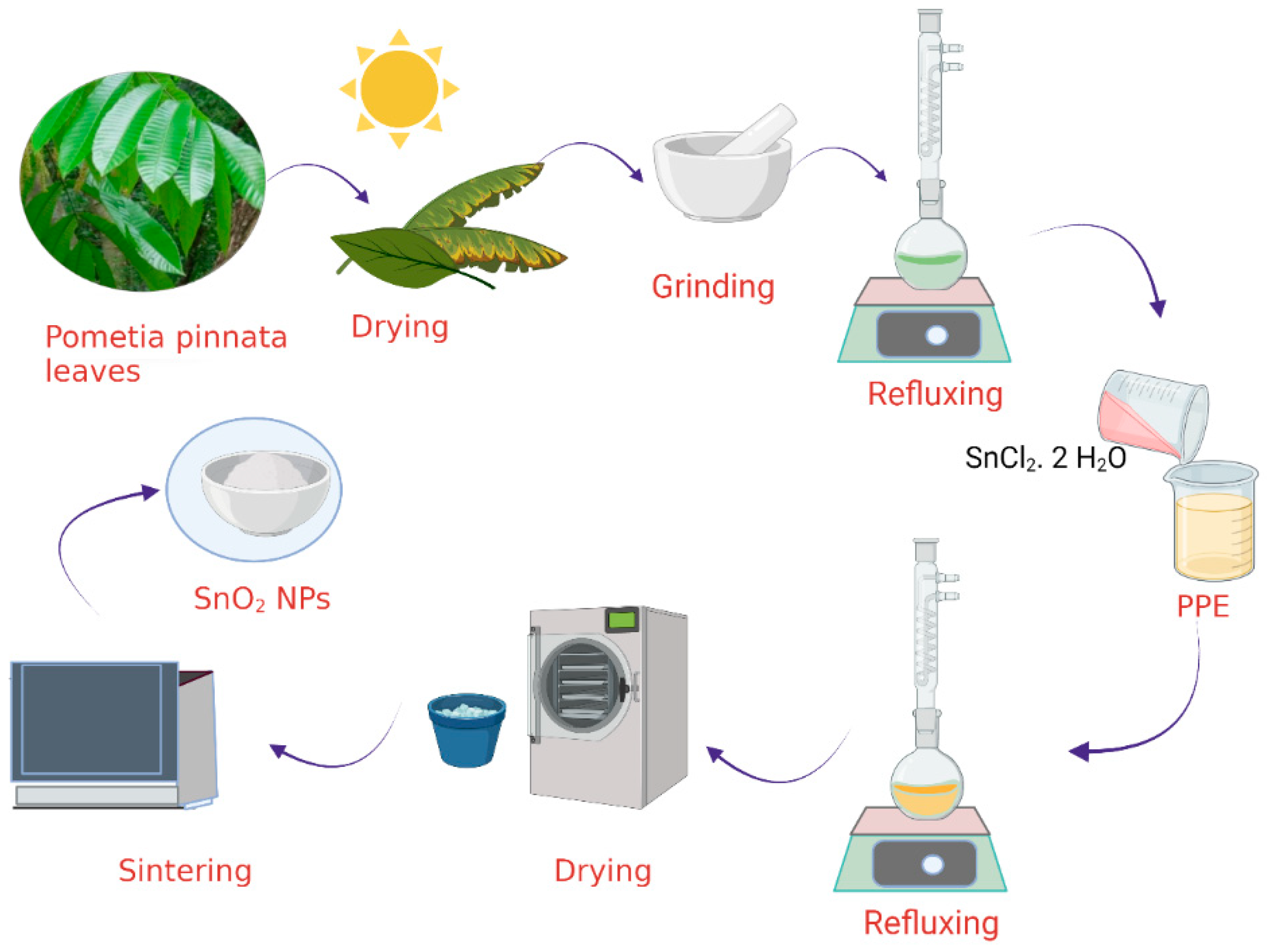
2.2. Extraction of Pometia pinnata Leaves

Fresh Pometia pinnata leaves were harvested from rural farms in Sleman, Yogyakarta Province, Indonesia, and then washed several times with distilled water to remove dirt. Afterwards, the leaves were air dried for a week at ambient temperature to reach the appropriate moisture. The dried leaves were ground into fine powders using a mortar and pestle; furthermore, 20 g of the dried leaves were refluxed with 100 mL of distilled water for 4 h. The Pometia pinnata leaf extract (PPE) was obtained by filtering the result using Whatman Grade 1 filter paper. To maintain the condition of the PPE, the PPE was kept at a temperature of 5 °C in a refrigerator.

2.3. Synthesis of SnO2 NPs

The synthesis of SnO2 NPs was performed by mixing 2 g of tin chloride (SnCl2·2H2O) with 50 mL of PPE, followed by the addition of 50 mL of distilled water. The mixture was then refluxed for an hour, and the monitoring of nanoparticle formation was performed by UV-visible spectrophotometry. As the formation of the nanoparticles was confirmed by UV visible spectroscopy, the precipitate was obtained by evaporating water in an oven at a constant temperature of 60 °C. The powder obtained from these steps was then sintered at 500 °C for 2 h to get SnO2 NPs. Figure 1 displays the schematic representation of SnO2 NP synthesis.

2.4. Characterization of SnO2 NPs

X-ray diffraction (XRD) measurement was carried out using a Shimadzu X6000 diffractometer with Ni-filtered Cu-Kα radiation (λ =1.5406 Å) operated at a voltage of 40 kV and current of 30 mA. The scanning was taken from 10 to 70°. The crystalline size of the NPs was measured based on the Scherer equation:
d = /Bcosθ
where d is the mean crystalline size of the NPs, λ is the wavelength of radiation (1.5406 Å), θ is the selected angle and B is the full width at half maximum (FWHM) intensity of the selected reflection.
The morphology of prepared nanoparticles was investigated on a Phenom-X field-emission scanning electron microscope (FE-SEM); meanwhile, the elemental composition of the SnO2 NPs was determined by X-ray Fluorescence spectroscopy (XRF) using the Shimadzu EDX-7000 instrument. Transmission electron microscopy (TEM) images and high-resolution TEM (HR-TEM) images were obtained with a JEOL TEM 2010 transition electron microscope operated at 200 kV. The sample was collected on a holey carbon grid. XPS analysis was performed on a V.G. Scientific ESKALAB MKII instrument with a monochromatic Al Kα radiation with photon energy of 1486.6 ± 0.2 eV. Prior to being analyzed, the sample was slightly pressed into a small size and then degassed to achieve a dynamic vacuum below 10−8 Pa.

2.5. Photocatalytic Activity of SnO2 NPs

To verify the photocatalytic performances of SnO2 NPs, experiments on photocatalytic oxidation (herein called photooxidation) of BPB were conducted under UV and visible light illumination. A 20 watt Philips UV light lamp was employed as the UV light source, and a 20 watt Philip Xenon lamp was utilized as the visible light source. The light intensities of UV light and Xenon lamps were 39.99 MW/Cm2 and 31.22 MW/Cm2, respectively; the radiometer was VLX-3W. The experiments were executed in a batch photocatalytic reactor equipped with a water-jacketed chamber to stabilize the temperature. In particular, 0.25 g of SnO2 NPs was dispersed in 100 mL of 20 ppm BPB solution, and about 1 mL of 5 × 10⁻³ M of H2O2 was then added. The suspension was stirred in the dark for 15 min before being exposed to light, and the samples of supernatant were then collected sequentially for colorimetric analysis using UV-visible spectrophotometry.
The degradation efficiency of the photocatalysis was calculated based on the initial concentration and the concentration at the time of sampling with reference to the following equation (Equation (2)):
Degradation   efficiency   ( DE ) ( % ) = 100 × ( C 0 C t C 0 )
where C0 and Ct are initial concentration and concentration of BPB at time t.

2.6. Antibacterial Assay of SnO2 NPs

Antibacterial activity of the SnO2 NPs was evaluated by disc diffusion method. The tested bacterial strains were S. aureus (ATCC 25923), S. pyogenes (ATCC 19615), K. pneumoniae (ATCC 13883) and E. coli (ATCC 11303). The medium for bacterial growth was prepared by suspending nutrient agar in distilled water and was autoclaved before use. Bacterial culture was evenly spread throughout the petri plate and a 6 mm sterile filter disc loaded with 100 μg/100 mL SnO2 NPs solution.

3. Results

3.1. SnO2 NP Synthesis and Characterization

The formation of SnO2 NPs was initially monitored by the identification of surface plasmon resonance (SPR) using UV-visible spectrophotometry analysis on a HITACHI U-2010 instrument.
Figure 2 shows that the bioreduction occurred as the change of peaks recorded values of from 200 to 800 nm. The PPE showed peaks at the range of 200–300 nm and 500–600 nm, indicating the presence of secondary metabolites which contain aromatic structures. The spectrum of SnO2 NPs shows the characteristic surface plasmon resonance (SPR) absorption band at 396 nm. This wavelength is consistent with the SPR absorption band of Sn NPs in previous literature mentioning the range of 295–400 nm. The disappearing peaks at the range of 500–600 nm is an indication of the reduction of some secondary metabolite. A previous study identified some secondary metabolites such as kaempferol-3-O-rhamnoside, glycolipid, epicatechin, quercetin-3-O-rhamnoside, steroid glycosides, and other saponin compounds as the components of PPE [23,24]; the reduction of Sn2+ possibly involves the functional groups of those compounds. Taking quercetin as one of the bioreductors, the reduction mechanism is as presented in Figure 3.
Moreover, other phenolic compounds work as singlet oxygen quenchers, hydrogen donators, metal chelating and reducing agents which furthermore act as capping agents for the nanoparticles [25].
Results from the XRD analysis presented in Figure 4 exhibit the peaks which are associated with the (110), (101), (200), (210), (211), (220), (002), (310), (112), (301) and (202) planes, respectively. All diffraction peaks coincide strongly with the planes of standard tetragonal rutile SnO2, which refers to JCPDS 41-1445, and there is no other peak detected, indicating the single phase of the nanoparticles [26,27]. Based on the Scherer equation, the average crystallite size of SnO2 NPs calculated from (110), (101) and (211) is about 18.2 nm. Complete oxide formation was observed by XPS analysis, and the spectra are presented in Figure 5.
The survey scan spectrum (Figure 5a) shows the major peaks of C, O and Sn, clearly indicating the successful fabrication of SnO2 by using PPE as the reducing agents. The deconvoluted Sn 3d spectrum (Figure 5b) shows two peaks at 487.1 and 495.9 eV, which are assigned as the Sn 3d5/2 and Sn 3d3/2 spin-orbit peaks, indicating the chemical state of the SnO2 NPs as Sn4+. In addition, the O1s spectrum can be deconvoluted into two peaks centered at 530.6 and 531.8 eV, associated with the Sn-O and Sn-OH, respectively (Figure 5c). These peaks are ascribed as the oxygen-containing functional groups bound to the SnO2 NPs as the caping agent [18,28,29].
Figure 6a shows the flower-like FE-SEM image of the SnO2 NPs, while Figure 6b represents the higher magnification of the image. By the higher magnification, the nanoparticle facets of the of the structures are smooth and discriminable in uniform spherical shapes.
Moreover, the TEM and HR-TEM investigations represent further insight into the morphologies and the structural features of SnO2 NPs. It can be seen from the TEM profile (Figure 7a–c) that the irregular spherical forms have diameters ranging from 8 to 18 nm. In addition, HR-TEM images (Figure 7c) represent the clear lattice fringes with a width of 0.34 nm, which corresponds to the interplanar distance of (1 1 0) planes SnO2 [16,30,31]. These data infer that the calculated average particle sizes from XRD, SEM and TEM measurements have a consistent value. The particle size obtained in this work is at the average and within the range found using other plants extracts, as presented in Table 1. Many researches have found the spherical form of nanoparticles to be within the range of 2–50 nm, except concerning Ficus carica with a size of 128 nm. The form and particle size are strongly influenced by several factors such as composition of the extract, reaction procedure and temperature of sintering [32].
The different compositions of secondary metabolites govern the bioreduction of Sn2+ and furthermore affect the particle’s aggregation after the sintering process, which has also been reported in the green syntheses of ZnO, Fe2O3 and SnO2 nanoparticles [28,32,38,39].

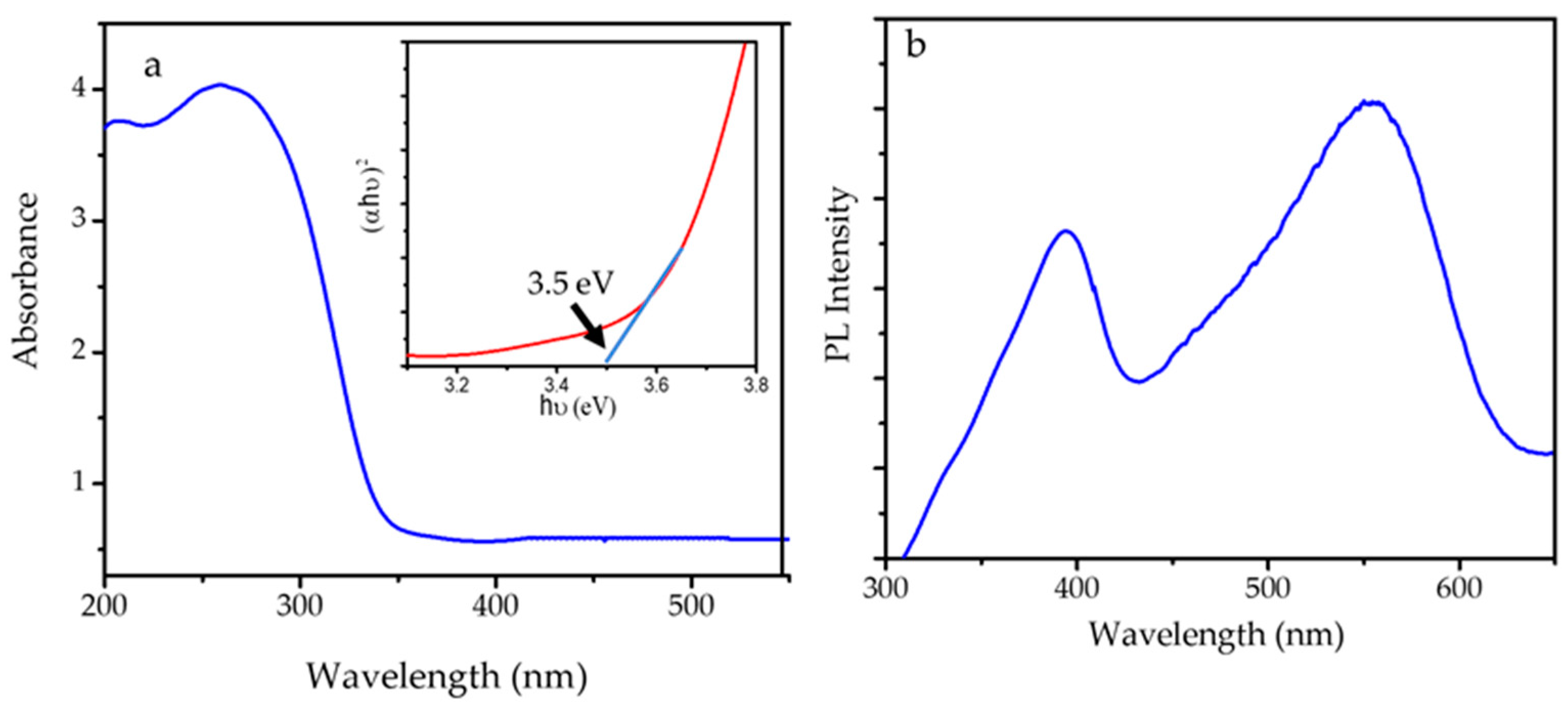
3.2. Optical Properties of SnO2 NPs

The optical properties of SnO2 NPs were studied by UV-DRS and photoluminescence spectroscopy analyses, and the spectra are presented in Figure 8. The band gap of SnO2 NPs was evaluated according to the UV-DRS spectrum with the following Equation (2):
(αhν)2=A(hν − Eg)
where α, h, ν, Eg and A are the absorption coefficient, Planck constant, light frequency, bandgap energy and Tauc constant, respectively. The extrapolation of (α h ν)2 versus hν to zero gives the band gap energy of 3.5 eV. This value lies within the range of 3.1–3.9 eV, as mentioned in the literatures, and is comparable to the band gap energy of SnO2 NPs synthesized using the hydrothermal method [17,40,41]. The photoluminescence (PL) spectrum was recorded with varying excitation wavelengths (l = 360~380 nm), and the spectrum of SnO2 NPs showed an excitation wavelength of 396 and 550 nm, of which the peak at 550 nm is attributed to 6A1→4T1(4G) ligand field transition of Sn4+. The data represents that SnO2 NPs have the capability to interact with photons in either the UV or the visible region.

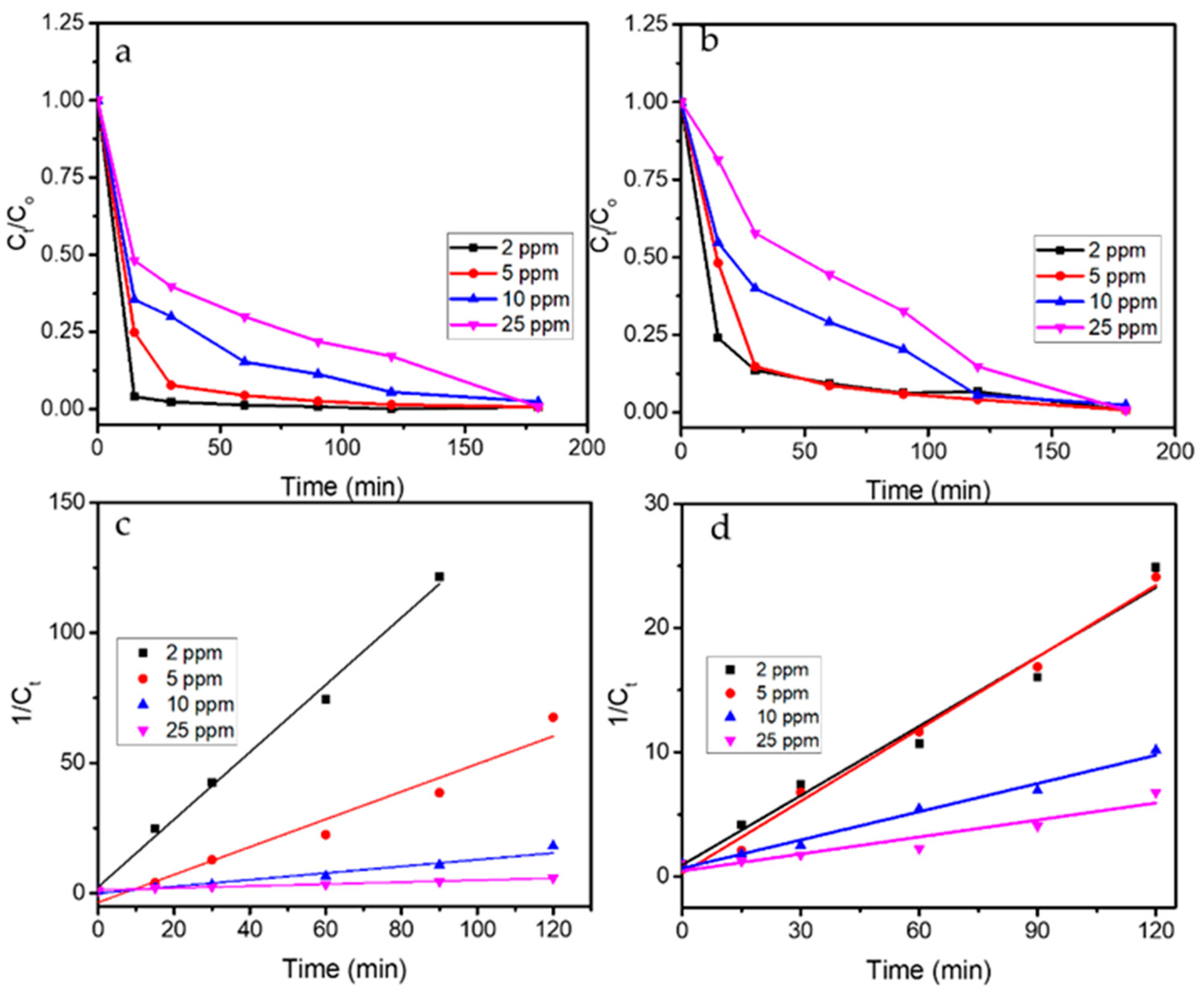
3.3. Photocatalytic Activity

Figure 9 shows the kinetics plot of BPB photooxidation under UV and visible light irradiation in comparison with blank experiments consisting of adsorption and photolytic treatments. The adsorption treatment was the treatment with the addition of SnO2 NPs in the solution without any light exposure, while the photolytic experiment was the UV light exposure to the solution with the addition of H2O2 in the absence of photocatalysts. It can be seen that the photocatalytic and photooxidation experiments significantly reduced BPB concentration attributed to the decolorization from the initial step of the reaction, while the blank experiments reveal insignificant decolorization. The decolorization of BPB reaches less than 5% with respect to the initial concentration by photolytic treatments; meanwhile, there is about 15% reduction concerning the adsorption experiment for 120 min. The decolorization for the adsorption experiment is attributed to the role of SnO2 NPs in providing surface interactions with the BPB molecules, as has been reported in previous experiments of dye adsorption using SnO2 and SnO2 NPs [42,43]. The photolytic mechanism depends on the capability of light to produce radicals from the homolytic cleavage of H2O2 molecules and, from the experiment, it was conclusively obtained that the reaction went slower compared to the formation of radicals over the catalytic mechanism.
It was also seen that the use of UV light illumination demonstrated a faster removal. While the PL intensity of SnO2 NPs supports the photoactivity within the visible light region, according to the band gap energy value which lays within the UV light range, the photon interaction with UV light produced the radicals for further propagation reactions.

3.4. Identification on Oxidation Mechanism

For the verification of the occurrence of the oxidation mechanism, LCMS analyses were performed. Results from LCMS analysis presented in Figure 10 validated the degradation mechanism, concerning which the chromatogram of the initial solution demonstrated a single peak at 12.8 min and later changed, displaying other peaks in the treated solution, indicating that the products degraded [44]. In addition, the MS spectra of the initial and treated solutions suggested the predicted reaction intermediates from the presence of m/z = 667.4, 665.7, 632.7, 603.5, 500.3, 523.2, 415.1 and 325.6. The degradation mechanism and products of azo dyes have been extensively discussed, and refer to the existence of the peak m/z = 667.4, which is higher compared to the m/z reflecting the presence of BPB (665.7), indicating the addition of the radical as the first step of the propagation [45,46].
Referring to these MS peaks, the possible degradation mechanism can be seen in Figure 11. A similar mechanism was reported for BPB removal over CuO-nano-clinoptilolite and BPB degradation by sono-catalytic treatment using SnO2/montmorillonite [47,48].
These data revealed the role of SnO2 NPs to accelerate the radical formation and propagation steps by the following mechanism:
SnO2 NPs + hν→SnO2 NPs* + hvb+ + ecb
H2O + h+→H+ + OH•
OH + h+→OH•
O2 + e→O2
O2 + 2H+ + 2e→H2O2
H2O2 + e→OH• + OH
BPB + •OH + O2→degradation products→→→CO2 + H2O
Furthermore, the kinetics plots of photooxidation reactions from varied concentrations are depicted in Figure 12. Generally speaking, the compared data are consistent with the trends of the higher degradation rate which were achieved under UV exposure rather than the use of visible light. The kinetics evaluation on these data suggests that the reactions obey the pseudo-second order kinetics with reference to following equation (Equation (4)):
1 C t = k t + 1 C 0
where C0 and Ct are initial concentration and concentration of BPB at time t, and k is kinetics constant.
The fitness of the data concerning pseudo-second-order kinetics is similar to the kinetics of BPB photocatalytic degradation over SnO2 synthesized using red spinach [37], which also represented the effect of the initial concentration of dye concerning the reaction rate. The kinetics parameters and DE values are presented in Table 2.

3.5. Effect of Scavenger

To study the role of ∙OH in photooxidation, the effect of scavenging agents consisting of EDTA as a hole scavenger and isopropanol as the radical’s scavenger on kinetics of BPB removal was examined. Figure 13a shows that isopropanol significantly suppressed the degradation of BPB as slower C/C0 reduction occurred. Isopropanol traps the radicals formed by the excitation produced by the interaction between SnO2 NPs and light so the propagation steps for producing further oxidation reaction are diminished. In contrast, the addition of EDTA enhanced the photooxidation with about 10% BPB reduction being observed at the initial part of the reaction. The kinetics reflected that EDTA inhibits the recombination of electrons and holes so more electrons can migrate to the surface of the SnO2 NPs and further react with O2 to form HOO• [48].

3.6. Reusability of Photocatalysts

The reusability of photocatalysts is an important character which governs the applicability for further scale. In order to check the reusability of photocatalysts, the spent photocatalyst was filtered and recycled by washing with ethanol, followed by drying at 200 °C for 2 h. A mass loss of about 5–10% wt. occurred due to technical steps such as incompleteness in recovery by filtration. The degradation efficiency of recycled photocatalysts compared to fresh ones is presented in a chart in Figure 13b. It is seen that DE remained stable in the range of 98.52–94.60% until the fifth cycle, which means that the reduced activity was no more than 10%. This performance is attributed to the stability of the nanoparticles which related to the maintained physicochemical characteristics concerning its role in the photooxidation mechanism. The reusability of the SnO2 is comparable with magnetite nanoparticles and SnO2 dispersed onto montmorillonite [47], but the crucial factor of recovery after the use of photocatalyst needs to be addressed for improvement in industrial or scaled-up levels.

3.7. Antibacterial Activity

Disk diffusion assay was performed to analyze the inhibition zone of SnO2 NPs against K. pneumoniae, E. coli, S. aureus and S. pyogenes. Figure 14 shows the inhibition zone diameter for each test in comparison with ampicillin as the positive control. It is also found that the inhibition zone reduced at prolonged incubation times for 48 and 72 h, suggesting that the nanoparticles’ capability is more for inhibiting rather than killing the tested bacteria.
The data show that the inhibition zone diameters for all tested bacteria are smaller compared to those of ampicillin as positive control. The different mechanism is a known factor in this, and ampicillin is a well-known pharmaceutical, being an antimicrobial agent. In more detail concerning SnO2 NPs, higher antibacterial effects against K. pneumoniae and S. pyogenes, which are gram-positive bacteria, compared to those of E. coli and S. aureus, are demonstrated. With regard to a previous study on the surface interaction among nanoparticles and the bacteria, the stronger surface interaction of nanoparticles with gram-positive bacteria is due to a partially less negative surface potential of the bacteria.
Metal oxide nanoparticles tend to have a negative zeta potential, which easily interacts with this surface potential according to the electrostatic force equilibrium. In contrast, the more negative the surface potential of the gram-negative bacteria, the less the surface interaction [49,50,51,52]. Ions can be released from the interaction between SnO2 NPs with the cell wall leading to the generation of reactive oxygen species (ROS) on the surface of the nanoparticles. The DNA damage causing cell destruction is a further step for bacteria inactivation. Figure 15 depicts the schematic representation of the mechanism [53].
The inhibition zone which identified the antibacterial activity of SnO2 NPs in this study was comparable to, and even greater than, the SnO2 NPs observed in other studies. For example, the activity against E coli was higher compared to that demonstrated by the SnO2 NP synthesized co-precipitation method using DNA as the capping agent [54], and was also higher compared to SnO2 NPs synthesized using Vitex altissima [52]. The inhibition zone of 12 mm in this research was achieved with a concentration of 200 μg/mL, higher than the inhibition zone from the DNA-synthesized SnO2 NPs (10 mm) and Vitex altissima-mediated SnO2 NPs (1.1 mm). Higher activity was also demonstrated against S. aureus and K. pneumoniae compared to Stevia rebaudiana-mediated SnO2 NPs [55]. The particle size of Stevia rebaudiana-mediated SnO2 NPs ranged from 20 to 30 nm, suggesting that the higher antibacterial activity in this work is strongly correlated with smaller particle size. Smaller particle size enables the intrusion of nanoparticles into the cell wall to further destroy the structure of the bacteria [32,56,57,58]. Generally speaking, the synthesized SnO2 NPs have pronounced antibacterial activity concerning tested gram-negative and gram-positive bacteria, so it has potentially further developed for environmental and biomedical applications.

4. Conclusions

This research, the first ever study on the synthesis of SnO2 nanoparticles using Pometia pinnata leaf extract, demonstrated the flower-like structure of nanoparticles as a single SnO2 tetragonal phase. TEM analysis proved the nanoparticles had uniform spherical shapes with size ranging from 8 to 20 nm, which was confirmed with XRD analysis, finding a crystallite size of 18.2 nm. Optical study of the nanoparticles showed that the band gap energy of SnO2 nanoparticles is 3.5 eV, contributing to the photocatalytic activity in bromophenol blue photooxidation. Kinetics study revealed the fitness of bromophenol blue photooxidation with second-order kinetics by either UV or visible light exposure. Degradation efficiencies ranging from 85 to 99% were found with dependency toward an initial concentration of BPB and the use of a light source. The antibacterial activity evaluation of SnO2 nanoparticles indicated an inhibition capability against Klebsiella pneumoniae, Escherichia coli Staphylococcus aureus and Streptococcus pyogenes which is comparable and even higher in comparison with SnO2 nanoparticles reported in previous studies.

Author Contributions

Conceptualisation, I.F. and H.H.; methodology, S.S., G.P. and R.-A.D.; validation, W.-C.O. and R.-A.D.; formal analysis, H.H. and G.P.; investigation, I.F. and H.H.; resources, I.F.; data curation, H.H., G.P. and S.A.I.S.M.G.; writing—original draft preparation, I.F.; writing—review and editing, R.-A.D.; visualisation, G.P.; supervision, W.-C.O.; project administration, W.-C.O.; funding acquisition, I.F. and R.-A.D. All authors have read and agreed to the published version of the manuscript.

Funding

This research was funded by the Ministry of Ministry of Education, Culture, Research, and Technology through the World Class Professor Program in 2021.

Data Availability Statement

This study did not report any data.

Acknowledgments

The authors would like to express appreciation for the support from the Ministry of Education, Culture, Research, and Technology through the World Class Professor Program in 2021, Contract No: 2817/E4.1/KK.04.05/2021.

Conflicts of Interest

The authors declare no conflict of interest.

References

  1. Frenzilli, G. Nanotechnology for environmental and biomedical research. Nanomaterials 2020, 10, 2220. [Google Scholar] [CrossRef] [PubMed]
  2. Corsi, I.; Winther-Nielsen, M.; Sethi, R.; Punta, C.; Della Torre, C.; Libralato, G.; Lofrano, G.; Sabatini, L.; Aiello, M.; Fiordi, L.; et al. Ecofriendly nanotechnologies and nanomaterials for environmental applications: Key issue and consensus recommendations for sustainable and ecosafe nanoremediation. Ecotoxicol. Environ. Saf. 2018, 154, 237–244. [Google Scholar] [CrossRef] [PubMed]
  3. Singh, J.; Dutta, T.; Kim, K.H.; Rawat, M.; Samddar, P.; Kumar, P. “Green” synthesis of metals and their oxide nanoparticles: Applications for environmental remediation. J. Nanobiotechnology 2018, 16, 1–24. [Google Scholar] [CrossRef] [PubMed]
  4. Liu, Y.; Nam Ong, C.; Xie, J. Emerging nanotechnology for environmental applications. Nanotechnol. Rev. 2016, 5, 1–2. [Google Scholar] [CrossRef]
  5. Zhang, D.; Ma, X.L.; Gu, Y.; Huang, H.; Zhang, G.W. Green Synthesis of Metallic Nanoparticles and Their Potential Applications to Treat Cancer. Front. Chem. 2020, 8, 1–18. [Google Scholar] [CrossRef]
  6. Akbari, A.; Sabouri, Z.; Hosseini, H.A.; Hashemzadeh, A.; Khatami, M.; Darroudi, M. Effect of nickel oxide nanoparticles as a photocatalyst in dyes degradation and evaluation of effective parameters in their removal from aqueous environments. Inorg. Chem. Commun. 2020, 115, 107867. [Google Scholar] [CrossRef]
  7. Sharma, N.; Kumar, J.; Thakur, S.; Sharma, S.; Shrivastava, V. Antibacterial study of silver doped zinc oxide nanoparticles against Staphylococcus aureus and Bacillus subtilis. Drug Invent. Today 2013, 5, 50–54. [Google Scholar] [CrossRef]
  8. Hassan, S.M.; Ahmed, A.I.; Mannaa, M.A. Preparation and characterization of SnO2 doped TiO2 nanoparticles: Effect of phase changes on the photocatalytic and catalytic activity. J. Sci. Adv. Mater. Devices 2019, 4, 400–412. [Google Scholar] [CrossRef]
  9. Khezrianjoo, S.; Lee, J.; Kim, K.-H.; Kumar, V. Eco-Toxicological and Kinetic Evaluation of TiO2 and ZnO Nanophotocatalysts in Degradation of Organic Dye. Catalysts 2019, 9, 871. [Google Scholar] [CrossRef] [Green Version]
  10. Suresh, K.C.; Balamurugan, A. Evaluation of structural, optical, and morphological properties of nickel oxide nanoparticles for multi-functional applications. Inorg. Nano-Metal. Chem. 2021, 51, 296–301. [Google Scholar] [CrossRef]
  11. Geetha, A.; Sakthivel, R.; Mallika, J.; Kannusamy, R.; Rajendran, R. Green Synthesis of Antibacterial Zinc Oxide Nanoparticles Using Biopolymer Azadirachta indica Gum. Orient. J. Chem. 2015, 32, 955–963. [Google Scholar] [CrossRef]
  12. Pugazhendhi, S.; Sathya, P.; Palanisamy, P.K.; Gopalakrishnan, R. Synthesis of silver nanoparticles through green approach using Dioscorea alata and their characterization on antibacterial activities and optical limiting behavior. J. Photochem. Photobiol. B Biol. 2016, 159, 155–160. [Google Scholar] [CrossRef] [PubMed]
  13. Lalithambika, K.C.; Thayumanavan, A.; Ravichandran, K.; Sriram, S. Photocatalytic and antibacterial activities of eco-friendly green synthesized ZnO and NiO nanoparticles. J. Mater. Sci. Mater. Electron. 2017, 28, 2062–2068. [Google Scholar] [CrossRef]
  14. Fatimah, I.; Hidayat, H.; Nugroho, B.H.; Husein, S. Ultrasound-assisted biosynthesis of silver and gold nanoparticles using Clitoria ternatea flower. S. Afr. J. Chem. Eng. 2020, 34, 97–106. [Google Scholar] [CrossRef]
  15. Haritha, E.; Roopan, S.M.; Madhavi, G.; Elango, G.; Al-Dhabi, N.A.; Arasu, M.V. Green chemical approach towards the synthesis of SnO2 NPs in argument with photocatalytic degradation of diazo dye and its kinetic studies. J. Photochem. Photobiol. B Biol. 2016, 162, 441–447. [Google Scholar] [CrossRef] [PubMed]
  16. Begum, S.; Ahmaruzzaman, M. Green synthesis of SnO2 quantum dots using Parkia speciosa Hassk pods extract for the evaluation of anti-oxidant and photocatalytic properties. J. Photochem. Photobiol. B Biol. 2018, 184, 44–53. [Google Scholar] [CrossRef] [PubMed]
  17. Sharif, M.S.; Aqeel, M.; Haider, A.; Naz, S.; Ikram, M.; Ul-Hamid, A.; Haider, J.; Aslam, I.; Nazir, A.; Butt, A.R. Photocatalytic, Bactericidal and Molecular Docking Analysis of Annealed Tin Oxide Nanostructures. Nanoscale Res. Lett. 2021, 16, 1–16. [Google Scholar] [CrossRef] [PubMed]
  18. Matussin, S.; Harunsani, M.H.; Tan, A.L.; Khan, M.M. Plant-Extract-Mediated SnO2 Nanoparticles: Synthesis and Applications. ACS Sustain. Chem. Eng. 2020, 8, 3040–3054. [Google Scholar] [CrossRef]
  19. Prihanti, G.S.; Katjasungkana, R.M.K.; Novitasari, B.R.; Amalia, S.R.; Nurfajriana, A.; Agustini, S.M.; Cakrawati, H.; Andari, D. Antidiabetic potential of matoa bark extract (pometia pinnata) in alloxan-induced diabetic male rat strain wistar (rattus norvegicus). Syst. Rev. Pharm. 2020, 11, 88–97. [Google Scholar] [CrossRef]
  20. Sauriasari, R.A.N.I.; Azizah, N.; Basah, K. Tyrosinase Inhibition 2,2-Diphenul-1-picrylhydraxyl Radical Scavening Activity, and Phytochemical Screening of Fractions and Ethanol Extract from Leaves and Steam Bark of Matoa (Pometia Pinnata). Asian J. Pharmacetical Clin. Res. 2017, 10, 85–89. [Google Scholar] [CrossRef] [Green Version]
  21. Handayani, W.; Ningrum, A.S.; Imawan, C. The Role of pH in Synthesis Silver Nanoparticles Using Pometia pinnata (Matoa) Leaves Extract as Bioreductor. J. Phys. Conf. Ser. 2020, 1428, 012021. [Google Scholar] [CrossRef]
  22. Sujatmiko, F.; Sahroni, I.; Fadillah, G.; Fatimah, I. Visible light-responsive photocatalyst of SnO2/rGO prepared using Pometia pinnata leaf extract. Open Chem. 2021, 19, 174–183. [Google Scholar] [CrossRef]
  23. Suedee, A.; Tewtrakul, S.; Panichayupakaranant, P. Anti-HIV-1 integrase compound from Pometia pinnata leaves. Pharm. Biol. 2013, 51, 1256–1261. [Google Scholar] [CrossRef] [PubMed]
  24. Purwidyaningrum, I.; Sukandar, E.Y.; Fidrianny, I. Diuretic activity of matoa leaves extracts and fractions (Pometia pinnata J.R. Forster & J.G Forster) and its influence on potassium and sodium levels. Asian J. Pharm. Clin. Res. 2017, 10, 31–34. [Google Scholar] [CrossRef] [Green Version]
  25. Bibi, I.; Kamal, S.; Ahmed, A.; Iqbal, M.; Nouren, S.; Jilani, K.; Nazar, N.; Amir, M.; Abbas, A.; Ata, S.; et al. Nickel nanoparticle synthesis using Camellia Sinensis as reducing and capping agent: Growth mechanism and photo-catalytic activity evaluation. Int. J. Biol. Macromol. 2017, 103, 783–790. [Google Scholar] [CrossRef]
  26. Wang, J.; Fan, H.Q.; Yu, H.W. Synthesis of Monodisperse Walnut-Like SnO2 Spheres and Their Photocatalytic Performances. J. Nanomater. 2015, 2015, 1–8. [Google Scholar] [CrossRef] [Green Version]
  27. Bhosale, T.T.; Shinde, H.M.; Gavade, N.L.; Babar, S.B.; Gawade, V.V.; Sabale, S.R.; Kamble, R.J.; Shirke, B.S.; Garadkar, K.M. Biosynthesis of SnO2 nanoparticles by aqueous leaf extract of Calotropis gigantea for photocatalytic applications. J. Mater. Sci. Mater. Electron. 2018, 29, 6826–6834. [Google Scholar] [CrossRef]
  28. Fatimah, I.; Sahroni, I.; Muraza, O.; Doong, R.A. One-pot biosynthesis of SnO2 quantum dots mediated by Clitoria ternatea flower extract for photocatalytic degradation of rhodamine B. J. Environ. Chem. Eng. 2020, 8, 103879. [Google Scholar] [CrossRef]
  29. Kwoka, M.; Lyson-Sypien, B.; Kulis, A.; Zappa, D.; Comini, E. Surface properties of SnO2 nanowires deposited on Si substrate covered by Au catalyst studies by XPS, TDS and SEM. Nanomaterials 2018, 8, 738. [Google Scholar] [CrossRef] [Green Version]
  30. Kim, S.P.; Choi, M.Y.; Choi, H.C. Photocatalytic activity of SnO2 nanoparticles in methylene blue degradation. Mater. Res. Bull. 2016, 74, 85–89. [Google Scholar] [CrossRef]
  31. Guan, X.; Luo, P.; Li, X.; Yu, Y.; Chen, D.; Zhang, L. One-step facile synthesis of hierarchically porous nitrogen-doped SnO2 nanoparticles with ultrahigh surface area for enhanced lithium storage performance. Int. J. Electrochem. Sci. 2018, 13, 5667–5680. [Google Scholar] [CrossRef]
  32. Gebreslassie, Y.T.; Gebretnsae, H.G. Green and Cost-Effective Synthesis of Tin Oxide Nanoparticles: A Review on the SYnthesis Methodologies, Mechanism of Formation, and Their Potential Applications. Nanoscale Res. Lett. 2021, 16, 1–16. [Google Scholar] [CrossRef] [PubMed]
  33. Awoke, N.; Pandey, D.; Habtemariam, A.B. Synthesis of Tin(IV) Oxide Nanoparticles Using Plant Leaf Extracts of Vernonia amygdalina and Mentha spicata. Regen. Eng. Transl. Med. 2021, 1–6. [Google Scholar] [CrossRef]
  34. Gomathi, E.; Jayapriya, M.; Arulmozhi, M. Environmental benign synthesis of tin oxide (SnO2) nanoparticles using Actinidia deliciosa (Kiwi) peel extract with enhanced catalytic properties. Inorg. Chem. Commun. 2021, 130, 108670. [Google Scholar] [CrossRef]
  35. Al-Enazi, N.M.; Ameen, F.; Alsamhary, K.; Dawoud, T.; Al-Khattaf, F.; AlNadhari, S. Tin oxide nanoparticles (SnO2-NPs) synthesis using Galaxaura elongata and its anti-microbial and cytotoxicity study: A greenery approach. Appl. Sci. 2021, 2. [Google Scholar] [CrossRef]
  36. Hu, J. Biosynthesis of SnO2 nanoparticles by fig (Ficus Carica) leaf extract for electrochemically determining Hg(II) in water samples. Int. J. Electrochem. Sci. 2015, 10, 10668–10676. [Google Scholar] [CrossRef] [Green Version]
  37. Wicaksono, W.P.; Sahroni, I.; Saba, A.K.; Rahman, R.; Fatimah, I. Biofabricated SnO2 nanoparticles using Red Spinach (Amaranthus tricolor L.) extract and the study on photocatalytic and electrochemical sensing activity. Mater. Res. Express 2020, 7, 075009. [Google Scholar] [CrossRef]
  38. Ong, C.B.; Ng, L.Y.; Mohammad, A.W. A review of ZnO nanoparticles as solar photocatalysts: Synthesis, mechanisms and applications. Renew. Sustain. Energy Rev. 2018, 81, 536–551. [Google Scholar] [CrossRef]
  39. Harjati, F.; Citradewi, P.W.; Purwiandono, G.; Fatimah, I. Green synthesis of hematite/TUD-1 nanocomposite as efficient photocatalyst for bromophenol blue and methyl violet degradation. Arab. J. Chem. 2020, 13, 8395–8410. [Google Scholar] [CrossRef]
  40. Ma, C.M.; Hong, G.B.; Lee, S.C. Facile synthesis of tin dioxide nanoparticles for photocatalytic degradation of Congo red dye in aqueous solution. Catalysts 2020, 10, 792. [Google Scholar] [CrossRef]
  41. Wongsaprom, K.; Winyayong, A.; Maensiri, S. Synthesis and room-temperature ferromagnetism in flower-like SnO2 nanostructures. J. Phys. Conf. Ser. 2018, 1144, 012042. [Google Scholar] [CrossRef]
  42. Abdelkader, E.; Nadjia, L.; Rose-noe, V. Adsorption of Congo red azo dye on nanosized SnO2 derived from sol-gel method. Int. J. Ind. Chem. 2016, 53–70. [Google Scholar] [CrossRef] [Green Version]
  43. Kumar, K.Y.; Muralidhara, H.B.; Nayaka, Y.A.; Balasubramanyam, J.; Hanumanthappa, H. Low-cost synthesis of metal oxide nanoparticles and their application in adsorption of commercial dye and heavy metal ion in aqueous solution. Powder Technol. 2013, 246, 125–136. [Google Scholar] [CrossRef]
  44. Houas, A.; Lachheb, H.; Ksibi, M.; Elaloui, E.; Guillard, C.; Herrmann, J.M. Photocatalytic degradation pathway of methylene blue in water. Appl. Catal. B Environ. 2001, 31, 145–157. [Google Scholar] [CrossRef]
  45. Shah, T.; Gul, T.; Saeed, K. Photodegradation of bromophenol blue in aqueous medium using graphene nanoplates-supported TiO2. Appl. Water Sci. 2019, 9, 1–7. [Google Scholar] [CrossRef] [Green Version]
  46. Nezamzadeh-Ejhieh, A.; Hushmandrad, S. Solar photodecolorization of methylene blue by CuO/X zeolite as a heterogeneous catalyst. Appl. Catal. A Gen. 2010, 388, 149–159. [Google Scholar] [CrossRef]
  47. Fatimah, I.; Rubiyanto, D.; Sahroni, I.; Putra, R.S.; Nurillahi, R.; Nugraha, J. Physicochemical characteristics and photocatalytic performance of Tin oxide/montmorillonite nanocomposites at various Sn/montmorillonite molar to mass ratios. Appl. Clay Sci. 2020, 193, 105671. [Google Scholar] [CrossRef]
  48. Nezamzadeh-Ejhieh, A.; Zabihi-Mobarakeh, H. Heterogeneous photodecolorization of mixture of methylene blue and bromophenol blue using CuO-nano-clinoptilolite. J. Ind. Eng. Chem. 2014, 20, 1421–1431. [Google Scholar] [CrossRef]
  49. Malarkodi, C.; Rajeshkumar, S.; Paulkumar, K.; Vanaja, M.; Gnanajobitha, G.; Annadurai, G. Biosynthesis and antimicrobial activity of semiconductor nanoparticles against oral pathogens. Bioinorg. Chem. Appl. 2014, 2014, 1–10. [Google Scholar] [CrossRef] [Green Version]
  50. Burdușel, A.C.; Gherasim, O.; Grumezescu, A.M.; Mogoantă, L.; Ficai, A.; Andronescu, E. Biomedical applications of silver nanoparticles: An up-to-date overview. Nanomaterials 2018, 8, 681. [Google Scholar] [CrossRef] [Green Version]
  51. Wikaningtyas, P.; Sukandar, E.Y. The antibacterial activity of selected plants towards resistant bacteria isolated from clinical specimens. Asian Pac. J. Trop. Biomed. 2016, 6, 16–19. [Google Scholar] [CrossRef] [Green Version]
  52. Bhavana, S.; Gubbiveeranna, V.; Kusuma, C.G.; Ravikumar, H.; Sumachirayu, C.K.; Nagabhushana, H.; Nagaraju, S. Facile Green Synthesis of SnO2 NPs Using Vitex altissima (L.) Leaves Extracts: Characterization and Evaluation of Antibacterial and Anticancer Properties. J. Clust. Sci. 2019, 30, 431–437. [Google Scholar] [CrossRef]
  53. Dizaj, S.M.; Lotfipour, F.; Barzegar-Jalali, M.; Zarrintan, M.H.; Adibkia, K. Antimicrobial activity of the metals and metal oxide nanoparticles. Mater. Sci. Eng. C 2014, 44, 278–284. [Google Scholar] [CrossRef] [PubMed]
  54. John, N.; Somaraj, M.; Tharayil, N.J. Synthesis, Characterization and Anti—Bacterial Activities of SnO2 Nanoparticles Using Biological Molecule. IOP Conf. Ser. Mater. Sci. Eng. 2021, 360, 012007. [Google Scholar] [CrossRef]
  55. Merlin, M.; Chitra, S.; Nalini Jayanthi, N. Synthesis and Characterization of Tin Oxide Nanoparticles Using Plant Extract. Pharma Chem. 2018, 10, 17–20. [Google Scholar]
  56. Naikoo, G.A.; Mustaqeem, M.; Hassan, I.U.; Awan, T.; Arshad, F.; Salim, H.; Qurashi, A. Bioinspired and green synthesis of nanoparticles from plant extracts with antiviral and antimicrobial properties: A critical review. J. Saudi Chem. Soc. 2021, 25, 101304. [Google Scholar] [CrossRef]
  57. Vidhu, V.K.; Philip, D. Materials Characterization Phytosynthesis and applications of bioactive SnO2 nanoparticles. Mater. Charact. 2015, 101, 97–105. [Google Scholar] [CrossRef]
  58. Tiwari, V.; Mishra, N.; Gadani, K.; Solanki, P.S.; Shah, N.A. Mechanism of Anti-bacterial Activity of Zinc Oxide Nanoparticle Against Acinetobacter baumannii. Front. Microbiol. 2018, 9, 1218. [Google Scholar] [CrossRef] [Green Version]
Figure 1. Schematic representation of SnO2 NP synthesis.
Nanomaterials 11 03012 g001
Figure 2. UV-visible spectra of produced SnO2 NPs in comparison with PPE.
Nanomaterials 11 03012 g002
Figure 3. Mechanism of Sn2+ bioreduction by quercetin in PPE.
Nanomaterials 11 03012 g003
Figure 4. XRD pattern of SnO2 NPs.
Nanomaterials 11 03012 g004
Figure 5. (a) Survey scan spectrum of SnO2 NPs. (b) Spectrum of 3d. (c) Deconvoluted spectrum of O1s.
Nanomaterials 11 03012 g005
Figure 6. (a). Flower-like form of FE-SEM images of the SnO2 NPs. (b) FE-SEM images of the SnO2 NPs with higher magnification.
Nanomaterials 11 03012 g006
Figure 7. (a,b) TEM images of the SnO2 NPs at different magnifications. (c) HR-TEM image of the particles (d). Particle size distribution of nanoparticles.
Nanomaterials 11 03012 g007
Figure 8. (a) UV-DRS spectrum, and (b) PL spectrum of SnO2 NPs.
Nanomaterials 11 03012 g008
Figure 9. Kinetics of BPB decolorization by varied treatments.
Nanomaterials 11 03012 g009
Figure 10. LCMS of initial and treated solutions.
Nanomaterials 11 03012 g010
Figure 11. Proposed degradation mechanism of BPB by photooxidation.
Nanomaterials 11 03012 g011
Figure 12. (a) Kinetics plots of BPB photooxidation under UV light. (b) Kinetics plots of BPB photooxidation under visible light. (c) Pseudo-second-order plot of BPB photooxidation under UV light. (d) Pseudo-second-order plot of BPB photooxidation under visible light.
Nanomaterials 11 03012 g012
Figure 13. (a) Kinetics plot of BPB photooxidation in the absence and presence of scavenger. (b) DE (%) of photooxidation process at 1st until 5th cycles.
Nanomaterials 11 03012 g013
Figure 14. Inhibition zone of SnO2 NPs against tested bacteria, in comparison with ampicillin.
Nanomaterials 11 03012 g014
Figure 15. Schematic representation of bacteria inactivation mechanism.
Nanomaterials 11 03012 g015
Table 1. Comparison of morphologies and particle size of SnO2 NPs synthesized using various plant extracts.
Bioreductor.MorphologyParticle Size (nm)References
Camellia sinensis flower extractspherical5–30[32]
Vernonia amygdalina leaf extract nanorod6.45[33]
Menta spicata leaf extractnanorod7.35[33]
Actinidia deliciosa (Kiwi) peel extract.spherical20[34]
Galaxaura elongataspherical35[35]
Ficus Carica leafspherical128[36]
Calotropis giganteairregular35[27]
Vitex altissima (L.) Leaf Extractspherical20[27]
Red spinach leaf extractSpherical20–40 [37]
Table 2. Kinetics constant and DE of BPB photooxidation at varied initial concentrations.
Initial Concentration Light R2 of the Second Order KineticsKinetics Constant k (L/mg.min)DE at 120 min (%)
2UV0.9973.4199.93
5UV0.9930.7298.52
10UV0.9950.6094.50
25UV0.9940.2192.85
2Visible0.9940.6793.29
5Visible0.9960.6395.85
10Visible0.9960.6094.50
25Visible0.9960.2185.16
Publisher’s Note: MDPI stays neutral with regard to jurisdictional claims in published maps and institutional affiliations.

