1. Introduction
Reconstruction of deficient alveolar ridges remains a major challenge in implant dentistry. Following tooth extraction, periodontal disease, trauma, tumor resection, or congenital defects, alveolar bone loss may compromise implant placement by reducing the available bone volume and altering the three-dimensional architecture of the residual ridge [
1,
2,
3,
4,
5,
6]. In these situations, bone augmentation procedures are frequently required to restore sufficient hard tissue volume and to allow prosthetically driven implant rehabilitation [
7,
8,
9,
10,
11,
12].
Autogenous bone has traditionally been considered the reference material for alveolar ridge augmentation because of its osteogenic, osteoinductive, and osteoconductive properties [
4,
5,
7,
8]. However, autogenous grafting is associated with relevant disadvantages, including donor-site morbidity, postoperative discomfort, increased surgical time, limited availability of graft volume, and possible graft resorption during healing [
4,
5,
12,
13,
14]. These limitations have stimulated the development and evaluation of alternative biomaterials, including allogeneic, xenogeneic, and synthetic bone substitutes, with the aim of reducing morbidity while maintaining adequate regenerative potential and dimensional stability [
12,
14,
15,
16,
17].
Block grafting has particular relevance in horizontal and vertical ridge augmentation because the block provides a stable three-dimensional scaffold that may contribute to space maintenance and reconstruction of the augmented contour [
18,
19,
20]. Nevertheless, graft incorporation is strongly influenced by the biological configuration of the recipient site, graft stability, vascular supply, adaptation to the recipient bed, and the osteoconductive properties of the material used [
1,
21,
22,
23,
24]. Previous experimental studies in rabbit mandibles comparing inlay and onlay block grafting showed that inlay grafts generally exhibit more favorable bone formation and incorporation than onlay grafts [
23,
24]. This advantage has been attributed to the self-contained morphology of the inlay defect, which provides increased contact with the parent bone and multiple sources for vascular and cellular ingrowth [
23,
24]. In contrast, onlay grafts are placed on a one-wall recipient bed and are therefore more dependent on graft stability, revascularization from the underlying bone, protection from soft tissue invasion, and the intrinsic osteoconductive and remodeling properties of the biomaterial [
1,
21,
23,
24].
In this context, the onlay model represents a biologically demanding condition for evaluating bone substitutes [
1,
21,
23,
24]. The recipient site offers limited bony contact, and bone formation must progress from the underlying cortical surface toward the external regions of the graft [
23,
24]. Previous experimental studies reported that deproteinized bovine bone mineral blocks used as onlay or inlay grafts may show limited incorporation, with newly formed bone mainly restricted to the basal region close to the recipient bone and, in some cases, connective tissue interposition between the graft and the parent bone [
1,
21]. Conversely, collagenic cancellous equine bone blocks have shown more favorable osteoconductive behavior, with newly formed bone growing along the trabecular structure of the graft and, in some specimens, reaching more peripheral regions [
23,
24].
The biological performance of bone grafting materials is influenced by their origin, composition, porosity, microarchitecture, degradation or resorption behavior, and space-maintaining capacity [
25,
26,
27]. Deproteinized bovine bone mineral is commonly used as a reference xenogeneic material because of its well-documented osteoconductive properties and slow resorption profile, which may contribute to maintenance of the augmented volume but also result in substantial long-term persistence of residual biomaterial [
28,
29,
30,
31].
Collagenic cancellous equine bone blocks, in which the native collagen component is preserved during processing, have been developed to provide an osteoconductive scaffold with the potential for progressive remodeling [
23,
24,
32]. Synthetic substitutes, including hydroxyapatite, β-tricalcium phosphate, biphasic calcium phosphates, and polymer–calcium phosphate composites, offer alternatives to animal-derived grafts and may exhibit markedly different patterns of scaffold persistence, degradation, bone ingrowth, and biological replacement [
33,
34,
35,
36,
37]. Their performance should therefore not be assessed solely according to the percentage of newly formed bone, but also by considering residual scaffold, remodeling behavior, mode of graft incorporation, spatial distribution of bone formation, and dimensional maintenance of the augmented region.
However, comparative information on the histological behavior of block biomaterials of different origins and compositions when used as onlay grafts on the lateral aspect of the mandible remains limited. Because the onlay configuration represents a less favorable biological environment than contained or interpositional defects, this model may help reveal differences in osteoconductive potential, graft persistence, remodeling pattern, and bone formation among scaffolds with different structures and degradation characteristics [
1,
21,
23,
24]. The comparison of deproteinized bovine bone mineral, collagenic cancellous equine bone with preserved native collagen, and synthetic hydroxyapatite-based substitutes may therefore provide relevant information on histological healing patterns associated with scaffolds differing in reported composition, structure, and degradation characteristics in a non-contained augmentation model [
1,
16,
17,
21,
23,
24].
Accordingly, four commercially available biomaterials were selected to represent contrasting block-graft concepts: a widely used deproteinized bovine bone mineral with a reported slow-resorption profile, serving as the reference material; a collagen-containing equine xenograft; a biphasic calcium phosphate ceramic; and a polymer–hydroxyapatite composite. The aim of the present experimental study was to determine whether these materials, when standardized to comparable external dimensions and evaluated under identical onlay conditions on the lateral aspect of the rabbit mandible, would exhibit different histological healing and remodeling patterns after 10 weeks. The histological and histomorphometric analyses focused on newly formed bone and its spatial distribution, residual biomaterial, soft-tissue components, graft incorporation, and maintenance of the augmented cross-sectional area.
2. Materials and Methods
2.1. Ethical Considerations
The experimental protocol was approved by the Animal Ethics Committee of the Faculty of Dentistry of Ribeirão Preto, University of São Paulo, Brazil, on 12 June 2024, under protocol number 0115/2024. All experimental procedures were performed in accordance with the Brazilian regulations for animal experimentation and followed the principles of the ARRIVE 2.0 guidelines.
2.2. Study Design and Experimental Unit
This was a prospective, randomized, exploratory preclinical study in rabbits. Four different block biomaterials were evaluated as onlay grafts on the lateral aspect of the mandible after 10 weeks of healing.
A total of 12 rabbits were included. Each animal received two blocks of biomaterials, one on each hemimandible, according to a randomized allocation sequence. Therefore, each biomaterial was represented by six experimental sites. The lateral surface of the mandibular angle was used bilaterally as the recipient site. In all sites, the cortical bone was perforated to promote vascular and cellular supply from the underlying medullary compartment. The assigned block biomaterial was fixed in an onlay position using a titanium fixation screw and was covered with a collagen membrane.
The four experimental groups were as follows:
Bio-Oss® Block (Geistlich Pharma AG, Wolhusen, Switzerland);
OsteoBiol® SP-Block (Tecnoss®, Giaveno, Italy);
ReproBone® Synthetic Bone Graft (Ceramisys Ltd., Sheffield, UK);
Alos Block (Allmed S.r.l., Lissone, Italy).
The primary outcome was the percentage of newly formed bone within the standardized regions of interest. Secondary outcomes included the percentages of residual biomaterial, interpenetrating bone network-like (IBN-like) tissue, intra- and extra-compartment soft tissues, and other components, as well as the cross-sectional area of the augmented region.
2.3. Experimental Animals and Eligibility Criteria
Twelve adult female New Zealand White rabbits, 5–6 months of age and weighing approximately 3.5–4.0 kg, were included in the experiment. The animals were housed individually at the Animal Facility of the Faculty of Dentistry of Ribeirão Preto, University of São Paulo, under controlled environmental conditions, with free access to water and a standard laboratory diet.
2.4. Acclimatization, Housing, Husbandry, and Environmental Enrichment
Following arrival at the University of São Paulo, the rabbits were acclimatized to the institutional animal facility for at least 7 days before surgery.
Throughout the study, all animals were housed individually in the same animal room under identical environmental conditions. Stainless-steel cages providing a surface area of 4500 cm2 per animal were maintained under a 12 h light/dark cycle, controlled temperature (20–22 °C), and 15–25 air changes per hour. Standard laboratory chow and filtered water were available ad libitum.
Environmental enrichment consisted of a plastic resting platform provided in each cage throughout the experimental period.
2.5. Sample Size Calculation
The sample size estimation was based on the percentage of newly formed bone, selected as the primary histomorphometric outcome. No previous data were available for a direct comparison of the four biomaterials evaluated as standardized onlay blocks on the lateral aspect of the rabbit mandible. Therefore, the expected variability was derived from a previous experimental study using a comparable rabbit mandibular block-graft model, in which the maximum standard deviation for newly formed bone was 7.4 percentage points [
24].
Because the principal planned comparisons evaluated each test biomaterial against Bio-Oss® Block, the sample size was approximated using a two-sided comparison between two independent group means. A difference of 15 percentage points was considered biologically relevant for this exploratory model, corresponding to a standardized effect size of 2.03. With a two-sided α level of 0.05 and a power of 80%, approximately five experimental sites per group were estimated to be required. Six sites per biomaterial were ultimately included to provide a modest allowance for uncertainty, variability, and possible specimen loss. The calculation was reproduced using the power.t.test function in R software, version 4.6.1 (R Foundation for Statistical Computing, Vienna, Austria).
Each rabbit contributed two hemimandibular experimental sites. Thus, 12 animals provided 24 sites, corresponding to six sites for each biomaterial. Allocation was performed at the site level and was not constrained within animals; consequently, some rabbits received the same biomaterial bilaterally. Because no reliable prior estimate of within-animal correlation was available for this experimental model, clustering of the two sites within each rabbit could not be incorporated into the sample-size calculation. The independent-groups calculation should therefore be regarded as an approximate planning estimate for detecting large material-related differences and may have overestimated the effective statistical power. The correlation between sites belonging to the same animal was subsequently accounted for in the analysis by including animal as a random intercept in the mixed-effects model.
2.6. Randomization, Allocation Concealment, and Blinding
Treatment allocation was generated electronically at the experimental-site level by an investigator not involved in animal selection, surgical procedures, or histological evaluation (S.P.X.). The allocation sequence was designed to obtain six sites for each biomaterial among the 24 available hemimandibular sites. No restriction was applied to force the placement of two different biomaterials within the same animal. Therefore, in some rabbits, the same biomaterial was assigned to both hemimandibles.
Allocation was concealed in opaque sealed envelopes, which were opened immediately before block placement. Histological specimens were coded before analysis. Complete masking of the examiner could not be guaranteed because of the different histological appearance of the tested biomaterials.
2.7. Biomaterials
Four commercially available block biomaterials were evaluated. The materials were selected a priori to represent two xenogeneic and two synthetic block-graft concepts with contrasting biological origins, manufacturer-reported compositions, and anticipated remodeling profiles, with Bio-Oss
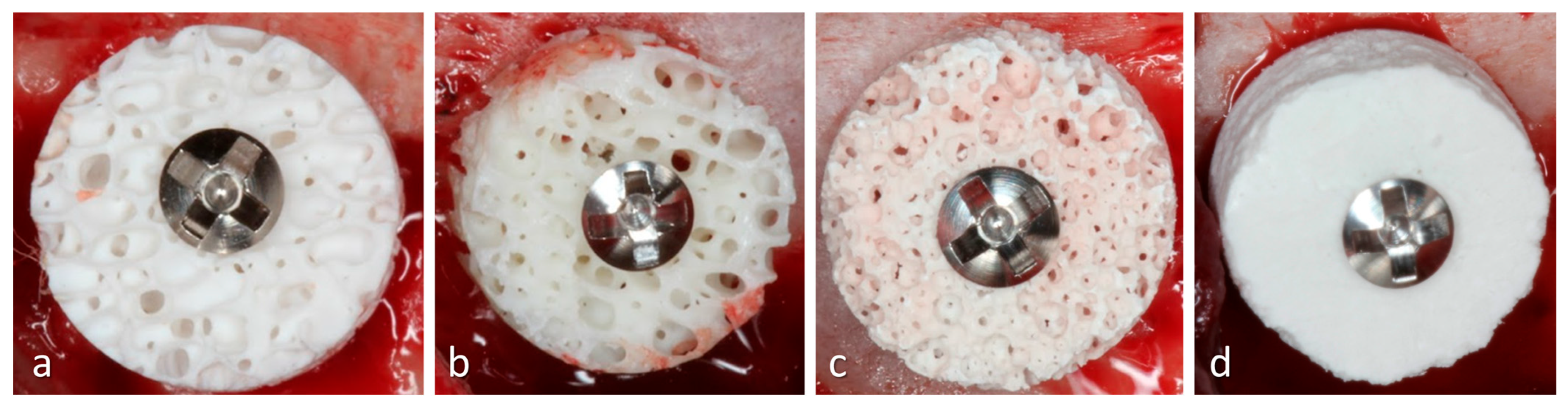
® Block serving as the reference material. Before implantation, each block was trimmed and shaped to obtain standardized cylindrical specimens measuring approximately 7 mm in diameter and 3 mm in height. Preparation was performed under copious sterile saline irrigation to limit overheating and to obtain comparable external dimensions among groups. A central hole was then prepared in each block to allow insertion of the titanium fixation screw. The following biomaterial blocks were used (
Figure 1):
Bio-Oss® Block (Geistlich Pharma AG, Wolhusen, Switzerland) was used as the deproteinized bovine bone mineral block. This xenogeneic biomaterial consists of a porous mineral matrix derived from bovine bone and was included as a slowly resorbing reference material.
OsteoBiol® SP-Block (Tecnoss®, Giaveno, Italy) was used as the collagenic equine-derived block. This material consists of cancellous xenogeneic bone in which the bone matrix is not ceramized and the collagen component is preserved within the scaffold structure. In the present study, according to the manufacturer, the blocks were hydrated with sterile saline solution before fixation.
ReproBone® Blocks (Ceramisys Ltd., Sheffield, UK) were used as the synthetic calcium phosphate block biomaterial. The material consists of a resorbable biphasic calcium phosphate ceramic composed of hydroxyapatite and β-tricalcium phosphate and is characterized by a highly interconnected porous structure.
Alos Block (Allmed S.r.l., Lissone, Italy) was used as the synthetic polymer–hydroxyapatite block. This material is described as a moldable, fully synthetic and resorbable block biomaterial, without components of biological origin. It consists of a polylactic/polyglycolic acid-based copolymer associated with porous non-sintered hydroxyapatite; the copolymer provides a temporary space-maintaining structure, whereas the hydroxyapatite phase acts as the mineral osteoconductive component.
After block fixation, all experimental sites were covered with an OsteoBiol
® Evolution Standard membrane (Tecnoss
®, Giaveno, Italy). This resorbable, non-cross-linked collagen membrane originates from xenogeneic pericardium tissue and is characterized by a dense collagen-fiber matrix intended to protect and stabilize the grafted region [
38]. Membrane permeability or cell occlusivity was not independently quantified in the present study. The membrane was trimmed with sterile scissors, hydrated when required, and adapted over the block and surrounding recipient bed.
The descriptions of the composition and structural characteristics of the four block graft materials were based on information provided by the manufacturers and the available literature. No independent physicochemical or microstructural characterization of the specific material batches was performed in the present study. Therefore, associations between the observed histological patterns and graft properties such as porosity, scaffold architecture, or degradation behavior should be regarded as interpretative hypotheses rather than as mechanisms directly demonstrated by the present experiment.
2.8. Anesthetic Protocol
Each rabbit was premedicated with acepromazine at 1.0 mg/kg IM (Acepran®, Vetnil; Louveira, SP, Brazil). Fifteen minutes later, general anesthesia was induced by the intramuscular administration of xylazine at 3.0 mg/kg (Dopaser®, Hertape Calier; Juatuba, MG, Brazil) combined with ketamine hydrochloride at 50.0 mg/kg (União Química Farmacêutica Nacional S/A; Embu-Guaçu, SP, Brazil). Once an adequate level of anesthesia had been achieved, oxytetracycline was administered intramuscularly at 0.2 mL/kg for antibiotic prophylaxis (Biovet; Vargem Grande Paulista, SP, Brazil).
The surgical field was subsequently clipped and prepared with a 1% povidone–iodine solution (Riodeíne® Tincture, Rioquímica; São José do Rio Preto, SP, Brazil). Additional local anesthesia was obtained by infiltrating 2% mepivacaine containing norepinephrine at a concentration of 1:100,000 (Mepinor®, Nova DFL; Rio de Janeiro, RJ, Brazil).
2.9. Surgical Procedures
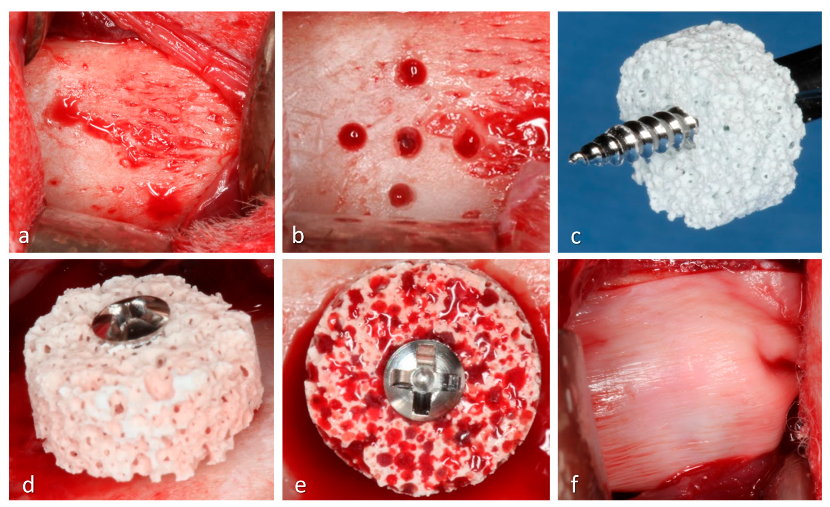
All surgeries were performed by a single experienced operator (E.R.S.). A linear skin incision of approximately 2.5–3.0 cm was made bilaterally along the lower border of the mandible. The muscles and periosteum were carefully reflected to expose the lateral surface of the mandibular angle (
Figure 2).
At each experimental site, the cortical recipient bed was prepared with five equidistant monocortical perforations using a 1.0 mm truncated-conical drill mounted on a straight handpiece under constant sterile saline irrigation. The perforations were performed using a stainless-steel template and extended to the medullary compartment to promote blood supply and cellular migration from the endosteal region.
Each biomaterial block was adapted to the prepared recipient bed. A central perforation was prepared through the block to allow insertion of the fixation screw. The block was positioned in an onlay configuration on the lateral surface of the mandible and fixed using a 1.5 × 10 mm titanium screw (Neodent, Curitiba, Brazil). After fixation, the experimental region was covered with an OsteoBiol® Evolution collagen membrane.
The muscle layers were repositioned and sutured using Vicryl 4-0 (Ethicon, Johnson & Johnson, Cincinnati, OH, USA). The skin was closed with Nylon 4-0 (Ethicon, Johnson & Johnson, USA) using simple interrupted sutures.
2.10. Postoperative Care, Welfare Monitoring, and Humane Endpoints
During the first three postoperative days, the animals received ketoprofen at 3.0 mg/kg every 12 h intramuscularly (Ketofen 10%, Merial, Campinas, São Paulo, Brazil) and tramadol hydrochloride 2% at 1.0 mg/kg every 12 h subcutaneously (Cronidor, Agener União Saúde Animal, Apucarana, Paraná, Brazil).
The animals underwent daily welfare checks performed jointly by the investigators, veterinary personnel, and animal-care staff. Monitoring focused on feeding and drinking behavior, urine and fecal production, activity and general behavior, clinical signs of discomfort or distress, overall health, and the condition of the surgical site, including edema, hemorrhage, wound separation, or signs suggestive of infection.
Criteria for early withdrawal were established before the start of the experiment. These included sustained loss of appetite or inadequate hydration, pain not adequately controlled by the prescribed analgesic regimen, serious infection, wound breakdown likely to interfere with healing, pronounced locomotor impairment, progressive worsening of the animal’s condition, or any other clinical situation judged incompatible with continued participation. When one of these conditions was detected, the rabbit was promptly assessed by the responsible veterinarian and, if a satisfactory recovery was considered improbable, euthanasia was performed in accordance with institutional procedures.
2.11. Euthanasia and Specimen Collection
After 10 weeks of healing, the animals were euthanized by administration of an overdose of intravenous thiopental 1.0 g, 2.0 mL (Thiopentax, Cristália, Itapira, São Paulo, Brazil), in accordance with the guidelines of the Animal Ethics Committee.
The experimental regions were carefully dissected and reduced into individual blocks containing the grafted region, the fixation screw, and the surrounding native bone. All specimens were fixed in 10% paraformaldehyde for 10 days, with regular changes of the fixative solution every two days.
2.12. Histological Processing
After fixation, the specimens were rinsed under running water to remove residual fixative solution. The samples were dehydrated in an ascending series of ethyl alcohol solutions, changed every three days under constant agitation: 60%, 80%, 96%, and absolute alcohol twice.
The specimens were then infiltrated and embedded in LR White™ HardGrid resin (London Resin Co. Ltd., Reading, Berkshire, UK). Polymerization was performed in an oven at 60 °C.
After polymerization, each specimen was sectioned through the center of the grafted region, using the fixation screw as the main reference landmark. One undecalcified ground section per experimental site, approximately 100–150 μm thick, was obtained using a precision cutting/grinding system (Exakt, Apparatebau, Norderstedt, Germany). Each section was then ground to a final thickness of approximately 60–80 μm and stained with Stevenel’s blue and alizarin red.
2.13. Histomorphometric Outcomes and Measurements
Photomicrographs were obtained using 1.6×, 10×, and 40× objectives on a trinocular upright microscope equipped for bright-field and polarized-light microscopy (DMLB; Leica Microsystems, Wetzlar, Germany). Images acquired using the 10× objective were used for the histomorphometric measurements. All measurements were performed using ImageJ 1.54g image-analysis software (National Institutes of Health, Bethesda, MD, USA).
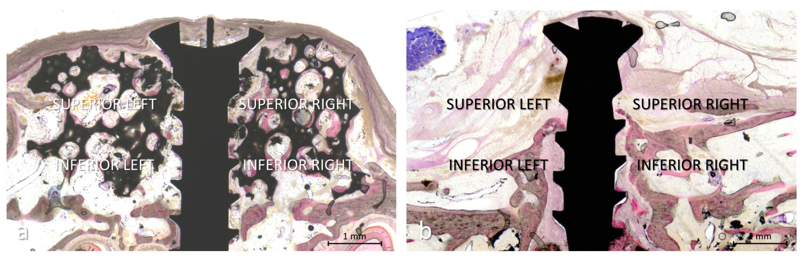
For each specimen, four standardized rectangular regions of interest were selected within the expected original grafted area (
Figure 3a,b). Each region measured 2 mm × 1 mm. Two rectangles were positioned on one side of the fixation screw and two on the opposite side. On each side, one rectangle was placed in the inferior region, immediately above the mandibular recipient bed, and the other in the superior region, toward the external surface of the original block. The inferior border of the lower regions was aligned with the recipient bone surface without including native bone.
The position of the regions of interest was standardized according to the recipient bone surface and fixation screw, independently of the final dimensions of the residual grafted or regenerated compartment. Consequently, in specimens showing marked graft contraction or resorption, portions of the standardized regions could extend beyond the remaining hard-tissue contour.
A point-counting method was applied by superimposing a lattice grid on each region of interest (ROI) using squares 75 µm in dimension. Each lattice intersection was assigned to one, and only one, of the following mutually exclusive categories:
A lattice intersection was classified as IBN-like only when newly formed bone and residual biomaterial were both visually identifiable at the same counting point but could not be reliably assigned to separate categories because they were closely intermingled or optically superimposed. Classification was performed using standardized transmitted-light conditions, followed, when necessary, by increased light intensity and adjustment of the focal plane. When a counting point could be unequivocally assigned to either newly formed bone or residual graft, it was recorded in the corresponding conventional category rather than as IBN-like.
Intra-compartment soft tissue included non-mineralized tissue located within the external contour of the residual grafted or regenerated compartment and not assigned to any of the other specifically identified categories.
Extra-compartment soft tissue included non-mineralized tissue located within the standardized region of interest (ROI) but outside the residual grafted or regenerated hard-tissue contour. Because the regions of interest were positioned according to fixed anatomical references rather than the final dimensions of the regenerated compartment, this category represented the proportion of the standardized region no longer occupied by the residual grafted or regenerated compartment and was therefore interpreted primarily as an indicator of compartment contraction or resorption.
Other components included vascular structures, inflammatory infiltrate, residual collagen membrane, muscle fibers, and osteoblast-rich areas. These structures were grouped because each represented a relatively limited and variably distributed proportion of the standardized regions and was not considered a separate primary histomorphometric outcome.
For each histological section, the two inferior regions of interest, located on opposite sides of the fixation screw, were averaged to obtain a single inferior value. Similarly, the two superior regions of interest were averaged to obtain a single superior value. The resulting inferior and superior values were used as the site-level values for each histomorphometric outcome. The overall site-level value was calculated as the mean of the inferior and superior site-level values. Thus, each hemimandibular experimental site contributed one inferior, one superior, and one overall value for each outcome.
2.14. Cross-Sectional Area of the Augmented Region
The same ground section used for the histomorphometric analysis was used to measure the cross-sectional area of the augmented region. The area was measured planimetrically on calibrated digital images using ImageJ 1.54g software. The external contour of the residual grafted or regenerated compartment was manually delineated, using the surface of the mandibular recipient bed as the basal boundary. The traced area comprised the entire space enclosed within this contour, including newly formed bone, residual biomaterial, IBN-like tissue, non-mineralized tissue, and the portion of the fixation screw located within the augmented compartment. Tissues located outside this contour were excluded. The resulting area was expressed in square millimeters (mm2) and was used solely as a two-dimensional indicator of the extent of the augmented region at 10 weeks. It was not interpreted as a direct measurement of augmented volume or volumetric stability.
2.15. Statistical Analysis
Site-level data were summarized as mean ± standard deviation. The percentage of newly formed bone within the standardized regions of interest was designated as the primary histomorphometric outcome. Secondary outcomes included the percentages of residual graft, IBN-like tissue, intra-compartment soft tissue, extra-compartment soft tissue, and other components, as well as the cross-sectional area of the augmented region. Comparisons between the inferior and superior regions were considered exploratory regional analyses.
Bio-Oss® Block was used as the reference material because deproteinized bovine bone mineral is extensively used as a xenogeneic scaffold in bone-augmentation procedures and is characterized by a slow resorption profile. The main planned comparisons therefore evaluated each test biomaterial against Bio-Oss® Block.
The hemimandibular site was considered the experimental unit. Because each rabbit contributed two experimental sites, observations obtained from the same animal were treated as correlated rather than fully independent. Moreover, site allocation was not constrained to ensure that each rabbit received two different biomaterials.
For overall site-level outcomes suitable for mixed-model analysis, comparisons among biomaterials were performed using linear mixed-effects models, with biomaterial included as a fixed factor and animal as a random intercept. Degrees of freedom were estimated using the Satterthwaite approximation. Planned contrasts comparing each test biomaterial with Bio-Oss® Block were estimated from the mixed model and adjusted for multiple comparisons using Dunnett’s method.
For the regional analyses, biomaterial, region (inferior or superior), and their interaction were included as fixed factors. Animal and experimental site nested within animal were included as random effects to account for the correlation between the two sites from the same rabbit and between the inferior and superior measurements obtained from the same site. Comparisons between each test material and Bio-Oss® Block were performed separately within the inferior and superior regions using Dunnett-adjusted contrasts. Inferior–superior comparisons within each material were adjusted using Holm’s method.
Because IBN-like tissue was absent from all Bio-Oss® Block and SP-Block sites and was present in all ReproBone® Block sites but only one Alos Block site, its distribution showed structural zeros and marked separation among materials. Extra-compartment soft tissue showed a similarly sparse distribution, being absent from the Bio-Oss® Block, SP-Block, and ReproBone® Block groups and present almost exclusively in the Alos Block group. These two outcomes were therefore summarized descriptively and were not analyzed using standard Gaussian mixed-effects models.
Model assumptions were evaluated by inspecting residual-versus-fitted plots and quantile-quantile plots of the residuals. The Shapiro–Wilk test was used only as a supplementary assessment and was not interpreted in isolation. When material deviations from model assumptions were identified, an appropriate transformation was considered. If an adequate model could not be obtained, the corresponding outcome was summarized descriptively and interpreted as exploratory rather than being analyzed using rank-based tests that did not account for within-animal clustering.
The statistical significance level was set at 5%. Multiplicity adjustment was applied to the planned comparisons between each test biomaterial and Bio-Oss® Block within each outcome. Because the secondary and regional outcomes were exploratory, their results were interpreted cautiously.
Statistical analyses were performed using R software, version 4.6.1 (R Foundation for Statistical Computing, Vienna, Austria), with the lme4, lmerTest, and emmeans packages.
4. Discussion
4.1. Principal Findings and Relevance of the Onlay Model
The present exploratory study compared the healing and remodeling patterns of two xenogeneic and two synthetic block biomaterials used as onlay grafts on the lateral aspect of rabbit mandibles. After 10 weeks of healing, all four materials supported new bone formation, with no significant differences in the overall percentage of newly formed bone. Nevertheless, marked differences were observed in scaffold persistence, tissue organization, mode of incorporation, and cross-sectional area observed at 10 weeks. These findings indicate that the biological performance of block biomaterials cannot be interpreted solely according to the percentage of newly formed bone but requires consideration of the residual scaffold, soft-tissue organization, IBN-like tissue, and cross-sectional augmented area at 10 weeks.
These differences are particularly relevant in an onlay model. Unlike an inlay graft placed within a contained defect, an onlay block has limited contact with native bone and therefore depends largely on vascular and osteogenic ingrowth from the underlying recipient bed, although the repositioned periosteum may provide an additional contribution from the outer aspect. In the present model, the collagen membrane was interposed between the block and the repositioned periosteum and could therefore have modified or delayed the periosteal contribution, particularly during early healing. Experimental evidence suggests that this effect may depend on the membrane and experimental model: a non-cross-linked collagen membrane did not substantially impair periosteal regenerative capacity in a rat calvarial model, whereas lower neoangiogenesis was observed beneath a resorbable barrier membrane than beneath a rapidly resorbing collagen fleece in rabbit calvarial defects [
39,
40]. In the present study, all sites received the same membrane, and neither a membrane-free control nor direct measurements of vascularization were included. Consequently, whether the membrane reduced revascularization or de novo osteogenesis cannot be determined. The cortical perforations were nevertheless intended to promote vascular and cellular supply from the underlying medullary compartment.
Bone formation must therefore progress from the mandibular surface toward regions increasingly distant from the native bone, while the graft must simultaneously resist soft-tissue pressure and preserve the intended augmentation contour. Previous experiments using the same rabbit mandibular model showed that inlay grafts generally provide more favorable conditions for bone formation because they are exposed to multiple surrounding bone surfaces [
23,
24]. Nevertheless, the present study demonstrated that newly formed bone could also reach the superior regions of onlay blocks when a stable and sufficiently persistent osteoconductive framework was maintained.
The absence of a significant material-by-region interaction for newly formed bone suggests that its regional distribution was not consistently different among materials. However, a significantly greater percentage of newly formed bone was observed in the inferior than in the superior region of the Alos Block group. Conversely, residual graft showed a significant material-by-region interaction, indicating that scaffold persistence differed not only among biomaterials but also according to position within the block. Overall, the findings suggest that onlay graft healing may be influenced by the balance among scaffold persistence, osteoconductive continuity, and tissue formation. Because the materials were not independently characterized and only one healing interval was evaluated, explanations involving scaffold architecture and degradation kinetics should be regarded as interpretative rather than mechanistically demonstrated.
4.2. Bio-Oss® Block: Structural Persistence and Surface-Guided Osteoconduction
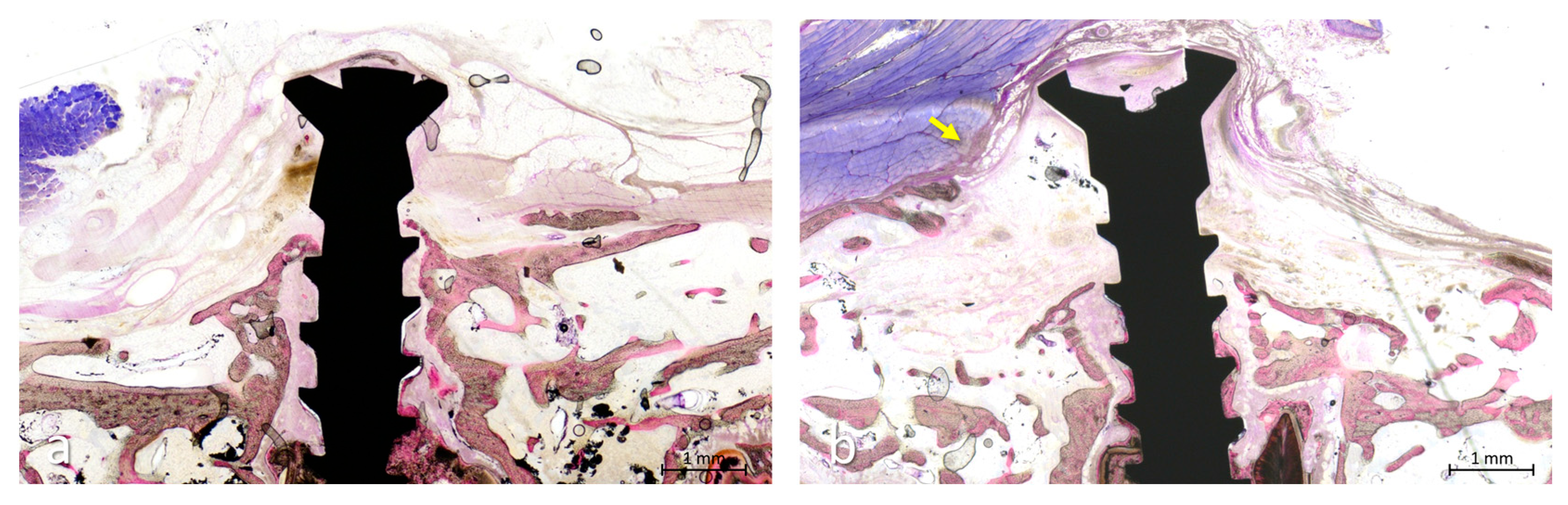
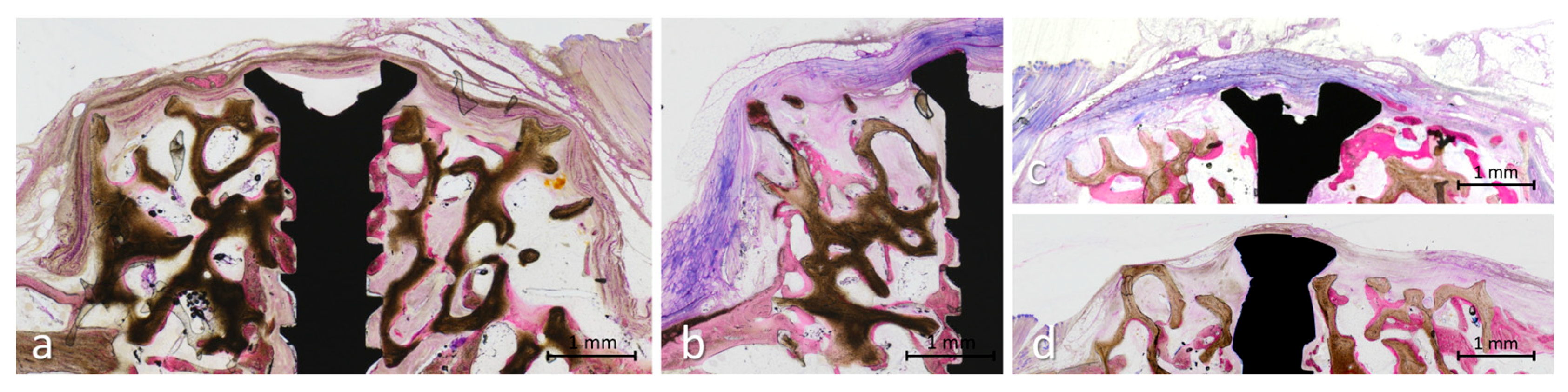
Bio-Oss® Block showed the greatest proportion of residual graft and maintained a cross-sectional augmented area comparable to those of SP-Block and ReproBone® Block. Despite the persistence of the scaffold, newly formed bone was not confined to the interface with the mandibular recipient bed. Bone developed along the surfaces of the residual trabeculae and, in several specimens, reached the superior and peripheral portions of the block. This pattern was characterized by preservation of a continuous trabecular framework, with bone growth occurring along its surfaces.
However, scaffold persistence should not be interpreted as biological replacement. Newly formed bone was frequently deposited as relatively thin layers along the residual biomaterial surfaces, and the overall percentage of bone did not differ significantly from that observed with the other materials. Thus, preservation of the cross-sectional augmented area at 10 weeks and extensive spatial distribution of bone did not necessarily correspond to replacement of the graft by vital tissue.
Previous canine studies reported more limited incorporation of DBBM blocks, with bone formation mainly restricted to the basal region or connective tissue interposed between the graft and the recipient bed [
1,
21]. These differences cannot be explained solely by the onlay or inlay configuration. Graft adaptation, uninterrupted stability at the recipient interface, vascular access, species-related healing dynamics, and the surgical procedures performed during healing may also influence incorporation.
In the present study, the blocks remained undisturbed throughout the 10-week healing period. In contrast, in the experiments by De Santis et al. [
21], implant osteotomies were subsequently prepared at the interface between the graft and native bone after only 3 months of healing. This relatively short healing interval may also have contributed to the observed outcome, as the newly established connections between the graft and native bone might not yet have reached sufficient maturity. Although this remains speculative, drilling and implant insertion may therefore have disrupted immature connections or displaced the brittle scaffold. Overall, Bio-Oss
® Block showed preservation of the cross-sectional augmented area and a surface-guided pattern of osteoconduction at 10 weeks, while the substantial amount of residual scaffold indicated limited biological substitution during the evaluated interval.
4.3. SP-Block: Heterogeneous Scaffold Remodeling
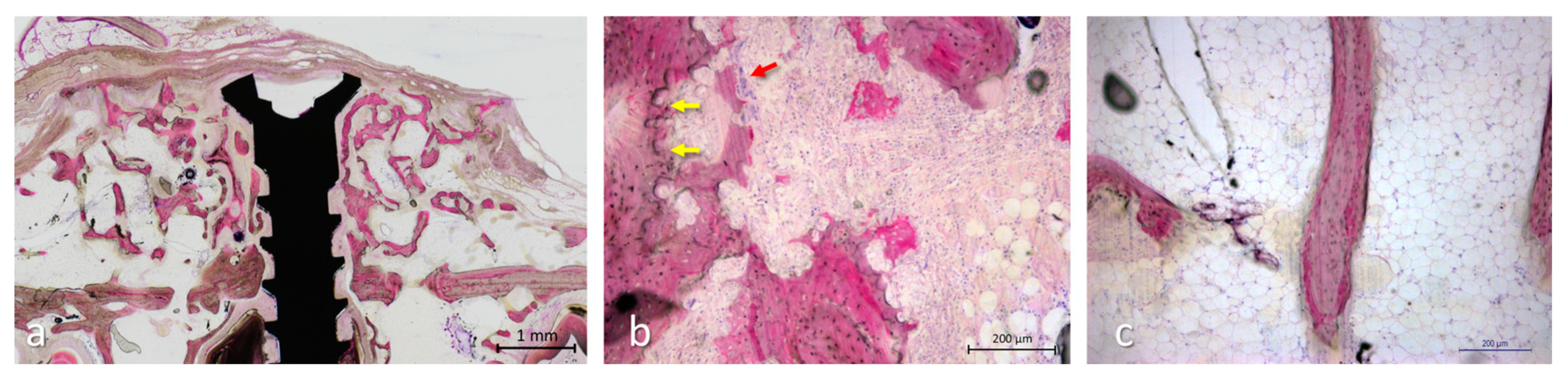
SP-Block showed significantly less residual graft than Bio-Oss® Block while maintaining a comparable cross-sectional augmented area. The original trabecular architecture was variably preserved, and residual trabeculae frequently displayed irregular surfaces and resorption bays (Howship’s lacunae). Newly formed bone developed along the remaining scaffold and occasionally reached the superior region, but its percentage and spatial distribution varied considerably among specimens.
The reduced persistence of the biomaterial was accompanied by a greater proportion of intra-compartment non-mineralized tissue, much of which exhibited a marrow-like appearance, rather than by a significantly greater percentage of newly formed bone. Thus, the lower proportion of recognizable residual graft was not accompanied by a proportional increase in mineralized bone, although part of the space created during remodeling appeared to undergo maturation toward bone marrow rather than being occupied simply by undifferentiated connective tissue. When loss of the trabecular framework occurred before sufficient bone deposition, however, the continuity of the mineralized osteoconductive pathways toward the superior region may have been reduced.
The sparse osteoclasts observed at 10 weeks, together with the presence of Howship’s lacunae, suggest that more active resorption may have occurred earlier during healing. Effective remodeling requires coordination between osteoclastic resorption and subsequent bone formation, whereas an imbalance between these processes may result in net loss of the mineralized scaffold [
41]. SP-Block therefore showed a more pronounced and heterogeneous remodeling pattern than Bio-Oss
® Block, characterized by reduced graft persistence, new bone formation, and marrow maturation.
4.4. ReproBone® Blocks: Interconnected Porosity and IBN-like Pattern
ReproBone® Blocks maintained a cross-sectional augmented area comparable to that of Bio-Oss® Block, despite showing a significantly lower proportion of conventionally recognizable residual graft. Newly formed bone was observed at different levels of the porous scaffold and reached the superior regions in several specimens. However, the overall percentage of newly formed bone did not differ significantly from that observed with Bio-Oss® Block or the other materials.
The most distinctive finding was the consistent presence of IBN-like tissue in all ReproBone
® specimens. These areas represented a close intermingling or optical superimposition of newly formed bone and residual calcium phosphate scaffold, rather than the clearly demarcated surface apposition typically observed around Bio-Oss
® trabeculae. Similar patterns have been described with calcium phosphate biomaterials in experimental sinus augmentation and human biopsies [
42,
43]. A comparable IBN-like pattern was also observed with ReproBone
® novo Paste in a rabbit sinus augmentation model, despite the different anatomical environment and material configuration [
44].
Nevertheless, IBN-like tissue should not be interpreted as pure newly formed bone or as evidence that bone penetrated directly through the solid scaffold matrix. In two-dimensional ground sections, bone occupying interconnected pores or structures located at different depths may appear continuous across residual biomaterial. Maintaining IBN-like tissue as a separate category therefore avoided overestimating either bone formation or graft persistence.
Overall, ReproBone® Blocks showed preservation of the cross-sectional augmented area and a frequent histological pattern in which newly formed bone and residual scaffold could not be reliably separated in two-dimensional sections. This IBN-like pattern should be regarded solely as a descriptive observation. Its biological maturation, mechanical competence, long-term fate, and potential relevance to implant osseointegration remain unknown.
4.5. Alos Block: Extensive Remodeling and Reduced Cross-Sectional Area
Alos Block showed the lowest proportion of residual graft and the smallest cross-sectional augmented area among the materials evaluated. Although the percentage of newly formed bone did not differ significantly from that observed in the other groups, the original scaffold architecture was largely lost, and the healing pattern varied considerably among specimens.
In some sites, scaffold degradation was accompanied by the formation of organized bone surrounding marrow-like compartments and extending toward the superior region. In others, bone remained predominantly confined to the inferior portion, while the superior region was replaced by connective or muscular tissue. This heterogeneous pattern was also reflected in the regional analysis, in which the percentage of newly formed bone was significantly higher in the inferior than in the superior region of the Alos Block group. Extra-compartment soft tissue was located predominantly in the superior region, a descriptive finding consistent with incomplete maintenance of the original augmentation contour.
Only limited IBN-like tissue was identified, coinciding with extensive scaffold loss and few residual areas in which bone and biomaterial could still be recognized simultaneously. Overall, Alos Block showed the greatest loss of recognizable residual scaffold; however, this was not accompanied by a greater percentage of newly formed bone, and maintenance of the cross-sectional augmented area was less consistent. These observations are compatible with pronounced remodeling, but they do not establish degradation kinetics or long-term material persistence.
A similarly pronounced degradation pattern was observed with Alos Granular in rabbit sinus augmentation, where extensive scaffold degradation was associated with marked reduction in the augmented compartment [
44]. However, IBN-like tissue was more evident with the granular formulation than with the Alos Block evaluated in the present study, suggesting that material configuration and the anatomical environment may influence the relationship between scaffold degradation and newly formed bone.
4.6. Translational Considerations
From a translational perspective, an ideal block biomaterial would provide initial structural support, permit vascular and bone ingrowth, and remodel in coordination with new bone formation. However, the present exploratory study was not designed to establish clinical performance or to determine the optimal biomaterial for a specific type of defect.
Within the limitations of this model, the findings suggest that the percentage of newly formed bone alone may not fully describe the histological healing pattern of a block biomaterial. Residual scaffold, spatial distribution of bone, tissue organization, IBN-like tissue, and cross-sectional augmented area at 10 weeks provided complementary information. Whether these histological findings translate into three-dimensional maintenance, mechanical competence, implant osseointegration, or performance under functional loading remains unknown.
Autogenous bone remains the biological reference for onlay augmentation because it provides vital, physiologically remodelable tissue. This advantage must be balanced against donor-site morbidity, limited availability, increased surgical time, and postoperative discomfort [
13,
14]. Substitute blocks avoid graft harvesting and provide standardized, readily available scaffolds, but the present findings do not demonstrate biological equivalence to autogenous bone.
Previous animal experiments provide relevant context for this comparison. In a rabbit mandibular onlay model, autogenous iliac-crest blocks and equine xenogeneic blocks yielded similar percentages of newly formed bone after 60 days; however, the autogenous blocks showed better incorporation into the recipient bed, greater bone volume, and superior overall quality of the grafted area [
45]. Similarly, in dogs, autogenous mandibular blocks became vital and integrated with the parent bone, whereas deproteinized bovine bone mineral blocks showed limited bone ingrowth [
21]. Nevertheless, these historical comparisons cannot replace a contemporaneous autogenous control because of differences in experimental models, biomaterials, surgical procedures, and healing intervals.
Accordingly, no recommendation regarding clinical material selection or superiority can be made on the basis of this experiment. Direct extrapolation to human lateral ridge augmentation should be made cautiously, and longer-term studies incorporating three-dimensional volumetric analysis, mechanical assessment, implant placement, and functional loading are required.
4.7. Study Limitations
Several limitations should be considered when interpreting the present findings. First, this was an exploratory preclinical study performed in rabbit mandibles, and the observed biological response cannot be directly extrapolated to human lateral ridge augmentation. Rabbits exhibit substantially faster bone turnover and healing than humans, and differences in mandibular anatomy, bone characteristics, local mechanical conditions, and the experimental environment should be considered when translating these findings to the clinical setting [
46,
47]. Moreover, only one healing interval of 10 weeks was evaluated; therefore, the study provides a cross-sectional representation of graft incorporation and remodeling but does not establish their temporal progression.
Second, the sample size was limited. Although the allocation provided six sites for each biomaterial, it was not constrained within animals, and some rabbits received the same material bilaterally. Consequently, comparisons among biomaterials were not uniformly paired within animals. Because no reliable prior estimate of the within-animal correlation was available, the approximate sample-size calculation assumed independent site-level observations and did not account for clustering of the two sites within each rabbit. The calculation may therefore have overestimated the effective statistical power. Although the mixed-effects models accounted for correlation between sites belonging to the same rabbit, the study remains primarily suited to identifying relatively large differences. In addition, complete masking of the examiner could not be maintained because the biomaterials had distinctive histological appearances.
Third, no autogenous bone control was included. Comparisons with previous studies of autogenous block grafts remain indirect because of differences in experimental models, surgical procedures, and healing intervals. The present experiment therefore allows comparison among the four substitute biomaterials but cannot determine whether any of them approaches the biological performance of an autogenous onlay graft under identical conditions.
Fourth, the histomorphometric assessment was based on ground sections and standardized regions of interest. Although this approach allowed consistent comparison of inferior and superior regions and identification of tissues located outside the remaining augmented contour, it cannot fully represent the three-dimensional distribution of bone, residual scaffold, pores, and soft tissues. The cross-sectional area measurements provided complementary information on cross-sectional augmented area at 10 weeks. No micro-computed tomographic or other volumetric assessment was available; therefore, three-dimensional maintenance or volumetric stability could not be determined.
Particular caution is required when interpreting IBN-like tissue. Its identification depended on the recognition of newly formed bone closely intermingled with, or optically superimposed on, residual biomaterial. Section thickness, optical density, overlapping pores, and structures located at different depths may have influenced its classification. Although predefined criteria and standardized illumination were used, IBN-like tissue remains a descriptive histological category rather than a validated biological entity and cannot be considered equivalent to either pure vital bone or residual graft.
The physicochemical and microstructural characteristics of the specific biomaterial batches were not independently assessed. Interpretations linking the observed histological behavior to material composition, porosity, scaffold architecture, or degradation characteristics were therefore based on manufacturer information and published evidence and should be considered hypothesis-generating.
Finally, the study did not assess the mechanical properties of the regenerated regions or their ability to support implant placement and functional loading. The behavior of the different scaffolds during drilling, their potential for fracture or displacement, and subsequent implant osseointegration therefore remain unknown. Future studies should include longer healing periods, three-dimensional volumetric analyses, mechanical assessment, and implant placement.





 ,
,