Comparative Histological and Histomorphometric Analysis of Xenografts and Synthetic Biomaterials in Rabbit Sinus Floor Elevation


 ,
,  ,
,  ,
, 
Abstract
1. Introduction
2. Materials and Methods
2.1. Ethical Considerations
2.2. Research Design
2.3. Animals and Eligibility Criteria
2.4. Acclimatization, Housing, and Environmental Enrichment
2.5. Sample Size Calculation
2.6. Randomization, Allocation Concealment, and Blinding
2.7. Biomaterials
2.8. Anesthetic Protocol
2.9. Surgical Procedures
2.10. Postoperative Care, Welfare Monitoring, and Humane Endpoints
2.11. Euthanasia
2.12. Histological Processing
2.13. Histomorphometric Outcomes and Measurements
2.14. Statistical Analysis
3. Results
3.1. Animal Flow and Adverse Events
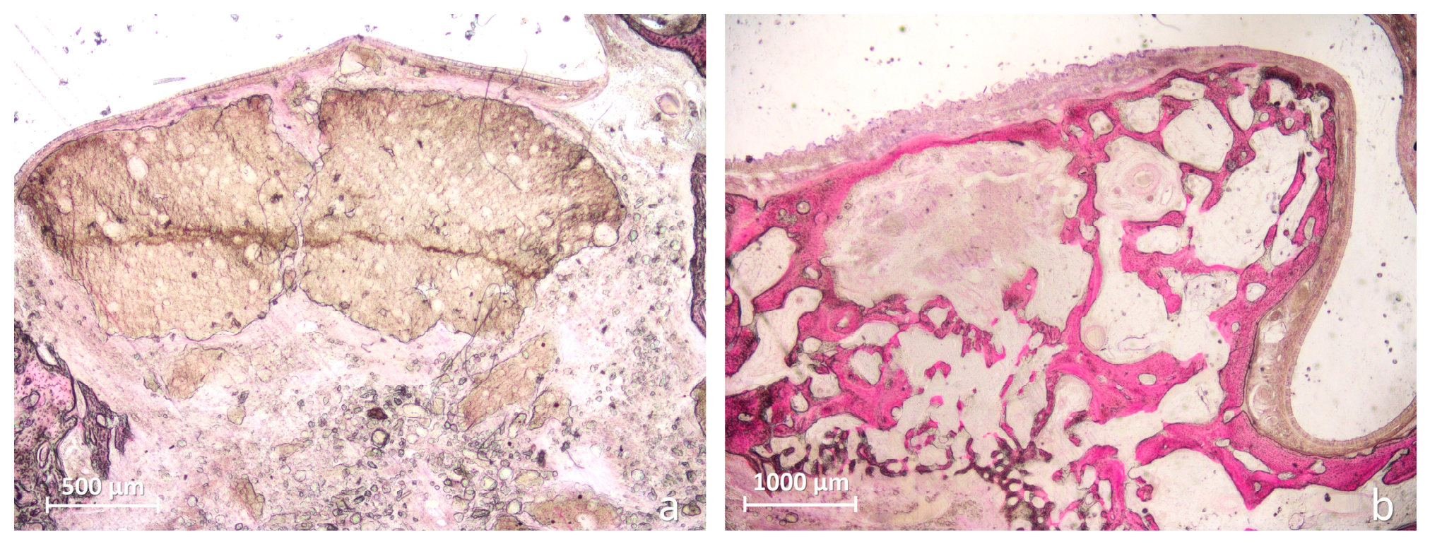
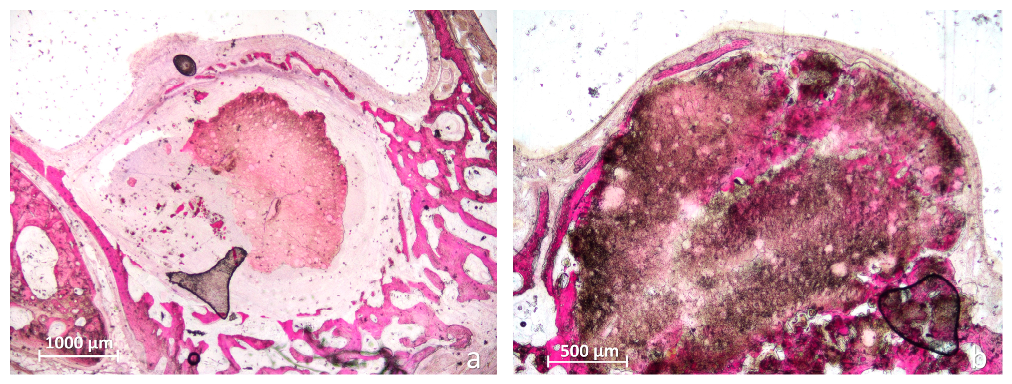
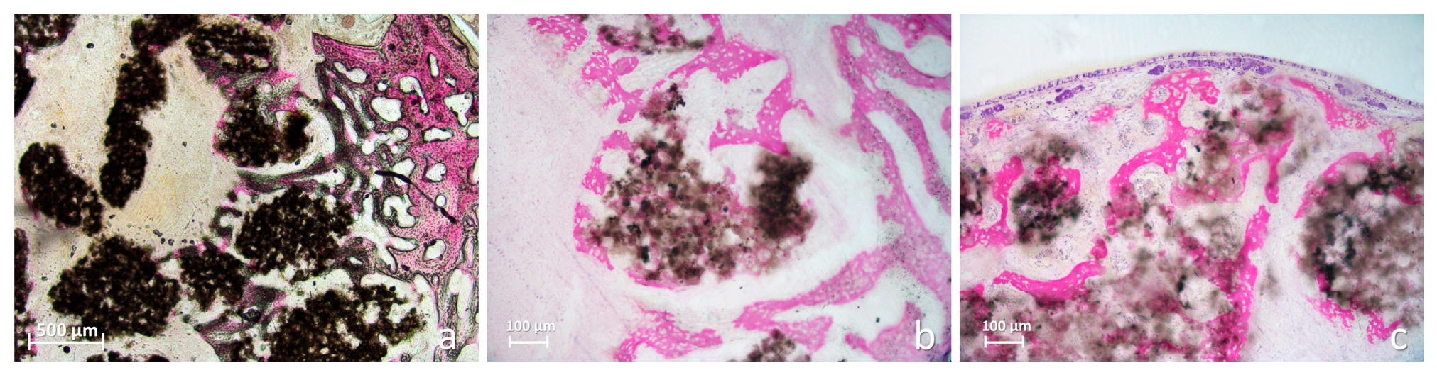
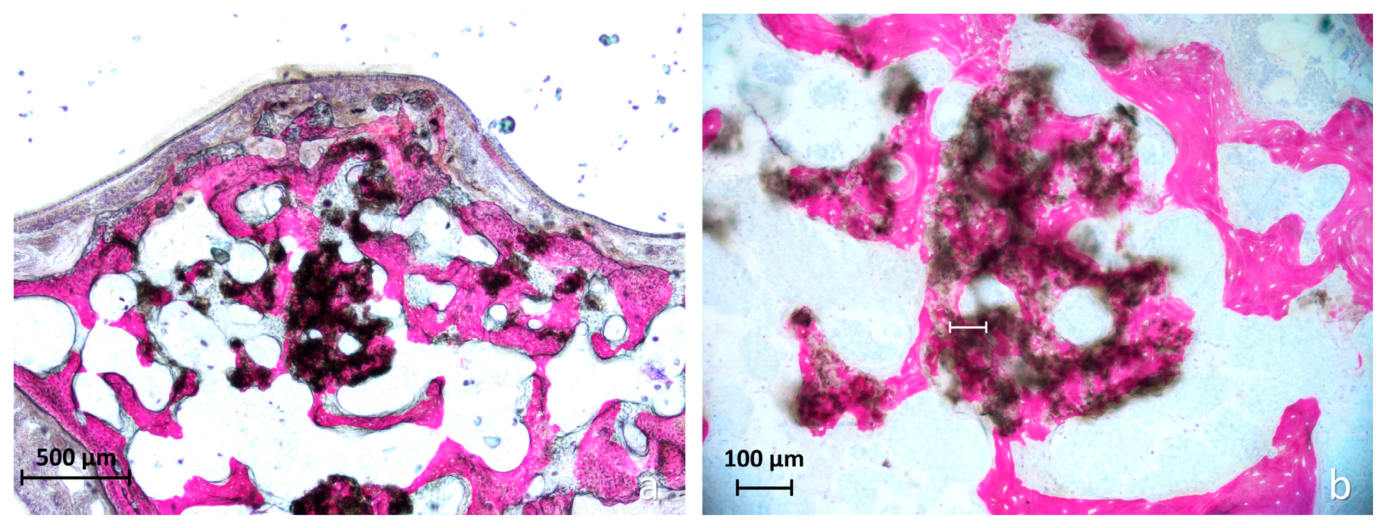
3.2. Qualitative Morphologic Evaluation
3.3. Quantitative Histomorphometric Evaluation
3.4. Elevated Compartment Cross-Sectional Area
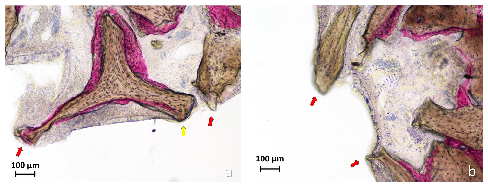
3.5. Descriptive Assessment of Schneiderian Membrane Alterations
4. Discussion
5. Conclusions
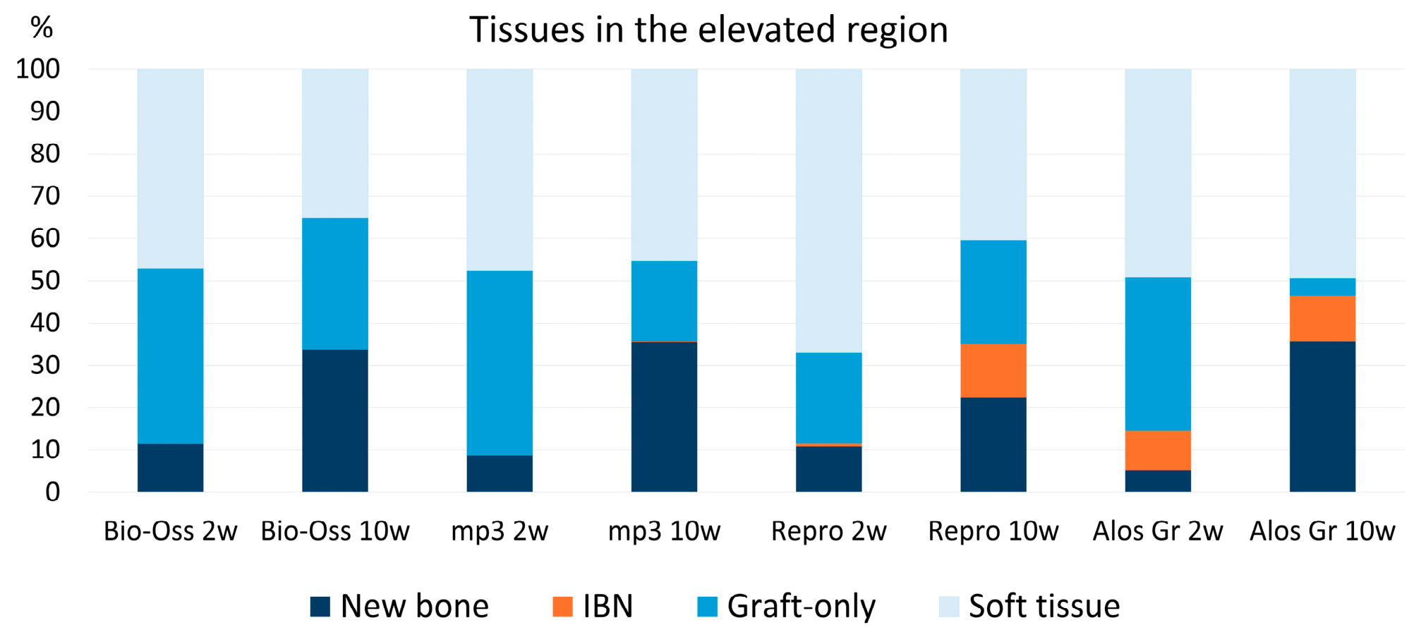
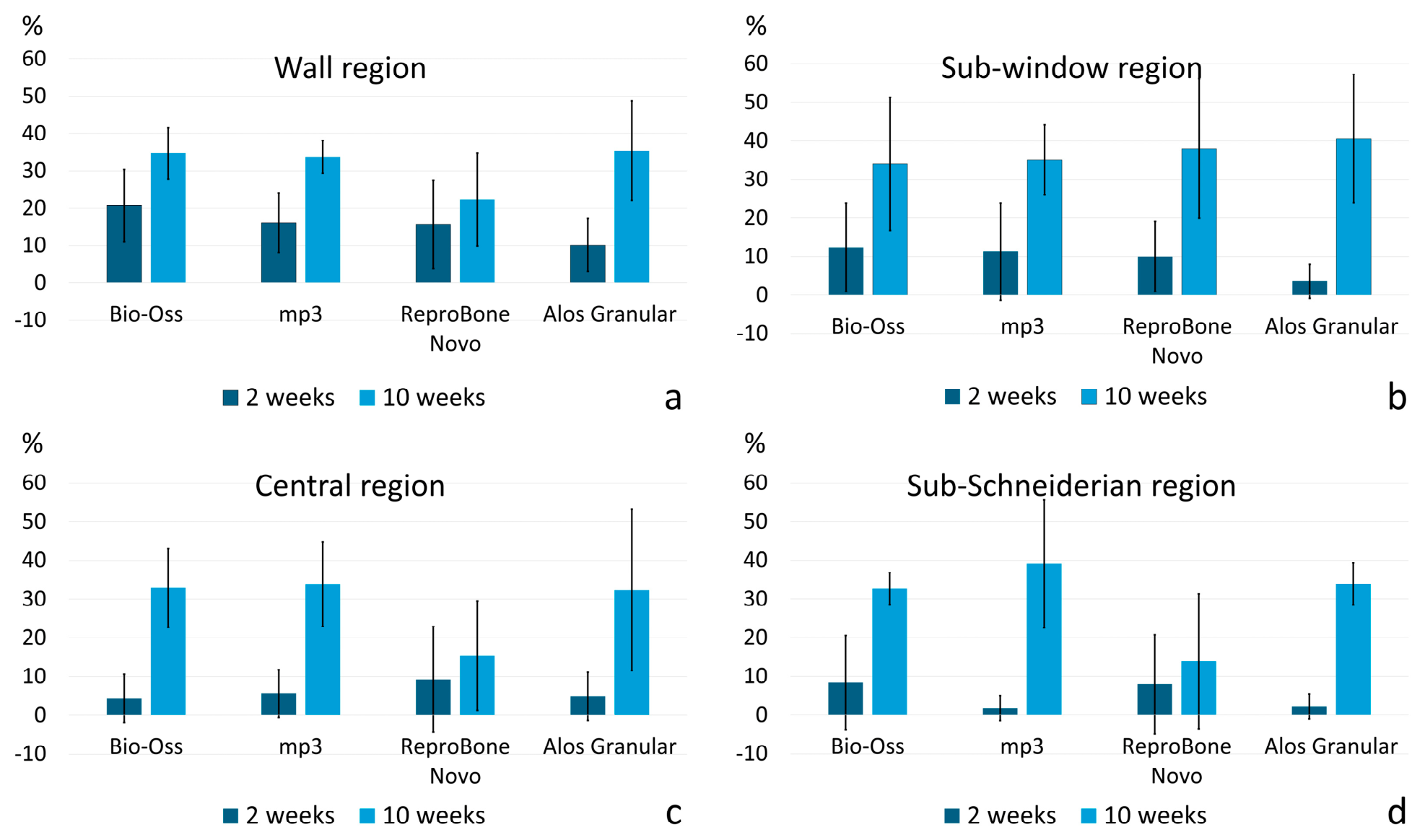
- Newly formed bone percentages did not differ significantly among the four biomaterials at either 2 or 10 weeks.
- At 10 weeks, Bio-Oss® showed significantly higher residual graft and combined graft and IBN percentages than Alos Granular, consistent with greater scaffold persistence. At the same healing interval, IBN-like tissue percentages were higher in ReproBone® novo Paste and Alos Granular than in Bio-Oss®.
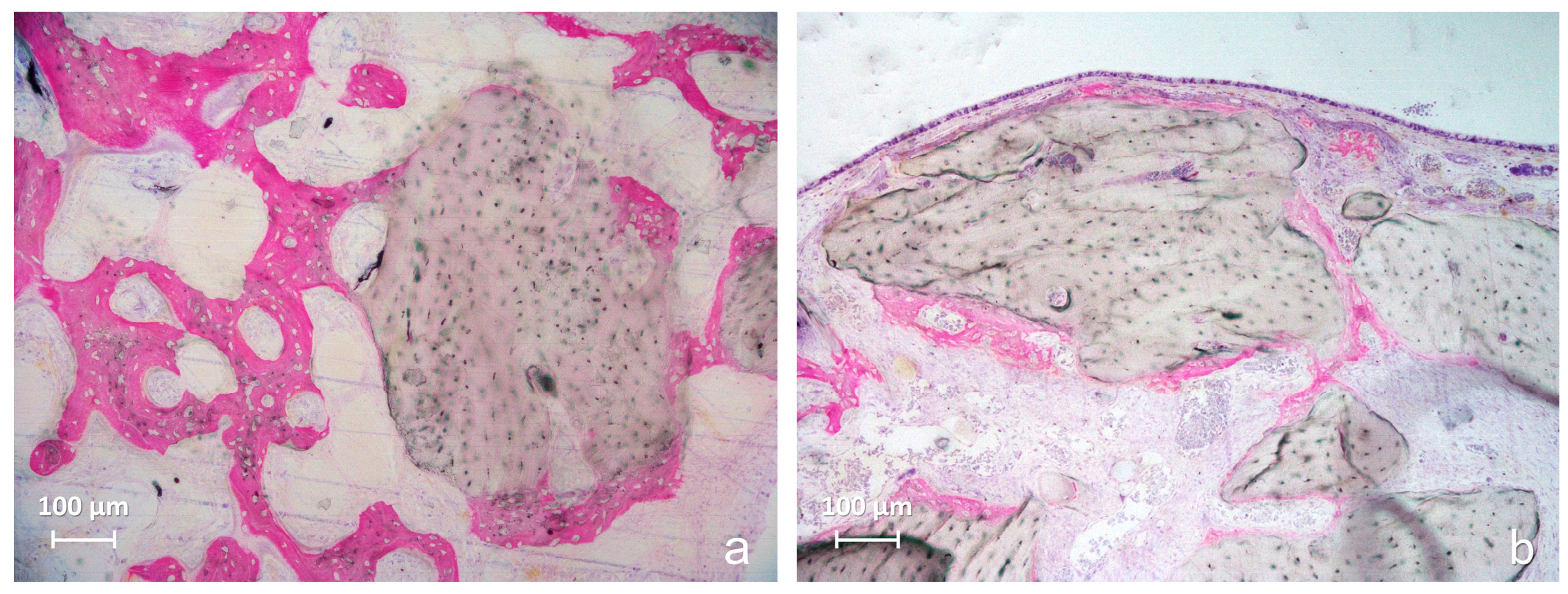
- Bio-Oss® and mp3® predominantly showed conventional surface-related osteoconduction within a recognizable trabecular network. Alos Granular showed extensive scaffold degradation and limited residual graft persistence, although the regenerated bone generally maintained a recognizable trabecular organization. ReproBone® novo Paste showed heterogeneous graft–bone intermingling, with extensive graft and IBN areas frequently associated with irregular and poorly trabeculated mineralized tissue.
- Overall, the findings indicate that the biological performance of sinus grafting materials cannot be characterized by newly formed bone percentage alone. Residual graft persistence, degradation pattern, graft–bone integration, tissue architecture, maintenance of the elevated compartment, and Schneiderian membrane response should also be considered. Further studies including implant placement and direct assessment of the bone-to-implant interface are required to determine the functional relevance of persistent IBN-like graft–bone complexes.
Author Contributions
Funding
Institutional Review Board Statement
Informed Consent Statement
Data Availability Statement
Acknowledgments
Conflicts of Interest
References
- Sabri, H.; Saleh, M.H.A.; Nava, P.; Scaini, R.; Testori, T.; Del Fabbro, M. Long-term outcomes of lateral sinus floor elevation: A machine-learning analysis, systematic review, and meta-analysis of predictive factors. Periodontol. 2000 2026, Epub ahead of print. [Google Scholar] [CrossRef] [Scilit] [PubMed]
- Sabri, H.; Saleh, M.H.A.; Zimmer, J.M.; Schwarz, F.; Wang, H.L. Patient-Reported Outcome Measures (PROMs) and Clinician-Reported Outcomes (ClinROs) in Sinus Floor Elevation for Implant Rehab in the Edentulous Maxilla: A Systematic Review and COSMIN Analysis. Clin. Oral Implant. Res. 2026, 37, S346–S371. [Google Scholar] [CrossRef] [Scilit] [PubMed] [PubMed Central]
- Ghodsian, D.; D’Jesús, S.; Sánchez-Labrador, L.; Cobo-Vázquez, C.M.; Cortés-Bretón Brinkmann, J.; Martínez-González, J.M.; Meniz-García, C. Maxillary Sinus Augmentation with Autogenous Tooth Grafting Material: A Systematic Review. Biomimetics 2024, 9, 518. [Google Scholar] [CrossRef] [Scilit] [PubMed] [PubMed Central]
- Busenlechner, D.; Huber, C.D.; Vasak, C.; Dobsak, A.; Gruber, R.; Watzek, G. Sinus augmentation analysis revised: The gradient of graft consolidation. Clin. Oral Implant. Res. 2009, 20, 1078–1083. [Google Scholar] [CrossRef] [Scilit] [PubMed]
- Lambert, F.; Léonard, A.; Drion, P.; Sourice, S.; Layrolle, P.; Rompen, E. Influence of space-filling materials in subantral bone augmentation: Blood clot vs. autogenous bone chips vs. bovine hydroxyapatite. Clin. Oral Implant. Res. 2011, 22, 538–545. [Google Scholar] [CrossRef] [Scilit] [PubMed]
- Xu, H.; Shimizu, Y.; Asai, S.; Ooya, K. Grafting of deproteinized bone particles inhibits bone resorption after maxillary sinus floor elevation. Clin. Oral Implant. Res. 2004, 15, 126–133. [Google Scholar] [CrossRef] [Scilit] [PubMed]
- Corbella, S.; Taschieri, S.; Weinstein, R.; Del Fabbro, M. Histomorphometric outcomes after lateral sinus floor elevation procedure: A systematic review of the literature and meta-analysis. Clin. Oral Implant Res. 2016, 27, 1106–1122. [Google Scholar] [CrossRef] [Scilit] [PubMed]
- Zhang, L.; Si, M.; Shi, J.; Yang, G.; Shi, Y. Evaluation of three-dimensional contraction of the volume of grafts after staged augmentation of the sinus floor. Br. J. Oral Maxillofac. Surg. 2019, 57, 323–329. [Google Scholar] [CrossRef] [Scilit] [PubMed]
- Temmerman, A.; Van Dessel, J.; Cortellini, S.; Jacobs, R.; Teughels, W.; Quirynen, M. Volumetric changes of grafted volumes and the Schneiderian membrane after transcrestal and lateral sinus floor elevation procedures. J. Clin. Periodontol. 2017, 44, 660–671. [Google Scholar] [CrossRef] [Scilit] [PubMed]
- Jensen, T.; Schou, S.; Svendsen, P.A.; Forman, J.L.; Gundersen, H.J.G.; Terheyden, H.; Holmstrup, P. Volumetric changes of the graft after maxillary sinus floor augmentation with Bio-Oss and autogenous bone in different ratios: A radiographic study in minipigs. Clin. Oral Implant. Res. 2012, 23, 902–910. [Google Scholar] [CrossRef] [Scilit] [PubMed]
- Canellas, J.V.D.S.; Drugos, L.; Ritto, F.G.; Fischer, R.G.; Medeiros, P.J.D. Xenograft materials in maxillary sinus floor elevation surgery: A systematic review with network meta-analyses. Br. J. Oral Maxillofac. Surg. 2021, 59, 742–751. [Google Scholar] [CrossRef] [Scilit] [PubMed]
- Li, X.; Lin, S.C.; Duan, S.Y. The impact of deproteinized bovine bone particle size on histological outcomes in sinus floor elevation: A systematic review and meta-analysis. Int. J. Implant Dent. 2023, 9, 35. [Google Scholar] [CrossRef] [Scilit] [PubMed] [PubMed Central]
- Li, Y.; Zhou, W.; Li, P.; Luo, Q.; Li, A.; Zhang, X. Comparison of the osteogenic effectiveness of an autogenous demineralised dentin matrix and Bio-Oss® in bone augmentation: A systematic review and meta-analysis. Br. J. Oral Maxillofac. Surg. 2022, 60, 868–876. [Google Scholar] [CrossRef] [Scilit] [PubMed]
- Figueiredo, M.; Henriques, J.; Martins, G.; Guerra, F.; Judas, F.; Figueiredo, H. Physicochemical characterization of biomaterials commonly used in dentistry as bone substitutes—Comparison with human bone. J. Biomed. Mater. Res. B Appl. Biomater. 2010, 92, 409–419. [Google Scholar] [CrossRef] [Scilit] [PubMed]
- Schmitt, C.M.; Wiesheu, S.; Schlegel, K.A.; Adler, W.; Kesting, M.R.; Matta, R.E.; Möst, T. Three-dimensional assessment after maxillary sinus grafting with a bovine, a porcine, and a synthetic bone substitute material. A randomized controlled clinical trial. Int. J. Comput. Dent. 2024, 27, 325–336. [Google Scholar] [CrossRef] [Scilit] [PubMed]
- Zamure-Damberga, L.; Radzins, O.; Salms, G.; Zolovs, M.; Bokvalde, Z.; Neimane, L. Long-Term Volumetric Stability of Maxillary Sinus Floor Augmentation Using a Xenograft Bone Substitute and Its Combination with Autologous Bone: A 6+ Year Retrospective Follow-Up Study Using Cone Beam Computed Tomography. Dent. J. 2024, 12, 121. [Google Scholar] [CrossRef] [Scilit] [PubMed] [PubMed Central]
- Piattelli, M.; Favero, G.A.; Scarano, A.; Orsini, G.; Piattelli, A. Bone reactions to anorganic bovine bone (Bio-Oss) used in sinus augmentation procedures: A histologic long-term report of 20 cases in humans. Int. J. Oral Maxillofac. Implant. 1999, 14, 835–840. [Google Scholar] [PubMed]
- Valentini, P.; Bosshardt, D.D. 20-Year Follow-up in Maxillary Sinus Floor Elevation Using Bovine-Derived Bone Mineral: A Case Report with Histologic and Histomorphometric Evaluation. Int. J. Oral Maxillofac. Implant. 2018, 33, 1345–1350. [Google Scholar] [CrossRef] [Scilit] [PubMed]
- Xu, H.; Shimizu, Y.; Onodera, K.; Ooya, K. Long-term outcome of augmentation of the maxillary sinus using deproteinised bone particles experimental study in rabbits. Br. J. Oral Maxillofac. Surg. 2005, 43, 40–45. [Google Scholar] [CrossRef] [Scilit] [PubMed]
- Barone, A.; Ricci, M.; Grassi, R.F.; Nannmark, U.; Quaranta, A.; Covani, U. A 6-month histological analysis on maxillary sinus augmentation with and without use of collagen membranes over the osteotomy window: Randomized clinical trial. Clin. Oral Implant. Res. 2013, 24, 1–6. [Google Scholar] [CrossRef] [Scilit] [PubMed]
- Correia, F.; Pozza, D.H.; Gouveia, S.; Felino, A.C.; Faria-Almeida, R. Advantages of Porcine Xenograft over Autograft in Sinus Lift: A Randomised Clinical Trial. Materials 2021, 14, 3439. [Google Scholar] [CrossRef] [Scilit] [PubMed] [PubMed Central]
- Pistilli, R.; Canullo, L.; Pesce, P.; Pistilli, V.; Caponio, V.C.A.; Sbricoli, L. Guided implant surgery and sinus lift in severely resorbed maxillae: A retrospective clinical study with up to 10 years of follow-up. J. Dent. 2022, 121, 104137. [Google Scholar] [CrossRef] [Scilit] [PubMed]
- Taniguchi, Z.; Esposito, M.; Xavier, S.P.; Silva, E.R.; Botticelli, D.; Buti, J.; Baba, S. On the Use of a Sticky Bone Substitute in the Presence of a Ruptured Sinus Membrane During Sinus Elevation Procedures: An Experimental Rabbit Study. Int. J. Oral Maxillofac. Implant. 2025, 40, 417–426. [Google Scholar] [CrossRef] [Scilit] [PubMed]
- Wenz, B.; Oesch, B.; Horst, M. Analysis of the risk of transmitting bovine spongiform encephalopathy through bone grafts derived from bovine bone. Biomaterials 2001, 22, 1599–1606. [Google Scholar] [CrossRef] [Scilit] [PubMed]
- Bucchi, C.; Del Fabbro, M.; Arias, A.; Fuentes, R.; Mendes, J.M.; Ordonneau, M.; Orti, V.; Manzanares-Céspedes, M.C. Multicenter study of patients’ preferences and concerns regarding the origin of bone grafts utilized in dentistry. Patient Prefer Adherence 2019, 13, 179–185. [Google Scholar] [CrossRef] [Scilit] [PubMed] [PubMed Central]
- Eliaz, N.; Metoki, N. Calcium phosphate bioceramics: A review of their history, structure, properties, coating technologies and biomedical applications. Materials 2017, 10, 334. [Google Scholar] [CrossRef] [Scilit] [PubMed]
- Bozza, B.; Pesce, P.; Baldi, D.; Bagnasco, F.; Migliorati, M.; De Angelis, N. Synthetic Biomaterials for Alveolar Bone Regeneration: A Systematic Review of Clinical Evidence. Materials 2025, 18, 5328. [Google Scholar] [CrossRef] [Scilit] [PubMed] [PubMed Central]
- Ivanova, N.; Ivanov, S.; Peev, S.; Dikova, T. Types of Bone Substitutes and Their Application in Regenerative Medicine: A Systematic Review. J. Funct. Biomater. 2025, 16, 341. [Google Scholar] [CrossRef] [Scilit] [PubMed] [PubMed Central]
- Somngam, C.; Samartkit, S.; Kanchanasurakit, S.; Strietzel, F.P.; Khongkhunthian, P. New bone formation of biphasic calcium phosphate bone substitute material: A systematic review and network meta-analysis of randomized controlled trials (RCTs). Int. J. Implant Dent. 2025, 11, 47. [Google Scholar] [CrossRef] [Scilit] [PubMed] [PubMed Central]
- Kerckhofs, G.; Chai, Y.C.; Luyten, F.P.; Geris, L. Combining microCT-based characterization with empirical modelling as a robust screening approach for the design of optimized CaP-containing scaffolds for progenitor cell-mediated bone formation. Acta Biomater. 2016, 49, 352–367. [Google Scholar] [CrossRef] [Scilit] [PubMed]
- Chai, Y.C.; Roberts, S.J.; Desmet, E.; Kerckhofs, G.; van Gastel, N.; Geris, L.; Carmeliet, G.; Schrooten, J.; Luyten, F.P. Mechanisms of ectopic bone formation by human osteoprogenitor cells on CaP biomaterial carriers. Biomaterials 2012, 33, 3127–3142. [Google Scholar] [CrossRef] [Scilit] [PubMed]
- Roberts, S.J.; Geris, L.; Kerckhofs, G.; Desmet, E.; Schrooten, J.; Luyten, F.P. The combined bone forming capacity of human periosteal derived cells and calcium phosphates. Biomaterials 2011, 32, 4393–4405. [Google Scholar] [CrossRef] [Scilit] [PubMed]
- Rezwan, K.; Chen, Q.Z.; Blaker, J.J.; Boccaccini, A.R. Biodegradable and bioactive porous polymer/inorganic composite scaffolds for bone tissue engineering. Biomaterials 2006, 27, 3413–3431. [Google Scholar] [CrossRef] [Scilit] [PubMed]
- Ceccarelli, G.; Presta, R.; Lupi, S.M.; Giarratana, N.; Bloise, N.; Benedetti, L.; Cusella De Angelis, M.G.; Rodriguez YBaena, R. Evaluation of Poly(Lactic-co-glycolic) Acid Alone or in Combination with Hydroxyapatite on Human-Periosteal Cells Bone Differentiation and in Sinus Lift Treatment. Molecules 2017, 22, 2109. [Google Scholar] [CrossRef] [Scilit] [PubMed] [PubMed Central]
- Félix Lanao, R.P.; Jonker, A.M.; Wolke, J.G.; Jansen, J.A.; van Hest, J.C.; Leeuwenburgh, S.C. Physicochemical properties and applications of poly(lactic-co-glycolic acid) for use in bone regeneration. Tissue Eng. Part B Rev. 2013, 19, 380–390. [Google Scholar] [CrossRef] [Scilit] [PubMed] [PubMed Central]
- Gentile, P.; Chiono, V.; Carmagnola, I.; Hatton, P.V. An overview of poly(lactic-co-glycolic) acid (PLGA)-based biomaterials for bone tissue engineering. Int. J. Mol. Sci. 2014, 15, 3640–3659. [Google Scholar] [CrossRef] [Scilit] [PubMed] [PubMed Central]
- Polimeni, G.; Koo, K.-T.; Pringle, G.A.; Agelan, A.; Safadi, F.F.; Wikesjö, U.M.E. Histopathological observations of a polylactic acid-based device intended for guided bone/tissue regeneration. Clin. Implant Dent. Relat. Res. 2008, 10, 99–105. [Google Scholar] [CrossRef] [Scilit] [PubMed]
- Herranz-Diez, C.; Crawford, A.; Goodchild, R.L.; Hatton, P.V.; Miller, C.A. Stimulation of Metabolic Activity and Cell Differentiation in Osteoblastic and Human Mesenchymal Stem Cells by a Nanohydroxyapatite Paste Bone Graft Substitute. Materials 2022, 15, 1570. [Google Scholar] [CrossRef] [Scilit] [PubMed] [PubMed Central]
- Kotsu, M.; Apaza Alccayhuaman, K.A.; Ferri, M.; Iezzi, G.; Piattelli, A.; Fortich Mesa, N.; Botticelli, D. Osseointegration at Implants Installed in Composite Bone: A Randomized Clinical Trial on Sinus Floor Elevation. J. Funct. Biomater. 2022, 13, 22. [Google Scholar] [CrossRef] [Scilit] [PubMed] [PubMed Central]
- Tanaka, K.; Botticelli, D.; Canullo, L.; Baba, S.; Xavier, S.P. New bone ingrowth into β-TCP/HA graft activated with argon plasma: A histomorphometric study on sinus lifting in rabbits. Int. J. Implant Dent. 2020, 6, 36. [Google Scholar] [CrossRef] [Scilit] [PubMed] [PubMed Central]
- Omori, Y.; Botticelli, D.; Migani, S.; Balan, V.F.; Pires Godoy, E.; Xavier, S.P. Sinus mucosal damage triggered by synthetic or xenogeneic bone substitutes: A histological analysis in rabbits. J. Funct. Biomater. 2022, 13, 257. [Google Scholar] [CrossRef] [Scilit] [PubMed]
- Nakajima, Y.; Botticelli, D.; De Rossi, E.F.; Ferreira Balan, V.; Pires Godoy, E.; Ricardo Silva, E.; Xavier, S.P. Schneiderian membrane collateral damage caused by collagenated and non-collagenated xenografts: A histological study in rabbits. Dent. J. 2023, 11, 31. [Google Scholar] [CrossRef] [Scilit] [PubMed]
- Percie du Sert, N.; Hurst, V.; Ahluwalia, A.; Alam, S.; Avey, M.T.; Baker, M.; Browne, W.J.; Clark, A.; Cuthill, I.C.; Dirnagl, U.; et al. The ARRIVE guidelines 2.0: Updated guidelines for reporting animal research. Br. J. Pharmacol. 2020, 177, 3617–3624. [Google Scholar] [CrossRef] [Scilit] [PubMed] [PubMed Central]
- Maniwa, N.; Xavier, S.P.; Scombatti de Souza, S.L.; Silva, E.R.; Botticelli, D.; Morinaga, K.; Baba, S. Sequential Bone Repair in Rabbit Sinus Lifts Using Bio-Oss and Hyaluronic Acid-Polynucleotide Gel (Regenfast). J. Funct. Biomater. 2024, 15, 361. [Google Scholar] [CrossRef] [Scilit] [PubMed] [PubMed Central]
- Faul, F.; Erdfelder, E.; Buchner, A.; Lang, A.G. Statistical power analyses using G*Power 3.1: Tests for correlation and regression analyses. Behav. Res. Methods 2009, 41, 1149–1160. [Google Scholar] [CrossRef] [Scilit] [PubMed]
- Mangano, C.; Perrotti, V.; Shibli, J.A.; Mangano, F.; Ricci, L.; Piattelli, A.; Iezzi, G. Maxillary sinus grafting with biphasic calcium phosphate ceramics: Clinical and histologic evaluation in man. Int. J. Oral Maxillofac. Implant. 2013, 28, 51–56. [Google Scholar] [CrossRef] [Scilit] [PubMed]
- Morimoto, A.; Kobayashi, N.; Ferri, M.; Iezzi, G.; Piattelli, A.; Fortich Mesa, N.; Botticelli, D. Influence on Implant Bone Healing of a Collagen Membrane Placed Subjacent the Sinus Mucosa-A Randomized Clinical Trial on Sinus Floor Elevation. Dent. J. 2022, 10, 105. [Google Scholar] [CrossRef] [Scilit] [PubMed] [PubMed Central]
- Busenlechner, D.; Tangl, S.; Mair, B.; Fugger, G.; Gruber, R.; Redl, H.; Watzek, G. Simultaneous in vivo comparison of bone substitutes in a guided bone regeneration model. Biomaterials 2008, 29, 3195–3200. [Google Scholar] [CrossRef] [Scilit] [PubMed]
- Korzinskas, T.; Schnettler, R.; Rimashevskiy, D.; Malik, B.; Dakenov, B.; Bagdoniene, D.; Jung, O.; Barbeck, M. Low-temperature Versus High-temperature Sintering: Regenerative and Osteoimmunological Insights from Bio-Oss®, Ti-Oss® in Sinus Lift Surgery. In Vivo 2026, 40, 322–332. [Google Scholar] [CrossRef] [Scilit] [PubMed] [PubMed Central]
- Barone, A.; Ricci, M.; Covani, U.; Nannmark, U.; Azarmehr, I.; Calvo-Guirado, J.L. Maxillary sinus augmentation using prehydrated corticocancellous porcine bone: Hystomorphometric evaluation after 6 months. Clin. Implant Dent. Relat. Res. 2012, 14, 373–379. [Google Scholar] [CrossRef] [Scilit] [PubMed]
- Giuliani, A.; Iezzi, G.; Mazzoni, S.; Piattelli, A.; Perrotti, V.; Barone, A. Regenerative properties of collagenated porcine bone grafts in human maxilla: Demonstrative study of the kinetics by synchrotron radiation microtomography and light microscopy. Clin. Oral Investig. 2018, 22, 505–513, Erratum in Clin Oral Investig. 2022, 26, 4241. https://doi.org/10.1007/s00784-022-04478-x. PMID: 28577053. [Google Scholar] [CrossRef] [Scilit] [PubMed]
- Falacho, R.I.; Palma, P.J.; Marques, J.A.; Figueiredo, M.H.; Caramelo, F.; Dias, I.; Viegas, C.; Guerra, F. Collagenated Porcine Heterologous Bone Grafts: Histomorphometric Evaluation of Bone Formation Using Different Physical Forms in a Rabbit Cancellous Bone Model. Molecules 2021, 26, 1339. [Google Scholar] [CrossRef] [Scilit] [PubMed] [PubMed Central]
- Otero-Cruz, A.R.; Pecci-Lloret, M.P.; Pérez-Guzmán, N.; El-Akrout, A.E.; Ruiz-Roca, J.A. Autologous Bone Versus Xenograft and Their Combination in Vertical Ridge Augmentation: An Analysis of Graft Resorption and Implant Survival-A Systematic Review. Dent. J. 2026, 14, 321. [Google Scholar] [CrossRef] [Scilit] [PubMed] [PubMed Central]
- Díaz-Olivares, L.A.; Cortés-Bretón Brinkmann, J.; Martínez-Rodríguez, N.; Martínez-González, J.M.; López-Quiles, J.; Leco-Berrocal, I.; Meniz-García, C. Management of Schneiderian membrane perforations during maxillary sinus floor augmentation with lateral approach in relation to subsequent implant survival rates: A systematic review and meta-analysis. Int. J. Implant Dent. 2021, 7, 91. [Google Scholar] [CrossRef] [Scilit] [PubMed] [PubMed Central]
- Becker, S.T.; Terheyden, H.; Steinriede, A.; Behrens, E.; Springer, I.; Wiltfang, J. Prospective observation of 41 perforations of the Schneiderian membrane during sinus floor elevation. Clin. Oral Implant. Res. 2008, 19, 1285–1289. [Google Scholar] [CrossRef] [Scilit] [PubMed]
- Ye, M.; Lin, X.; Liu, W.; Calatrava, J.; Huang, W.; Wang, H.L. Residual ridge height as a potential risk factor for membrane perforation during lateral-window sinus elevation surgery: A systematic review and meta-analysis. BMC Oral Health 2025, 25, 1522. [Google Scholar] [CrossRef] [Scilit] [PubMed] [PubMed Central]
- Urban, I.A.; Nagursky, H.; Church, C.; Lozada, J.L. Incidence, diagnosis, and treatment of sinus graft infection after sinus floor elevation: A clinical study. Int. J. Oral Maxillofac. Implant. 2012, 27, 449–457. [Google Scholar] [PubMed]
- Park, W.B.; Han, J.Y.; Kang, P.; Momen-Heravi, F. The clinical and radiographic outcomes of Schneiderian membrane perforation without repair in sinus elevation surgery. Clin. Implant Dent. Relat. Res. 2019, 21, 931–937. [Google Scholar] [CrossRef] [Scilit] [PubMed]
- Doud Galli, S.K.; Lebowitz, R.A.; Giacchi, R.J.; Glickman, R.; Jacobs, J.B. Chronic sinusitis complicating sinus lift surgery. Am. J. Rhinol. 2001, 15, 181–186. [Google Scholar] [CrossRef] [Scilit] [PubMed]
- Casteleyn, C.; Cornillie, P.; Hermens, A.; Van Loo, D.; Van Hoorebeke, L.; van den Broeck, W.; Simoens, P. Topography of the rabbit paranasal sinuses as a prerequisite to model human sinusitis. Rhinology 2010, 48, 300–304. [Google Scholar] [CrossRef] [Scilit] [PubMed]





| BioMaterial | New Bone | Graft-Only Tissue | IBN-Like Tissue | Soft Tissue | Combined Graft and IBN |
|---|---|---|---|---|---|
| Bio-Oss | 11.5 ± 6.0 | 41.4 ± 8.7 | 0.0 ± 0.0 | 47.1 ± 5.6 | 41.4 ± 8.7 |
| mp3 | 8.7 ± 4.4 | 43.7 ± 9.2 | 0.0 ± 0.0 | 47.6 ± 9.1 | 43.7 ± 9.2 |
| ReproBone | 10.8 ± 8.3 | 21.3 ± 26.8 | 0.9 ± 2.1 | 67.1 ± 24.3 | 22.2 ± 28.4 |
| Alos | 5.2 ± 2.5 | 36.2 ± 17.1 | 9.4 ± 11.7 | 49.3 ± 26.8 | 45.6 ± 25.1 |
| BioMaterial | New Bone | Graft-Only Tissue | IBN-like Tissue | Soft Tissue | Combined Graft and IBN |
|---|---|---|---|---|---|
| Bio-Oss | 33.6 ± 7.1 | 31.4 ± 6.0 | 0.0 ± 0.0 | 35.1 ± 8.0 | 31.4 ± 6.0 |
| mp3 | 35.4 ± 5.7 | 19.2 ± 8.8 | 0.2 ± 0.5 | 45.2 ± 12.1 | 19.4 ± 9.1 |
| ReproBone | 22.4 ± 10.9 | 24.5 ± 24.3 | 12.6 ± 10.9 | 40.4 ± 22.2 | 37.2 ± 30.0 |
| Alos | 35.6 ± 11.2 | 4.2 ± 3.6 | 10.8 ± 4.9 | 49.4 ± 13.4 | 15.0 ± 6.3 |
| Thinned Mucosal Sites | Perforations (n) | ||||
|---|---|---|---|---|---|
| Mean Thickness (µm) | Events (n) | Events | Sinuses with ≥1 Perforation | ||
| Bio-Oss | 2 weeks | 25 ± 3 | 18 | 2 | 1 |
| 10 weeks | 26 ± 9 | 19 | 6 | 4 | |
| mp3 | 2 weeks | 28 ± 4 | 9 | 0 | 0 |
| 10 weeks | 20 ± 10 | 6 | 0 | 0 | |
| ReproBone Novo | 2 weeks | 31 ± 3 | 7 | 0 | 0 |
| 10 weeks | 17 ± 6 | 3 | 0 | 0 | |
| Alos Granular | 2 weeks | 20 ± 6 | 13 | 0 | 0 |
| 10 weeks | 22 ± 11 | 3 | 0 | 0 | |
Disclaimer/Publisher’s Note: The statements, opinions and data contained in all publications are solely those of the individual author(s) and contributor(s) and not of MDPI and/or the editor(s). MDPI and/or the editor(s) disclaim responsibility for any injury to people or property resulting from any ideas, methods, instructions or products referred to in the content. |
© 2026 by the authors. Licensee MDPI, Basel, Switzerland. This article is an open access article distributed under the terms and conditions of the Creative Commons Attribution (CC BY) license.
Share and Cite
Yanagiuchi, H.; Botticelli, D.; Silva, E.R.; Xavier, S.P.; Iezzi, G.; Uwazumi, T.; Baba, S. Comparative Histological and Histomorphometric Analysis of Xenografts and Synthetic Biomaterials in Rabbit Sinus Floor Elevation. J. Funct. Biomater. 2026, 17, 437. https://doi.org/10.3390/jfb17090437
Yanagiuchi H, Botticelli D, Silva ER, Xavier SP, Iezzi G, Uwazumi T, Baba S. Comparative Histological and Histomorphometric Analysis of Xenografts and Synthetic Biomaterials in Rabbit Sinus Floor Elevation. Journal of Functional Biomaterials. 2026; 17(9):437. https://doi.org/10.3390/jfb17090437
Chicago/Turabian StyleYanagiuchi, Hirotomo, Daniele Botticelli, Erick Ricardo Silva, Samuel Porfirio Xavier, Giovanna Iezzi, Takahito Uwazumi, and Shunsuke Baba. 2026. "Comparative Histological and Histomorphometric Analysis of Xenografts and Synthetic Biomaterials in Rabbit Sinus Floor Elevation" Journal of Functional Biomaterials 17, no. 9: 437. https://doi.org/10.3390/jfb17090437
APA StyleYanagiuchi, H., Botticelli, D., Silva, E. R., Xavier, S. P., Iezzi, G., Uwazumi, T., & Baba, S. (2026). Comparative Histological and Histomorphometric Analysis of Xenografts and Synthetic Biomaterials in Rabbit Sinus Floor Elevation. Journal of Functional Biomaterials, 17(9), 437. https://doi.org/10.3390/jfb17090437