Article

Co-Cultivation of Schizosaccharomyces japonicus and Fusarium graminearum Reveals the Biocontrol Effect of Yeast and Its Potential Genes for Detoxification


1
Department of Genetics and Applied Microbiology, Faculty of Science and Technology, Institute of Biotechnology, University of Debrecen, Egyetem tér 1, H-4032 Debrecen, Hungary
2
Central Laboratory of Agricultural and Food Products, Faculty of Agricultural and Food Sciences and Environmental Management, University of Debrecen, Böszörményi Street 138, H-4032 Debrecen, Hungary
3
Department of Botany, Faculty of Science and Technology, Institute of Biology and Ecology, University of Debrecen, Egyetem tér 1, H-4032 Debrecen, Hungary
4
Department of Molecular Biotechnology and Microbiology, Faculty of Science and Technology, Institute of Biotechnology, University of Debrecen, Egyetem tér 1, H-4032 Debrecen, Hungary
5
HUN-REN-UD Fungal Stress Biology Research Group, Egyetem tér 1, H-4032 Debrecen, Hungary
*
Author to whom correspondence should be addressed.
Agriculture 2026, 16(5), 494; https://doi.org/10.3390/agriculture16050494
Submission received: 10 January 2026 / Revised: 15 February 2026 / Accepted: 19 February 2026 / Published: 24 February 2026
(This article belongs to the Section Agricultural Product Quality and Safety)

Abstract

Fusarium graminaerum causes Fusarium Head Blight (FHB) on wheat, reduces yield, and contaminates food and feed. It is therefore of paramount importance to control its growth or convert its harmful mycotoxins. This study aimed to find yeasts with biocontrol activity against F. graminearum, and to identify genes with potential detoxifying activities, using microbiological, molecular methods and bioinformatics. Co-cultivation tests showed that Schizosaccharomyces japonicus was able to inhibit the growth of F. graminearum. Transcriptomic analysis of the yeast cells co-cultured with F. graminearum highlighted differentially expressed genes (DEGs) encoding various enzymes, such as oxidoreductases, transferases, hydrolases, or genes involved in transmembrane transport. Three trichothecene-3-O-acetyltransferase homologous genes, which can convert trichothecenes to less toxic forms, were also among them. A database search showed that several yeast species contained this gene, including S. japonicus, which unexpectedly had seven copies. Real-time PCR analysis and mycotoxin tolerance tests confirmed that some of these genes could be induced by deoxynivalenol (DON), and S. japonicus had stronger DON tolerance than the related S. pombe, whose genome did not contain such a gene. This study is the first to report the biocontrol efficacy of S. japonicus against F. graminearum and the identification of its potential detoxification genes, offering promising new avenues for biotechnological applications in food safety.

1. Introduction

The plant pathogenic fungus F. graminearum, the main causal agent of Fusarium Head Blight (FHB), which causes bleached spikelets, is spread worldwide and reduces yields in most countries [1,2,3]. In addition, it often produces mycotoxins such as trichothecenes and zearalenone (ZEA), depending on the environmental conditions [4,5,6,7,8], which can enter the food chain through food and feed contamination [3,9,10]. Previous studies have shown that these mycotoxins have several negative effects on cells, such as inhibiting protein and DNA synthesis [11,12]. Trichothecenes can alter the membrane structure or cell division of infected cells [13] or induce apoptosis (programmed cell death) [14,15,16]. Deoxynivalenol (DON), also known as a vomitoxin, can activate neurons [17] and disrupt important signaling pathways and reactive oxygen species (ROS) production, thereby turning important biological functions on or off [18]. It has been shown that ZEA is hepatotoxic and immunotoxic; furthermore, it has estrogenic and carcinogenic effects in animals [11]. These phenotypic changes may be the result of altered gene functions, as mycotoxins can affect the expression of various genes, e.g., MAPK-pathway genes [19], and membrane-associated [20], immune [21], or cancer-promoting genes [16].
Because of these adverse effects, various agricultural practices have been developed to ensure food safety, including the use of resistant crop varieties, burial of crop residues, crop rotation, proper harvesting procedures, well-adjusted combine harvesters, appropriate soil management, and chemical control. However, since these approaches have not produced fully satisfactory results, alternative strategies—such as biological control—have also been investigated [8,10,22]. Biological control is an alternative form of defense where the ability of microorganisms to inhibit the growth of other microbes is exploited [23,24,25,26,27]. This process includes competition for nutrients or the production of metabolites and volatile compounds [22,28], resulting in a reduction in the number of harmful microorganisms.
In addition to inhibiting the growth of moulds, the removal or transformation of mycotoxins is another possible way to ensure food safety when food and feed are contaminated. Physical, chemical, and biological methods can be used for mycotoxin reduction [29]. In the latter case, microbes are used, as there are some, such as yeasts, whose cell walls can bind mycotoxins and thus reduce their concentration [30,31,32,33]. Other microbes can produce enzymes that convert mycotoxins into less toxic forms [33,34,35,36]. Bacillus, Lactobacillus, or Saccharomyces strains, for example, can be used to detoxify ZEA [37]. Sphingopyxis species can produce fumonisin B-degrading enzymes, while Sphingomonas species may be good for degrading trichothecenes [29]. The trichotechene-3-O-acetyltransferases can modify DON and reduce its toxicity [38], while the Trichomonascus clade is capable of bioconversion of T2 toxins [39]. These types of microbes can be suitable for post-harvest detoxification, as has been shown in small-scale silage fermentations [40]. In addition to using the microorganisms themselves for detoxification, their genes can also be exploited. If a microorganism produces enzymes suitable for detoxification, the corresponding enzyme-encoding genes (once identified) can be cloned and introduced into a host cell to enable the production of recombinant enzymes. Examples include chitinase from Metschnikowia fructicola [41] and peroxidase from Bacillus subtilis [42,43]. These results have also led to the development of commercially available biotransforming feed additives, such as FUMzyme and Biomin Mycofix [29].
As the control of Fusaria is still a challenge and different agricultural strategies have little effect on their infectivity, our primary goal was to find a potential yeast species with antagonistic (growth-inhibitory) ability against F. graminearum. We were also interested in the transcriptomic changes caused by Fusarium in yeast cells, as the latter data could lead to the identification of genes encoding enzymes involved in the detoxification and biotransformation of mycotoxins. Since S. japonicus was isolated from soil [44], it was hypothesized that this yeast may have encountered soilborne pathogenic fungi, including F. graminearum, and therefore, this yeast species may have a higher tolerance and/or inhibitory effect against Fusarium. To test this hypothesis, we used molecular and in silico approaches to investigate S. japonicus and F. graminearum co-cultures.

2. Materials and Methods

2.1. Strains and Media

S. japonicus var. japonicus (7-1) (CCY-44-5-1, CBS354, ATCC 10660) (hereafter referred to as S. japonicus) and F. graminearum (FGSC9075) strains were used in this study. The strains were purchased from the American Type Culture Collection (ATCC, Manassas, VA, USA) and the Fungal Genetic Stock Center (FGSC, Manhattan, KS, USA), respectively. S. japonicus var. japonicus was isolated by Yukawa and Maki [45], while the depositor of F. graminearum was F. Trail [46].
YEA (2% glucose (J0188), 1% yeast extract (J850), 2% agar (J637); VWR, Radnor, PA, USA), and YEL (YEA without agar) were used as standard culture media. MXGB (malt extract, glucose, and agar (Oxoid, Basingstoke, UK) medium [47] were used for the F. graminearum yeast co-cultures. PDA (Potato Dextrose Agar; 01-483-500; Scharlau, Barcelona, Spain) and the PDB (Potato Dextrose liquid; 02-483-500; Scharlau, Barcelona, Spain), as well as YEA, YEL, and MXGB, were used in the antagonistic tests.

2.2. Test of Growth Inhibition

The growth inhibitory capacity of yeast cells was tested on solid PDA medium. F. graminaerum mycelium (4-day-old, 5 × 5 mm cube of mycelium prepared with a sterile spatula) was inoculated into 2 mL sterile water. After vigorous vortexing, 20 µL of the suspension was dropped onto the surface of agar plates (PDA) in the center and incubated at 25 °C. After 1 day, S. japonicus cells were streaked around Fusarium (2.5 cm from the center). The Petri dishes were incubated at 25 °C for 6 days, and the growth of F. graminearum was monitored [48].
Growth inhibition was also tested in liquid media (PDB, MXGB, YEL). A total of 200 mL of culture medium was inoculated with a cube of Fusarium hyphae (5 × 5 mm). 500 µL S. japonicus cell suspension prepared in sterile Milli-Q water (OD595: 0.2, 5 × 10⁶ cells/mL) was added to the cultures [48]. The Erlenmeyer flasks were incubated at room temperature without shaking for 5 days. The growth of S. japonicus and F. graminearum was monitored, and their wet weight was measured after filtration of Fusarium hyphae through Miracloth (Merck, Darmstadt, Germany) and centrifugation of yeast cells. The data are the means of three separate experiments. Within an experiment, the experimental conditions and culture media were identical for the co-culture and monoculture.

2.3. Preparation of Yeast-Fusarium Co-Cultures

F. graminearum cells were cultured in 100 mL MXGB medium for 3 days at room temperature. Hyphal cubes measuring 5 × 5 mm were removed and transferred into 50 mL of fresh MXGB. A volume of 100 µL S. japonicus cells from an overnight MXGB preculture was added to the hyphae, and the co-cultures were incubated at 25 °C without shaking. After 2 days, the Fusarium hyphae were removed, while the yeast cells (settled on the bottom of the flask) were harvested by centrifugation and used for RNA extraction. The cultures were made in triplicate.

2.4. Analysis of the Supernatants of Co-Cultures

Since yeast cells can produce ethanol, which can have a growth-inhibitory effect, the volatile organic compounds were also measured. The culture supernatants were filtered through pleated filter paper (Ahlstrom-Munksjö, Helsinki, Finland; Grade 292) and with a Spartan 30 mm syringe filter (GE Healthcare Life Sciences, Little Chalfont, UK) (regenerated cellulose, 0.45 μm). A volume of 1 μL was injected into a Varian CP 3800GC gas chromatograph (Varian Inc, Walnut Creek, CA, USA) loaded with Varian CP-Wax 52 CB, 30 m × 0.25 mm ID; 0.25 μm, Colonna and Flame Ionization Detector at 230 °C. The carrier gas was 1.0 mL/min helium in constant flow; the injector set was 230 °C with a 1:30 split. The schedule: 30 °C (3.0 min), 100 °C/min, 80 °C (hold: 1.0 min); methyl acetate, ethyl acetate, methanol, ethanol, and n-propanol. LOD (limit of detection) was 0.005 (v/v)% [48].

2.5. Growth of F. graminearum in the Presence of Ethanol

Since analysis of the supernatant showed that S. japonicus produced ethanol, we aimed to determine whether the ethanol produced would inhibit the growth of F. graminearum, as has been observed for Fusarium oxysporum and Aspergillus flavus [49,50]. Therefore, a 5 × 5 mm cube of 4-day-old Fusarium graminearum hyphae was inoculated in 2 mL of sterile water. After vigorous vortexing, 100 µL of the suspension was inoculated into 5 mL MXGB; MXGB was supplemented with 0.5, 1, 1.5, and 2% (v/v) ethanol. The Falcon tubes (15 mL) were incubated at 25 °C without shaking. Fusarium hyphae were collected after 5 days, and their wet weight was measured. The cultures were made in triplicate.

2.6. RNA Isolation and Sequencing

The co-cultured yeast cells were harvested, and their RNA contents were extracted according to the Lyne method [51], sent for RNA sequencing, and used for RT-PCR validation. The quantity and quality of the total RNA samples were checked on the Agilent BioAnalyzer using the Eukaryotic Total RNA Nano Kit, according to the manufacturer’s protocol (Agilent Technologies, Santa Clara, CA, USA). Samples with RNA integrity number (RIN) value ≥ 7 were used for library preparation. RNA sequencing libraries were prepared from 500 ng total RNA using the Ultra II RNA Sample Prep kit (New England BioLabs, Ipswich, MA, USA) according to the manufacturer’s protocol. Briefly, poly-A RNAs were captured by oligo-dT-conjugated magnetic beads, then the mRNAs were eluted and fragmented at 94 °C. The first strand cDNA was generated by random priming reverse transcription, and after the synthesis of the second strand, double-stranded cDNA was generated. After repairing ends, A-tailing, and adapter ligation steps, the adapter-ligated fragments were amplified in enrichment PCR, and sequencing libraries were finally generated. Sequencing runs were performed on the Illumina NextSeq 500 instrument (Illumina, San Diego, CA, USA) using single-end 75-cycle sequencing. A total of 18–20 million passing filter reads were generated per sample, with Q30 > 95% basecalling accuracy [52].
Raw sequencing reads were aligned to the S. japonicus reference genome [SJ5-GCF_000149845.2] using the HISAT2 aligner (ver. 2.2.1) with default parameters. The resulting alignment files were processed and assigned to genomic features using featureCounts (ver. 2.1.1), generating a raw count matrix for downstream analysis. Differential gene expression analysis was performed in R (ver. 4.2.1) using the DESeq2 package (ver. 1.38.3). Log2 fold-change values were calculated for comparisons between experimental conditions, and statistical significance was determined using adjusted p-values corrected for multiple testing.
The library preparations and the sequencing run were performed by UD-GenoMed Ltd. and the Genomic Medicine and Bioinformatics Core Facility of the Department of Biochemistry and Molecular Biology, Faculty of Medicine, University of Debrecen (University of Debrecen, Debrecen, Hungary). The data came from two independent biological replicates and three technical replicates. RNA samples were also used for RT-PCR to validate the generated data. The primers were listed in Table S1.

2.7. DON Treatment and RT-PCR

The S. japonicus cells from an overnight MXGB culture were inoculated into 20 mL of fresh MXGB (OD595: 0.4), and MXGB + 0.5 ppm DON (sublethal concentration of DON was determined in preliminary experiments). The BIOPURE deoxynivalenol was purchased from Romer Labs (Romer Lab, Tulln, Austria). The flasks were incubated at 25 °C, and after 1 h, the cells were harvested for RNA extraction. The isolated RNA was subjected to DNase treatment (M0303; New England Biolabs GmbH, Frankfurt am Main, Germany), while the cDNA synthesis was performed with the High-Capacity cDNA Reverse Transcription Kit (Thermo-4368814; Thermo Fisher Scientific Inc, Waltham, MA, USA) following the manufacturer’s protocol.
For the RT-PCR, Bio-Rad IQ5 real-time PCR system (Bio-Rad, Hercules, CA, USA), and SsoAdvanced Universal SYBR® Green Supermix (Bio-Rad, 1725272) reagent were used with the final primer concentration of 0.2 µM. The primers are listed in Table S1. Serial dilutions of genomic DNA (1/10, 1/100, 1/1000, 1/10,000) were prepared to generate standard curves for each reaction. All PCR reactions (toxin-treated and non-treated control) were performed in triplicate. PCR conditions were: 98 °C for 2 min; 40 cycles at 98 °C for 5 s, and 57 °C for 20 s. The melt curve was also generated according to the company’s instructions to confirm the amplification specificity. Data were analyzed with the software (Bio-Rad iQ5 2.0) supplied with the qPCR instrument. Gene expression was normalized using a double normalization strategy, using the wild-type non-treated samples and transcription data of the act1 gene (SJAG_03145.1; internal household gene). Relative expression levels were calculated using the 2−ΔΔCt method. The outlying data were removed during analysis.

2.8. Effect of DON on Yeast-Cell Division

To test the effect of Fusarium mycotoxin on the yeast cell division, 1 × 10⁹ yeast cells from a 16 h S. japonicus and S. pombe pre-culture were inoculated into 2 mL MXGB prepared with DON (0.1–1 ppm). The cells were incubated at 25 °C, at 100 r.p.m. for 24 h. The optical density was measured at 570 nm and compared to the control medium (prepared without toxin) [48]. The experiment was repeated four times.

2.9. Bioinformatics Analyses

2.9.1. Searching for Trichothecene-3-O-Acetyltransferase Homologous Genes

To find the trichothecene-3-O-acetyltransferase homologous genes in the yeast species, the protein sequence of the F. graminearum Tri101 gene (ACCESSION AB000874, VER. AB000874.1) (ADQ52713.1) was downloaded from the NCBI database and was used for BLASTp analysis as a query. The BLASTp searches were performed on the NCBI website (https://blast.ncbi.nlm.nih.gov/) (accessed on 18 August 2023) after selecting the budding yeasts (taxid:4892) or Schizosaccharomycetes (taxid:147554) categories. The BLASTp search was also performed in the Ensemble Fungi database (http://fungi.ensembl.org/) (accessed on 21 August 2023). The reciprocal BLASTp analyses were also carried out, where the homologous protein sequences of the yeasts (listed in Table S3) were used as a query. Sequence alignments were performed at the Clustal Omega website (https://www.ebi.ac.uk/Tools/msa/clustalo/) (accessed on 10 September 2023).

2.9.2. Analysis of the S. japonicus Tri101 Homologous Sequences

Chromosomal localisation and coordinates of the S. japonicus Tri101 homologs were obtained from the S. japonicus database (https://www.japonicusdb.org/) (accessed on 12 May 2024) [53]. Information on the localisation of genes regarding ancestral collinear blocks (aLCBs) was obtained from [54]. To investigate amino acid identity, similarity, and gaps, pairwise sequence alignments were performed using a Needleman–Wunsch algorithm on the EMBL-EBI website (https://www.ebi.ac.uk/jdispatcher/psa/emboss_needle/) (accessed on 13 June 2024) [55].

2.9.3. Phylogenetic Analysis of the S. japonicus Tri101 Homologs

Phylogenetic trees were constructed on the Phylogeny.fr website [56] using the “A la carte” option to create a custom workflow. The custom workflow consisted of MUSCLE 3.8.31. for the alignments [57], GBLOCKS v.091b for the curation of alignments [58], and PhyML 3.0 for the phylogenies [59]. The MUSCLE algorithm was adjusted to ‘full mode’, and the GBLOCKS was set to ‘allow smaller final blocks’. For the PhyML phylogeny, the WAG substitution model was used, the gamma shape parameter and the proportion of invariable sites were both estimated, and the number of substitution rate categories was adjusted to 4. The approximate likelihood ratio test (aLRT SH-like) was used for the branch support estimation [59]. The created tree files were downloaded in Newick format and edited in FigTree v1.4.4. (http://tree.bio.ed.ac.uk/software/figtree/) (accessed on 24 July 2024).

2.9.4. Gene Ontology Enrichment Analyses

Functions and GO (Gene Ontology) categories were obtained from the S. japonicus database [53]. GO enrichment was performed with the ShinyGO 0.80 (http://bioinformatics.sdstate.edu/go/) (accessed on 2 May 2024) [60], using default settings. Definitions of GO categories were obtained from https://www.ebi.ac.uk/QuickGO/) (accessed on 10 May 2024).

2.10. Statistical Analyses

Normal distributions of the data were tested by Shapiro–Wilk and Anderson–Darling tests. Normally distributed pairwise data were tested with a two-sample t-test, and the effect size was measured by Cohen’s D. When a two-sample t-test was used as a statistical analysis p < 0.05 value was regarded as a significant inhibition. In the absence of normal distribution, the non-parametric Mann–Whitney U test was used for related pairwise data, followed by Vargha–Delaney A for testing effect size. All statistical analyses were performed using the Past (v5.2.1) program (https://www.nhm.uio.no/english/research/resources/past/, accessed on 10 July 2025) [61].

3. Results

3.1. S. japonicus Inhibits the Growth of F. graminearum

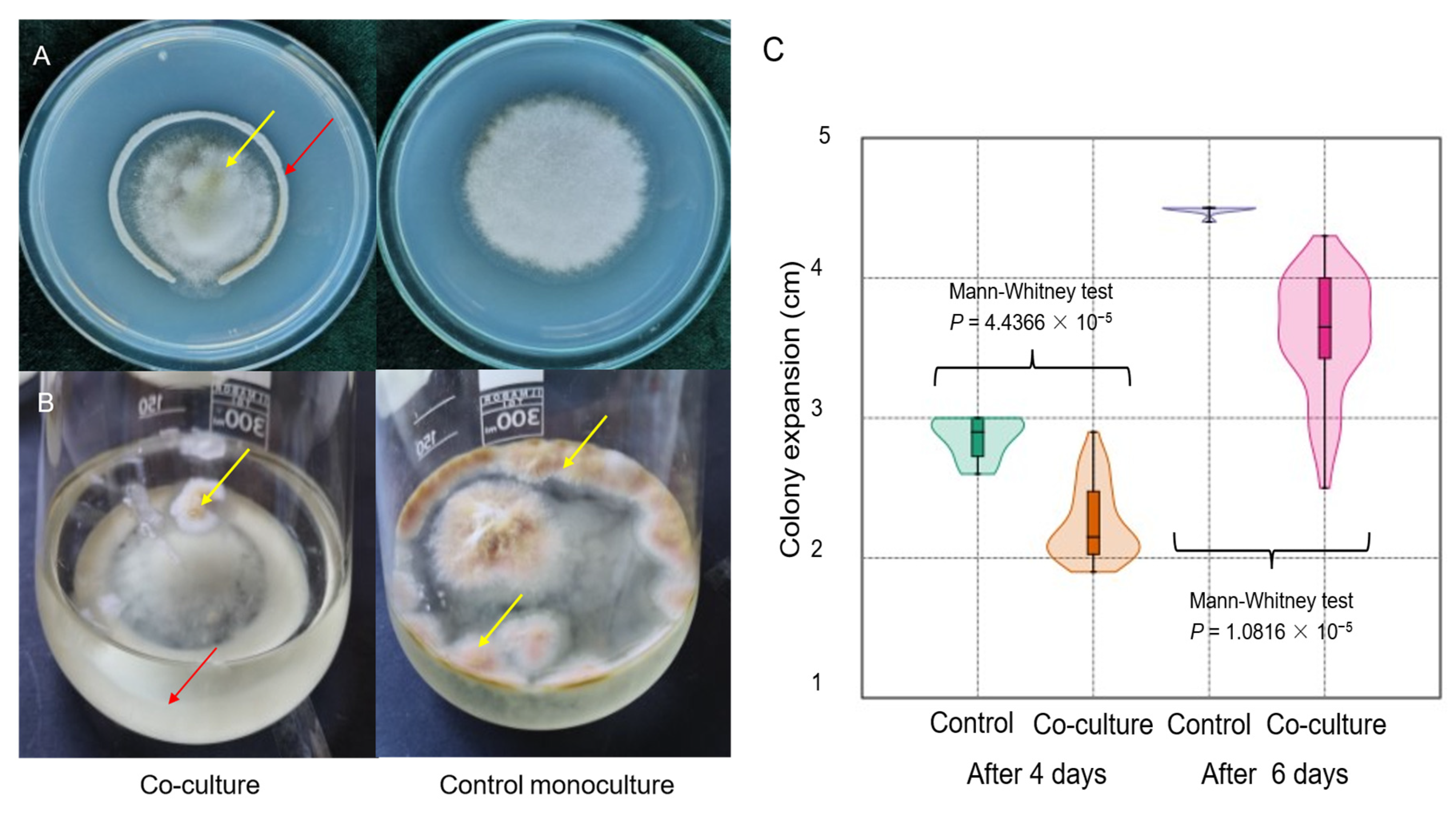
Since we hypothesized that S. japonicus might exhibit antagonistic activity, and our preliminary experiments suggested that this yeast can inhibit F. graminearum, we established co-cultures on various media (PDA, MXGB, YEA) and determined the growth of Fusarium in the presence of yeast cells. These experiments demonstrated that the yeast significantly inhibited the growth of F. graminearum in both solid (Figure 1A,C) and liquid media (Figure 1B), which was confirmed by wet weight measurements. The wet weight of Fusarium hyphae produced in the liquid medium was lower in the co-culture than in the control monoculture (average weight was 1.44 g in the co-culture and 6.92 g in the F. graminearum monoculture) (Two-sample t test, p = 7.025 × 10⁻¹³, Cohen’s D effect size = 50.25 (huge effect size).

3.2. S. japonicus Produces Ethanol, Which Did Not Inhibit the Growth of F. graminearum

Since yeast cells are generally capable of producing ethanol, which can have a growth-inhibitory effect [49,50], we measured the ethanol content of the supernatant of liquid cultures. The yeast cells produced ethanol (1.46%), (n-propanol, n-butanol, and methanol were not detected in the supernatants), and slightly reduced the pH compared to Fusarium monocultures (Table 1). Interestingly, F. graminearum also produced some ethanol in static liquid culture (Table 1).
To test the possible inhibitory effect of ethanol on the growth of F. graminearum, Fusarium hyphae were inoculated into ethanol-containing MXGB medium (0.5, 1, 1.5, 2 (v/v) % ethanol), and the wet weight of hyphae was measured. No significant decrease in hyphae production was observed at any ethanol concentration compared to the control.

3.3. RNA Sequencing of S. japonicus Cells Grown in Co-Culture with F. graminearum Revealed the DEGs

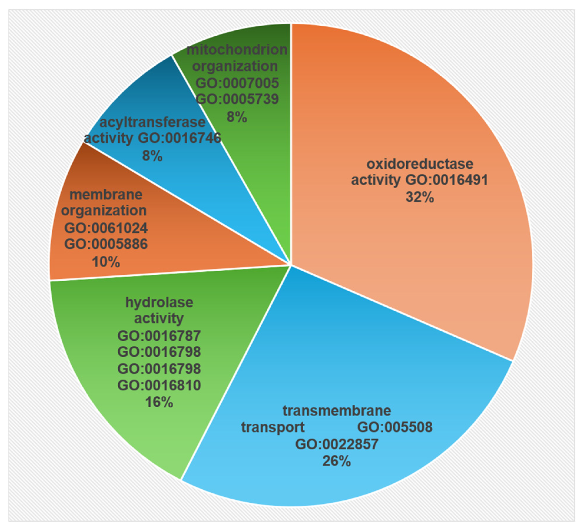
To determine the yeast genes that are differentially expressed in the presence of Fusarium, S. japonicus, and F. graminearum cells were co-cultured (incubated for two days in MXGB medium, at room temperature), and mRNA levels of S. japonicus genes were determined by RNA sequencing. mRNA levels of 135 genes changed significantly during co-culture (109 were upregulated, 24 downregulated) compared to the control yeast culture (Table S2). RNA sequencing data were also validated by RT-PCR, as shown in Figure S1, where randomly selected genes yielded results similar to those obtained from RNA sequencing. Later, the functions and GO categories of genes were obtained from the Japonicus database (JaponicusDB) [53]. According to our data, several transport and membrane-associated genes (e.g., GO:0055085, GO:0022857, GO:0061024) and oxidoreductases (GO:0016491) were among the DEGs (Figure 2, Table S2). Table 2 also shows that most of these genes were upregulated.
In addition, genes with hydrolase activity (GO:0016787, GO:0016798, GO:0016788, GO:0016810), or transferases (GO:0016746, GO:0016741, GO:0016769) (Figure 2), including a glutathione S-transferase gene (GST) (SJAG_00238) (protects cells from toxins) appeared among the DEGs (Table S2). GO enrichment analysis also confirmed the changes in the gene expression of transporters and oxidoreductases (Table 3).

3.4. Study of the Trichothecene-3-O-Acetyltransferase Genes

After a detailed investigation of the DEGs, we noticed that they contain three genes belonging to the CoA-dependent acyltransferase superfamily (marked in red letters in Table S2), whose sequence analysis revealed that they are homologs of the TRI101 trichothecene-3-O-acetyltransferase gene. Further BLASTp analyses also showed that several yeast species (Lipomyces starkeyi, Torulaspora delbrueckii, Saccharomyces cerevisiae, Yarrowia lipolytica, Saccharomyces pastorianus, Sugiyamaella lignohabitans, Debaryomyces hansenii, Trichomonascus ciferrii, Brettanomyces naardenensis) possess such a gene (Table 4 and Table S3). While these species had only a single homologous gene, S. japonicus unexpectedly contained seven copies of the trichothecene-3-O-acetyltransferase homologous gene (Table 4 and Table S3). In contrast, we did not find homologs in its closely related yeast species, S. pombe (Table 4).
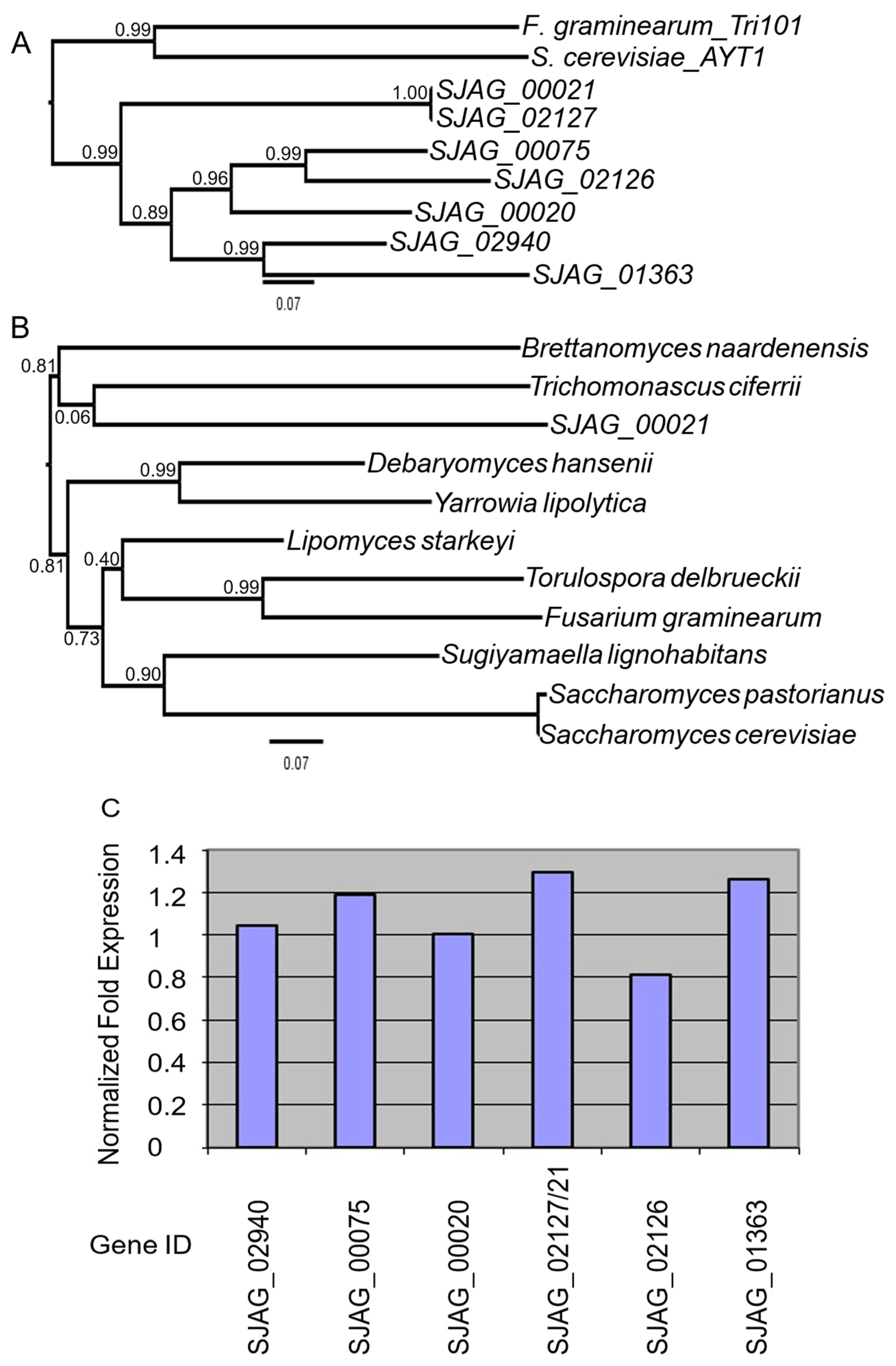
To obtain further information on seven copies of the S. japonicus trichotecene-3-O-acetyltransferase homologous genes, their protein sequences were investigated by pairwise and multiple sequence alignments, as well as phylogenetic analyses. The sequences were very similar to each other (Figure 3) and were localized in all three chromosomes of S. japonicus, mainly in the subtelomeric regions, which are the DNA segments between chromosomal telomeric caps and chromatin (Table S4). Their phylogenetic analysis also showed that SJAG_01363-SJAG_02940, SJAG_02126-SJAG_00075, and SJAG_02127-SJAG_00021 sequences were more similar to each other than to the others (Figure 3A), while further phylogenetic analysis, extended to other species, confirmed their relationship to Tri101 homologs identified in other yeasts (Figure 3B).

3.5. The Closely Related S. japonicus and S. pombe Have Different DON Tolerance

Since, unexpectedly, we have found a large difference in the number of trichotecene-3-O-acetyltransferase homologous genes between S. japonicus and S. pombe (Table 4), we compared the DON tolerance of the two closely related species. The inhibition of their cell division was different, i.e., the DON tolerance of S. japonicus was better than that of S. pombe, whose genome did not contain this gene (Table 5). RT-PCR analysis also revealed that most of the S. japonicus TR101 homologous genes can be induced by DON (Figure 3C).

4. Discussion

Since F. graminearum infection causes significant agricultural problems by reducing crop yields and frequently contaminating food and feed with mycotoxins, and because biological methods are suitable for controlling pathogenic fungi, the objectives of this study were to find a yeast strain that inhibits F. graminearum and to identify genes that may be capable of converting Fusarium mycotoxins into less harmful compounds.
Here, we provide the first evidence that the fission yeast S. japonicus exhibits growth-inhibitory capacity (Figure 1), thereby expanding the range of yeasts known to exhibit antagonistic activity against Fusarium species [24,63,64,65,66]. To determine whether the yeast produced ethanol (as yeasts are often capable of producing ethanol) and whether this could have caused growth inhibition, as in the case of Fusarium oxysporum and Aspergillus flavus [49,50], we measured the alcohol concentrations in the supernatant of the co-cultures. As expected, S. japonicus cells produced ethanol (1.4%) in static liquid cultures (Table 1); however, when we examined the growth-inhibiting effect of ethanol on F. graminearum, we found no significant growth inhibition in the presence of 0.5–2% ethanol. This may also be related to the fact that, interestingly, F. graminearum was also able to produce small amounts of ethanol, similar to F. oxysporum and F. verticillioides [48,67,68]. Therefore, it can be assumed that nutrient competition or other antifungal compounds produced by fission yeast may have caused the growth inhibition. Antifungal compounds can be diverse, including volatile compounds [28] as well as secreted toxic molecules. For example, Cochliobolus sativus is able to produce and secrete toxic molecules, one of which is similar to prehelminthosporol, and these compounds were shown to inhibit the growth of F. graminearum [69]. Another study reported that the expression of mannosidase and glucosidase genes in Aureobasidium pullulans was enhanced in the presence of F. oxysporum [68]. These enzymes may contribute to cell wall degradation and, consequently, to growth inhibition. Since growth inhibition may arise from multiple mechanisms and may reflect synergistic effects, further experiments are necessary to identify the inhibitory compounds involved. Nevertheless, our results indicated that S. japonicus exhibits antifungal potential and, pending further field evaluation, could be a suitable candidate for the biological control of F. graminearum.
Since the co-cultivation of Fusarium and a yeast species may induce different gene expression in microorganisms due to numerous biological and environmental factors resulting from the interaction between the species [48,70], we also examined gene expression changes in yeast cells in the hope that the presence of Fusarium triggers detoxification and activation of toxin-modifying/defense-related genes in the yeast. According to our results, S. japonicus is not only capable of inhibiting the growth of F. graminearum, but also presumably biologically transforms its mycotoxins. When we examined the transcriptomic changes induced by the presence of F. graminearum in yeast cells (co-culture), we identified several enzyme-encoding genes among the 135 genes whose mRNA levels were significantly altered compared to the control yeast monoculture (DEGs) (Table S2). Most genes were upregulated (109 out of 135) and several of them encoded oxidoreductases (GO:0016491) (Figure 2, Table 2 and Table S2), which was also confirmed by GO enrichment (Table 3). In addition, DEGs included genes encoding hydrolases (GO:0016787, GO:0016798, GO:0016788, GO:0016810) and transferases (GO:0016746, GO:0016741, GO:0016769) (Table S2, Figure 2). Since oxidoreductases are generally known as enzymes that can remove contaminants [71], and for example, peroxidases can reduce mycotoxins [70], or hydrolases such as carboxypeptidases degrade ochratoxin [72,73], we assume that these genes may have played an important role in detoxification. This is supported by the fact that several oxidoreductase and hydrolase genes also appeared among the DEGs during the co-cultivation of other species, such as S. pombe and F. verticillioides, or A. pullulans and F. oxysporum [48,70]. This means that these genes represent a promising opportunity to identify new Fusarium toxin–degrading genes, whose products could later be used as recombinant enzymes following gene cloning. This is important because such enzymes are needed, yet only a few of them are currently commercially available [29]. Potential detoxifying role of these enzymes is supported by the fact that the glutathione S-transferase (GST) (SJAG_00238) gene was among them, and its expression was upregulated in co-culture (Table S2) (confirmed by RT-PCR validation; Figure S1). This result is consistent with the enzyme being involved in detoxification [74], and with previous results that showed overexpression of the barley GST13 gene in Arabidopsis resulted in an increased resistance to F. graminearum [75], or introduction of another GST gene into wheat caused resistance to Fusarium Head Blight [76].
The transcriptome analysis also highlighted additional enzyme-coding genes, as three genes belonging to the CoA-dependent acyltransferase superfamily were identified among the DEGs (Table S2). Sequence alignments of the encoded proteins and subsequent phylogenetic analyses revealed that these three acetyltransferases are homologs of the trichothecene 3-O-acetyltransferase Tri101 (Figure 3A).
Their functions may also be supported by the fact that the homologs included the S. cerevisiae Ayt1 protein (Figure 3B), which is capable of trichothecene-3-O-acetylation [62]. In addition, it was also demonstrated that expressing a trichothecene-3-O-acetyltransferase gene in S. pombe and in plant cells could protect the host cells from DON [77,78,79]. Therefore, we further investigated these acetyltransferase genes. In silico analyses also revealed that, unexpectedly, the genome of S. japonicus contained seven copies of the trichothecene-3-O-acetyltransferase homologous gene sequence, in contrast to other yeast species and the closely related S. pombe (Table 4). While the genomes of several yeast species, such as Lipomyces starkeyi, Torulaspora delbrueckii, Saccharomyces cerevisiae, Yarrowia lipolytica, Saccharomyces pastorianus, Sugiyamaella lignohabitans, Debaryomyces hansenii, Trichomonascus ciferrii, Brettanomyces naardenensis, contain only a single TRI101 homologous gene, the S. pombe genome contains no homologous gene (Table 4). This latter data is also remarkable because, in general, the gene content and order of the two closely related fission yeast species (S. japonicus and S. pombe) are highly conserved [54,80]. One possible explanation for the difference in copy number of trichothecene 3-O-acetyltransferase homologous genes is that the two related species occupy different habitats. S. pombe is frequently detected on fruits, whereas S. japonicus is mainly found in soil [44], where it may encounter Fusarium species and therefore needs to defend itself. This assumption is supported by the observation that several species possessing TRI101 homologs—such as Debaryomyces hansenii, Yarrowia lipolytica, Saccharomyces pastorianus, Lipomyces starkeyi, and Torulaspora delbrueckii—have also been isolated from soil [81,82,83,84,85,86].
Further examination and alignment of the S. japonicus TRI101 homologous gene sequences and their encoded proteins indicated that some of these genes are more similar to each other, and are localized in subtelomeric regions (Figure 3A, Table S4). These findings suggest that segmental duplication and translocation events may have contributed to the high copy number observed in the examined strain. Consequently, this strain represents a non–genetically modified yeast that naturally harbors a trichothecene 3-O-acetyltransferase gene at high copy number, without the involvement of artificial genetic modification.
The question then arose as to whether these genes are functional or not. To obtain further evidence for the functionality of trichothecene-3-O-acetyltransferase genes, S. japonicus cells were treated with purified DON, and changes in mRNA levels were measured by RT-PCR. DON treatment caused changes in mRNA levels of all but two genes, compared to controls (Figure 3C). Based on these data, we hypothesized that at least five of the seven trichothecene-3-O-acetyltransferase copies would be functional, and that S. japonicus cells should have greater DON tolerance than S. pombe cells lacking the TRI101 homolog gene. Experiments on DON-supplemented media confirmed that S. japonicus has a stronger DON tolerance than S. pombe (Table 5). This is in good agreement with previous studies, which showed that yeast and plant cells exhibit increased resistance to DON when the trichothecene 3-O-acetyltransferase gene is overexpressed [77,78].
Our results not only highlighted the genes encoding the above-mentioned enzymes (which may offer good opportunities for data mining and further gene cloning) but also showed that genes related to transport and membranes (e.g., GO:0055085, GO:0022857) were strongly influenced by the presence of F. graminearum or its mycotoxin (Figure 2, Table 2 and Table S2). This is consistent with the findings of Kosawang, who also identified several transporter genes in the transcriptome profile of Clonostachys rosea after treatment with Fusarium mycotoxins [87], as well as with our previous findings, which suggested that transporters are important components of mycotoxin defense in yeasts [48].
In summary, we demonstrated that the fission yeast S. japonicus exhibits antagonistic activity against F. graminearum. In addition, we identified genes that are differentially expressed in the presence of Fusarium and may be involved in the detoxification and biotransformation of mycotoxins, including genes homologous to the trichothecene-3-O-acetyltransferase gene. We also showed that there are seven such genes in the S. japonicus genome and that this species displays high tolerance to deoxynivalenol (DON). Although gene expression may differ depending on whether yeast cells are cultured with F. graminearum under in vivo or in vitro conditions, and therefore further experiments in plants are required, we believe that these findings contribute to a better understanding of the mechanisms underlying interactions between Fusarium (and Fusarium-derived mycotoxins) and yeast, and may ultimately lead to promising biotechnological approaches for the production of safer foods.

5. Conclusions

S. japonicus may be suitable for the biological control of F. graminearum. Transcriptomic results from co-cultivation showed a sophisticated genetic response and highlighted genes sensitive to Fusarium graminearum or its mycotoxins, while bioinformatic analyses indicated the presence of multiple trichothecene 3-O-acetyltransferase homologs.
These findings represent more than just a list of genes; the differentially expressed enzyme-encoding genes identified in this study offer data mining opportunities. By using this data set to identify and clone new genes, we are paving the way for the next generation of highly efficient, biotransforming feed additives.

Supplementary Materials

The following supporting information can be downloaded at: https://www.mdpi.com/article/10.3390/agriculture16050494/s1, Figure S1. RT-PCR validation of RNA sequencing data. The randomly selected upregulated (SJAG_00238, 02988,03723,03526,03640, 04796) and down–regulated genes (SJAG_03266, 04677,04675,03724) gave similar results as in RNA sequencing. Their normalized gene expression was above 1 or below 1, respectively; Table S1: Primers used for RT-PCR; Table S2: Differentially expressed S. japonicus genes in the presence of F. graminearum; Table S3: Tri101 homologous protein sequences; Table S4: Chromosomal localization and characteristic data of the S. japonicus Tri101 homologs.

Author Contributions

Conceptualization, L.A.P., L.A.-S. and I.M.; investigation, H.C. and C.A.; validation, G.B.; supervision, I.M.; writing—original draft preparation, I.M.; writing—review and editing, T.P. and I.P.; project administration and funding acquisition, I.P. All authors have read and agreed to the published version of the manuscript.

Funding

The project no. 2019-2.1.13-TÉT_IN-2020-00056 has been implemented with the support provided by the National Research, Development and Innovation Fund of Hungary, financed under the 2019-2.1.13-TÉT_IN funding scheme. Project no. TKP2021-EGA-18 has been implemented with the support provided by the Ministry of Culture and Innovation of Hungary from the National Research, Development and Innovation Fund, financed under the TKP2021-EGA funding scheme. This project has received funding from the HUN-REN Hungarian Research Network and was supported by the University of Debrecen Program for Scientific Publication and for Scientific Research Bridging Fund (DETKA).

Data Availability Statement

All data generated or analyzed during this study are included in this published article and its Supplementary Information Files. The transcriptome data sets are also available in the Gene Expression Omnibus database (GEO; http://www.ncbi.nlm.nih.gov/geo; accessed on 25 July 2024) with the following accession number: GSE273231. Data collection: 2024.

Acknowledgments

We thank Ilona Lakatos for her technical assistance. The authors thank Sebastien Santini (CNRS/AMU IGS UMR7256) and the PACA Bioinfo platform for the availability and management of the phylogeny.fr website. The authors apologize to the authors whose articles have not been cited for reasons of length.

Conflicts of Interest

The authors declare no conflicts of interest.

References

  1. Backhouse, D. Global Distribution of Fusarium graminearum, F. aesiaticum and F. boothii from Wheat in Relation to Climate. Eur. J. Plant Pathol. 2014, 139, 161–173. [Google Scholar] [CrossRef]
  2. O’Donnell, K.; Kistler, H.C.; Tacke, B.K.; Casper, H.H. Gene Genealogies Reveal Global Phylogeographic Structure and Reproductive Isolation among Lineages of Fusarium graminearum, the Fungus Causing Wheat Scab. Proc. Natl. Acad. Sci. USA 2000, 97, 7905–7910. [Google Scholar] [CrossRef] [PubMed]
  3. Polak-Śliwińska, M.; Paszczyk, B. Trichothecenes in Food and Feed, Relevance to Human and Animal Health and Methods of Detection: A Systematic Review. Molecules 2021, 26, 454. [Google Scholar] [CrossRef] [PubMed]
  4. Garcia-Cela, E.; Kiaitsi, E.; Medina, A.; Sulyok, M.; Krska, R.; Magan, N. Interacting Environmental Stress Factors Affects Targeted Metabolomic Profiles in Stored Natural Wheat and That Inoculated with F. graminearum. Toxins 2018, 10, 56. [Google Scholar] [CrossRef]
  5. Gardiner, D.M.; Osborne, S.; Kazan, K.; Manners, J.M. Low pH Regulates the Production of Deoxynivalenol by Fusarium graminearum. Microbiology 2009, 155, 3149–3156. [Google Scholar] [CrossRef]
  6. Merhej, J.; Boutigny, A.L.; Pinson-Gadais, L.; Richard-Forget, F.; Barreau, C. Acidic pH as a Determinant of TRI Gene Expression and Trichothecene B Biosynthesis in Fusarium graminearum. Food Addit. Contam. Part A 2010, 27, 710–717. [Google Scholar] [CrossRef]
  7. Nakajima, Y.; Maeda, K.; Jin, Q.; Takahashi-Ando, N.; Kanamaru, K.; Kobayashi, T.; Kimura, M. Oligosaccharides Containing an α-(1 → 2) (Glucosyl/Xylosyl)-Fructosyl Linkage as Inducer Molecules of Trichothecene Biosynthesis for Fusarium graminearum. Int. J. Food Microbiol. 2016, 238, 215–221. [Google Scholar] [CrossRef]
  8. Ferrigo, D.; Raiola, A.; Causin, R. Fusarium Toxins in Cereals: Occurrence, Legislation, Factors Promoting the Appearance and Their Management. Molecules 2016, 21, 627. [Google Scholar] [CrossRef]
  9. Kushiro, M. Effects of Milling and Cooking Processes on the Deoxynivalenol Content in Wheat. Int. J. Mol. Sci. 2008, 9, 2127–2145. [Google Scholar] [CrossRef]
  10. Battilani, P.; Palumbo, R.; Giorni, P.; Dall’Asta, C.; Dellafiora, L.; Gkrillas, A.; Toscano, P.; Crisci, A.; Brera, C.; De Santis, B.; et al. Mycotoxin Mixtures in Food and Feed: Holistic, Innovative, Flexible Risk Assessment Modelling Approach: MYCHIF. EFSA Support. Publ. 2020, 17, 1757E. [Google Scholar] [CrossRef]
  11. Adam, M.A.A.; Tabana, Y.M.; Musa, K.B.; Sandai, D.A. Effects of Different Mycotoxins on Humans, Cell Genome and Their Involvement in Cancer. Oncol. Rep. 2017, 37, 1321–1336. [Google Scholar] [CrossRef] [PubMed]
  12. Gurdaswani, V.; Ghag, S.B. Toxins from Fusarium Species and Their Role in Animal and Plant Diseases. In New and Future Developments in Microbial Biotechnology and Bioengineering; Elsevier: Amsterdam, The Netherlands, 2020; pp. 7–27. ISBN 978-0-12-821006-2. [Google Scholar]
  13. Rocha, O.; Ansari, K.; Doohan, F.M. Effects of Trichothecene Mycotoxins on Eukaryotic Cells: A Review. Food Addit. Contam. 2005, 22, 369–378. [Google Scholar] [CrossRef] [PubMed]
  14. Shifrin, V.I.; Anderson, P. Trichothecene Mycotoxins Trigger a Ribotoxic Stress Response That Activates C-Jun N-Terminal Kinase and P38 Mitogen-Activated Protein Kinase and Induces Apoptosis. J. Biol. Chem. 1999, 274, 13985–13992. [Google Scholar] [CrossRef] [PubMed]
  15. Ye, W.; Chen, Y.; Li, H.; Zhang, W.; Liu, H.; Sun, Z.; Liu, T.; Li, S. Two Trichothecene Mycotoxins from Myrothecium roridum Induce Apoptosis of HepG-2 Cells via Caspase Activation and Disruption of Mitochondrial Membrane Potential. Molecules 2016, 21, 781. [Google Scholar] [CrossRef]
  16. Song, J.-L.; Zhang, G.-L. Deoxynivalenol and Zearalenone: Different Mycotoxins with Different Toxic Effects in the Sertoli Cells of Equus asinus. Cells 2021, 10, 1898. [Google Scholar] [CrossRef]
  17. Csikós, V.; Varró, P.; Bódi, V.; Oláh, S.; Világi, I.; Dobolyi, A. The Mycotoxin Deoxynivalenol Activates GABAergic Neurons in the Reward System and Inhibits Feeding and Maternal Behaviours. Arch. Toxicol. 2020, 94, 3297–3313. [Google Scholar] [CrossRef]
  18. Habrowska-Górczyńska, D.E.; Kowalska, K.; Urbanek, K.A.; Domińska, K.; Sakowicz, A.; Piastowska-Ciesielska, A.W. Deoxynivalenol Modulates the Viability, ROS Production and Apoptosis in Prostate Cancer Cells. Toxins 2019, 11, 265. [Google Scholar] [CrossRef]
  19. Chung, Y.-J.; Zhou, H.-R.; Pestka, J.J. Transcriptional and Posttranscriptional Roles for P38 Mitogen-Activated Protein Kinase in Upregulation of TNF-α Expression by Deoxynivalenol (Vomitoxin). Toxicol. Appl. Pharmacol. 2003, 193, 188–201. [Google Scholar] [CrossRef]
  20. Nossol, C.; Diesing, A.K.; Kahlert, S.; Kersten, S.; Kluess, J.; Ponsuksili, S.; Hartig, R.; Wimmers, K.; Dänicke, S.; Rothkötter, H.J. Deoxynivalenol Affects the Composition of the Basement Membrane Proteins and Influences En Route the Migration of CD16+ Cells into the Intestinal Epithelium. Mycotoxin Res. 2013, 29, 245–254. [Google Scholar] [CrossRef]
  21. Pierron, A.; Mimoun, S.; Murate, L.S.; Loiseau, N.; Lippi, Y.; Bracarense, A.-P.F.L.; Schatzmayr, G.; He, J.W.; Zhou, T.; Moll, W.-D.; et al. Microbial Biotransformation of DON: Molecular Basis for Reduced Toxicity. Sci. Rep. 2016, 6, 29105. [Google Scholar] [CrossRef]
  22. Zhang, X.; Li, B.; Zhang, Z.; Chen, Y.; Tian, S. Antagonistic Yeasts: A Promising Alternative to Chemical Fungicides for Controlling Postharvest Decay of Fruit. J. Fungi 2020, 6, 158. [Google Scholar] [CrossRef]
  23. Khan, N.I.; Schisler, D.A.; Boehm, M.J.; Slininger, P.J.; Bothast, R.J. Selection and Evaluation of Microorganisms for Biocontrol of Fusarium Head Blight of Wheat Incited by Gibberella zeae. Plant Dis. 2001, 85, 1253–1258. [Google Scholar] [CrossRef] [PubMed]
  24. Laitila, A.; Sarlin, T.; Kotaviita, E.; Huttunen, T.; Home, S.; Wilhelmson, A. Yeasts Isolated from Industrial Maltings Can Suppress Fusarium Growth and Formation of Gushing Factors. J. Ind. Microbiol. Biotechnol. 2007, 34, 701–713. [Google Scholar] [CrossRef] [PubMed]
  25. Wachowska, U.; Głowacka, K. Antagonistic Interactions between Aureobasidium pullulans and Fusarium culmorum, a Fungal Pathogen of Winter Wheat. BioControl 2014, 59, 635–645. [Google Scholar] [CrossRef]
  26. Papp, L.A.; Horváth, E.; Peles, F.; Pócsi, I.; Miklós, I. Insight into Yeast–Mycotoxin Relations. Agriculture 2021, 11, 1291. [Google Scholar] [CrossRef]
  27. Modrzewska, M.; Błaszczyk, L.; Stępień, Ł.; Urbaniak, M.; Waśkiewicz, A.; Yoshinari, T.; Bryła, M. Trichoderma versus Fusarium—Inhibition of Pathogen Growth and Mycotoxin Biosynthesis. Molecules 2022, 27, 8146. [Google Scholar] [CrossRef]
  28. Alasmar, R.; Ul-Hassan, Z.; Zeidan, R.; Al-Thani, R.; Al-Shamary, N.; Alnaimi, H.; Migheli, Q.; Jaoua, S. Isolation of a Novel Kluyveromyces marxianus Strain QKM-4 and Evidence of Its Volatilome Production and Binding Potentialities in the Biocontrol of Toxigenic Fungi and Their Mycotoxins. ACS Omega 2020, 5, 17637–17645. [Google Scholar] [CrossRef]
  29. Loi, M.; Fanelli, F.; Liuzzi, V.; Logrieco, A.; Mulè, G. Mycotoxin Biotransformation by Native and Commercial Enzymes: Present and Future Perspectives. Toxins 2017, 9, 111. [Google Scholar] [CrossRef]
  30. Acs-Szabo, L.; Pfliegler, W.P.; Kovács, S.; Adácsi, C.; Rácz, H.V.; Horváth, E.; Papp, L.A.; Murvai, K.P.; Király, S.; Miklós, I.; et al. Striking Mycotoxin Tolerance and Zearalenone Elimination Capacity of the Decaying Wood Associated Yeast Sugiyamaella novakii (Trichomonascaceae). BMC Microbiol. 2025, 25, 422. [Google Scholar] [CrossRef]
  31. Joannis-Cassan, C.; Tozlovanu, M.; Hadjeba-Medjdoub, K.; Ballet, N.; Pfohl-Leszkowicz, A. Binding of Zearalenone, Aflatoxin B1, and Ochratoxin A by Yeast-Based Products: A Method for Quantification of Adsorption Performance. J. Food Prot. 2011, 74, 1175–1185. [Google Scholar] [CrossRef]
  32. Solovyov, V.V.; Marhunova, A.M.; Permiakova, O.L.; Voblikova, T.V.; Semenova, Y.O. Yeast Cell Walls Adsorption Capacity. IOP Conf. Ser. Earth Environ. Sci. 2020, 613, 012143. [Google Scholar] [CrossRef]
  33. Xiao, J.; Tan, J.; Guo, R.; Dai, J.; Xiu, Z.; Sun, Y.; Liu, H.; Li, Y.; Tong, Y.; Quan, C. Deoxynivalenol Detoxification by a Novel Strain of Pichia kudriavzevii via Enzymatic Degradation and Cell Wall Adsorption. Appl. Biochem. Biotechnol. 2024, 196, 3102–3114. [Google Scholar] [CrossRef] [PubMed]
  34. Zhou, T.; He, J.; Gong, J. Microbial Transformation of Trichothecene Mycotoxins. World Mycotoxin J. 2008, 1, 23–30. [Google Scholar] [CrossRef]
  35. Ahundov, R.; Lyagin, I.; Senko, O.; Maslova, O.; Makhlis, T.; Efremenko, E. Biocatalytic Transformation of Various Mycotoxins: Modern Problems and Existing Potential. IOP Conf. Ser. Mater. Sci. Eng. 2019, 525, 012090. [Google Scholar] [CrossRef]
  36. Azam, M.S.; Yu, D.; Wu, A. Enzymes for Degradation of Fusarium mycotoxins. In Food Safety & Mycotoxins; Wu, A., Ed.; Springer: Singapore, 2019; pp. 113–135. ISBN 978-981-329-037-2. [Google Scholar]
  37. Wang, N.; Wu, W.; Pan, J.; Long, M. Detoxification Strategies for Zearalenone Using Microorganisms: A Review. Microorganisms 2019, 7, 208. [Google Scholar] [CrossRef]
  38. Khatibi, P.A.; Newmister, S.A.; Rayment, I.; McCormick, S.P.; Alexander, N.J.; Schmale, D.G. Bioprospecting for Trichothecene 3-O-Acetyltransferases in the Fungal Genus Fusarium Yields Functional Enzymes with Different Abilities To Modify the Mycotoxin Deoxynivalenol. Appl. Env. Microbiol. 2011, 77, 1162–1170. [Google Scholar] [CrossRef]
  39. McCormick, S.P.; Price, N.P.J.; Kurtzman, C.P. Glucosylation and Other Biotransformations of T-2 Toxin by Yeasts of the Trichomonascus Clade. Appl. Environ. Microbiol. 2012, 78, 8694–8702. [Google Scholar] [CrossRef]
  40. Krishnan, S.V.; Anaswara, P.A.; Nampoothiri, K.M.; Kovács, S.; Adácsi, C.; Szarvas, P.; Király, S.; Pócsi, I.; Pusztahelyi, T. Biocontrol Activity of New Lactic Acid Bacteria Isolates Against Fusaria and Fusarium mycotoxins. Toxins 2025, 17, 68. [Google Scholar] [CrossRef]
  41. Banani, H.; Spadaro, D.; Zhang, D.; Matic, S.; Garibaldi, A.; Gullino, M.L. Postharvest Application of a Novel Chitinase Cloned from Metschnikowia fructicola and Overexpressed in Pichia pastoris to Control Brown Rot of Peaches. Int. J. Food Microbiol. 2015, 199, 54–61. [Google Scholar] [CrossRef]
  42. Qin, X.; Su, X.; Tu, T.; Zhang, J.; Wang, X.; Wang, Y.; Wang, Y.; Bai, Y.; Yao, B.; Luo, H.; et al. Enzymatic Degradation of Multiple Major Mycotoxins by Dye-Decolorizing Peroxidase from Bacillus subtilis. Toxins 2021, 13, 429. [Google Scholar] [CrossRef]
  43. Takahashi-Ando, N.; Ohsato, S.; Shibata, T.; Hamamoto, H.; Yamaguchi, I.; Kimura, M. Metabolism of Zearalenone by Genetically Modified Organisms Expressing the Detoxification Gene from Clonostachys rosea. Appl. Environ. Microbiol. 2004, 70, 3239–3245. [Google Scholar] [CrossRef] [PubMed]
  44. Brysch-Herzberg, M.; Jia, G.-S.; Seidel, M.; Assali, I.; Du, L.-L. Insights into the Ecology of Schizosaccharomyces Species in Natural and Artificial Habitats. Antonie Leeuwenhoek 2022, 115, 661–695. [Google Scholar] [CrossRef] [PubMed]
  45. Yukawa, M.; Maki, T. Regarding the new fission yeast Schizosaccharomyces japonicus. Kyushu Daigaku Kiyou 1931, 218–226. (In Japanese) [Google Scholar]
  46. Trail, F.; Common, R. Perithecial Development by Gibberella zeae: A Light Microscopy Study. Mycologia 2000, 92, 130–138. [Google Scholar] [CrossRef] [PubMed]
  47. Boeira, L.S.; Bryce, J.H.; Stewart, G.G.; Flannigan, B. The Effect of Combinations of Fusarium mycotoxins (Deoxynivalenol, Zearalenone and Fumonisin B1) on Growth of Brewing Yeasts. J. Appl. Microbiol. 2000, 88, 388–403. [Google Scholar] [CrossRef]
  48. Papp, L.A.; Acs-Szabo, L.; Kovács, S.; Adácsi, C.; Batta, G.; Pusztahelyi, T.; Pócsi, I.; Miklós, I. Transcriptome Changes of Fission Yeast Cells Exposed to Fumonisin B1 or Co-Cultured with Fusarium verticillioides. Appl. Microbiol. Biotechnol. 2025, 109, 211. [Google Scholar] [CrossRef]
  49. Ren, Y.; Jin, J.; Zheng, M.; Yang, Q.; Xing, F. Ethanol Inhibits Aflatoxin B1 Biosynthesis in Aspergillus flavus by Up-Regulating Oxidative Stress-Related Genes. Front. Microbiol. 2020, 10, 2946. [Google Scholar] [CrossRef]
  50. Paschos, T.; Xiros, C.; Christakopoulos, P. Ethanol Effect on Metabolic Activity of the Ethalogenic Fungus Fusarium oxysporum. BMC Biotechnol. 2015, 15, 15. [Google Scholar] [CrossRef]
  51. Lyne, R.; Burns, G.; Mata, J.; Penkett, C.J.; Rustici, G.; Chen, D.; Langford, C.; Vetrie, D.; Bähler, J. Whole-Genome Microarrays of Fission Yeast: Characteristics, Accuracy, Reproducibility, and Processing of Array Data. BMC Genom. 2003, 4, 27. [Google Scholar] [CrossRef]
  52. Kumar, R.; Ichihashi, Y.; Kimura, S.; Chitwood, D.H.; Headland, L.R.; Peng, J.; Maloof, J.N.; Sinha, N.R. A High-Throughput Method for Illumina RNA-Seq Library Preparation. Front. Plant Sci. 2012, 3, 202. [Google Scholar] [CrossRef]
  53. Rutherford, K.M.; Harris, M.A.; Oliferenko, S.; Wood, V. JaponicusDB: Rapid Deployment of a Model Organism Database for an Emerging Model Species. Genetics 2022, 220, iyab223. [Google Scholar] [CrossRef]
  54. Acs-Szabo, L.; Papp, L.A.; Sipiczki, M.; Miklos, I. Genome Comparisons of the Fission Yeasts Reveal Ancient Collinear Loci Maintained by Natural Selection. J. Fungi 2021, 7, 864. [Google Scholar] [CrossRef]
  55. Madeira, F.; Pearce, M.; Tivey, A.R.N.; Basutkar, P.; Lee, J.; Edbali, O.; Madhusoodanan, N.; Kolesnikov, A.; Lopez, R. Search and Sequence Analysis Tools Services from EMBL-EBI in 2022. Nucleic Acids Res. 2022, 50, W276–W279. [Google Scholar] [CrossRef] [PubMed]
  56. Dereeper, A.; Guignon, V.; Blanc, G.; Audic, S.; Buffet, S.; Chevenet, F.; Dufayard, J.-F.; Guindon, S.; Lefort, V.; Lescot, M.; et al. Phylogeny.Fr: Robust Phylogenetic Analysis for the Non-Specialist. Nucleic Acids Res. 2008, 36, W465–W469. [Google Scholar] [CrossRef] [PubMed]
  57. Edgar, R.C. MUSCLE: Multiple Sequence Alignment with High Accuracy and High Throughput. Nucleic Acids Res. 2004, 32, 1792–1797. [Google Scholar] [CrossRef] [PubMed]
  58. Castresana, J. Selection of Conserved Blocks from Multiple Alignments for Their Use in Phylogenetic Analysis. Mol. Biol. Evol. 2000, 17, 540–552. [Google Scholar] [CrossRef]
  59. Guindon, S.; Dufayard, J.-F.; Lefort, V.; Anisimova, M.; Hordijk, W.; Gascuel, O. New Algorithms and Methods to Estimate Maximum-Likelihood Phylogenies: Assessing the Performance of PhyML 3.0. Syst. Biol. 2010, 59, 307–321. [Google Scholar] [CrossRef]
  60. Ge, S.X.; Jung, D.; Yao, R. ShinyGO: A Graphical Gene-Set Enrichment Tool for Animals and Plants. Bioinformatics 2020, 36, 2628–2629. [Google Scholar] [CrossRef]
  61. Hammer, Ø.; Harper, D.A.T.; Ryan, P.D. PAST: Paleontological statistics software package for education and data analysis. Palaeontol. Electron. 2001, 4, 9. [Google Scholar]
  62. Alexander, N.J.; McCormick, S.P.; Hohn, T.M. The Identification of the Saccharomyces cerevisiae Gene AYT1 (ORF-YLL063c) Encoding an Acetyltransferase. Yeast 2002, 19, 1425–1430. [Google Scholar] [CrossRef]
  63. Korres, A.M.N.; Buss, D.S.; Ventura, J.A.; Fernandes, P.M.B. Candida krusei and Kloeckera apis Inhibit the Causal Agent of Pineapple Fusariosis, Fusarium guttiforme. Fungal Biol. 2011, 115, 1251–1258. [Google Scholar] [CrossRef]
  64. Armando, M.R.; Dogi, C.A.; Poloni, V.; Rosa, C.A.R.; Dalcero, A.M.; Cavaglieri, L.R. In Vitro Study on the Effect of Saccharomyces cerevisiae Strains on Growth and Mycotoxin Production by Aspergillus carbonarius and Fusarium graminearum. Int. J. Food Microbiol. 2013, 161, 182–188. [Google Scholar] [CrossRef] [PubMed]
  65. Fallah, B.; Zaini, F.; Daei Ghazvini, R.; Kachuei, R.; Kordbacheh, P.; Safara, M.; Mahmoudi, S. The Antagonistic Effects of Candida parapsilosis on the Growth of Fusarium Species and Fumonisin Production. Curr. Med. Mycol. 2016, 2, 1–6. [Google Scholar] [CrossRef] [PubMed]
  66. Jamal, A.; Farhat, H.; Urooj, F.; Rahman, A.; Irfan, M.; Ehteshamul-Haque, S. Characterization of Endophytic Yeast and Its Suppressive Effect on Root Rotting Fungi of Tomato under Neem Cake Soil Amendment. Egypt. J. Biol. Pest Control. 2021, 31, 152. [Google Scholar] [CrossRef]
  67. Xiros, C.; Christakopoulos, P. Enhanced Ethanol Production from Brewer’s Spent Grain by a Fusarium oxysporum Consolidated System. Biotechnol. Biofuels 2009, 2, 4. [Google Scholar] [CrossRef]
  68. De Almeida, M.N.; Guimarães, V.M.; Falkoski, D.L.; Visser, E.M.; Siqueira, G.A.; Milagres, A.M.F.; De Rezende, S.T. Direct Ethanol Production from Glucose, Xylose and Sugarcane Bagasse by the Corn Endophytic Fungi Fusarium verticillioides and Acremonium zeae. J. Biotechnol. 2013, 168, 71–77. [Google Scholar] [CrossRef]
  69. Rampitsch, C.; Bacala, R.; Tekauz, A.; McCallum, B.D. Inhibition of Fusarium graminearum and Other Fusarium Species by Cochlioblolus sativus in Culture and on Barley Plants. Can. J. Plant Pathol. 2016, 38, 422–429. [Google Scholar] [CrossRef]
  70. Rueda-Mejia, M.P.; Nägeli, L.; Lutz, S.; Hayes, R.D.; Varadarajan, A.R.; Grigoriev, I.V.; Ahrens, C.H.; Freimoser, F.M. Genome, Transcriptome and Secretome Analyses of the Antagonistic, Yeast-like Fungus Aureobasidium pullulans to Identify Potential Biocontrol Genes. Microb. Cell 2021, 8, 184–202. [Google Scholar] [CrossRef]
  71. Bilal, M.; Bagheri, A.R.; Vilar, D.S.; Aramesh, N.; Eguiluz, K.I.B.; Ferreira, L.F.R.; Ashraf, S.S.; Iqbal, H.M.N. Oxidoreductases as a Versatile Biocatalytic Tool to Tackle Pollutants for Clean Environment—A Review. J. Chem. Technol. Biotechnol. 2022, 97, 420–435. [Google Scholar] [CrossRef]
  72. Fu, X.; Fei, Q.; Zhang, X.; Li, N.; Zhang, L.; Zhou, Y. Two Different Types of Hydrolases Co-Degrade Ochratoxin A in a Highly Efficient Degradation Strain Lysobacter sp. CW239. J. Hazard. Mater. 2024, 473, 134716. [Google Scholar] [CrossRef]
  73. Zhang, X.; Yang, H.; Apaliya, M.T.; Zhao, L.; Gu, X.; Zheng, X.; Hu, W.; Zhang, H. The Mechanisms Involved in Ochratoxin A Elimination by Yarrowia lipolytica Y-2. Ann. Appl. Biol. 2018, 173, 164–174. [Google Scholar] [CrossRef]
  74. Aloke, C.; Onisuru, O.O.; Achilonu, I. Glutathione S-Transferase: A Versatile and Dynamic Enzyme. Biochem. Biophys. Res. Commun. 2024, 734, 150774. [Google Scholar] [CrossRef] [PubMed]
  75. Wahibah, N.N.; Tsutsui, T.; Tamaoki, D.; Sato, K.; Nishiuchi, T. Expression of Barley Glutathione S-Transferase13 Gene Reduces Accumulation of Reactive Oxygen Species by Trichothecenes and Paraquat in Arabidopsis Plants. Plant Biotechnol. 2018, 35, 71–79. [Google Scholar] [CrossRef] [PubMed]
  76. Wang, H.; Sun, S.; Ge, W.; Zhao, L.; Hou, B.; Wang, K.; Lyu, Z.; Chen, L.; Xu, S.; Guo, J.; et al. Horizontal Gene Transfer of Fhb7 from Fungus Underlies Fusarium Head Blight Resistance in Wheat. Science 2020, 368, eaba5435. [Google Scholar] [CrossRef] [PubMed]
  77. Kimura, M.; Kaneko, I.; Komiyama, M.; Takatsuki, A.; Koshino, H.; Yoneyama, K.; Yamaguchi, I. Trichothecene 3-O-Acetyltransferase Protects Both the Producing Organism and Transformed Yeast from Related Mycotoxins. J. Biol. Chem. 1998, 273, 1654–1661. [Google Scholar] [CrossRef]
  78. Ohsato, S.; Ochiai-Fukuda, T.; Nishiuchi, T.; Takahashi-Ando, N.; Koizumi, S.; Hamamoto, H.; Kudo, T.; Yamaguchi, I.; Kimura, M. Transgenic Rice Plants Expressing Trichothecene 3-O-Acetyltransferase Show Resistance to the Fusarium Phytotoxin Deoxynivalenol. Plant Cell Rep. 2007, 26, 531–538. [Google Scholar] [CrossRef]
  79. Hao, G.; McCormick, S.; Tiley, H.; Usgaard, T. Detoxification and Excretion of Trichothecenes in Transgenic Arabidopsis thaliana Expressing Fusarium graminearum Trichothecene 3-O-Acetyltransferase. Toxins 2021, 13, 320. [Google Scholar] [CrossRef]
  80. Rhind, N.; Chen, Z.; Yassour, M.; Thompson, D.A.; Haas, B.J.; Habib, N.; Wapinski, I.; Roy, S.; Lin, M.F.; Heiman, D.I.; et al. Comparative Functional Genomics of the Fission Yeasts. Science 2011, 332, 930–936. [Google Scholar] [CrossRef]
  81. Barnett, J.A.; Payne, R.W.; Yarrow, D. Yeasts: Characteristics and Identification, 3rd ed.; Cambridge Univ. Press: Cambridge, UK, 2007; ISBN 978-0-521-57396-2. [Google Scholar]
  82. Devillers, H.; Brunel, F.; Połomska, X.; Sarilar, V.; Lazar, Z.; Robak, M.; Neuvéglise, C. Draft Genome Sequence of Yarrowia lipolytica Strain A-101 Isolated from Polluted Soil in Poland. Genome Announc. 2016, 4, e01094-16. [Google Scholar] [CrossRef]
  83. Naganuma, T.; Katsumata, K.; Ando, T.; Watanabe, H.; Nishimura, K.; Uzuka, Y. An Improved Method for Isolating Yeasts in the Genus Lipomyces and Related Genera from Soil. Biosci. Biotechnol. Biochem. 1999, 63, 195–198. [Google Scholar] [CrossRef]
  84. Silva-Sousa, F.; Fernandes, T.; Pereira, F.; Rodrigues, D.; Rito, T.; Camarasa, C.; Franco-Duarte, R.; Sousa, M.J. Torulaspora delbrueckii Phenotypic and Metabolic Profiling towards Its Biotechnological Exploitation. J. Fungi 2022, 8, 569. [Google Scholar] [CrossRef]
  85. Wu, C.-H.; Lin, H.-T.; Wu, G.-J.; Wang, S.-H.; Tsai, G.-J. Effects of Cultural Medium and Conditions on the Proliferation and Hypoglycemic Activity of Saccharomyces Pastorianus No. 54. J. Biosci. Bioeng. 2011, 112, 159–165. [Google Scholar] [CrossRef]
  86. Yurkov, A.M. Yeasts of the Soil—Obscure but Precious. Yeast 2018, 35, 369–378. [Google Scholar] [CrossRef]
  87. Kosawang, C.; Karlsson, M.; Jensen, D.; Dilokpimol, A.; Collinge, D.B. Transcriptomic Profiling to Identify Genes Involved in Fusarium Mycotoxin Deoxynivalenol and Zearalenone Tolerance in the Mycoparasitic Fungus Clonostachys rosea. BMC Genom. 2014, 15, 55. [Google Scholar] [CrossRef]
Figure 1. Growth inhibition test. S. japonicus was able to inhibit the growth of F. graminearum. F. graminearum (yellow arrow) did not grow on yeast cells (red arrow) in co-culture and was smaller in size (PDA, at 25 °C, photographed after 5 days) (A), which was significant (N = 11) (C) (Colony expansion after 4 days: Mann–Whitney test, p = 4.4366 × 10⁻⁵, Vargha–Delaney A effect size = 0.9635 (large effect size); Colony expansion after 6 days: Mann–Whitney test, p = 1.0816 × 10⁻⁵, Vargha–Delaney A effect size = 1 (large effect size). F. graminearum also produced fewer mycelia compared to the control in liquid medium (B) (PDB, incubated at room temperature, for 5 days, without shaking) (Similar results were obtained in MXGB and YEL media). The yellow arrows show F. graminearum, while the red arrow indicates the yeast cells settled to the bottom of the Erlenmeyer flask.
Agriculture 16 00494 g001
Figure 2. The most important GO categories of differentially expressed S. japonicus genes after co-culturing with F. graminearum.
Agriculture 16 00494 g002
Figure 3. Study of the putative trichothecene-3-O-acetyltransferase homologous protein sequences. (A) Phylogenetic analysis of the S. japonicus protein sequences and the experimentally validated F. graminearun and S. cerevisiae trichothecene-3-O-acetyltransferase protein sequences. The S. japonicus genes are clustered together and separated from the F. graminearum and S. cerevisiae sequences because they are more similar to each other than to the sequences of other species. (B) Phylogenetic analysis of the S. japonicus SJAG_00021 and Tri101 putative homologous sequences identified in other yeasts. To demonstrate that the identified S. japonicus sequences are putative orthologs of the Tri101 gene family, one of the most basally located sequences of S. japonicus (SJAG_00021) was included in a phylogenetic analysis comprising many other putative orthologous protein sequences. Although SJAG_00021 is not located on the main branch of the tree, it is clustered with other putative trichothecene-3-O-acetyltransferase homologous protein sequences. (C) Normalized fold expression of the Tri101 homologs shows that DON treatment of S. japonicus cells caused changes in mRNA levels of all but two genes, compared to controls.
Agriculture 16 00494 g003
Table 1. The alcohol concentration and pH of the supernatants.
SpeciesEthanol Concentrations (v/v%)pH
F. graminearum0.22 ± 0.174.06 ± 0.02
S. japonicus1.53 ± 0.213.41 ± 0.05
S. japonicus + F. graminearum1.46 ± 0.083.46 ± 0.04
Average of three experiments; alcohol LOD: 0.005 (v/v)%.
Table 2. Genes belonging to transmembrane transport (GO:0055085) and oxidoreductase activity (GO:0016491).
Gene SymbolLog2 FCRegulationFunction of the Gene
SJAG_021331.8282354upAmino acid/polyamine transporter
SJAG_010961.7833421upAmino acid/polyamine transporter
SJAG_021131.5267855upAmino acid/polyamine transporter
SJAG_030791.5025475upMFS transporter superfamily
SJAG_02150−1.5304266downAmino acid transmembrane transporter
SJAG_04675−2.4129047downPlasma membrane iron transmembrane transporter Fip1
SJAG_019031.8415602upProline dehydrogenase Put1
SJAG_006741.7543724upSuccinate–semialdehyde dehydrogenase
SJAG_002371.7031807upHexitol dehydrogenase
SJAG_007041.7962087upSerine/threonine protein kinase Ppk31
SJAG_029601.6098309upMethylglyoxyl reductase (NADPH-dependent)
SJAG_014921.518742upShort-chain dehydrogenase, human DHRS7 family
SJAG_018501.5178025upShort-chain dehydrogenase, human DHRS7 family
SJAG_04743−1.6851736downPlasma membrane ferric-chelate reductase
SJAG_03266−2.1648088downNADP-specific glutamate dehydrogenase Gdh1
SJAG_04677−2.8037887downPlasma membrane iron transport multicopper oxidase Fio1
Log2FC: Log2 Fold Change; up and down: the gene was up-regulated or down-regulated compared to the control.
Table 3. GO enrichment of the differentially expressed S. japonicus genes obtained from cells co-cultured with F. graminearum.
GO Biological ProcessesGO Molecular Function
Upregulated genes
No significant enrichment was foundOxidoreductase activity 15/232 (GO:0016491)
Downregulated genes
Transmembrane transport 8/245 (GO:0055085)Oxidoreductase acting on metal ions 2/3 (GO:0016722)
Transport 10/652 (GO:0006810)
Amino acid transport 4/25 (GO:0006865)
Organic acid transport 4/35 (GO:0015849)
Transition metal ion transport 3/21 (GO:0000041)
Cation transport 4/96 (GO:0006812)
Import into cell 4/35 (GO:0098657)
Glutamine fam. amino acid metabolic proc. 4/33 (GO:0009064)
Glutamine metabolic proc. 3/15 (GO:0006541)
Glutamate metabolic proc. 3/7 (GO:0006536)
Alpha-amino acid metabolic proc. 4/102 (GO:1901605)
Cellular amino acid metabolic proc. 5/156 (GO:0006520)
Iron import into cell 2/3 (GO:0033212)
Iron ion homeostasis 2/11 (GO:0055072)
Iron ion transport 3/9 (GO:0006826)
Cellular iron ion homeostasis 2/11 (GO:0006879)
Nitrogen utilization 2/4 (GO:0019740)
Ammonia assimilation cycle 2/3 (GO:0019676)
Dicarboxylic acid metabolic proc. 3/25 (GO:0043648)
Establishment of localization 10/690 (GO:0051234)
Oxidoreductase activity: Catalysis of an oxidation-reduction (redox) reaction. Transport: The directed movement of substances (such as macromolecules, small molecules, ions) or cellular components (such as complexes and organelles) into, out of, or within a cell. Metabolic proc.: metabolic process: A cellular process consisting of the biochemical pathways by which a living organism transforms chemical substances. Iron ion homeostasis: A homeostatic process involved in the maintenance of a steady state level of iron ions within a cell. Nitrogen utilization: A series of processes that form an integrated mechanism by which a cell or an organism detects the depletion of the primary nitrogen source, usually ammonia, and then activates genes to scavenge the last traces of the primary nitrogen source and to transport and metabolize alternative nitrogen sources. Establishment of localization: Any process that localizes a substance or cellular component. Ammonia assimilation cycle: The pathway by which ammonia is processed and incorporated into a cell. (Number of Genes/Pathway Genes) (proc.: processes).
Table 4. Yeast species with homologous Tri101 protein sequences.
SpeciesProtein Name and LengthScoreE-ValueIdentitiesPositives
Schizosaccharomyces japonicusCoA-dependent acyltransferase superfamily (predicted) SJAG_00021.1, length: 4573424 × 10−113182/455 (40%)254/455 (55%)
Schizosaccharomyces japonicusCoA-dependent acyltransferase superfamily (predicted) SJAG_02940.1, length: 4563872 × 10−130208/455 (46%)278/455 (61%)
Schizosaccharomyces japonicusCoA-dependent acyltransferase superfamily (predicted) SJAG_00020.1, length: 4573508 × 10−116193/458 (42%)265/458 (57%)
Schizosaccharomyces japonicusCoA-dependent acyltransferase superfamily (predicted) SJAG_00075.1, length: 4583678 × 10−123196/452 (43%)273/452 (60%)
Schizosaccharomyces japonicusCoA-dependent acyltransferase superfamily (predicted) SJAG_01363.1, length: 4533582 × 10−119193/454 (43%)276/454 (60%)
Schizosaccharomyces japonicusCoA-dependent acyltransferase superfamily (predicted) SJAG_02126.1, length: 4523245 × 10−106182/452 (40%)261/452 (57%)
Schizosaccharomyces japonicus *CoA-dependent acyltransferase superfamily (predicted) SJAG_02127.1, length: 457
Saccharomyces cerevisiae ** YJM1402, YJM1208Acetyltransferase Ayt1p, length: 4743887 × 10−130204/454 (45%)286/454 (62%)
Lipomyces starkeyi NRRL Y-11557Hypothetical protein LIPSTDRAFT_86407, length:4664541 × 10−155233/456 (51%)297/456 (65%)
Torulaspora delbrueckiiHypothetical protein TDEL_0G04720, length: 4274349 × 10−149223/450 (50%)298/450 (66%)
Yarrowia lipolyticaTransferase, length: 4433467 × 10−114188/450 (42%)265/450 (58%)
Saccharomyces pastorianusTrichothecene 3-O-acetyltransferase, length: 4743864 × 10−129203/454 (45%)286/454 (62%)
Sugiyamaella lignohabitansAyt1p, length: 4743737 × 10−124197/468 (42%)280/468 (59%)
Debaryomyces hansenii CBS767Transferase DEHA2D07326p, length: 4543678 × 10−122203/457 (44%)280/457 (61%)
Trichomonascus ciferriiHypothetical protein TRICI_005126, length: 4443661 × 10−121194/449 (43%)276/449 (61%)
Brettanomyces naardenensisDEKNAAC103449, length: 4403342 × 10−109183/449 (41%)266/449 (59%)
Schizosaccharomyces pombe *** -
Query: F. graminearum Tri101p (ACCESSION AB000874, VERSION AB000874.1) (ADQ52713.1) (14.12.2023 data) (NCBI database, BLASTp). * This gene was found in Ensemble. ** This gene is capable of trichothecene 3-O-acetylation [62]. *** BLASTp did not find homologous proteins.
Table 5. Growth inhibition of yeast cells by DON.
DON Concentrations
0.1 ppm0.5 ppm0.7 ppm1 ppm
SpeciesInhibition (%)
S. japonicus<10<10<10<10
S. pombe<10203752
LOD: 10%.
Disclaimer/Publisher’s Note: The statements, opinions and data contained in all publications are solely those of the individual author(s) and contributor(s) and not of MDPI and/or the editor(s). MDPI and/or the editor(s) disclaim responsibility for any injury to people or property resulting from any ideas, methods, instructions or products referred to in the content.