Article

Effects of Aerobic Exercise on Cortisol Stress Reactivity in Response to the Trier Social Stress Test in Inpatients with Major Depressive Disorders: A Randomized Controlled Trial

1
Sport Science Section, Department of Sport, Exercise and Health, University of Basel, CH-4052 Basel, Switzerland
2
Psychiatric Services Solothurn, 4503 Solothurn, Switzerland
3
Private Clinic Wyss, 3053 Muenchenbuchsee, Switzerland
4
Clinic Sonnenhalde, 4125 Riehen, Switzerland
5
University Psychiatric Clinics (UPK), Center for Affective, Stress and Sleep Disorders, University of Basel, 4002 Basel, Switzerland
6
Substance Abuse Prevention Research Center and Sleep Disorders Research Center, Kermanshah University of Medical Sciences, Kermanshah 6715847141, Iran
7
University Psychiatric Clinics (UPK), Neurobiology Laboratory for Brain Aging and Mental Health, University of Basel, 4002 Basel, Switzerland
*
Author to whom correspondence should be addressed.
These authors have contributed equally to this study.
J. Clin. Med. 2020, 9(5), 1419; https://doi.org/10.3390/jcm9051419
Submission received: 2 April 2020 / Revised: 30 April 2020 / Accepted: 5 May 2020 / Published: 11 May 2020
(This article belongs to the Section Mental Health)

Abstract

:
Physical activity is associated with a favourable (blunted) cortisol stress reactivity in healthy people. However, evidence from experimental study and with psychiatric patients is missing. This study examines whether exercise training impacts on cortisol stress reactivity in inpatients with major depressive disorder (MDD). These new insights are important because the stress reactivity of healthy people and patients with severe symptoms of depression might differ. Methods: The study was designed as a randomized controlled trial (trial registration number: NCT02679053). In total, 25 patients (13 women, 12 men, mean age: 38.1 ± 12.0 years) completed a laboratory stressor task before and after a six-week intervention period. Nine samples of salivary free cortisol were taken before and after the Trier social stress test (TSST). Fourteen participants took part in six weeks of aerobic exercise training, while 11 patients were allocated to the control condition. While the primary outcome of the study was depressive symptom severity, the focus of this paper is on one of the secondary outcomes (cortisol reactivity during the TSST). The impact of aerobic exercise training was examined with a repeated-measures analysis of variance. We also examined the association between change in depression and cortisol response via correlational analysis. Cortisol reactivity did not change from baseline to post-intervention, either in the intervention or the control group. Participation in six weeks of aerobic exercise training was not associated with participants’ cortisol reactivity. Moreover, depressive symptom change was not associated with change in cortisol response. Aerobic exercise training was not associated with patients’ stress reactivity in this study. Because many patients initially showed a relatively flat/blunted cortisol response curve, efforts might be needed to find out which treatments are most efficient to promote a normalization of HPA axis reactivity.

1. Introduction

Major depression is among the most common and burdensome disorders worldwide. In Switzerland [1], as in other Western countries [2], more than 20% of the population are affected by major depression once in a lifetime. Currently, major depressive disorders (MDDs) constitute the leading cause of years lived with disability (YLDs) [3] and in 2030, MDDs are expected to cause the greatest number of disability-adjusted life years (DALYs) [4].
Depression is often referred to as a stress-related disorder [5]. In line with this notion, exposure to critical life events and chronic stress play an important role in the onset and course of MDDs [6]. Accordingly, most individuals who develop a depressive episode experience a major adverse life event before depression onset [7]. Chronic stress exposure also increases the risk of developing a new depressive disorder [8]. Moreover, scholars have highlighted that people with depressive disorders are more likely to perceive their lives as stressful [9].
The relationship between stress and depressive disorders has often been attributed to a dysregulation of the hypothalamic-pituitary-adrenal (HPA) axis [10,11], and it is expected that a normalization of HPA axis activity will lead to an improvement in depressive symptom severity [12,13]. In line with this notion, previous studies have shown that chronically high stress levels are associated with a dysregulated diurnal HPA axis activity [14], an increased cortisol awakening response [15], and an increased reactivity in response to experimentally induced stress [16,17]. Similarly, evidence suggests that MDD might be associated with a dysregulation of HPA axis activity [18]. For instance, researchers observed a hyperactivity of the HPA axis and a lower suppression of cortisol after a dexamethasone suppression test (DST) among individuals with MDD [19,20,21]. Nevertheless, findings have not always been consistent [20]. For instance, in a systematic review and meta-analysis with 20 case-control studies, Knorr et al. [22] were unable to detect a clear difference in diurnal salivary cortisol levels between depressed patients and non-depressed controls, and some studies found that patients might even have lower cortisol levels [23,24]. The latter has been interpreted as an indication of HPA axis exhaustion, following chronic or recurrent depression [23,24].
Researchers have also tested whether people with depression differ from non-depressed counterparts with regard to their stress reactivity. If exposed to an acute stressor, healthy people generally show a dynamic and responsive pattern of cortisol secretion, with stress reactivity typically peaking 20 to 40 min after onset of the stressor [25]. In previous research, different patterns of cortisol secretion were found between depressed and non-depressed participants when they were exposed to an experimentally induced psychosocial stressor task [26]. However, the pattern seems to be moderated by a variety of factors, including participants’ age. Thus, in child and adolescent samples, no clear pattern emerged. While depressed participants exhibited a higher cortisol reactivity and a delay in cortisol recovery in some studies [27,28], the findings of other investigations pointed towards a blunted cortisol reaction [29,30]. Among adults, a meta-analysis indicated a similar cortisol reactivity, but higher pre-stress cortisol levels and increased cortisol levels during the recovery period in clinically depressed individuals compared to non-depressed individuals [31]. A more recent meta-analysis [32] with 12 studies focusing on HPA axis responses to the Trier social stress test (TSST: 33) (or comparable distressing paradigms) confirmed that depressed participants showed a reactivity pattern similar to that of healthy controls, after controlling for cortisol levels prior to the TSST. However, the meta-analysis of Burke et al. [31] suggested that stress reactivity might depend on depression severity since participants with more severe depressive symptoms tended to show a reduced cortisol response (flat and unresponsive pattern of cortisol secretion) in response to laboratory stress. The notion of a blunted cortisol reactivity is in line with other experimental and naturalistic studies [32,33,34,35,36]. In summary, previous research suggests that the cortisol reactivity of people with depressive disorders may differ from healthy individuals, however these differences seem to depend on several extraneous factors. Lopez-Duran et al. [37] argued that a larger variability in stress reactivity might exist in depressed participants than healthy controls, which is in line with Peeters et al. [38], who observed a higher variability in circadian cortisol secretion among patients with MDDs compared to healthy controls, if participants were assessed in their everyday environment.
Meanwhile, scholars have identified several sources of individual variability in stress reactivity [39]. One such factor is participants’ physical activity and fitness level. As shown in a recent systematic review [40], about 60% of all existing studies suggest that higher physical activity and fitness levels are associated with an attenuated cortisol response to the TSST, one of the most widely used and validated laboratory stressors [25]. However, Mücke et al. [40] also showed that most evidence is based on cross-sectional observations, whereas longitudinal and experimental studies are lacking. The impact of physical activity and fitness on adrenocortical activity has been explained with the so-called cross-stressor-adaptation (CSA) hypothesis [41]. This hypothesis suggests that exposure to physical stress (e.g., vigorous exercise) elicits a comparable stress reaction as the one following exposure to psychosocial stressors. With other words, the CSA posits that the (favourable) adaptations of HPA axis activity due to regular exercise participation generalize to situations where people are confronted with other, non-physical (e.g., cognitive or psychosocial) stressors. So far, however, only one experimental study has been carried out to test the CSA, with the TSST as an experimental stress trigger [42]. In this study, physically inactive (but generally healthy) male office workers recruited from banking and insurance companies were assigned to a 12-week running training program or a wait-list control condition. The findings of this study show that the cortisol stress reactivity decreased from pre- to post-intervention in the running group, which was not the case in the wait-list control group. While this study indicates that regular exercise training may indeed lead to a reduced stress reactivity, it is premature to draw firm conclusions about cause and effect until more experimental evidence has been collected. In view of the potential differences between healthy people and individuals suffering from MDDs, it also remains unclear whether these findings can be generalized to other (less healthy) populations.
A better understanding of whether such a generalization is possible seems important for several reasons: First, recent meta-analyses have shown that exercise training has the potential to decrease symptom severity [43,44,45,46,47,48,49] and to improve physical fitness [50] among patients suffering from mild to more severe depression. Second, findings from single trials suggest that exercise training may have effects comparable to those of pharmacological treatment [51], that exercise training has a positive impact if used as an add-on to standard care (compared with standard care alone) [52], and that even treatment-resistant patients might benefit from regular exercise training [53]. Third, researchers have shown increasing interest in the neurobiological mechanisms that may explain the benefits of exercise training in individuals with MDD [54,55]. For instance, Schuch et al. [54] recently argued that the benefits associated with exercise might be due to both acute and chronic effects, and be attributable to a variety of mediating factors including atrial natriuretic peptide, brain natriuretic peptide, copeptin, cortisol, growth hormone, prolactin, brain-derived neurotrophic factor, insulin like growth factor 1, vascular endothelial growth factor, pro-inflammatory cytokines, anti-inflammatory cytokines, oxidative stress, and changes on cortical structure and activity. In their review, Kandola et al. [55] also identified neuroendocrine response, neuroplasticity, inflammation and oxidative stress as the main underlying biological mechanisms. With regard to the neuroendocrine response, Kandola et al. [55] argued that prolonged and excessive cortisol exposure can have neurotoxic effects, lead to unfavourable changes of brain structures, and to cognitive deficits that are linked with depression. Accordingly, they conclude that “interventions that normalise HPA axis tone may minimise the corresponding neural harms and support the treatment of depression” (p. 529). This is in line with Matta Mello Portugal et al. [56] and Wegener et al. [57], who suggest that exercise training might result in precisely such a normalisation of the HPA axis activity.
Despite this, the evidence regarding the question of whether exercise leads to neuroendocrine adaptations in patients with MDD is still very limited. Kiive et al. [58] observed no differences with regard to the initial cortisol levels and responses to an incremental bicycle ergometer test between clinically depressed patients and healthy controls. This was partly corroborated by Krogh et al. [59], who found that resting plasma levels of cortisol did not differ between clinically depressed patients and healthy controls. However, their findings indicated that depressed patients showed a blunted cortisol response during an incremental bicycle ergometer test. Ida et al. [60] found that saliva free cortisol levels decreased after a single 15-min aerobic exercise session among depressed patients; and this decrease was accompanied by an improvement of subjective depressive symptoms. Krogh et al. [59] further reported that changes in cortisol levels did not differ between patients assigned to either 12-week exercise training or an active control condition (relaxation exercises). Nevertheless, this contrasts with a study by Foley et al. [61], who found that 12 weeks of aerobic exercise training resulted in a decreased cortisol awakening response (CAR), whereas the active control condition (stretching) was associated with an increased CAR. This is in line with the findings of Krogh et al. [59], who observed a significant decrease in the stress hormone copeptin [62] in the aerobic exercise condition (compared to the active control), particularly among patients with high compliance to the exercise intervention [63].
Taken together, an inadequate cortisol secretion as a consequence of a disturbed HPA axis activity seems to play an important role in the onset and maintenance of MDD [64]. Cortisol is considered as a mediating factor linking chronic psychosocial stress with depression [5]. Although chronic stress and exercise both trigger a cortisol response [65,66], chronic stress has a detrimental effect on cognition, neural plasticity and well-being, whereas exercise seems to have a beneficial impact [67]. It is therefore possible that both acute and chronic exercise might contribute to a normalization of HPA axis activity [56,57]. However, research in patients with MDD is very limited, and results are not conclusive. Recent reviews therefore collectively come to the conclusion that more research is needed in this area [54,68,69]. Although the TSST is the most widely used laboratory stressor to experimentally induce a stress response [70], and previous studies suggest that regular exercise participation might positively affect the cortisol response to the TSST among healthy people [40,42], the effects of chronic exercise on the cortisol reactivity in response to the TSST have not yet been tested among patients with MDD.
Against this background, the main purpose of our study was to test for the first time whether an exercise intervention has an impact on cortisol stress reactivity in a psychiatric patient population (inpatients with MDD). Based on the existing body of research, it was not possible to formulate a clear-cut hypothesis: While the only existing exercise trial with healthy participants indicates that exercise training results in a blunted cortisol secretion in response to the TSST [42], evidence with participants with more severe depressive symptoms suggests that this population shows a flat and unresponsive pattern of cortisol secretion in response to laboratory stress. If the cortisol response is blunted, it seems unlikely that a further reduction in cortisol response as consequence of exercise training will be observable. In this case, it might also be that exercise training may lead to a normalization of cortisol secretion under laboratory stress conditions.

2. Materials and Methods

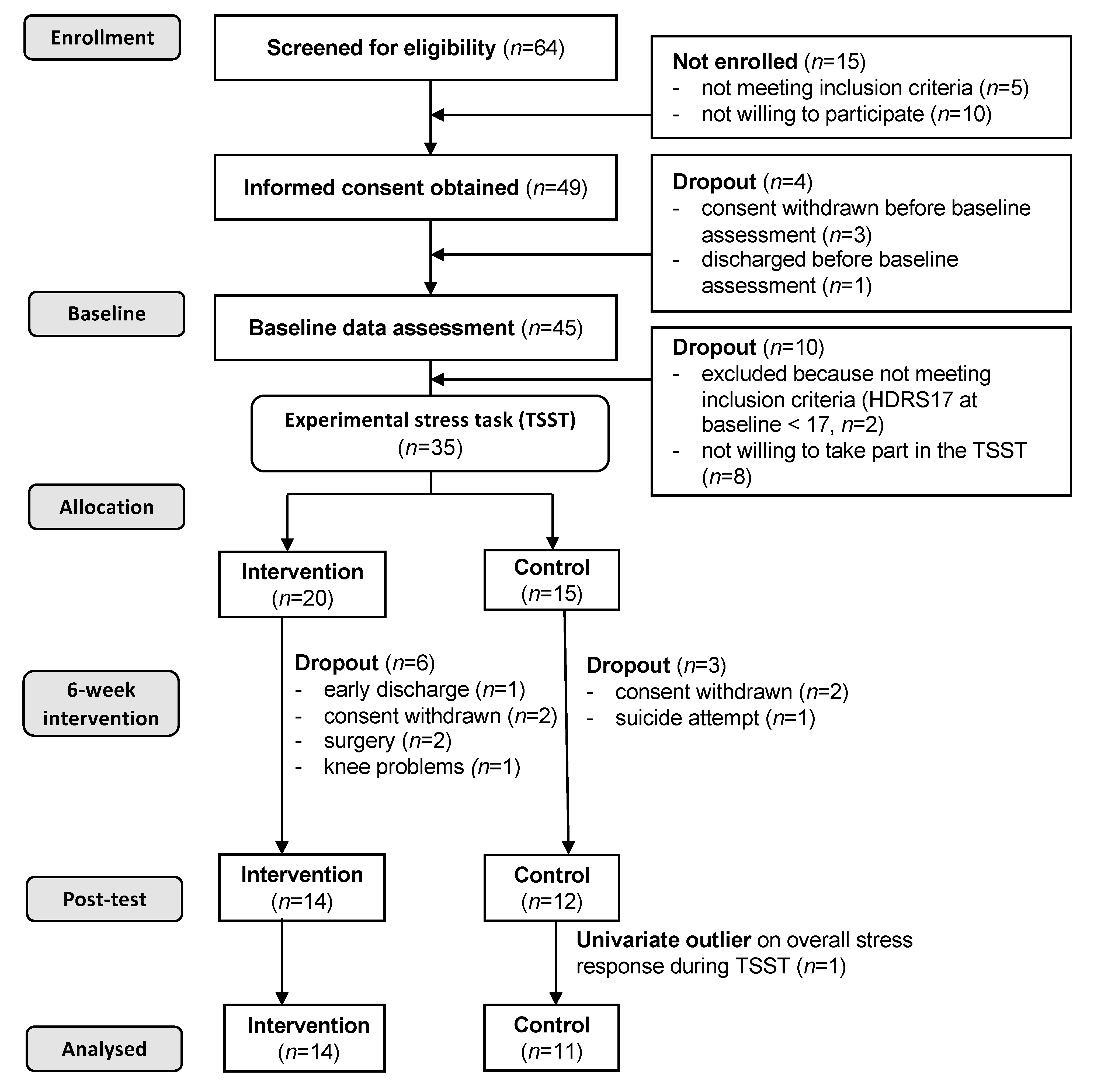
2.1. Participants and Procedures

Participants were recruited from two (public) psychiatric clinics in the Northwestern, German-speaking part of Switzerland. The following inclusion criteria were assessed by the consultant psychiatrist of the respective depression ward as part of the initial screening (in the first week of hospitalization), prior to providing more specific information about the study to the eligible patients: (a) aged >18 and <61 years, (b) be an inpatient in one of the two depression wards, (c) ICD-10 diagnosis of depression (first episode, recurrent or bipolar; F32, F33, F31). Moreover, the following exclusion criteria were applied: (a) a score <17 on the Hamilton depression rating scale 17 (HDRS17) at the baseline data assessment, (b) presence of a somatic condition not permitting regular exercise, (c) body mass index (BMI) > 35 kg/m2, (d) pregnant at the baseline data assessment, (e) acute suicidal ideation, (f) comorbid substance dependence (except nicotine), (g) comorbid major psychiatric disorder, and (h) participation in regular vigorous-intensity exercise (i.e., participating in long distance runs).
The study was designed as a two-centre, two-armed randomised controlled trial (RCT), with a 1:1 allocation ratio (intervention:control group). The study was registered at https://www.clinicaltrials.gov (trial number: NCT02679053). As highlighted in the study protocol [71], the primary outcome of this study was depression severity. While the effects of the intervention on the primary outcome are presented elsewhere [72], the focus of the present paper is on one of the secondary outcomes (cortisol response during the TSST).
Recruitment took place between October 2013 and January 2016, and was done by the local study coordinators. A graphical representation of the study schedule including information about which measurements were performed at which timepoint is presented in the study protocol [71]. As shown in Figure 1, written informed consent was obtained from all participants that were eligible for the study, and they were allocated to either an aerobic exercise condition (intervention group) or a standardised stretching and mobility program (active control group) after the baseline data assessment. The last author was responsible for the randomisation and assignment of the participants to the intervention and control group. Simple randomisation was achieved by drawing lots for the intervention and control condition, separately for males and females. Blinding of patients with regard to group allocation was achieved by informing them that two different exercise programs were to be compared, but not letting them know which intervention was considered the intervention or control condition. Data assessors and local study coordinators were fully blinded to group allocation (by anonymized ID). The baseline data assessment took place during the first or second week of hospitalization.
To detect a moderate effect (f = 0.25) in the RCT on the primary outcome (depressive symptoms severity), a power analysis for repeated measures analyses of variance (ANOVAs) (using G*Power 3.1; alpha error probability: 0.05, power: 0.90, correlations among measures: 0.50) revealed that at least n = 36 (overall) participants are needed. We therefore originally aimed to include 40 patients with a roughly equal gender distribution.
Before the beginning of the study, ethical clearance was obtained from the Ethics Committee of both Basels (EKBB, Basel, Switzerland; reference no. 62/13) and the Ethics Committee Aargau/Solothurn (Aarau, Switzerland; reference no. 2013/029) and all study procedures were carried out in line with the ethical principles defined in the 1964 Declaration of Helsinki and its later amendments.

2.2. Intervention vs. Control Condition

The intervention consisted of supervised aerobic exercise on indoor bicycles three times per week for six consecutive weeks. The target heartrate (HR) was set at 60–75% of maximal heartrate (HRmax) monitored with Polar™ RS800CX. We used the following formula (220–age (in years)) to compute HRmax. Following Dunn et al. [73], the targeted exercise-based energy expenditure was 17.5 kcal per kg bodyweight.
Participants assigned to the active control condition, engaged in a program consisting of coordination and stretching activities for all major muscle groups using a medium strength Theraband®, a gymnastics ball (diameter 65 cm) and juggling balls, which also took place three times per week for six consecutive weeks. To ensure that the intensity in the control group was kept at a low level during the stretching sessions, supervisors reminded the participants of the control group that they should not get out of breath. This was important to avoid an overlap in activity intensity between the intervention and the control group. To minimize the influence of social contact, the coordination and stretching activities were also carried out individually or in groups of two patients.
All sessions (intervention and active control group) were scheduled in the late afternoon (between 4 and 6 p.m.) for approximately 40–50 min. Additionally, all patients received standard inpatient treatment consisting of pharmacological treatment according to Swiss national standards [74], individual and group-psychotherapy supported by an array of creative group therapies. Pharmacological treatment was limited to antidepressant treatment with selective serotonine-reuptake-inhibitors (SSRI) or selective serotonine-norepinephrine-reuptake-inhibitors (SNRI) and lithium as augmentation strategy, whereas antidepressant combination therapy, tricyclic antidepressants, MAO-inhibitors, and antipsychotics other than low-dose quetiapine for sedation were not allowed. Participants were asked not to engage in any additional vigorous exercise activities during their stay at the hospital. However, engagement in additional vigorous exercise was not systematically monitored. Evidence regarding compliance with the intervention program has been reported previously [71].

2.3. Trier Social Stress Test

We used the Trier Social Stress Test (TSST) [33] to experimentally induce stress. The TSST consists of two standardised 5-min tasks including a free speech task (job interview) and a mental arithmetic task (counting backwards in steps of 13 from a 4-digit number). At both measurement occasions, the TSST was performed in front of a jury of two people unknown to the patient. Patients were also informed that the test would be videotaped to evaluate their behaviour. Patients had 5 min to prepare themselves for the speech task, before they entered the test room. The test was completed in a standing position. After completion of the TSST, participants recovered in a separate room for a period of 60 min. In the present study, the TSST was carried out exactly at 2 p.m. to control for circadian variations of cortisol levels [75]. The participants were also asked to refrain from eating, drinking (except for water) and from any moderate or vigorous intensity physical activity 2 h prior to the beginning of the stress protocol. While previous research has shown that participation in physical activity prior to the TSST might have acute effects on stress reactivity [76], researchers also showed that cortisol levels typically return to the level prior to the TSST within two hours after individual have stopped exercise participation [77]. All participants were unaware of the experimental protocol. Because stress reactions depend on the novelty of the stressor task [25], responses might be less strong if the same psychosocial stressor task is performed a second time [78]. However, researchers showed that the stressfulness of the TSST can be maintained if the stressor tasks are modified [79]. We therefore slightly changed the content of the free speech task (application for a promotion/prize) and mental arithmetic tasks (counting backwards in steps of 13 from a different number), but ensured that the socio-evaluative pressure of the stressor protocol was maintained. According to Mazurka et al. [80], the TSST is useful for examining the stress reactivity in depression “because it provokes the HPA axis response by activating the same corticolimbic circuitry implicated in the pathology of major depression” (…), whereas “pharmacologic challenge tests and studies of the cortisol awakening response bypass this important circuitry and directly activate components of the axis” (pp. 301–302).

2.4. Assessment of the Adrenocortical Stress Response

The adrenocortical stress response was measured via salivary cortisol samples. To minimize an anticipation bias, we gathered the first saliva sample before participants were informed about the nature of the experimental stress protocol. At both the baseline data assessment and at post-intervention, salivary cortisol was obtained at −20, −5, +1, +5, +10, +20, +30, +45, and +60 min prior to the onset/after completion of the TSST, using commercially available devices (Salivette®, Sarstedt, Germany). All samples were stored at −20 °C, before they were sent to the Biochemical Laboratory of the University of Trier for analyses (approximately every three months). Free salivary cortisol levels (nmol/L) were established, based on a time-resolved fluorescence immunoassay (for more details see: [81]).

2.5. Assessment of Covariates

2.5.1. Depressive Symptom Severity

Symptom severity of depression was measured with the 21-item Beck Depression Inventory [82,83]. The BDI measures a range of affective, behavioural, cognitive, and somatic symptoms, which are characteristic for unipolar depression (e.g., “I am so unhappy/sad that I can’t stand it.”). Response options are anchored on a scale from 0 to 3, with higher scores being indicative of stronger depressive symptomatology. A sum score was calculated to generate an overall index with a potential range from 0 to 63 and higher scores reflecting more severe depressive symptoms. Scores between 0 and 9 are considered as no depression, whereas scores of 10–18, 19–29, and 30–63 can be interpreted as mild, moderate and severe depression, respectively. The Cronbach’s alpha in the present sample was 0.72 at baseline.

2.5.2. Physical Activity

To gather information about participants’ physical activity levels prior to hospitalization, participants filled in a written version of the German International Physical Activity Questionnaire Short Form (IPAQ-SF) [84]. The IPAQ-SF assesses time spent in moderate physical activity (e.g., bicycling at a regular pace, low-intensity sports such as doubles tennis) and vigorous physical activity (e.g., fast bicycling, running, aerobics), using a frequency-by-duration format. Thus, participants first reported the number of days per week they engaged in these activities (from 0 to 7 days), and then indicated the average duration (in minutes) for the days they engaged in these activities. Participants were asked to refer to the last week prior to admission to the hospital. Multiplication of frequency and duration scores resulted in an estimate of weekly hours invested in moderate and vigorous physical activity. Summing up these two scores resulted in a total score for time (in min/week) spent in moderate-to-vigorous physical activity (MVPA). Evidence for the validity of the IPAQ-SF has been shown previously in adult samples [85].

2.5.3. Further Potential Confounders

We assessed sex, age, smoking status, educational status, duration of current depressive episode, number of prior depressive episodes, and age of onset of depression. Additionally, all patients underwent a physical examination including the assessment of blood pressure (BP), resting heart rate (HR), as well as body weight and height. Body weight (kg) and height (m) were used to calculate participants’ body mass index (BMI; kg/m2).

2.6. Statistical Analyses

We first examined whether results of the different variables are normally distributed (using Kolmogorov–Smirnov and Shapiro–Wilk tests). To examine whether baseline differences existed between the intervention and control groups, in the case of normally distributed (metric) variables, ANOVAs were conducted, and descriptive statistics were reported as M and SD. If either the Kolmogorov–Smirnov or the Shapiro–Wilk test indicated a non-normal distribution of a metric variable, nonparametric tests were used (independent samples median test (Kruskal–Wallis) and independent samples Mann–Whitney U Test). Moreover, in case of non-normally distributed (metric) variables, we reported the median (Mdn) and range (min; max). For categorical variables, we used χ2 tests to examine group differences, and we reported the number (n) and frequency (%) as descriptive statistics.
To assess bivariate associations between the potential covariates and the cortisol responses at baseline and post-intervention, correlational analyses were conducted. Pearson’s correlations (r) were used if covariates were normally distributed. Spearman’s correlations (Rho) were used if covariates were non-normally distributed. In case of non-normally distributed cortisol values, cortisol scores were logarithmized to achieve a normal distribution prior to calculating the correlations. Moreover, in case of categorical covariates, analyses of variance (ANOVAs) was conducted.
To examine whether the TSST elicited a significant stress response in the total sample and the intervention and control groups, a repeated measure analysis of variance (ANOVA) was calculated, with the factors time (two measures from 20 min prior to the onset of the TSST to 20 min after completion of the stressor task (peak) and group (intervention vs. control group), and the interaction effect (time x group). The time point 20 min after completion of the stressor task was chosen as a peak, as most studies show that cortisol levels tend to increase approximately 20 min after the end of a stressor task [40]. Again, in case of non-normally distributed cortisol values, the logarithmized scores were used. We also descriptively analysed how many participants responded to the TSST, hereby following Petrowski et al. [86] who defined a response to the TSST as a salivary cortisol increase of ≥ 2.5 mmol/L from prior to the TSST to the individual peak.
As an index of overall cortisol stress reactivity, we calculated the area under the total response curve with respect to ground (AUCG), by using the trapezoid formula described by Pruessner et al. [87], including the entire time period from 20 min prior to the onset to 60 min after completion of the TSST. We did not use the total response curve with respect to increase (AUCI), because this index results in negative values if the repeated measures are lower than the cortisol level prior to the TSST.
To examine whether six weeks of aerobic exercise training had an effect on participants’ overall stress reactivity, we employed a repeated measures analysis of covariance (ANCOVA) with AUCG as an outcome, with the factors time (baseline vs. post-intervention), group (intervention vs. control group), and the interaction effect (time × group), and controlling for depressive symptom severity, physical activity and further relevant covariates (age, depressive symptom severity, duration of current depressive episode, number of prior depressive episodes, age at onset of depression, resting heart rate, blood pressure, height, weight, BMI, MVPA, sex, smoking status, educational background, and diagnoses). However, covariates were only considered if they were significantly associated with the cortisol stress response either at baseline or post-intervention. To interpret the magnitude of the effects, η2 values were shown for both main and interaction effects. Finally, we calculated a Pearson’s correlation to examine whether changes in depressive symptom severity were associated with changes in cortisol response. The level of significance was set at p < 0.05 across all analyses. All tests were carried with SPSS 24 for Mac (SPSS Inc., Chicago, IL).

3. Results

3.1. Sample Characteristics and Descriptive Statistics

As shown in the participant flow chart (Figure 1), 49 participants (25 women, 24 men, M = 38.9 years, SD = 11.3) were willing to take part in the study. Thereof, four participants (three women, one man, M = 33.5 years, SD = 13.2) dropped out before the beginning of the data assessment (three withdrew consent, one was discharged). Of the remaining 45 participants, 10 participants (five women, five men, M = 37.0 years, SD = 9.8) were not willing to take part in the experimental stress test (n = 8) or did not meet the inclusion criteria with regard to depression severity (n = 2). Of the remaining 35 participants with valid baseline data, nine dropped out before post-intervention due to the following reasons: early discharge (n = 1), withdrawn consent (n = 4), surgery (n = 2), knee problems (n = 1), and suicide attempt (n = 1). The attempted suicide was considered as a severe adverse event and immediately reported to the local ethical review committee. However, since there was no reason to believe that the suicide attempt was caused by the study intervention, no further actions were deemed necessary other than a more rigorous assessment of suicidal behaviour prior to study inclusion. One participant was identified as a univariate outlier because stress reactivity exceeded three standard deviations around the mean. This person was excluded from all further analyses. Among the patients who took part in the first TSST assessment, no significant differences were found between dropouts and study completers in any of the study variables. The post-intervention assessment took place immediately after completion of the six-week intervention/control period.
As shown in Table 1, not all (metric) study variables were normally distributed in the present sample. This is taken into consideration when sample characteristics and descriptive statistics for the baseline and post-intervention data assessment are presented (Table 1).
On average, the total sample (n = 25) was 38.1 (SD = 12.0) years old, and consisted of 13 women and 12 men. Most of the participants were diagnosed with a first unipolar depressive episode (n = 10, 40%) or recurrent depressive episode (n = 14, 56%); only one participant (4%) was diagnosed with bipolar depression. Based on the BDI scores, three participants (12%) reported mild depression, 14 (56%) reported moderate depression, and eight (32%) reported severe depression. Based on participant self-reports, five participants (20%) reported that they were completely inactive (0 min/week of MVPA) prior to hospitalization. Fourteen participants (56%) reported some MVPA, but according to the standards of the American College of Sports Medicine [88], did not meet current MVPA recommendations (≥150 min/week). Six participants (24%) accomplished these standards. As shown in Table 1, no significant baseline differences were found between participants assigned to the intervention and control group in any of the study variables.

3.2. Reactivity in Response to the TSST at the Baseline Data Assessment

At the baseline data assessment, a repeated measures ANOVA showed that in the total sample, a significant time effect occurred prior to the onset of the TSST (−20 min) to peak (+20 min after completion of the TSST), F(2,23) = 4.4, p = 0.046.05, η2 = 0.161, indicating that the TSST elicited a cortisol response. However, no significant time x group interaction effect was observed, F(2,23) = 0.1, p = 0.769, η2 = 0.004, and the response curve was flat in both the intervention and control group (see Figure 2). At the baseline assessment, only a 1.6-fold increase was observed from 20 min prior to the TSST to peak in the total sample (see Table 1). Moreover, at the baseline data assessment, 52 percent (n = 13) of the participants responded to the TSST (increase in cortisol from 20 min prior to the TSST to individual peak of ≥2.5 mmol/l), whereas 48 percent of the participants were classified as non-responders (n = 12).

3.3. Associations Between Potential Confounders and Stress Reactivity

As shown in Table 2, bivariate correlations and univariate ANOVAs revealed that none of the potential confounders was significantly associated with the overall cortisol stress response during the TSST (neither at baseline nor at post-intervention), as measured with AUCG. Therefore, none of these factors were considered as a potential confounder in the subsequent repeated measures ANOVAs.

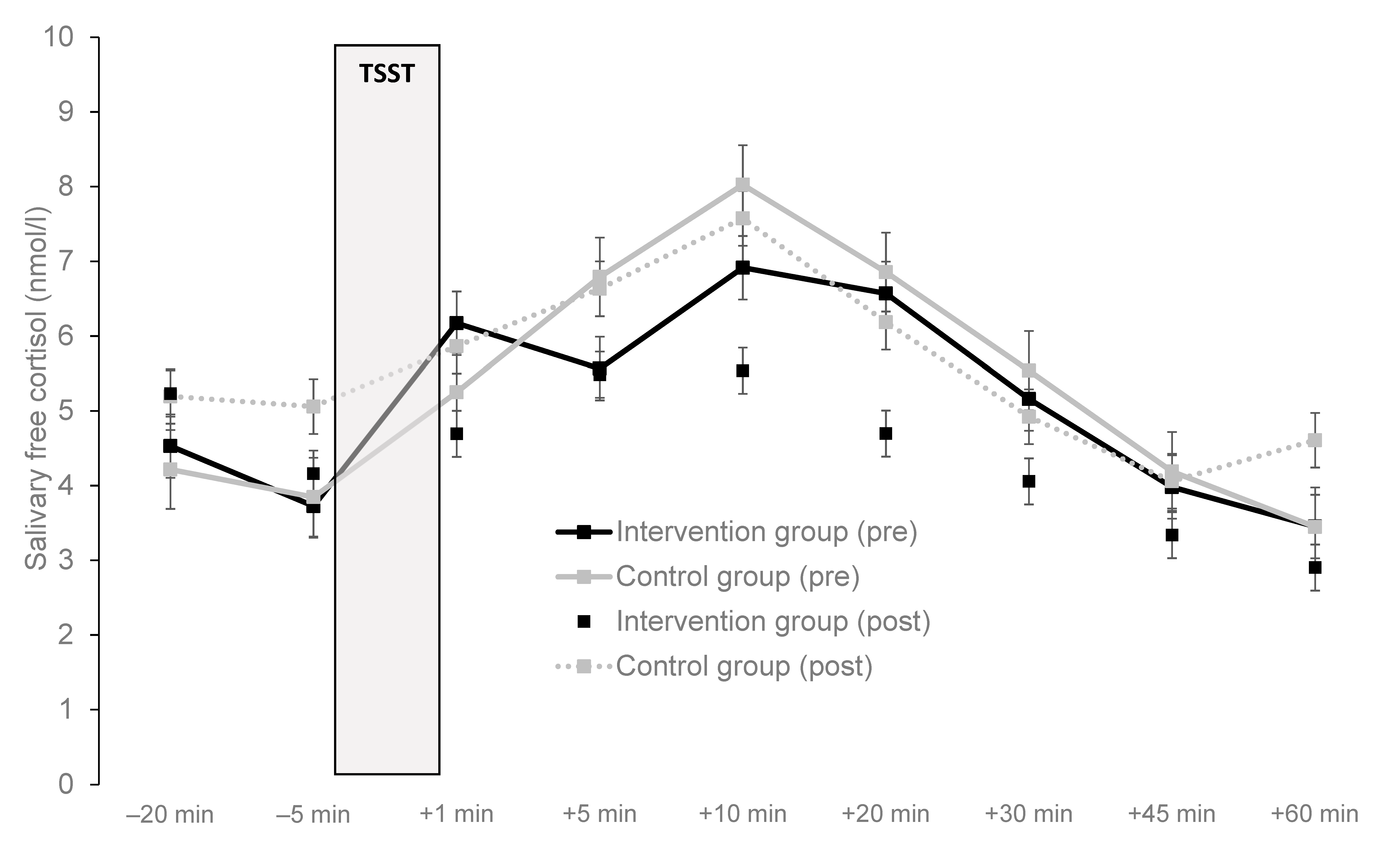
3.4. Impact on Aerobic Exercise Training on Stress Reactivity

The results of the repeated measures ANOVAs, addressing the main study question, are displayed in Table 3. No significant main and interaction effects occurred. As shown in Figure 2, no major differences were found in cortisol response pattern at baseline and post-intervention between patients of the intervention and control group.

3.5. Correlations of Change Between Depressive Symptoms and Cortisol Response

Although not the focus of the present paper, Table 3 shows that depressive symptom severity considerably decreased from baseline to post-intervention in both the intervention and the control group. Accordingly, a strong main effect for time was found in the repeated measures ANOVA. However, no significant time × group interaction was found in the present sample.
To examine the relationship between change in depressive symptom severity and change in cortisol response, a Pearson’s correlation was computed (based on Kolmogorov–Smirnov and Shapiro–Wilk tests, both change scores were normally distributed). The Pearson’s correlation between these variables was r = 0.02, p = 0.944.

4. Discussion

The present study shows that six weeks of exercise training did not result in an altered pattern of cortisol secretion in response to an acute psychosocial stressor among inpatients with MDD. The findings are novel because this is the first study worldwide testing the validity of the cross-stressor adaption (CSA) hypothesis in psychiatric patients [40]. Moreover, only one further intervention trial exists in which the impact of an exercise training program was tested on the cortisol reactivity to the TSST [42]. Changes in depressive symptom severity were not correlated with changes in cortisol response to the TSST.
In contrast with the work of Klaperski et al. [42] with generally healthy, but physically inactive employees, regular aerobic exercise training did not result in a blunted stress reactivity in our sample. Figure 2 indicates that, in our sample, the cortisol response pattern remained nearly unchanged from pre- to post-intervention in both participants assigned to the intervention and control condition. This is remarkable as depressive symptom severity considerably decreased in both the intervention and control group between the two assessment periods. A more detailed discussion regarding the effects of the intervention on the primary outcome (depression symptom severity) is presented elsewhere [72]. Obviously, changes in depressive symptom severity and cortisol secretion during the TSST do not change in parallel among patients with MDD undergoing inpatient treatment. While our findings suggest that exercise training is more suited to impact on stress reactivity among healthy people, these results do not allow too far-reaching conclusions. As highlighted in the introduction section, research on the neurobiological mechanisms that explain the benefits of exercise training in individuals with MDD is still in a very early stage [69], and research with neuroendocrine markers is sparse [54,68]. Therefore, further research is needed to find out whether different effects occur if the training is maintained over longer periods of time, and if patients with differing psychiatric disorders are assessed.
Beyond the main research question addressed in this paper, some further observations were made. Our first observation was that as reported in the meta-analysis by Burke et al. [31], patients with MDD tend to show a relatively non-responsive cortisol secretion pattern if exposed to the TSST. This notion was supported in the present study. Although patients exhibited a significant increase in free salivary cortisol concentration, on average, we found only a 1.6-fold increase from 20 min prior to the TSST to peak, which is considerably lower than the response to socio-evaluative stressors in healthy populations [25]. Moreover, the median cortisol concentrations at the peak (+20 min after completion of the TSST) varied between 5.2 nmol/L (at the baseline data assessment) and 4.7 nmol/L (at post-intervention) in the total sample. Again, this concentration is lower compared to studies with healthy participants where group peak cortisol concentrations reached levels of >15 nmol/L [33,89]. Moreover, no evidence was found that patients’ non-responsiveness was due to heightened pre-stress cortisol concentrations. As a putative mechanism for the blunted cortisol reactivity, Suzuki et al. [90] pointed towards a downregulation of HPA axis activity among patients with MDD [91]. As highlighted by McEwen and Stellar [92] and Juster et al. [93], exposure to chronic stress might lead to a repeated overactivation and impairment of the HPA axis. This notion is in line with the fact that chronic stress and critical life events play a major role in the development and maintenance of MDD [5,6]. This finding raises important questions regarding the interpretation of low cortisol responses. Thus, while a low stress reactivity to acute laboratory stressors is generally regarded adaptive among healthy participants [94,95], this is not necessarily the case among patients with MDD. In a model focussing on brain structures involved in the regulation of stress reactivity, Lovallo [96] argued that a dysregulated stress reactivity on the central nervous level (prefrontal cortex, limbic system, hypothalamus and brain stem) will lead to poor behavioral homeostasis. Accordingly, Lovallo assumes that stress reactivity may range from very low to very high and that both exaggerated and diminished stress reactivity may have a negative impact on health, because both tendencies are indicative of a loss of homeostatic regulation.
Our second observation was that self-reported physical activity levels prior to hospitalization were not associated with patients’ cortisol responses (either at the baseline data assessment or at post-intervention). This is in contrast to previous results with healthy population, in which the CSA hypothesis associated with physical activity and fitness was supported in about 60% of all studies [40]. The lack of a significant relationship can be attributed to various factors. First, it is difficult to detect significant differences in a non-responsive sample. Second, most of the participants in our sample were physically inactive or reported low physical activity levels. Thus, individual variability was limited in the independent variable, as well.
Our third observation was that physical activity levels were relatively low among patients with depressive disorders. In a previous meta-analysis, Schuch et al. [97] highlighted that among patients with MDD, more than two thirds (68%) did not reach current levels of recommended physical activity (≥150 min of MVPA per week). Moreover, Schuch et al. [97] showed that patients with MDD were significantly more likely to miss MVPA recommendations than healthy controls. In the present study, only 18% of the participants reached recommended MVPA standards. Given this background, it is encouraging that sport and exercise training are increasingly used in Swiss psychiatric clinics as an additional treatment approach [98], and that many MDD patients are sufficiently physically active during their stay at the clinics [99]. Nevertheless, more systematic efforts are needed to increase patients’ lifestyle physical activity sustainably [100,101], because the beneficial effects of exercise and sport therapy may dissipate if the training is stopped after discharge from the clinic [102,103].
Finally, in contrast to previous studies [104], we did not find a significant association between BMI and participants’ cortisol reactivity. Moreover, we did not find a link between age and stress reactivity although previous studies suggested that the level of HPA axis dysregulation might increase with age among people with depression [20,21]. We also did not observe a difference in cortisol stress reactivity between women and men. While it is well established that women are more prone to develop depressive symptoms and MDD compared to men [105], studies examining sex differences in stress reactivity have yielded equivocal findings [106], with men responding more strongly [107], less strongly [108] or similarly [109] if compared to female participants. Chopra et al. [110] found in a study comparing patients with chronic MDD with healthy controls that female patients exhibited a higher reactivity, whereas a blunted reactivity was found in male patients. Nevertheless, the opposite pattern of results was found in a study with depressed and non-depressed adolescents [80]. Thus, depressed boys showed a higher cortisol reactivity than depressed girls, and the latter had a blunted cortisol response in comparison to non-depressed girls.
The strengths of the present study can be summarized as follows. As mentioned previously, this is the first study testing the CSA hypothesis in psychiatric patients, a population in which HPA axis activity is often dysregulated. In the present study, all participants were diagnosed with MDD according to ICD−10 definitions, were in-patients, and most of them reported moderate-to-severe levels of depression. This is noteworthy, as Burke et al. [31] showed that cortisol reactivity is particularly low in in-patients and in more severely depressed people. Moreover, few intervention studies have been carried out so far to examine whether exercise training positively impacts on stress reactivity. Another strength was that we first tested bivariate relationships with potential confounders to rule out that these factors confound the association between self-reported physical activity and cortisol reactivity. We used an established psychosocial stressor, which is known to trigger a stronger stress response compared to other cognitive stressors due to its socio-evaluative character. Gender distribution was almost equal in our sample. Finally, we systematically assessed energy expenditure in the intervention group, showing that the compliance rate was high in the intervention group [71].
Despite these strengths, our findings need to be interpreted in light of several limitations. First, the sample size was small. Thus, the low sample size did not allow for a consideration of further moderating factors (such as sex, baseline physical activity levels, symptom severity, etc.). For instance, we did not control atypical depression, a specific type of depression that has been associated with stronger appetite, hyperphagia and hypersomnia, and a lower stimulation of the HPA axis [111]. We also acknowledge that our power calculation was based on the primary outcome (depressive symptom severity) and not on cortisol response, although these are two different outcomes. Moreover, a posteriori power analyses showed that with a repeated measures design (within-between interaction, alpha error = 0.05, Power = 0.80, two groups, two measurements, correlations among repeated measures of r = 0.35), we would have needed a total sample size of 258 participants to detect small effects (f = 0.1) or 44 participants to detect moderate effects (f = 0.25). Because we only included 25 patients, our study was somewhat underpowered. Second, we were not able to control all influences that may have an influence on individuals’ stress response (such as previous critical life events). Moreover, for future studies with a repeated-measures design using two slightly different forms of the TSST, it seems advisable to assign half of the group to the modified test at pre-intervention and then switch tests at post-intervention to rule out the possibility that different tests produce different stress reactions. Third, we also acknowledge that physical activity was based on participants’ self-reports that might be negatively biased, especially since we studied a clinically depressed population. Fourth, the intervention period was relatively short (six weeks). Thus, exercise training might have had a stronger effect if carried out across a longer period of time. Fifth, a considerable number of eligible participants were not willing to take part in the TSST, or dropped out from the baseline data assessment to post-intervention, which may have entailed a selection and dropout bias. Nevertheless, we did not find substantial differences between patients who declined participation, dropouts and patients who volunteered for the TSST. Moreover, while we initially employed multiple imputation to substitute missing values, this yielded highly improbable values, which we attributed to the small sample size. We therefore proceed with a “per-protocol analysis” and did not carry out intention-to-treat analyses, as originally foreseen in the study protocol [71]. Sixth, our study does not allow a direct comparison with healthy controls. Such analyses may be important to more definitely establish whether the validity of the CSA hypothesis holds in both psychiatric and healthy participants. Seventh, our sample included a fairly broad age-range. Eighth, we did not systematically assess the physical activity levels of participants in the control group during the intervention period. Thus, we cannot exclude that these patients also engaged in moderate-to-vigorous physical activities while they were hospitalized. Ninth, we acknowledge that our control group also got some form of physical activity intervention (standardized coordination and stretching activities). While the instructors made efforts to keep the intensity level low, we did not monitor the intensity level systematically in this group. Moreover, an additional “treatment as usual” (TAU) control group could have been helpful to further assess the effect of the aerobic exercise intervention and the active control condition. Tenth, although we have carried out a six-month follow-up assessment in order to assess the longer-term effects on the primary and secondary outcomes, due to limited resources, we had to refrain from running the TSST a third time. Finally, no generalization is possible to other stress reactivity indicators (e.g., cardiovascular, emotional) and to stress reactivity in real-life conditions. The latter is important as researchers have shown that cortisol reactivity following the TSST and in response to a real-life stressor are only weakly related. In line with this notion, scholars assumed that stress reactions might be less strong in a laboratory setting [112], because emotional involvement is limited during an experimental stressor test [113].

5. Conclusions

Patients with MDD tend to self-report insufficient physical activity levels and show a blunted cortisol response pattern in following socio-evaluative stress. Therefore, systematic efforts are needed to increase participants’ physical activity levels and foster their cardiorespiratory fitness and cardiovascular health. More research is needed to find out which treatments are most efficient to promote a normalization of HPA axis reactivity in patients with MDD. The present study suggests that six weeks of aerobic exercise training are insufficient to impact on patients’ cortisol stress reactivity. Nevertheless, since this was the first study on the CSA hypothesis with psychiatric patients, the findings of our randomized controlled trial need to interpreted with caution until a broader empirical evidence base exists.

Author Contributions

M.G., C.I., J.B., A.E., U.P., E.H.-T. and M.H. developed the study design. C.I. and J.B. coordinated the study at the two study sites. A.E. coordinated all laboratory analyses. M.G. and C.I. were responsible for data assessment. M.G. and S.B. conducted the statistics. M.G. wrote the manuscript. All authors contributed to the data interpretation and the internal revision of the manuscript draft. F.C. was responsible for the final language check. All authors approved the final draft version. M.G. and C.I. have contributed equally to this study. All authors have read and agreed to the published version of the manuscript.

Funding

This study was supported by a grant of the “Gottfried und Julia Bangerter-Rhyner Stiftung”, the Helsana insurance company, and the “Lotteriefonds des Kantons Solothurn”. The funding sources had no influence on the study design, on the collection, analysis and interpretation of the data, and in the decision to submit this report for publication. The funding sources had no influence on the study design, the data collection, the analysis and interpretation of the data, the writing of the report, and the decision to submit the article for publication.

Acknowledgments

We thank Xenia Fischer, Simon Fritsche, Michel Hutter, Alexandra Kuhn, Sonja Kulka, Fabienne Ott and Lukas Zumsteg for their support regarding the implementation of the study and for their contribution to the data assessment. We also thank all participants for their time and willingness to take part in our study.

Conflicts of Interest

The authors declare no conflict of interests.

References

  1. Baer, N.; Schuler, D.; Füglister-Dousse, S.; Moreau-Gruet, F. Depressionen in der Schweizer Bevölkerung. Daten zur Epidemiologie, Behandlung und Sozialberuflichen Integration (No. 56); Schweizerisches Gesundheitsobservatorium: Neuchâtel, Switzerland, 2013. [Google Scholar]
  2. Kessler, R.C.; Berglund, P.; Demler, O.; Jein, R.; Koretz, D.; Merikangas, K.R.; Rush, A.J.; Walters, E.E.; Wang, P.S. The epidemiology of Major Depressive Disorder. Results from the National Comorbidity Survey Replication (NCS-R). JAMA 2003, 18, 3095–3105. [Google Scholar] [CrossRef]
  3. WHO. The World Health Report 2001-mental Health: New Understanding, New Hope; World Health Organization: Geneva, Switzerland, 2001. [Google Scholar]
  4. Lépine, J.-P.; Briley, M. The increasing burden of depression. Neuropsychiatr. Dis. Treat. 2011, 7, 3–7. [Google Scholar] [PubMed] [Green Version]
  5. Qin, D.-D.; Rizak, J.; Feng, X.-L.; Yang, X.-C.; Lü, L.-B.; Pan, L.; Yin, Y.; Hu, Y.-T. Prolonged secretion of cortisol as a possible mechanism underlying stress and depressive behaviour. Sci. Rep. 2016, 6. [Google Scholar] [CrossRef] [PubMed]
  6. Hammen, C. Stress and depression. Annu. Rev. Clin. Psychol. 2005, 1, 293–319. [Google Scholar] [CrossRef] [PubMed] [Green Version]
  7. Phillips, A.C.; Carroll, D.; Der, G. Negative life events and symptoms of depression and anxiety: Stress causation and/or stress generation. Anxiety Stress Coping 2015, 28, 357–371. [Google Scholar] [CrossRef] [Green Version]
  8. Siegrist, J. Chronic psychosocial stress at work and risk of depression: Evidence from prospective studies. Eur. Arch. Psychiatry Clin. Neurosci. 2008, 258, 115–119. [Google Scholar] [CrossRef]
  9. Monroe, K.L.; Harkness, S.M. Life stress, the “kindling” hypothesis, and the recurrence of depression: Considerations from a life stress perspective. Psychol. Rev. 2005, 112, 417–445. [Google Scholar] [CrossRef] [Green Version]
  10. Ising, M.; Horstmann, S.; Kloiber, S.; Lucae, S.; Binder, E.B.; Kern, N.; Künzel, H.E.; Pfenning, A.; Uhr, M.; Holsboer, F. Combined dexamethasone/corticotropin releasing hormone test predicts treatment response in major depression-a potential biomarker? Biol. Psychiatry 2007, 62, 47–54. [Google Scholar] [CrossRef]
  11. Gotlib, I.H.; Joormann, J.; Minor, K.L.; Hallmayer, J. HPA axis reactivity: A mechanism underlying the associations among 5-HTTLPR, stress, and depression. Biol. Psychiatry 2008, 63, 847–851. [Google Scholar] [CrossRef] [Green Version]
  12. Schüle, C. Neuroendocrinological mechanisms of actions of antidepressantdrugs. J. Neuroendocrinol. 2007, 19, 213–226. [Google Scholar] [CrossRef] [PubMed]
  13. Willner, P.; Scheel-Krüger, J.; Belzung, C. The neurobiology of depression andantidepressant action. Neurosci. Biobehav. Rev. 2013, 37, 2331–2371. [Google Scholar] [CrossRef] [PubMed]
  14. Miller, G.E.; Chen, E.; Zhou, E.S. If it goes up, must it come down? Chronic stress and the hypothalamic-pituitary-adrenocortical axis in humans. Psychol. Bull. 2007, 133, 25–45. [Google Scholar] [CrossRef] [Green Version]
  15. Chida, Y.; Steptoe, A. Cortisol awakening response and psychosocial factors: A systematic review and meta-analysis. Biol. Psychol. 2009, 80, 265–278. [Google Scholar] [CrossRef] [PubMed]
  16. Chida, Y.; Hamer, M. Chronic psychosocial factors and acute physiological responses to laboratory-induced stress in healthy populations: A quantitative review of 30 years of investigations. Psychol. Bull. 2008, 134, 829–885. [Google Scholar] [CrossRef] [PubMed]
  17. Pike, J.L.; Smith, T.L.; Hauger, R.L.; Nicassio, P.M.; Patterson, T.L.; McClintick, J.; Costlow, C.; Irwin, M.R. Chronic life stress alters sympathetic, neuroendocrine, and immune responsivity to an acute psychological stressor in humans. Psychosom. Med. 1997, 59, 447–457. [Google Scholar] [CrossRef]
  18. Holsboer, F. The corticosteroid receptor hypothesis of depression. Neuropsychopharmacology 2000, 23, 477–501. [Google Scholar] [CrossRef] [Green Version]
  19. Modell, S.; Yassouridis, A.; Huber, J.; Holsboer, F. Cortico-steroid receptor function is decreased in depressed patients. Neuroendocrinology 1997, 65, 216–222. [Google Scholar] [CrossRef]
  20. Stetler, C.; Miller, G.E. Depression and hypothalamic-pituitary-adrenal activation: A quantitative summary of four decades of research. Psychosom. Med. 2011, 73, 114–126. [Google Scholar] [CrossRef]
  21. Belvederi Murri, M.; Pariante, C.; Mondelli, V.; Masotti, M.; Atti, A.R.; Mellacqua, Z.; Antonioli, M.; Ghio, L.; Menchetti, M.; Zanetidiou, S.; et al. HPA axis and aging in depression: Systematic review and meta-analysis. Psychoneuroendocrinology 2014, 41, 46–62. [Google Scholar] [CrossRef]
  22. Knorr, U.; Vinberg, M.; Kessing, L.V.; Wetterslev, J. Salivary cortisol in depressed patients versus control persons: A systematic review and meta-analysis. Psychoneuroendocrinology 2010, 35, 1275–1286. [Google Scholar] [CrossRef]
  23. Bremmer, M.A.; Deeg, D.J.; Beekman, A.T.; Penninx, B.W.; Lips, P.; Hoogendijk, W.J. Major depression in late life is associated with both hypo- and hypercortisolemia. Biol. Psychiatry 2007, 62, 479–486. [Google Scholar] [CrossRef] [PubMed]
  24. Oldehinkel, A.J.; van den Berg, M.D.; Flentge, F.; Bouhuys, A.L.; ter Horst, G.J.; Ormel, J. Urinary free cortisol excretion in elderly persons with minor and major depression. Psychiatry Res. 2001, 104, 39–47. [Google Scholar] [CrossRef]
  25. Dickerson, S.S.; Kemeny, M.E. Acute stressors and cortisol responses: A theoretical integration and synthesis of laboratory research. Psychol. Bull. 2004, 130, 355–391. [Google Scholar] [CrossRef] [PubMed] [Green Version]
  26. Handwerger, K. Differential patterns of HPA activity and reactivity in adult post-traumatic stress disorder and major depressive disordr. Harv. Rev. Psychiatry 2009, 17, 184–205. [Google Scholar] [CrossRef] [PubMed]
  27. Rao, U.; Hammen, C.; Ortiz, L.R.; Chen, L.A.; Poland, R.E. Effects of early and recent adverse experiences on adrenal response to psychosocial stress in depressed adolescents. Biol. Psychiatry 2008, 64, 521–526. [Google Scholar] [CrossRef] [PubMed] [Green Version]
  28. Stewart, J.G.; Mazurka, R.; Bond, L.; Wynne-Edwards, K.E.; Harkness, K.L. Rumination and impaired contisol recovery following a social stressor in adolescent depression. J. Abnorm. Child Psychol. 2013, 41, 1015–1026. [Google Scholar] [CrossRef]
  29. Harkness, K.L.; Stewart, J.G.; Wynne-Edwards, K.E. Cortisol reactivity to social stress in adolescents: Role of depression severity and child maltreatment. Psychoneuroendocrinology 2011, 36, 173–181. [Google Scholar] [CrossRef]
  30. Luby, J.L.; Heffelfinger, A.; Mrakotsky, C.; Brown, K.W.; Hessler, M.; Spitznagel, E. Alterations in stress cortisol reactivity in depressed preschoolers relative to psychiatric no-disorder comparison groups. Arch. Gen. Psychiatry 2003, 60, 1248–1255. [Google Scholar] [CrossRef]
  31. Burke, H.M.; Davis, S.C.; Otte, C.; Mohr, D.C. Depression and cortisol response to psychological stress: A meta-analysis. Psychoneuroendocrinology 2005, 30, 846–856. [Google Scholar] [CrossRef]
  32. Ciufolini, S.; Dazzan, P.; Kempton, M.J.; Pariante, C.; Mondelli, V. HPA axis response to social stress is attenuated in schizophrenia but normal in depression: Evidence from a meta-analysis of existing studies. Neurosci. Biobehav. Rev. 2014, 47, 359–368. [Google Scholar] [CrossRef]
  33. Kirschbaum, C.; Pirke, K.M.; Hellhammer, D.H. The Trier Social Stress Test: A tool for investigating psychobiological stress responses in a laboratory setting. Neuropsychobiology 1993, 28, 76–81. [Google Scholar] [CrossRef] [PubMed]
  34. Morris, M.C.; Rao, U.; Wang, L.; Garber, J. Cortisol reactivity to experimentally manipulated psychosocial stress in young adults at varied risk for depression. Depress. Anxiety 2014, 31, 44–52. [Google Scholar] [CrossRef] [Green Version]
  35. Burke, H.M.; Fernald, L.C.; Gertler, P.J.; Adler, N.E. Depressive symptoms are associated with blunted cortisol stress responses in very low income women. Psychosom. Med. 2005, 67, 211–216. [Google Scholar] [CrossRef] [PubMed]
  36. Peeters, F.; Nicholson, N.A.; Berkhof, J. Cortisol responses to daily events in major depressive disorder. Psychosom. Med. 2003, 65, 836–841. [Google Scholar] [CrossRef] [PubMed] [Green Version]
  37. Lopez-Duran, N.L.; Kovacs, M.; George, C.J. Hypothalamic-pituitary-adrenal axis dysregulation in depressed children and adolescents: A meta-analysis. Psychoneuroendocrinology 2009, 34, 1272–1283. [Google Scholar] [CrossRef] [PubMed] [Green Version]
  38. Peeters, F.; Nicolson, N.A.; Berkhof, J. Levels and variability of daily life cortisol secretion in major depression. Psychiatry Res. 2004, 126, 1–13. [Google Scholar] [CrossRef]
  39. Von Dawans, B.; Heinrichs, M. Physiologische Stressreaktionen. In Stressregulation und Sport; Fuchs, R., Gerber, M., Eds.; Springer: Berlin/Heidelberg, Germany, 2017. [Google Scholar]
  40. Mücke, M.; Ludyga, S.; Colledge, F.; Gerber, M. Influence of regular physical activity and fitness on stress reactivity as measured with the Trier Social Stress Test protocol: A systematic review. Sports Med. 2018, 48, 2607–2622. [Google Scholar] [CrossRef]
  41. Sothmann, M.S. The cross-stressor adaptation hypothesis and exercise training. In Psychobiology of Physical Activity; Acevedo, E.O., Ekkekakis, P., Eds.; Human Kinetics: Champaign, IL, USA, 2006; pp. 149–160. [Google Scholar]
  42. Klaperski, S.; von Dawans, B.; Heinrichs, M.; Fuchs, R. Effects of a 12-week endurance training program on the physiological response to psychosocial stress in men: A randomized controlled trial. J. Behav. Med. 2014, 37, 1118–1133. [Google Scholar] [CrossRef]
  43. Schuch, F.B.; Dunn, A.L.; Kanitz, A.C.; Delevatti, R.S.; Fleck, M.P. Moderators of response in exercise treatment for depression: A systematic review. J. Affect. Disord. 2016, 195, 40–49. [Google Scholar] [CrossRef]
  44. Rethorst, C.D.; Wipfli, B.M.; Landers, D.M. The antidepressive effects of exercise: A meta-analysis of randomized trials. Sports Med. 2009, 39, 491–511. [Google Scholar] [CrossRef]
  45. Krogh, J.; Hjorthøj, C.; Speyer, H.; Gluud, C.; Nordentoft, M. Exercise for patients with major depression: A systematic review with meta-analysis and trial sequential analysis. BMJ Open 2017, 7, e014820. [Google Scholar] [CrossRef] [PubMed]
  46. Nebiker, L.; Lichtenstein, E.; Minghetti, A.; Zahner, L.; Gerber, M.; Faude, O.; Donath, L. Moderating effects of exercise duration and intensity in neuromuscular versus endurance exercise interventions for the treatment of depression: A meta-analytical review with meta-regression and sensitivity analyses. Front. Psychiatry 2018, 9. [Google Scholar] [CrossRef] [PubMed]
  47. Cooney, G.M.; Dwan, K.; Greig, C.A.; Lawlor, D.A.; Rimer, J.; Waugh, F.R.; McMurdo, M.E.T.; Mead, G.E. Exercise for depression. Cochrane Database Syst. Rev. 2013, 9. [Google Scholar] [CrossRef]
  48. Josefsson, T.; Lindwall, M.; Archer, T. Physical exercise intervention in depressive disorders: Meta-analysis and systematic review. Scand. J. Med. Sci. Sports 2013, 24, 259–272. [Google Scholar] [CrossRef]
  49. Schuch, F.B.; Vancampfort, D.; Richards, J.; Rosenbaum, S.; Ward, P.B.; Stubbs, B. Exercise as a treatment for depression: A meta-analysis adjusting for publication bias. J. Psychiatr. Res. 2016, 77, 42–51. [Google Scholar] [CrossRef] [PubMed] [Green Version]
  50. Stubbs, B.; Rosenbaum, S.; Vancampfort, D.; Ward, P.B.; Schuch, F.B. Exercise improves cardiorespiratory fitness in people with depression: A meta-analysis of randomized controlled trials. J. Affect. Disord. 2016, 190, 249–253. [Google Scholar] [CrossRef]
  51. Blumenthal, J.A.; Michael, A.; Babjak, A. Exercise and pharmacotherapy in the treatment of major depressive disorder. Psychosom. Med. 2007, 69, 587–596. [Google Scholar] [CrossRef] [Green Version]
  52. Schuch, F.B.; Vasconcelos-Moreno, M.P.; Borowsky, C.; Fleck, M.P. Exercise and severe depression: Preliminary results of an add-on study. J. Affect. Disord. 2011, 133, 615–618. [Google Scholar] [CrossRef]
  53. Mota-Pereira, J.; Silverio, J.; Carvalho, S.; Ribeiro, J.C.; Fonte, D.; Ramos, J. Moderate exercise improves depression parameters in treatment-resistant patients with major depressive disorder. J. Psychiatr. Res. 2011, 45, 1005–1011. [Google Scholar] [CrossRef]
  54. Schuch, F.B.; Camaz Deslandes, A.; Stubbs, B.; Pereira Gosmann, N.; Tschiedel Belem da Silva, C.; de Almeida Fleck, M.P. Neurobiological effects of exercise on major depressive disorder: A systematic review. Neurosci. Biobehav. Rev. 2016, 61, 1–11. [Google Scholar] [CrossRef]
  55. Kandola, A.; Vancampfort, D.; Herring, M.P.; Rebar, A.L.; Hallgren, M.; Firth, J.; Stubbs, B. Moving to beat anxiety: Epidemiology and therapeutic issues with physical activity for anxiety. Curr. Psychiatry Rep. 2018, 20. [Google Scholar] [CrossRef] [PubMed] [Green Version]
  56. Matta Mello Portugal, E.; Cevada, T.; Sobral Monteiro-Junior, R.; Teixeira Guimarães, T.; da Cruz Rubini, E.; Lattari, E.; Blois, C.; Camaz Deslandes, A. Neuroscience of exercise: From neurobiology mechanisms to mentalhealth. Neuropsychobiology 2013, 68, 1–14. [Google Scholar] [CrossRef] [PubMed]
  57. Wegner, M.; Helmich, I.; Machado, S.; Nardi, A.E.; Arias-Carrion, O.; Budde, H. Effects of exercise on anxiety and depression disorders: Review of meta-analyses and neurobiological mechanisms. CNS Neurol. Disord. Drug Targets 2014, 13, 1002–1014. [Google Scholar] [CrossRef] [PubMed] [Green Version]
  58. Kiive, E.; Maaroos, J.; Shlik, J.; Toru, I.; Harro, J. Growth hormone, cortisol and prolactin responses to physical exercise: Higher prolactin response in depressed patients. Prog. Neuro-Psychopharmacol. Biol. Psychiatry 2004, 28, 1007–1013. [Google Scholar] [CrossRef]
  59. Krogh, J.; Nordentoft, M.; Mohammad-Nezhad, M.; Westrin, A. Growth hormone, prolactin and cortisol response to exercise in patients with depression. J. Affect. Disord. 2010, 125, 189–197. [Google Scholar] [CrossRef]
  60. Ida, M.; Ida, I.; Wada, N.; Sohmiya, M.; Tazawa, M.; Shirakura, K. A clinical study of the efficacy of a single session of individual exercise for depressive patients, assessed by the change in saliva free cortisol level. Biopsychosoc. Med. 2013, 7. [Google Scholar] [CrossRef] [Green Version]
  61. Foley, L.S.; Prapavessis, H.; Osuch, E.A.; De Pace, J.A.; Murphy, B.A.; Podolinsky, N.J. An examination of potential mechanisms for exercise as a treatment fordepression: A pilot study. Ment. Health Phys. Act. 2008, 1, 69–73. [Google Scholar] [CrossRef]
  62. Katan, M.; Christ-Crain, M. The stress hormone copeptin: A new prognostic biomarker in acute illness. Swiss Med. Wkly. 2010, 140. [Google Scholar] [CrossRef]
  63. Krogh, J.; Gøtze, J.P.; Jørgensen, M.B.; Kristensen, L.Ø.; Kistorp, C.; Nordentoft, M. Copeptin during rest and exercise in major depression. J. Affect. Disord. 2013, 151, 284–290. [Google Scholar] [CrossRef]
  64. Pariante, C.; Miller, A.H. Glucocorticoid receptors in major depression: Relevance to pathophysiology and treatment. Biol. Psychiatry 2001, 49, 391–404. [Google Scholar] [CrossRef]
  65. Kanaley, J.A.; Hartman, M.L. Cortsiol and growth hormone responses to exercise. Endocrinologist 2002, 12, 421–435. [Google Scholar] [CrossRef]
  66. Stranahan, A.M.; Lee, K.; Mattson, M.P. Central mechanisms of HPA axis regulation by voluntary exercise. Neuromolecular Med. 2008, 10, 118–127. [Google Scholar] [CrossRef] [PubMed] [Green Version]
  67. Chen, C.; Nakagawa, S.; An, Y.; Ito, K.; Kitaichi, Y.; Kusumi, I. The exercise glucocorticoid paradox: How exercise is beneficial to cognition, mood, and the brain while increasing glucocorticoid levels. Front. Neuroendocrinol. 2017, 44, 83–102. [Google Scholar] [CrossRef] [PubMed]
  68. Al-Qahtani, A.M.; Shaikh, M.A.K.; Shaikh, I.B. Exercise as a treatment modality for depression: A narrative review. Alex. J. Med. 2018, 54, 429–435. [Google Scholar] [CrossRef] [Green Version]
  69. Kandola, A.; Ashdown-Franks, G.; Hendrikse, J.; Sabiston, C.M.; Stubbs, B. Physical activity and depression: Towards understanding the antidepressant mechanisms of physical activity. Neurosci. Biobehav. Rev. 2019, 107, 525–539. [Google Scholar] [CrossRef] [PubMed]
  70. Allen, A.P.; Kennedy, P.J.; Dockray, S.; Cryan, J.F.; Dinan, T.G.; Clarke, G. The Trier Social Stress Test: Principles and practice. Neurobiol. Stress 2016, 6, 113–126. [Google Scholar] [CrossRef] [Green Version]
  71. Imboden, C.; Gerber, M.; Beck, J.; Eckert, A.; Pühse, U.; Holsboer-Trachsler, E.; Hatzinger, M. Effects of aerobic exercise as add-on treatment for inpatients with moderate to severe depression on depression severity, sleep, cognition, psychological well-being, and biomarkers: Study protocol, description of study population, and manipulation-check. Front. Psychiatry 2019. [Google Scholar] [CrossRef]
  72. Imboden, C.; Gerber, M.; Beck, J.; Holsboer-Trachsler, E.; Pühse, U.; Hatzinger, M. Aerobic exercise or stretching as add-on to inpatient treatment of depression: Similar antidepressant effects on depressive symptoms and larger effects on working memory for aerobic exercise alone. In revision.
  73. Dunn, A.L.; Trivedi, M.H.; Kampert, J.B.; Clark, C.G.; Chambliss, H.O. Exercise treatment for depression: Efficacy and dose response. Am. J. Prev. Med. 2005, 28, 1–8. [Google Scholar] [CrossRef]
  74. Holsboer-Trachsler, E.; Hättenschwiler, J.; Beck, J.; Brand, S.; Hemmeter, U.M.; Keck, M.E.; Rennhard, S.; Hatzinger, M.; Merlo, M.; Bondolfi, G.; et al. Die somatische Behandlung der unipolaren depressiven Störung-Teil 1. Schweiz. Med. Forum 2010, 10, 802–809. [Google Scholar]
  75. Kudielka, B.M.; Schommer, N.C.; Hellhammer, D.H.; Kirschbaum, C. Acute HPA axis responses, heart rate, and mood changes to psychosocial stress (TST) in humans at different times of day. Psychoneuroendocrinology 2004, 29, 983–992. [Google Scholar] [CrossRef]
  76. Taylor, A.C. Physical activity, anxiety, and stress. In Physical Activity and Psychological Well-being; Biddle, S.J., Fox, K.R., Boutcher, S.H., Eds.; Routledge: London, UK, 2000; pp. 10–45. [Google Scholar]
  77. Kanaley, J.A.; Weltman, J.Y.; Pieper, K.S.; Weltman, A.; Hartman, M.L. Cortisol and growth hormone responses to exercise at different times of day. J. Clin. Endocrinol. Metab. 2001, 86, 2881–2889. [Google Scholar] [CrossRef] [PubMed]
  78. Boesch, M.; Sefidan, S.; Ehlert, U.; Annen, H.; Wyss, T.; Steptoe, A.; La Marca, R. Mood and autonomic responses to repeated exposure to the Trier Social Stress Test for Groups (TSST-G). Psychoneuroendocrinology 2014, 43, 41–51. [Google Scholar] [CrossRef] [PubMed] [Green Version]
  79. Petrowski, K.; Winteramnn, G.B.; Siepmann, M. Cortisol responses to repeated psychosocial stress. Appl. Psychophysiol. Feedback 2012, 37, 103–107. [Google Scholar] [CrossRef] [PubMed]
  80. Mazurka, R.; Wynne-Edwards, K.E.; Harkness, K.L. Sex differences in the cortisol response to the Trier Social Stress Test in depressed and nondepressed adolescents. Clin. Psychol. Sci. 2018, 6, 301–314. [Google Scholar] [CrossRef]
  81. Dressendörfer, R.A.; Kirschbaum, C.; Rohde, W.; Stahl, F.; Strasburger, C.J. Synthesis of a cortisol-biotin conjugate and evaluation as a tracer in an immunoassay for salivary cortisol measurement. Clin. Psychol. Sci. 1992, 43, 683–692. [Google Scholar] [CrossRef]
  82. Beck, A.T.; Ward, C.H.; Mendelson, M.; Mock, J.; Erbaugh, J. An inventory for measuring depression. J. Steroid Biochem. Mol. Boil. 1961, 4, 561–571. [Google Scholar] [CrossRef] [Green Version]
  83. Beck, A.T.; Steer, R.A.; Carbin, M.G. Psychometric properties of the Beck Depression Inventory: Twenty-five years of evaluation. Clin. Psychol. Rev. 1988, 8, 77–100. [Google Scholar] [CrossRef]
  84. Craig, C.L.; Marshall, A.L.; Sjöström, M.; Bauman, A.E.; Booth, M.L.; Ainsworth, B.E.; Pratt, M.; Ekelund, U.; Yngve, A.; Sallis, J.P.; et al. International Physical Activity Questionnaire: 12-country reliability and validity. Med. Sci. Sports Exerc. 2003, 35, 1381–1395. [Google Scholar] [CrossRef] [Green Version]
  85. Lee, P.H.; Macfarlane, D.J.; Lam, T.H.; Stewart, S.M. Validity of the International Physical Activity Questionnaire Short Form (IPAQ-SF): A systematic review. Int. J. Behav. Nutr. Phys. Act. 2011, 81. [Google Scholar] [CrossRef] [Green Version]
  86. Petrowski, K.; Herold, U.; Joraschky, P.; Wittchen, H.U.; Kirschbaum, C. A striking pattern of cortisol non-responsiveness to psychosocial stress in patients with panic disorder with concurrent normal cortisol awakening responses. Psychoneuroendocrinology 2010, 35, 414–421. [Google Scholar] [CrossRef] [Green Version]
  87. Pruessner, J.C.; Kirschbaum, C.; Meinlschmid, G.; Hellhammer, D.H. Two formulas for computation of the area under the curve rpresent measures of total hormone concentration versus time-dependent change. Psychoneuroendocrinology 2003, 28, 916–931. [Google Scholar] [CrossRef]
  88. Haskell, W.L.; Lee, I.M.; Pate, R.R.; Powell, W.W.; Blair, S.N.; Franklin, B.A.; Macera, C.A.; Heath, G.W.; Thompson, A.; Bauman, A. Physical activity and public health. Updated recommendation for adults from the American College of Sports Medicine and the American Heart Association. Med. Sci. Sport Exerc. 2007, 39, 1423–1434. [Google Scholar] [CrossRef] [PubMed] [Green Version]
  89. Von Dawans, B.; Kirschbaum, C.; Heinrichs, M. The Trier Social Stress Test for Groups (TSST-G): A new research tool for controlled simultaneous social stress exposure in a group format. Psychoneuroendocrinology 2011, 36, 514–522. [Google Scholar] [CrossRef] [PubMed]
  90. Suzuki, H.; Belden, A.C.; Spitznagel, E.; Dietrich, R.; Luby, J.L. Blunted stress cortisol reactivity and failure to acclimate to familiar stress in depressed and sub-syndromal children. Psychiatry Res. 2013, 210, 575–583. [Google Scholar] [CrossRef] [Green Version]
  91. Gunnar, M.R.; Vazquez, D.M. Low cortisol and a flattening of expected daytime rhythm: Potential indices of risk in human development. Dev. Psychopathol. 2001, 13, 515–538. [Google Scholar] [CrossRef]
  92. McEwen, B.S.; Stellar, E. Stress and the individual. Mechanisms leading to disease. Arch. Intern. Med. 1993, 153, 2093–2101. [Google Scholar] [CrossRef]
  93. Juster, R.P.; McEwen, B.S.; Lupien, S.J. Allostatic load biomarkers of chronic stress and impact on health and cognition. Neurosci. Biobehav. Rev. 2010, 35, 2–16. [Google Scholar] [CrossRef]
  94. Lambert, G.; Schlaich, M.; Lambert, E.; Dawood, T.; Esler, M. Stress reactivity and its association with increased cardiovascular risk: A role for the sympathetic nervous system? Hypertension 2010, 55, e20. [Google Scholar] [CrossRef] [Green Version]
  95. Chida, Y.; Steptoe, A. Greater cardiovascular responses to laboratory mental stress are associated with poor subsequent cardiovascular risk status: A meta-analysis of prospective evidence. Hypertension 2010, 55, 1026–1032. [Google Scholar] [CrossRef] [Green Version]
  96. Lovallo, W.R. Do low levels of stress reactivity signal poor states of helath. Biol. Psychol. 2011, 86, 121–128. [Google Scholar] [CrossRef] [Green Version]
  97. Schuch, F.B.; Vancampfort, D.; Firth, J.; Rosenbaum, S.; Ward, P.B.; Reichert, T.; Bagatini, N.; Bgeginski, R.; Stubbs, B. Physical activity and sedentary behavior in people with major depressive disorder: A systematic review and meta-analysis. J. Affect. Disord. 2017, 210, 139–150. [Google Scholar] [CrossRef] [PubMed] [Green Version]
  98. Brand, S.; Colledge, F.; Beeler, N.; Pühse, U.; Kalak, N.; Sadeghi Bahmani, D.; Mikoteit, T.; Holsboer-Trachsler, E.; Gerber, M. The current state of physical activity and exercise programs in German-speaking, Swiss psychiatric hospitals: Results from a brief online survey. Neuropsychiatr. Dis. Treat. 2016, 12, 1309–1317. [Google Scholar] [CrossRef] [PubMed] [Green Version]
  99. Ehrbar, J.; Brand, S.; Colledge, F.; Donath, L.; Egger, S.T.; Hatzinger, M.; Holsboer-Trachsler, E.; Imboden, C.; Schweinfurth, N.; Vetter, S.; et al. Psychiatric in-patients are more likely to meet recommended levels of health-enhancing physical activity if they engage in exercise and sport therapy programs. Front. Psychiatry 2018, 9. [Google Scholar] [CrossRef] [PubMed]
  100. Gerber, M.; Holsboer-Trachsler, E.; Pühse, U.; Brand, S. Exercise is medicine for patients with major depressive disorders. But only if the “pill” is taken! Neuropsychiatr. Dis. Treat. 2016, 12, 1977–1981. [Google Scholar] [CrossRef] [Green Version]
  101. Chalder, M.; Wiles, N.J.; Campbell, J.; Hollinghurst, S.P.; Haase, A.M.; Taylor, A.H.; Fox, K.R.; Costelloe, C.; Searle, A.; Baxter, H.; et al. Facilitated physical activity as a treatment for depressed adults: Randomized controlled trial. BMJ 2012, 344, e2758. [Google Scholar] [CrossRef] [Green Version]
  102. Hoffman, B.M.; Babyak, M.A.; Craighead, E.; Sherwood, A.; Doraiswamy, P.M.; Coons, M.J.; Blumenthal, J.A. Exercise and pharmacotherapy in patients with major depression: One-year follow-up of the SMILE study. Psychosom. Med. 2011, 73, 127–133. [Google Scholar] [CrossRef] [Green Version]
  103. Lindegård, A.; Jonsdottir, I.H.; Börjesson, M.; Lindwall, M.; Gerber, M. Changes in mental health in compliers and non-compliers with physical activity recommendations in patients with stress-related exhaustion. BMC Psychiatry 2015, 15. [Google Scholar] [CrossRef] [Green Version]
  104. Dockray, S.; Susman, E.J.; Dorn, L.D. Depression, cortisol reactivity and obesity in childhood and adolescence. J. Adolesc. Health 2009, 45, 344–350. [Google Scholar] [CrossRef] [Green Version]
  105. Hilt, L.M.; Nolen-Hoeksema, S. Gender differences in depression. In Handbook of Depression; Gotlib, I.H., Hammen, C.L., Eds.; The Quilford Press: New York, NY, USA, 2014; pp. 355–373. [Google Scholar]
  106. Liu, J.J.W.; Ein, N.; Peck, K.; Huang, V.; Pruessner, J.C.; Vickers, K. Sex differences in salivary cortisol reactivity to the Trier Social Stress Test (TSST): A meta-analysis. Psychoneuroendocrinology 2017, 82, 26–37. [Google Scholar] [CrossRef] [Green Version]
  107. Childs, E.; de Wit, H. Regular exercise is associated with emotional resilience to acute stress in healthy adults. Front. Physiol. 2014, 5, 161. [Google Scholar] [CrossRef]
  108. Kudielka, B.M.; Federenko, I.S.; Hellhammer, D.H.; Wüst, S. Morningness and eveningness: The free cortisol rise after awakening in “early birds” and “night owls”. Biol. Psychol. 2006, 72, 141–146. [Google Scholar] [CrossRef] [PubMed]
  109. Gerber, M.; Ludyga, S.; Mücke, M.; Colledge, F.; Brand, S.; Pühse, U. Low vigorous physical activity is associated with increased adrenocortical reactivity to psychosocial stress in students with high stress perceptions. Psychoneuroendocrinology 2017, 80, 104–113. [Google Scholar] [CrossRef] [PubMed]
  110. Chopra, K.K.; Ravindran, A.; Kennedy, S.H.; Mackenzie, B.; Matthews, S.; Anisman, H.; Bagby, R.M.; Farvolden, P.; Levitan, R.D. Sex differences in hormonal responses to a social stressor in chronic major depression. Psychoneuroendocrinology 2009, 34, 1235–1241. [Google Scholar] [CrossRef]
  111. Murck, H. Atypical depression spectrum disorder – Neurobiology and treatment. Acta Neuropsychiatr. 2003, 15, 227–241. [Google Scholar] [CrossRef] [PubMed]
  112. Zanstra, Y.J.; Johnston, D.W. Cardiovascular reactivity in real life settings: Measurement, mechanisms and meaning. Biol. Psychol. 2011, 86, 98–105. [Google Scholar] [CrossRef] [Green Version]
  113. Campbell, J.; Ehlert, U. Acute psychosocial stress: Does the emotional stress response correspond with physiological responses? Psychoneruoendocrinology 2012, 37, 1111–1134. [Google Scholar] [CrossRef]
Figure 1. Participant flow chart.
Jcm 09 01419 g001
Figure 2. Stress reactivity pattern before and after six weeks of exercise training, in the total sample, the intervention group and the control group, from 20 min prior to the onset of the TSST until 60 min after completion of the TSST. Note. Error bars represent the standard errors (SE).
Jcm 09 01419 g002
Table 1. Sample characteristics and descriptive statistics of participants (n = 25) with complete baseline and post-intervention data.
Total Sample
(n = 25)
Intervention Group
(n = 14)
Control Group
(n = 11)
Test of between-Group Differences (ANOVAs)
Metric variables (normally distributed)MSDMSDMSDFpη2
Age38.112.039.49.736.414.80.390.5390.017
Depressive symptom severity (BDI) at baseline26.48.627.610.024.96.60.610.4430.026
Depressive symptom severity (BDI) at post-intervention15.711.117.112.514.09.20.460.5030.020
Age at onset of depression33.112.933.812.132.214.50.090.7660.004
Resting heart rate (bpm)75.012.878.114.371.09.81.950.1760.078
Systolic blood pressure (mmHg)123.918.2128.418.9118.316.31.970.1730.079
Diastolic blood pressure (mmHg)75.99.978.67.072.412.11.950.1760.078
Height (cm)172.18.1171.67.4172.79.30.120.7320.005
Weight (kg)71.620.374.122.268.418.20.470.5000.020
BMI (kg/m2)23.74.824.75.422.63.81.200.2850.050
Metric variables (non-normally distributed)Mdnmin; maxMdnmin; maxMdnmin; maxIndependent Samples MedianTest (p-Value)Independent Samples Kruskal-Wallis Test (p-Value)
Duration of current depressive episode123; 52103; 52135; 500.2020.703
Number of prior depressive episodes10; 510; 510; 50.6660.649
Moderate-to-vigorous physical activity (min/week)600; 540600; 360450; 5400.6090.582
Cortisol level –20 min prior to TSST onset (nmol/L) at baseline3.21.2; 8.83.02.1; 8.84.31.2; 7.90.6090.582
Cortisol level +20 min after completion of TSST (nmol/L) at baseline5.21.1; 31.94.21.7; 31.98.11.1; 16.10.3440.324
Overall cortisol response (AUCG) at baseline380.8114.5; 1225.6299.2157.4; 1225.6521.0114.8; 805.00.2670.250
Cortisol level –20 min prior to TSST onset (nmol/L) at post-intervention5.02.0; 13.94.72.0; 13.95.92.4; 9.10.6870.661
Cortisol level + 20 min after completion of TSST (nmol/L) at post-intervention4.72.2; 21.43.02.2; 16.15.22.5; 21.40.0950.090
Overall cortisol response (AUCG) at post-intervention392.3200.2; 1252.0351.1200.2; 992.5429.6217.6; 1252.00.2020.189
Categorial variablesn%n%n%χ2p
Sex (females)13526437641.070.302
Smoking status (smokers)9364295460.760.383
Educational background
Compulsory school313218193.300.194
High school1464546982
Higher education52343619
Diagnoses
F31 (Bipolar affective disorder)1417000.900.625
F32 (Unipolar depressive episode)1040536546
F33 (Recurrent depressive disorder)1456857654
Notes. Normal distribution was tested with Kolmogorov-Smirnov test and Shapiro-Wilk test. BDI = Beck Depression Inventory. bpm = Beats per minute. BMI = Body Mass Index. AUCG = Area under the curve with respect to ground. TSST = Trier Social Stress Test. M = Mean. SD = Standard deviation. Mdn = Median.
Table 2. Bivariate associations between potential confounders and stress reactivity (AUCG) at the baseline data assessment and at post-intervention.
Bivariate Correlations with AUCG a
Metric VariablesBaselinePost-Intervention
r/Rhopr/Rhop
Age b0.090.671−0.070.744
Depressive symptom severity (BDI) at baseline b0.170.430−0.170.419
Duration of current depressive episode c0.1550.458−0.100.631
Number of prior depressive episodes c0.110.601−0.350.095
Age at onset of depression b−0.040.849−0.050.817
Resting heart rate (bpm) b−0.220.3010.140.514
Systolic blood pressure (mmHg) b0.090.6620.040.856
Diastolic blood pressure (mmHg) b0.280.1840.260.214
Height (cm) b0.340.0910.200.337
Weight (kg) b0.330.1040.220.300
BMI (kg/m2) b0.270.1850.170.410
Moderate-to-vigorous physical activity (min/week) c
Group differences in AUCG d
Categorial variablesBaselinePost-intervention
MSDFpη2MSDFpη2
Sex 2.00.1740.100 0.00.9370.000
Males538312 413135
Females373192 466315
Smoking status 1.30.2680.053 0.00.8360.002
Smokers506223 425201
Non-smokers421224 468315
Educational background 0.10.9420.006 0.10.9390.007
Compulsory school421274 405172
High school467197 480300
Higher education573440 413153
Diagnoses 0.60.5370.055 0.10.9090.009
F31 (Bipolar affective disorder)7200.0 4400.0
F32 (Unipolar depressive episode)443340 433142
F33 (Recurrent depressive disorder)439209 445242
Notes. BDI = Beck Depression Inventory. BMI = Body Mass Index. AUCG = Area under the response curve with respect to ground of salivary free cortisol. Bpm = Beats per minute. a Correlations based on logarithmized AUCG scores. b Pearson’s correlations (r). c Spearman’s correlations (Rho). d F-, p- and η2-statistics are based on logarithmized AUCG scores, M and SD are shown as non-logarithmized values.
Table 3. Stress reactivity and depressive symptoms severity before and after six weeks of aerobic exercise training.
BaselinePost-InterventionTimeGroupTime × Group
Interaction
Stress reactivityas assessed via …nMSDMSDFpη2Fpη2Fpη2
AUCG a in the … 0.10.7980.0031.70.2090.0680.00.8600.001
Total sample25452265441242
Intervention group14423301393215
Control group11489219501272
Depressive symptoms as assessed via the …nMSDMSDFpη2Fpη2Fpη2
BDI in the … 34.80.0000.6020.60.4290.0270.00.9270.000
Total sample2526.48.615.711.1
Intervention group1427.610.017.112.5
Control group1124.96.614.09.2
Notes. AUCG = Area under the response curve with respect to ground. BDI = Beck Depression Inventory. a F-, p- and η2-statistics are based on logarithmized AUCG scores, M and SD are shown as non-logarithmized values.

