Review

Oral Appliance Therapy for Obstructive Sleep Apnoea: State of the Art

by
Kate Sutherland
1,2,* and
Peter A. Cistulli
1,2
1
Sleep Research Group, Charles Perkins Centre and Northern Clinical School, Faculty of Medicine and Health, University of Sydney, Sydney, NSW 2006, Australia
2
Department of Respiratory Medicine, Royal North Shore Hospital, Sydney, NSW 2065, Australia
*
Author to whom correspondence should be addressed.
J. Clin. Med. 2019, 8(12), 2121; https://doi.org/10.3390/jcm8122121
Submission received: 1 November 2019 / Revised: 26 November 2019 / Accepted: 27 November 2019 / Published: 2 December 2019

Abstract

:
Obstructive sleep apnoea (OSA) represents a significant global health burden, with impact on cardiometabolic health, chronic disease, productivity loss and accident risk. Oral appliances (OA) are an effective therapy for OSA and work by enlarging and stabilising the pharyngeal airway to prevent breathing obstructions during sleep. Although recommended in clinical guidelines for OSA therapy, they are often considered only as second-line therapy following positive airway pressure (PAP) therapy failure. There has been a long-standing barrier to selecting OA over PAP therapy due to the inability to be certain about the level of efficacy in individual OSA patients. A range of methods to select OSA patients for OA therapy, based on the outcome of a single sleep study night, have been proposed, although none has been widely validated for clinical use. Emergent health outcome data suggest that equivalent apnoea–hypopnea index reduction may not be necessary to produce the same health benefits of PAP. This may be related to the more favourable adherence to OA therapy, which can now be objectively verified. Data on longer term health outcomes are needed, and there are additional opportunities for device improvement and combination therapy approaches. OAs have an important role in precision care of OSA as a chronic disorder through a multi-disciplinary care team. Future studies on real-world health outcomes following OA therapy are needed.

1. Introduction

Obstructive sleep apnoea (OSA) is characterised by the repetitive collapse of the pharyngeal airway during sleep leading to complete or partial airflow obstruction, resulting in perturbations (i.e., intermittent hypoxia, sleep fragmentation, intra-thoracic pressure swings) which provide mechanistic links to a range of neurobehavioral and cardio-metabolic consequences. OSA is an independent risk factor for a number of conditions including hypertension, cardiac disease, stroke, type 2 diabetes, as well as motor vehicle accident risk and all-cause mortality [1,2,3,4,5]. OSA is common, and recent estimates suggest over one billion people globally are affected [6].
Although a potential global health crisis, OSA management is challenging. Positive airway pressure (PAP) therapy has long been the mainstay of treatment and is still first line in the majority of cases, particularly as OSA severity increases. PAP is highly efficacious in preventing pharyngeal collapse while in use; however, improvement in health outcomes is dependent on the number of hours of nightly usage [7,8]. Although many OSA patients successfully use PAP in the long term, many others are suboptimal PAP users, limiting real-world effectiveness and creating a need for alternate treatment options. The most common alternative OSA treatment device is an oral appliance (OA). Evidence-based clinical guidelines recommend OA for OSA if PAP therapy is not successful or if a patient prefers OA treatment, regardless of OSA severity [9]. Our understanding of the effectiveness of OA as an OSA therapy has increased over many years of research. This review aims to provide an up-to-date overview of OA therapy for OSA.

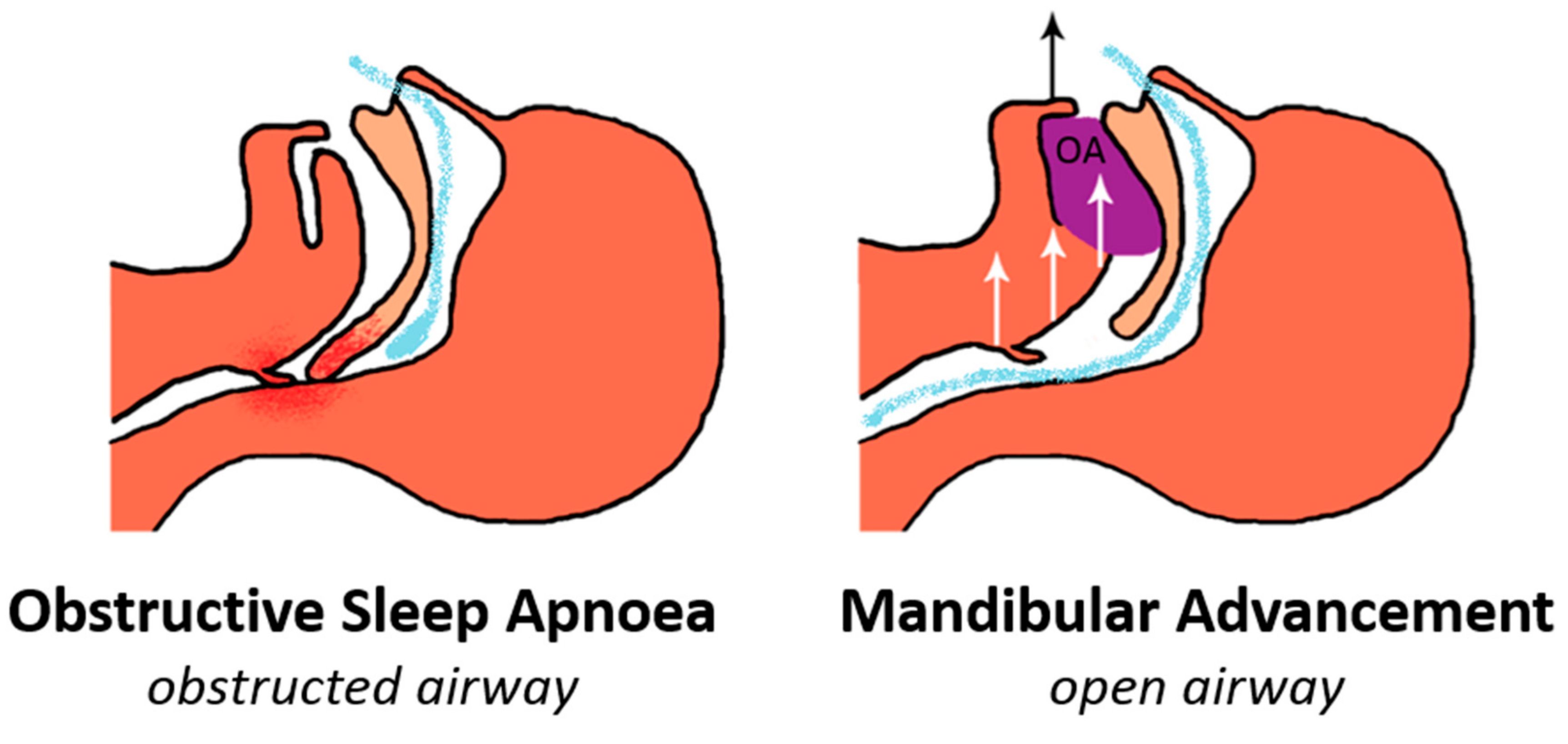
2. Mechanism of Action

OAs can be broadly classified as devices that protrude the tongue (tongue-retaining devices, TRD) and devices that advance the lower jaw (commonly termed mandibular advancement splints or devices, MAS or MAD). Although there is evidence for TRD use in the treatment of OSA [10], and this may be the only option in some edentulous patients, clinical guidelines specifically relate to mandibular advancement devices [9]. It is these devices which are discussed in this review.
Protrusion of the lower jaw is the essential therapeutic mechanism of action [11] (Figure 1), which has been shown to enlarge the airway space, as visualised by MRI and nasopharyngoscopy [12,13]. The most prominent feature is a widening of the lateral airway diameter, particularly in the velopharyngeal region (behind the soft palate) [12,13], likely through soft tissue connections between the lateral airway wall muscles and the mandible [14]. The tongue shows evidence of anterior repositioning with the mandibular advancement, although to varying degrees in different individuals [14]. In addition, the mandibular advancement decreases the upper airway collapsibility, variously demonstrated using pharyngeal critical pressure (Pcrit) [15,16] and pharyngeal closing pressure (Pclose) methods [17], optimal positive airway pressure (PAP) pressure (a surrogate measure of Pcrit) [18] and reduction in spontaneous collapse during sedation [19]. This stabilisation of pharyngeal airway is likely largely attributable to the anatomical enlargement of the airway and the anterior movement of the tongue [13,14].
Whether there are additional direct effects on pharyngeal dilator muscle activity is less clear. The activity of the major pharyngeal dilator, the genioglossus, in response to mandibular advancement has only been assessed in limited studies. Using intra-oral surface or intra-muscular fine-needle electrodes, an increase in genioglossus activity with mandibular advancement has been observed during wakefulness [20,21]. An increase in muscle activity could contribute to the effectiveness of the device. In a single case, intra-oral surface electrodes showed a decrease in both tonic and phasic genioglossus activity with progressive mandibular advancement during sleep [22], suggesting the muscles could switch off in response to the more favourable anatomy created by the advancement. In a slightly larger study of 12 participants during sleep, intramuscular fine-needle electrodes detected no overall effect of progressive mandibular advancement on genioglossus muscle responsiveness to induced apnoea or no effectiveness in restoring flow [15]. This would support a purely passive anatomical mechanism of action. However, this experimental condition required participants to simultaneously be on therapeutic PAP to maintain airflow, and the muscles could therefore be in a passive state. It is yet unclear if this reflects the behaviour of the genioglossus response to mandibular advancement in the natural sleep state. However, it is evident that OA reduces pharyngeal collapsibility and improves airway geometry.

3. Efficacy of Oral Appliance Therapy

OA improves indices of OSA as measured by polysomnography. Respiratory event indices as well as oxygen saturation, arousal index and sleep efficiency have demonstrated improvements compared to their baseline values in a sleep study [23]. The clinical metric to categorise OSA presence and severity is the apnoea–hypopnea index (AHI). It has long been recognised that the average AHI reduction induced by OA therapy use is less than that seen when PAP is applied, with residual OSA often present (AHI > 5 events/hour) [24]. Unlike PAP, which will lead to AHI normalisation if applied at adequate pressure, OA use is results in a large inter-individual variability of the AHI [23]. An example of efficacy variation from a sample of 425 OSA patients treated with a customized OA is shown in Table 1. The definition of an OA “responder” varies in the literature, from a complete response (AHI < 5, or no OSA) to a minimum 50% AHI reduction from baseline (often termed a ‘partial response’) and definitions related to values between these two extremes (e.g., AHI <10 plus 50% reduction from baseline). These three definitions are most commonly presented, but additional definitions also exist. On average, AHI reduction across the range of OSA severity is around 50%, but with a wide variation [23]. Approximately two-thirds of OA-treated patients will achieve greater than 50% reduction in AHI, with at least one-third achieving a complete response [23]. Therefore, many OSA patients will receive a highly efficacious OSA therapy with an OA alone. However, uncertainty around the amount of AHI reduction an individual will achieve has been a barrier to the implementation of this mode of therapy, and OA is still commonly reserved for second-line therapy, particularly in more severe OSA.

4. Factors Related to Oral Appliance Efficacy and Prediction Methods

The uncertainty around the level of AHI reduction achieved following OA therapy has remained a clinical barrier to the implementation of OAs in routine clinical practice. A clinical prediction method has been a focus of much research, but this ‘holy grail’ remains elusive. A complexity in comparing prediction studies is the variation in treatment response definitions. Many studies have started to present the three commonly used definitions of treatment response (described above) to improve generalisability, but others use a single definition. Understanding individual variations in efficacy has been the focus of much research [25]. A summary of prediction methods and their performance is given in Table 2. Group clinical characteristics such as less obesity, lower age, and female gender are associated with treatment success [23,26,27]. A higher baseline AHI is a feature of non-responders at the group level, but nearly a quarter of severe OSA patients will achieve a complete response (AHI < 5) [23]. While clinical and polysomnographic information reveals characteristic patterns for treatment responders and non-responders, these are not adequate predictors to rule someone in or out for therapy [23,28,29]. Therefore, there is a need for additional tests or information above routine clinical information.
Craniofacial structure, predominately assessed by lateral cephalometry, is associated with OA treatment response. However, there is very little consistency between studies of craniofacial features, which vary in sample populations, treatment response definitions and craniofacial measurements assessed [29,30]. Cephalometric investigations include skeletal and dental measurements, upper airway soft tissues and airway space. Even features most commonly reported as beneficial, such as hyoid bone position, cranial base angle, soft palate length, retruded mandible, have no association in other studies [29,30]. There is some suggestion that the amount of soft tissue relative to craniofacial bony enclosure (‘anatomical balance’ [44]) relates to treatment response [45,46], but this requires further exploration. Oropharyngeal crowding, assessed by the Mallampati score, particularly when combined with obesity, has been identified as a negative predictor [31]. A posteriorly located tongue observed during natural sleep by nasendoscopy has also been noted as an anatomical marker of favourable response [16].
Some physiological features have also been associated with greater OA efficacy. A more collapsible pharyngeal airway at baseline is a negative indicator [42]. Optimal PAP pressure, a surrogate for pharyngeal collapsibility [47], has predictive value, with higher pressure requirement associated with greater likelihood of being a non-responder [33,34,35,48]. However, the cut-off pressure values for identifying non-responders differ somewhat between studies, as does its predictive value, with or without other clinical variables. This could reflect differences in pressure titration methods, with ethnic and gender differences in the studied populations likely being an important factor.
The site of pharyngeal collapse, oropharyngeal rather than velopharyngeal collapse, is predictive of a good treatment response [39]. Different methods for estimating the site of collapse have been assessed as prediction methods. Nasendoscopy to observe pharyngeal behaviour during induced sleep (drug-induced sleep endoscopy, DISE) has shown collapse behind the palate to be a positive indicator of OA treatment response, while hypopharyneal collapse tends to be a negative indicator [19]. An awake assessment of the site of collapse by measuring velopharyngeal and oropharyngeal pressure via a pharyngeal catheter (under condition of phrenic nerve stimulation to simulate pharyngeal obstruction during sleep) found oropharyngeal collapse to be associated with complete response [49]. Flow-volume loop metrics were explored as a surrogate measure of site of airway collapse and showed predictive utility for OA outcome in a derivation sample [36]. However, this was not able to be validated in a subsequent sample [37], illustrating the importance of the validation of prediction models derived from small clinical samples.
The technique of observing changes in the pharyngeal airway with mandibular advancement via nasendoscopy has also been applied during wakefulness [12], a simpler assessment than DISE in some clinical environments. A qualitative assessment of pharyngeal changes with mandibular advancement and performance of the Mueller manoeuvre on a scoring chart found inadequate discrimination between responders and non-responders for prediction [50]. A Japanese prediction study used voluntary mandibular protrusion to measure the pharyngeal cross-sectional area (CSA) at different levels using an image analysis software [38]. The CSA during mandibular advancement was compared to the CSA with centric occlusion (CSA expansion ratio). This ratio in the velopharyngeal region, in combination with baseline AHI, had predictive utility across the three response definitions, with accuracy varying across prediction models (area under the receiver operating characteristic (ROC) curve ranging from 0.74 to 0.87).
In recent years, there has been interest in non-anatomical contributors to pharyngeal obstruction [51]. One of these traits, the stability of the ventilatory system, is quantified using a value termed ‘loop gain’. A lower loop gain, or more stable ventilatory control, has been associated with treatment response in long-term user of the treatment, alone and in combination with less collapsibility [42]. Recent work has estimated pathophysiological traits, or endotypes, both pharyngeal (collapsibility and muscle compensation) and non-pharyngeal (loop gain, arousal threshold, ventilator response to arousal) traits, using algorithms applied to clinical polysomnography. This study found a complement of traits that showed some relationship to OA treatment response, including lower loop gain, higher arousal threshold, moderate pharyngeal collapsibility and weaker muscle compensation. However, its predictive utility was not above that of other published models, and there remains a need for prospective validation before any clinical utility can be confirmed.
Most prediction models add data from a single type of assessment to clinical information, which may not capture the complexity of OA action in preventing sleep-disordered breathing. A multimodal phenotyping approach was assessed as a way to improve prediction by combing assessments of site of collapse surrogates (flow-volume loop), craniofacial structure, and nasendoscopy investigation [52]. The prediction modelling was structured using clinical information (age, obesity, AHI) as readily available variables and subsequently assessed the benefit of adding the results from three phenotypic tests. Although multimodal phenotyping improved the prediction model, the magnitude of the improvement may not justify the additional effort and cost. These models were not tested beyond the original derivation sample, but accuracy ranged from fair to excellent, depending on which response definition was used.
A question often asked is whether an inexpensive, ‘off-the-shelf’ or non-customised appliance can be used to indicate treatment outcome with a customised device. No studies have been specifically designed to assess a prefabricated non-customised appliance as a prediction tool. However, two studies have compared a customised and a non-customised appliance in a randomised cross-over design [53,54]. In one study, 63% of patients who did not receive any benefit using the non-customised device were treatment responders when using the customised device [53]; in another study, 44% of patients who did not respond to a non-customised device were treatment responders to customised ones [54]. This suggests that the results from a night study using a non-customised device are not comparable to those of a study with a customised device. Another a sleep test method is based on the use of a remotely controlled mandibular protrusion device enabling the manipulation of the jaw position to eliminate obstructive events in real time during sleep monitoring, additionally determining the amount of advancement required [55,56,57]. There have been two studies using prototype devices [55,56], and two studies using a commercial remotely control mandibular protrusion (RCMP) device [28,40]. The commercial device used in-laboratory polysomnography and a prediction algorithm based on events in supine REM (rapid eye movement) sleep, which is essential to its predictive capability [28,40]. Predictive accuracy was shown to be relatively stable by two laboratories, but higher test failure rates and less capacity to identify a sub-maximal mandibular protrusion level were found in a validation study by an independent sleep laboratory [40]. A home-testing approach has also been assessed, using a feedback-controlled mandibular positioner for two nights [58].
To date, there are no robust and widely validated prediction methods [25]. Other issues to keep in mind in regard to the achievement of a universal prediction model for OA therapy, is that the outcome is based on the total AHI on a single night. We know AHI varies in polysomnography scoring, from night to night and depending on sleep stage and body position [59,60]. Additionally, its efficacy varies over time (e.g., with weight gain) [61,62]. Therefore, prediction of efficacy, based on an initial, single, sleep study night, is not a robustly accomplished, and this has likely contributed to the limited success of the prediction models. An additional complication is that we do not know the level of efficacy that is needed to attain the desired health outcomes, and treatment adherence has an important influence on treatment effectiveness.

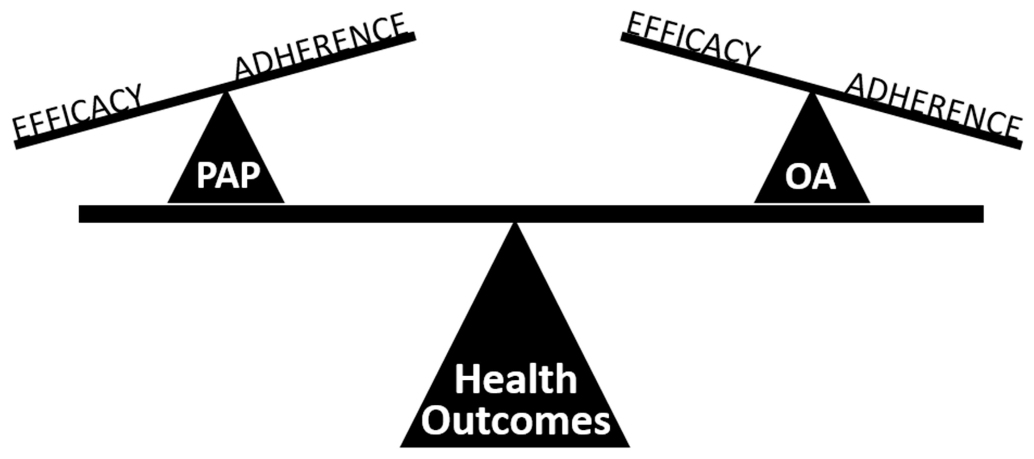
5. Treatment Effectiveness of Oral Appliance Therapy

It is increasingly recognised that AHI changes have limitations in terms of indicating the health benefits of OSA therapy [63]. Ultimately, the goal of OSA therapy is to improve symptoms, and sleep quality and reduce health risks. Efficacy (assessed by the AHI on a single treatment night) reflects the outcome of a treatment administered under ideal circumstances and is only one component of treatment effectiveness. How OSA treatment is administered in the real world has impact on health outcomes. In the case of PAP, hours of nightly usage have been shown to be important in evoking positive health changes in terms of sleepiness, daily functioning, blood pressure and cardiovascular disease [7,8,64].
There is growing evidence that despite residual AHI with OA therapy versus PAP, inferior health outcomes do not necessarily result. For example, CPAP has a well-documented small but significant effect in reducing blood pressure (in the order of 2 mm Hg, greater in resistant hypertension) [65,66]. A recent network meta-analysis has confirmed that both PAP and OA therapies reduce short-term blood pressure to the same extent versus respective sham therapies, with no difference in blood pressure reduction between therapies [7]. A short-term randomised controlled trial of one month of optimised OA and PAP therapies assessed a range of health outcomes (cardiovascular symptoms, quality of life, driving simulator performance) after each therapy period in a sleep clinic population enriched in severe OSA cases [67]. OA therapy was found to be non-inferior to PAP across all outcomes and even superior to it for some domains of health-related quality of life. As predicted, AHI with PAP treatment was reduced to normal levels, while mild residual OSA was evident with OA therapy [67]. However, OA therapy was reported to be used longer than PAP therapy during the trial (by >1 h night), and more participants chose to continue using OA therapy at the end of the trial.
The potential explanation for equivalent health outcomes is that a highly efficacious therapy with moderate usage (PAP) results in the same effectiveness as a moderately efficacious therapy with higher usage (OA) [68] (Figure 2). Therefore, adherence is a key component of treatment effectiveness, which is not captured by traditional metrics of treatment efficacy (AHI). Metrics which incorporate time on and off treatment to describe treatment effectiveness have been proposed and evaluate ‘mean disease alleviation’ or ‘sleep-adjusted residual AHI’ [68,69,70,71]. However, formulas which aim to adjust for adherence over longer treatments are reliant on some assumptions, particularly, that AHI reverts to diagnostic polysomnography levels when therapy is removed. This is not true at least for PAP, for which it has been demonstrated that AHI does increase in partial users when PAP treatment is removed in the night although AHI may not revert to baseline levels, AHI values can still correspond to severe OSA [72]. An in-laboratory study of the objective measurement of AHI in partial PAP users has shown that effectiveness measures could be collected using simultaneous measurements with a portable monitor [72]. New wearable technologies for monitoring of sleep-disordered breathing, sleep time, and treatment compliance, may in the future allow objective treatment effectiveness measures to be explored in relation to health outcomes.

6. Cardiovascular Health

OSA is an independent risk factor for hypertension and cardio- and cerebro-vascular diseases including coronary heart disease, heart failure and stroke [1,73,74,75]. However, demonstrating a beneficial effect of OSA treatment on cardiovascular endpoints has proved elusive. The largest randomised study of PAP therapy (the Sleep Apnoea Cardiovascular Endpoints, or SAVE, study), with an average of 3.7 years follow-up, showed no benefit of PAP in secondary prevention of composite cardiovascular events [76]. One of the potential confounders to this trial was that average PAP usage was only 3.3 h/night in this cardiovascular disease population. A recent meta-analysis of patients using PAP for more than 4 h per night, found a positive effect on the occurrence of a composite outcome including stroke, cerebrovascular disease, cardiac death or acute myocardial infarction [64]. This suggests OSA treatment adherence is key in cardiovascular prevention. It is unknown whether OA therapy may be a more acceptable therapy in cardiovascular clinical populations and therefore may have a potential role in cardiovascular disease prevention.
A systematic review [77] has summarised the cardiovascular effects of OA therapy. As discussed above, blood pressure reduction appears to be similar to that achieved with PAP [7]. A single observational study suggests OA therapy is as effective as PAP in reducing the risk of death related to cardiovascular mortality [78]. In terms of intermediary markers of cardiovascular health (biomarkers, heart rate variability, arterial stiffness, endothelial function), most studies are very small and short-term and have heterogeneous designs, which makes any results about OA effects inconclusive [77]. A more recent randomised controlled trial of 6 months OA treatment vs sham treatment (single plate providing no mandibular advancement) did not find a beneficial effect on microvascular endothelial function assessed by the reactive hyperaemia index [79]. However, the study population was relatively healthy from a vascular point of view at baseline and free of cardiovascular disease. Therefore, studies of populations with cardiovascular risk factors are warranted. More trials of intermediary cardiovascular markers are needed to assess the potential of OA therapy in this field.

7. Devices and Customisation

With more than 150 devices with FDA approval for the treatment of snoring and OSA patients, there is considerable debate about the efficacy of different devices. In reality, only a small number of commercially available devices have been subjected to robust clinical scrutiny, and head-to-head comparison studies are also limited. Customised devices which are titratable (allow adjustment of the mandibular advancement) and implemented by a qualified dentist are suggested in the clinical guidelines [9]. Non-customised devices (prefabricated, ‘off-the-shelf’ devices which mould the teeth when heated) tend to be poorly tolerated and less effective than customised devices [53,54,80,81].
There is a relationship between ‘dose’ of mandibular advancement and effects of sleep-disordered breathing, with further advancement generally leading to better results. However, there is also some evidence that greater advancement may not always provide further benefit. There may be some individuals that have most benefit at a submaximal level of mandibular advancement, for whom further advancement is detrimental [15,18,82]. Avoiding unnecessary advancement may be a way to reduce side effects such as tooth movements and improve treatment acceptance.
The methods used for OA titration vary and include mandibular advancement to patients’ physical limits, monitoring symptom resolution, or using polysomnography or single-channel devices [83,84]. Other proposed titration protocols have used nasendoscopy to observe effects on the pharyngeal area for different levels of mandibular advancement (provided by an occlusal registration tool) [85]. The RCMP device has been used during both polysomnography and DISE to work out the effective therapeutic target protrusion, and was successful for most, but not all, patients in these studies [86].
Another parameter distinguishing different OAs regards the vertical dimension, i.e., the amount of mouth opening permitted by the device. Vertical opening without mandibular advancement has been shown to have a negative effect on AHI [87] and pharyngeal dimensions in some patients [88]. In the context of mandibular advancement, a cross-over study examining the effect of 4 mm vs. 14 mm of vertical mouth opening showed no effect of vertical height on AHI improvement [89]. However, patients preferred the reduced opening, suggesting that vertical dimension may impact patient acceptance and adherence. Permissible mouth movement is another design difference for OAs. Some two-piece appliances allow the lower jaw to move, while one-piece (monobloc) appliances or some two-pieces with an anterior titration apparatus, for example, do not permit vertical jaw movement. There is some suggestion that devices which allow mouth opening may be detrimental for OSA patients in the supine position. Controlling the amount of permissible mouth opening, such as with elastic bands, may be important in some patients, particularly those with supine-predominant OSA [90,91].
In recent years, devices are emerging which rely on computer-aided design or computer-aided manufacturing technology. Instead of the traditional impressions, digital intraoral scans can now be used to design appliances, and digital manufacturing techniques are increasingly being used. These can streamline the process and result in more rapid access to treatment and better fit of appliances, which could improve acceptance and efficacy [92].

8. Long-term Side Effects

There are initial side effects when starting an OA therapy, such as dry mouth, excessive salivation or soft tissue irritation, but these are generally temporary and resolve [93]. The forces on the teeth resulting from the action of the OA in keeping the lower jaw forward can result in tooth movements and bite changes. Some bite changes are actually favourable and, in any case, often not noticed by the patient and considered subclinical [94]. There have now been studies that track the effects of oral appliance wear on bite changes in twenty years of use [95,96,97]. These studies show that tooth movements are continuous and relate to the duration and frequency of OA use and to the mandibular advancement level [95,96]. The extent of tooth movements has some relationship to the initial bite or craniofacial structures [95,98]. Material and design may also influence the side effects. A study comparing OA devices constructed of flexible material without incisor coverage to rigid appliances with full occlusal coverage, found greater short-term tooth movement resulting in irregularity in the lower teeth in patients using the former [99]. Guidelines recommend that patients have regular follow-ups with qualified dentists to survey dental side effects or occlusal changes [9].

9. Objective Compliance

The observed similarity in health outcomes between OA and PAP therapies, despite the greater PAP efficacy, could be explained by a greater adherence to OA treatment. However, this has largely been based on self-reported adherence data for OA. Objective compliance monitors have become available for OA, but the literature about this is limited to date. Compliance monitors are small temperature-sensing data chips which can be embedded in the OA material. Three commercial brands have been independently tested and found to be reliable for determining usage time [100]. In a three-month study with covert objective compliance monitoring, subjective (daily diary) and objective compliance (temperature microsensor) did not differ [69]. The average nightly usage across the initial three months of OA therapy was 6.7 ± 1.3 h/day, with usage on >90% of days. At one-year follow-up, the mean usage was 6.4 ± 1.7 h/night, with 83% of patients using OA for more than 4 h on more than 70% of days, with a 9.8% discontinuation rate [101]. A randomised trial of two months of OA therapy versus sham therapy in severe OSA patients found OA use for 6.6 ± 1.4 h/night, with 96.1% compliant users, compared to sham device use for 5.6 ± 2.3 h/night, with 73.6% compliant users [79]. In this study, OA therapy was used for around 90% of the reported sleep duration, but the lack of objective sleep measurements is a limitation in assessing OA therapy usage. Although initial reports of objective compliance indicated good average usage, there are likely to be different subtypes of users, which requires further investigation and longer-term data.
Objective compliance data in the short term has some relationship to self-report snoring improvement and the presence and severity of side effects [102,103]. Interestingly, AHI improvement has not been shown to be related to short-term compliance in these small studies, neither have anthropometric data or sleepiness [103].

10. Combination Therapy Strategies

With increasing recognition of OSA as a heterogenous disorder and the possibility of tailoring treatments to the individuals on the basis of their pathophysiology and preferences, the field has opened up to combination therapy possibilities to address efficacy and adherence deficiencies and optimise outcomes [104]. OA and PAP have been proposed to be combined in different ways. Using both treatments simultaneously can reduce the PAP pressure requirement [105,106,107] and have an additive effect on AHI in those who are PAP-intolerant [107]. Alternatively, the treatments can be used interchangeably, according to patient preference and circumstance (e.g., travel) [108]. The combination of OA and positional therapy resulted more beneficial than either therapy alone for supine OSA patients [109]. A study of uvulopalatopharyngoplasty (UPPP) surgery for OSA treatment with or without OA therapy found greater long-term control of OSA with the combination approach compared to surgery alone [110]. Other more contemporary surgical approaches for OSA have not been assessed. However, since the two approaches of upper airway surgery and OA therapy are often associated with residual AHI, a combination therapy could be beneficial. Weight loss improves OSA and, when combined with PAP therapy, leads to greater cardiometabolic benefits than weight loss or PAP alone [111]. Obesity and weight gain are known to decrease OA efficacy [23,61], thus, combined weight loss and OA therapy could be beneficial. Recently, the combination of OA therapy and expiratory positive airway pressure (EPAP) valves has been shown to reduce AHI versus either treatment alone, albeit at the cost of decreasing sleep efficiency [112]. More studies on combination therapy approaches are needed.

11. Models of Care

With increasing recognition that patients with OSA are phenotypically diverse and have individual needs, preferences and values, the field is undergoing a shift in OSA management, from a focus on diagnosis to a focus on chronic care management. This mandates that treatment decisions are informed by individual patients’ values and preferences in order to optimize patients’ outcomes [113]. Since sleep medicine is highly multidisciplinary, there is a need to develop efficient and cost-effective care models, centred around the patient. In the case of OA therapy, an effective working relationship between the sleep physician and the dentist is required, founded on an understanding of their respective roles in the care process. Sleep physicians currently have the primary responsibility for diagnosis and discussion of treatment options relevant to each individual patient as well as for assessment of treatment outcomes. Dentists have the responsibility for delivering the OA therapy, including assessment of dental suitability, impressions, appliance selection and fitting, titration and side effects monitoring. Ideally, the dentists should be credentialed in dental sleep medicine. Furthermore, since dentists routinely see patients for dental health check-ups, they may play a useful role in raising patient awareness of the condition and screening modalities. As practice in the field evolves, we can expect to see interdisciplinary models of care in which physicians and dentists may work within the same clinic, alongside other disciplinary experts (e.g., surgeons, psychologists). With adequate training and credentialing, it is possible that the scope of practice for dentists and other practitioners will expand in the future to help address the enormous burden of OSA.

12. Toward a Precision Medicine Approach

Precision medicine is an emerging approach to healthcare that aims to identify the appropriate intervention for a specific patient, based on the phenotypic characteristics of the individuals and their disease. There is increasing recognition that OSA is a highly heterogeneous disorder in terms of the pathways to the disease, the expression of the disease, and the response to treatment [114]. Individual differences include variability in genome, environment, and lifestyle of each person [63]. Precision sleep medicine represents a shift away from an outdated one-size-fits-all approach of treatment toward a tailored approach that acknowledges alternative methods of diagnosis and management [43]. Personalization of treatment is particularly important in OSA, since it is a chronic condition requiring long-term therapy. While it is important that the therapy effectively controls the disorder, the importance of patient acceptance for adherence and effectiveness must be emphasized. OA therapy is the leading alternative treatment to PAP and provides an important role for dentists in the management of OSA in collaboration with medical practitioners. The uncertainty around OA treatment efficacy has been a barrier to its prescription as first-line therapy. This has spurred a major research effort aimed at identifying predictors of treatment response, thereby promoting the ideal of personalized medicine in OA therapy.

13. Conclusions

Oral appliances are the main alternative to PAP for the treatment of OSA. There has been an expansion of the research evidence supporting the use of oral appliances in clinical practice, and key professional bodies now recommend oral appliances for use in patients with OSA who are intolerant to PAP therapy or prefer an alternative therapy. Patient preference is a key component of patient-centred care and has a pivotal role in treatment acceptance, usage, and health outcomes. OA therapy requires a multidisciplinary approach, involving a sleep physician and a dental practitioner with expertise in the management of sleep disorders, in order to achieve optimal outcomes for the patients. It is time for the sleep field to work collaboratively to address the barriers to the effective implementation of this evidence-based treatment into routine clinical practice.

Author Contributions

K.S. and P.A.C. drafted and reviewed the manuscript.

Funding

No external funding was received for this review.

Conflicts of Interest

PAC has an appointment to an endowed academic Chair at the University of Sydney that was established from ResMed funding. He has received research support from ResMed, SomnoMed, Zephyr Sleep Technologies, and Bayer. He is a consultant to Zephyr Sleep Technologies and ResMed Narval. He has a pecuniary interest in SomnoMed related to a previous role in R&D (2004). KS has no conflict of interest to disclose.

References

  1. Gottlieb, D.J.; Yenokyan, G.; Newman, A.B.; O’Connor, G.T.; Punjabi, N.M.; Quan, S.F.; Redline, S.; Resnick, H.E.; Tong, E.K.; Diener-West, M.; et al. Prospective study of obstructive sleep apnea and incident coronary heart disease and heart failure: The sleep heart health study. Circulation 2010, 122, 352–360. [Google Scholar] [CrossRef] [PubMed]
  2. Heinzer, R.; Vat, S.; Marques-Vidal, P.; Marti-Soler, H.; Andries, D.; Tobback, N.; Mooser, V.; Preisig, M.; Malhotra, A.; Waeber, G.; et al. Prevalence of sleep-disordered breathing in the general population: The HypnoLaus study. Lancet Respir. Med. 2015, 3, 310–318. [Google Scholar] [CrossRef]
  3. Marshall, N.S.; Wong, K.K.; Phillips, C.L.; Liu, P.Y.; Knuiman, M.W.; Grunstein, R.R. Is sleep apnea an independent risk factor for prevalent and incident diabetes in the Busselton Health Study? J. Clin. Sleep Med. 2009, 5, 15–20. [Google Scholar] [PubMed]
  4. Sassani, A.; Findley, L.J.; Kryger, M.; Goldlust, E.; George, C.; Davidson, T.M. Reducing motor-vehicle collisions, costs, and fatalities by treating obstructive sleep apnea syndrome. Sleep 2004, 27, 453–458. [Google Scholar] [CrossRef]
  5. Fu, Y.; Xia, Y.; Yi, H.; Xu, H.; Guan, J.; Yin, S. Meta-analysis of all-cause and cardiovascular mortality in obstructive sleep apnea with or without continuous positive airway pressure treatment. Sleep Breath 2017, 21, 181–189. [Google Scholar] [CrossRef]
  6. Benjafield, A.V.; Ayas, N.T.; Eastwood, P.R.; Heinzer, R.; Ip, M.S.M.; Morrell, M.J.; Nunez, C.M.; Patel, S.R.; Penzel, T.; Pepin, J.L.; et al. Estimation of the global prevalence and burden of obstructive sleep apnoea: A literature-based analysis. Lancet Respir. Med. 2019, 7, 687–698. [Google Scholar] [CrossRef]
  7. Bratton, D.J.; Gaisl, T.; Wons, A.M.; Kohler, M. CPAP vs Mandibular Advancement Devices and Blood Pressure in Patients With Obstructive Sleep Apnea: A Systematic Review and Meta-analysis. JAMA 2015, 314, 2280–2293. [Google Scholar] [CrossRef]
  8. Weaver, T.E.; Maislin, G.; Dinges, D.F.; Bloxham, T.; George, C.F.; Greenberg, H.; Kader, G.; Mahowald, M.; Younger, J.; Pack, A.I. Relationship between hours of CPAP use and achieving normal levels of sleepiness and daily functioning. Sleep 2007, 30, 711–719. [Google Scholar] [CrossRef]
  9. Ramar, K.; Dort, L.C.; Katz, S.G.; Lettieri, C.J.; Harrod, C.G.; Thomas, S.M.; Chervin, R.D. Clinical Practice Guideline for the Treatment of Obstructive Sleep Apnea and Snoring with Oral Appliance Therapy: An Update for 2015. J. Clin. Sleep Med. 2015, 11, 773–827. [Google Scholar] [CrossRef]
  10. Chang, E.T.; Fernandez-Salvador, C.; Giambo, J.; Nesbitt, B.; Liu, S.Y.; Capasso, R.; Kushida, C.A.; Camacho, M. Tongue retaining devices for obstructive sleep apnea: A systematic review and meta-analysis. Am. J. Otolaryngol. 2017, 38, 272–278. [Google Scholar] [CrossRef]
  11. Mehta, A.; Qian, J.; Petocz, P.; Darendeliler, M.A.; Cistulli, P.A. A randomized, controlled study of a mandibular advancement splint for obstructive sleep apnea. Am. J. Respir. Crit. Care Med. 2001, 163, 1457–1461. [Google Scholar] [CrossRef] [PubMed]
  12. Chan, A.S.; Lee, R.W.; Srinivasan, V.K.; Darendeliler, M.A.; Grunstein, R.R.; Cistulli, P.A. Nasopharyngoscopic evaluation of oral appliance therapy for obstructive sleep apnoea. Eur. Respir. J. 2010, 35, 836–842. [Google Scholar] [CrossRef] [PubMed]
  13. Chan, A.S.; Sutherland, K.; Schwab, R.J.; Zeng, B.; Petocz, P.; Lee, R.W.; Darendeliler, M.A.; Cistulli, P.A. The effect of mandibular advancement on upper airway structure in obstructive sleep apnoea. Thorax 2010, 65, 726–732. [Google Scholar] [CrossRef] [PubMed]
  14. Brown, E.C.; Cheng, S.; McKenzie, D.K.; Butler, J.E.; Gandevia, S.C.; Bilston, L.E. Tongue and lateral upper airway movement with mandibular advancement. Sleep 2013, 36, 397–404. [Google Scholar] [CrossRef]
  15. Bamagoos, A.A.; Cistulli, P.A.; Sutherland, K.; Ngiam, J.; Burke, P.G.R.; Bilston, L.E.; Butler, J.E.; Eckert, D.J. Dose-dependent effects of mandibular advancement on upper airway collapsibility and muscle function in obstructive sleep apnea. Sleep 2019, 42. [Google Scholar] [CrossRef]
  16. Marques, M.; Genta, P.R.; Azarbarzin, A.; Taranto-Montemurro, L.; Messineo, L.; Hess, L.B.; Demko, G.; White, D.P.; Sands, S.A.; Wellman, A. Structure and severity of pharyngeal obstruction determine oral appliance efficacy in sleep apnoea. J. Physiol. 2019. [Google Scholar] [CrossRef]
  17. Kato, J.; Isono, S.; Tanaka, A.; Watanabe, T.; Araki, D.; Tanzawa, H.; Nishino, T. Dose-dependent effects of mandibular advancement on pharyngeal mechanics and nocturnal oxygenation in patients with sleep-disordered breathing. Chest 2000, 117, 1065–1072. [Google Scholar] [CrossRef]
  18. Bamagoos, A.A.; Eckert, D.J.; Sutherland, K.; Ngiam, J.; Cistulli, P.A. Dose-dependent effects of mandibular advancement on optimal positive airway pressure requirements in obstructive sleep apnoea. Sleep Breath 2019. [Google Scholar] [CrossRef]
  19. Vroegop, A.V.; Vanderveken, O.M.; Dieltjens, M.; Wouters, K.; Saldien, V.; Braem, M.J.; Van de Heyning, P.H. Sleep endoscopy with simulation bite for prediction of oral appliance treatment outcome. J. Sleep Res. 2013, 22, 348–355. [Google Scholar] [CrossRef]
  20. Johal, A.; Gill, G.; Ferman, A.; McLaughlin, K. The effect of mandibular advancement appliances on awake upper airway and masticatory muscle activity in patients with obstructive sleep apnoea. Clin. Physiol. Funct. Imaging 2007, 27, 47–53. [Google Scholar] [CrossRef]
  21. Tsuiki, S.; Ono, T.; Kuroda, T. Mandibular Advancement Modulates Respiratory-Related Genioglossus Electromyographic Activity. Sleep Breath 2000, 4, 53–58. [Google Scholar] [CrossRef] [PubMed]
  22. Almeida, F.R.; Tsuiki, S.; Hattori, Y.; Takei, Y.; Inoue, Y.; Lowe, A.A. Dose-dependent effects of mandibular protrusion on genioglossus activity in sleep apnoea. Eur. Respir. J. 2011, 37, 209–212. [Google Scholar] [CrossRef] [PubMed]
  23. Sutherland, K.; Takaya, H.; Qian, J.; Petocz, P.; Ng, A.T.; Cistulli, P.A. Oral Appliance Treatment Response and Polysomnographic Phenotypes of Obstructive Sleep Apnea. J. Clin. Sleep Med. 2015, 11, 861–868. [Google Scholar] [CrossRef] [PubMed]
  24. Sharples, L.D.; Clutterbuck-James, A.L.; Glover, M.J.; Bennett, M.S.; Chadwick, R.; Pittman, M.A.; Quinnell, T.G. Meta-analysis of randomised controlled trials of oral mandibular advancement devices and continuous positive airway pressure for obstructive sleep apnoea-hypopnoea. Sleep Med. Rev. 2016, 27, 108–124. [Google Scholar] [CrossRef] [PubMed]
  25. Okuno, K.; Pliska, B.T.; Hamoda, M.; Lowe, A.A.; Almeida, F.R. Prediction of oral appliance treatment outcomes in obstructive sleep apnea: A systematic review. Sleep Med. Rev. 2016, 30, 25–33. [Google Scholar] [CrossRef] [PubMed]
  26. Marklund, M.; Stenlund, H.; Franklin, K.A. Mandibular advancement devices in 630 men and women with obstructive sleep apnea and snoring: Tolerability and predictors of treatment success. Chest 2004, 125, 1270–1278. [Google Scholar] [CrossRef] [PubMed]
  27. Vecchierini, M.F.; Attali, V.; Collet, J.M.; d’Ortho, M.P.; Goutorbe, F.; Kerbrat, J.B.; Leger, D.; Lavergne, F.; Monaca, C.; Monteyrol, P.J.; et al. Sex differences in mandibular repositioning device therapy effectiveness in patients with obstructive sleep apnea syndrome. Sleep Breath 2019, 23, 837–848. [Google Scholar] [CrossRef]
  28. Remmers, J.; Charkhandeh, S.; Grosse, J.; Topor, Z.; Brant, R.; Santosham, P.; Bruehlmann, S. Remotely controlled mandibular protrusion during sleep predicts therapeutic success with oral appliances in patients with obstructive sleep apnea. Sleep 2013, 36, 1517–1525. [Google Scholar] [CrossRef]
  29. Denolf, P.L.; Vanderveken, O.M.; Marklund, M.E.; Braem, M.J. The status of cephalometry in the prediction of non-CPAP treatment outcome in obstructive sleep apnea patients. Sleep Med. Rev. 2016, 27, 56–73. [Google Scholar] [CrossRef]
  30. Alessandri-Bonetti, G.; Ippolito, D.R.; Bartolucci, M.L.; D’Anto, V.; Incerti-Parenti, S. Cephalometric predictors of treatment outcome with mandibular advancement devices in adult patients with obstructive sleep apnea: A systematic review. Korean J. Orthod. 2015, 45, 308–321. [Google Scholar] [CrossRef]
  31. Tsuiki, S.; Ito, E.; Isono, S.; Ryan, C.F.; Komada, Y.; Matsuura, M.; Inoue, Y. Oropharyngeal crowding and obesity as predictors of oral appliance treatment response to moderate obstructive sleep apnea. Chest 2013, 144, 558–563. [Google Scholar] [CrossRef] [PubMed]
  32. Ng, A.T.; Darendeliler, M.A.; Petocz, P.; Cistulli, P.A. Cephalometry and prediction of oral appliance treatment outcome. Sleep Breath 2012, 16, 47–58. [Google Scholar] [CrossRef] [PubMed]
  33. Storesund, A.; Johansson, A.; Bjorvatn, B.; Lehmann, S. Oral appliance treatment outcome can be predicted by continuous positive airway pressure in moderate to severe obstructive sleep apnea. Sleep Breath 2018, 22, 385–392. [Google Scholar] [CrossRef] [PubMed] [Green Version]
  34. Dort, L.C.; Savard, N.; Dort, E.; Dort, J. Does CPAP pressure predict treatment outcome with oral appliances? J. Dent. Sleep Med. 2016, 3, 113–117. [Google Scholar] [CrossRef]
  35. Sutherland, K.; Phillips, C.L.; Davies, A.; Srinivasan, V.K.; Dalci, O.; Yee, B.J.; Darendeliler, M.A.; Grunstein, R.R.; Cistulli, P.A. CPAP pressure for prediction of oral appliance treatment response in obstructive sleep apnea. J. Clin. Sleep Med. 2014, 10, 943–949. [Google Scholar] [CrossRef] [PubMed]
  36. Zeng, B.; Ng, A.T.; Darendeliler, M.A.; Petocz, P.; Cistulli, P.A. Use of flow-volume curves to predict oral appliance treatment outcome in obstructive sleep apnea. Am. J. Respir. Crit. Care Med. 2007, 175, 726–730. [Google Scholar] [CrossRef] [PubMed] [Green Version]
  37. Chan, A.S.; Lee, R.W.; Srinivasan, V.K.; Darendeliler, M.A.; Cistulli, P.A. Use of flow-volume curves to predict oral appliance treatment outcome in obstructive sleep apnea: A prospective validation study. Sleep Breath 2011, 15, 157–162. [Google Scholar] [CrossRef]
  38. Okuno, K.; Sasao, Y.; Nohara, K.; Sakai, T.; Pliska, B.T.; Lowe, A.A.; Ryan, C.F.; Almeida, F.R. Endoscopy evaluation to predict oral appliance outcomes in obstructive sleep apnoea. Eur. Respir. J. 2016, 47, 1410–1419. [Google Scholar] [CrossRef]
  39. Ng, A.T.; Qian, J.; Cistulli, P.A. Oropharyngeal collapse predicts treatment response with oral appliance therapy in obstructive sleep apnea. Sleep 2006, 29, 666–671. [Google Scholar]
  40. Sutherland, K.; Ngiam, J.; Cistulli, P.A. Performance of Remotely Controlled Mandibular Protrusion Sleep Studies for Prediction of Oral Appliance Treatment Response. J. Clin. Sleep Med. 2017, 13, 411–417. [Google Scholar] [CrossRef] [Green Version]
  41. Tsai, W.H.; Vazquez, J.C.; Oshima, T.; Dort, L.; Roycroft, B.; Lowe, A.A.; Hajduk, E.; Remmers, J.E. Remotely controlled mandibular positioner predicts efficacy of oral appliances in sleep apnea. Am. J. Respir. Crit. Care Med. 2004, 170, 366–370. [Google Scholar] [CrossRef] [PubMed]
  42. Edwards, B.A.; Andara, C.; Landry, S.; Sands, S.A.; Joosten, S.A.; Owens, R.L.; White, D.P.; Hamilton, G.S.; Wellman, A. Upper-Airway Collapsibility and Loop Gain Predict the Response to Oral Appliance Therapy in Patients with Obstructive Sleep Apnea. Am. J. Respir. Crit. Care Med. 2016, 194, 1413–1422. [Google Scholar] [CrossRef] [PubMed] [Green Version]
  43. Sutherland, K.; Almeida, F.R.; de Chazal, P.; Cistulli, P.A. Prediction in obstructive sleep apnoea: Diagnosis, comorbidity risk, and treatment outcomes. Expert Rev. Respir. Med. 2018, 12, 293–307. [Google Scholar] [CrossRef] [PubMed]
  44. Tsuiki, S.; Isono, S.; Ishikawa, T.; Yamashiro, Y.; Tatsumi, K.; Nishino, T. Anatomical balance of the upper airway and obstructive sleep apnea. Anesthesiology 2008, 108, 1009–1015. [Google Scholar] [CrossRef] [Green Version]
  45. Mostafiz, W.; Dalci, O.; Sutherland, K.; Malhotra, A.; Srinivasan, V.; Darendeliler, M.A.; Cistulli, P.A. Influence of oral and craniofacial dimensions on mandibular advancement splint treatment outcome in patients with obstructive sleep apnea. Chest 2011, 139, 1331–1339. [Google Scholar] [CrossRef]
  46. Sutherland, K.; Chan, A.S.; Cistulli, P.A. Three-dimensional assessment of anatomical balance and oral appliance treatment outcome in obstructive sleep apnoea. Sleep Breath 2016, 20, 903–910. [Google Scholar] [CrossRef]
  47. Landry, S.A.; Joosten, S.A.; Eckert, D.J.; Jordan, A.S.; Sands, S.A.; White, D.P.; Malhotra, A.; Wellman, A.; Hamilton, G.S.; Edwards, B.A. Therapeutic CPAP Level Predicts Upper Airway Collapsibility in Patients With Obstructive Sleep Apnea. Sleep 2017, 40. [Google Scholar] [CrossRef]
  48. Tsuiki, S.; Kobayashi, M.; Namba, K.; Oka, Y.; Komada, Y.; Kagimura, T.; Inoue, Y. Optimal positive airway pressure predicts oral appliance response to sleep apnoea. Eur. Respir. J. 2010, 35, 1098–1105. [Google Scholar] [CrossRef] [Green Version]
  49. Bosshard, V.; Masse, J.F.; Series, F. Prediction of oral appliance efficiency in patients with apnoea using phrenic nerve stimulation while awake. Thorax 2011, 66, 220–225. [Google Scholar] [CrossRef] [Green Version]
  50. Sutherland, K.; Chan, A.S.L.; Ngiam, J.; Darendeliler, M.A.; Cistulli, P.A. Qualitative assessment of awake nasopharyngoscopy for prediction of oral appliance treatment response in obstructive sleep apnoea. Sleep Breath 2018, 22, 1029–1036. [Google Scholar] [CrossRef]
  51. Eckert, D.J. Phenotypic approaches to obstructive sleep apnoea—New pathways for targeted therapy. Sleep Med. Rev. 2018, 37, 45–59. [Google Scholar] [CrossRef] [PubMed]
  52. Sutherland, K.; Chan, A.S.L.; Ngiam, J.; Dalci, O.; Darendeliler, M.A.; Cistulli, P.A. Awake Multimodal Phenotyping for Prediction of Oral Appliance Treatment Outcome. J. Clin. Sleep Med. 2018, 14, 1879–1887. [Google Scholar] [CrossRef] [PubMed] [Green Version]
  53. Vanderveken, O.M.; Devolder, A.; Marklund, M.; Boudewyns, A.N.; Braem, M.J.; Okkerse, W.; Verbraecken, J.A.; Franklin, K.A.; De Backer, W.A.; Van de Heyning, P.H. Comparison of a custom-made and a thermoplastic oral appliance for the treatment of mild sleep apnea. Am. J. Respir. Crit. Care Med. 2008, 178, 197–202. [Google Scholar] [CrossRef] [PubMed]
  54. Johal, A.; Haria, P.; Manek, S.; Joury, E.; Riha, R. Ready-Made Versus Custom-Made Mandibular Repositioning Devices in Sleep Apnea: A Randomized Clinical Trial. J. Clin. Sleep Med. 2017, 13, 175–182. [Google Scholar] [CrossRef] [Green Version]
  55. Dort, L.C.; Hadjuk, E.; Remmers, J.E. Mandibular advancement and obstructive sleep apnoea: A method for determining effective mandibular protrusion. Eur. Respir. J. 2006, 27, 1003–1009. [Google Scholar] [CrossRef] [Green Version]
  56. Petelle, B.; Vincent, G.; Gagnadoux, F.; Rakotonanahary, D.; Meyer, B.; Fleury, B. One-night mandibular advancement titration for obstructive sleep apnea syndrome: A pilot study. Am. J. Respir. Crit. Care Med. 2002, 165, 1150–1153. [Google Scholar] [CrossRef]
  57. Kastoer, C.; Dieltjens, M.; Oorts, E.; Hamans, E.; Braem, M.J.; Van de Heyning, P.H.; Vanderveken, O.M. The Use of Remotely Controlled Mandibular Positioner as a Predictive Screening Tool for Mandibular Advancement Device Therapy in Patients with Obstructive Sleep Apnea through Single-Night Progressive Titration of the Mandible: A Systematic Review. J. Clin. Sleep Med. 2016, 12, 1411–1421. [Google Scholar] [CrossRef] [Green Version]
  58. Remmers, J.E.; Topor, Z.; Grosse, J.; Vranjes, N.; Mosca, E.V.; Brant, R.; Bruehlmann, S.; Charkhandeh, S.; Zareian Jahromi, S.A. A Feedback-Controlled Mandibular Positioner Identifies Individuals With Sleep Apnea Who Will Respond to Oral Appliance Therapy. J. Clin. Sleep Med. 2017, 13, 871–880. [Google Scholar] [CrossRef] [Green Version]
  59. Alshaer, H.; Ryan, C.; Fernie, G.R.; Bradley, T.D. Reproducibility and predictors of the apnea hypopnea index across multiple nights. Sleep Sci. 2018, 11, 28–33. [Google Scholar] [CrossRef]
  60. Younes, M.; Raneri, J.; Hanly, P. Staging Sleep in Polysomnograms: Analysis of Inter-Scorer Variability. J. Clin. Sleep Med. 2016, 12, 885–894. [Google Scholar] [CrossRef]
  61. Vuorjoki-Ranta, T.R.; Aarab, G.; Lobbezoo, F.; Tuomilehto, H.; Ahlberg, J. Weight gain may affect mandibular advancement device therapy in patients with obstructive sleep apnea: A retrospective study. Sleep Breath 2019, 23, 531–534. [Google Scholar] [CrossRef] [PubMed]
  62. Marklund, M. Long-term efficacy of an oral appliance in early treated patients with obstructive sleep apnea. Sleep Breath 2016, 20, 689–694. [Google Scholar] [CrossRef] [PubMed]
  63. Lim, D.C.; Sutherland, K.; Cistulli, P.A.; Pack, A.I. P4 medicine approach to obstructive sleep apnoea. Respirology 2017, 22, 849–860. [Google Scholar] [CrossRef] [PubMed] [Green Version]
  64. Javaheri, S.; Martinez-Garcia, M.A.; Campos-Rodriguez, F.; Muriel, A.; Peker, Y. CPAP Adherence for Prevention of Major Adverse Cerebrovascular and Cardiovascular Events in Obstructive Sleep Apnea. Am. J. Respir. Crit. Care Med. 2019. [Google Scholar] [CrossRef] [PubMed]
  65. Bakker, J.P.; Edwards, B.A.; Gautam, S.P.; Montesi, S.B.; Duran-Cantolla, J.; Aizpuru, F.; Barbe, F.; Sanchez-de-la-Torre, M.; Malhotra, A. Blood pressure improvement with continuous positive airway pressure is independent of obstructive sleep apnea severity. J. Clin. Sleep Med. 2014, 10, 365–369. [Google Scholar] [CrossRef] [PubMed] [Green Version]
  66. Lei, Q.; Lv, Y.; Li, K.; Ma, L.; Du, G.; Xiang, Y.; Li, X. Effects of continuous positive airway pressure on blood pressure in patients with resistant hypertension and obstructive sleep apnea: A systematic review and meta-analysis of six randomized controlled trials. J. Bras. Pneumol. 2017, 43, 373–379. [Google Scholar] [CrossRef] [PubMed] [Green Version]
  67. Phillips, C.L.; Grunstein, R.R.; Darendeliler, M.A.; Mihailidou, A.S.; Srinivasan, V.K.; Yee, B.J.; Marks, G.B.; Cistulli, P.A. Health outcomes of continuous positive airway pressure versus oral appliance treatment for obstructive sleep apnea: A randomized controlled trial. Am. J. Respir. Crit. Care Med. 2013, 187, 879–887. [Google Scholar] [CrossRef]
  68. Sutherland, K.; Phillips, C.L.; Cistulli, P.A. Efficacy versus Effectiveness in the treatment of Obstructive Sleep Apnea: CPAP and oral appliances. J. Dent. Sleep Med. 2015, 2, 175–181. [Google Scholar] [CrossRef]
  69. Vanderveken, O.M.; Dieltjens, M.; Wouters, K.; De Backer, W.A.; Van de Heyning, P.H.; Braem, M.J. Objective measurement of compliance during oral appliance therapy for sleep-disordered breathing. Thorax 2013, 68, 91–96. [Google Scholar] [CrossRef] [Green Version]
  70. Ravesloot, M.J.; de Vries, N. Reliable calculation of the efficacy of non-surgical and surgical treatment of obstructive sleep apnea revisited. Sleep 2011, 34, 105–110. [Google Scholar] [CrossRef] [Green Version]
  71. Ravesloot, M.J.; de Vries, N.; Stuck, B.A. Treatment adherence should be taken into account when reporting treatment outcomes in obstructive sleep apnea. Laryngoscope 2014, 124, 344–345. [Google Scholar] [CrossRef] [PubMed]
  72. Boyd, S.B.; Upender, R.; Walters, A.S.; Goodpaster, R.L.; Stanley, J.J.; Wang, L.; Chandrasekhar, R. Effective Apnea-Hypopnea Index (“Effective AHI”): A New Measure of Effectiveness for Positive Airway Pressure Therapy. Sleep 2016, 39, 1961–1972. [Google Scholar] [CrossRef] [PubMed] [Green Version]
  73. Hla, K.M.; Young, T.; Hagen, E.W.; Stein, J.H.; Finn, L.A.; Nieto, F.J.; Peppard, P.E. Coronary heart disease incidence in sleep disordered breathing: The Wisconsin Sleep Cohort Study. Sleep 2015, 38, 677–684. [Google Scholar] [CrossRef] [PubMed] [Green Version]
  74. Nieto, F.J.; Young, T.B.; Lind, B.K.; Shahar, E.; Samet, J.M.; Redline, S.; D’Agostino, R.B.; Newman, A.B.; Lebowitz, M.D.; Pickering, T.G. Association of sleep-disordered breathing, sleep apnea, and hypertension in a large community-based study. Sleep Heart Health Study. JAMA 2000, 283, 1829–1836. [Google Scholar] [CrossRef] [PubMed] [Green Version]
  75. Redline, S.; Yenokyan, G.; Gottlieb, D.J.; Shahar, E.; O’Connor, G.T.; Resnick, H.E.; Diener-West, M.; Sanders, M.H.; Wolf, P.A.; Geraghty, E.M.; et al. Obstructive sleep apnea-hypopnea and incident stroke: The sleep heart health study. Am. J. Respir. Crit. Care Med. 2010, 182, 269–277. [Google Scholar] [CrossRef] [PubMed]
  76. McEvoy, R.D.; Antic, N.A.; Heeley, E.; Luo, Y.; Ou, Q.; Zhang, X.; Mediano, O.; Chen, R.; Drager, L.F.; Liu, Z.; et al. CPAP for Prevention of Cardiovascular Events in Obstructive Sleep Apnea. N. Engl. J. Med. 2016, 375, 919–931. [Google Scholar] [CrossRef]
  77. De Vries, G.E.; Wijkstra, P.J.; Houwerzijl, E.J.; Kerstjens, H.A.M.; Hoekema, A. Cardiovascular effects of oral appliance therapy in obstructive sleep apnea: A systematic review and meta-analysis. Sleep Med. Rev. 2018, 40, 55–68. [Google Scholar] [CrossRef]
  78. Anandam, A.; Patil, M.; Akinnusi, M.; Jaoude, P.; El-Solh, A.A. Cardiovascular mortality in obstructive sleep apnoea treated with continuous positive airway pressure or oral appliance: An observational study. Respirology 2013, 18, 1184–1190. [Google Scholar] [CrossRef]
  79. Gagnadoux, F.; Pepin, J.L.; Vielle, B.; Bironneau, V.; Chouet-Girard, F.; Launois, S.; Meslier, N.; Meurice, J.C.; Nguyen, X.L.; Paris, A.; et al. Impact of Mandibular Advancement Therapy on Endothelial Function in Severe Obstructive Sleep Apnea. Am. J. Respir. Crit. Care Med. 2017, 195, 1244–1252. [Google Scholar] [CrossRef]
  80. Gagnadoux, F.; Nguyen, X.L.; Le Vaillant, M.; Priou, P.; Meslier, N.; Eberlein, A.; Kun-Darbois, J.D.; Chaufton, C.; Villiers, B.; Levy, M.; et al. Comparison of titrable thermoplastic versus custom-made mandibular advancement device for the treatment of obstructive sleep apnoea. Respir. Med. 2017, 131, 35–42. [Google Scholar] [CrossRef] [Green Version]
  81. Johal, A.; Agha, B. Ready-made versus custom-made mandibular advancement appliances in obstructive sleep apnea: A systematic review and meta-analysis. J. Sleep Res. 2018, 27, e12660. [Google Scholar] [CrossRef] [PubMed]
  82. Aarab, G.; Lobbezoo, F.; Hamburger, H.L.; Naeije, M. Effects of an oral appliance with different mandibular protrusion positions at a constant vertical dimension on obstructive sleep apnea. Clin. Oral Investig. 2010, 14, 339–345. [Google Scholar] [CrossRef] [PubMed]
  83. Dieltjens, M.; Vanderveken, O.M.; Heyning, P.H.; Braem, M.J. Current opinions and clinical practice in the titration of oral appliances in the treatment of sleep-disordered breathing. Sleep Med. Rev. 2012, 16, 177–185. [Google Scholar] [CrossRef] [PubMed]
  84. Metz, J.E.; Attarian, H.P.; Harrison, M.C.; Blank, J.E.; Takacs, C.M.; Smith, D.L.; Gozal, D. High-Resolution Pulse Oximetry and Titration of a Mandibular Advancement Device for Obstructive Sleep Apnea. Front. Neurol. 2019, 10, 757. [Google Scholar] [CrossRef] [PubMed] [Green Version]
  85. Okuno, K.; Ikai, K.; Matsumura-Ai, E.; Araie, T. Titration technique using endoscopy for an oral appliance treatment of obstructive sleep apnea. J. Prosthet. Dent. 2018, 119, 350–353. [Google Scholar] [CrossRef] [PubMed]
  86. Kastoer, C.; Dieltjens, M.; Op de Beeck, S.; Braem, M.J.; Van de Heyning, P.H.; Vanderveken, O.M. Remotely Controlled Mandibular Positioning During Drug-Induced Sleep Endoscopy Toward Mandibular Advancement Device Therapy: Feasibility and Protocol. J. Clin. Sleep Med. 2018, 14, 1409–1413. [Google Scholar] [CrossRef] [Green Version]
  87. Nikolopoulou, M.; Naeije, M.; Aarab, G.; Hamburger, H.L.; Visscher, C.M.; Lobbezoo, F. The effect of raising the bite without mandibular protrusion on obstructive sleep apnoea. J. Oral Rehabil. 2011, 38, 643–647. [Google Scholar] [CrossRef]
  88. Vroegop, A.V.; Vanderveken, O.M.; Van de Heyning, P.H.; Braem, M.J. Effects of vertical opening on pharyngeal dimensions in patients with obstructive sleep apnoea. Sleep Med. 2012, 13, 314–316. [Google Scholar] [CrossRef]
  89. Pitsis, A.J.; Darendeliler, M.A.; Gotsopoulos, H.; Petocz, P.; Cistulli, P.A. Effect of vertical dimension on efficacy of oral appliance therapy in obstructive sleep apnea. Am. J. Respir. Crit. Care Med. 2002, 166, 860–864. [Google Scholar] [CrossRef]
  90. Norrhem, N.; Marklund, M. An oral appliance with or without elastic bands to control mouth opening during sleep-a randomized pilot study. Sleep Breath 2016, 20, 929–938. [Google Scholar] [CrossRef]
  91. Milano, F.; Mutinelli, S.; Sutherland, K.; Milioli, G.; Scaramuzzino, G.; Cortesi, A.B.; Siciliani, G.; Lombardo, L.; Cistulli, P.A. Influence of vertical mouth opening on oral appliance treatment outcome in positional obstructive sleep apnea: A pilot study. J. Dent. Sleep Med. 2018, 5, 17–23. [Google Scholar] [CrossRef]
  92. Vecchierini, M.F.; Attali, V.; Collet, J.M.; d’Ortho, M.P.; El Chater, P.; Kerbrat, J.B.; Leger, D.; Monaca, C.; Monteyrol, P.J.; Morin, L.; et al. A custom-made mandibular repositioning device for obstructive sleep apnoea-hypopnoea syndrome: The ORCADES study. Sleep Med. 2016, 19, 131–140. [Google Scholar] [CrossRef] [PubMed] [Green Version]
  93. Schell, T.G. Avoiding and Managing Oral Appliance Therapy Side Effects. Sleep Med. Clin. 2018, 13, 503–512. [Google Scholar] [CrossRef] [PubMed]
  94. Marklund, M. Subjective versus objective dental side effects from oral sleep apnea appliances. Sleep Breath 2019. [Google Scholar] [CrossRef] [Green Version]
  95. Hamoda, M.M.; Almeida, F.R.; Pliska, B.T. Long-term side effects of sleep apnea treatment with oral appliances: Nature, magnitude and predictors of long-term changes. Sleep Med. 2019, 56, 184–191. [Google Scholar] [CrossRef]
  96. Minagi, H.O.; Okuno, K.; Nohara, K.; Sakai, T. Predictors of Side Effects With Long-Term Oral Appliance Therapy for Obstructive Sleep Apnea. J. Clin. Sleep Med. 2018, 14, 119–125. [Google Scholar] [CrossRef]
  97. Pliska, B.T.; Nam, H.; Chen, H.; Lowe, A.A.; Almeida, F.R. Obstructive sleep apnea and mandibular advancement splints: Occlusal effects and progression of changes associated with a decade of treatment. J. Clin. Sleep Med. 2014, 10, 1285–1291. [Google Scholar] [CrossRef] [Green Version]
  98. Marklund, M. Predictors of long-term orthodontic side effects from mandibular advancement devices in patients with snoring and obstructive sleep apnea. Am. J. Orthod. Dentofac. Orthop. 2006, 129, 214–221. [Google Scholar] [CrossRef]
  99. Norrhem, N.; Nemeczek, H.; Marklund, M. Changes in lower incisor irregularity during treatment with oral sleep apnea appliances. Sleep Breath 2017, 21, 607–613. [Google Scholar] [CrossRef] [Green Version]
  100. Kirshenblatt, S.; Chen, H.; Dieltjens, M.; Pliska, B.T.; Almeida, F.R. Accuracy of thermosensitive microsensors intended to monitor patient use of removeable oral appliances. J. Can. Dent. Assoc. 2018, 84, i2. [Google Scholar]
  101. Dieltjens, M.; Braem, M.J.; Vroegop, A.; Wouters, K.; Verbraecken, J.A.; De Backer, W.A.; Van de Heyning, P.H.; Vanderveken, O.M. Objectively measured vs self-reported compliance during oral appliance therapy for sleep-disordered breathing. Chest 2013, 144, 1495–1502. [Google Scholar] [CrossRef] [PubMed]
  102. Mullane, S.; Loke, W. Influence of short-term side effects on oral sleep appliance compliance among CPAP-intolerant patients: An objective monitoring of compliance. J. Oral Rehabil. 2019, 46, 715–722. [Google Scholar] [CrossRef] [PubMed]
  103. Dieltjens, M.; Verbruggen, A.E.; Braem, M.J.; Wouters, K.; Verbraecken, J.A.; De Backer, W.A.; Hamans, E.; Van de Heyning, P.H.; Vanderveken, O.M. Determinants of Objective Compliance During Oral Appliance Therapy in Patients With Sleep-Disordered Breathing: A Prospective Clinical Trial. JAMA Otolaryngol. Head Neck Surg. 2015, 141, 894–900. [Google Scholar] [CrossRef] [PubMed]
  104. Sutherland, K.; Kairaitis, K.; Yee, B.J.; Cistulli, P.A. From CPAP to tailored therapy for obstructive sleep Apnoea. Multidiscip. Respir. Med. 2018, 13, 44. [Google Scholar] [CrossRef]
  105. De Vries, G.E.; Doff, M.H.; Hoekema, A.; Kerstjens, H.A.; Wijkstra, P.J. Continuous positive airway pressure and oral appliance hybrid therapy in obstructive sleep apnea: Patient comfort, compliance and preference: A pilot study. J. Dent. Sleep Med. 2016, 3, 5–10. [Google Scholar] [CrossRef]
  106. El-Solh, A.A.; Moitheennazima, B.; Akinnusi, M.E.; Churder, P.M.; Lafornara, A.M. Combined oral appliance and positive airway pressure therapy for obstructive sleep apnea: A pilot study. Sleep Breath 2011, 15, 203–208. [Google Scholar] [CrossRef]
  107. Liu, H.W.; Chen, Y.J.; Lai, Y.C.; Huang, C.Y.; Huang, Y.L.; Lin, M.T.; Han, S.Y.; Chen, C.L.; Yu, C.J.; Lee, P.L. Combining MAD and CPAP as an effective strategy for treating patients with severe sleep apnea intolerant to high-pressure PAP and unresponsive to MAD. PLoS ONE 2017, 12, e0187032. [Google Scholar] [CrossRef] [Green Version]
  108. Almeida, F.R.; Mulgrew, A.; Ayas, N.; Tsuda, H.; Lowe, A.A.; Fox, N.; Harrison, S.; Fleetham, J.A. Mandibular advancement splint as short-term alternative treatment in patients with obstructive sleep apnea already effectively treated with continuous positive airway pressure. J. Clin. Sleep Med. 2013, 9, 319–324. [Google Scholar] [CrossRef] [Green Version]
  109. Dieltjens, M.; Vroegop, A.V.; Verbruggen, A.E.; Wouters, K.; Willemen, M.; De Backer, W.A.; Verbraecken, J.A.; Van de Heyning, P.H.; Braem, M.J.; de Vries, N.; et al. A promising concept of combination therapy for positional obstructive sleep apnea. Sleep Breath 2015, 19, 637–644. [Google Scholar] [CrossRef] [Green Version]
  110. Yang, D.; Zhou, H.F.; Xie, Y. Efficacy of uvulopalatopharyngoplasty combined with oral appliance in treatment of obstructive sleep apnea-hypopnea syndrome. Ir. J. Med. Sci. 2015, 184, 329–334. [Google Scholar] [CrossRef]
  111. Chirinos, J.A.; Gurubhagavatula, I.; Teff, K.; Rader, D.J.; Wadden, T.A.; Townsend, R.; Foster, G.D.; Maislin, G.; Saif, H.; Broderick, P.; et al. CPAP, weight loss, or both for obstructive sleep apnea. N. Engl. J. Med. 2014, 370, 2265–2275. [Google Scholar] [CrossRef] [PubMed]
  112. Lai, V.; Tong, B.K.; Tran, C.; Ricciardiello, A.; Donegan, M.; Murray, N.P.; Carberry, J.C.; Eckert, D.J. Combination therapy with mandibular advancement and expiratory positive airway pressure valves reduces obstructive sleep apnea severity. Sleep 2019, 42. [Google Scholar] [CrossRef] [PubMed]
  113. Hilbert, J.; Yaggi, H.K. Patient-centered care in obstructive sleep apnea: A vision for the future. Sleep Med. Rev. 2018, 37, 138–147. [Google Scholar] [CrossRef] [PubMed]
  114. Cistulli, P.A.; Sutherland, K. Phenotyping obstructive sleep apnoea-Bringing precision to oral appliance therapy. J. Oral Rehabil. 2019. [Google Scholar] [CrossRef] [PubMed]
Figure 1. Oral appliance (OA) therapy for obstructive sleep apnoea (OSA). OAs allow the mandible to be retained in a forward position relative to the maxilla. This action enlarges and stabilises the pharyngeal airway, preventing pharyngeal collapse and obstructed breathing.
Jcm 08 02121 g001
Figure 2. Treatment effectiveness and treatment profiles of OA, and standard PAP therapy. There is evidence that at least the short-term health outcomes of OA and PAP are similar, despite mild residual sleep apnoea with OA treatment. Although PAP is highly efficacious, adherence to it outside of the sleep laboratory is often suboptimal. Treatment effectiveness, in terms of health benefits, is a composite of efficacy and adherence. OA and PAP have different profiles of efficacy and adherence. However, the end result in terms of treatment effectiveness may be the same.
Jcm 08 02121 g002
Table 1. OA therapy efficacy variation.
Treatment Response DefinitionsAll OSAOSA Severity
MildModerateSevere
‘Complete response’Treatment AHI < 5/h 36.5%52.2%38.3%23.6%
‘Near-complete response’Treatment AHI < 10/h + ≥ 50% AHI reduction52.2%52.2%59.6%42.1%
‘Partial response’≥50% AHI reduction63.8%52.2%64.8%70.0%
These representative data of OA efficacy variation regard a sample of 425 OSA patients treated with a two-piece customised device set to the maximum comfortable protrusive limit [23]. These data represent an individual-level analysis of research participants in studies from a single research centre. No upper limits for apnoea–hypopnea index (AHI) or body mass index (BMI) were set as entry criteria for the studies. Proportions of responders are shown for three commonly used definitions based on changes in the AHI. OA: oral appliances; OSA: obstructive sleep apnoea.
Table 2. Comparison of published methods for the prediction of oral appliance treatment outcome.
Prediction TestRange of Diagnostic AccuracyAccuracy ClassificationApplicability ConcernsReference
AUCSensitivitySpecificityAccuracy
Craniofacial (cephalometry)0.73–0.860.960.72 Fair–PoorRadiation, poor prediction[30]
Clinical factors (age, BMI) and OSA severity0.66 58%PoorClinically applicable, but poor prediction[23]
Obesity and Mallampati score 0.850.55 Easy to perform, no prospective studies[31]
Clinical factors and craniofacial (cephalometry)0.730.61–0.780.55–0.8251%PoorRadiation, poor prediction[32]
PAP optimal pressure0.65–0.700.86–0.870.32–0.62 PoorRequires available pressure value, clinically applicable but variation between studies[33,34,35]
Spirometry0.910.36–0.800.30–0.8045–57%Excellent–PoorExcellent performance in derivation study, but poor on prospective validation[36,37]
Drug-induced sleep endoscopy0.820.490.7858%GoodCostly and not widely available[19]
Awake nasendoscopy0.74–0.870.65–0.880.68–0.8080%GoodExcellent only in a small study of Japanese patients[38]
Site of pharyngeal collapse (multisensory catheter) 0.57–0.800.73–1.0 GoodInvasive, not clinically applicable[39]
Remotely controlled mandibular protrusion sleep studies 0.60–0.860.89–0.9288%Good–ExcellentExcellent if based on ODI, good if based on AHI. Potentially poor if accounting for inconclusive tests[28,40,41]
Pathophysiology (airway collapsibility and unstable ventilator control)0.86–0.961.00.8763%GoodSmall sample, no prospective validation, not clinically applicable[42]
This table provides a summary of a range of prediction tools and their diagnostic accuracy. Accuracy classification is based on the receiver operating characteristic (ROC) area under the curve (AUC) as excellent (AUC 0.90–1.0), good (AUC 0.80–0.90), fair (AUC 0.70–0.80), poor (AUC 0.60–0.70). Table reproduced with permission from [43]. ODI: Oxygen Desaturation Index; PAP: positive airway pressure.

