Review

Neonatal Esophageal Perforation: A Comprehensive Review of the Literature

by
Gregorio Serra
1,
Veronica Notarbartolo
1,
Maria Rita Di Pace
2,*,
Ingrid Anne Mandy Schierz
1,
Valeria Guarneri
1,
Marco Pensabene
2,*,
Maria Sergio
2,
Mario Giuffrè
1 and
Giovanni Corsello
1
1
Neonatal Intensive Care Unit, Department of Health Promotion, Mother and Child Care, Internal Medicine and Medical Specialties “G. D’Alessandro”, University of Palermo, 90133 Palermo, Italy
2
Pediatric Surgery Unit, Department of Health Promotion, Mother and Child Care, Internal Medicine and Medical Specialties “G. D’Alessandro”, University of Palermo, 90133 Palermo, Italy
*
Authors to whom correspondence should be addressed.
J. Clin. Med. 2026, 15(4), 1603; https://doi.org/10.3390/jcm15041603
Submission received: 15 January 2026 / Revised: 8 February 2026 / Accepted: 11 February 2026 / Published: 19 February 2026
(This article belongs to the Special Issue New Insights in Neonatal Intensive Care)

Abstract

Background/Objectives: Neonatal esophageal perforation (EP) is a rare but potentially life-threatening condition, primarily affecting preterm and very low birth weight infants. Iatrogenic injury—most commonly related to malpositioned naso- or orogastric tubes—represents the leading cause. Methods: We conducted a comprehensive review of EP cases diagnosed within the first 28 days of life and reported between 2004 and October 2025 in PubMed and Scopus databases. The analysis focused on clinical presentation, risk factors, diagnostic modalities, management strategies, and outcomes. Only English-language case reports, case series, and observational studies (retrospective, cross-sectional and multicenter analyses) were included. Previously published narrative and systematic reviews were screened for relevant primary studies and contextual comparison, but were not included as primary data sources. Additionally, the emotional impact of iatrogenic complications on neonatal teams was also explored, through a focus on the importance of safety culture, reflective practice, and professional learning. Results: A total of 84 neonatal EP cases, were identified across 11 publications. The literature consistently indicates that iatrogenic EP predominantly affects infants born <28 weeks of gestational age and weighing <1000 g. Conservative management is effective in the majority of cases, whereas surgical intervention is reserved for complicated or refractory presentations. Prevention relies on standardized tube insertion techniques and early imaging verification. Conclusions: Although rare, neonatal EP demands high clinical vigilance, timely imaging-based diagnosis, and cautious conservative treatment. This review aims to consolidate available evidence while emphasizing the role of preventive strategies, safety culture, and team awareness in neonatal intensive care. By integrating clinical findings with reflections on iatrogenic risk, it seeks to support standardized practices, multidisciplinary learning, and continuous improvement in patient safety.

1. Introduction

Esophageal perforation (EP) in neonates is an uncommon but potentially life-threatening complication, posing both diagnostic and therapeutic challenges. Reported incidence rates range from 0.05% to 0.15% in the general neonatal population, but the condition is considerably more prevalent among very low birth weight (VLBW, <1500 g) and extremely low birth weight (ELBW, <1000 g) infants [1,2,3]. Mortality rates have been reported between 21% and 30%, reflecting the inherent fragility of this patient population [2]. Most neonatal EPs are iatrogenic in origin, typically resulting from traumatic or misplaced insertion of naso- or orogastric tubes (NGT/OGT), endotracheal intubation, or forceful pharyngeal suctioning [1,4,5]. The esophageal wall in preterm neonates is extremely delicate, with minimal submucosal support and a lack of serosa, making it particularly susceptible to perforation. Additional risk is conferred by anatomical factors (e.g., narrow esophageal lumen, high cervical angulation), repeated procedural attempts, and the frequent use of mechanical ventilation [6,7]. Despite the clinical significance of neonatal EP, existing reviews provide useful insights but are limited by heterogeneous study populations, inclusion of mixed pediatric age groups, and lack of attention to safety-culture considerations. Available data remain confined to case reports, case series, and a small number of retrospective studies—mostly single-center—and a single cross-sectional study, which collectively hamper the development of standardized diagnostic and therapeutic guidelines.
The present comprehensive review aims to (i) synthesize and critically appraise the available literature on neonatal esophageal perforation published between 2004 and 2025, and (ii) outline current diagnostic and therapeutic approaches.

2. Materials and Methods

A comprehensive narrative review was conducted to identify all published cases and case series of neonatal EP between January 2004 and October 2025. Two databases—PubMed and Scopus—were searched using combinations of the following keywords: “esophageal perforation”, “newborn”, “neonate”, “preterm”, “iatrogenic injury”, “nasogastric tube”, “orogastric tube”, and “tube malposition.” The search was limited to articles in English. Reference lists of relevant papers were also screened to identify additional publications. Specifically, secondary searches identified large case series and reports of uncommon anatomical sites that were not captured in the initial database queries. Included studies comprised case reports, case series, observational studies (retrospective and cross-sectional analyses) and a multicenter study, reporting EP in neonates aged ≤28 days, regardless of gestational age or birth weight.
Exclusion criteria were non-peer-reviewed materials, editorials, animal studies, and reports describing esophageal perforation beyond the neonatal period. From each eligible publication, data were extracted on:
  • gestational age (GA) and birth weight (BW);
  • sex, delivery mode, and maternal risk factors;
  • associated neonatal conditions;
  • etiology and mechanism of perforation;
  • diagnostic modalities;
  • management strategy (conservative vs. surgical);
  • complications and outcomes.
Data were tabulated and synthesized in order to highlight epidemiological trends, diagnostic evolution, and therapeutic outcomes over time. Due to the heterogeneity of the available reports, no formal meta-analysis was performed.
Furthermore, two previously published narrative reviews were explicitly screened during the study selection process. These studies were considered for context; however, they were not included in the quantitative and qualitative synthesis of our results, as they did not meet the predefined inclusion criteria for primary studies reporting original neonatal cases. Such narrative reviews differ from the present work mainly in methodology and structure. Narrative reviews, indeed, typically do not report explicit inclusion or exclusion criteria, and lack a systematic search strategy. Their selection of studies is based on author expertise and contextual discussion, which may introduce subjective or confirmation bias. In contrast, the present review follows a systematic and structured approach, with a clearly defined, replicable search strategy, predefined inclusion and exclusion criteria, structured data extraction, and critical appraisal of included studies. This methodology reduces the risk of bias and ensures a comprehensive, robust, and clinically applicable synthesis of the available evidence.

3. Results

3.1. Review of Reported Cases (2004–2025)

Between 2004 and October 2025, a total of 84 neonatal cases of esophageal perforation were identified in the literature, across 11 publications. The findings are summarized in Table 1 and Table 2, and a qualitative analysis is discussed in the following paragraphs of this Results section. Nearly all cases involved preterm ELBW and/or VLBW infants (55 and 21 neonates, i.e., 65% and 25%, respectively); 7 were LBW and only 1 was a term newborn with birth weight > 2500 g (9% and 1%, respectively). All were attributable to iatrogenic injury. Over the past two decades, clinical presentation and outcomes have remained broadly stable, apart from a decline in mortality, as diagnostic and management practices have evolved considerably.

3.2. Epidemiology and Risk Factors

The cumulative data confirm that neonatal EP predominantly affects male (53.8%), extremely preterm (GA < 28 weeks) and ELBW (<1000 g) infants (65.5%). All perforations were iatrogenic, primarily due to traumatic or malpositioned NGT/OGT insertion (97.6%), although endotracheal intubation and vigorous suctioning were also reported causes (65.5%) [1,2,3,4,5,6,7]. Maternal factors such as preeclampsia, gestational diabetes, premature rupture of membranes (PROM), and chorioamnionitis were frequent antecedents (38%), likely reflecting the perinatal context of preterm birth; twinning, although reported in only two studies and not quantifiable, was also present among maternal risk factors. Among neonatal comorbidities, respiratory distress syndrome (RDS), patent ductus arteriosus (PDA), and sepsis were consistently reported, all contributing to tissue hypoxia and fragility [1,2,3,4,5,6,7,8,9,10,11,12,13,14,15,16,17]. Collectively, these factors exacerbate esophageal vulnerability and complicate recovery. Prematurity and low birth weight, in particular, represent major risk factors that increase tissue susceptibility to iatrogenic injury and to the adverse effects of postnatal therapies. In addition, barotrauma from non-invasive high positive-pressure ventilation—well recognized in the literature as a contributor to EP—may further aggravate injury risk [18]. Other predisposing conditions associated with ischemic or iatrogenic gastrointestinal perforation are also well documented, including inflammatory or infectious diseases (e.g., necrotizing enterocolitis [NEC], congenital infections/tumors directly involving or indirectly compromising the digestive system), malformations (e.g., Boerhaave syndrome, Bochdalek hernia, tracheoesophageal atresia/fistula, diverticula, and strictures), and hypoxia secondary to asphyxia or congenital heart disease [5,19,20,21,22,23,24,25].
Table 1. The findings of selected papers.
AuthorsStudy DesignCases (n)GA
(Weeks and Median)
BW
(Range and Median, gr)
SexType of DeliveryMaternal FactorsNeonatal FactorsEtiology and Additional Risk
Factors
Complications and ComorbiditiesDiagnostic ModalitiesManagementOutcome
Emil, 2004
[8]
Case report126900FVaginalN/SPreterm
ELBW
Intubation
OGT
RDS, Gastroesophageal refluxSurgical exploration,
Esophagogram,
Chest X-ray
Operative
[Thoracotomy]
Survival
Suryawanshi et al., 2014 [9]Case report127900MCesarean sectionN/SPreterm, ELBWOGTSepsis, RDSEsophagogramConservativeSurvival
Hesketh et al., 2015
[2]
Case series724–36
(25+6)
450–2315
(600)
M (4)
F (3)
N/SAbruptio placentae
Chorioamnionitis
Preterm, LBW
ELBW, LBW
IUGR
OGT, NGT
Intubation
Sepsis, NEC, RDS, PDA
PNX, PNM, IVH, PNP,
Cardiac arrest
Chest X-ray,
Tube contrast study,
Esophagoscopy,
Esophagogram
Conservative
(7)
Survival (4),
Exitus (3)
Onwuka et al., 2016 [4]Observational retrospective study2524–29 (26+5)540–1410 (900)M (13) F (12)Cesarean section (21) Vaginal (4)TwinningPreterm, ELBW, VLBW, twinsOGT, NGTSepsis, Pneumonia, PNX, Respiratory failure, Prolonged ventilation, nosocomial infections, NEC, IVHEsophagogramConservative (25)Survival (21), Exitus (4)
Yong et al., 2016
[12]
Case series323–27
(25)
585–995
(650)
N/SCesarean Section (2)
Vaginal (1)
History of Multiple
Abortions
Preterm
ELBW
Intubation
NGT
Bacteremia, Cerebral ventriculomegaly
PNP, Subependymal cyst,
RDS, ROP, IVH, PNX
Chest X-rayConservative
(3)
Survival (3)
Lithoxopoulou et al., 2019 [13]Case report1281100MCesarean sectionChorioamnionitis, PROMPreterm, VLBWNGT (posterior cervical esophageal injury)Sepsis, RDS, thrombocytopenia EsophagogramConservativeSurvival
Adel et al., 2023
[3]
Observational cross-sectional study1527–34
(30)
780–1800
(1100)
M (8)
F (7)
Cesarean Section (12)
Vaginal (3)
Hypertension, PROM
Decollement, GDM
Placenta previa
Preeclampsia
Preterm, LBW
ELBW, IUGR
VLBW
Intubation
OGT
Pneumonia, Sepsis, Tricuspid Regurgitation,
LVH, RDS, PDA, ASD, VSD, PNX
EsophagogramConservative
(15)
Survival (11)
Exitus (4)
Mikołajczak et al., 2023
[14]
Multicenter retrospective study1023–35
(24+3)
430–1100
(640)
M (5)
F (5)
Cesarean Section (4)
Vaginal (6)
Chorioamnionitis
ELBW, VLBW
Preterm, SGA
IUGR
OGT or NGT
Intubation
BPD, PE, Milky PE, Ileum perforation, PDA,
Airl leak [PNX, PNM], Sepsis, Renal failure Fungemia, Pneumonia,
Cardiorespiratory decompression, IVH,
Peritonitis, MOF, RDS, NEC
Chest-X-RayConservative
(8) Surgical (2)
Survival (7)
Exitus (3)
Sorensen et al., 2023
[15]
Multicenter retrospective study823+4–39
(26+4)
511–3500
(636)
M (7)
F (1)
N/SN/SPreterm, LBW
ELBW
OGT or NGT
Intubation
Acute on Chronic Renal Failure, Septic Shock, Pleural effusion, BPD, IVH
Mediastinal abscess, RDS, PNX
Chest X-rayConservative
(8)
Survival (7)
Exitus (1)
Aljadaan et al., 2025 [16]Case series722+5–27
(24+4)
460–840
(576)
M (3)
F (4)
N/SN/SPreterm, ELBW, IUGR Ambiguous genitaliaIntubation
OGT
Periventricular leukomalacia, PDA, NEC, Sepsis, Left atrium and ventricle dilatation, BPD, PNX,
Lung collapse, Renal failure, ASD, IVH, RDS,
PNM, TPN-related liver disease
Surgical exploration
Esophagogram
Chest X-ray
Conservative
(7)
Still hospitalized (1)
Survival (2)
Exitus (4)
Eguchi et al., 2025
[17]
Observational retrospective study623+5–28 +6
(27+1)
630–1232
(823)
M (4)
F (2)
N/SN/SPreterm, ELBW
VLBW, Triplet
SGA
OGT or NGT
Intubation
Mediastinitis,
PNX, PDA, RDS, IVH
Laryngoscopy
Esophagogram
Chest X-ray
Conservative
(6)
Survival (6)
Note. ASD = Atrial Septal Defect; BPD = Bronchopulmonary Dysplasia; GDM = Gestational Diabetes Mellitus; HCM = hypertrophic cardiomyopathy; LBW = Low Birth Weight; LVH = Left ventricular hypertrophy; MOF = Multi-organ Failure; NEC = Necrotizing Enterocolitis; N/S = Not specified; PE = Pleural effusion; PNM = Pneumomediastinum; PNP= Pneumoperitoneum; PNX = Pneumothorax; PROM= Premature Rupture of Membranes; RDS = Respiratory Distress Syndrome; SGA = Small for Gestational Age; TPN = Total parenteral nutrition VSD = Ventricular Septal Defect.
Table 2. Synthesis of demographics, etiology, clinical presentation, management and outcomes of the reviewed 84 neonatal EP cases.
Variablen (%)
Gestational age (weeks)Median: 26
Range: 22+5–39
Preterm (<37 weeks)83 (98.8%)
Term (≥37 weeks)1 (1.2%)
Birth weight (g)Median: ~650–700
Range: 430–3500
Birth weight categoriesELBW (<1000 g): 55 (65.5%)
VLBW (1000–1499 g): 21 (25.0%)
LBW (1500–2499 g): 7 (8.3%)
>2500 g: 1 (1.2%)
Sex (available for 78/84 cases)Male: 42 (53.8%)
Female: 36 (46.2%)
NR: 6 (7.1%)
Maternal risk factorsMaternal infection/inflammation (chorioamnionitis, PROM, abruptio placentae, GDM, hypertension, preeclampsia): 32 (38%), multiple gestation reported in 2 studies (Onwuka et al.; Eguchi et al.) but not quantifiable
Neonatal risk factorsPrematurity/ELBW/VLBW/LBW: 83 (98.8%)
Presumed etiologyOGT/NGT insertion: 82 (97.6%)
Endotracheal intubation: 55 (65.5%)
Clinical presentationRespiratory distress/RDS: 62 (73.8%)
Sepsis: 45 (53.6%)
Air leak (PNX/PNM): 30 (35.7%)
Mediastinitis/pleural effusion: 8 (9.5%)
Age at diagnosisNR (not consistently reported)
Diagnostic modalitiesEsophagogram: 81 (96.4%)
Chest X-ray: 76 (90.5%)
Endoscopy/Laryngoscopy: 6 (7.1%)
ManagementConservative: 81 (96.4%)
Surgical: 3 (3.6%)
OutcomeSurvival 65 (77.4%)
Mortality/Exitus 19 (22.6%)
Note. ELBW = Extremely low birth weight; GDM = Gestational Diabetes Mellitus; LBW = Low Birth Weight; N/R = Not reported; NGT = nasogastric tube; OGT = orogastric tube; PNM = Pneumomediastinum; PNX = Pneumothorax; PROM = Premature Rupture of Membranes; RDS = Respiratory Distress Syndrome; VLBW = Very low birth weight.
Anatomically, perforations were most frequently located at the pharyngoesophageal junction or in the distal third of the esophagus, where angulation and compression by adjacent vertebral structures create areas of mechanical vulnerability. The absence of a serosal layer facilitates the spread of inflammation and infection into the mediastinum or pleural cavities, occasionally resulting in mediastinitis, pneumothorax, or sepsis (see Discussion section below) [5,11]. While the pharyngoesophageal junction represents the most common site of perforation, clinicians should remain vigilant for atypical locations. Thoracic esophageal perforations, although rare, have been reported following routine orogastric tube placement or replacement in premature infants [13].

3.3. Diagnostic Approaches

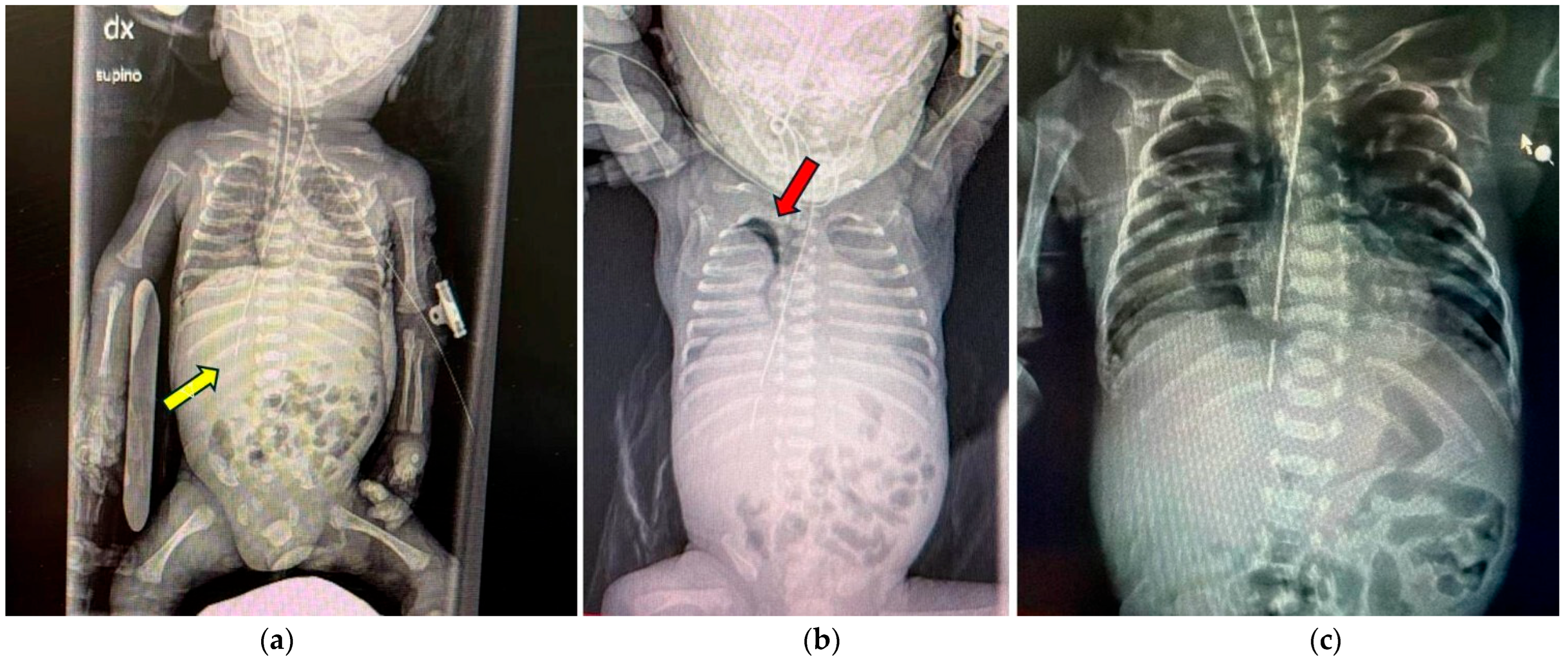
Clinical signs were typically nonspecific, including respiratory distress, cyanosis, abdominal distension, or difficulty advancing a feeding tube. Chest and abdominal radiographs remain the first-line diagnostic tool, revealing misplaced tubes, pneumothorax, or pneumomediastinum (Figure 1a–c).
However, up to one-third of cases initially display normal plain films, underscoring the need for confirmatory imaging [5]. Specifically, plain X-rays may fail to identify subtle mediastinal air or minor perforations, particularly in the cervical region. Over the past decade, point-of-care ultrasonography (POCUS) has emerged as a valuable, radiation-free, and real-time alternative for verifying tube placement and detecting pneumothorax or mediastinal air [26]. Recent reports document sensitivity rates exceeding 90% when performed by trained operators. Contrast esophagography has been found to be reserved for unclear cases or assessment of healing prior to feeding resumption [2].

3.4. Management and Outcomes

Conservative treatment—consisting of tube removal, nil per os, broad-spectrum antibiotics, parenteral nutrition, and supportive care—was the predominant approach in over 95% of cases [3,15,27]. Surgical treatment was performed in three cases (3.5%) included in the review [8,14]. The surgical approach consisted of direct suturing of the esophageal perforation. No cases of esophagostomy were reported. In patients presenting with abscess or mediastinitis, management was predominantly conservative (including antibiotic therapy, drainage, and nutritional support), while surgery was reserved for complicated and early presentations, prior to the development of diffuse infection. Although the literature suggests that esophageal diversion (esophagostomy) may be required in the presence of mediastinitis or abscess [15], no cases treated with this technique were reported in the included studies. In cases complicated by air leakage (pneumothorax, pneumomediastinum, or pneumoperitoneum), management consisted of thoracic or abdominal drainage combined with conservative treatment, without the need for major surgical intervention [3,4]. Endoscopic treatment, including vacuum-assisted therapy, was not reported as a management option in the neonatal patients described; however, the literature documents its use in selected neonatal and pediatric cases (persistent leakage or anastomotic dehiscence not responsive to conservative management, first-line approach in the presence of contained perforations without signs of sepsis or extensive contamination, rescue therapy following failure of surgical suturing) [28]. The median duration of fasting ranged between 10 and 14 days, with reintroduction of enteral feeding guided by repeat imaging [5,8]. The overall survival rate exceeded 75%, with most fatalities attributable to extreme prematurity and comorbid sepsis. Temporal analysis revealed a decline in mortality from ~35% before 2010 to <20% after 2018, reflecting the increasing use of early imaging (especially US), standardization of feeding tube protocols, and emphasis on operator training. Complications most frequently reported include pneumothorax, pneumomediastinum, empyema, sepsis, and mediastinitis. While the direct mortality attributable to EP is relatively low, the overall prognosis is significantly influenced by gestational age, comorbidities, and infection severity [1,2,3,4,10,11]. Infants with extreme prematurity (<26 weeks) or birth weight <700 g have been detected to have poorer outcomes, largely due to multisystem immaturity rather than the perforation itself.

4. Discussion

Neonatal EP remains a rare but critical event, particularly among extremely preterm infants, whose tissue immaturity and fragility predispose them to procedural trauma. The pathophysiological substrate involves the combination of immature connective tissue, lack of a serosal layer, and poor vascularization of the neonatal esophagus, rendering it susceptible even to minimal mechanical stress. The latter, indeed, when associated with routine procedures, such as scheduled tube replacement every 3–5 days, may precipitate sudden esophageal perforation even after previously uncomplicated insertions, as demonstrated in reported neonatal cases [13]. The delicate balance between required intensive interventions—such as airway management and enteral nutrition—and the risk of iatrogenic harm constitutes a recurring dilemma in neonatal intensive care [1,2,3]. The present review explicitly contrasts with prior studies in several key aspects. First, it encompasses a broader spectrum of study designs, including case reports, case series, retrospective observational studies, an observational cross-sectional study, and two multicenter retrospective studies, thereby providing a more comprehensive overview of the literature. Second, it focuses specifically on neonatal patients, whereas previous analyses often included mixed pediatric populations. Third, the time frame of the included papers has been updated to capture the most recent evidence. Finally, the review incorporates considerations related to clinical safety and risk management, highlighting the role of iatrogenic factors and strategies to prevent complications. Indeed, the available evidence emphasizes that iatrogenic injury continues to account for the vast majority of cases. This observation further underscores the necessity of procedural standardization and continuous professional training, particularly for less experienced healthcare providers working in high-stress NICU environments. Traditional radiographic confirmation of tube placement has long been considered the diagnostic cornerstone, but it carries limitations. Furthermore, contrast-enhanced imaging remains indispensable for complex cases, not only confirming the diagnosis but also guiding the decision to resume enteral feeding. However, its use must be judicious, balancing diagnostic yield against the risk of aspiration or further mucosal injury. The growing integration of multimodal imaging aligns with the trend toward minimally invasive diagnostics in neonatology, promoting earlier detection and safer management [26].
The predominant treatment paradigm for neonatal EP is conservative management, which has demonstrated high success rates when initiated promptly. The safety and effectiveness of nonoperative management are further supported by Onwuka et al., who reported a 100% survival rate in a cohort of 25 neonates managed conservatively [4]. Supportive endotracheal ventilation and fluid balance are essential components of care. In contrast, surgical intervention should be reserved for cases complicated by abscess, mediastinitis, or uncontrolled leakage. Surgical repair in this population is technically challenging and associated with higher mortality; therefore, it is justified only when conservative therapy fails or life-threatening complications occur [3,4,5,11]. Recent literature indicates that the duration of fasting and timing of feeding resumption can be individualized based on imaging findings [5,8]. Early follow-up with contrast studies or US to confirm healing is now considered best practice. Importantly, interdisciplinary coordination among neonatologists, pediatric surgeons, and radiologists plays a pivotal role in optimizing outcomes. Notably, several recent series demonstrate a progressive decline in mortality since 2015. While earlier reports have credited this improvement to the increasing use of early imaging, infection control, and nutritional support [1,7], it is likely that advances in general aspects of neonatal intensive care—including respiratory management, hemodynamic support, analgesia and sedation, the use of less invasive devices and life-support systems, and the implementation of less traumatic procedural tools—have played a more critical role in enhancing survival.
Considering its predominantly iatrogenic etiology, prevention remains the most effective strategy against neonatal esophageal perforation (EP). Recommended measures include:
  • Gentle NGT/OGT insertion techniques, using minimal force and adequate lubrication;
  • Extending intervals between routine tube replacements, which may reduce cumulative mechanical stress on the fragile neonatal esophageal wall;
  • Accurate tube length estimation based on measurement methods (e.g., NEMU [Nose–Ear–Mid–Xiphoid–Umbilicus] or NEX [Nose–Earlobe–Xiphoid]);
  • Routine post-insertion verification via radiography or ultrasonography before initiating feeding;
  • Minimizing repeated insertion attempts, particularly in unstable or extremely preterm infants;
  • Simulation-based procedural training for NICU staff to reinforce safe handling of fragile neonates [18,29].
While NGT/OGT insertion has traditionally been described as the predominant trigger event, endotracheal intubation is equally important and should be given similar consideration. As both procedures are typically performed within a similar timeframe on the first day of life, it is often difficult to ascribe the perforation to one procedure over the other. Prevention strategies should therefore factor in both procedures. Moreover, the use of less traumatic devices for intubation, such as plastic blades (e.g., Parker Neonatal Video Laryngoscope), may further reduce the risk of iatrogenic injury.
Despite these recommendations [30], the literature still lacks standardized diagnostic algorithms and unified management guidelines for neonatal EP. Previous reviews on neonatal esophageal perforation have provided valuable insights but also exhibit notable limitations. Many included only a small number of cases, restricting the generalizability of their conclusions. Several reviews combined neonatal and broader pediatric populations, limiting the ability to draw neonatal-specific inferences. Outcome reporting was often inconsistent, with incomplete data on management strategies, procedural details, and clinical follow-up. In particular, incomplete follow-up data hinder a full understanding of potential late sequelae, such as esophageal strictures, dysmotility, or persistent feeding difficulties. Taken together, the limited number of reported cases, heterogeneity in reporting, and underrepresentation of long-term outcomes have, to date, constrained the generalizability of the findings.

Safety Culture, Reflective Practice, and Professional Learning

In addition to technical precautions, fostering a strong safety culture—one in which adverse events are discussed transparently and reframed as learning opportunities—has been shown to reduce procedural complications. Institutional strategies such as standardized checklists, peer review, and non-punitive error-reporting systems promote accountability and continuous improvement. Every case of neonatal EP should, then, also prompt reflective practice—a structured process through which healthcare professionals analyze the event, identify modifiable factors, and translate lessons into improved clinical protocols [29,30]. The emotional impact of iatrogenic complications on neonatal teams can be substantial, often leading to self-doubt or moral distress. Implementing peer-support programs and clinical supervision frameworks helps mitigate these effects, enabling clinicians to regain confidence and professional balance. Furthermore, incorporating clinical simulation, morbidity-and-mortality conferences, and ethical reflection groups into NICU routines can transform individual errors into institutional learning, ultimately strengthening both patient safety and team resilience [19,29,30,31,32].

5. Conclusions

Esophageal perforation in ELBW preterm infants remains a rare but potentially life-threatening complication, often associated with iatrogenic interventions such as NGT/OGT placement. The management of neonatal EP has evolved from high surgical dependency to a conservative, multidisciplinary, and ethically aware approach [33,34,35,36,37,38,39,40]. Early recognition (imaging-guided diagnosis, especially bedside US), prompt withdrawal of the offending device, and individualized supportive care have significantly improved survival outcomes. However, the overall prognosis in such patients is strongly influenced by prematurity-related vulnerabilities, including respiratory immaturity, susceptibility to infections, and multiorgan fragility. Therefore, prevention remains the cornerstone of care. Continued reporting of such cases enriches the body of evidence guiding best practices in neonatal intensive care. It also underscores the relevance of vigilance, standardized NICU protocols, including early detection, multidisciplinary coordination and meticulous procedural execution, as well as ongoing staff training to prevent complications.

Author Contributions

Conceptualization, G.S. and V.G.; methodology, V.N. and M.S.; formal analysis, I.A.M.S.; investigation, V.N.; resources, M.G.; data curation, V.G.; writing—original draft preparation, G.S. and V.G.; writing—review and editing, G.S. and M.P.; supervision, G.C., M.G. and M.R.D.P. All authors have read and agreed to the published version of the manuscript.

Funding

This research received no external funding.

Institutional Review Board Statement

Not applicable.

Informed Consent Statement

Not applicable. The paper is a review of already published papers.

Data Availability Statement

The original data presented in the study are openly online available at https://pubmed.ncbi.nlm.nih.gov.

Conflicts of Interest

The authors declare no conflicts of interest.

Abbreviations

BWbirth weight
ELBWextremely low birth weight
E.PEsophageal perforation
GAgestational age
NECnecrotizing enterocolitis
NGT/OGTnaso- or orogastric tubes
PDApatent ductus arteriosus
POCUSpoint-of-care ultrasonography
PROMpremature rupture of membranes
RDSespiratory distress syndrome
VLBWvery low birth weight

References

  1. Elgendy, M.M.; Othman, H.; Aly, H. Esophageal Perforation in Very Low Birth Weight Infants. Eur. J. Pediatr. 2021, 180, 513–518. [Google Scholar] [CrossRef] [Scilit]
  2. Hesketh, A.J.; Behr, C.A.; Soffer, S.Z.; Hong, A.R.; Glick, R.D. Neonatal esophageal perforation: Nonoperative management. J. Surg. Res. 2015, 198, 1–6. [Google Scholar] [CrossRef] [Scilit]
  3. Adel, M.G.; Sabagh, V.G.; Sadeghimoghadam, P.; Albazal, M. The Outcome of Esophageal Perforation in Neonates and Its Risk Factors: A 10-Year Study. Pediatr. Surg. Int. 2023, 39, 127. [Google Scholar] [CrossRef] [Scilit] [PubMed]
  4. Onwuka, E.A.; Saadai, P.; Boomer, L.A.; Nwomeh, B.C. Nonoperative Management of Esophageal Perforations in the Newborn. J. Surg. Res. 2016, 205, 102–107. [Google Scholar] [CrossRef] [Scilit] [PubMed]
  5. Rentea, R.M.; St Peter, S.D. Neonatal and Pediatric Esophageal Perforation. Semin. Pediatr. Surg. 2017, 26, 87–94. [Google Scholar] [CrossRef] [Scilit] [PubMed]
  6. Seefelder, C.; Elango, S.; Rosbe, K.W.; Jennings, R.W. Oesophageal Perforation Presenting as Oesophageal Atresia in a Premature Neonate Following Difficult Intubation. Paediatr. Anaesth. 2001, 11, 112–118. [Google Scholar] [CrossRef] [Scilit]
  7. Hermans, K.E.; Witvliet, M.J.; van den Hoogen, A.; de Bijl-Marcus, K.A. A Case Series of Iatrogenic Gastrointestinal Perforations in Premature Infants Below 30 Weeks’ Gestation. J. Pediatr. Surg. 2025, 60, 162590. [Google Scholar] [CrossRef] [Scilit]
  8. Emil, S.G.S. Neonatal esophageal perforation. J. Pediatr. Surg. 2004, 39, 1296–1298. [Google Scholar] [CrossRef] [Scilit]
  9. Suryawanshi, P.; Dahat, A.; Nagpal, R.; Malshe, N.; Kalrao, V. A rare case of accidental esophageal perforation in an extremely low birth weight neonate. J. Clin. Diagn. Res. 2014, 8, PD01–PD02. [Google Scholar] [CrossRef] [Scilit]
  10. Hodgson, K.; Togo, A.; Moore, A.M.; Moody, A.; King, S.K.; Zani, A. Neonatal Oesophageal Perforation: The Role for Non-Operative Management. J. Paediatr. Child Health 2018, 54, 872–874. [Google Scholar] [CrossRef] [Scilit]
  11. Gander, J.W.; Berdon, W.E.; Cowles, R.A. Iatrogenic esophageal perforation in children. Pediatr. Surg. Int. 2009, 25, 395–401. [Google Scholar] [CrossRef] [Scilit]
  12. Yong, S.B.; Ma, J.S.; Chen, F.S.; Chung, M.Y.; Yang, K.D. Nasogastric Tube Placement and Esophageal Perforation in Extremely Low Birth Weight Infants. Pediatr. Neonatol. 2016, 57, 427–430. [Google Scholar] [CrossRef] [Scilit]
  13. Lithoxopoulou, M.; Gkampeta, A.; Babatseva, E.; Simeoforidou, E.; Chatzitoliou, E.; Georgiadou, P.; Anastasiadis, K.; Kaselas, C.; Kepertis, C.; Spyridakis, I.; et al. Unusual site for iatrogenic esophageal perforation in a premature neonate. J. Pediatr. Neonat. Individual. Med. 2019, 8, e080113. [Google Scholar] [CrossRef]
  14. Mikołajczak, A.; Kufel, K.; Żytyńska-Daniluk, J.; Rutkowska, M.; Bokiniec, R. Iatrogenic Esophageal Perforation in Premature Infants: A Multicenter Retrospective Study from Poland. Children 2023, 10, 1399. [Google Scholar] [CrossRef] [Scilit]
  15. Sorensen, E.; Yu, C.; Chuang, S.L.; Midrio, P.; Martinez, L.; Nash, M.; Jester, I.; Saxena, A.K. Iatrogenic Neonatal Esophageal Perforation: A European Multicentre Review on Management and Outcomes. Children 2023, 10, 217. [Google Scholar] [CrossRef] [Scilit]
  16. Aljadaan, S.A.; Alharbi, N.S.; Alnamshan, M.K.; Alaqeel, S.M.; Abaas, A.O. Iatrogenic esophageal perforation in extremely premature babies with low birth weight: A case series at a single tertiary-care center. Cureus 2025, 17, e94197. [Google Scholar] [CrossRef] [Scilit] [PubMed]
  17. Eguchi, S.; Hisaeda, Y.; Ukawa, T.; Koto, M.; Hosokawa, M.; Tsurisawa, C.; Takeda, T.; Amagata, S.; Nakao, A. Clinical Features of iatrogenic Pharyngo-esophageal perforation in very low birth weight infants. Pediatr. Neonatol. 2025, 66, 25–30. [Google Scholar] [CrossRef] [Scilit] [PubMed]
  18. Sapin, E.; Gumpert, L.; Bonnard, A.; Carricaburu, E.; Sava, E.; Contencin, P.; Helardot, P. Iatrogenic pharyngoesophageal perforation in premature infants. Eur. J. Pediatr. Surg. 2000, 10, 83–87. [Google Scholar] [CrossRef] [Scilit]
  19. Kan, S.Y.; Ngeow, A.J.H.; Tan, M.G.; Jacobsen, A.S.; Sanamandra, S.K.; Poon, W.B. Esophageal Perforation in VLBW Infants in a Singapore Tertiary Hospital: A Case-Control Study. Ann. Pediatr. Res. 2024, 8, 1081. [Google Scholar]
  20. Savarino, G.; Carta, M.; Cimador, M.; Corsello, A.; Giuffrè, M.; Schierz, I.A.M.; Serra, G.; Corsello, G. Necrotizing enterocolitis in the preterm: Newborns medical and nutritional Management in a Single-Center Study. Ital. J. Pediatr. 2021, 47, 226. [Google Scholar] [CrossRef] [Scilit]
  21. Giuffrè, M.; Lo Verso, C.; Serra, G.; Moceri, G.; Cimador, M.; Corsello, G. Study Group of Neonatal Infectious Diseases Affiliated to the Italian Society of Neonatology Portal Vein Thrombosis in a Preterm Newborn with Mutation of the MTHFR and PAI-1 Genes and Sepsis by Candida parapsilosis. Am. J. Perinatol. 2016, 33, 1099–1103. [Google Scholar]
  22. Schierz, I.A.M.; Giuffrè, G.; Cimador, M.; D’Alessandro, M.M.; Serra, G.; Favata, F.; Antona, V.; Piro, E.; Corsello, G. Hypertrophic pyloric stenosis masked by kidney failure in a male infant with a contiguous gene deletion syndrome at Xp22.31 involving the steroid sulfatase gene: Case report. Ital. J. Pediatr. 2022, 48, 19. [Google Scholar] [CrossRef] [Scilit] [PubMed]
  23. Serra, G.; Antona, V.; Giuffré, M.; Li Pomi, F.; Lo Scalzo, L.; Piro, E.; Schierz, I.A.M.; Corsello, G. Novel missense mutation of the TP63 gene in a newborn with Hay-Wells/Ankyloblepharon-Ectodermal Defects-Cleft Lip/Palate (AEC) syndrome: Clinical report and follow-up. Ital. J. Pediatr. 2021, 47, 196. [Google Scholar] [CrossRef] [Scilit]
  24. Grasso, F.; Baldanza, F.; Pernicone, S.; Pensabene, M.; Sergio, M.; Di Pace, M.R. The Role of Endoscopy in the Postoperative Management of Patients Treated for Esophageal Atresia: 20 Years of Experience. Diagnostics 2025, 15, 843. [Google Scholar] [CrossRef] [Scilit] [PubMed]
  25. Serra, G.; Cimador, M.; Giuffrè, M.; Insinga, V.; Montante, C.; Pensabene, M.; Piro, E.; Salerno, S.; Schierz, I.A.M.; Corsello, G. Report and follow-up on two new patients with congenital mesoblastic nephroma. Ital. J. Pediatr. 2023, 49, 124. [Google Scholar] [CrossRef] [Scilit]
  26. Atalay, Y.O.; Polat, A.V.; Ozkan, E.O.; Tomak, L.; Aygun, C.; Tobias, J.D. Bedside ultrasonography for the confirmation of gastric tube placement in the neonate. Saudi J. Anaesth. 2019, 13, 23–27. [Google Scholar] [CrossRef] [Scilit] [PubMed]
  27. Thanhaeuser, M.; Lindtner-Kreindler, C.; Berger, A.; Haiden, N. Conservative Treatment of Iatrogenic Perforations Caused by Gastric Tubes in Extremely Low Birth Weight Infants. Early Hum. Dev. 2019, 137, 104836. [Google Scholar] [CrossRef] [Scilit]
  28. Kaczmarek, D.J.; Heling, D.J.; Strassburg, C.P.; Katzer, D.; Düker, G.; Strohm, J.; Müller, A.; Heydweiller, A.; Weismüller, T.J. Management of esophageal perforations in infants by endoscopic vacuum therapy: A single center case series. BMC Gastroenterol. 2022, 22, 282. [Google Scholar] [CrossRef] [Scilit]
  29. Panagos, P.G.; Pearlman, S.A. Creating a Highly Reliable Neonatal Intensive Care Unit Through Safer Systems of Care. Clin. Perinatol. 2017, 44, 645–662. [Google Scholar] [CrossRef] [Scilit]
  30. Caeymaex, L.; Astruc, D.; Biran, V.; Marcus, L.; Flamein, F.; Le Bouedec, S.; Guillois, B.; Remichi, R.; Harbi, F.; Durrmeyer, X.; et al. An Educational Programme in Neonatal Intensive Care Units (SEPREVEN): A Stepped-Wedge, Cluster-Randomised Controlled Trial. Lancet 2022, 399, 384–392. [Google Scholar] [CrossRef] [Scilit]
  31. Hybinette, K.; Pukk Härenstam, K.; Ekstedt, M. A First-line management team’s strategies for sustaining resilience in a specialised intensive care unit-a qualitative observational study. BMJ Open 2021, 11, e040358. [Google Scholar] [CrossRef] [Scilit]
  32. Bondurant, P.G.; Nielsen-Farrell, J.; Armstrong, L. The Journey to High Reliability in the NICU. J. Perinat. Neonatal Nurs. 2015, 29, 170–178. [Google Scholar] [CrossRef] [Scilit] [PubMed]
  33. Quandt, D.; Schraner, T.; Ulrich Bucher, H.; Arlettaz Mieth, R. Malposition of feeding tubes in neonates: Is it an issue? J. Pediatr. Gastroenterol. Nutr. 2009, 48, 608–611. [Google Scholar] [CrossRef] [Scilit]
  34. Ottosen, M.J.; Engebretson, J.; Etchegaray, J.; Arnold, C.; Thomas, E.J. An Ethnography of Parents’ Perceptions of Patient Safety in the Neonatal Intensive Care Unit. Adv. Neonatal Care 2019, 19, 500–508. [Google Scholar] [CrossRef] [Scilit]
  35. Arimitsu, T.; Hatayama, K.; Gaughwin, K.; Kusuda, S. Ethical Considerations Regarding the Treatment of Extremely Preterm Infants at the Limit of Viability: A Comprehensive Review. Eur. J. Pediatr. 2025, 184, 140. [Google Scholar] [CrossRef] [Scilit] [PubMed]
  36. Janvier, A.; Barrington, K.; Farlow, B. Multidisciplinary ethics support in neonatal intensive care. J. Med. Ethics 2020, 46, 428–432. [Google Scholar]
  37. Albersheim, S. The extremely preterm infant: Ethical considerations in life and death decision making. Front. Pediatr. 2020, 8, 55. [Google Scholar] [CrossRef] [Scilit]
  38. Boss, R.D.; Hutton, N.; Sulpar, L.J.; West, A.M.; Donohue, P.K. Family-centered communication in neonatal intensive care. J. Perinatol. 2017, 37, 1175–1181. [Google Scholar]
  39. Wilkinson, D.; Pike, K.; Savulescu, J. Decision-making in extremely preterm infants. Arch. Dis. Child. Fetal Neonatal Ed. 2019, 104, F190–F195. [Google Scholar]
  40. Bluebond-Langner, M.; Beecham, E.; Candy, B.; Langner, R.; Jones, L. Shared decision-making in the NICU. Pediatrics 2018, 141, e20172924. [Google Scholar]
Figure 1. (a) Abnormal position of the nasogastric tube, with its tip located in the right hypochondrium at the level of the twelfth rib (yellow arrow). (b) In the upright image, a right-sided pneumothorax is visible, with partial lung collapse. The thickness of the air pocket indicated by the red arrow measures 5 mm. (c) Pneumothorax collection located in the right paramediastinal region following esophageal perforation in an extremely preterm neonate.
Jcm 15 01603 g001
Disclaimer/Publisher’s Note: The statements, opinions and data contained in all publications are solely those of the individual author(s) and contributor(s) and not of MDPI and/or the editor(s). MDPI and/or the editor(s) disclaim responsibility for any injury to people or property resulting from any ideas, methods, instructions or products referred to in the content.