Systematic Review

Association of Porphyromonas gingivalis with Acute Myocardial Infarction: A Systematic Review

by
Elina Ghondaghsaz
1,
Edward E. Putnins
2 and
Ahmed Hieawy
2,3,4,*
1
Department of Neuroscience, University of British Columbia, Vancouver, BC V6T 1Z3, Canada
2
Department of Oral Biological and Medical Sciences, Faculty of Dentistry, University of British Columbia, Vancouver, BC V6T 1Z3, Canada
3
Vancouver Coastal Health Research Institute, Vancouver General Hospital, Vancouver, BC V5Z 1M9, Canada
4
BC Children’s Hospital Research Institute, BC Children’s Hospital, Vancouver, BC V5Z 4H4, Canada
*
Author to whom correspondence should be addressed.
J. Clin. Med. 2026, 15(14), 5689; https://doi.org/10.3390/jcm15145689
Submission received: 24 June 2026 / Revised: 17 July 2026 / Accepted: 18 July 2026 / Published: 20 July 2026
(This article belongs to the Special Issue Interaction Between Systemic Diseases and Oral Diseases: 2nd Edition)

Abstract

Background/Objectives: Periodontitis is a chronic oral infection in which Porphyromonas gingivalis (Pg) acts as a keystone pathogen capable of systemic dissemination and immune evasion. A possible association between Pg and acute myocardial infarction (AMI) has been proposed; this systematic review is, to our knowledge, among the first to evaluate this association through a pathogen-specific synthesis integrating both microbial and serological evidence. Methods: Five electronic databases (PubMed, Scopus, Embase, Web of Science, Cochrane Library) were searched from inception to February 2025, with a supplementary top-up search performed in July 2026 that identified no additional eligible studies. Observational studies assessing Pg presence or anti-Pg antibody levels in participants with and without AMI were eligible. Methodological quality was assessed using the Newcastle–Ottawa Scale (NOS). Findings were synthesised narratively due to substantial clinical and methodological heterogeneity. The protocol was registered in PROSPERO (CRD42025644043). Results: Twelve case-control studies (5147 participants; 2518 AMI cases, 2629 controls) were included. Six evaluated serum anti-Pg antibodies and six used direct microbial or molecular detection. Three studies in each category reported a significant Pg–AMI association; three in each category did not. NOS scores ranged from 6 to 9 (eight studies rated good quality; four fair quality). Heterogeneity in antigen selection, sampling site, immunoglobulin isotype, and confounder adjustment precluded meta-analysis. Conclusions: The available evidence suggests a possible but inconsistent association between Pg and AMI, insufficient to establish a causal relationship. Standardised detection protocols and prospective longitudinal studies with comprehensive confounder adjustment are needed. The detection of Pg in isolation is not currently validated for AMI risk stratification, and the available evidence does not establish a causal relationship between Pg and AMI.

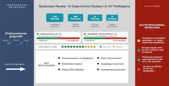
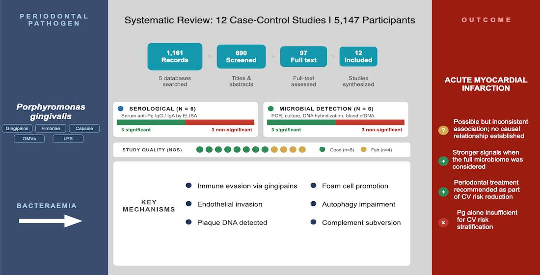
Graphical Abstract

1. Introduction

Acute myocardial infarction (AMI) typically results from the rupture of a vulnerable atherosclerotic plaque and the consequent thrombotic occlusion of a coronary artery [1]. Coronary artery disease is the principal underlying condition and remains the leading cause of death worldwide [2]. Well-established modifiable risk factors for AMI include hypertension, dyslipidaemia, diabetes mellitus, tobacco use, physical inactivity, and obesity [3,4]. Evidence from large longitudinal cohorts suggests that the majority of AMI events could be prevented through sustained lifestyle modification and pharmacological risk factor control [5,6]. Beyond conventional risk factors, systemic infections have increasingly been recognised as contributors to AMI risk. A large Danish cohort study spanning three decades found that common bacterial infections were associated with significantly elevated AMI rates, with inflammatory and procoagulant mechanisms proposed as the underlying pathways [7]. Autopsy and experimental data further indicate that systemic infection can destabilise existing atheromatous plaques through heightened inflammatory activity and oxidative stress [8,9].
Periodontitis is a chronic inflammatory disease of the tooth-supporting structures and one of the most prevalent oral infectious diseases globally [10]. Global burden-of-disease estimates indicate that severe periodontitis affects approximately 11% of adults worldwide, making it the sixth most prevalent condition globally, with prevalence rising markedly with age [11]. Porphyromonas gingivalis (Pg), a Gram-negative anaerobic bacterium, is regarded as a keystone pathogen in periodontitis owing to its capacity to subvert innate immune responses and disseminate systemically via bacteraemia [12,13,14]. It frequently co-exists with Treponema denticola and Tannerella forsythia—collectively termed the red complex—whose synergistic interactions amplify local tissue destruction and systemic inflammatory burden [15,16,17]. Serum immunoglobulin responses against Pg have been proposed as a surrogate marker of periodontal disease severity and systemic pathogen burden [18,19,20,21,22].
Mechanistically, Pg possesses a broad repertoire of virulence factors, including fimbriae, capsule, arginine- and lysine-specific gingipain proteases, outer membrane vesicles, and atypical lipopolysaccharide, which collectively facilitate immune evasion, complement dysregulation, tissue invasion, and transient bacteraemia [13,14,23]. Following entry into the bloodstream, Pg has been shown to invade vascular endothelial cells, vascular smooth muscle cells, and macrophages, where it contributes to persistent vascular inflammation. In addition, Pg DNA has been detected within atherosclerotic plaques, supporting its presence in vascular lesions and providing biological plausibility for a contributory role in atherogenesis and plaque instability [24,25]. Experimental studies further suggest that Pg promotes macrophage foam-cell formation through enhanced oxidised low-density lipoprotein uptake and impairs cardiomyocyte autophagy following myocardial infarction, mechanisms that could contribute to the development and progression of acute coronary events [26,27]. Collectively, these mechanistic findings provide biological plausibility for the epidemiological associations between Pg exposure and acute myocardial infarction examined in this review.
Several meta-analyses have examined the association between periodontitis and cardiovascular outcomes. Qin et al. reported a 13% higher relative risk of AMI in patients with periodontal disease across ten cohort studies, though without a pathogen-specific subgroup analysis [23]. Joshi et al. conducted a meta-analysis of 20 studies in coronary artery disease patients, reporting a pooled odds ratio of 1.23 (95% CI 1.09–1.38) for anti-Pg immunoglobulin G (IgG), though these studies broadly sampled coronary artery disease rather than AMI specifically [24]. Larvin et al. and Leng et al. confirmed elevated cardiovascular disease risk in periodontitis patients across large cohorts [25,26]. To date, no systematic review has focused specifically on the association between Pg—assessed by direct microbial detection or anti-Pg antibody measurement—and the occurrence of AMI. Such specificity is warranted given the unique virulence profile of Pg [13,27]. Antibody-based and microbial detection methods capture complementary aspects of Pg exposure: serum anti-Pg antibody titres reflect cumulative or chronic host exposure to Pg antigens over time, whereas direct microbial or molecular detection in subgingival plaque or blood reflects current bacterial presence or bacteraemic dissemination [18,19,20,21,22]. This review deliberately incorporated both serological and microbial evidence to capture complementary dimensions of Pg exposure by integrating markers of cumulative host immune response with evidence of current bacterial detection, both of which may be relevant to AMI risk. The present systematic review aims to evaluate the evidence linking Pg exposure to AMI risk, enabling a more pathogen-specific analysis than has previously been undertaken.

2. Materials and Methods

2.1. Protocol and Reporting

This systematic review was designed and reported in accordance with the Preferred Reporting Items for Systematic Reviews and Meta-Analyses (PRISMA) 2020 guidelines [28]. The completed PRISMA checklist is provided in Supplementary Table S1. The review protocol was prospectively registered in PROSPERO (registration number CRD42025644043) on 7 February 2025, prior to the commencement of title/abstract screening and data extraction.

2.2. Search Strategy

A comprehensive literature search was conducted across five international databases—PubMed, Scopus, Embase, Web of Science, and the Cochrane Library—from inception to February 2025. Search terms were organised into two categories: Category A (myocardial infarction and related cardiovascular terms) and Category B (Pg and synonyms), combined using the Boolean operator AND. Full search strings are provided in Supplementary Table S2. Grey literature were not searched; this is acknowledged as a potential source of publication bias. A supplementary top-up search using the same terms was performed in July 2026 to identify any eligible studies published between February 2025 and submission. This identified no additional clinical studies meeting the review’s inclusion criteria; the studies published in this interval were mechanistic or in vitro investigations of Pg–atherosclerosis pathways rather than clinical studies of Pg exposure and AMI outcome. The evidence base underlying this review’s conclusions is therefore considered current as of submission.

2.3. Eligibility Criteria

Inclusion criteria: (1) observational studies or randomised controlled trials assessing Pg presence or anti-Pg antibody levels in participants with and without AMI; (2) human subjects of any age or sex; (3) clearly defined diagnostic criteria for both Pg exposure and AMI; and (4) English-language publications. The restriction to English-language publications was applied to allow accurate extraction of diagnostic and outcome data without reliance on translation; however, it is acknowledged that this restriction may have excluded relevant non-English studies and introduced potential language bias, which is discussed further as a limitation.
Exclusion criteria: (1) preclinical studies (in vivo or in vitro); (2) cardiovascular outcomes other than AMI unless AMI data were extractable; (3) narrative reviews, case reports; (4) duplicate studies; (5) incomplete outcome data; and (6) conference abstracts or preprints.

2.4. Study Selection

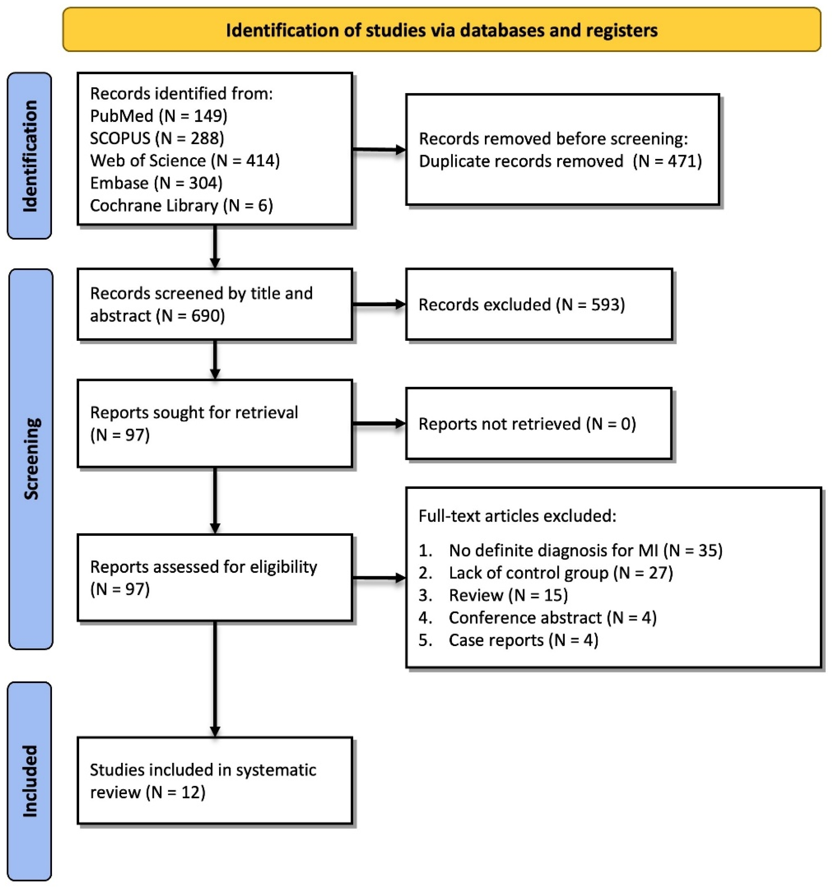
Records were imported into EndNote 20 and deduplicated. Two reviewers (E.G. and A.H.) independently screened titles and abstracts, followed by full-text assessment of potentially eligible studies. Disagreements were resolved by consensus; where consensus could not be reached, a third reviewer was consulted. Eligibility was structured around a PECO framework (Population: individuals with and without AMI; Exposure: Pg presence or anti-Pg antibody level; Comparator: AMI-free controls; Outcome: occurrence of AMI). Screening was performed manually without automation or machine learning-assisted tools. The reference lists of all included studies were manually screened for additional eligible records; no further studies were identified through this process. Inter-rater agreement was quantified using Cohen’s kappa (k = 0.82), indicating strong agreement. The study selection process is summarized in the PRISMA flow diagram (Figure 1).

2.5. Data Extraction

Two reviewers independently extracted data covering: (1) study characteristics; (2) Pg detection method; (3) AMI diagnostic criteria; (4) primary outcomes; and (5) adjusted confounders. For studies reporting medians and interquartile ranges, conversion to means and standard deviations followed the methods of Luo et al. and Wan et al. [29,30]. Discrepancies were resolved by consensus. Data extraction used a standardised, piloted extraction form capturing the exact wording of each study’s AMI diagnostic definition, the effect estimates and measures of variability reported (odds ratios, relative risks, or p-values, as available), and the specific confounders entered into each adjusted model, to support the narrative and comparative synthesis presented in Section 3.

2.6. Quality Assessment

Methodological quality was assessed using the Newcastle–Ottawa Scale (NOS) for case-control studies, evaluating selection (maximum 4 stars), comparability (maximum 2 stars), and exposure (maximum 3 stars), yielding a maximum total of 9 stars. Studies scoring 7–9 were rated good quality, 5–6 fair quality, and 0–4 poor quality. Two reviewers assessed each study independently. Across studies, comparability stars were most often lost due to non-adjustment for smoking or diabetes status, and exposure stars were most often lost where Pg status was ascertained by self-report rather than direct clinical or laboratory verification. A formal GRADE assessment was not performed. Although GRADE can be applied without a pooled estimate, we judged this impractical given the marked heterogeneity in AMI definitions and Pg exposure metrics across studies (Section 3.5), which limits a coherent summary judgement; this is noted as a limitation. In addition, since the NOS assesses risk of bias at the study level rather than the outcome level, and several studies reported multiple Pg-related exposure metrics (e.g., IgG/IgA, or oral versus blood detection), residual outcome-level bias (particularly selective outcome reporting) cannot be ruled out and is also acknowledged as a limitation.

2.7. Data Synthesis

Meta-analysis was not feasible due to substantial clinical and methodological heterogeneity, including variation in Pg detection platforms, exposure types (antibody-based vs. direct microbial detection), outcome definitions, and population characteristics. Specifically, meta-analysis was precluded by a combination of inconsistent exposure definitions (antibody isotype and antigen target differed across studies), non-comparable effect metrics (several studies reported only p-values or descriptive comparisons without an extractable odds ratio, relative risk, or standard error), and heterogeneous outcome ascertainment (AMI defined variably across studies; see Section 3.5). As a complementary approach, structured vote-counting by direction of effect was considered; however, given the small number of studies (n = 12) and the fact that several reported multiple, partly overlapping exposure metrics within the same cohort, this was judged unlikely to add meaningfully to the narrative synthesis and risked overstating precision. This decision is discussed further as a limitation. Findings were therefore synthesised narratively. No formal publication bias assessment was conducted owing to the small number of included studies (n = 12) and the absence of a pooled statistical estimate; this is acknowledged as a limitation.
Where multiple publications originated from the same cohort, studies were retained separately in the qualitative synthesis when they reported distinct outcomes or analyses; however, participant numbers were counted only once when calculating the overall sample size to avoid double-counting.

3. Results

3.1. Study Selection Results

The systematic search retrieved 1161 records: 149 from PubMed, 288 from Scopus, 414 from Web of Science, 304 from Embase, and 6 from the Cochrane Library. After the removal of 471 duplicates, 690 records were screened, and 97 full-text articles were assessed for eligibility. Reasons for exclusion included: no definitive AMI diagnosis (n = 35), no control group (n = 27), and being a review article (n = 15), conference abstract (n = 4), or case report (n = 4). Twelve studies were retained for synthesis [12,31,32,33,34,35,36,37,38,39,40,41]. The PRISMA flow diagram is presented as Figure 1. Because the included studies varied in how AMI was ascertained, the specific diagnostic criteria used by each study (e.g., cardiac biomarkers, ECG findings, angiography, clinical records, or ICD coding) are summarised in Section 3.5 to facilitate the interpretation of differences in AMI outcome ascertainment across the included studies.

3.2. Baseline Characteristics of Included Studies

All 12 included studies used a case-control design; no cohort studies or randomised controlled trials met the inclusion criteria. A total of 5147 participants were enrolled (2518 AMI cases; 2629 controls). Two studies by Lund Haheim et al. [34,35] were derived from the same longitudinal cohort and were retained because they reported different analyses and outcomes. To avoid double-counting, participants from this cohort were included only once in the calculation of the overall sample size. Mean participant age ranged from 51.9 to 70.8 years. The proportion of male participants ranged from 53.1% to 100%. All studies were published between 2004 and 2023. Study characteristics are presented in Table 1.
Six studies employed serological analysis, quantifying serum anti-Pg IgG and/or immunoglobulin A (IgA) by enzyme-linked immunosorbent assay (ELISA), with heterogeneous antigen selection ranging from Pg arginine gingipain (Rgp) to a single reference strain (ATCC 3277) or multiple Pg serotypes. Six studies assessed Pg directly in biological samples using polymerase chain reaction (PCR), indirect immunofluorescence, dot-blot DNA hybridisation, or bacterial culture, primarily from subgingival plaque. One study additionally analysed circulating cell-free DNA (cfDNA) and genomic DNA (gDNA) in peripheral blood. Periodontal disease (PD) status was formally assessed in eight of the 12 studies.

3.3. Quality Assessment of Included Studies

NOS scores for all 12 studies are presented in Table 2. Eight studies were rated good quality (scores 7–9) and four were rated fair quality (scores 5–6). No study met the criteria for poor quality (scores 0–4). All 12 studies were retained for narrative synthesis. Discordance in findings was present even among studies achieving the highest NOS scores (score = 9), indicating that inconsistency reflects genuine biological and methodological heterogeneity rather than poor study design alone.

3.4. Association Between Pg and AMI

Where reported, effect estimates (odds ratios with 95% confidence intervals) are presented in Table 3 alongside the narrative summary below; however, most included studies reported only p-values or descriptive comparisons without an extractable odds ratio, relative risk, or standard error, which is itself part of the reporting heterogeneity underlying the decision not to pool data (Section 2.7).

3.4.1. Serological Studies-Serum Antibody Analysis

Six studies compared anti-PgPg antibody levels between AMI cases and controls using ELISA [12,32,34,35,36,37]. Three reported a statistically significant association; three found no significant association in Table 3.
de Vries et al. [32] used Pg arginine gingipain as the antigen in 779 AMI cases and 719 healthy controls. Anti-Rgp IgG was significantly elevated in the AMI group overall (p = 0.035); however, this difference was non-significant when restricted to participants with confirmed periodontitis (p > 0.05). Lysek et al. [36] reported comparable median anti-PgPg gingipain IgG between 97 AMI cases and 113 controls (p = 0.36); a subgroup analysis identified that a moderate antibody titre was associated with approximately threefold higher odds of past AMI (odds ratio [OR] = 2.82, 95% confidence interval [CI] 1.02–7.84).
Holmlund et al. [12] reported elevated anti-Pg IgG in 100 AMI cases compared with 101 controls (p = 0.043), alongside higher periodontal pathogen counts; IgA levels were comparable between groups. Conversely, Pussinen et al. [37] found no significant IgG difference between 63 AMI cases and 63 controls (p = 0.876) but detected a significantly higher IgA in AMI patients (p = 0.035). Lund Haheim et al. published two analyses from the same longitudinal male cohort [34,35]. Anti-Pg IgG alone was non-significant in both analyses; however, a combined four-pathogen antibody panel was independently associated with AMI risk in the 2008 paper [34].

3.4.2. Microbial Detection Studies-Plaque and Blood Samples

Six studies assessed Pg using direct microbial or molecular detection methods [31,33,38,39,40,41]. Three reported a significant positive association with AMI; three did not.
Stein et al. [40] reported the strongest positive association in this review, identifying Pg as an independent predictor of AMI after full adjustment for age, sex, smoking, body mass index, hypertension, plaque index, statin use, and cholesterol/high-density lipoprotein (HDL) ratio (OR = 13.6, 95% CI 3.1–59.8, p = 0.0005). Wu et al. [41] found oral swab PCR positivity was non-significant between 382 AMI cases and 78 controls, whereas circulating Pg cfDNA and gDNA in peripheral blood were significantly more frequent in AMI patients (p < 0.05 each), correlating with coronary artery disease (CAD) lesion severity. Pasupuleti et al. [38] reported significantly higher Pg load in AMI with generalised chronic periodontitis (GCP) compared with AMI alone (p = 0.043), suggesting the association may be contingent on concurrent PD.
Andriankaja et al. [31] found comparable Pg prevalence between 313 non-fatal AMI cases and 747 controls (18.5% vs. 15.8%; p > 0.05), with no independent association in adjusted models. Tannerella forsythensis and Prevotella intermedia independently predicted AMI in the same study, and an increasing number of co-detected pathogens elevated risk progressively. Dogan et al. [33] reported a significantly lower mean proportion of PgPg in patients with GCP and AMI compared with GCP alone (p = 0.05), suggesting an inverse rather than positive relationship. Seoane et al. [39] found Pg detection was more strongly associated with PD status than with AMI; no independent Pg–AMI association was identified after accounting for periodontal status.

3.5. AMI Diagnostic Criteria Across Included Studies

To facilitate the interpretation of the included evidence, Table 4 summarizes the myocardial infarction outcome label and diagnostic criteria reported in each study, including the methods used for outcome ascertainment (e.g., cardiac biomarkers, electrocardiographic findings, coronary angiography, clinical records, or ICD coding). Because the included studies varied in their definitions and ascertainment of myocardial infarction, the table also provides comments relevant to the review eligibility criteria and highlights sources of clinical heterogeneity that should be considered when interpreting the findings.
When the included studies are considered according to the certainty of AMI ascertainment (Table 4), seven studies diagnosed clinically confirmed acute myocardial infarction using cardiac biomarkers, electrocardiographic findings, and/or coronary angiography [12,32,33,38,39,40,41]. The remaining five did not meet this strict definition and instead used broader ascertainment: self-reported history of myocardial infarction [34,35], previously documented myocardial infarction obtained from medical records [36], and registry-identified incident myocardial infarction [37]; in one of the self-reported studies, cardiovascular mortality was the primary outcome [35]. A further study [31] diagnosed confirmed non-fatal acute myocardial infarction by WHO criteria but was restricted to survivors. When interpretation was restricted to the seven studies with clinically confirmed acute events, the overall pattern was unchanged: findings remained inconsistent, with both significant and null associations reported across serological and microbial detection methods. The direction and inconsistency of the evidence were therefore not driven by studies using less rigorous AMI ascertainment; nonetheless, this heterogeneity in outcome definition remains an important source of clinical heterogeneity and is acknowledged as a limitation.

4. Discussion

4.1. Summary of Main Findings

This systematic review is, to our knowledge, among the first to evaluate the association between Pg and AMI specifically, integrating both direct microbial detection and serological evidence within a single pathogen-focused synthesis. Across both approaches, findings were inconsistent: three of six studies in each category reported a significant Pg–AMI association, while three in each did not. Discordance persisted even among studies achieving the highest NOS scores, suggesting that inconsistency reflects genuine biological and methodological heterogeneity.
A notable pattern was that associations were most consistent when analyses incorporated the broader subgingival microbiome rather than Pg alone. Studies examining combined antibody responses to multiple red complex pathogens [34], total subgingival bacterial burden [38], or the co-presence of several periodontitis-associated bacteria [31] tended to show stronger links to AMI risk. This supports the polymicrobial model of periodontitis and raises the possibility that Pg contributes to cardiovascular risk synergistically with co-infecting organisms rather than as a sole driver [17].
From a clinical standpoint, these findings do not currently support using Pg detection or anti-Pg antibody titres as a stand-alone biomarker for AMI risk prediction or stratification in practice. Even where statistically significant associations were reported, effect sizes were generally modest, derived from case-control designs that cannot establish temporality, and inconsistent across studies using comparable methods. Any clinical significance of Pg exposure for AMI risk should therefore be considered alongside, rather than in place of, established cardiovascular risk factors, and any translation into risk-stratification tools or treatment recommendations should await confirmation in prospective, adequately confounder-adjusted studies.

4.2. Virulence Mechanisms and Cardiovascular Pathobiology

Pg possesses an extensive arsenal of virulence factors—including capsule, fimbriae, gingipains (arginine-specific and lysine-specific cysteine proteases), outer membrane vesicles, and atypical lipopolysaccharides—that collectively enable immune evasion and systemic dissemination [13,14]. Bacteraemia following periodontal manipulation enables Pg to invade endothelial cells, smooth muscle cells, and macrophages, perpetuating chronic intravascular inflammation [42]. The detection of Pg DNA within atherosclerotic plaques supports a role in direct plaque destabilisation [43], consistent with Wu et al. [41], who linked circulating Pg DNA to CAD severity. Additional mechanistic pathways include the Pg-induced impairment of cardiomyocyte autophagy [44] and the promotion of macrophage foam cell formation through oxidised low-density lipoprotein uptake [45,46]. Gingipains further contribute to complement subversion, helping Pg evade macrophage-mediated phagocytic clearance [47].

4.3. Sources of Heterogeneity

Several distinct sources of heterogeneity account for inconsistency across included studies. First, antigen selection for ELISA varied considerably: some studies targeted Pg gingipain [32,36], others used a single reference strain [34,35], and others employed multiple Pg serotypes [12,37]. Second, only two of the six serological studies quantified IgA in addition to IgG [12,37], and the two isotypes yielded divergent findings in both, highlighting the importance of isotype-resolved immunological analysis. Evidence that IgG2 responses to Pg gingipains correlate positively with periodontal pocket depth while IgG4 correlates negatively further underscores this complexity [48].
Third, the number of subgingival sampling sites per participant ranged from 3 to 12, and sampling criteria were not standardised. Studies employing oral swabs rather than subgingival plaque sampling were less likely to identify significant associations [40], confirming that sampling site and depth are critical variables. Fourth, confounder adjustment varied from minimal (age and sex only) to comprehensive, limiting the comparability of adjusted effect estimates across studies.

4.4. Comparison with Prior Literature

Prior meta-analyses on periodontitis and cardiovascular outcomes have reported modest but significant associations [25,26], though none have examined Pg specifically as the exposure of interest. Joshi et al. [24] provided the nearest precedent, reporting a pooled odds ratio of 1.23 (95% CI 1.09–1.38) for anti-Pg IgG in coronary artery disease patients across 20 studies; however, the inclusion of heterogeneous cardiovascular disease subtypes limits direct comparison. The present review contributes a more granular, Pg-specific analysis, revealing that even when the same organism is targeted, the direction and magnitude of association vary substantially depending on the exposure metric and detection platform.

4.5. Periodontitis as a Confounder, Mediator, or Effect Modifier

A conceptual issue central to interpreting this body of evidence is the relationship between periodontitis and Pg exposure itself. Periodontitis could plausibly act as a confounder (a shared cause of both Pg exposure and AMI risk through systemic inflammation), a mediator (if Pg contributes to AMI risk by inducing periodontal tissue destruction and the resulting systemic inflammation), or an effect modifier (if the Pg–AMI association differs according to periodontal disease severity). These three roles imply different, and partly incompatible, analytic strategies. Treating periodontitis solely as a confounder and adjusting for it risks over-adjustment and the attenuation of a true mediated effect, whereas failing to account for it risks residual confounding. Among the included studies, adjustment for periodontal disease status was inconsistent. Periodontal status was assessed in only eight of the 12 studies (Table 1), and none explicitly modelled periodontitis as a mediator or effect modifier of the Pg–AMI relationship. The stronger associations observed when analyses incorporated the broader periodontal microbiome or combined antibody panels (Section 4.1) are compatible with a model in which Pg acts partly through, rather than independently of, periodontitis, although the current evidence base cannot distinguish between these possibilities. Future studies should explicitly pre-specify whether periodontitis is being modelled as a confounder, mediator, or effect modifier and select statistical approaches, such as mediation analysis, accordingly.

4.6. Certainty of the Available Evidence

The overall certainty of the evidence underlying this review is low. All 12 included studies were case-control in design, a design inherently vulnerable to reverse causation and selection bias. Although NOS ratings indicated generally sound methodological quality within individual studies (eight rated good and four rated fair), they cannot overcome the inherent limitations of the case-control design in establishing temporality. As noted in Section 2.6, a formal GRADE assessment was not undertaken given the heterogeneity in outcomes and exposure metrics across studies, so certainty could not be graded across the evidence base as a whole. Accordingly, the association between Pg and AMI reported here should be regarded as hypothesis-generating rather than as evidence sufficient to guide clinical practice, and statements in this review regarding the existence, direction, or strength of a Pg–AMI association should be read as provisional, subject to revision by higher-certainty prospective evidence.

4.7. Strengths and Limitations

Strengths include comprehensive five-database searching with a prospectively registered protocol, restriction of the eligible population to myocardial infarction rather than the broader coronary artery disease spectrum (acknowledging that the method of AMI ascertainment varied across the included studies; see Table 4 and Section 3.5), focus on a single keystone periodontal pathogen, high inter-rater reliability in study selection (Cohen’s kappa = 0.82), and adherence to PRISMA 2020 reporting standards.
Limitations include: (1) all included studies were case-control in design, precluding causal inference; (2) no formal publication bias assessment was conducted—null findings may be under-represented in the literature; (3) GRADE certainty-of-evidence grading was not applied; (4) meta-analysis was not feasible due to clinical and methodological heterogeneity; (5) the exclusion of grey literature and non-English publications may introduce selection bias; and (6) heterogeneity in the ascertainment of AMI across the included studies—ranging from biomarker-, ECG-, and angiography-confirmed acute events to self-reported or previously documented myocardial infarction—limits the comparability of the assembled evidence base.

5. Conclusions

This systematic review is, to our knowledge, among the first to evaluate the association between Porphyromonas gingivalis and acute myocardial infarction specifically, integrating both direct microbial detection and serological evidence within a single pathogen-focused synthesis. Current evidence from 12 case-control studies is insufficient to support a consistent, independent association between Pg and AMI. Findings varied substantially across studies depending on detection platform, antigen selection, anatomical sampling site, immunoglobulin isotype, and degree of confounder adjustment. Stronger and more consistent associations were observed in studies incorporating the broader periodontal microbiome, underscoring the polymicrobial character of periodontitis-associated cardiovascular risk.
Methodological standardisation is urgently needed, including consensus on Pg antigen selection, standardised subgingival sampling protocols, immunoglobulin subclass resolution, and comprehensive confounder sets. Prospective cohort studies and, where feasible, interventional trials examining the effect of periodontal treatment on AMI incidence are needed. Comprehensive periodontal assessment and management may contribute to overall systemic health and should be encouraged as part of routine preventive care; however, current evidence does not support the use of Pg detection, in isolation, as a validated tool for AMI risk stratification, nor does it establish a causal relationship between Pg and AMI.

Supplementary Materials

The following supporting information can be downloaded at: https://www.mdpi.com/article/10.3390/jcm15145689/s1, Table S1: PRISMA 2020 Checklist; Table S2: Full Database Search Strategies.

Author Contributions

Conceptualization, A.H.; Methodology, E.G. and A.H.; Investigation, E.G.; Data Curation, E.G.; Formal Analysis, E.G.; Writing—Original Draft Preparation, E.G.; Writing—Review and Editing, E.E.P. and A.H.; Supervision, A.H. All authors have read and agreed to the published version of the manuscript.

Funding

This research received no external funding.

Data Availability Statement

No new data were created or analyzed in this study. Data sharing is not applicable to this article.

Conflicts of Interest

The authors declare no conflicts of interest.

References

  1. Frangogiannis, N.G. Pathophysiology of myocardial infarction. Compr. Physiol. 2015, 5, 1841–1875. [Google Scholar] [CrossRef] [Scilit] [PubMed]
  2. Mensah, G.A.; Fuster, V.; Murray, C.J.L.; Roth, G.A. Global burden of cardiovascular diseases and risks, 1990–2022. J. Am. Coll. Cardiol. 2023, 82, 2350–2473. [Google Scholar] [CrossRef] [Scilit] [PubMed]
  3. Anand, S.S.; Islam, S.; Rosengren, A.; Franzosi, M.G.; Steyn, K.; Yusufali, A.H.; Yusuf, S. Risk factors for myocardial infarction in women and men: Insights from the INTERHEART study. Eur. Heart J. 2008, 29, 932–940. [Google Scholar] [CrossRef] [Scilit] [PubMed]
  4. Thygesen, K.; Alpert, J.S.; Jaffe, A.S.; Chaitman, B.R.; Bax, J.J.; Morrow, D.A.; Windecker, S. Fourth universal definition of myocardial infarction (2018). Glob. Heart 2018, 13, 305–338. [Google Scholar] [CrossRef] [Scilit] [PubMed]
  5. Akesson, A.; Larsson, S.C.; Discacciati, A.; Wolk, A. Low-risk diet and lifestyle habits in the primary prevention of myocardial infarction in men. J. Am. Coll. Cardiol. 2014, 64, 1299–1306. [Google Scholar] [CrossRef] [Scilit] [PubMed]
  6. Caldwell, M.; Martinez, L.; Foster, J.G.; Sherling, D.; Hennekens, C.H. Prospects for the primary prevention of myocardial infarction and stroke. J. Cardiovasc. Pharmacol. Ther. 2019, 24, 207–214. [Google Scholar] [PubMed]
  7. Pedersen, E.M.J.; Yonis, H.; Egelund, G.B.; Lohse, N.; Torp-Pedersen, C.; Lindegaard, B.; Kjaer, A. Severe infections as risk factors for acute myocardial infarction: A nationwide Danish cohort study from 1987 to 2018. Eur. J. Prev. Cardiol. 2025, 32, 661–670. [Google Scholar] [CrossRef] [Scilit] [PubMed]
  8. Gopal, R.; Marinelli, M.A.; Alcorn, J.F. Immune mechanisms in cardiovascular diseases associated with viral infection. Front. Immunol. 2020, 11, 570681. [Google Scholar] [CrossRef] [Scilit] [PubMed]
  9. Musher, D.M.; Abers, M.S.; Corrales-Medina, V.F. Acute infection and myocardial infarction. N. Engl. J. Med. 2019, 380, 171–176. [Google Scholar] [CrossRef] [Scilit] [PubMed]
  10. Nazir, M.A. Prevalence of periodontal disease, its association with systemic diseases and prevention. Int. J. Health Sci. 2017, 11, 72–80. [Google Scholar] [CrossRef] [Scilit] [PubMed]
  11. Kassebaum, N.J.; Bernabé, E.; Dahiya, M.; Bhandari, B.; Murray, C.J.L.; Marcenes, W. Global burden of severe periodontitis in 1990–2010: A systematic review and meta-regression. J. Dent. Res. 2014, 93, 1045–1053. [Google Scholar] [CrossRef] [Scilit] [PubMed]
  12. Holmlund, A.; Hedin, M.; Pussinen, P.J.; Lerner, U.H.; Lind, L. Porphyromonas gingivalis (Pg) a possible link between impaired oral health and acute myocardial infarction. Int. J. Cardiol. 2011, 148, 148–153. [Google Scholar] [CrossRef] [Scilit] [PubMed]
  13. Aleksijevic, L.H.; Aleksijevic, M.; Skrlec, I.; Sram, M.; Talapko, J. Porphyromonas gingivalis virulence factors and clinical significance in periodontal disease and coronary artery diseases. Pathogens 2022, 11, 1173. [Google Scholar] [CrossRef] [Scilit] [PubMed]
  14. Silva, I.L.; Cascales, E. Molecular strategies underlying Porphyromonas gingivalis virulence. J. Mol. Biol. 2021, 433, 166836. [Google Scholar] [CrossRef] [Scilit] [PubMed]
  15. Liccardo, D.; Cannavo, A.; Spagnuolo, G.; Ferrara, N.; Cittadini, A.; Rengo, C.; Rengo, G. Periodontal disease: A risk factor for diabetes and cardiovascular disease. Int. J. Mol. Sci. 2019, 20, 1414. [Google Scholar] [CrossRef] [Scilit] [PubMed]
  16. Shaddox, L.M.; Huang, H.; Lin, T.; Hou, W.; Harrison, P.L.; Aukhil, I.; Wallet, S.M. Microbiological characterization in children with aggressive periodontitis. J. Dent. Res. 2012, 91, 927–933. [Google Scholar] [CrossRef] [Scilit] [PubMed]
  17. Suzuki, N.; Yoneda, M.; Hirofuji, T. Mixed red-complex bacterial infection in periodontitis. Int. J. Dent. 2013, 2013, 587279. [Google Scholar] [CrossRef] [Scilit] [PubMed]
  18. Bui, F.Q.; Almeida-da-Silva, C.L.C.; Huynh, B.; Trinh, A.; Liu, J.; Woodward, J.; Bhatt, D.L. Association between periodontal pathogens and systemic disease. Biomed. J. 2019, 42, 27–35. [Google Scholar] [CrossRef] [Scilit] [PubMed]
  19. Martinez-Garcia, M.; Hernandez-Lemus, E. Periodontal inflammation and systemic diseases: An overview. Front. Physiol. 2021, 12, 709438. [Google Scholar] [CrossRef] [Scilit] [PubMed]
  20. Aoyama, N.; Kure, K.; Minabe, M.; Izumi, Y. Increased heart failure prevalence in patients with a high antibody level against periodontal pathogen. Int. Heart J. 2019, 60, 1142–1146. [Google Scholar] [CrossRef] [Scilit] [PubMed]
  21. Brun, A.; Nuzzo, A.; Prouvost, B.; Diallo, D.; Hamdan, S.; Meseguer, E.; Bouchard, P. Oral microbiota and atherothrombotic carotid plaque vulnerability in periodontitis patients. J. Periodontal Res. 2021, 56, 339–350. [Google Scholar] [PubMed]
  22. Park, D.G.; Woo, B.H.; Lee, B.J.; Yoon, S.; Cho, Y.; Kim, Y.D.; Park, H.R. Serum interleukin-6 and antibody titers against Porphyromonas gingivalis as potential biomarkers for oral squamous cell carcinoma. Int. J. Mol. Sci. 2019, 20, 2749. [Google Scholar] [CrossRef] [Scilit] [PubMed]
  23. Qin, X.; Zhao, Y.; Guo, Y. Periodontal disease and myocardial infarction risk: A meta-analysis of cohort studies. Am. J. Emerg. Med. 2021, 48, 103–109. [Google Scholar] [CrossRef] [Scilit] [PubMed]
  24. Joshi, C.; Bapat, R.; Anderson, W.; Dawson, D.; Cherukara, G.; Hijazi, K. Serum antibody response against periodontal bacteria and coronary heart disease: Systematic review and meta-analysis. J. Clin. Periodontol. 2021, 48, 1570–1586. [Google Scholar] [CrossRef] [Scilit] [PubMed]
  25. Larvin, H.; Kang, J.; Aggarwal, V.R.; Pavitt, S.; Wu, J. Risk of incident cardiovascular disease in people with periodontal disease: A systematic review and meta-analysis. Clin. Exp. Dent. Res. 2021, 7, 109–122. [Google Scholar] [PubMed]
  26. Leng, Y.; Hu, Q.; Ling, Q.; Yao, X.; Liu, M.; Chen, J.; Luo, X. Periodontal disease is associated with the risk of cardiovascular disease independent of sex: A meta-analysis. Front. Cardiovasc. Med. 2023, 10, 1114927. [Google Scholar] [CrossRef] [Scilit] [PubMed]
  27. Ruan, Q.; Guan, P.; Qi, W.; Li, J.; Xi, M.; Xiao, L.; Ye, D. Porphyromonas gingivalis regulates atherosclerosis through an immune pathway. Front. Immunol. 2023, 14, 1103592. [Google Scholar] [CrossRef] [Scilit] [PubMed]
  28. Page, M.J.; McKenzie, J.E.; Bossuyt, P.M.; Boutron, I.; Hoffmann, T.C.; Mulrow, C.D.; Moher, D. The PRISMA 2020 statement: An updated guideline for reporting systematic reviews. Syst. Rev. 2021, 10, 89. [Google Scholar] [CrossRef] [Scilit] [PubMed]
  29. Luo, D.; Wan, X.; Liu, J.; Tong, T. Optimally estimating the sample mean from the sample size, median, mid-range, and/or mid-quartile range. Stat. Methods Med. Res. 2018, 27, 1785–1805. [Google Scholar] [CrossRef] [Scilit] [PubMed]
  30. Wan, X.; Wang, W.; Liu, J.; Tong, T. Estimating the sample mean and standard deviation from the sample size, median, range and/or interquartile range. BMC Med. Res. Methodol. 2014, 14, 135. [Google Scholar] [CrossRef] [Scilit] [PubMed]
  31. Andriankaja, O.; Trevisan, M.; Falkner, K.; Dorn, J.; Hovey, K.; Sarikonda, S.; Genco, R. Association between periodontal pathogens and risk of nonfatal myocardial infarction. Community Dent. Oral Epidemiol. 2011, 39, 177–185. [Google Scholar] [CrossRef] [Scilit] [PubMed]
  32. de Vries, C.; Ruacho, G.; Kindstedt, E.; Potempa, B.A.; Potempa, J.; Klinge, B.; Lundberg, P. Antibodies to Porphyromonas gingivalis are increased in patients with severe periodontitis, and associate with presence of specific autoantibodies and myocardial infarction. J. Clin. Med. 2022, 11, 1008. [Google Scholar] [CrossRef] [Scilit] [PubMed]
  33. Dogan, B.; Buduneli, E.; Emingil, G.; Atilla, G.; Akilli, A.; Antinheimo, J.; Asikainen, S. Characteristics of periodontal microflora in acute myocardial infarction. J. Periodontol. 2005, 76, 740–748. [Google Scholar] [CrossRef] [Scilit] [PubMed]
  34. Lund Haheim, L.; Olsen, I.; Nafstad, P.; Schwarze, P.; Ronningen, K.S. Antibody levels to single bacteria or in combination evaluated against myocardial infarction. J. Clin. Periodontol. 2008, 35, 473–478. [Google Scholar] [CrossRef] [Scilit] [PubMed]
  35. Lund Haheim, L.; Schwarze, P.E.; Thelle, D.S.; Nafstad, P.; Ronningen, K.S.; Olsen, I. Low levels of antibodies for the oral bacterium Tannerella forsythia predict cardiovascular disease mortality in men with myocardial infarction. Med. Hypotheses 2020, 138, 109575. [Google Scholar] [CrossRef] [Scilit] [PubMed]
  36. Lysek, R.P.; Szafraniec, K.; Polak, M.; Jankowski, P.; Micek, A.; Wolfshaut-Wolak, R.; Pajak, A. Relationship between past myocardial infarction, periodontal disease and Porphyromonas gingivalis serum antibodies. Cardiol. J. 2018, 25, 386–392. [Google Scholar] [CrossRef] [Scilit] [PubMed]
  37. Pussinen, P.J.; Alfthan, G.; Tuomilehto, J.; Asikainen, S.; Jousilahti, P. High serum antibody levels to Porphyromonas gingivalis predict myocardial infarction. Eur. J. Cardiovasc. Prev. Rehabil. 2004, 11, 408–411. [Google Scholar] [CrossRef] [Scilit] [PubMed]
  38. Pasupuleti, M.K.; Nagireddy, R.R.; Dinahalli, R.; Anumala, D.; Kishore Kumar, A.; Chavan, V. Microbiological tests to identify a link between periodontitis and acute myocardial infarction. Iran. J. Microbiol. 2013, 5, 391–395. [Google Scholar] [PubMed]
  39. Seoane, T.; Bullon, B.; Fernandez-Riejos, P.; Garcia-Rubira, J.C.; Garcia-Gonzalez, N.; Villar-Calle, P.; Serrano-Aguilar, P. Periodontitis and other risk factors related to myocardial infarction and its follow-up. J. Clin. Med. 2022, 11, 2618. [Google Scholar] [CrossRef] [Scilit] [PubMed]
  40. Stein, J.M.; Kuch, B.; Conrads, G.; Fickl, S.; Chrobot, J.; Schulz, S.; Schara, R. Clinical periodontal and microbiologic parameters in patients with acute myocardial infarction. J. Periodontol. 2009, 80, 1581–1589. [Google Scholar] [CrossRef] [Scilit] [PubMed]
  41. Wu, Y.; Wang, Y.; Du, L.; Wang, K.; Wang, S.; Li, G. The link between different infection forms of Porphyromonas gingivalis and acute myocardial infarction: A cross-sectional study. BMC Oral Health 2023, 23, 63. [Google Scholar] [CrossRef] [Scilit] [PubMed]
  42. Li, L.; Michel, R.; Cohen, J.; Decarlo, A.; Kozarov, E. Intracellular survival and vascular cell-to-cell transmission of Porphyromonas gingivalis. BMC Microbiol. 2008, 8, 26. [Google Scholar] [CrossRef] [Scilit] [PubMed]
  43. Huang, X.; Xie, M.; Lu, X.; Mei, F.; Song, W.; Liu, Y.; Chen, L. The roles of periodontal bacteria in atherosclerosis. Int. J. Mol. Sci. 2023, 24, 12916. [Google Scholar] [CrossRef] [Scilit] [PubMed]
  44. Shiheido-Watanabe, Y.; Maejima, Y.; Nakagama, S.; Fan, Q.; Tamura, N.; Sasano, T. Porphyromonas gingivalis impairs post-infarcted myocardium by inhibiting autophagosome-lysosome fusion. Int. J. Oral Sci. 2023, 15, 42. [Google Scholar] [CrossRef] [Scilit] [PubMed]
  45. Afzoon, S.; Amiri, M.A.; Mohebbi, M.; Hamedani, S.; Farshidfar, N. A systematic review of the impact of Porphyromonas gingivalis on foam cell formation. BMC Oral Health 2023, 23, 481. [Google Scholar] [CrossRef] [Scilit] [PubMed]
  46. Choi, H.; Dey, A.K.; Priyamvara, A.; Aksentijevich, M.; Bandyopadhyay, D.; Dey, D.; Mehta, N.N. Role of periodontal infection, inflammation and immunity in atherosclerosis. Curr. Probl. Cardiol. 2021, 46, 100638. [Google Scholar] [CrossRef] [Scilit] [PubMed]
  47. Widziolek, M.; Mieszkowska, A.; Marcinkowska, M.; Salamaga, B.; Folkert, J.; Rakus, K.; Potempa, J. Gingipains protect Porphyromonas gingivalis from macrophage-mediated phagocytic clearance. PLoS Pathog. 2025, 21, e1012821. [Google Scholar] [CrossRef] [Scilit] [PubMed]
  48. O’Brien-Simpson, N.M.; Black, C.L.; Bhogal, P.S.; Cleal, S.M.; Slakeski, N.; Higgins, T.J.; Reynolds, E.C. Serum IgG and IgG subclass responses to the RgpA-Kgp proteinase-adhesin complex of Porphyromonas gingivalis in adult periodontitis. Infect. Immun. 2000, 68, 2704–2712. [Google Scholar] [CrossRef] [Scilit] [PubMed]
Figure 1. Study selection flow diagram (PRISMA 2020) [28]. From 1161 records retrieved across five databases (13 February 2025), 690 titles and abstracts were screened, and 12 studies met the inclusion criteria following full-text review of 97 articles.
Jcm 15 05689 g001
Table 1. Characteristics of the Studies Included in the Review.
StudyYearPopulationnCases (n)Controls (n)Age Mean (SD)Male (%)Pg Antigen/SourceDetection Method
(A) Serological Studies-Serum Antibody Analysis
de Vries et al. [32]2022MI/controls with or without PD149877971963.2 (6.8)81.3Pg Rgp gingipainSerum ELISA/IgG
Holmlund et al. [12]2011MI/controls; PD assessed20110010157.5 (5.3)79.6Pg multi-serotypesSerum ELISA/IgA and IgG
Lund Haheim et al. [34]2008MI/controls; PD not assessed117354862570.8 (4.6)100.0ATCC 3277Serum ELISA/IgG
Lund Haheim et al. [35]2020MI/controls; PD not assessed117254862470.8 (4.6)100.0ATCC 3277Serum ELISA/IgG
Lysek et al. [36]2018MI/controls; PD assessed2109711360.4 (8.7)76.7Pg gingipainSerum ELISA/IgG
Pussinen et al. [37]2004MI/controls; PD not assessed126636347.6 (6.9)100.0Pg multi-serotypesSerum ELISA/IgA and IgG
(B) Microbial Detection Studies-Plaque and Blood Samples
Andriankaja et al. [31]2011Non-fatal MI/controls; PD not assessed106031374754.8 (9.6)53.1Subgingival plaque (12 sites)Indirect immunofluorescence
Dogan et al. [33]2005GCP with and without AMI; PD assessed22111151.9 (6.7)90.9Subgingival plaque (6 sites)Bacterial culture and PCR
Pasupuleti et al. [38]2013Acute MI with and without GCP; PD assessed504010NRNRSubgingival plaqueDNA hybridisation
Seoane et al. [39]2022MI/controls with or without PD Stage III/IV24313111259.5 (8.3)100.0Subgingival plaque (3 sites)PCR
Stein et al. [40]2009Acute MI/controls; PD assessed104545051.2 (6.4)93.3Subgingival plaque (4 deepest sites)Dot-blot DNA hybridisation
Wu et al. [41]2023Acute MI/controls; PD not assessed4603827858.1 (10.0)70.0Oral swab and bloodPCR; cfDNA; gDNA
Note. PD = periodontal disease; PgPg = Porphyromonas gingivalis; MI = myocardial infarction; AMI = acute myocardial infarction; GCP = generalised chronic periodontitis; ELISA = enzyme-linked immunosorbent assay; PCR = polymerase chain reaction; IgA = immunoglobulin A; IgG = immunoglobulin G; cfDNA = circulating cell-free DNA; gDNA = genomic DNA; NR = not reported; SD = standard deviation.
Table 2. Methodological quality assessment using the Newcastle–Ottawa Scale.
StudyYearSelection (0–4)Comparability (0–2)Exposure (0–3)Total (0–9)Quality
Andriankaja et al. [31]20113227Good
de Vries et al. [32]20224239Good
Dogan et al. [33]20053126Fair
Holmlund et al. [12]20114239Good
Lund Haheim et al. [34]20083126Fair
Lund Haheim et al. [35]20203126Fair
Lysek et al. [36]20184138Good
Pasupuleti et al. [38]20133238Good
Pussinen et al. [37]20044239Good
Seoane et al. [39]20222226Fair
Stein et al. [40]20093227Good
Wu et al. [41]20234228Good
Note. Three domains evaluated: selection (maximum 4 stars), comparability (maximum 2 stars), exposure (maximum 3 stars). Total maximum = 9. Good quality = 7–9; fair quality = 5–6; poor quality = 0–4.
Table 3. Summary of Findings: Association between Porphyromonas gingivalis and acute myocardial infarction in included studies.
StudyNOSDetection MethodKey Pg MeasureMain FindingAMI AssociationConfounders Adjusted
de Vries et al. [32]9Serum ELISA (IgG)Anti-Rgp IgG vs. MI/controlsHigher in MI overall (p = 0.035); non-significant in PD-only subgroupPartialAge, sex, smoking, PD
Holmlund et al. [12]9Serum ELISA (IgA, IgG)Anti-Pg IgG and IgAElevated IgG in MI (p = 0.043); IgA comparableYes-IgGAge, sex, smoking
Pussinen et al. [37]9Serum ELISA (IgA, IgG)Anti-Pg IgA and IgGIgA higher in MI (p = 0.035); IgG comparableYes-IgAAge, sex, smoking, diabetes
Lysek et al. [36]8Serum ELISA (IgG)Anti-Pg gingipain IgGMedian IgG comparable; moderate titre OR = 2.82 (95% CI 1.02–7.84)Partial-subgroupAge, sex, PD status
Lund Haheim et al. [34]6Serum ELISA (IgG)Pg IgG; multi-pathogen panelPg alone non-significant; combined 4-pathogen panel associated with MI riskNo (Pg alone)Cardiovascular risk factors
Lund Haheim et al. [35]6Serum ELISA (IgG)Pg IgG; 12.5-year mortalityPg alone non-significant; low anti-T. forsythia predicted CAD mortalityNoCV risk factors; mortality
Stein et al. [40]7Dot-blot DNA hybridisationPg in subgingival plaquePg independent predictor of AMI (OR = 13.6, 95% CI 3.1–59.8, p = 0.0005)Yes-strongest in reviewAge, sex, smoking, BMI, hypertension, statins, cholesterol/HDL
Wu et al. [41]8PCR; cfDNA; gDNA (blood)Oral Pg; circulating Pg DNAOral swab non-significant; blood cfDNA and gDNA higher in AMI (p < 0.05)Partial-blood onlyAge, sex, CAD severity
Seoane et al. [39]6PCR (subgingival, 3 sites)Pg detection vs. MI/controlsPg driven by PD status; no independent AMI associationNoPD status; CV risk factors
Andriankaja et al. [31]7Immunofluorescence (12 sites)Pg prevalence vs. non-fatal MI/controlsPg comparable (18.5% vs. 15.8%, p > 0.05); no association in adjusted modelNoAge, sex, smoking, diabetes, BMI
Pasupuleti et al. [38]8DNA hybridisation (subgingival)Pg load in MI with vs. without GCPHigher Pg in MI with GCP vs. MI alone (p = 0.043)Partial-via PDPD status
Dogan et al. [33]6Bacterial culture and PCRPg proportion in GCP with vs. without AMILower Pg in GCP with AMI vs. GCP alone (p = 0.05)No-inversePD severity
Note. NOS = Newcastle–Ottawa Scale; Pg = Porphyromonas gingivalis; AMI = acute myocardial infarction; MI = myocardial infarction; ELISA = enzyme-linked immunosorbent assay; PCR = polymerase chain reaction; cfDNA = circulating cell-free DNA; gDNA = genomic DNA; PD = periodontal disease; GCP = generalised chronic periodontitis; OR = odds ratio; CI = confidence interval; CAD = coronary artery disease; HDL = high-density lipoprotein; BMI = body mass index; CV = cardiovascular. Reference numbers are given in square brackets.
Table 4. Acute myocardial infarction (AMI) diagnostic criteria used in each included study.
StudyYearOutcome Label (Table 1)AMI/MI Diagnostic CriteriaComments Relevant to Eligibility
Andriankaja et al. [31]2011Non-fatal MI/controlsWHO criteria: ≥2 of chest pain, CK-MB elevation, or ECG changes; hospital records (ICD-9 code 410) used for confirmation.Partial. Included patients with confirmed non-fatal acute myocardial infarction using WHO diagnostic criteria. Restricted to survivors of non-fatal events.
de Vries et al. [32]2022MI/controls (with or without PD)First hospitalized MI (PAROKRANK cohort); previous MI excluded.Yes. Included patients hospitalized with a first myocardial infarction. Previous myocardial infarction was excluded.
Dogan et al. [33]2005GCP with and without AMIAcute MI diagnosed by ECG changes and elevated cardiac enzymes.Yes. Acute myocardial infarction diagnosed using ECG findings and cardiac enzyme elevation.
Holmlund et al. [12]2011MI/controls; PD assessedAcute MI confirmed by ECG findings plus elevated CK-MB and troponin T.Yes. Acute myocardial infarction confirmed using ECG findings together with elevated cardiac biomarkers.
Lund Haheim et al. [34]2008MI/controls; PD not assessedSelf-reported history of MI obtained by questionnaire; no clinical verification reported.Partial. Included participants with a self-reported history of myocardial infarction rather than clinically confirmed acute events.
Lund Haheim et al. [35]2020MI/controls; PD not assessed (CVD mortality outcome)Self-reported history of MI at baseline; primary outcome was cardiovascular mortality.Partial. Baseline myocardial infarction status was self-reported, while the primary outcome was cardiovascular mortality rather than incident acute myocardial infarction.
Lysek et al. [36]2018Past MI/controls; PD assessedPrevious MI identified from cardiology medical records; specific diagnostic criteria not reported.Partial. Included participants with a documented history of previous myocardial infarction. Time from infarction to assessment was not reported.
Pasupuleti et al. [38]2013Acute MI with and without GCPAcute MI diagnosed by ECG findings and elevated cardiac enzymes.Yes. Acute myocardial infarction diagnosed using ECG findings and cardiac enzyme measurements.
Pussinen et al. [37]2004MI/controls; PD not assessedIncident fatal and non-fatal MI identified through national hospital and death registries.Partial. Prospective study of future myocardial infarction identified through national registry data, including fatal and non-fatal events.
Seoane et al. [39]2022MI/controls with or without PD Stage III/IVFirst acute MI confirmed by ECG, high-sensitivity troponin I, and coronary angiography.Yes. Included first acute myocardial infarction confirmed using clinical evaluation, cardiac biomarkers, ECG findings, and coronary angiography.
Stein et al. [40]2009Acute MI/controls; PD assessedAcute MI diagnosed according to the 2000 ESC/ACC Universal Definition (symptoms, ECG, and troponin).Yes. Acute myocardial infarction diagnosed according to the ESC/ACC Universal Definition using clinical symptoms, ECG changes, and troponin elevation.
Wu et al. [41]2023Acute MI/controls; PD not assessedAcute MI diagnosed according to the Fourth Universal Definition with ECG, cardiac biomarkers, and coronary angiography.Yes. Acute myocardial infarction diagnosed according to the Fourth Universal Definition with biomarker, ECG, and angiographic confirmation.
Note. NOS = Newcastle–Ottawa Scale; PD = periodontal disease; GCP = generalised chronic periodontitis; MI = myocardial infarction; AMI = acute myocardial infarction; CVD = cardiovascular disease.
Disclaimer/Publisher’s Note: The statements, opinions and data contained in all publications are solely those of the individual author(s) and contributor(s) and not of MDPI and/or the editor(s). MDPI and/or the editor(s) disclaim responsibility for any injury to people or property resulting from any ideas, methods, instructions or products referred to in the content.