1. Introduction
Stroke remains the leading cause of death and permanent disability among neurological disorders worldwide, and early diagnosis, rapid treatment, and accurate prognostic assessment constitute the cornerstones of contemporary stroke management. Although spontaneous intracerebral hemorrhage (ICH) accounts for only approximately 10–15% of all stroke cases, it is associated with substantially higher mortality rates and more severe functional impairment than other stroke subtypes [
1]. The 30-day mortality rate of ICH has been reported as approximately 40%, with considerable variation across populations [
2]. Among patients requiring admission to the intensive care unit (ICU), the clinical burden becomes even more critical, with early mortality rates exceeding 30%. Consequently, the search for reliable and practical tools for mortality prediction in this patient population remains an important clinical priority.
Several prognostic scoring systems integrating clinical and radiological parameters have been developed to predict outcomes in patients with ICH. Among the most widely used tools in clinical practice are the National Institutes of Health Stroke Scale (NIHSS) and the Glasgow Coma Scale (GCS), both of which provide objective assessments of neurological status at presentation and serve as important predictors of short- and long-term outcomes [
3,
4]. In critical care settings, general ICU scoring systems such as the Sequential Organ Failure Assessment (SOFA) score and the Acute Physiology and Chronic Health Evaluation (APACHE) II score are commonly employed [
5,
6]. However, calculation of these scores requires the simultaneous evaluation of multiple physiological and laboratory variables, which may limit their practical applicability, particularly in acute care settings. Moreover, SOFA and APACHE II were originally developed for heterogeneous ICU populations and were not specifically designed to capture the pathophysiological processes unique to ICH, including neuroinflammation, coagulation abnormalities, and remote organ injury. This limitation may inherently restrict their prognostic utility in patients with ICH. Therefore, there remains a need for novel prognostic tools that can comprehensively reflect the pathophysiology of ICH while maintaining simplicity and clinical practicality.
Recent studies have demonstrated that, in addition to clinical assessment scales, biochemical biomarkers may provide valuable prognostic information in stroke. Stroke is increasingly recognized not merely as an isolated neurological event but as a systemic disease process characterized by neuronal injury, neuroinflammation, and oxidative stress, accompanied by peripheral immune dysregulation, autonomic nervous system dysfunction, and distant organ damage. Within this framework, biomarkers reflecting remote organ injury, such as lactate dehydrogenase (LDH), creatine kinase-MB (CK-MB), and alanine aminotransferase (ALT), as well as markers of inflammation and coagulation, including neutrophil percentage, D-dimer, C-reactive protein (CRP), and procalcitonin, have been reported to be significantly associated with clinical outcomes and mortality in patients with ICH [
7,
8].
Based on these findings, integrated prognostic models combining clinical assessment parameters with multisystem biomarkers have been developed and evaluated. One such model is the CNS-LAND score, a 9-point composite scoring system incorporating CK-MB, NIHSS score, systolic blood pressure, LDH, ALT, neutrophil percentage, and D-dimer levels. The score was originally designed to predict early neurological deterioration (END) following intravenous thrombolytic therapy (IVT) in patients with acute ischemic stroke and demonstrated strong discriminative performance in both derivation and evaluation cohorts [
9]. By simultaneously assessing neurological status and systemic biological responses, the CNS-LAND score represents a practical prognostic tool that relies solely on routinely available clinical and laboratory data, without requiring advanced diagnostic infrastructure.
Nevertheless, the validity of the CNS-LAND score in patients with ICH, particularly within critically ill ICU populations, has not yet been investigated. Given that systemic inflammation, organ injury, and coagulation disturbances are often more pronounced in ICH than in ischemic stroke, it may be hypothesized that the CNS-LAND score could also capture key pathophysiological features of ICH. Indeed, previous studies have demonstrated the independent prognostic significance of several CNS-LAND components—including LDH, CK-MB, and D-dimer—in patients with ICH [
7,
8,
10]. However, the contribution of these variables within a unified scoring framework for predicting mortality in ICH has not been systematically evaluated.
Accordingly, in the present study, we investigated whether the CNS-LAND score, originally developed for acute ischemic stroke, could predict ICU mortality in patients with spontaneous ICH, a distinct clinical entity, particularly among severe cases requiring intensive care management. To this end, we compared the prognostic performance of the CNS-LAND score with that of the GCS, which directly reflects neurological severity; the SOFA score, which assesses multiorgan dysfunction; and the APACHE II score, a widely used predictor of ICU mortality.
2. Materials and Methods
2.1. Study Design and Ethical Approval
This study was designed as a single-center, retrospective observational cohort study. The study protocol was approved by the local Ethics Committee (Decision No: 2026-06/1), and all procedures were conducted in accordance with the principles of the Declaration of Helsinki. Patient data were obtained from the institution’s electronic health record system and medical files. Due to the retrospective nature of the study, the requirement for individual informed consent was waived by the Ethics Committee.
2.2. Patient Selection and Study Population
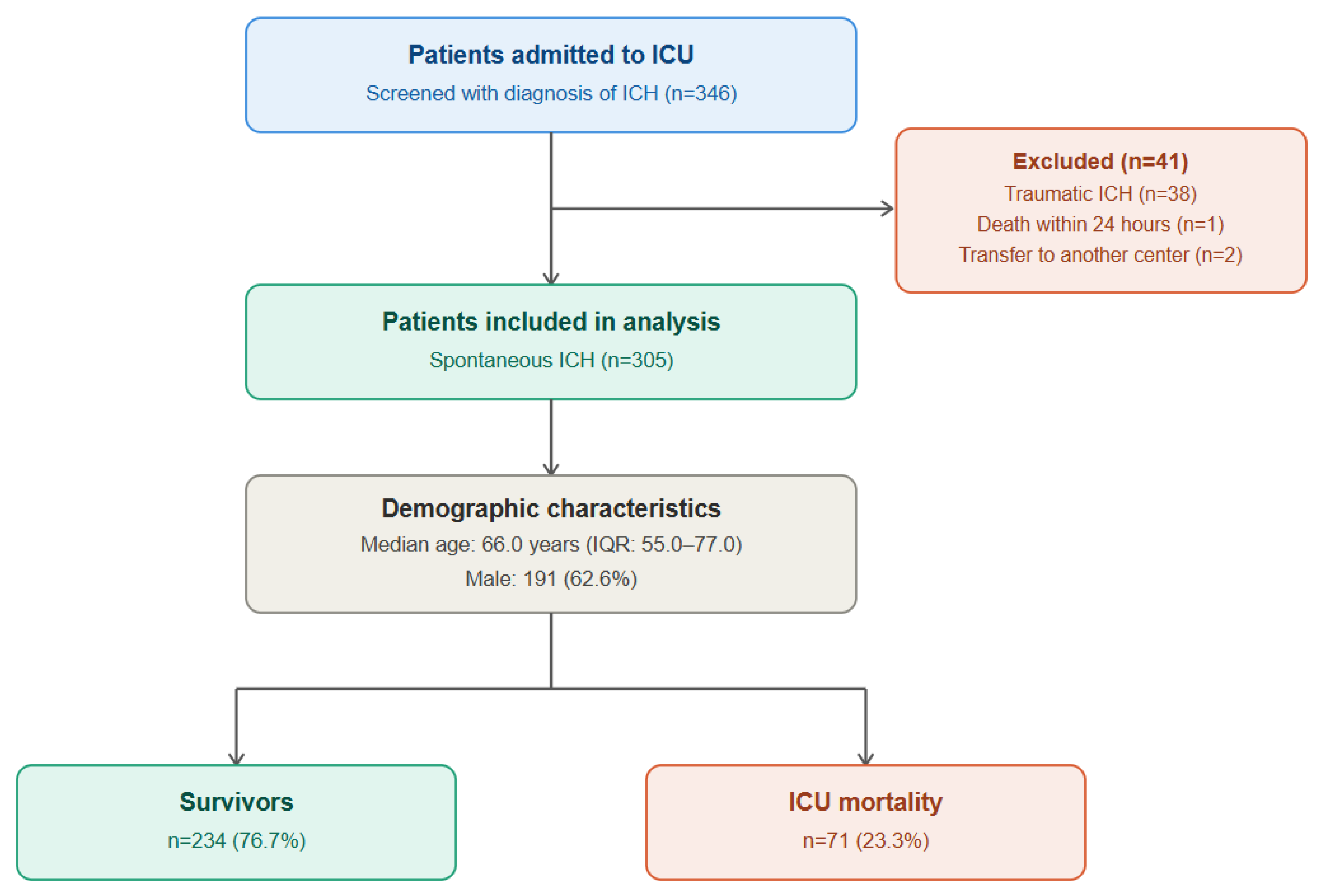
All consecutive adult patients admitted to the intensive care unit (ICU) of our hospital with a diagnosis of spontaneous intracerebral hemorrhage (ICH) between 1 September 2022 and 1 March 2026 were screened for eligibility. The diagnosis of ICH was confirmed through the combined assessment of clinical findings at presentation and neuroimaging results obtained by brain computed tomography (CT) and/or magnetic resonance imaging (MRI).
Inclusion criteria: Patients were eligible for inclusion if they were aged 18 years or older, had neuroimaging-confirmed supratentorial spontaneous (non-traumatic) intracerebral hemorrhage, required admission to the intensive care unit (ICU), and had complete routine biochemical and hematological laboratory data available at the time of admission.
Exclusion criteria: Patients were excluded if they had traumatic intracerebral hemorrhage, ischemic stroke with hemorrhagic transformation, secondary intracerebral hemorrhage caused by a brain tumor, arteriovenous malformation (AVM), or cavernoma, incomplete clinical or laboratory data, or death within the first 24 h after admission.
2.3. Collection of Demographic and Clinical Data
Demographic data, including age and sex, were recorded for all patients. Information regarding concomitant systemic diseases, including hypertension, diabetes mellitus, atrial fibrillation, coronary artery disease, and chronic kidney disease, was retrospectively obtained from medical records. Comorbidity burden was categorized into three groups: no comorbidity, a single comorbidity, and multiple comorbidities (≥2).
Neurological assessment included the Glasgow Coma Scale (GCS) score, National Institutes of Health Stroke Scale (NIHSS) score, and systolic blood pressure (mmHg) at admission, all of which were recorded simultaneously. Intensive care unit (ICU) follow-up parameters, including total ICU length of stay (days) and the requirement for mechanical ventilation (MV status was recorded at ICU admission), were also documented for each patient.
2.4. Laboratory Parameters
For all analyses, the worst laboratory values recorded within the first 24 h following ICU admission were used. Hematological parameters included white blood cell count (WBC, ×109/L), (%) platelet count (×109/L). Biochemical parameters included blood urea nitrogen (BUN), creatinine, aspartate aminotransferase (AST), alanine aminotransferase (ALT), C-reactive protein (CRP), procalcitonin, lactate dehydrogenase (LDH) and creatine kinase-MB isoenzyme (CK-MB, ng/mL). Coagulation parameters included prothrombin time (PT), international normalized ratio (INR), activated partial thromboplastin time (aPTT), and D-dimer (mg/L).
2.5. Calculation of Prognostic Scores
The CNS-LAND, APACHE II, and SOFA scores were calculated for each patient using the worst clinical and laboratory data obtained during the first 24 h after admission. The Glasgow Coma Scale (GCS) was included as a fourth comparator because it is a component of both the APACHE II and SOFA scoring systems and is also widely recognized as an independent indicator of neurological severity.
The CNS-LAND score was calculated as a 9-point scale based on seven independent variables, as originally described by Jin et al. [
9]. The cutoff values and corresponding point assignments were as follows: CK-MB ≥ 5 ng/mL (+1 point), NIHSS score 5–15 (+1 point) or 16–42 (+3 points), systolic blood pressure ≥ 160 mmHg (+1 point), LDH ≥ 220 U/L (+1 point), ALT ≥ 40 U/L (+1 point), neutrophil percentage ≥ 80% (+1 point), and D-dimer ≥ 0.5 mg/L (+1 point). Total scores were categorized as low risk (0–3 points) and high risk (4–9 points).
It should be noted that the CNS-LAND score was originally developed to predict early neurological deterioration (END) in patients with acute ischemic stroke treated with intravenous thrombolytic therapy (IVT). In the present study, however, the score was applied to a different patient population (spontaneous ICH requiring ICU admission) and evaluated against a different clinical endpoint (ICU mortality).
2.6. Primary and Secondary Endpoints
The primary endpoint was ICU mortality.
The secondary endpoints included total ICU length of stay (days) and the requirement for mechanical ventilation, both of which were compared between survivors and non-survivors. In addition, the prognostic performance of the CNS-LAND, GCS, APACHE II, and SOFA scores in predicting ICU mortality was evaluated as a secondary outcome.
2.7. Statistical Analysis
The normality of continuous variables was assessed using the Kolmogorov–Smirnov test. All continuous variables failed to meet the assumption of normality; therefore, they are presented as median (interquartile range [IQR]: Q1–Q3). Categorical variables are expressed as number and percentage (n, %). Between-group comparisons of continuous variables (survivors vs. non-survivors) were performed using the Mann–Whitney U test, while categorical variables were compared using the chi-square test or Fisher’s exact test, as appropriate.
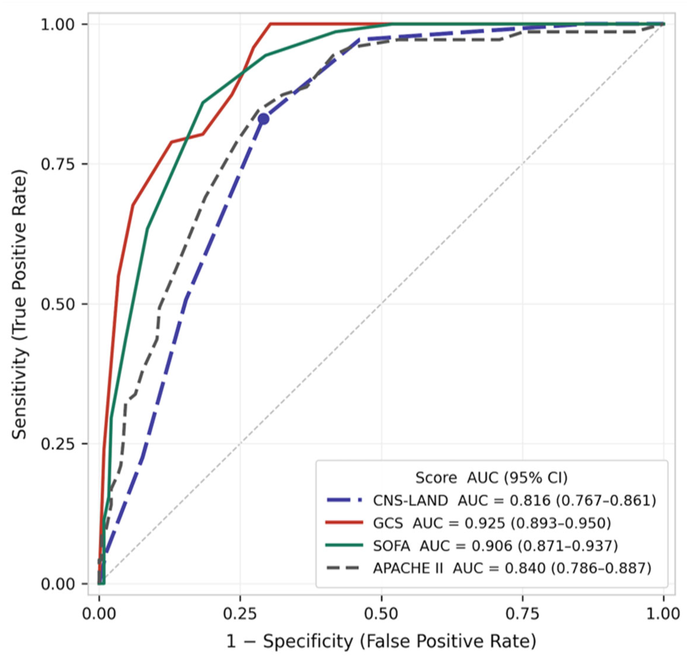
The discriminative performance of each prognostic score (CNS-LAND, GCS, SOFA, and APACHE II) for predicting ICU mortality was evaluated by receiver operating characteristic (ROC) curve analysis. The area under the ROC curve (AUC) with 95% confidence intervals (CI) was calculated using the nonparametric method. The optimal cut-off value for each score was determined by maximizing the Youden J index (J = sensitivity + specificity − 1). Sensitivity, specificity, positive predictive value (PPV), and negative predictive value (NPV) were reported at the optimal cut-off. ROC curves were generated using Python (version 3.12.3) with the following libraries: SciPy (version 1.17.1) and Matplotlib (version 3.10.8). Pairwise comparisons of AUC values between CNS-LAND and each comparator score were performed using the bootstrap-based DeLong method (2000 iterations) in MedCalc Statistical Software. Positive and negative likelihood ratios (PLR and NLR) were calculated at the optimal cut-off for each score, as these measures of diagnostic accuracy are independent of disease prevalence, unlike PPV and NPV.
Associations between prognostic scores, and between CNS-LAND and ICU mortality, were assessed using Spearman’s rank correlation coefficient (rs). To determine whether CNS-LAND provided discriminatory information independent of GCS, partial Spearman correlation analysis was performed with GCS as the controlling variable, using a two-step rank-transformation approach. The incremental contribution of CNS-LAND beyond GCS was further quantified using the Integrated Discrimination Improvement (IDI).
To evaluate whether CNS-LAND provided independent prognostic information beyond established clinical predictors, two complementary logistic regression analyses were performed. In the primary analysis, a GCS-only model (Model 1) was compared with a model additionally incorporating CNS-LAND (Model 2). In the extended analysis, a broader multivariable base model incorporating GCS, age, and comorbidity burden (Model A) was compared with a model additionally including CNS-LAND (Model B). Mechanical ventilation was excluded from all regression models due to complete separation—all non-survivors required mechanical ventilation, precluding parameter estimation. SOFA and APACHE II were likewise excluded as covariates because both scores incorporate GCS directly, which would introduce collinearity with the primary predictor of interest. Incremental model performance was assessed using the likelihood ratio test (LRT).
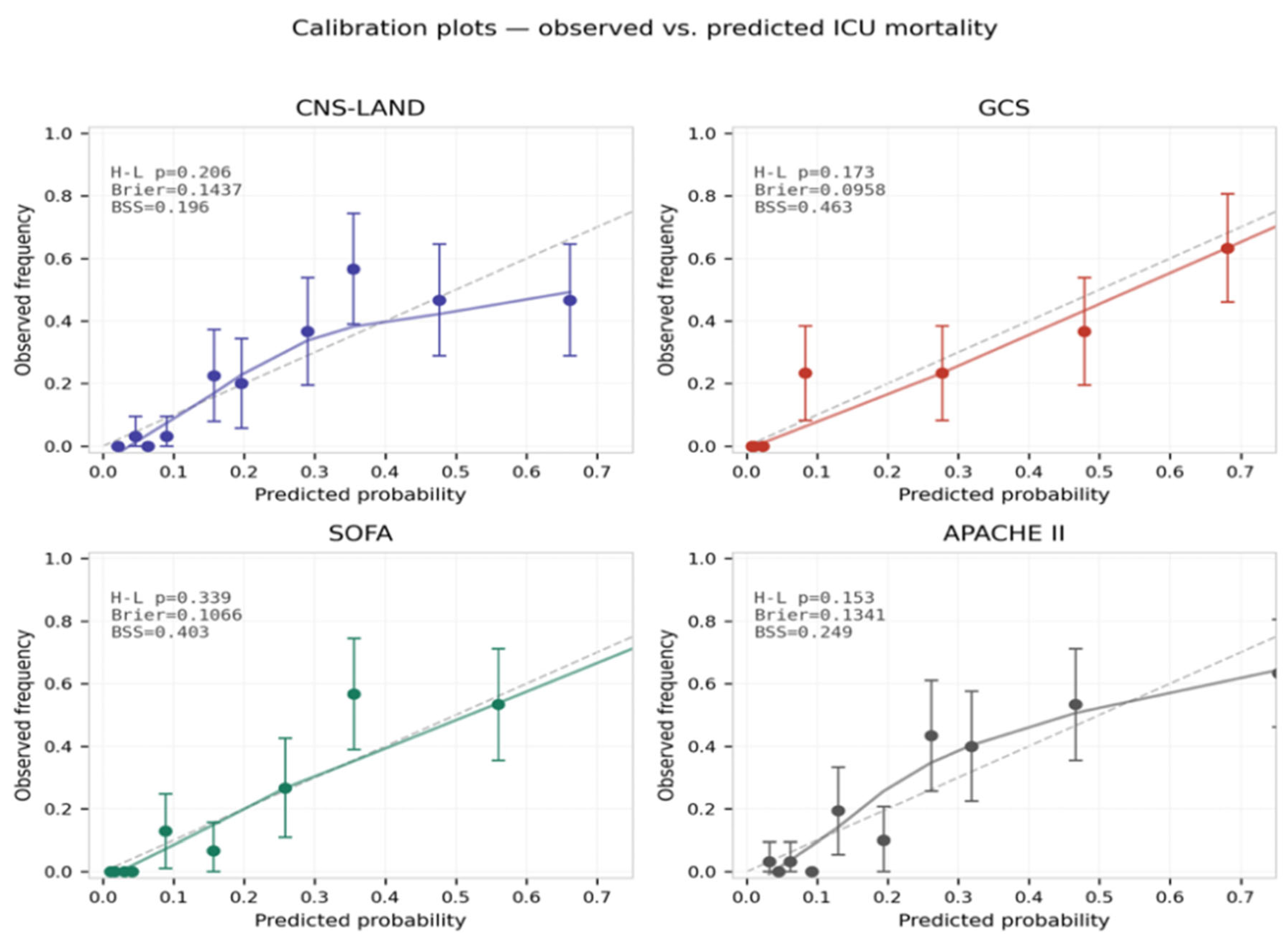
To assess model calibration, the Hosmer-Lemeshow goodness-of-fit test (10 groups, df = 8) was applied to each prognostic score, and calibration plots depicting observed versus predicted ICU mortality across deciles of predicted probability were constructed. The Brier score was calculated as a composite measure of calibration and discrimination.
Decision curve analysis (DCA) was performed to evaluate the net clinical benefit of each prognostic score across a range of threshold probabilities (5–80%), relative to the ‘treat-all’ and ‘treat-none’ strategies.
All analyses were performed using IBM SPSS Statistics version 26.0 (IBM Corp., Armonk, NY, USA) and MedCalc Statistical Software version 23.5.5 (MedCalc Software bv, Ostend, Belgium). A two-tailed p value < 0.05 was considered statistically significant.
4. Discussion
The present study investigated whether the CNS-LAND score—originally developed to predict early neurological deterioration following intravenous thrombolysis in acute ischemic stroke—could be extended to forecast ICU mortality in a clinically distinct population of patients with spontaneous intracerebral hemorrhage. Our analyses yielded several important findings. The CNS-LAND score demonstrated clinically meaningful performance in predicting mortality in this patient population, and the difference in AUC compared with APACHE II did not reach statistical significance. Considering their biological proximity to the outcome and their structural characteristics, the superior discriminative performance of the GCS and the SOFA score may be regarded as an expected finding. Perhaps the most important observation of this study, and the one that most directly qualifies CNS-LAND’s apparent performance, was that the association between CNS-LAND and mortality lost statistical significance after adjustment for GCS.
To provide a prevalence-independent assessment of diagnostic accuracy, we additionally calculated the positive and negative likelihood ratios for CNS-LAND (PLR = 2.86; NLR = 0.238). The NLR of 0.238 indicates that a CNS-LAND score below the optimal cut-off produces a moderate, clinically meaningful reduction in the post-test probability of ICU mortality, independent of the underlying event rate. By comparison, GCS demonstrated a markedly lower NLR (0.00) at its optimal threshold, reflecting its perfect sensitivity in this cohort, while SOFA showed the highest PLR (4.68) among the four scores. These likelihood ratios should be applied to the pre-test probability relevant to each clinical setting to derive a more clinically interpretable post-test probability of mortality, rather than relying on NPV alone.
The independent prognostic value of several CNS-LAND components, including LDH, CK-MB, and D-dimer, has previously been demonstrated in patients with ICH [
7,
8,
10]. Unlike ischemic stroke, ICH is characterized by intraparenchymal blood accumulation, the toxic effects of hemoglobin degradation products, and acute disruption of the blood–brain barrier, all of which trigger a profound systemic response that promotes neuroinflammation, coagulopathy, and remote organ injury [
11]. This biological framework provides a mechanistic basis for the observed associations. However, not all five components appear to contribute equally. LDH, CK-MB, and D-dimer levels were significantly higher in non-survivors, whereas ALT levels and neutrophil percentage did not differ significantly between groups. This asymmetry suggests that the individual components of the score do not provide equivalent prognostic contributions within the ICH population.
The three CNS-LAND components found to be significant are consistent with the existing literature. The median LDH level in the non-survivor group (296 U/L) markedly exceeded that of the survivor group (214 U/L), aligning with Feng et al.’s study of 406 ICH patients from the MIMIC-IV database, which demonstrated that the highest LDH quartile was associated with a significantly increased risk of 28-day mortality (HR: 3.054; 95% CI: 1.522–6.126;
p = 0.002) [
7]. The higher CK-MB levels observed in the non-survivor group (3.4 vs. 2.1 ng/mL) likely reflect the neurocardiac stress response following cerebral injury, as recent studies have highlighted that brain injury predisposes to catecholamine-mediated myocardial damage [
8]. D-dimer levels were found to be approximately twice as high in non-survivors, consistent with Delgado et al.’s prospective study of 98 acute ICH patients, which demonstrated an independent association between elevated D-dimer levels and both early neurological deterioration and first-week mortality [
10]. In contrast, the lack of a significant difference in ALT between groups suggests a limited role for this biomarker in ICH prognosis. This observation aligns with Liu et al.’s study of 639 spontaneous ICH patients, which reported that among liver function markers, AST and ALP were associated with clinical outcomes, whereas ALT did not emerge as an independent predictor [
12]. Regarding neutrophil percentage, the already elevated baseline neutrophil activation in critically ill ICH patients may have masked intergroup differences. Indeed, the ICH literature suggests that the neutrophil-to-lymphocyte ratio, rather than neutrophil percentage alone, serves as a stronger prognostic indicator; in a meta-analysis encompassing 21 studies and 7176 patients, Guo et al. found that elevated neutrophil-to-lymphocyte was significantly associated with mortality (OR: 1.10; 95% CI: 1.04–1.17) and poor functional outcome (OR: 1.29; 95% CI: 1.17–1.41) [
13]. These findings underscore the need to re-evaluate the relative biological weighting of CNS-LAND components in the ICH population in future studies.
Taken together, these findings indicate that the apparent discriminative performance of CNS-LAND in this cohort is not attributable to the score as a composite construct, but is almost entirely driven by a single component: the NIHSS. To state this as plainly as possible: CNS-LAND does not “work” in ICH because its biochemical components (CK-MB, LDH, ALT, neutrophil percentage, D-dimer) meaningfully track mortality; it appears to work only because it inherits, through its NIHSS sub-item, information that GCS already provides. Once that shared information is removed, no independent signal remains. Consequently, when GCS is accounted for, CNS-LAND does not provide independent discriminative information regarding mortality: the partial Spearman correlation with mortality decreased to −0.051 (
p = 0.373), and binary logistic regression confirmed that adding CNS-LAND to a model containing GCS alone did not significantly improve discriminative performance (ΔAUC = 0.000; LRT χ
2 = 0.121,
p = 0.728; IDI = 0.001). This pattern contrasts sharply with the context in which CNS-LAND was originally developed—acute ischemic stroke—where NIHSS can be markedly elevated due to aphasia or hemiplegia while consciousness remains largely intact, allowing for meaningful dissociation between NIHSS and GCS. In ICH, however, the presence of mass effect and progressive herniation leads to parallel progression of neurological deficits and impaired consciousness, rendering GCS and NIHSS functionally overlapping scales. Indeed, previous studies have reported a strong correlation between NIHSS and GCS in ICH patients (r = −0.773;
p < 0.001) and suggested that this relationship may be even more pronounced in severe ICH cases requiring intensive care [
14]. The higher correlation coefficient observed in our study (r = −0.964) can be attributed to our cohort, which predominantly included severe ICU-level cases where deficits in consciousness and focal neurological function progressed concurrently.
Similarly, the superior discriminative performance of the SOFA score (AUC: 0.906) is biologically plausible and consistent with its intended clinical application. In a prospective study of 352 mixed ICU patients, Ferreira et al. demonstrated that SOFA is a robust predictor of mortality across the general critical care population [
5]. The present findings extend this observation to patients with spontaneous ICH, further supporting the utility of SOFA as a reliable prognostic tool in neurocritical care settings. It is worth acknowledging that SOFA incorporates GCS as its neurological component, and APACHE II includes neurological assessment indirectly. This overlap may partially explain their superior performance; nonetheless, both scores reflect multiorgan dysfunction beyond neurological status alone, which likely accounts for their incremental discriminative value over CNS-LAND in this critically ill population.
The absence of a statistically significant difference between CNS-LAND and APACHE II (AUC: 0.816 vs. 0.840;
p = 0.467) represents one of the notable findings of our study. However, this result should not be interpreted as evidence of statistical equivalence. Given the relatively wide confidence interval (95% CI: −0.088 to +0.040), the possibility that APACHE II may be truly superior cannot be excluded. Nevertheless, the lack of a significant performance difference between CNS-LAND—comprising only seven routinely available biochemical parameters—and APACHE II, a comprehensive scoring system incorporating 12 physiological variables in addition to age and chronic health status, may suggest a practical advantage in terms of ease of calculation and clinical applicability [
6]. Two principal mechanisms may explain the comparable performance of CNS-LAND and APACHE II. First, the significantly higher levels of inflammatory biomarkers not included in the CNS-LAND framework, namely C-reactive protein (CRP) and procalcitonin, in the non-survivor group—CRP: 12.5 vs. 8.6 mg/L (
p = 0.013); procalcitonin: 0.2 vs. 0.1 ng/mL (
p < 0.001)—suggest that the systemic inflammatory response in severe ICH extends beyond the biochemical domains captured by currently available scoring systems. This observation is consistent with the meta-analysis by Guo and Zou involving 25,928 patients, which demonstrated that elevated neutrophil-to-lymphocyte ratio (NLR), white blood cell count (WBC), and CRP levels were associated with poor functional outcomes and increased mortality [
15]. Second, the inclusion within CNS-LAND of biomarkers such as LDH, CK-MB, and D-dimer—which directly reflect the systemic response triggered by hemorrhagic stroke—may explain why the broader physiological coverage of APACHE II fails to provide additional discriminative value in this population. This finding is also in agreement with the observations of Nilsson et al., who demonstrated that common ICU scoring systems did not outperform age and GCS in predicting mid-term mortality in ICH patients [
16]. In this context, CNS-LAND may be considered a simpler and potentially more practical alternative to APACHE II.
Calibration analysis confirmed that CNS-LAND demonstrated adequate goodness-of-fit across deciles of predicted probability (Hosmer-Lemeshow χ2 = 10.931; p = 0.206), comparable to GCS (p = 0.173), SOFA (p = 0.339), and APACHE II (p = 0.153). However, the Brier score for CNS-LAND (0.144) was notably higher than those of GCS (0.096) and SOFA (0.107), reflecting its comparatively lower composite predictive accuracy—a finding consistent with its inferior AUC. While all four scores were adequately calibrated, these results reinforce that calibration alone does not distinguish CNS-LAND from its comparators; it is the discrimination gap that limits its clinical utility in this population.
Decision curve analysis provided further insight into the comparative clinical utility of these scores. CNS-LAND offered meaningful net benefit primarily at threshold probabilities ≤25%, consistent with a rule-out role at lower risk thresholds. At threshold probabilities ≥30%—which are more clinically relevant for decisions to escalate monitoring or intervention—its net benefit diminished markedly and approached zero at 50%, at which point GCS continued to demonstrate substantial net benefit. This divergence provides additional evidence that GCS offers more robust and consistent decision-making utility across the full spectrum of clinically relevant threshold probabilities in ICU patients with spontaneous ICH.
Several limitations of this study should be acknowledged. First, the retrospective, single-center design introduces the potential for selection bias, and the findings cannot be generalized without prospective evaluation in external cohorts. Second, key radiological variables with well-established, independent prognostic weight in ICH—including hematoma volume, hematoma location, presence of intraventricular hemorrhage, hydrocephalus, and midline shift—were not systematically extracted from imaging reports and could not be incorporated into the analysis. Because these variables are precisely the determinants that structure the ICH Score, and none of them is captured by CNS-LAND, GCS, SOFA, or APACHE II, their omission is a substantive limitation rather than an incidental one [
4]. It prevented calculation of the ICH Score as a direct comparator, and it leaves open the possibility that CNS-LAND’s residual, GCS-independent association with mortality—however small in this cohort—partly reflects unmeasured radiological severity rather than the biochemical processes the score was designed to capture. Furthermore, the cohort comprised exclusively supratentorial ICH cases, which limits the generalizability of the findings to infratentorial hemorrhages such as cerebellar and brainstem bleeding. Third, biomarkers were measured only at admission, and their temporal dynamics were not assessed. Fourth, because CNS-LAND was originally developed to predict early neurological deterioration, its evaluation against a different outcome measure—ICU mortality—in the present study inevitably introduces a degree of outcome mismatch. Finally, patients who died within the first 24 h of ICU admission were excluded because complete laboratory and clinical data required for score calculation were not available within this timeframe. Although only one patient was excluded on this basis, this subgroup likely represents the most critically ill cases, and their exclusion may have led to an overestimation of the predictive performance of the evaluated scoring systems. Another point that warrants consideration is the use of ICU mortality as the primary outcome. In the Turkish healthcare system, intensive care services are organized according to the level of organ support provided (Level 1–3), rather than by a distinction between acute and chronic care. For patients with severe neurological injury who survive the acute phase but require prolonged life support, such as tracheostomy or home mechanical ventilation, all preparations for transfer to long-term or palliative care facilities are generally completed within the ICU. Consequently, ICU mortality is expected to closely approximate hospital mortality in our setting, minimizing the potential for bias arising from inter-facility transfers or early discharge. Nevertheless, this organizational structure may differ across healthcare systems, and this should be taken into account when interpreting and generalizing our findings to other settings.