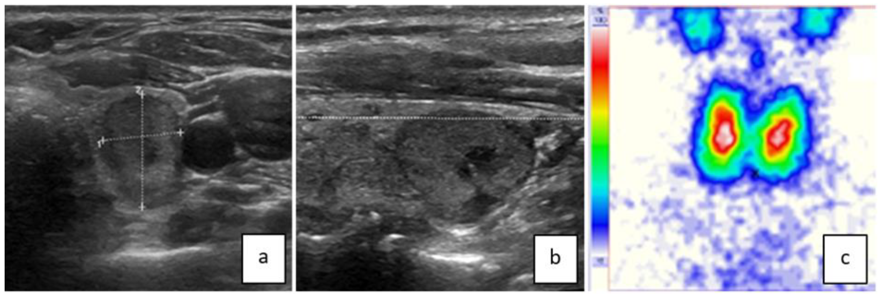
Thyroid Imaging Reporting and Data Systems: Applicability of the “Taller than Wide” Criterium in Primary/Secondary Care Units and the Role of Thyroid Scintigraphy
Abstract
1. Introduction
2. Materials and Methods
2.1. Patients
2.2. Reference Standard
2.3. Nodules
2.4. Ethics
2.5. Examinations
2.6. Statistical Analysis
3. Results
3.1. Center A
3.1.1. Overall Characteristics of Patients and Nodules
3.1.2. Nodules with Available Reference Standard (n = 200)
3.2. Center B
4. Discussion
Limitations
5. Conclusions
Author Contributions
Funding
Institutional Review Board Statement
Informed Consent Statement
Data Availability Statement
Acknowledgments
Conflicts of Interest
References
- Horvath, E.; Majlis, S.; Rossi, R.; Franco, C.; Niedmann, J.P.; Castro, A.; Dominguez, M. An Ultrasonogram Reporting System for Thyroid Nodules Stratifying Cancer Risk for Clinical Management. J. Clin. Endocrinol. Metab. 2009, 94, 1748–1751. [Google Scholar] [CrossRef] [PubMed]
- Kwak, J.Y.; Han, K.H.; Yoon, J.H.; Moon, H.J.; Son, E.J.; Park, S.H.; Jung, H.K.; Choi, J.S.; Kim, B.M.; Kim, E.K. Thyroid imaging reporting and data system for US features of nodules: A step in establishing better stratification of cancer risk. Radiology 2011, 260, 892–899. [Google Scholar] [CrossRef] [PubMed]
- Shin, J.H.; Baek, J.H.; Chung, J.; Ha, E.J.; Kim, J.-H.; Lee, Y.H.; Lim, H.K.; Moon, W.-J.; Na, D.G.; Park, J.S.; et al. Ultrasonography Diagnosis and Imaging-Based Management of Thyroid Nodules: Revised Korean Society of Thyroid Radiology Consensus Statement and Recommendations. Korean J. Radiol. 2016, 17, 370–395. [Google Scholar] [CrossRef]
- Russ, G.; Bonnema, S.J.; Erdogan, M.F.; Durante, C.; Ngu, R.; Leenhardt, L. European Thyroid Association Guidelines for Ultrasound Malignancy Risk Stratification of Thyroid Nodules in Adults: The EU-TIRADS. Eur. Thyroid. J. 2017, 6, 225–237. [Google Scholar] [CrossRef] [PubMed]
- Tessler, F.N.; Middleton, W.D.; Grant, E.G.; Hoang, J.K.; Berland, L.L.; Teefey, S.A.; Cronan, J.J.; Beland, M.D.; Desser, T.S.; Frates, M.C.; et al. ACR Thyroid Imaging, Reporting and Data System (TI-RADS): White Paper of the ACR TI-RADS Committee. J. Am. Coll. Radiol. 2017, 14, 587–595. [Google Scholar] [CrossRef] [PubMed]
- Haugen, B.R.; Alexander, E.K.; Bible, K.C.; Doherty, G.M.; Mandel, S.J.; Nikiforov, Y.E.; Pacini, F.; Randolph, G.W.; Sawka, A.M.; Schlumberger, M.; et al. 2015 American Thyroid Association Management Guidelines for Adult Patients with Thyroid Nodules and Differentiated Thyroid Cancer: The American Thyroid Association Guidelines Task Force on Thyroid Nodules and Differentiated Thyroid Cancer. Thyroid 2016, 26, 1–133. [Google Scholar] [CrossRef]
- Ha, E.J.; Chung, S.R.; Na, D.G.; Ahn, H.S.; Chung, J.; Lee, J.Y.; Park, J.S.; Yoo, R.-E.; Baek, J.H.; Baek, S.M.; et al. 2021 Korean Thyroid Imaging Reporting and Data System and Imaging-Based Management of Thyroid Nodules: Korean Society of Thyroid Radiology Consensus Statement and Recommendations. Korean J. Radiol. 2021, 22, 2094–2123. [Google Scholar] [CrossRef] [PubMed]
- Seifert, P.; Schenke, S.; Zimny, M.; Stahl, A.; Grunert, M.; Klemenz, B.; Freesmeyer, M.; Kreissl, M.C.; Herrmann, K.; Görges, R. Diagnostic Performance of Kwak, EU, ACR, and Korean TIRADS as Well as ATA Guidelines for the Ultrasound Risk Stratification of Non-Autonomously Functioning Thyroid Nodules in a Region with Long History of Iodine Deficiency: A German Multicenter Trial. Cancers 2021, 13, 4467. [Google Scholar] [CrossRef]
- Fu, C.; Cui, Y.; Li, J.; Yu, J.; Wang, Y.; Si, C.; Cui, K. Effect of the categorization method on the diagnostic performance of ultrasound risk stratification systems for thyroid nodules. Front. Oncol. 2023, 13, 1073891. [Google Scholar] [CrossRef]
- Petersen, M.; Schenke, S.A.; Zimny, M.; Görges, R.; Grunert, M.; Groener, D.; Seifert, P.; Stömmer, P.E.; Kreissl, M.C.; Stahl, A.R.; et al. Introducing a Pole Concept for Nodule Growth in the Thyroid Gland: Taller-than-Wide Shape, Frequency, Location and Risk of Malignancy of Thyroid Nodules in an Area with Iodine Deficiency. J. Clin. Med. 2022, 11, 2549. [Google Scholar] [CrossRef]
- Remonti, L.R.; Kramer, C.K.; Leitão, C.B.; Pinto, L.C.F.; Gross, J.L. Thyroid Ultrasound Features and Risk of Carcinoma: A Systematic Review and Meta-Analysis of Observational Studies. Thyroid 2015, 25, 538–550. [Google Scholar] [CrossRef]
- Brito, J.P.; Gionfriddo, M.R.; Al Nofal, A.; Boehmer, K.R.; Leppin, A.L.; Reading, C.; Callstrom, M.; Elraiyah, T.A.; Prokop, L.J.; Stan, M.N.; et al. The Accuracy of Thyroid Nodule Ultrasound to Predict Thyroid Cancer: Systematic Review and Meta-Analysis. J. Clin. Endocrinol. Metab. 2014, 99, 1253–1263. [Google Scholar] [CrossRef]
- Seifert, P.; Görges, R.; Zimny, M.; Kreissl, M.C.; Schenke, S. Interobserver agreement and efficacy of consensus reading in Kwak-, EU-, and ACR thyroid imaging recording and data systems and ATA guidelines for the ultrasound risk stratification of thyroid nodules. Endocrine 2020, 67, 143–154. [Google Scholar] [CrossRef] [PubMed]
- Cibas, E.S.; Ali, S.Z. The Bethesda System for Reporting Thyroid Cytopathology. Am. J. Clin. Pathol. 2009, 132, 658–665. [Google Scholar] [CrossRef]
- Giovanella, L.; Avram, A.M.; Iakovou, I.; Kwak, J.; Lawson, S.A.; Lulaj, E.; Luster, M.; Piccardo, A.; Schmidt, M.; Tulchinsky, M.; et al. EANM practice guideline/SNMMI procedure standard for RAIU and thyroid scintigraphy. Eur. J. Nucl. Med. 2019, 46, 2514–2525. [Google Scholar] [CrossRef]
- Grussendorf, M.; Ruschenburg, I.; Brabant, G. Malignancy rates in thyroid nodules: A long-term cohort study of 17,592 patients. Eur. Thyroid. J. 2022, 11, e220027. [Google Scholar] [CrossRef]
- Treglia, G.; Caldarella, C.; Saggiorato, E.; Ceriani, L.; Orlandi, F.; Salvatori, M.; Giovanella, L. Diagnostic performance of 99mTc-MIBI scan in predicting the malignancy of thyroid nodules: A meta-analysis. Endocrine 2013, 44, 70–78. [Google Scholar] [CrossRef] [PubMed]
- Hurtado-López, L.-M.; Martínez-Duncker, C. Negative MIBI thyroid scans exclude differentiated and medullary thyroid cancer in 100% of patients with hypofunctioning thyroid nodules. Eur. J. Nucl. Med. 2007, 34, 1701–1703. [Google Scholar] [CrossRef] [PubMed]
- Kim, S.-J.; Lee, S.-W.; Jeong, S.Y.; Pak, K.; Kim, K. Diagnostic Performance of Technetium-99m Methoxy-Isobutyl-Isonitrile for Differentiation of Malignant Thyroid Nodules: A Systematic Review and Meta-Analysis. Thyroid 2018, 28, 1339–1348. [Google Scholar] [CrossRef] [PubMed]
- Middleton, W.D.; Teefey, S.A.; Reading, C.C.; Langer, J.E.; Beland, M.D.; Szabunio, M.M.; Desser, T.S. Comparison of Performance Characteristics of American College of Radiology TI-RADS, Korean Society of Thyroid Radiology TIRADS, and American Thyroid Association Guidelines. Am. J. Roentgenol. 2018, 210, 1148–1154. [Google Scholar] [CrossRef]
- Ospina, N.S.; Brito, J.P.; Maraka, S.; de Ycaza, A.E.E.; Rodriguez-Gutierrez, R.; Gionfriddo, M.R.; Castaneda-Guarderas, A.; Benkhadra, K.; Al Nofal, A.; Erwin, P.; et al. Diagnostic accuracy of ultrasound-guided fine needle aspiration biopsy for thyroid malignancy: Systematic review and meta-analysis. Endocrine 2016, 53, 651–661. [Google Scholar] [CrossRef] [PubMed]
- Bongiovanni, M.; Spitale, A.; Faquin, W.C.; Mazzucchelli, L.; Baloch, Z.W. The Bethesda System for Reporting Thyroid Cytopathology: A Meta-Analysis. Acta Cytol. 2012, 56, 333–339. [Google Scholar] [CrossRef]
- Schenke, S.A.; Kreissl, M.C.; Grunert, M.; Hach, A.; Haghghi, S.; Kandror, T.; Peppert, E.; Rosenbaum-Krumme, S.; Ruhlmann, V.; Stahl, A.; et al. Distribution of Functional Status of Thyroid Nodules and Malignancy Rates of Hyperfunctioning and Hypofunctioning Thyroid Nodules in Germany. Nuklearmedizin 2022, 61, 376–384. [Google Scholar] [CrossRef] [PubMed]
- Schenke, S.; Seifert, P.; Zimny, M.; Winkens, T.; Binse, I.; Goerges, R. Risk Stratification of Thyroid Nodules Using the Thyroid Imaging Reporting and Data System (TIRADS): The Omission of Thyroid Scintigraphy Increases the Rate of Falsely Suspected Lesions. J. Nucl. Med. 2019, 60, 342–347. [Google Scholar] [CrossRef]
- Hoang, J.K.; Middleton, W.D.; Tessler, F.N. Update on ACR TI-RADS: Successes, Challenges, and Future Directions, From the AJR Special Series on Radiology Reporting and Data Systems. Am. J. Roentgenol. 2021, 216, 570–578. [Google Scholar] [CrossRef] [PubMed]
- Kim, P.H.; Suh, C.H.; Baek, J.H.; Chung, S.R.; Choi, Y.J.; Lee, J.H. Unnecessary thyroid nodule biopsy rates under four ultrasound risk stratification systems: A systematic review and meta-analysis. Eur. Radiol. 2021, 31, 2877–2885. [Google Scholar] [CrossRef]
- Grani, G.; Del Gatto, V.; Cantisani, V.; Mandel, S.J.; Durante, C. A Reappraisal of Suspicious Sonographic Features of Thyroid Nodules: Shape Is Not an Independent Predictor of Malignancy. J. Clin. Endocrinol. Metab. 2023, 108, e816–e822. [Google Scholar] [CrossRef]
- Frilling, A.; Autschbach, R.; Gomez, J.R.; Kremer, K. Cold struma nodule—An absolute surgical indication? Langenbeck’s Arch. Surg. 1986, 369, 207–208. [Google Scholar] [CrossRef]
- Ardakani, A.A.; Mohammadzadeh, A.; Yaghoubi, N.; Ghaemmaghami, Z.; Reiazi, R.; Jafari, A.H.; Hekmat, S.; Shiran, M.B.; Bitarafan-Rajabi, A. Predictive quantitative sonographic features on classification of hot and cold thyroid nodules. Eur. J. Radiol. 2018, 101, 170–177. [Google Scholar] [CrossRef]
- Carlé, A.; Krejbjerg, A.; Laurberg, P. Epidemiology of nodular goitre. Influence of iodine intake. Best Pr. Res. Clin. Endocrinol. Metab. 2014, 28, 465–479. [Google Scholar] [CrossRef]


| Patients (n = 223) | Nodules ttw + Solid (n = 259) | Diagnostics | Reference Standard n = 200 |
|---|---|---|---|
| female/male 144/79 age (years) 61 ± 14 | max. size (mm) 20 ± 10 | FNB: 149 scintigraphy: 252 MIBI scan: 8 | unequivocal FNB: 133 surgery: 36 other (MIBI imaging, clinical course, laboratory values): 33 |
| After Exclusion of Hyperfunctioning Nodules (n = 176) | |||||
|---|---|---|---|---|---|
| Benign | Malignant | p | Benign | Malignant | |
| number | 195 | 5 →ROM 2.5% 95% CI: 5.1% | 171 | 5 ROM 2.8% upper limit 95% CI: 5.7% | |
| main size (mm) | 20 | 23 | |||
| nodules ≥15 mm | 127 | 3 | 0.81 | 113 | 3 (p = 0.085) |
| hypoechoic | 47 | 1 | 0.75 | ||
| irregular margin | 19 | 1 | 1 | ||
| microcalcification | 23 | 4 | <<0.01 | ||
| number additional US risk features per nodule | 89/195 | 6/5 | 0.02 | ||
| nodules without additional US risk features, i.e., exclusively solid and ttw | 131 | 1 | 0.09 | 119 (of these 84 ≥15 mm) | 1 (p = 0.06) ROM 0.9% upper limit 95% CI: 3.7% (of these, 0 carcinomas ≥15 mm) ROM 0% upper limit 95% CI: 3.5% |
| scintigraphically | |||||
| hyperfunctioning | 23 | 0 | 0.97 | ||
| hypofunctioning | 47 | 2 →ROM 4% upper limit 95% CI: 9.0% | 0.58 | ||
| indifferent | 118 | 2 | 1 |
| #1 | #2 | #3 | #4 | #5 | |
|---|---|---|---|---|---|
| size (mm) transversal × sagittal × vertical | 39 × 43 × 53 | 10 × 11 × 18 | 19 × 20 × 24 | 9 × 10 × 11 | 10 × 11 × 11 |
| hypoechoic | no | yes | no | no | no |
| irregular margin/microlobulation | no | no | yes | no | no |
| microcalcification | yes | yes | yes | no | yes |
| scintigraphy | hypo-functioning | - | hypo-functioning | indifferent | indifferent |
| Benign n (%) | Malignant n (%) | p | Reference Standard n (%) | ||
|---|---|---|---|---|---|
| number | 46 (79) | 12 (21) | →ROM 21% lower border 95% CI: 12% | FNB 45 (78) | |
| mean size (mm) | 29 | 25 | surgery 39 (67) | ||
| ≥15 mm | 46 (79) | 10 (17) | 0.05 | ||
| hypoechoic | 29 (50) | 12 (21) | 0.03 | ||
| irregular margin | 6 (10) | 7 (12) | 0.003 | ||
| microcalcification | 8 (14) | 6 (10) | 0.05 | ||
| solid | 33 (57) | 12 (21) | 0.09 | ||
| hypofunctioning + ttw + solid * | 10 (17) | 0 (0) | 0.18 |
Disclaimer/Publisher’s Note: The statements, opinions and data contained in all publications are solely those of the individual author(s) and contributor(s) and not of MDPI and/or the editor(s). MDPI and/or the editor(s) disclaim responsibility for any injury to people or property resulting from any ideas, methods, instructions or products referred to in the content. |
© 2024 by the authors. Licensee MDPI, Basel, Switzerland. This article is an open access article distributed under the terms and conditions of the Creative Commons Attribution (CC BY) license (https://creativecommons.org/licenses/by/4.0/).
Share and Cite
Petersen, M.; Schenke, S.A.; Veit, F.; Görges, R.; Seifert, P.; Zimny, M.; Croner, R.S.; Kreissl, M.C.; Stahl, A.R. Thyroid Imaging Reporting and Data Systems: Applicability of the “Taller than Wide” Criterium in Primary/Secondary Care Units and the Role of Thyroid Scintigraphy. J. Clin. Med. 2024, 13, 514. https://doi.org/10.3390/jcm13020514
Petersen M, Schenke SA, Veit F, Görges R, Seifert P, Zimny M, Croner RS, Kreissl MC, Stahl AR. Thyroid Imaging Reporting and Data Systems: Applicability of the “Taller than Wide” Criterium in Primary/Secondary Care Units and the Role of Thyroid Scintigraphy. Journal of Clinical Medicine. 2024; 13(2):514. https://doi.org/10.3390/jcm13020514
Chicago/Turabian StylePetersen, Manuela, Simone A. Schenke, Franziska Veit, Rainer Görges, Philipp Seifert, Michael Zimny, Roland S. Croner, Michael C. Kreissl, and Alexander R. Stahl. 2024. "Thyroid Imaging Reporting and Data Systems: Applicability of the “Taller than Wide” Criterium in Primary/Secondary Care Units and the Role of Thyroid Scintigraphy" Journal of Clinical Medicine 13, no. 2: 514. https://doi.org/10.3390/jcm13020514
APA StylePetersen, M., Schenke, S. A., Veit, F., Görges, R., Seifert, P., Zimny, M., Croner, R. S., Kreissl, M. C., & Stahl, A. R. (2024). Thyroid Imaging Reporting and Data Systems: Applicability of the “Taller than Wide” Criterium in Primary/Secondary Care Units and the Role of Thyroid Scintigraphy. Journal of Clinical Medicine, 13(2), 514. https://doi.org/10.3390/jcm13020514


