
Metal Allergy: State-of-the-Art Mechanisms, Biomarkers, Hypersensitivity to Implants


Abstract
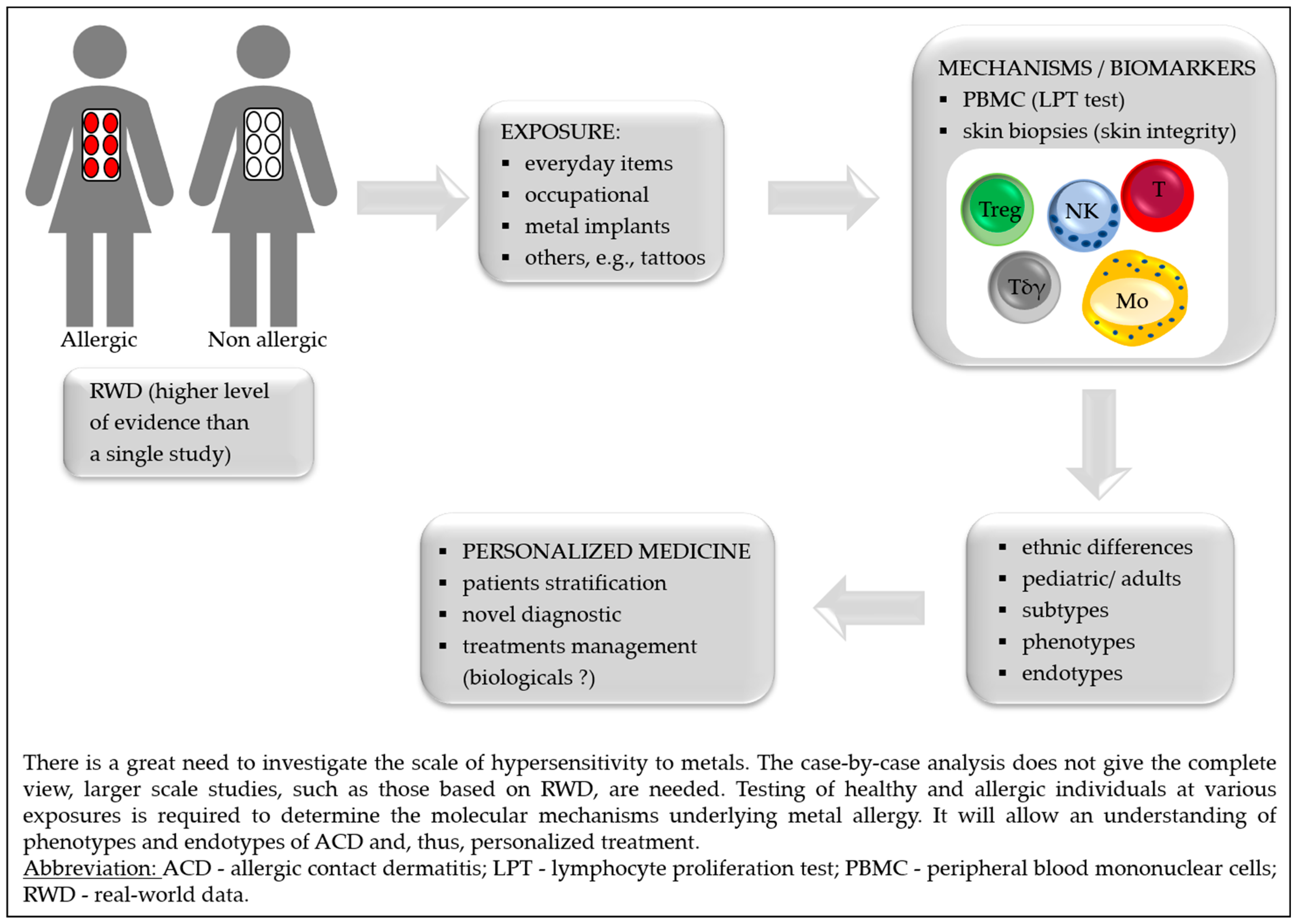
1. Introduction
2. Exposure
3. Diagnostics
4. Mechanisms and Biomarkers

| Outcomes | Exposition to Metal | Ref. |
|---|---|---|
| ↑ metal-specific CD154+ CD4+ Tmem (overexpression of TRAV9-2 and CDR3 histidine) | Allergic and non-allergic subjects stimulated with Ni, Co or Pd (PBMC) | [67,68] |
| ↑ IL-5 | Ni-allergic patients (PBMC) | [69] |
| Ni-allergy patients, differentiation between independent Ni or cross-reactivity of Ni/Pd allergy (PBMC) | [71] | |
| Cross reactivity between Ni/Cr and Pd | Sensitization with Ni or Cr, challenge with Pd (mice model) | [70] |
| Skin barrier defects: ↓ terminal differentiation—FLG, FLG2, LOR, LCEs, tight junction—CLDN1/CLDN8, lipid metabolism—FA2H, FABP7 | Biopsies from healthy subjects after Ni-topical application | [73] |
| Cellular infiltrates: ↑ CD3+ T, CD11c+ myeloid DC, DC-LAMP+ mature DC, MBP+ eosinophils, FOXP3+ Treg | ||
| ↑ M1, mast cells, neutrophils, NK, CD4+ Tmem, CD8+ T | Biopsies from Ni-allergic patients | [74] |
| ↓ M2, resting mast cells, Tγδ, Treg | ||
| ↓ SBSN | Ni-allergic patients (serum) | [75] |
| ↑ Sema3A (activates MAPK and TNF-α) | Ni-induced allergy (mouse ear tissue) | [76] |
| ↑ TSLP in keratinocytes and TNF-α in epithelium | OLPs metal-allergy patients | [77] |
| ↑ IL-6, CXCL8, CCL2, CCL5, and CCL20 | RHS exposure to Ni and Streptococcus mitis exposure | [78] |
| Lipid profile: ↑ cholesterol, DAG, MAG | Non-allergic skin exposed to Co | [79] |
| Lipid profile: ↑ DAG | Non-allergic skin exposed to Cr | |
| Lipid profile: ↓ DAG and MAG | Non-allergic skin exposed to Ni | |
| LC emigration of the epidermis (in an IL-10 but not IL-1B dependent way) | RHS exposure to Ti | [80] |
5. Implants
| Metal that Caused Hypersensitivity/Implant Type | Symptoms | Ref. |
|---|---|---|
| Multiple metals/orthopedic implants: hip, knee shoulder joint | Different; the most common—delayed wound healing and/or recurrent wound issues after the implantation, joint failure or loosening | [86,92,93,98,106] |
| Ni/endovascular implants (stents) | In-stent restenosis or prominent eczematous reaction overlying the endovascular implant, eosinophilia | [102,107] |
| Co, Ni/drug-eluting stents | Pruritic rash with hypereosinophilia | [108] |
| Au and Pd/dental implant | Oral lichenoid contact lesion (OLCLs) | [103] |
| Ti/dental implants | Rash, urticaria, pruritus, redness, dermatitis and facial eczema, pain, hyperaemia of soft tissues, swelling in submental and labial sulcus, gingival hyperplasia acne-like facial inflammation | [105] |
| Ni/metal anchors | Erythematous and vesicular lesions around the grafted tattoo skin, but the tattoo was not affected (placed with clips or anchors) | [59] |
| Ni/stainless-steel skull pins | Erythema on sites of the head where the skull pins inserted | [109] |
| Ti/cervical implant | Persistent refractory neck pain; subsequently, after eight years, a planter rush | [110] |
| Ti/metal clips for cholecystectomy | Right upper quadrant pain, diarrhea, and nausea | [111] |
| Low-grade fever, nausea, vomiting, joint pain, bloody diarrhea | [112] | |
| Co, Ni, Hg/metal clips for cholecystectomy | Myalgia, joint pain and tenderness, mental fogginess, mild forgetfulness, irritable bowel syndrome, stomach cramps, dry skin and hair, hair loss | [113] |
| Cu/intrauterine device | Cutaneous eruption | [114] |
| Multiple-metals/dental implant | Palmoplantar pustulosis (PPP) and periodontitis | [104,115] |
| Au/dental implants | Oral lesions with characteristic Wickham’s striae | [116] |
| Ti/temporary tissue expander | Well-demarcated, erythematous plaque over the left breast reconstructive breast surgery | [117] |
6. Treatment with Biologicals
7. Conclusions
Funding
Institutional Review Board Statement
Informed Consent Statement
Conflicts of Interest
References
- Johansen, J.D.; Bonefeld, C.M.; Schwensen, J.F.B.; Thyssen, J.P.; Uter, W. Novel insights into contact dermatitis. J. Allergy Clin. Immunol. 2022, 149, 1162–1171. [Google Scholar] [CrossRef] [PubMed]
- Luger, T.; Amagai, M.; Dreno, B.; Dagnelie, M.-A.; Liao, W.; Kabashima, K.; Schikowski, T.; Proksch, E.; Elias, P.M.; Simon, M.; et al. Atopic dermatitis: Role of the skin barrier, environment, microbiome, and therapeutic agents. J. Dermatol. Sci. 2021, 102, 142–157. [Google Scholar] [CrossRef] [PubMed]
- Fonacier, L.; Frankel, D.; Mawhirt, S. Contact allergens for the allergist. Ann. Allergy Asthma Immunol. 2022, 128, 629–644. [Google Scholar] [CrossRef]
- Agache, I.; Akdis, C.A. Precision medicine and phenotypes, endotypes, genotypes, regiotypes, and theratypes of allergic diseases. J. Clin. Investig. 2019, 129, 1493–1503. [Google Scholar] [CrossRef] [PubMed]
- Tokura, Y.; Hayano, S. Subtypes of atopic dermatitis: From phenotype to endotype. Allergol. Int. 2022, 71, 14–24. [Google Scholar] [CrossRef]
- Wang, T.; Yin, L.; Ma, Z.; Zhang, Y. Chlorogenic Acid-Loaded Mesoporous Silica Nanoparticles Modified with Hexa-Histidine Peptides Reduce Skin Allergies by Capturing Nickel. Molecules 2022, 27, 1430. [Google Scholar] [CrossRef]
- Quintana, B.R.; Hernández, A.F.; Guedes, A.S.; Borrego, L. Contact Dermatitis to Allergens in the Spanish Standard Series: PT Findings in the South of Gran Canaria. Actas Dermosifiliogr. 2022, 113, T555–T562. [Google Scholar] [CrossRef]
- Linauskienė, K.; Malinauskienė, L.; Blažienė, A. Metals Are Important Contact Sensitizers: An Experience from Lithuania. BioMed Res. Int. 2017, 2017, 3964041. [Google Scholar] [CrossRef]
- Silverberg, J.I.; Patel, N.; Warshaw, E.M.; Maibach, H.I.; Belsito, D.V.; DeKoven, J.G.; Zug, K.A.; Taylor, J.S.; Sasseville, D.; DeLeo, V.A.; et al. Patch testing with cobalt in children and adolescents: North American contact dermatitis group experience, 2001–2018. Contact Dermat. 2022, 87, 420–429. [Google Scholar] [CrossRef]
- Mercan, S.; Vehid, H.; Semen, S.; Celik, U.; Yayla, M.; Engin, B. An ICP-MS Study for Quantitation of Nickel and Other Inorganic Elements in Urine Samples: Correlation of Patch Test Results with Lifestyle Habits. Biol. Trace Element Res. 2021, 200, 49–58. [Google Scholar] [CrossRef]
- Linauskiene, K.; Dahlin, J.; Ezerinskis, Z.; Isaksson, M.; Sapolaite, J.; Malinauskiene, L. The Penetration of Chromium: An Up-To-Date 0.5% Potassium Dichromate Vehicle Comparison. Dermatitis 2022, 33, 368–372. [Google Scholar] [CrossRef]
- Kim, D.; Kim, A.R.; Kim, H.; Lee, S.; Seo, B.; Suh, H.S.; Sim, C.S.; Lee, H.; Yoo, C. Nickel dust-induced occupational contact dermatitis by welding and grinding work in shipyard workers: A report of nine cases. Ann. Occup. Environ. Med. 2022, 34, e7. [Google Scholar] [CrossRef] [PubMed]
- Özkaya, E.; Aslan, M.S.E. Occupational allergic contact dermatitis: A 24-year, retrospective cohort study from Turkey. Contact Dermat. 2021, 85, 503–513. [Google Scholar] [CrossRef] [PubMed]
- Linde, S.J.L.; Franken, A.; du Plessis, J.L. Skin and respiratory exposure to soluble lead, cobalt, nickel, copper, arsenic and silver at two South African precious metals refineries. Int. Arch. Occup. Environ. Health 2022, 1–12. [Google Scholar] [CrossRef] [PubMed]
- Babaahmadifooladi, M.; Jacxsens, L.; De Meulenaer, B.; Du Laing, G. Nickel in foods sampled on the Belgian market: Identification of potential contamination sources. Food Addit. Contam. Part A Chem. Anal. Control. Expo. Risk Assess. 2020, 37, 607–621. [Google Scholar] [CrossRef]
- Babaahmadifooladi, M.; Jacxsens, L. Chronic dietary exposure to nickel from selected foods consumed in Belgium. Food Addit. Contam. Part A Chem. Anal. Control. Expo. Risk Assess. 2021, 38, 95–112. [Google Scholar] [CrossRef]
- Cubadda, F.; Iacoponi, F.; Ferraris, F.; D’Amato, M.; Aureli, F.; Raggi, A.; Sette, S.; Turrini, A.; Mantovani, A. Dietary exposure of the Italian population to nickel: The national Total Diet Study. Food Chem. Toxicol. 2020, 146, 111813. [Google Scholar] [CrossRef] [PubMed]
- Mania, M.; Rebeniak, M.; Orshulyak, O.; Postupolski, J. Assessment of exposure to nickel intake with selected cereal grains and cereal-based products. Rocz. Panstw. Zakl. Hig. 2020, 71, 371–376. [Google Scholar] [CrossRef] [PubMed]
- EFSA Panel on Contaminants in the Food Chain (CONTAM); Schrenk, D.; Bignami, M.; Bodin, L.; Chipman, J.K.; Del Mazo, J.; Grasl-Kraupp, B.; Hogstrand, C.; Hoogenboom, L.; Leblanc, J.; et al. Update of the risk assessment of nickel in food and drinking water. EFSA J. 2020, 18, e06268. [Google Scholar] [CrossRef]
- Pearson, A.J.; Ashmore, E. Risk assessment of antimony, barium, beryllium, boron, bromine, lithium, nickel, strontium, thallium and uranium concentrations in the New Zealand diet. Food Addit. Contam. Part A Chem. Anal. Control. Expo. Risk Assess. 2020, 37, 451–464. [Google Scholar] [CrossRef]
- Picarelli, A.; Greco, N.; Sciuttini, F.; Marini, C.; Meacci, A. High consumption of Nickel-containing foods and IBS-like disorders: Late events in a gluten-free diet. Ecotoxicol. Environ. Saf. 2021, 222, 112492. [Google Scholar] [CrossRef] [PubMed]
- Borghini, R.; De Amicis, N.; Bella, A.; Greco, N.; Donato, G.; Picarelli, A. Beneficial Effects of a Low-Nickel Diet on Relapsing IBS-Like and Extraintestinal Symptoms of Celiac Patients during a Proper Gluten-Free Diet: Nickel Allergic Contact Mucositis in Suspected Non-Responsive Celiac Disease. Nutrients 2020, 12, 2277. [Google Scholar] [CrossRef] [PubMed]
- Borghini, R.; Porpora, M.G.; Casale, R.; Marino, M.; Palmieri, E.; Greco, N.; Donato, G.; Picarelli, A. Irritable Bowel Syndrome-Like Disorders in Endometriosis: Prevalence of Nickel Sensitivity and Effects of a Low-Nickel Diet. An Open-Label Pilot Study. Nutrients 2020, 12, 341. [Google Scholar] [CrossRef] [PubMed]
- Yousaf, A.; Hagen, R.; Mitchell, M.; Ghareeb, E.; Fang, W.; Correa, R.; Zinn, Z.; Gayam, S. The effect of a low-nickel diet and nickel sensitization on gastroesophageal reflux disease: A pilot study. Indian J. Gastroenterol. 2021, 40, 137–143. [Google Scholar] [CrossRef]
- Kageyama, Y.; Shimokawa, Y.; Kawauchi, K.; Morimoto, M.; Aida, K.; Akiyama, T.; Nakamura, T. Higher Prevalence of Nickel and Palladium Hypersensitivity in Patients with Ulcerative Colitis. Int. Arch. Allergy Immunol. 2020, 181, 456–461. [Google Scholar] [CrossRef]
- Rizzi, A.; Chini, R.; Inchingolo, R.; Carusi, V.; Pandolfi, F.; Gasbarrini, A.; Nucera, E. Nickel allergy in lipid transfer protein sensitized patients: Prevalence and clinical features. Int. J. Immunopathol. Pharmacol. 2020, 34, 2058738420974895. [Google Scholar] [CrossRef]
- Risi, R.; Masieri, S.; Poggiogalle, E.; Watanabe, M.; Caputi, A.; Tozzi, R.; Gangitano, E.; Masi, D.; Mariani, S.; Gnessi, L.; et al. Nickel Sensitivity Is Associated with GH-IGF1 Axis Impairment and Pituitary Abnormalities on MRI in Overweight and Obese Subjects. Int. J. Mol. Sci. 2020, 21, 9733. [Google Scholar] [CrossRef]
- Kluger, N. Nickel and tattoos: Where are we? Contact Dermat. 2021, 85, 136–140. [Google Scholar] [CrossRef]
- Blaser, P.; Rothmund, B.; Schmid, P.; Stadler, R.; Gemperle, C.; McCombie, G. Nickel release from metal items in contact with skin: A comparison of methods and practical implications for regulation in Europe. J. Environ. Sci. Health Part A Tox/Hazard. Subst. Environ. Eng. 2022, 57, 45–51. [Google Scholar] [CrossRef]
- Wennervaldt, M.; Ahlström, M.G.; Menné, T.; Thyssen, J.P.; Johansen, J.D. Copper release from metals may mask positive nickel spot test results. Contact Dermat. 2022, 86, 431–433. [Google Scholar] [CrossRef]
- Pavesi, T.; Moreira, J.C. A comprehensive study of nickel levels in everyday items in Brazil. Contact Dermat. 2020, 83, 88–93. [Google Scholar] [CrossRef] [PubMed]
- Nayak, S.U.K.; Amala, D.; Shenoi, S.D. Nickel release from laptop detected by dimethylglyoxime (DMG) test. Indian J. Dermatol. 2021, 66, 696–697. [Google Scholar] [CrossRef]
- Margulies, S.; Samia, A.M.; Montañez-Wiscovich, M.; Saikaly, S.K. Microneedling in the nickel-allergic patient. JAAD Int. 2022, 9, 48–49. [Google Scholar] [CrossRef]
- Symanzik, C.; Uter, W.; Becker, S.; Skudlik, C.; John, S.M. Nickel and cobalt release from beauty tools: A field study in the German cosmetics trade. Contact Dermat. 2022, 87, 162–169. [Google Scholar] [CrossRef]
- Symanzik, C.; Skudlik, C.; John, S.M. Nickel and cobalt: Underestimated contact allergens in hairdressers? Allergol. Sel. 2022, 6, 98–103. [Google Scholar] [CrossRef]
- Symanzik, C.; Skudlik, C.; John, S.M. Experimental evaluation of nickel and cobalt release from tools and self-reported prevalence of nickel and cobalt allergy in the German hairdressing trade. J. Eur. Acad. Dermatol. Venereol. 2021, 35, 965–972. [Google Scholar] [CrossRef] [PubMed]
- Available online: https://eur-lex.europa.eu/LexUriServ/LexUriServ.do?uri=OJ:L:2004:301:0051:0052:EN:PDF (accessed on 14 October 2022).
- Wennervaldt, M.; Ahlström, M.G.; Menné, T.; Thyssen, J.P.; Johansen, J.D. Nickel release from metallic earrings: A survey of the Danish market and validation of the nickel spot test. Contact Dermat. 2021, 85, 178–185. [Google Scholar] [CrossRef] [PubMed]
- Mercan, S. A Comprehensive Artificial Sweat Study for Quantitation of Nickel and Other Inorganic Elements Released from Imitation Earrings Purchased in Istanbul Market. Biol. Trace Element Res. 2020, 194, 303–312. [Google Scholar] [CrossRef]
- Wennervaldt, M.; Ahlström, M.G.; Menné, T.; Haulrig, M.B.; Alinaghi, F.; Thyssen, J.P.; Johansen, J.D. Chromium and cobalt release from metallic earrings from the Danish market. Contact Dermat. 2021, 85, 523–530. [Google Scholar] [CrossRef]
- Saxena, S.; Tiwari, S. Effects of pH and time on Nickel Ion release from pediatric stainless-steel crowns: An In-Vitro Comparative Study. J. Pharm. Bioallied Sci. 2022, 14 (Suppl. S1), S545–S549. [Google Scholar] [CrossRef]
- Hayakawa, M.; Suzuki, C.; Zhu, Y.; Anzai, H. Allergic contact dermatitis to gold in the parts of in-ear headphones. Contact Dermat. 2022, 86, 328–330. [Google Scholar] [CrossRef]
- Oppel, E.; Kapp, F.; Böhm, A.S.; Pohl, R.; Thomas, P.; Summer, B. Contact sensitization to iron: A potentially underestimated metal allergen and elicitor of complications in patients with metal implants. Contact Dermat. 2022, 86, 531–538. [Google Scholar] [CrossRef]
- Mistry, B.D.; DeKoven, J.G. Widespread cutaneous eruption after aluminum-containing vaccination: A case report and review of current literature. Pediatr. Dermatol. 2021, 38, 872–874. [Google Scholar] [CrossRef]
- Aquino, M.R.; Bingemann, T.A.; Nanda, A.; Maples, K.M. Delayed allergic skin reactions to vaccines. Allergy Asthma Proc. 2022, 43, 20–29. [Google Scholar] [CrossRef]
- Hoffmann, S.S.; Wennervaldt, M.; Alinaghi, F.; Simonsen, A.B.; Johansen, J.D. Aluminium contact allergy without vaccination granulomas: A systematic review and meta-analysis. Contact Dermat. 2021. [Google Scholar] [CrossRef] [PubMed]
- Novack, D.E.; Yu, J.; Adler, B.L. Aluminum: The 2022 American Contact Dermatitis Society Allergen of the Year. Cutis 2022, 110, 21–24. [Google Scholar] [CrossRef] [PubMed]
- Hoffmann, S.S.; Elberling, J.; Thyssen, J.P.; Hansen, K.S.; Johansen, J.D. Does aluminium in sunscreens cause dermatitis in children with aluminium contact allergy: A repeated open application test study. Contact Dermat. 2021, 86, 9–14. [Google Scholar] [CrossRef] [PubMed]
- Bruze, M.; Netterlid, E.; Siemund, I. Aluminum—Allergen of the Year 2022. Dermatitis 2022, 33, 10–15. [Google Scholar] [CrossRef]
- Fonacier, L.; Noor, I. Contact dermatitis and patch testing for the allergist. Ann. Allergy Asthma Immunol. 2018, 120, 592–598. [Google Scholar] [CrossRef]
- Tupker, R.A.; Stapper, W.G.C.; Kelder, J.C. Predictive factors for Day 7 positive patch test readings at a secondary referral centre. Ski. Health Dis. 2021, 2, e79. [Google Scholar] [CrossRef] [PubMed]
- Forkel, S.; Schubert, S.; Dickel, H.; Gina, M.; Schröder-Kraft, C.; Vieluf, D.; Brans, R.; Kreft, B.; Wurpts, G.; Geier, J.; et al. The benefit of late readings in patch testing depends both on allergen and patient characteristics. Allergy 2022, 77, 1477–1485. [Google Scholar] [CrossRef]
- Kato, M.; Oiso, N.; Yanagihara, S.; Kawada, A. Long-lasting allergic patch test reactions to dental metal allergens in a patient with palmoplantar pustulosis and pustulotic arthro-osteitis. J. Dermatol. 2020, 47, e324–e325. [Google Scholar] [CrossRef] [PubMed]
- Mufti, A.; Lu, J.D.; Sachdeva, M.; Zaaroura, H.; Kashetsky, N.; Yeung, J.; Maibach, H.I.; DeKoven, J. Patch Testing During Immunosuppressive Therapy: A Systematic Review. Dermatitis 2021, 32, 365–374. [Google Scholar] [CrossRef] [PubMed]
- de Wijs, L.E.M.; van der Waa, J.D.; Nijsten, T.; Silverberg, J.I.; Kunkeler, A.C.M.; Hijnen, D.J. Effects of dupilumab treatment on patch test reactions: A retrospective evaluation. Clin. Exp. Allergy 2021, 51, 959–967. [Google Scholar] [CrossRef]
- Raffi, J.; Suresh, R.; Botto, N.; Murase, J.E. The impact of dupilumab on patch testing and the prevalence of comorbid allergic contact dermatitis in recalcitrant atopic dermatitis: A retrospective chart review. J. Am. Acad. Dermatol. 2020, 82, 132–138. [Google Scholar] [CrossRef]
- Johnson, H.; Adler, B.L.; Yu, J. Dupilumab for Allergic Contact Dermatitis: An Overview of Its Use and Impact on patch testing. Cutis 2022, 109, 265–267. [Google Scholar] [CrossRef] [PubMed]
- Todberg, T.; Zachariae, C.; Krustrup, D.; Skov, L. The effect of anti-IL-17 treatment on the reaction to a nickel patch test in patients with allergic contact dermatitis. Int. J. Dermatol. 2019, 58, e58–e61. [Google Scholar] [CrossRef]
- Marcant, P.; Alcaraz, I.; Beauval, N.; de Lassalle, E.M.; Chantelot, C.; Staumont-Sallé, D. Metal implant allergy: A diagnostic challenge illustrating the limits of the nickel spot test. Contact Dermat. 2021, 85, 251–253. [Google Scholar] [CrossRef]
- Rodriguez, R.L.; Bujan, J.G. Silver: An underdiagnosed allergen? Contact Dermat. 2021, 84, 464–466. [Google Scholar] [CrossRef]
- Bach, R.O.; Svendsen, M.T.; Mose, K.F.; Bruze, M.; Svedman, C.; Andersen, K.E. A comparison of patch testing with nickel sulfate in TRUE Test and in petrolatum at 2.5% and 5% concentrations. Contact Dermat. 2022, 86, 233–234. [Google Scholar] [CrossRef] [PubMed]
- De Graaf, N.P.J.; Bontkes, H.J.; Roffel, S.; Kleverlaan, C.J.; Rustemeyer, T.; Gibbs, S.; Feilzer, A.J. Non–heat inactivated autologous serum increases accuracy of in vitro CFSE lymphocyte proliferation test (LPT) for nickel. Clin. Exp. Allergy 2020, 50, 722–732. [Google Scholar] [CrossRef]
- Sachs, B.; Fatangare, A.; Sickmann, A.; Glässner, A. Lymphocyte transformation test: History and current approaches. J. Immunol. Methods 2021, 493, 113036. [Google Scholar] [CrossRef] [PubMed]
- Glässner, A.; Dubrall, D.; Weinhold, L.; Schmid, M.; Sachs, B. Lymphocyte transformation test for drug allergy detection: When does it work? Ann. Allergy Asthma Immunol. 2022, 129, 497–506.e3. [Google Scholar] [CrossRef] [PubMed]
- Blom, L.H.; Elrefaii, S.A.; Zachariae, C.; Thyssen, J.P.; Poulsen, L.K.; Johansen, J.D. Memory T helper cells identify patients with nickel, cobalt, and chromium metal allergy. Contact Dermat. 2021, 85, 7–16. [Google Scholar] [CrossRef] [PubMed]
- Renert-Yuval, Y.; Thyssen, J.P.; Bissonnette, R.; Bieber, T.; Kabashima, K.; Hijnen, D.; Guttman-Yassky, E. Biomarkers in atopic dermatitis—A review on behalf of the International Eczema Council. J. Allergy Clin. Immunol. 2021, 147, 1174–1190.e1. [Google Scholar] [CrossRef]
- Aparicio-Soto, M.; Riedel, F.; Leddermann, M.; Bacher, P.; Scheffold, A.; Kuhl, H.; Timmermann, B.; Chudakov, D.M.; Molin, S.; Worm, M.; et al. TCRs with segment TRAV9-2 or a CDR3 histidine are overrepresented among nickel-specific CD4+ T cells. Allergy 2020, 75, 2574–2586. [Google Scholar] [CrossRef]
- Riedel, F.; Aparicio-Soto, M.; Curato, C.; Münch, L.; Abbas, A.; Thierse, H.; Peitsch, W.K.; Luch, A.; Siewert, K. Unique and common TCR repertoire features of Ni2+-, Co2+-, and Pd2+-specific human CD154+ CD4+ T cells. Allergy 2022. [Google Scholar] [CrossRef]
- De Graaf, N.P.J.; Roffel, S.; Gibbs, S.; Kleverlaan, C.J.; Gonzalez, M.L.; Rustemeyer, T.; Feilzer, A.J.; Bontkes, H.J. Nickel allergy is associated with a broad spectrum cytokine response. Contact Dermat. 2022. [Google Scholar] [CrossRef]
- Shigematsu, H.; Kumagai, K.; Suzuki, M.; Eguchi, T.; Matsubara, R.; Nakasone, Y.; Nasu, K.; Yoshizawa, T.; Ichikawa, H.; Mori, T.; et al. Cross-Reactivity of Palladium in a Murine Model of Metal-induced Allergic Contact Dermatitis. Int. J. Mol. Sci. 2020, 21, 4061. [Google Scholar] [CrossRef]
- Kapp, F.; Summer, B.; Thomas, P. Usefulness of lymphocyte transformation test and in vitro cytokine release in differentiating between independent and cross-reacting nickel/palladium allergy. Immun. Inflamm. Dis. 2020, 8, 483–492. [Google Scholar] [CrossRef]
- Tsushima, F.; Sakurai, J.; Shimizu, R.; Harada, H. A case report of oral lichenoid lesions related to cross-reactivity between nickel and palladium. Contact Dermat. 2021, 85, 700–701. [Google Scholar] [CrossRef] [PubMed]
- Pavel, A.B.; Del Duca, E.; Cheng, J.; Wu, J.; Ungar, B.; Estrada, Y.D.; Jack, C.; Maari, C.; Proulx, E.S.-C.; Ramirez-Valle, F.; et al. Delayed type hypersensitivity reactions to various allergens may differently model inflammatory skin diseases. Allergy 2022. [Google Scholar] [CrossRef] [PubMed]
- Wisgrill, L.; Werner, P.; Jalonen, E.; Berger, A.; Lauerma, A.; Alenius, H.; Fyhrquist, N. Integrative transcriptome analysis deciphers mechanisms of nickel contact dermatitis. Allergy 2021, 76, 804–815. [Google Scholar] [CrossRef] [PubMed]
- Nakazawa, S.; Phadungsakswasdi, P.; Kageyama, H.; Fukuchi, K.; Shimauchi, T.; Fujiyama, T.; Ito, T.; Honda, T. Decreased serum level of suprabasin in patients with nickel allergy. J. Dermatol. 2022, 49, e189–e190. [Google Scholar] [CrossRef] [PubMed]
- Liu, L.; Watanabe, M.; Minami, N.; Yunizar, M.F.; Ichikawa, T. Semaphorin 3A: A potential target for prevention and treatment of nickel allergy. Commun. Biol. 2022, 5, 671. [Google Scholar] [CrossRef]
- Yunizar, M.F.; Watanabe, M.; Liu, L.; Minami, N.; Ichikawa, T. Metal Allergy Mediates the Development of Oral Lichen Planus via TSLP-TSLPR Signaling. J. Clin. Med. 2022, 11, 519. [Google Scholar] [CrossRef] [PubMed]
- Shang, L.; Deng, D.; Roffel, S.; Gibbs, S. Differential influence of Streptococcus mitis on host response to metals in reconstructed human skin and oral mucosa. Contact Dermat. 2020, 83, 347–360. [Google Scholar] [CrossRef] [PubMed]
- Knox, S.; Hagvall, L.; Malmberg, P.; O’Boyle, N.M. Topical Application of Metal Allergens Induces Changes to Lipid Composition of Human Skin. Front. Toxicol. 2022, 4, 867163. [Google Scholar] [CrossRef] [PubMed]
- Neves, C.T.R.; Spiekstra, S.W.; de Graaf, N.P.; Rustemeyer, T.; Feilzer, A.J.; Kleverlaan, C.J.; Gibbs, S. Titanium salts tested in reconstructed human skin with integrated MUTZ-3-derived Langerhans cells show an irritant rather than a sensitizing potential. Contact Dermat. 2020, 83, 337–346. [Google Scholar] [CrossRef]
- Müller-Heupt, L.K.; Schiegnitz, E.; Kaya, S.; Jacobi-Gresser, E.; Kämmerer, P.W.; Al-Nawas, B. Diagnostic tests for titanium hypersensitivity in implant dentistry: A systematic review of the literature. Int. J. Implant Dent. 2022, 8, 29. [Google Scholar] [CrossRef]
- Corazza, M.; Bencivelli, D.; Lauriola, M.M.; Scuderi, V.; Borghi, A. Allergic contact dermatitis to nickel: Can systemic isotretinoin therapy promote sensitization? Contact Dermat. 2020, 83, 125–126. [Google Scholar] [CrossRef]
- Zigante, M.; Peternel, S.; Urek, M.M.; Mlinaric, M.R.; Acev, D.P.; Spalj, S. Smell and taste in titanium and nickel allergic sensitization in orthodontic patients. Orthod. Craniofacial Res. 2020, 23, 517–522. [Google Scholar] [CrossRef] [PubMed]
- Schalock, P.C.; Thyssen, J.P. Patch Testers’ Opinions Regarding Diagnostic Criteria for Metal Hypersensitivity Reactions to Metallic Implants. Dermatitis 2013, 24, 183–185. [Google Scholar] [CrossRef] [PubMed]
- Shahid, M.; Vassileva, S.; Drenovska, K. Nickel and Skin: From Allergy to Autoimmunity. Endocr. Metab. Immune Disord.-Drug Targets 2020, 20, 1032–1040. [Google Scholar] [CrossRef]
- Peacock, C.J.H.; Fu, H.; Asopa, V.; Clement, N.D.; Kader, D.; Sochart, D.H. The effect of Nickel hypersensitivity on the outcome of total knee arthroplasty and the value of skin patch testing: A systematic review. Arthroplasty 2022, 4, 40. [Google Scholar] [CrossRef]
- Desai, M.M.; Shah, K.A.; Mohapatra, A.; Patel, D.C. Prevalence of metal hypersensitivity in total knee replacement. J. Orthop. 2019, 16, 468–472. [Google Scholar] [CrossRef] [PubMed]
- Postler, A.; Beyer, F.; Lützner, C.; Tille, E.; Lützner, J. Similar outcome during short-term follow-up after coated and uncoated total knee arthroplasty: A randomized controlled study. Knee Surg. Sports Traumatol. Arthrosc. Off. J. ESSKA 2018, 26, 3459–3467. [Google Scholar] [CrossRef]
- Schmidt, K.J.; Huang, P.S.; Colwell, C.W.; McCauley, J.C.; Pulido, P.A.; Bugbee, W.D. Self-Reported Metal Allergy and Early Outcomes After Total Knee Arthroplasty. Orthopedics 2019, 42, 330–334. [Google Scholar] [CrossRef] [PubMed]
- Walker, T.; Rutkowski, L.; Innmann, M.; Panzram, B.; Herre, J.; Gotterbarm, T.; Aldinger, P.R.; Merle, C. Unicondylar knee arthroplasty using cobalt-chromium implants in patients with self-reported cutaneous metal hypersensitivity. Bone Jt. J. 2019, 101-B, 227–232. [Google Scholar] [CrossRef]
- Rossi, S.M.P.; Perticarini, L.; Mosconi, M.; Ghiara, M.; Benazzo, F. Ten-year outcomes of a nitrided Ti-6Al-4V titanium alloy fixed-bearing total knee replacement with a highly crosslinked polyethylene-bearing in patients with metal allergy. Knee 2020, 27, 1519–1524. [Google Scholar] [CrossRef]
- Baumann, C.A.; Crist, B.D. Nickel allergy to orthopaedic implants: A review and case series. J. Clin. Orthop. Trauma 2020, 11 (Suppl. S4), S596–S603. [Google Scholar] [CrossRef]
- Yang, S.; Choi, E.; Ng, Y.H. Cutaneous Metal Hypersensitivity Reaction. Case Rep. Dermatol. 2022, 14, 61–65. [Google Scholar] [CrossRef]
- Boutefnouchet, T.; Vallières, F.; Delisle, J.; Benderdour, M.; Fernandes, J.C. Lymphocyte transformation test reveals low prevalence of true metal hypersensitivity among pre-operative total knee arthroplasty patients. Knee Surg. Sports Traumatol. Arthrosc. 2022, 30, 4123–4133. [Google Scholar] [CrossRef]
- Sato, E.; Maeyama, A.; Yamasaki, Y.; Yamamoto, T.; Imafuku, S. Impact of Preoperative Metal Patch Testing on Surgery Using Metal Implants. Arthroplast. Today 2022, 14, 170–174. [Google Scholar] [CrossRef] [PubMed]
- Keller, L.; Hogan, C.; Schocket, A. The role of metal patch testing in evaluating patients for metallic prosthetic joint failure. Ann. Allergy Asthma Immunol. 2021, 126, 542–547.e1. [Google Scholar] [CrossRef] [PubMed]
- Shanmugham, H.A.; Handa, S.; De, D.; Dhillon, M.S.; Aggarwal, S. An observational study to determine the sensitizing potential of orthopedic implants. Indian J. Dermatol. Venereol. Leprol. 2021, 87, 826–830. [Google Scholar] [CrossRef] [PubMed]
- Matar, H.E.; Porter, P.J.; Porter, M.L. Metal allergy in primary and revision total knee arthroplasty. Bone Jt. Open 2021, 2, 785–795. [Google Scholar] [CrossRef]
- Zondervan, R.L.; Vaux, J.J.; Blackmer, M.J.; Brazier, B.G.; Jr, C.J.T. Improved outcomes in patients with positive metal sensitivity following revision total knee arthroplasty. J. Orthop. Surg. Res. 2019, 14, 182. [Google Scholar] [CrossRef]
- Sasseville, D.; Alfalah, K.; Savin, E. Patch Test Results and Outcome in Patients with Complications from Total Knee Arthroplasty: A Consecutive Case Series. J. Knee Surg. 2021, 34, 233–241. [Google Scholar] [CrossRef] [PubMed]
- Yang, S.; Dipane, M.; Lu, C.H.; Schmalzried, T.P.; McPherson, E.J. Lymphocyte Transformation Testing (LTT) in Cases of Pain Following Total Knee Arthroplasty: Little Relationship to His-topathologic Findings and Revision Outcomes. J. Bone Jt. Surg. 2019, 101, 257–264. [Google Scholar] [CrossRef]
- Guéroult, A.M.; Al-Balah, A.; Davies, A.H.; Shalhoub, J. Nickel hypersensitivity and endovascular devices: A systematic review and meta-analysis. Heart 2021, 108, 1707–1715. [Google Scholar] [CrossRef]
- Tsushima, F.; Sakurai, J.; Shimizu, R.; Kayamori, K.; Harada, H. Oral lichenoid contact lesions related to dental metal allergy may resolve after allergen removal. J. Dent. Sci. 2022, 17, 1300–1306. [Google Scholar] [CrossRef]
- Takaoka, Y.; Akiba, Y.; Nagasawa, M.; Ito, A.; Masui, Y.; Akiba, N.; Eguchi, K.; Miyazawa, H.; Tabeta, K.; Uoshima, K. The relationship between dental metal allergy, periodontitis, and palmoplantar pustulosis: An observational study. J. Prosthodont. Res. 2022, 66, 438–444. [Google Scholar] [CrossRef] [PubMed]
- Comino-Garayoa, R.; Brinkmann, J.C.-B.; Peláez, J.; López-Suárez, C.; Martínez-González, J.M.; Suárez, M.J. Allergies to Titanium Dental Implants: What Do We Really Know about Them? A Scoping Review. Biology 2020, 9, 404. [Google Scholar] [CrossRef] [PubMed]
- Lamb, L.; Dass, R.; Dass, K. M311 An unusual presentation of metal hypersensitivity symptom recurrence during omalizumab treatment successfully treated with dupilumab. Ann. Allergy Asthma Immunol. 2021, 127, S128–S129. [Google Scholar] [CrossRef]
- Joshi, S.R.; Khan, D.A. Effective Use of Dupilumab in Managing Systemic Allergic Contact Dermatitis. Dermatitis 2018, 29, 282–284. [Google Scholar] [CrossRef] [PubMed]
- Faybusovich, P.; Lim, J.; Ioffreda, M.D.; Al-Shaikhly, T. Mepolizumab for treating systemic allergic dermatitis with hypereosinophilia likely secondary to a nickel/cobalt-containing coronary artery stent. Contact Dermat. 2022, 86, 123–125. [Google Scholar] [CrossRef]
- Matsudate, Y. Case of allergic contact dermatitis due to nickel contained in stainless steel skull pins. J. Dermatol. 2022, 49, e307–e308. [Google Scholar] [CrossRef]
- Aoyama, R.; Anazawa, U.; Hotta, H.; Watanabe, I.; Takahashi, Y.; Matsumoto, S. Cervical Implant Allergy With Chronic Neck Pain: A Case Report. Cureus 2022, 14, e28293. [Google Scholar] [CrossRef]
- Yabit, F.; Hughes, L.; Sylvester, B.; Tiesenga, F. Hypersensitivity Reaction Post Laparoscopic Cholecystectomy Due to Retained Titanium Clips. Cureus 2022, 14, e26167. [Google Scholar] [CrossRef]
- Jain, M.S.; Lingarajah, S.; Luvsannyam, E.; Somagutta, M.R.; Jagani, R.P.; Sanni, J.; Ebose, E.; Tiesenga, F.M.; Jorge, J.M. Delayed Titanium Hypersensitivity and Retained Foreign Body Causing Late Abdominal Complications. Case Rep. Surg. 2021, 2021, 5515401. [Google Scholar] [CrossRef] [PubMed]
- Shah, R.N.; Tiesenga, F.; Jorge, J.; Chaudhry, A.F. Surgical clips metal allergy postlaparoscopic cholecystectomy. Int. J. Surgery Glob. Health 2020, 4, e48. [Google Scholar] [CrossRef]
- Gara, S.; Litaiem, N.; Bacha, T.; Jones, M.; Houas, A.; Zeglaoui, F. Systemic allergic dermatitis caused by a copper-containing intra-uterine device. Contact Dermat. 2021, 84, 132–134. [Google Scholar] [CrossRef] [PubMed]
- Nagura, S.; Sakai, M.; Obi, H.; Fukahara, K. Aortic valve replacement in a patient with self-reported systemic multiple metal allergy. Gen. Thorac. Cardiovasc. Surg. 2021, 70, 79–82. [Google Scholar] [CrossRef] [PubMed]
- Rasul, T.F.; Anderson, J.; Bergholz, D.R.; Faiz, A.; Prasad, R.R. Gold Dental Implant-Induced Oral Lichen Planus. Cureus 2022, 14, e21852. [Google Scholar] [CrossRef] [PubMed]
- Buonomo, M.; Ruggiero, J.L.; Hylwa, S. Titanium allergy as a likely cause of post-reconstruction dermatitis of the breast. Contact Dermat. 2022, 86, 142–143. [Google Scholar] [CrossRef]
- Bhatia, J.; Sarin, A.; Wollina, U.; Lotti, T.; Navarini, A.A.; Mueller, S.M.; Grabbe, S.; Saloga, J.; Rokni, G.R.; Goldust, M. Review of biologics in allergic contact dermatitis. Contact Dermat. 2020, 83, 179–181. [Google Scholar] [CrossRef]
- Slodownik, D.; Levi, A.; Lapidoth, M.; Moshe, S. Occupational Chronic Contact Dermatitis Successfully Treated with Dupilumab: A Case Series. Dermatology 2022, 238, 1073–1075. [Google Scholar] [CrossRef]
- Wilson, B.; Balogh, E.; Rayhan, D.; Shitabata, P.; Yousefzadeh, D.; Feldman, S. Chromate-Induced Allergic Contact Dermatitis Treated With Dupilumab. J. Drugs Dermatol. 2021, 20, 1340–1342. [Google Scholar] [CrossRef]

Publisher’s Note: MDPI stays neutral with regard to jurisdictional claims in published maps and institutional affiliations. |
© 2022 by the author. Licensee MDPI, Basel, Switzerland. This article is an open access article distributed under the terms and conditions of the Creative Commons Attribution (CC BY) license (https://creativecommons.org/licenses/by/4.0/).
Share and Cite
Zemelka-Wiacek, M. Metal Allergy: State-of-the-Art Mechanisms, Biomarkers, Hypersensitivity to Implants. J. Clin. Med. 2022, 11, 6971. https://doi.org/10.3390/jcm11236971
Zemelka-Wiacek M. Metal Allergy: State-of-the-Art Mechanisms, Biomarkers, Hypersensitivity to Implants. Journal of Clinical Medicine. 2022; 11(23):6971. https://doi.org/10.3390/jcm11236971
Chicago/Turabian StyleZemelka-Wiacek, Magdalena. 2022. "Metal Allergy: State-of-the-Art Mechanisms, Biomarkers, Hypersensitivity to Implants" Journal of Clinical Medicine 11, no. 23: 6971. https://doi.org/10.3390/jcm11236971
APA StyleZemelka-Wiacek, M. (2022). Metal Allergy: State-of-the-Art Mechanisms, Biomarkers, Hypersensitivity to Implants. Journal of Clinical Medicine, 11(23), 6971. https://doi.org/10.3390/jcm11236971