
60-S Retrogated Compressed Sensing 2D Cine of the Heart: Sharper Borders and Accurate Quantification

 , , , and
Abstract
1. Introduction
2. Materials and Methods
2.1. Study Population
2.2. Imaging Protocol
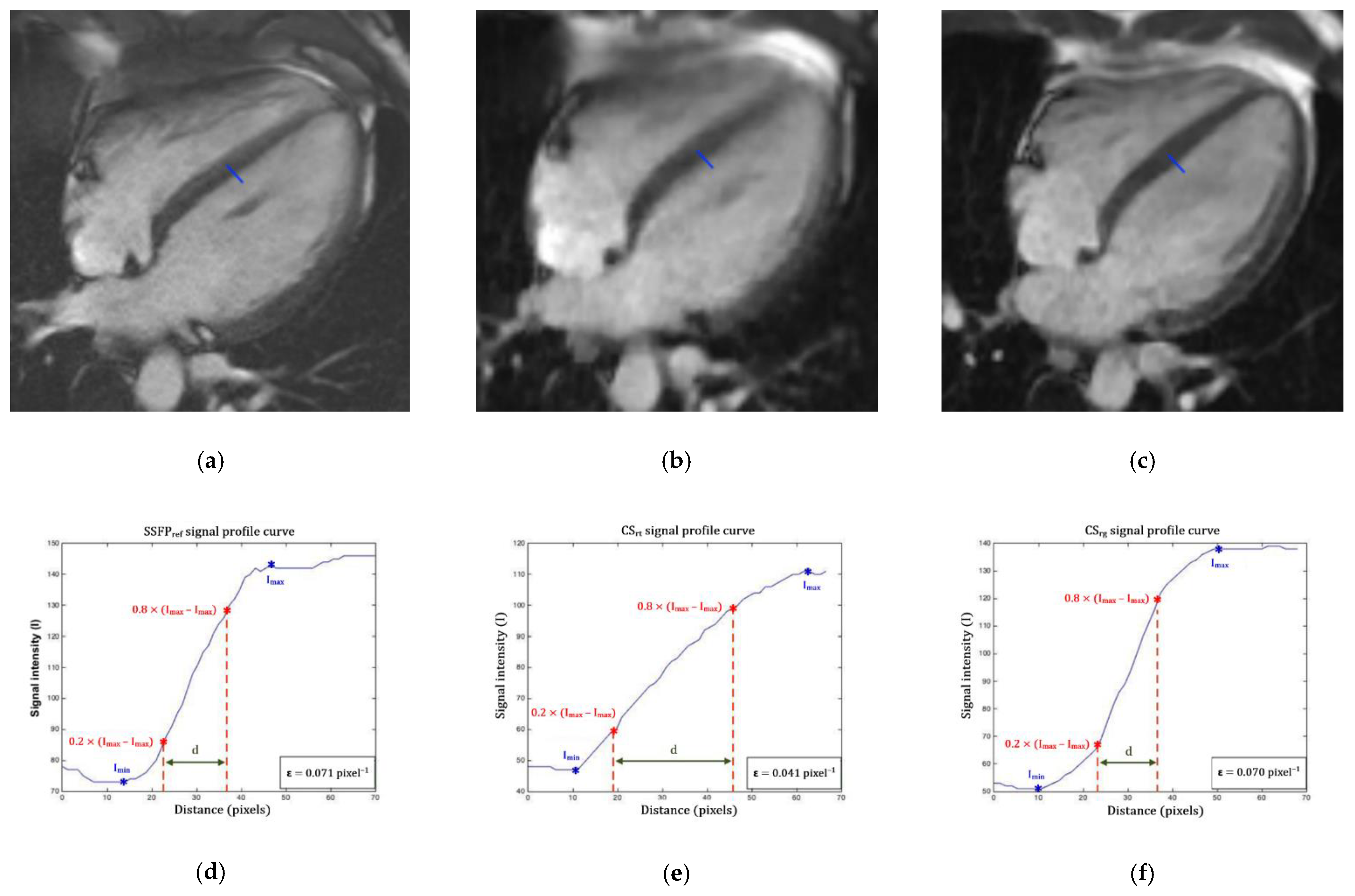
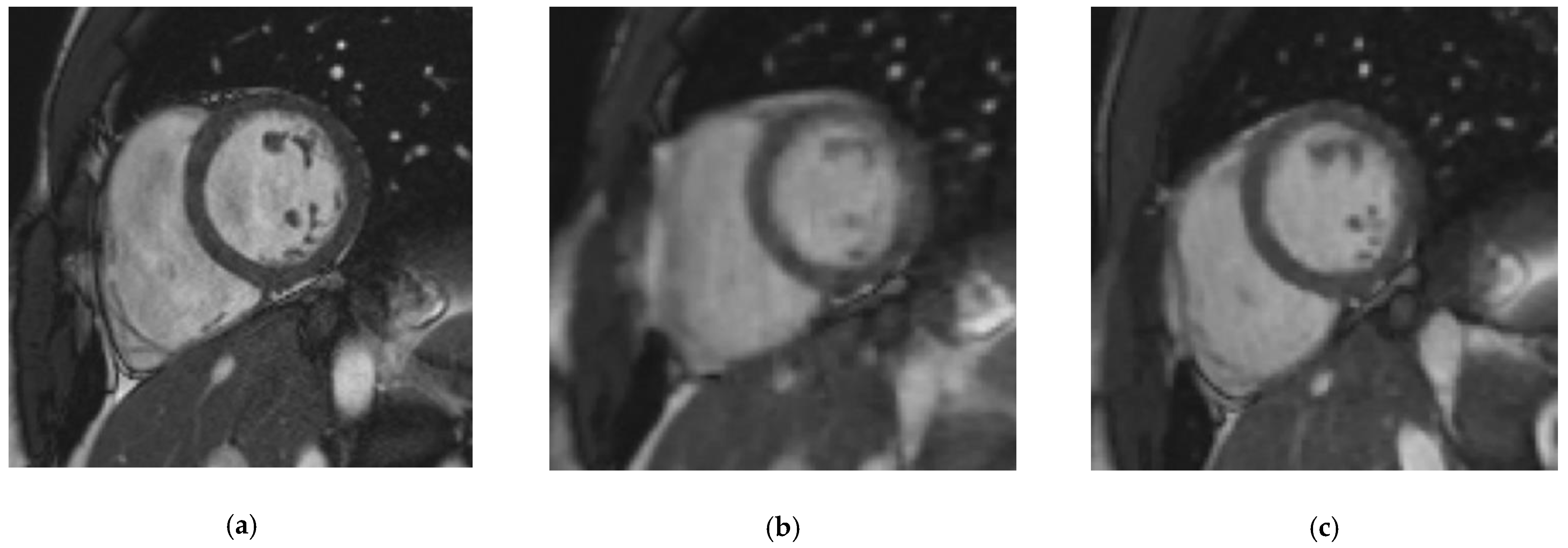
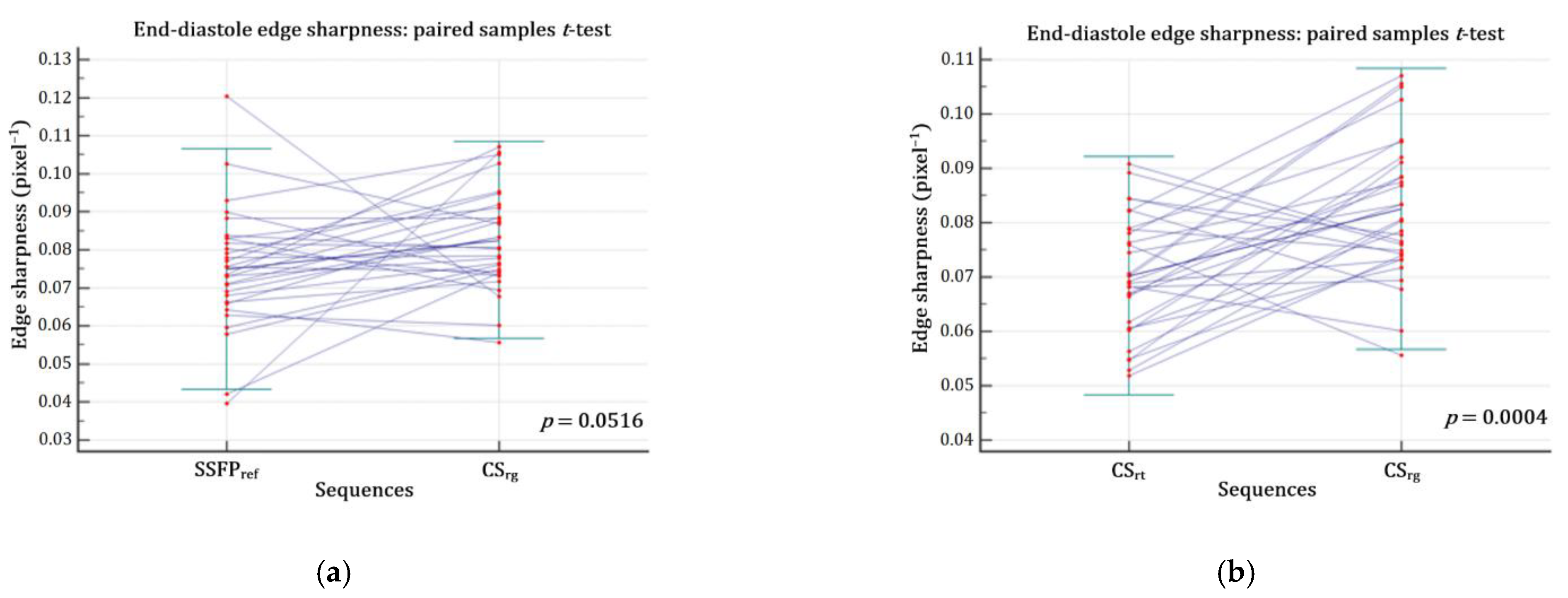
2.3. Cine Images Quality Assessment
2.4. Functional Evaluation
2.5. Conditions of Image Analysis
2.6. Statistics Analysis
3. Results
3.1. Population Description
3.2. Scan Time and Image Quality
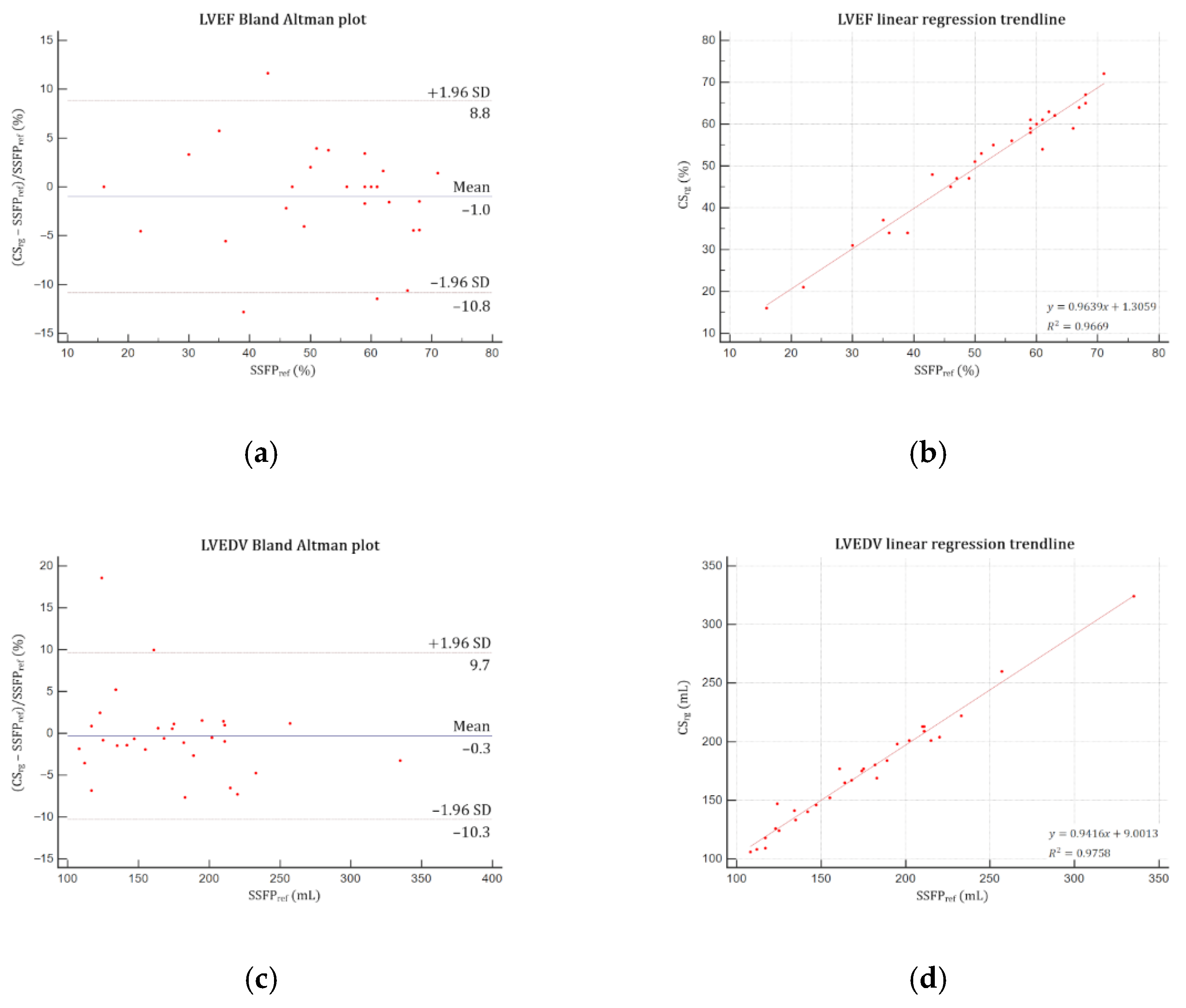
3.3. Volumes, Functions and Mass Quantification
4. Discussion
Limitations
5. Conclusions
Supplementary Materials
Author Contributions
Funding
Institutional Review Board Statement
Informed Consent Statement
Data Availability Statement
Conflicts of Interest
References
- Pennell, D.J.; Sechtem, U.P.; Higgins, C.B.; Manning, W.J.; Pohost, G.M.; Rademakers, F.E.; van Rossum, A.C.; Shaw, L.J.; Yucel, E.K.; Society for Cardiovascular Magnetic Resonance; et al. Clinical indications for cardiovascular magnetic resonance (CMR): Consensus panel report. Eur. Heart J. 2004, 25, 1940–1965. [Google Scholar] [CrossRef]
- Maceira, A.M.; Prasad, S.K.; Khan, M.; Pennell, D.J. Normalized left ventricular systolic and diastolic function by steady state free precession cardiovascular magnetic resonance. J. Cardiovasc. Magn. Reson. 2006, 8, 417–426. [Google Scholar] [CrossRef]
- Maceira, A.M.; Prasad, S.K.; Khan, M.; Pennell, D.J. Reference right ventricular systolic and diastolic function normalized to age, gender and body surface area from steady-state free precession cardiovascular magnetic resonance. Eur. Heart J. 2006, 27, 2879–2888. [Google Scholar] [CrossRef]
- Curtis, J.P.; Sokol, S.I.; Wang, Y.; Rathore, S.S.; Ko, D.T.; Jadbabaie, F.; Portnay, E.L.; Marshalko, S.J.; Radford, M.J.; Krumholz, H.M. The association of left ventricular ejection fraction, mortality, and cause of death in stable outpatients with heart failure. J. Am. Coll. Cardiol. 2003, 42, 736–742. [Google Scholar] [CrossRef]
- Karamitsos, T.D.; Francis, J.M.; Myerson, S.; Selvanayagam, J.B.; Neubauer, S. The role of cardiovascular magnetic resonance imaging in heart failure. J. Am. Coll. Cardiol. 2009, 54, 1407–1424. [Google Scholar] [CrossRef]
- Knauth, A.L.; Gauvreau, K.; Powell, A.J.; Landzberg, M.J.; Walsh, E.P.; Lock, J.E.; del Nido, P.J.; Geva, T. Ventricular size and function assessed by cardiac MRI predict major adverse clinical outcomes late after tetralogy of Fallot repair. Heart Br. Card. Soc. 2008, 94, 211–216. [Google Scholar] [CrossRef]
- Kramer, C.M.; Barkhausen, J.; Flamm, S.D.; Kim, R.J.; Nagel, E. Society for Cardiovascular Magnetic Resonance Board of trustees task force on standardized protocols standardized cardiovascular magnetic resonance (CMR) protocols 2013 update. J. Cardiovasc. Magn. Reson. 2013, 15, 91. [Google Scholar] [CrossRef]
- Vincenti, G.; Monney, P.; Chaptinel, J.; Rutz, T.; Coppo, S.; Zenge, M.O.; Schmidt, M.; Nadar, M.S.; Piccini, D.; Chèvre, P.; et al. Compressed sensing single-breath-hold CMR for fast quantification of lv function, volumes, and mass. J. Am. Coll. Cardiol. Imaging 2014, 7, 882–892. [Google Scholar] [CrossRef]
- Candès, E.J.; Romberg, J.K.; Tao, T. Stable signal recovery from incomplete and inaccurate measurements. Commun. Pure Appl. Math. 2006, 59, 1207–1223. [Google Scholar] [CrossRef]
- Donoho, D.L. Compressed Sensing. IEEE Trans. Inf. Theory 2006, 52, 1289–1306. [Google Scholar] [CrossRef]
- Lustig, M.; Donoho, D.; Pauly, J.M. Sparse MRI: The application of compressed sensing for rapid MR imaging. Magn. Reson. Med. 2007, 58, 1182–1195. [Google Scholar] [CrossRef]
- Lustig, M.; Santos, J.M.; Donoho, D.L.; Pauly, J.M. K-t SPARSE: High frame rate dynamic MRI exploiting spatio-temporal sparsity. In Proceedings of the 14th International Society for Magnetic Resonance in Medecine annual meeting, Seattle, WA, USA, 6–12 May 2006. [Google Scholar]
- Feng, L.; Srichai, M.B.; Lim, R.P.; Harrison, A.; King, W.; Adluru, G.; Dibella, E.V.R.; Sodickson, D.K.; Otazo, R.; Kim, D. Highly Accelerated real-time cardiac cine MRI using k-t SPARSE-SENSE. Magn. Reson. Med. 2013, 70, 64–74. [Google Scholar] [CrossRef]
- Jahnke, C.; Nagel, E.; Gebker, R.; Bornstedt, A.; Schnackenburg, B.; Kozerke, S.; Fleck, E.; Paetsch, I. Four-dimensional single breathhold magnetic resonance imaging using kt-BLAST enables reliable assessment of left- and right-ventricular volumes and mass. J. Magn. Reson. Imaging 2007, 25, 737–742. [Google Scholar] [CrossRef] [PubMed]
- Klinke, V.; Muzzarelli, S.; Lauriers, N.; Locca, D.; Vincenti, G.; Monney, P.; Lu, C.; Nothnagel, D.; Pilz, G.; Lombardi, M.; et al. Quality assessment of cardiovascular magnetic resonance in the setting of the European CMR Registry: Description and validation of standardized criteria. J. Cardiovasc. Magn. Reson. 2013, 15, 55. [Google Scholar] [CrossRef]
- Bogachkov, A.; Ayache, J.B.; Allen, B.D.; Murphy, I.; Carr, M.L.; Spottiswoode, B.; Schmidt, M.; Zenge, M.O.; Nadar, M.S.; Zuehlsdorff, S.; et al. Right ventricular assessment at cardiac MRI: Initial clinical experience utilizing an IS-SENSE reconstruction. Int. J. Cardiovasc. Imaging 2016, 32, 1081–1091. [Google Scholar] [CrossRef]
- Goebel, J.; Nensa, F.; Bomas, B.; Schemuth, H.P.; Maderwald, S.; Gratz, M.; Quick, H.H.; Schlosser, T.; Nassenstein, K. Real-Time SPARSE-SENSE Cardiac Cine MR Imaging: Optimization of image reconstruction and sequence validation. Eur. Radiol. 2016, 26, 4482–4489. [Google Scholar] [CrossRef]
- Goebel, J.; Nensa, F.; Schemuth, H.P.; Maderwald, S.; Gratz, M.; Quick, H.H.; Schlosser, T.; Nassenstein, K. Compressed sensing cine imaging with high spatial or high temporal resolution for analysis of left ventricular function. J. Magn. Reson. Imaging 2016, 44, 366–374. [Google Scholar] [CrossRef]
- Kido, T.; Kido, T.; Nakamura, M.; Watanabe, K.; Schmidt, M.; Forman, C.; Mochizuki, T. Compressed sensing real-time cine cardiovascular magnetic resonance: Accurate assessment of left ventricular function in a single-breath-hold. J. Cardiovasc. Magn. Reson. 2016, 18, 50–60. [Google Scholar] [CrossRef]
- Kido, T.; Kido, T.; Nakamura, M.; Watanabe, K.; Schmidt, M.; Forman, C.; Mochizuki, T. Assessment of left ventricular function and mass on free-breathing compressed sensing real-time cine imaging. Circ. J. 2017, 81, 1463–1468. [Google Scholar] [CrossRef] [PubMed]
- Lin, A.C.W.; Strugnell, W.; Riley, R.; Schmitt, B.; Zenge, M.; Schmidt, M.; Morris, N.R.; Hamilton-Craig, C. Higher resolution cine imaging with compressed sensing for accelerated clinical left ventricular evaluation. J. Magn. Reson. Imaging 2017, 45, 1693–1699. [Google Scholar] [CrossRef]
- Vermersch, M.; Longère, B.; Coisne, A.; Schmidt, M.; Forman, C.; Monnet, A.; Pagniez, J.; Silvestri, V.; Simeone, A.; Montaigne, D.; et al. Compressed sensing real-time cine imaging for assessment of ventricular function, volumes and mass in clinical practice. Eur. Radiol. 2020, 30, 609–619. [Google Scholar] [CrossRef]
- Haubenreisser, H.; Henzler, T.; Budjan, J.; Sudarski, S.; Zenge, M.O.; Schmidt, M.; Nadar, M.S.; Borggrefe, M.; Schoenberg, S.O.; Papavassiliu, T. Right ventricular imaging in 25 seconds: Evaluating the use of sparse sampling cine with iterative reconstruction for volumetric analysis of the right ventricle. Investig. Radiol. 2016, 51, 379–386. [Google Scholar] [CrossRef]
- Camargo, G.C.; Erthal, F.; Sabioni, L.; Penna, F.; Strecker, R.; Schmidt, M.; Zenge, M.O.; de Lima, R.S.L.; Gottlieb, I. Real-time cardiac magnetic resonance cine imaging with sparse sampling and iterative reconstruction for left-ventricular measures: Comparison with gold-standard segmented steady-state free precession. Magn. Reson. Imaging 2017, 38, 138–144. [Google Scholar] [CrossRef]
- Sudarski, S.; Henzler, T.; Haubenreisser, H.; Dösch, C.; Zenge, M.O.; Schmidt, M.; Nadar, M.S.; Borggrefe, M.; Schoenberg, S.O.; Papavassiliu, T. Free-breathing sparse sampling cine mr imaging with iterative reconstruction for the assessment of left ventricular function and mass at 3.0 T. Radiology 2017, 282, 74–83. [Google Scholar] [CrossRef]
- Vincenti, G.; Piccini, D.; Monney, P.; Chaptinel, J.; Rutz, T.; Coppo, S.; Zenge, M.O.; Schmidt, M.; Nadar, M.S.; Wang, Q.; et al. Preliminary experiences with compressed sensing multislice cine acquisitions for the assessment of left ventricular function: CV_sparse WIP. Magn. Flash 2013, 55, 26–34. [Google Scholar]
- Goebel, J.; Nensa, F.; Schemuth, H.P.; Maderwald, S.; Quick, H.H.; Schlosser, T.; Nassenstein, K. Real-time SPARSE-SENSE cine MR imaging in atrial fibrillation: A feasibility study. Acta Radiol. 2017, 58, 922–928. [Google Scholar] [CrossRef]
- Weiger, M.; Pruessmann, K.P.; Boesiger, P. Cardiac real-time imaging using SENSE. SENSitivity Encoding Scheme. Magn. Reson. Med. 2000, 43, 177–184. [Google Scholar] [CrossRef]
- Miller, S.; Simonetti, O.P.; Carr, J.; Kramer, U.; Finn, J.P. MR imaging of the heart with cine true fast imaging with steady-state precession: Influence of spatial and temporal resolutions on left ventricular functional parameters. Radiology 2002, 223, 263–269. [Google Scholar] [CrossRef]
- Roussakis, A.; Baras, P.; Seimenis, I.; Andreou, J.; Danias, P.G. Relationship of number of phases per cardiac cycle and accuracy of measurement of left ventricular volumes, ejection fraction, and mass. J. Cardiovasc. Magn. Reson. 2004, 6, 837–844. [Google Scholar] [CrossRef]
- Wetzl, J.; Schmidt, M.; Pontana, F.; Longère, B.; Lugauer, F.; Maier, A.; Hornegger, J.; Forman, C. Single-breath-hold 3-D CINE imaging of the left ventricle using cartesian sampling. Magma 2017, 31, 19–31. [Google Scholar] [CrossRef]
- Larson, A.C.; Kellman, P.; Arai, A.; Hirsch, G.A.; McVeigh, E.; Li, D.; Simonetti, O.P. Preliminary investigation of respiratory self-gating for free-breathing segmented cine MRI. Magn. Reson. Med. 2005, 53, 159–168. [Google Scholar] [CrossRef]
- Richard, S.; Husarik, D.B.; Yadava, G.; Murphy, S.N.; Samei, E. Towards Task-based assessment of CT performance: System and object MTF across different reconstruction algorithms. Med. Phys. 2012, 39, 4115–4122. [Google Scholar] [CrossRef] [PubMed]
- Forman, C.; Kroeker, R.; Schmidt, M. Accelerated 2D cine MRI featuring compressed sensing and ECG-triggered retro-gating. In Proceedings of the 25th International Society for Magnetic Resonance in Medicine annual meeting, Honolulu, HI, USA, 22–27 April 2017. [Google Scholar]
- Semelka, R.C.; Tomei, E.; Wagner, S.; Mayo, J.; Kondo, C.; Suzuki, J.; Caputo, G.R.; Higgins, C.B. Normal left ventricular dimensions and function: Interstudy reproducibility of measurements with cine MR imaging. Radiology 1990, 174, 763–768. [Google Scholar] [CrossRef] [PubMed]
- Voit, D.; Zhang, S.; Unterberg-Buchwald, C.; Sohns, J.M.; Lotz, J.; Frahm, J. Real-time cardiovascular magnetic resonance at 1.5 T using balanced SSFP and 40 ms resolution. J. Cardiovasc. Magn. Reson. 2013, 15, 79–86. [Google Scholar] [CrossRef] [PubMed]
- Eberle, H.C.; Nassenstein, K.; Jensen, C.J.; Schlosser, T.; Sabin, G.V.; Naber, C.K.; Bruder, O. Rapid MR assessment of left ventricular systolic function after acute myocardial infarction using single breath-hold cine imaging with the temporal parallel acquisition technique (TPAT) and 4D guide-point modelling analysis of left ventricular function. Eur. Radiol. 2010, 20, 73–80. [Google Scholar] [CrossRef]
- Nacif, M.S.; Zavodni, A.; Kawel, N.; Choi, E.-Y.; Lima, J.A.C.; Bluemke, D.A. Cardiac magnetic resonance imaging and its electrocardiographs (ECG): Tips and tricks. Int. J. Cardiovasc. Imaging 2012, 28, 1465–1475. [Google Scholar] [CrossRef]
- Kotwinski, P.; Smith, G.; Sanders, J.; Cooper, J.; Kotwinski, D.; Teis, A.; Mythen, M.; Monty, G.; Jones, A.; Montgomery, H.E.; et al. CMR shows that anthracycline cardiotoxicity is common in women treated for early breast cancer and associated with undiagnosed hypertension; but cannot be reliably detected using late-gadolinium enhancement imaging. J. Cardiovasc. Magn. Reson. 2013, 15, 276. [Google Scholar] [CrossRef]
- Patel, A.R.; Kramer, C.M. Role of cardiac magnetic resonance in the diagnosis and prognosis of nonischemic cardiomyopathy. J. Am. Coll. Cardiol. Imaging 2017, 10, 1180–1193. [Google Scholar] [CrossRef]
- Ponikowski, P.; Voors, A.A.; Anker, S.D.; Bueno, H.; Cleland, J.G.F.; Coats, A.J.S.; Falk, V.; González-Juanatey, J.R.; Harjola, V.-P.; Jankowska, E.A.; et al. 2016 ESC Guidelines for the diagnosis and treatment of acute and chronic heart failure: The task force for the diagnosis and treatment of acute and chronic heart failure of the European Society of Cardiology (ESC). Developed with the special contribution of the Heart Failure Association (HFA) of the ESC. Eur. J. Heart Fail. 2016, 18, 891–975. [Google Scholar] [CrossRef]
- Li, T.; Feng, H.; Xu, Z. A new analytical edge spread function fitting model for modulation transfer function measurement. Chin. Opt. Lett. 2011, 9, 031101. [Google Scholar] [CrossRef]
- Rösner, A.; Barbosa, D.; Aarsæther, E.; Kjønås, D.; Schirmer, H.; D’hooge, J. The influence of frame rate on two-dimensional speckle-tracking strain measurements: A study on silico-simulated models and images recorded in patients. Eur. Heart J. Cardiovasc. Imaging 2015, 16, 1137–1147. [Google Scholar] [CrossRef] [PubMed]
- Longère, B.; Chavent, M.H.; Coisne, A.; Gkizas, C.; Pagniez, J.; Simeone, A.; Silvestri, V.; Schmidt, M.; Forman, C.; Montaigne, D.; et al. Single breath-hold compressed sensing real-time cine imaging to assess left ventricular motion in myocardial infraction. Diagn. Interv. Imaging 2020. [Google Scholar] [CrossRef]
- Wintersperger, B.J.; Nikolaou, K.; Dietrich, O.; Rieber, J.; Nittka, M.; Reiser, M.F.; Schoenberg, S.O. Single breath-hold real-time cine MR imaging: Improved temporal resolution using generalized autocalibrating partially parallel acquisition (GRAPPA) algorithm. Eur. Radiol. 2003, 13, 1931–1936. [Google Scholar] [CrossRef]


| Parameters | SSFPref | CSrt | CSrg |
|---|---|---|---|
| Repetition time—ms | 3.16 | 2.70 | 2.70 |
| Echo time—ms | 1.23 | 1.14 | 1.14 |
| Flip angle—degrees | 57 | 60 | 60 |
| Field of view—mm2 | 375 × 280 | 360 × 270 | 360 × 270 |
| Matrix—pixels2 | 288 × 216 | 224 × 168 | 224 × 168 |
| Spatial resolution—mm2 | 1.3 × 1.3 | 1.6 × 1.6 | 1.6 × 1.6 |
| Temporal resolution—ms | 37 | 49 | 37 |
| Slice thickness/gap—mm | 8/2 | 8/2 | 8/2 |
| Bandwidth—Hz/pixel | 915 | 900 | 900 |
| Reconstructed cardiac phases—n Number of acquired cardiac phases | - 25 a | 25 a 16.8 ± 3.9 | - 25 a |
| Number of breath-holds | 15.0 ± 1.2 | 2 a | 3 a |
| Cycles per slice—n | 8 a | 1 a | 2 a |
| Cycles of iterative reconstruction—n | - | 40 | 40 |
| Items | 0 | 1 | 2 | 3 | Maximum Score |
|---|---|---|---|---|---|
| 1. LV coverage | Full | - | No apex | Base or ≥1 slice missing | 5 |
| 2. Wrap around | No | 1 slice | 2 slices | ≥3 slices | 3 |
| 3. Respiratory ghost | No | 1 slice | 2 slices | ≥3 slices | |
| 4. Cardiac ghost | No | 1 slice | 2 slices | ≥3 slices | |
| 5. Blurring/Mistriggering | No | 1 slice | 2 slices | ≥3 slices | |
| 6. Metallic artifacts | No | 1 slice | 2 slices | ≥3 slices | |
| 7. Shimming artifacts | No | 1 slice | 2 slices | ≥3 slices | |
| 8. Signal loss (coil inactive) | Activated | - | Not activated | 2 | |
| 9. Orientation of stack | Correct | - | Incorrect | - | 2 |
| 10. Slice thickness | ≤10 mm | 11–15 mm | - | >15 mm | 3 |
| 11. Gap | ≤3 mm | 3–4 mm | - | >4 mm | 3 |
| 12. Correct LV long axes | ≥2 mm | 1 | - | None | 3 |
| Score | 21 | ||||
| Modified score (items 1 to 8) | 10 |
| Mean ± SD (95% CI) | Minimum Value | Maximum Value | |
|---|---|---|---|
| Age—years | 48.0 ± 21.0 (40.2–55.9) | 18 | 87 |
| Weight—kg | 73.9 ± 12.1 (69.4–78.5) | 53 | 105 |
| Height—cm | 172.5 ± 8.3 (169.4–175.6) | 157 | 189 |
| Body surface area—m2 | 1.87 ± 0.17 (1.80–1.93) | 1.55 | 2.22 |
| Body mass index—kg/m2 | 24.8 ± 3.6 (23.5–26.2) | 19.8 | 33.7 |
| Heart rate—beats per minute | 73.8 ± 13.5 (68.7–78.9) | 54 | 101 |
| SSFPref Sequence (Mean ± SD (95% CI)) | CSrg Sequence (Mean ± SD (95% CI)) | Difference (Mean ± SD (95% CI)) | Paired t Test p | ICC | ||
|---|---|---|---|---|---|---|
| Inter | Intra | |||||
| LVEF—% | 52.7 ± 14.1 (47.5–58.0) | 52.1 ± 13.8 (47.0–57.3) | −0.6 ± 2.6 (−1.6 to −0.4) | 0.21 | 0.99 | 0.9996 |
| LVEDV—mL | 174.1 ± 50.3 (155.3–192.9) | 173.0 ± 48.0 (155.1–190.9) | −1.2 ± 8.0 (−4.2 to −1.8) | 0.43 | 0.99 | 0.9994 |
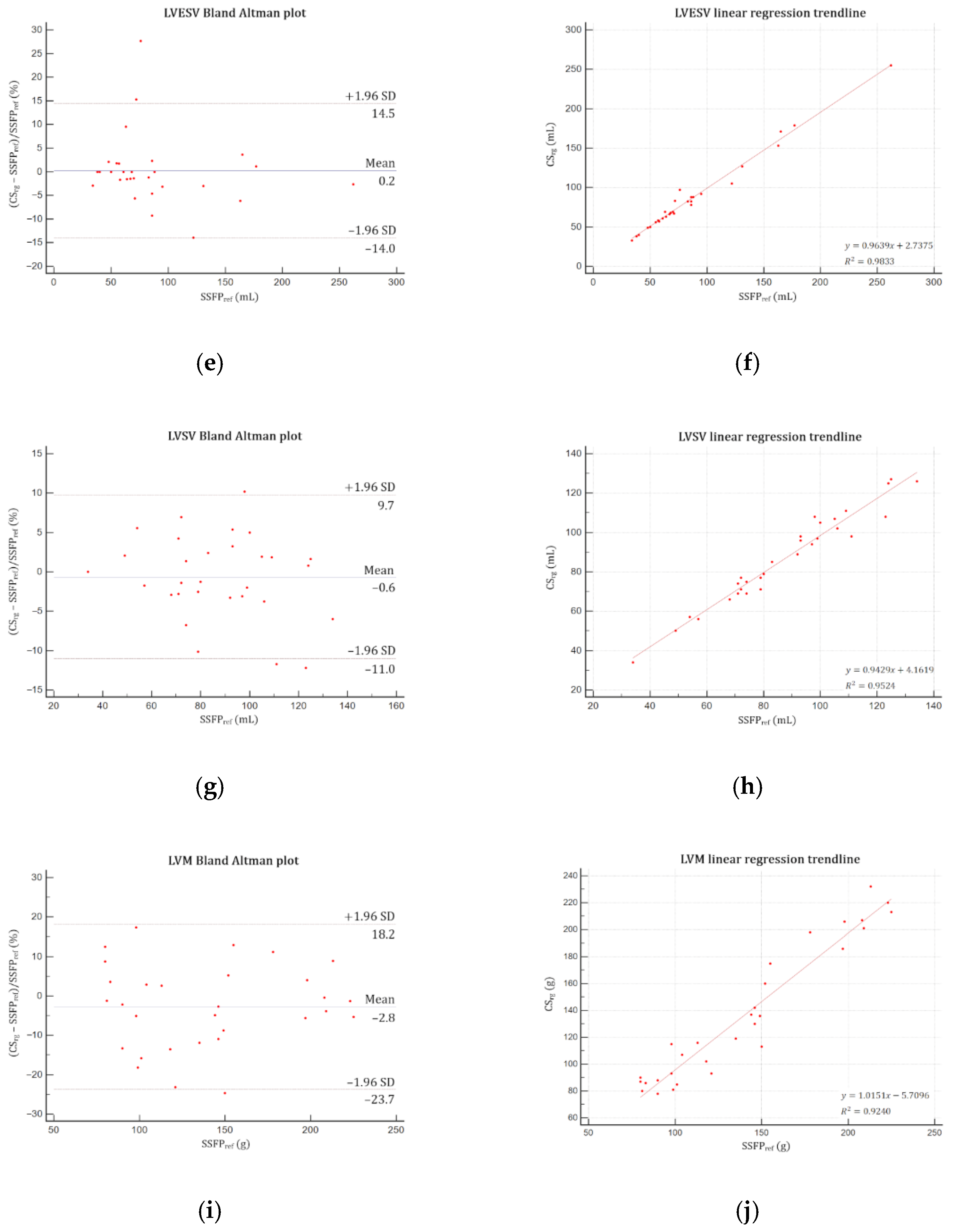
| LVESV—mL | 86.8 ± 49.4 (68.4–105.2) | 86.4 ± 48.0 (68.5–104.3) | −0.4 ± 6.5 (−2.8 to −2.0) | 0.74 | 0.98 | 0.9906 |
| LVSV—mL | 87.5 ± 24.0 (78.6–96.5) | 86.7 ± 23.2 (78.0–95.4) | −0.8 ± 5.3 (−2.8 to −1.1) | 0.39 | 0.98 | 0.9932 |
| LVM—g | 139.5 ± 47.7 (121.6–157.3) | 135.9 ± 50.4 (117.0–154.7) | −3.6 ± 13.9 (−8.8 to 1.6) | 0.17 | 0.97 | 0.9994 |
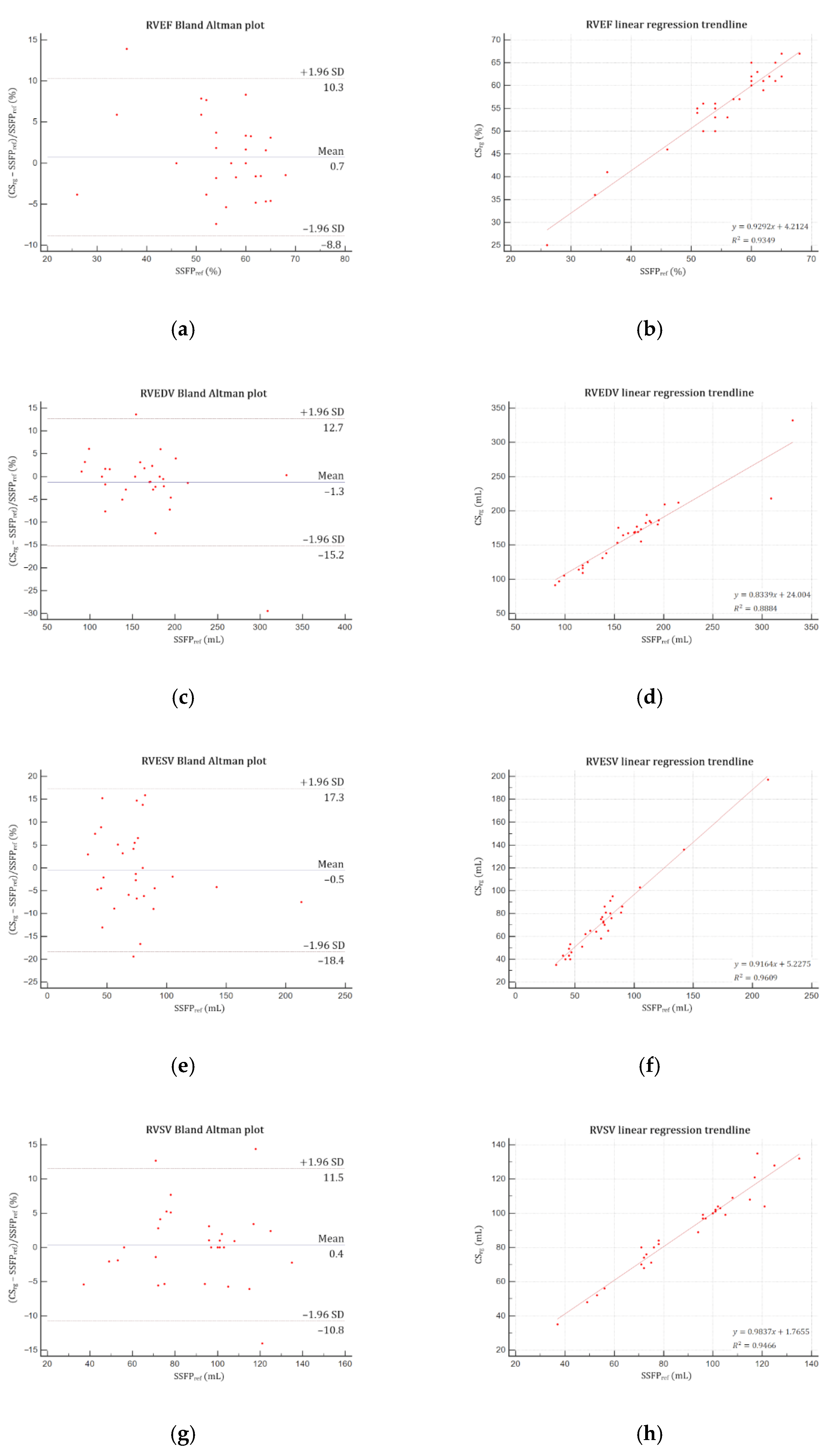
| RVEF—% | 55.8 ± 9.7 (52.2–59.4) | 56.0 ± 9.3 (52.6–59.5) | 0.3 ± 2.4 (−0.7 to 1.2) | 0.56 | 0.96 | 0.9965 |
| RVEDV—mL | 167.0 ± 53.6 (147.0–187.0) | 163.2 ± 47.4 (145.5–180.9) | −3.8 ± 18.2 (−10.5 to 3.05) | 0.27 | 0.98 | 0.9968 |
| RVESV—mL | 74.1 ± 34.3 (61.3–86.9) | 73.1 ± 32.0 (61.1–85.0) | −1.0 ± 6.9 (−3.6 to 1.6) | 0.45 | 0.96 | 0.9934 |
| RVSV—mL | 89.8 ± 24.3 (80.8–98.9) | 90.1 ± 24.5 (81.0–99.3) | −0.3 ± 5.7 (−1.8 to 2.4) | 0.77 | 0.97 | 0.9961 |
Publisher’s Note: MDPI stays neutral with regard to jurisdictional claims in published maps and institutional affiliations. |
© 2021 by the authors. Licensee MDPI, Basel, Switzerland. This article is an open access article distributed under the terms and conditions of the Creative Commons Attribution (CC BY) license (https://creativecommons.org/licenses/by/4.0/).
Share and Cite
Longère, B.; Gkizas, C.V.; Coisne, A.; Grenier, L.; Silvestri, V.; Pagniez, J.; Simeone, A.; Hennicaux, J.; Schmidt, M.; Forman, C.; et al. 60-S Retrogated Compressed Sensing 2D Cine of the Heart: Sharper Borders and Accurate Quantification. J. Clin. Med. 2021, 10, 2417. https://doi.org/10.3390/jcm10112417
Longère B, Gkizas CV, Coisne A, Grenier L, Silvestri V, Pagniez J, Simeone A, Hennicaux J, Schmidt M, Forman C, et al. 60-S Retrogated Compressed Sensing 2D Cine of the Heart: Sharper Borders and Accurate Quantification. Journal of Clinical Medicine. 2021; 10(11):2417. https://doi.org/10.3390/jcm10112417
Chicago/Turabian StyleLongère, Benjamin, Christos V. Gkizas, Augustin Coisne, Lucas Grenier, Valentina Silvestri, Julien Pagniez, Arianna Simeone, Justin Hennicaux, Michaela Schmidt, Christoph Forman, and et al. 2021. "60-S Retrogated Compressed Sensing 2D Cine of the Heart: Sharper Borders and Accurate Quantification" Journal of Clinical Medicine 10, no. 11: 2417. https://doi.org/10.3390/jcm10112417
APA StyleLongère, B., Gkizas, C. V., Coisne, A., Grenier, L., Silvestri, V., Pagniez, J., Simeone, A., Hennicaux, J., Schmidt, M., Forman, C., Toupin, S., Montaigne, D., & Pontana, F. (2021). 60-S Retrogated Compressed Sensing 2D Cine of the Heart: Sharper Borders and Accurate Quantification. Journal of Clinical Medicine, 10(11), 2417. https://doi.org/10.3390/jcm10112417