


Abstract
Larrea ameghinoi Speg., an endemic species of Argentine Patagonia traditionally used in folk medicine to treat fever, stomach disorders, respiratory conditions, back pain, and as an emmenagogue, among others, still remains chemically and biologically underexplored compared to the other four members of the genus. This study aimed to perform a comprehensive metabolomic characterization of methanolic extracts from two populations (EMLaSAO and EMLaMAQ) using ultra-high-resolution liquid chromatography coupled with electrospray ionization quadrupole time-of-flight mass spectrometry (UHPLC–ESI–QTOF–MS) and to evaluate their antioxidant, antimicrobial, and enzyme-inhibitory activities of relevance to human health. Thirty-three compounds were tentatively identified by extensive UHPLC–MS analysis, including flavones, two major lignans, and oleanane-type triterpenes. Both extracts exhibited high phenolic content (215–239 mg of gallic acid equivalents (GAE)/g extract) and strong free radical scavenging activity, as evidenced by 2,2-diphenyl-1-picrylhydrazyl (DPPH, EC50 ≈ 10 μg/mL), ferric-reducing antioxidant power (FRAP), and Trolox equivalent antioxidant activity (TEAC) assays. In addition, significant inhibition of butyrylcholinesterase (IC50 ≈ 50 μg extract/mL) and α-glucosidase, together with selective antibacterial activity against methicillin-sensitive and resistant Staphylococcus aureus (MIC = 125 μg extract/mL), were recorded. These findings suggest that L. ameghinoi possesses a distinctive phytochemical composition conferring multitarget bioactivity, differing from other Larrea species dominated by lignans such as nordihydroguaiaretic acid (NDGA) and its derivatives. Overall, this work supports the potential of L. ameghinoi as a novel source of bioactive metabolites for managing oxidative stress-related disorders and opportunistic infections. This warrants future in vivo studies investigating biological activities associated with oxidative stress and their relevance to human health.
1. Introduction
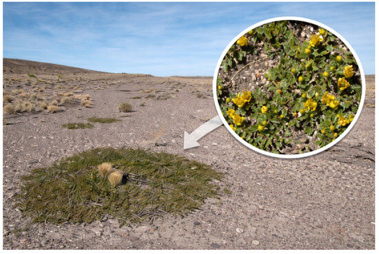
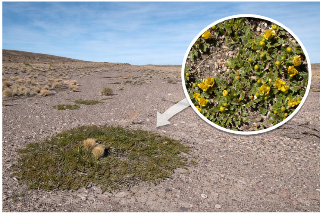
Medicinal resinous species belonging to the genus Larrea Cav. (Zygophyllaceae) are distributed amphitropically in arid environments of Argentina, Chile, Bolivia, Peru, Mexico, and the southwestern United States. The genus is composed of five species: Larrea ameghinoi Speg., L. cuneifolia Cav., L. divaricata Cav., and L. nitida Cav., commonly known as “jarillas”, occurring in South America, while L. tridentata (DC.) Coville grows only in Mexico and the USA [1,2,3]. The species are used extensively in traditional medicine in Argentina for the treatment of injuries and bruises, and are a good disinfectant for wounds and a repellent against insects; they are also used for roof construction in rural areas and as a plant-based fuel for cooking [4]. Extracts and infusions of aerial parts of most species of the genus Larrea have displayed several biological activities, such as antimicrobial, antioxidant, enzyme-inhibitory, anti-inflammatory, and antitumor activities, among others [4]. A notable research outcome involving Larrea divaricata was the recent development of a natural lotion composed of jarilla and decaffeinated coffee extracts to stimulate hair growth, reduce hair loss, and permanently control dandruff. The said product has been registered and obtained a patent [5]. Larrea ameghinoi Speg., commonly known as “jarilla rastrera”(Figure 1), is a shrub endemic to Argentine Patagonia that lives in lowland areas from Neuquén to Chubut between 200 and 800 m above sea level [6]. It is a resinous, branchy, prostrate woody plant with twisted branches and measures approximately 10 cm in height. It flowers from October to the end of November, a period in which its yellow flowers can be seen. It is distinguished from the rest of the species of the genus by having subsessile leaves (5–8 × 2–3 mm), with 3–7 unequal, imparipinnate leaflets, with a smaller, fused terminal leaflet. Its fruit is a schizocarp, verrucose and brown-red in color, which has five mericarps that remain associated at maturity; the seeds are small, smooth, and kidney-shaped [7,8]. The plant adopts a cushion-shaped structure, which gives it the ability to thrive in areas exposed to the westerlies, the characteristic winds of Patagonia, even when this implies a reduction in exposure to sunlight. In addition to its cultural and symbolic importance, L. ameghinoi constitutes a promising source of bioactive compounds with potential benefits for human health. It is valued by indigenous peoples, and the uses of the species of this genus have been transmitted through popular knowledge. The bark and leaf are popularly used to treat different ailments in the form of various preparations, to reduce fever, stomach disorders, and respiratory conditions, to treat back pain, and as an emmenagogue since it stimulates and promotes menstrual flow. In addition, it soothes rheumatic pain due to its application in the form of a poultice [8].
Figure 1.
L. ameghinoi Speg. habit and typical habitat (Patagonian steppe).
To date, studies on the chemical characterization and potential biological activity of this species are limited.
The main goals and novelty of this work are the evaluation of the antioxidant and antibacterial effects, as well as the inhibition of enzymes of interest to human health, complemented by the complete polyphenolic metabolome profile by UHPLC-ESI-QTOF-MS analysis of the methanolic extracts of L. ameghinoi from Argentina.
2. Materials and Methods
2.1. Chemicals
Ultra-pure water, with total organic carbon (TOC) levels below 5 µg/L, was sourced using an Arium 126 61316-RO purification system coupled with an Arium 611 UV unit (Sartorius, Goettingen, Germany). High-purity methanol (HPLC grade) and mass spectrometry-grade formic acid were supplied by J. T. Baker (Phillipsburg, NJ, USA), while HPLC-grade chloroform was provided by Merck (Santiago, Chile). Commercial Folin–Ciocalteu (FC) reagent, 2,2-diphenyl-1-picrylhydrazyl (DPPH), ferric chloride hexahydrate, 2,4,6-tris(2-pyridyl)-s-triazine, trolox, quercetin, gallic acid, and dimethyl sulfoxide (DMSO) were purchased from Sigma-Aldrich Chem. Co. (St Louis, MO, USA). Nordihydroguaiaretic acid (NDGA), 3′-methyl-nordihydroguaiaretic acid (MNDGA), and other lignans previously isolated from Larrea nitida and some HPLC standards were acquired from Sigma-Aldrich Chem. Co. (St Louis, MO, USA) or Extrasynthèse (Genay, France) were used for UHPLCMS analysis.
2.2. Plant Material, Methanolic Extracts, and the Total Phenol and Flavonoid Content
Considering the vulnerability of L ameghinoi, aerial parts (10 g) were collected in two locations in the province of Rio Negro, Maquinchao (MAQ) and a location near Las Grutas (SAO). The plant samples were dried at room temperature (25 °C) and stored in conditions devoid of light and heat. A voucher specimen for each locality has been deposited in the herbarium of Instituto de Botánica Darwinion (CONICET-ANCEFN) under the reference numbers Sede & Calcagno 885 (SAO) and Sede & Abraham 915 (MAQ).
From dried and ground plant material, the methanolic extracts of each sample were prepared as follows: 10 g of plants from SAO and MAQ were weighed, and successive extractions were carried out with methanol under sonication at a temperature of 30 °C. The process was repeated twice, each time. The resulting extracts were subjected to a filtration stage to eliminate solid residues. Subsequently, they were concentrated at reduced pressure in a rotary evaporator until constant weight was obtained, thus obtaining both methanolic extracts called EMLaSAO and EMLaMAQ, which yielded 12.79% and 16.69% w/w, respectively. The extracts were stored at −20 °C until they were analyzed via UHPLC–PDA–QTOF-MS, as well as for the spectrophotometric quantification of phenolics and flavonoids, and in vitro assays.
The total phenol content [9] and the flavonoid content were expressed as milligrams of gallic acid equivalents (GAE) or milligrams of quercetin equivalents (QE) per gram of EMLaSAO and EMLaMAQ (mg GAE/g EMLaSAO and EMLaMAQ), respectively. The values were obtained in quadruplicate using a Multiskan FC Microplate Photometer (Thermo Scientific, Waltham, MA, USA). The results are expressed as mean ± SD.
2.3. In Vitro Studies
2.3.1. Antioxidant Activity
Radical Scavenging Capacity Assay of 2,2-Diphenyl-1-Picrylhydrazyl (DPPH)
The free radical scavenging activity of EMLaSAO and EMLaMAQ was assessed using the DPPH assay. The concentration required to achieve 50% radical scavenging (EC50) was determined from a graph plotting inhibition percentage at 517 nm against concentrations of EMLaSAO and EMLaMAQ. Catechin (Sigma-Aldrich, ≥98%, St. Louis, MO, USA) served as the reference compound (EC50 4.1 μg/mL). Each test was conducted in quadruplicate [10].
Ferric-Reducing Antioxidant Power Assay (FRAP)
Results from the FRAP assay were calculated using linear regression based on the FRAP–Trolox calibration curve and are expressed as milligrams of Trolox equivalents per gram of EMLaSAO and EMLaMAQ (mg ETrolox/gof EMLaSAO and EMLaMAQ). All tests were conducted in quadruplicate [11].
Trolox Equivalent Antioxidant Activity Assay (TEAC)
The TEAC assay results were obtained through linear regression from a calibration curve created using Trolox concentrations ranging from 0 to 1 mM and are reported as milligrams of Trolox equivalents per gram of EMLaSAO and EMLaMAQ (mg ETrolox/g of EMLaSAO and EMLaMAQ. Each test was performed in quadruplicate [12].
2.4. Enzymatic Activity
2.4.1. Acetylcholinesterase and Butyrylcholinesterase Inhibitory Bioassay
Cholinesterase inhibitory activities were evaluated according to Ellman et al. (1961) [13] with some minor modifications. Enzyme inhibitory activity was calculated as a percentage compared to an assay using a buffer without any inhibitor. The values were expressed as half-maximal inhibitory concentration IC50 (µg/mL for extracts and galantamine), and were calculated as means ± SD of 3 individual determinations. The galantamine concentrations used to calculate the IC50 values were 0–1, 0.5, 1, 1.5, and 2 µg/mL in both AChE and BuChE assays, while for the EMLaSAO and EMLaMAQ extracts, they were 25, 50, 100, 150, 200, 250, and 300 µg/mL, respectively. The enzymes AChE and BuChE from Sigma-Aldrich were used. Galanthamine (Sigma-Aldrich) was used as a positive control in both assays.
2.4.2. Amylase and Glucosidsase Inhibition
The amylase inhibition was implemented by agreeing to the method previously described by [14]. On the other hand, the glucosidase inhibition assay was evaluated according to the method described by [15]. Both assays are shown as IC50 in µg/mlas means ± SD.
2.5. Antibacterial Activity
2.5.1. Antibacterial Susceptibility Testing
The minimum inhibitory concentration (MIC) and minimum bactericidal concentration (MBC) of EMLaSAO and EMLaMAQ and the antibiotic Imipecil (Imipenem, from Laboratory Northia, Buenos Aires, Argentina) were determined by broth microdilution techniques, according to Clinical and Laboratory Standards Institute (CLSI) [16]. Both extracts were tested from 1000 to 0.98 µg/mL using an inoculum of each bacterium adjusted to 5 × 10⁵ cells with colony-forming units (CFU)/mL. The following strains from the American Type Culture Collection (ATCC, Rockville, MD, USA) and clinical isolates from Laboratorio de Micro-biología, Hospital Marcial Quiroga, San Juan, Argentina (MQ) were employed: Staphylococcus aureus methicillin-sensitive ATCC 25923 (MSSA), Staphylococcus aureus methicillin-resistant ATCC 43300 (MRSA), Staphylococcus aureus methicillin-resistant-MQ1, Staphylococcus aureus methicillin-resistant-MQ2, Salmonella sp-MQ3, and Escherichia coli ATCC 25922.
2.5.2. Antifungal Susceptibility Testing
Minimum inhibitory concentration (MIC) of EMLaSAO and EMLaMAQ was determined using broth microdilution techniques, according to the guidelines of the CLSI [17]. For the development of the essay, stock solutions of EMLaSAO and EMLaMAQ were two-fold diluted with RPMI medium from 1000 to 0.98 µg/mL (final volume 100 µL and final dimethyl sulfoxide (DMSO) concentration ≤ 1%). A 100 µL volume of inoculum suspension was added to each well, except for the sterility control well, where sterile water was added instead. Ketoconazole (Sigma-Aldrich) was used as a positive control. Inoculum of cell or spore suspensions was obtained according to reported procedures and adjusted to 1–5 × 10³ cells/spores with colony-forming units (CFU)/mL. The methodology for the determination of MIC100 and MFC has been reported in detail in [18].
The following strains from the ATCC, MQ, and from CEREMIC (CCC), Reference Center in Mycology, Faculty of Biochemical and Pharmaceutical Sciences, Suipacha 531, 2000-Rosario, Argentina, and from were employed: Candida albicans-MQ 1924, Candida glabrata MQ1234, Candida parapsilosis MQ3, Candida tropicalis CCC 131-2000, and Cryptococcus neoformans ATCC 32264.
2.6. Ultra-High-Resolution Liquid Chromatography Analysis of EMLaSAO and EMLaMAQ
The analysis of EMLaSAO and EMLaMAQ was performed using a UHPLC-ESI-QTOF-MS system, integrated by a UHPLC Ultimate 3000 RS with Chromeleon 6.8 software (Dionex GmbH, Idstein, Germany), and a Bruker maXis ESI-QTOF-MS. The analysis of both extracts was performed on an Acclaim Thermo 5 µm C18 80 Å (150 mm × 4.6 mm) column at a flow rate of 1.0 mL/min, using a two-solution system: eluent (A) 0.1% formic acid in water and eluent (B) 0.1% formic acid in acetonitrile. The solution elution program was as follows: 1% isocratic B (0–2 min), 1–5% B (2–3 min), 5% isocratic B (3–5 min), 5–10% B (5–8 min), 10–30% B (8–30 min), 30–95% B (31–38 min), and 1% isocratic B (39–50 min). ESI-QTOF-MS experiments in negative ion mode were recorded, and the scanning range was between 100 and 1200 m/z. For the analysis, 5 mg of each extract was dissolved in 2 mL of methanol, passed through a polytetrafluoroethylene (PTFE) filter, and 10 µL was injected into the apparatus. MS data were analyzed using Bruker Data Analysis 4.0 (Bruker Daltonik GmbH, Bremen, Germany) and ACD lab spectrum processor (New York, NY, USA) software v2013.
2.7. Statistical Analysis
The results were analyzed with GraphPad Prism (GraphPad Software Inc., v.9, San Diego, CA, USA). The comparisons between two groups were conducted using Student’s t-test, whereas the comparisons between three groups were assessed using one-way ANOVA followed by Duncan or Newman–Keuls post hoc tests. The in vitro results were expressed as mean standard deviation (SD). All differences were considered significant when p < 0.05.
3. Results
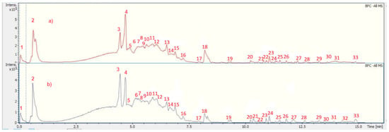
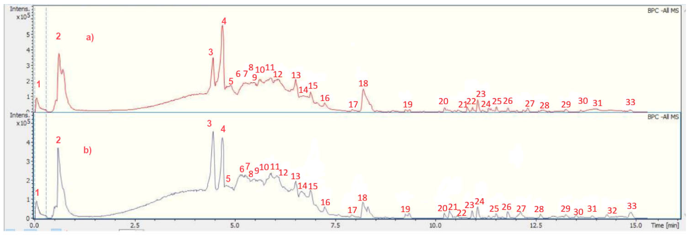
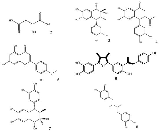
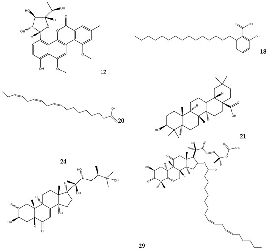
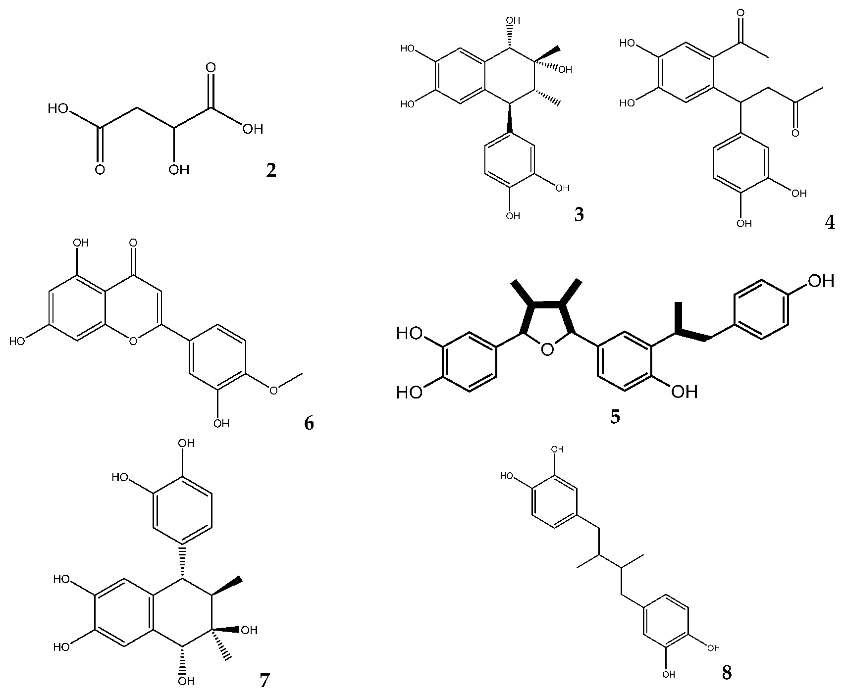
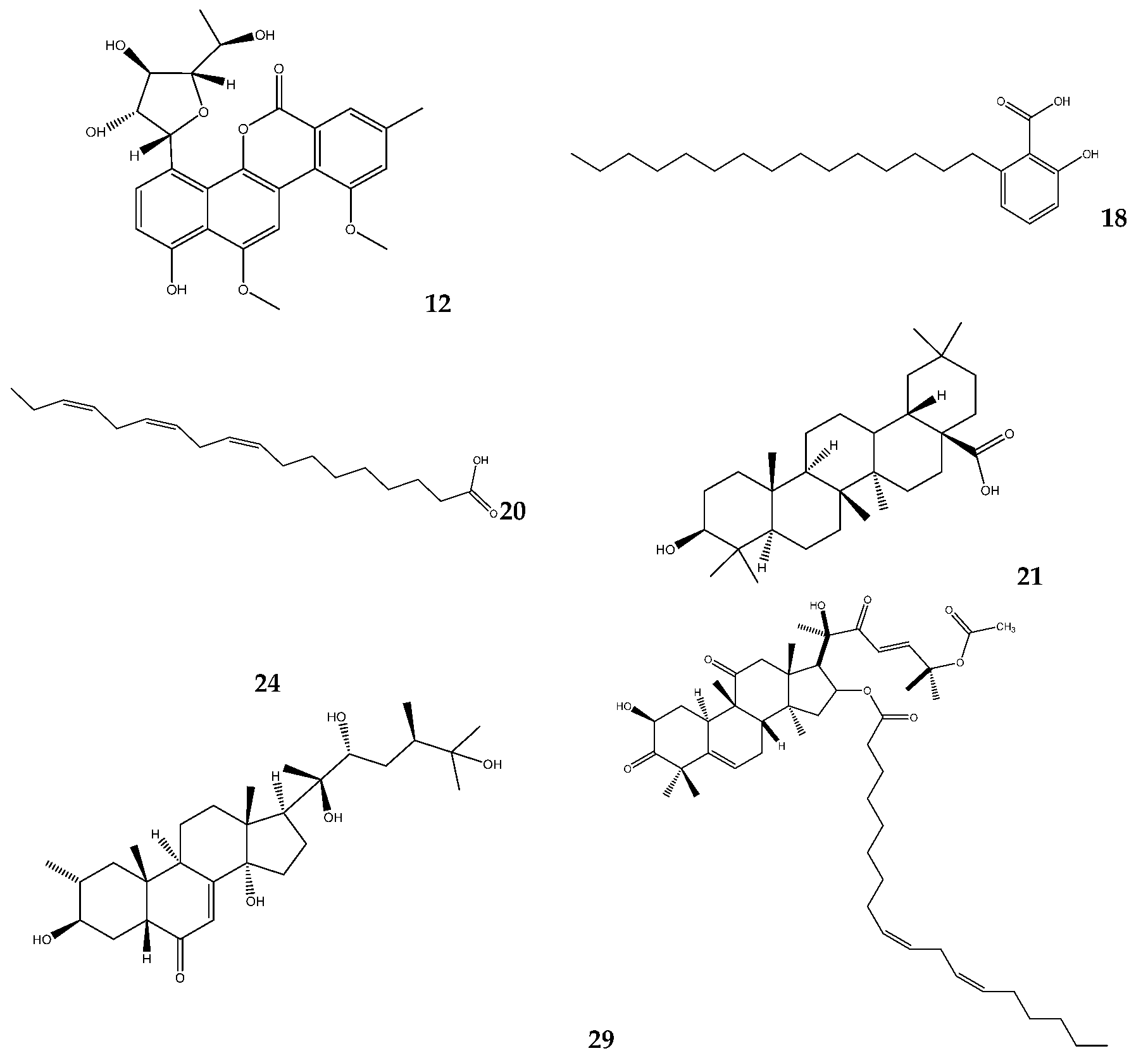
The characterization strategy using UHPLC-ESI-QTOF-MS of chemical compounds present in EMLaSAO and EMLaMAQ involved, initially, data analysis carried out using Metaboscape 4 software (Bruker, Billerica, MA, USA), a tool that allows the identification of metabolites based on their mass, fragmentation pattern, and isotopic pattern. The exhaustive UHPLC-ESI-QTOF-MS analysis in negative mode revealed, in this first analysis, the presence of 33 peaks (2–33) in EMLaSAO and EMLaMAQ, corresponding to 24 tentatively identified compounds (2,3,4,5,6,7,8,9,10,11,12,15,16,18,19,20,21,22,23,24,25,28,29); 8 unknown compounds (14,15,26,27,30–33) and the peak 1 (internal standard). These included flavones, aromatics, terpenes, sterols, furans, and several fatty acids, all of which are tentatively identified here for the first time in L. ameghinoi. The identification strategy employed involved spiking experiments with available standards. Additionally, a comprehensive search of several databases, including MassBank of North America (MONA) and Metaboscape, was performed, along with the use of specific software such as Data Analysis 4.0 (Bruker Daltonik GmbH, Bremen, Germany) and ACD/Spectrum Processor (ACD/Labs, Toronto, Canada), as well as previous reports on the genus Larrea [18,19]. The high-resolution UHPLC–PDA–QTOF analysis results for metabolite identification in EMLaSAO and EMLaMAQ are displayed in Figure 2 and Table 1. Furthermore, the chemical structures of some of the main compounds identified are shown in Figure 3.
Figure 2.
UHPLC Q-TOF-TIC chromatogram of EMLaMAQ (a) and EMLaSAO (b).
Table 1.
UHPLC-PDA-Q-TOF identification of metabolites from EMLaSAO and EMLaMAQ (Rt = Retention time in minutes).
Figure 3.
Molecular structures of representative compounds identified in EMLaMAQ and EMLaSAO.
Table 2 shows the antioxidant properties and total phenolic and flavonoid content of EMLaSAO and EMLaMAQ, which were identified using standardized colorimetric methods and following standardized protocols previously reported in research on the medicinal flora of Argentina and Chile.
Table 2.
Antioxidant properties; total phenolics and flavonoids content of L ameghinoi extracts.
The results obtained from the enzymatic inhibitory activity evaluation of EMLaSAO and EMLaMAQ indicate that EMLaSAO exhibited greater inhibitory activity against AChE compared to EMLaMAQ, while both extracts showed similar inhibition of BChE (Table 3). Regarding digestive enzymes, EMLaSAO and EMLaMAQ demonstrated higher inhibitory efficacy against glucosidase.
Table 3.
Cholinesterases, Amylase, and Glucosidase Enzyme Inhibitory Activities of EMLaSAO and EMLaMAQ.
The plant extracts EMLaSAO and EMLaMAQ from L. ameghinoi exhibited selective antimicrobial activity (Table 4), with EMLaSAO showing the best performance, particularly against Gram-positive Staphylococcus aureus strains, with MIC values ranging from 125 to 250 µg/mL. In contrast, activity against Gram-negative bacteria was weak or absent.
Table 4.
Antimicrobial activity of L. ameghinoi and antibiotic references (MICs and MBCs in µg extract/mL).
4. Discussion
In this article, the enzymatic inhibition, antioxidant and antibacterial effects, and polyphenolic profile of the methanolic extract of L. ameghinoi from Argentina are reported for the first time, supporting the potential of the species as a sustainable source of pharmacologically relevant biomolecules. While other species of the genus, including L. divaricata, L. cuneifolia, and L. nitida, have been extensively studied and associated with diverse biological activities, studies on L. ameghinoi remain limited. In this work, methanolic extracts from two Argentine populations of L. ameghinoi (SAO and MAQ) showed comparable total phenolic content (239.5 and 215.6 mg GAE/g of extract, respectively), with significantly higher flavonoid content in SAO (28.44 mg QE/g vs. 15.28 mg QE/g). Both extracts exhibited strong antioxidant activity, with IC50 values of 10 µg/mL in the DPPH assay, comparable to those of reference compounds such as catechin and quercetin. Consistent results were observed in the FRAP and TEAC assays, with no significant differences between the two samples. In a recent report, resins from Larrea divaricata and L. nitida showed strong DPPH radical scavenging with values around 8.4 µg resin/mL [4], similar to that exhibited by L. ameghinoi extracts (10 µg/mL). The Larrea species that grow in Argentina, including L. cuneifolia, L. divaricata, and L. nitida, are characterized by a high lignan content, notably NDGA and 3′-MNDGA, as well as their respective isomers, which have been identified as markers of these species and are partly responsible for the strong antioxidant and antimicrobial activity of their polar exudates and extracts. Additionally, the synergistic antifungal activity of some of these species, such as Larrea nitida, has also been associated with the content of the marker lignans NDGA and 3′-MNDGA [20]. Regarding the estimated chemical composition of EMLaMAQ and EMLaSAO, UHPLC Q-TOF-TIC chromatograms support that both populations stand out for the presence of three major compounds: malic acid (2), 1,2,4-trihydro-1-(3,4-dihydroxyphenyl)-2,3-dimethyl-3,4,6,7-naphthalenetetrol (3), and 4-(2-acetyl-4,5-dihydroxyphenyl)-4-(3,4-dihydroxyphenyl)butan-2-one (4). Unlike other species of the genus Larrea, both samples show relatively low content of key marker compounds of the genus, such as NDGA (8) and one of its isomers (11), while 3′-MNDGA and its isomers were not detected in the present analyses.Recently, it has been reported that the ability of phenolic compounds as free radical scavengers depends on the quantity and position of the hydroxyl and methoxy groups in their molecules. Additionally, compounds with a catechol group rather than a single hydroxyl group at position 4 have higher reducing capacities, as is the case for caffeic acid compared with p-coumaric acid, and for 3,4-dihydroxybenzoic acid compared with 4-hydroxybenzoic acid. One suggested justification is that stabilizing the phenoxyl radical through an intramolecular hydrogen bond enhances antioxidant activity [21]. Considering the solid and recent bibliography mentioned above, the potent DPPH radical scavenging activity of both samples could be partly explained by the predominant presence of compounds 3 and 4, characterized by two catechol groups in their structures. A contribution from other compounds present in smaller proportions, which also have catechol systems in their structures, such as compounds 5, 6, and 7–11, is also expected. Compounds 3, 4, and 7 have recently been reported from the aerial parts of Larrea tridentata, along with an evaluation of their cytotoxic activity. Compound 3, identified as (1R,2R,3R,4S)-1,2,4-trihydro-1-(3,4-dihydroxyphenyl)-2,3-dimethyl-3,4,6,7-naphthalenetetrol, has been reported to exhibit cytotoxic activity in HL-60 cells with IC50 values of 16 ± 0.95 µM [19]. The flora of Argentina, including various genera of Asteraceae, has attracted attention due to its potential benefits in preventing chronic or non-communicable diseases, such as chronic respiratory diseases, cardiovascular diseases, diabetes, and cancer. These conditions, along with major degenerative pathologies (e.g., Parkinson’s and Alzheimer’s diseases), are leading causes of mortality worldwide, making the search for natural preventive alternatives a priority [22,23]. In this study, L. ameghinoi extracts showed low AChE inhibition but moderate BChE inhibition (IC50 ≈ 49–50 µg/mL), suggesting potential selective therapeutic applications. Previous studies have demonstrated cholinesterase inhibitory activity in other Larrea species, such as L. tridentata, with this activity being associated with species-specific marker compounds like NDGA (compound 8) [24]. Furthermore, antioxidant compounds from dietary sources can neutralize reactive oxygen species, which are key mediators of functional decline and neuronal damage in Alzheimer’s disease, Parkinson’s disease, and other neurodegenerative disorders [25]. This suggests a mechanistic link between the antioxidant properties of the extracts and their ability to inhibit cholinesterase enzymes, supporting a potential neuroprotective effect. Overall, the biological effects observed for L. ameghinoi methanolic extracts can plausibly be explained by the synergy between phenolic metabolites (particularly flavones) and terpenoid/triterpenoid compounds detected by UHPLC. Among polyphenols, flavones stand out due to their potent electron-donating capacity, as well as their ability to modulate pro-oxidant targets, specifically diosmetin (compound 6) and related analogs, which exhibit potent antioxidant activity and a multitarget profile relevant for neuroprotection (AChE/BuChE inhibition and Cu2+ chelation), consistent with the cholinesterase inhibition recorded in our enzymatic assays [26,27]. Similarly, long-chain phenols such as anacardic acids (compounds 18 and 19) possess both antioxidant and anticholinesterase properties [28], providing an additional mechanistic link between phenolic content and the AChE/BuChE inhibitory effects observed in this study. It has recently been reported that anacardic acid exhibits anti-proliferative, pro-apoptotic, anti-inflammatory, and antinociceptive actions and also reduces oxidative stress in acute experimental models, suggesting compound 19 as a promising natural compound of interest for human health [29,30]. The results shown by L ameghinoi suggest potential and justify further research aimed at identifying the active principles responsible for the demonstrated biological activities. We are currently isolating and purifying the major compounds reported here, which will allow us to quickly evaluate their real potential in the biological activities reported here, and further investigate enzyme inhibition studies, including butyrylcholinesterase, and conduct molecular modeling studies to estimate the real potential against this enzyme and others.
Regarding other biological activities, the antimicrobial activity of EMLaSAO and EMLaMAQ was evaluated via MIC, MBC, and MFC (Table 4) and ranked according to the criteria reported in references [31,32]. EMLaSAO showed marked activity against Gram-positive bacteria, including methicillin-sensitive and methicillin-resistant Staphylococcus aureus (MIC = 125 µg/mL; MBC = 250 µg/mL), whereas Gram-negative bacteria were less susceptible (MIC = 1000 µg/mL), indicating a bacteriostatic effect. These results align with previous observations in other Larrea species. Zampini et al. (2007) [33] reported high activity of ethanolic extracts of L. divaricata and L. cuneifolia against Escherichia coli (MIC = 50 µg/mL), while Moreno et al. (2020) [34] observed moderate effects for L. nitida, L. divaricata, and L. cuneifolia (MIC = 800 µg/mL; MBC > 1000 µg/mL). Similarly, Gómez et al. (2021) [4] demonstrated that resinous exudates from L. divaricata and L. nitida effectively inhibited E. coli MQ 586 (MIC = 62.5 µg/mL) and methicillin-sensitive and resistant S. aureus (MIC = 16–32 µg/mL). On the other hand, Martins (2013) [35] found that specific methanolic fractions of L. tridentata, particularly the ethyl acetate fraction, exhibited an MIC of 31.3 µg/mL against methicillin-resistant S. aureus, surpassing even tetracycline (64 µg/mL). These interspecies differences suggest that antibacterial potency is strongly influenced by the particular chemical profile of each species, as well as by the nature of active metabolites, such as lignans and phenolic compounds, which have been associated with antimicrobial activity in Larrea.
With respect to antifungal activity, both extracts displayed moderate inhibition against clinical Candida species, with EMLaSAO generally more active (MIC = 125–500 μg/mL) than EMLaMAQ (MIC = 250–1000 μg/mL). The most susceptible strain was C. tropicalis C131, for which EMLaSAO displayed an MFC of 500 μg/mL, while other yeasts did not show significant fungicidal effects. Compared with extracts from other Larrea species, such as L. nitida [18], the antifungal activity of L. ameghinoi is lower, likely due to differences in chemical composition. Although the observed values of antimicrobial activity are moderate, it is possible and expected that the ongoing partitioning process of the extracts and the subsequent isolation of the major compounds will allow the identification of molecules that show better values with respect to the extracts of origin.
EMLaMAQ and EMLaSAO show a significant content of malic acid (compound 2), recognized for its important role in energy metabolism. Its potential capacity to intervene in the regulation of redox balance and cellular signaling links it to the prevention of cardiovascular diseases and conditions related to oxidative stress. Additionally, its antimicrobial properties support its potential as a versatile metabolite for the therapy of various pathologies [36]. Regarding carbohydrate-hydrolyzing enzymes, oleanane-type triterpenes provide a plausible explanation for the observed α-glucosidase and α-amylase inhibition: oleanolic acid (compound 20) and its derivatives have been shown to significantly inhibit α-glucosidase in vitro, consistent with the IC50 values obtained in our assays [37]. The antimicrobial activity of oleanolic acid has also been reported [38]. In summary, the results support that, unlike other Larrea species whose chemical profile is mainly associated with a high concentration of lignans such as NDGA, its isomers and derivatives, and flavonoids as major constituents, L. ameghinoi presents a distinctive combination of predominant polyphenols and a low concentration of NDGA and some of its isomers. This particular composition, associated with antioxidant, anticholinesterase, and α-glucosidase inhibitory activities, as well as antibacterial action against Gram-positive bacteria, represents the most probable chemical basis for the observed bioactive profile. The contrast in predominant chemistry suggests that L. ameghinoi may offer a different functional spectrum than its congeners, reinforcing its potential as a source of phytochemicals of interest for human health and guiding future research toward marker compound validation and bioactivity-guided fractionation specific to this species.
5. Conclusions
This study provides the first chemical and biological characterization of extracts from Larrea ameghinoi, a vulnerable medicinal species that grows in Argentine Patagonia. The results support the need to design a strategy to protect this species, given its potential as a source of antimicrobial, antioxidant, antitumor, and enzyme-inhibiting compounds of interest for human health. The results also support the medicinal use of this species in traditional Argentine medicine. These findings differentiate L. ameghinoi from other species in the genus, in which NDGA lignans and derivatives predominate, and suggest that its unique phytochemical composition could underlie its multitarget bioactivity. The results shown by L ameghinoi suggest potential and justify further research aimed at identifying the active principles responsible for the demonstrated biological activities. Bioactivity-guided isolation studies of the species’ major compounds are currently underway to confirm their contribution to the observed biological activities.
Author Contributions
Conceptualization, J.G., S.M.S., B.L., A.T. and M.J.S.; methodology, J.G., S.M.S., B.A.S., D.Z.-P., M.E.B.M., D.C., B.L., A.T. and M.J.S.; software, J.G., A.T. and M.J.S.; validation, J.G., S.M.S., B.A.S., D.Z.-P., M.E.B.M., D.C., B.L., A.T. and M.J.S.; formal analysis, J.G., S.M.S., B.A.S., D.Z.-P., M.E.B.M., D.C., B.L., A.T. and M.J.S.; investigation, J.G., S.M.S., B.A.S., D.Z.-P., M.E.B.M., D.C., B.L., A.T. and M.J.S.; resources, J.G., B.L., A.T. and M.J.S.; data curation, J.G., S.M.S., B.A.S., D.Z.-P., M.E.B.M., D.C., B.L., A.T. and M.J.S.; writing—original draft preparation, J.G., S.M.S., B.L., A.T. and M.J.S.; writing—review and editing, J.G., S.M.S., B.L., A.T. and M.J.S.; visualization, J.G., S.M.S., B.L., A.T. and M.J.S.; supervision, J.G., B.L., A.T. and M.J.S.; project administration, J.G., B.L., A.T. and M.J.S.; funding acquisition, J.G., B.L., A.T. and M.J.S. All authors have read and agreed to the published version of the manuscript.
Funding
This research was supported by grants from Argentina and Chile: PROJOVI (J.G.) Resolución Nº 1500-23-R and (PDTS) Resolución Nº 1499-23-R; CICITCAUNSJ, Argentina. M.J.S. received financial support from Fondecyt (Grant 1260005) and Fondequip (EQM170172), Chile.
Institutional Review Board Statement
Not applicable.
Informed Consent Statement
Not applicable.
Data Availability Statement
Data are contained within the article. The assay protocols are available in detail upon request from the authors.
Acknowledgments
The authors express their gratitude for the funding received from UNSJ (Argentina) and Fondecyt (Chile). S. Sede thanks J. A. Calcagno, A. E. Sede and A. G. Abraham for assistance during collection trips in Río Negro, Argentina. J.G., B.A.S. and M.E.B. thank CONICET-Argentina for their doctoral and postdoctoral fellowships.
Conflicts of Interest
The authors declare no conflicts of interest.
References
- Hunziker, J.H.; Palacios, R.A.; Poggio, L.; Naranjo, C.A.; Yang, T.W. Geographic Distribution, Morphology, Hybridization and Evolution. In Creosote Bush: Biology and Chemistry of Larrea in New World Deserts; Mabry, T.J., Hunziker, J., DiFeo, D.R., Jr., Eds.; Dowden, Hutchinson, and Ross: Stroudsburg, PA, USA, 1977; pp. 10–47. [Google Scholar]
- World Flora Online. Larrea. Available online: https://www.worldfloraonline.org/search?query=Larrea+&limit=24&start=0&sort= (accessed on 12 December 2025).
- Kiesling, R. Flora de San Juan, República Argentina; Estudio Sigma: Valladolid, Spain, 2003; Volume II. [Google Scholar]
- Gómez, J.; Simirgiotis, M.J.; Manrique, S.; Piñeiro, M.; Lima, B.; Bórquez, J.; Feresin, G.E.; Tapia, A. UHPLC-ESI-OT-MS phenolics profiling, free radical scavenging, antibacterial and nematicidal activities of “yellow-brown resins” from Larrea spp. Antioxidants 2021, 10, 185. [Google Scholar] [CrossRef] [Scilit] [PubMed]
- CONICET. Científicos Desarrollan un Producto para la Calvicie que ya Está Disponible en Farmacias. Available online: https://www.conicet.gov.ar/cientificos-desarrollan-un-producto-para-la-calvicie-que-ya-esta-disponible-en-farmacias/ (accessed on 9 February 2026).
- Available online: https://sib.gob.ar/especies/larrea-ameghinoi?tab=sitios (accessed on 9 February 2026).
- Palacios, R.A.; Hunziker, J.H. Observaciones sobre la taxonomía del género Larrea (Zygophyllaceae). Darwiniana 1972, 17, 473–476. [Google Scholar]
- Cabrera, A.A. Valorización de la “Jarilla rastrera” (Larrea ameghinoi Speg.): Introducción al Cultivo de Tejidos in vitro y Bioprospección Química. Doctoral Thesis, Universidad Nacional de Río Negro, Viedma, Argentina, 2023. [Google Scholar]
- Heldrich, K. Official Methods of Analysis of the Association of Official Analytical Chemists; Association of Official Chemists: Arlington, VA, USA, 1990. [Google Scholar]
- Brand-Williams, W.; Cuvelier, M.E.; Berset, C. Use of a free radical method to evaluate antioxidant activity. LWT Food Sci. Technol. 1995, 28, 25–30. [Google Scholar] [CrossRef] [Scilit]
- Benzie, I.F.F.; Strain, J.J. The ferric reducing ability of plasma (FRAP) as a measure of “antioxidant power”: The FRAP assay. Anal. Biochem. 1996, 239, 70–76. [Google Scholar] [CrossRef] [Scilit]
- Antolovich, M.; Prenzler, P.D.; Patsalides, E.; McDonald, S.; Robards, K. Methods for testing antioxidant activity. Analyst 2002, 127, 183–198. [Google Scholar] [CrossRef] [Scilit]
- Ellman, G.L.; Courtney, K.D.; Andres, V.; Featherstone, R.M. A new and rapid colorimetric determination of acetylcholinesterase activity. Biochem. Pharmacol. 1961, 7, 88–95. [Google Scholar] [CrossRef] [Scilit]
- Ali, H.; Houghton, P.J.; Soumyanath, A. α-Amylase inhibitory activity of some Malaysian plants used to treat diabetes; with particular reference to Phyllanthus amarus. J. Ethnopharmacol. 2006, 107, 449–455.18. [Google Scholar] [CrossRef] [Scilit]
- Liu, L.; Deseo, M.A.; Morris, C.; Winter, K.M.; Leach, D.N. Investigation of α-glucosidase inhibitory activity of wheat bran and germ. Food Chem. 2011, 126, 553–561. [Google Scholar] [CrossRef] [Scilit]
- Clinical and Laboratory Standards Institute (CLSI). Performance Standards for Antimicrobial Susceptibility Testing, Twenty-Third Informational Supplement, CLSI Document M100-S23; CLSI: Wayne, PA, USA, 2013. [Google Scholar]
- Clinical and Laboratory Standards Institute (CLSI). Reference Method for Broth Dilution Antifungal Susceptibility Testing of Yeasts, Approved Standard. In CLSI Document M27-A3, 3rd ed.; CLSI: Wayne, PA, USA, 2008. [Google Scholar]
- Agüero, M.B.; Svetaz, L.; Sánchez, M.; Luna, L.; Lima, B.; López, M.L.; Zacchino, S.; Palermo, J.A.; Wunderlin, D.; Feresin, G.E.; et al. Argentinean propolis from Larrea nitida Cav. (Zygophyllaceae) exudates: Phytochemical characterization and antifungal activity. Food Chem. Toxicol. 2011, 49, 1970–1978. [Google Scholar] [CrossRef] [Scilit]
- Yokosuka, A.; Iguchi, T.; Jitsuno, M.; Mimaki, Y. Structure and cytotoxicity of novel lignans and lignan glycosides from the aerial parts of Larrea tridentata. Molecules 2021, 26, 6186. [Google Scholar] [CrossRef] [Scilit] [PubMed]
- Butassi, E.; Svetaz, L.; Ivancovich, J.J.; Feresin, G.; Tapia, A.; Zacchino, S. Synergistic antifungal fixed-ratio combinations ofZuccagnia punctata Cav. and Larrea nitida Cav., using mixed-effects loewe (MixLow) method. Phytomedicine 2015, 22, 666–678. [Google Scholar] [CrossRef] [Scilit]
- Pérez, M.; Dominguez-López, I.; Lamuela-Raventós, R.M. The Chemistry Behind the Folin–Ciocalteu Method for the Estimation of (Poly)phenol Content in Food: Total Phenolic Intake in a Mediterranean Dietary Pattern. J. Agric. Food Chem. 2023, 71, 17543–17553. [Google Scholar] [CrossRef] [Scilit]
- Siddiqi, K. Noncommunicable diseases. In Public Health: An Action Guide to Improving Health; Walley, J., Wright, J., Eds.; Oxford University Press: Oxford, UK, 2010. [Google Scholar]
- WHO. Noncommunicable Diseases; WHO: Geneva, Switzerland, 2019. [Google Scholar]
- Reyes-Melo, K.Y.; Galván-Rodrigo, A.A.; Martínez-Olivo, I.E.; Núñez-Mojica, G.; Ávalos-Alanís, F.G.; García, A.; Del Rayo Camacho-Corona, M. Larrea tridentata and its biological activities. Curr. Top. Med. Chem. 2021, 21, 2352. [Google Scholar] [CrossRef] [Scilit]
- Lee, K.H.; Cha, M.; Lee, B.H. Neuroprotective effect of antioxidants in the brain. Int. J. Mol. Sci. 2020, 21, 7152. [Google Scholar] [CrossRef] [Scilit]
- Yang, A.; Yi, X.; Zhang, H.; Shen, R.; Kou, X. Diosmetin derivatives as multifunctional anti-AD ligands: Design, synthesis, and biological evaluation. Chem. Biol. Drug Des. 2024, 103, e14529. [Google Scholar] [CrossRef] [Scilit]
- Chagas, M.D.S.S.; Behrens, M.D.; Moragas-Tellis, C.J.; Penedo, G.X.M.; Silva, A.R.; Gonçalves-de-Albuquerque, C.F. Flavonols and flavones as potential anti-inflammatory, antioxidant, and antibacterial compounds. Oxid. Med. Cell Longev. 2022, 2022, 9966750. [Google Scholar] [CrossRef] [Scilit]
- Da Silva, W.M.B.; Pinheiro, S.O.; Alves, D.R.; de Menezes, J.E.S.A.; Magalhães, F.E.A.; Silva, F.C.O.; Marinho, M.M.; Marinho, E.S.; de Morais, S.M. Anacardic acid complexes as possible agents against Alzheimer’s disease through their antioxidant, in vitro, and in silico anticholinesterase and anxiolytic actions. Neurotox. Res. 2021, 39, 467–476. [Google Scholar] [CrossRef] [Scilit] [PubMed]
- Schultz, D.J.; Krishna, A.; Vittitow, S.L.; Alizadeh-Rad, N.; Muluhngwi, P.; Rouchka, E.C.; Klinge, C.M. Transcriptomic response of breast cancer cells to anacardic acid. Sci. Rep. 2018, 8, 8063. [Google Scholar] [CrossRef] [Scilit]
- Gomes Junior, A.L.; Islam, M.T.; Nicolau, L.A.D.; de Souza, L.K.M.; Araújo, T.D.S.L.; Lopes de Oliveira, G.A.; Melo-Cavalcante, A.A.D.C. Anti-inflammatory, antinociceptive, and antioxidant properties of anacardic acid in experimental models. ACS Omega 2020, 5, 19506–19515. [Google Scholar] [CrossRef] [Scilit]
- Rios, J.-L.; Recio, M.C. Medicinal plants and antimicrobial activity. J. Ethnopharmacol. 2005, 100, 80–84. [Google Scholar] [CrossRef] [Scilit]
- Tamokou, J.D.D.; Mbaveng, A.T.; Kuete, V. Medicinal spices and vegetables from Africa: Therapeutic potential against metabolic, inflammatory, infectious and systemic diseases. In Medicinal Spices and Vegetables from Africa; Academic Press: Cambridge, MA, USA, 2017; pp. 207–237. [Google Scholar]
- Zampini, I.C.; Cudmani, N.; Isla, M.I. Actividad antimicrobiana de plantas medicinales argentinas sobre bacterias antibiótico-resistentes. Acta Bioquím. Clín. Latinoam. 2007, 41, 385–393. [Google Scholar]
- Moreno, M.A.; Bojorges, H.; Falco, I.; Sanchez, G.; Lopez-Carballo, G.; Lopez-Rubio, A.; Zampini, I.C.; Isla, M.I.; Fabra, M.J. Activeproperties of edible marine polysaccharide-based coatings containing Larrea nitida polyphenols enriched extract. Food Hydrocoll. 2020, 102, 105595. [Google Scholar] [CrossRef] [Scilit]
- Martins, S.; Amorim, E.L.C.; da Silva Peixoto Sobrinho, T.J.; Saraiva, A.M.; Pisciottano, M.N.; Aguilar, C.N.; Teixeira, J.A.; Mussatto, S.I. Antibacterial activity of crude methanolic extract and fractions obtained from Larrea tridentata leaves. Ind. Crops Prod. 2013, 41, 306–311. [Google Scholar] [CrossRef] [Scilit]
- Lu, J.; Zhang, S.; Wu, S.; Gao, C. L-malic acid: A multifunctional metabolite at the crossroads of redox signaling, microbial symbiosis, and therapeutic innovation. Arch. Biochem. Biophys. 2025, 772, 110554. [Google Scholar] [CrossRef] [Scilit] [PubMed]
- Ali, M.S.; Jahangir, M.; Hussan, S.S.; Choudhary, M.I. Inhibition of alpha-glucosidase by oleanolic acid and its synthetic derivatives. Phytochemistry 2002, 60, 295–299. [Google Scholar] [CrossRef] [Scilit] [PubMed]
- Jesus, J.A.; Lago, J.H.; Laurenti, M.D.; Yamamoto, E.S.; Passero, L.F. Antimicrobial activity of oleanolic and ursolic acids: An update. Evid. Based Complement. Altern. Med. 2015, 2015, 620472. [Google Scholar] [CrossRef] [Scilit]

Disclaimer/Publisher’s Note: The statements, opinions and data contained in all publications are solely those of the individual author(s) and contributor(s) and not of MDPI and/or the editor(s). MDPI and/or the editor(s) disclaim responsibility for any injury to people or property resulting from any ideas, methods, instructions or products referred to in the content. |
© 2026 by the authors. Licensee MDPI, Basel, Switzerland. This article is an open access article distributed under the terms and conditions of the Creative Commons Attribution (CC BY) license.