Article

Autologous Tooth Graft after Endodontical Treated Used for Socket Preservation: A Multicenter Clinical Study

1
University of Bari Aldo Moro, 70121 Bari, Italy
2
Private Practice, 20100 Milan, Italy
3
College of Medicine and Dentistry Birmingham, University of Birmingham, Birmingham B15 2TT, UK
4
Private Practice, 73100 Lecce, Italy
5
Private practice, Singapore 238863
6
University of Naples Federico II, 80100 Naples, Italy
7
Visiting Professor University of Genoa, 16126 Genova, Italy
8
Private Practice, 10121 Turin, Italy
9
Private Practice, 50002 Hradec Kralove, Czech Republic
10
Private Practice, 34121 Trieste, Italy
11
Olomouc University, 77147 Olomouc, Czech Republic
12
School of Medicine, University of Bari, 70121 Bari, Italy
13
School of Dentistry, University of Foggia, 71122 Foggia, Italy
14
Private Practice - Biomaterial Clinical and Histological Research Association
15
Clinical and Experimental Medicine Dept. School of Dentistry University of Foggia, 71122 Foggia, Italy
*
Authors to whom correspondence should be addressed.
Appl. Sci. 2019, 9(24), 5396; https://doi.org/10.3390/app9245396
Submission received: 22 October 2019 / Revised: 13 November 2019 / Accepted: 29 November 2019 / Published: 10 December 2019
(This article belongs to the Special Issue Bio-Inspired Biodegradable Scaffold Constructs for Bone Tissue Repair)

Abstract

:
The aim of the study was to evaluate the tooth extracted use as autologous tooth graft after endodontic root canal therapies used for socket preservation. To this purpose, the Tooth Transformer shredding and decontamination machine has been used. The graft obtained in this way, was inserted at the time of the extraction or at a second surgery altogether with the chosen regenerative therapy. This clinical trial enrolled patients with post-estractive defects requiring the restoration bone dimension and shape in the maxillary and mandibular zone. In addition, 98 patients with 119 extraction sockets were enrolled across 10 standardized centers. An innovative preparation method, using the dedicated automated device Tooth Transformer, able to transform autologous teeth in suitable grafting material, has been used. The extracted tooth was cleaned and treated using a Tooth Transformer and made a socket preservation. Thirteen Biopsies were realized to analyze the histologic outcomes at the average time of four months to demonstrate that the autologous tooth graft made from root after endodontic therapy should be used in human bone regeneration as graft for dental implant placement.

1. Introduction

Alveolar socket preservation techniques were introduced for ridge augmentation and prevention of the alveolar bone resorption after tooth extraction with bone substitute materials with or without membrane barrier use [1,2,3]. The tooth grafting procedure has been introduced by Urist et al. more than 50 years ago, when they discovered the osteoinduction potential of demineralized dentin matrix [4,5]. Recently, Bessho et al. demonstrated the presence of bone morphogenetic proteins (BMPs) in human dentin matrix. In particular, bone formation and osteoblasts’ presence in rat muscles after demineralized human dentin matrix graft were observed [6]. It was clear that both bone and dentin matrix contained fundamental growth factors for bone regeneration. It represented an efficient reserve of BMPs and bioactive growth factors (GFs), such transforming growth factor-B (TGF-B), which are well known to be involved in bone repairing processes [7]. Some authors theorized that the demineralization process allows better bone augmentation than non-demineralized dentin [8]. Moreover, the chemical composition of bone and dentin was almost the same as the presence of an inorganic portion made of hydroxyapatite and an organic one, mainly composed of collagen type 1 and other secondary proteins. Heterologous or alloplastic grafting materials, on the other hand, have been used for bone augmentation procedures from more than 35 years, but they work as a mechanical scaffold for host cells and do not offer any osteoinductive stimulus [9,10,11,12]. The efficacy and safety of autogenous partially demineralized dentin matrix prepared chairside, for clinical application in bone regeneration procedures related to implant dentistry, including socket preservation, alveolar ridge augmentation, and maxillary sinus floor augmentation were recently demonstrated in some human studies [13,14]. Recently, an innovative medical device (TT TOOTH TRANSFORMER SRL, Via Washington, 59—Milan, Italy) to obtain suitable tooth graft materials starting from the whole tooth of the patient was introduced to the market. This device ensures completely automated disinfection, grinding, and demineralizing processes without any possible mistake induced by human manipulation of the process. This new device represents an advanced system in the area of tissue engineering because it is able to process and transform extracted tooth into useful bone graft material in a short time. The graft material, produced starting from the whole tooth, showed high wettability that allowed an easy handling and positioning on a host site [15]. A previous case series described the successful clinical outcomes of bone regeneration after autologous tooth grafting using this new device and demonstrated the complete filling of bone defects by hard tissue without any complications [16].
The aim of the study is to evaluate the tooth extracted use for the first time as autologous tooth graft after endodontic root canal therapies used for socket preservation with histological analysis. The second outcome is to evaluate the implant insertion in regenerated vital bone with six months of follow up.

2. Materials and Methods

2.1. Study Design

The primary objective of this prospective study was to analyze the tooth extracted use as autologous tooth graft after endodontic root canal therapies used for socket preservation with histological analysis. The second outcome is to evaluate the implant insertion in regenerated vital bone with six months of follow up. After tooth extraction, all patients received a socket preservation procedure using autologous tooth as a graft after the TT Tooth Transformer device procedure. One-hundred six sites received an implant insertion after 4 months of bone healing with 3i Certain implants and 13 biopsies were carried out for histological control (Table 1).

2.2. Inclusion Criteria

All patients were recruited for socket preservation procedure after tooth extraction and implant rehabilitation. Nine-eight patients (29 men and 69 women) with an average age 53.7 years (range, 22–83 years) in health conditions, after written informed consent, were treated between November 2016 and January 2019 in private dental clinics in Italy, Czech Republic, and Singapore. The study group included 119 socket sites and 106 implants were placed.

2.3. Exclusion Criteria

Patients with an history of allergies, tobacco (use within the last 6 months), healing disorders, diabetes, cancer, HIV, bone diseases, metabolic diseases, systemic corticosteroids use, intramuscular or intravenous bisphosphonates use, immunosuppressive agents use, radiation therapy, and chemotherapy (within the last two months) were excluded. Pregnant subjects were also excluded.

2.4. Preoperative Work-Up

Preliminary clinical and radiographic examination had been performed. Two weeks before surgery, all patients received a professional oral hygiene session, and the patients were also instructed about common oral hygiene procedures. Chlorhexidine 0.2% mouth rinses, twice a day for 2 weeks, were prescribed. Cone beam computed tomography (CBCT) scans were taken (53 cases). In the other cases, periapical X-rays or panoramic X-rays were performed. The periapical X-rays or panoramic X-rays were performed in cases with young patients and especially in female subjects of childbearing age.

2.5. Surgical Procedures and Follow-Up

Following administration of antibiotics, extractions were performed. Extraction socket morphology dimensions were recorded through direct measures, and baseline extraction socket buccal-lingual and vertical dimensions were recorded. Bone defects were filled with particulate Tooth grafts made from the Tooth Transformer. The whole extracted tooth was first cleaned from residual calculus using a piezoelectric instrument (Mectron, Carasco (GE), Italy). The root surface was polished using a diamond drill (ref.6855 Dentsply Maillefer, Ballaigues, Switzerland) with abundant irrigation. Any filling materials (gutta-percha, composite, etc.) were carefully removed from the tooth. The tooth was cut in small pieces and they were inserted in the mill of the device (Tooth Transformer, Milan, Italy). The single use unit was open, and a small box containing disposable liquids was inserted in the device in its correct position (indicated by arrows). According to the manufacturer, these solutions guarantee maximum release of BMP-2 and collagen as well as decontamination of the root. When all the components were inserted and the cover of the machine was closed, the device was started using the general switch button. Demineralized dentin graft was ready in 25 min to be placed in the patient’s mouth (TT TOOTH TRANSFORMER SRL, Via Washington, 59—Milan, Italy). The graft was confined to the existing alveolar ridge dimensions, making no attempt to go outside the confines of ridge. The defect was covered with absorbable collagen membranes’ porcine pericardium (bego oss). placed to cover the grafts and extend slightly 2–3 mm beyond bone defect margins. Amoxicillin (Pfizer, New York, NY USA) was provided 1000 mg every 12 h; alternatively, 2000 mg BID for 10–14 days (with clindamycin (Pfizer, New York, NY USA) 300 mg for 7–10 days for subjects with penicillin allergies. At 1-week post-surgery, the sutures were removed. The dental implants were placed after a 4-month healing period. The dental implants were composed of commercially pure titanium, made available by certified companies (Biomet 3i, Palm Beach, FL USA). The implants were performed using the milling kit of the specific implant house. For the first cutter, said pilot had a diameter of 1.2 mm. It was continued with burs of caliber gradually slightly greater to delicately and progressively widen the small hole, until reaching the desired width. Once the hole has been completed, the implant that we previously selected as diameter and length was taken from its sterile package and transported into the hole, screwing it very slowly with the help of a low speed motor (W&H, Bürmoos, Austria) or manually. Biopsies were performed using trephine cylindrical drills (Meisinger, Neuss, Germany) graduated to indicate the depth (from 5 to 18 mm) under copious sterile saline irrigation.

2.6. Performing Biopsies for Histological Evaluation

Biopsies were performed after waiting a variable healing time in relation to the therapeutic decision previously adopted in the treatment plan on the times of the second surgical phase. Before performing the biopsies, radiographs of the affected site were detected. The biopsies took place with cylindrical trephine or with a piezo device to cause the least possible biological damage to the area of the explant.

2.7. Collection and Statistical Analysis of Data

Clinical, histological, and morphometric data and patient records were collected in special files and stored and processed in accordance with the laws on privacy. A simple statistical analysis was used to obtain average values of the morphometric data, in order to obtain valid reference values for a comparison with future studies and for a comparison between the various groups.

2.8. Histological Technique

All samples were washed, fixed, and included in the resin for histological evaluation. The sample was dehydrated by a series of solutions with increasing alcohol concentration (Sigma-Aldrich, St.Louis, Missouri, USA), up to pure alcohol and then infiltrated into methacrylic resin ( Sigma-Aldrich, St.Louis, Missouri, USA). After the light curing of the resin, the sample was processed to obtain non-decalcified sections due to wear, using a disk abrasion system (LS2—Remet, Remet, Bologna, Italy) and a diamond disk cutting system (Micromet—Remet, Bologna, Italy). In the first phase, the inclusion in resin was abraded to eliminate the resin component that covers the sample, and the area of the biopsy to be observed was thus brought to the surface. Then, the surface was glued to a showcase with cyanacrylate (Sigma-Aldrich, St.Louis, Missouri, USA) based adhesives. Subsequently, a cutting with a high speed and cooling diamond blade (Bueheler, Lake Bluff, Illinois USA) was performed. In this way, a slide with a sample of about 200 microns thick was obtained which must be thinned by abrasion. With low abrasive paper, the sample was then abraded on the lapping machine (Bueheler, Lake Bluff, Illinois USA) with thickness control that allows for progressively reducing the sample thickness up to about 40–50 microns. At this point, the slide was polished with polishing papers and colored with basic fuchsin and blue toluidine for the final observation in light and polarized light microscopy (Olympus, Shinjuku, Tokyo, Japan). For histomorphometric measurements, the histological images obtained from the transmitted light microscope (Olympus, Shinjuku, Tokyo, Japan) were digitized through a digital camera and analyzed by means of an image analysis software IAS 2000 (QEA, Billerica, MA, USA). For each sample, BV% = Percentage of residual bone volume with exclusion of medullary tissues; Graft% = percentage of the remaining graft, excluding bone and marrow; and VB% = percentage of vital bone with exclusion of the medulla and residual graft were calculated.

3. Results

In addition, 98 patients were enrolled and treated and 119 sockets were carried out with autologous tooth graft material. All defects were classified with number of walls presents. In addition, 43% had three walls, 42% had four walls, 12% had one wall and 3% had two walls (Table 2).
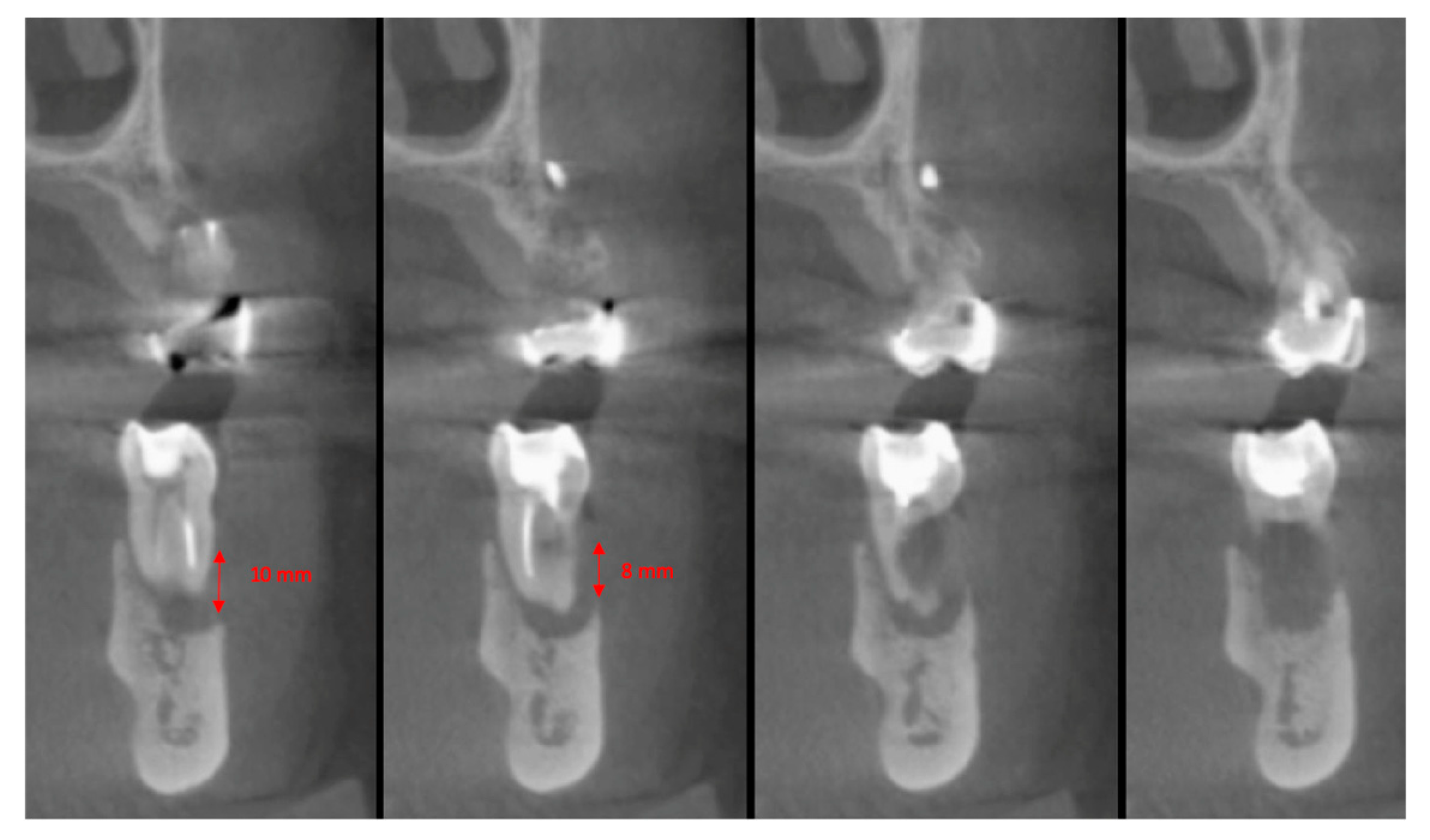
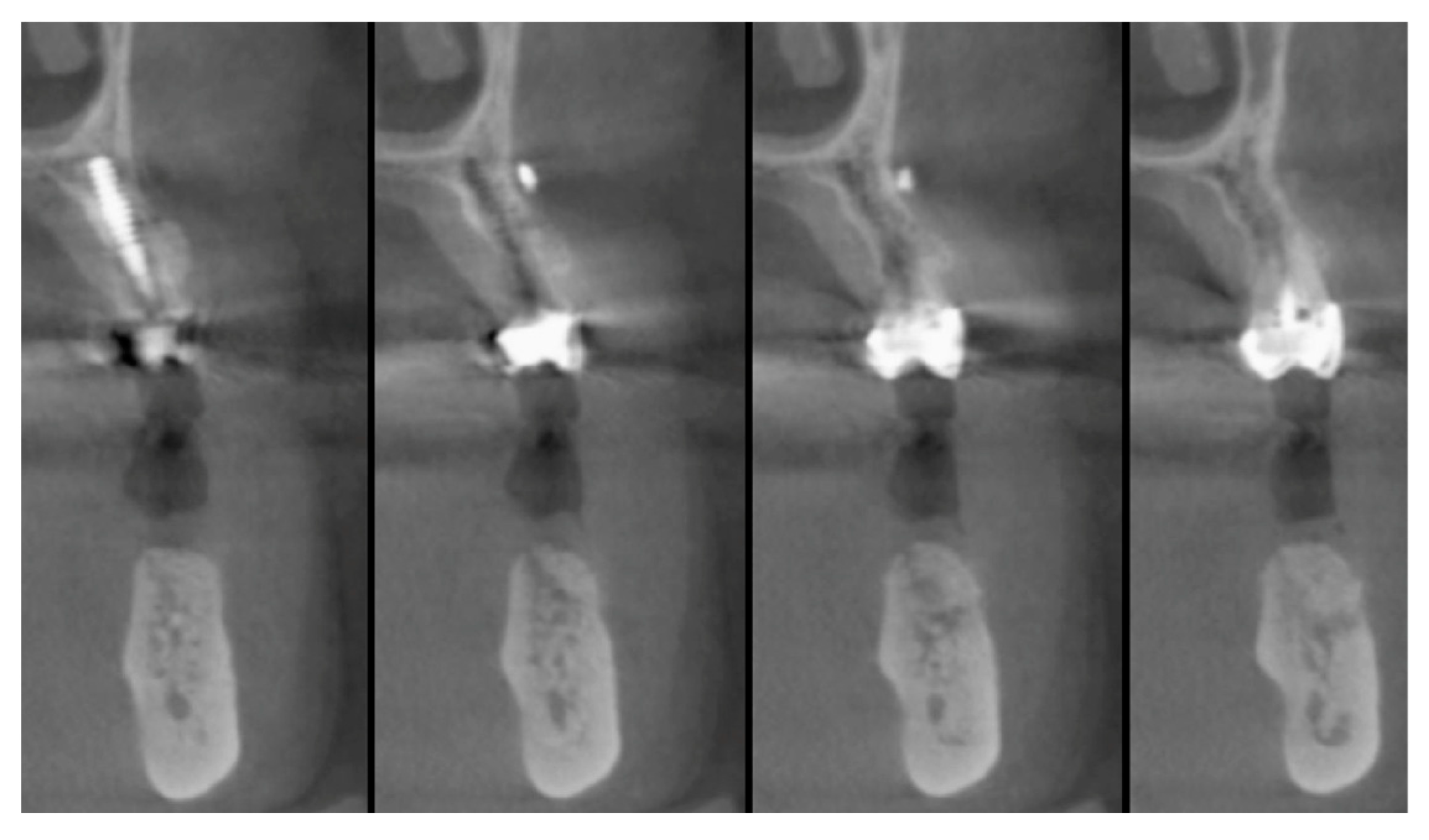
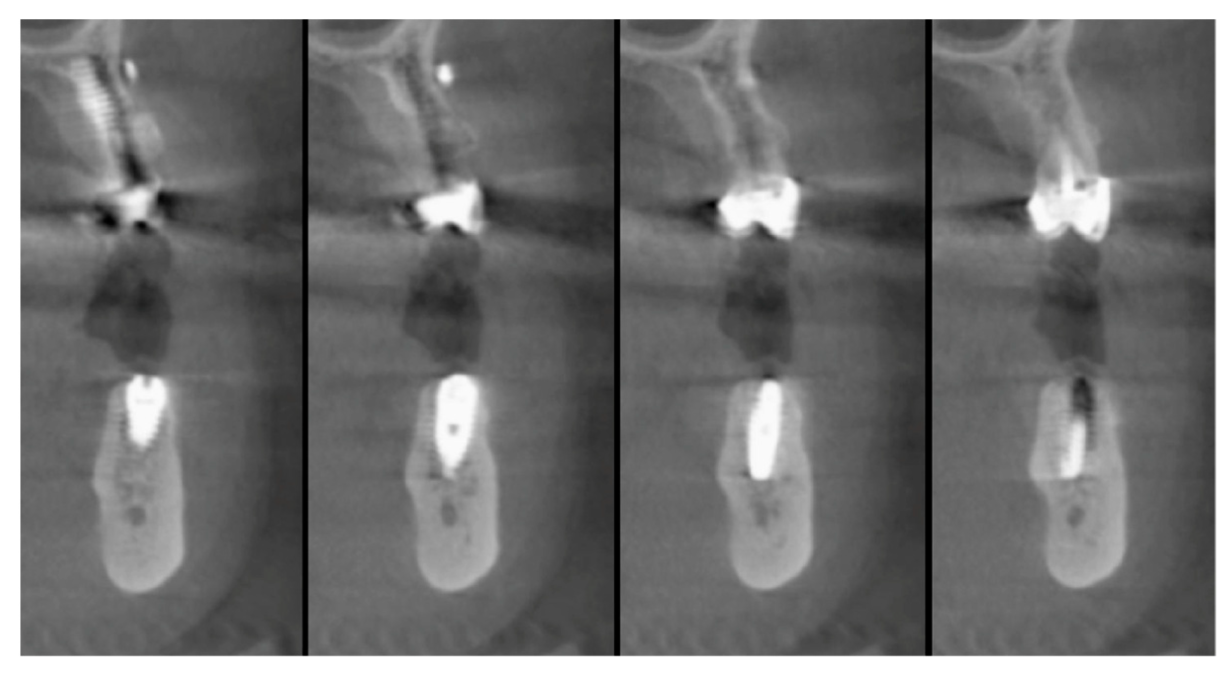
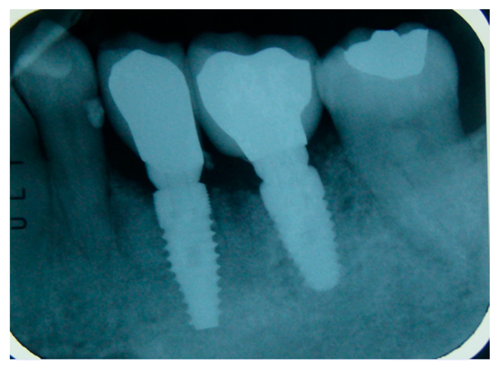
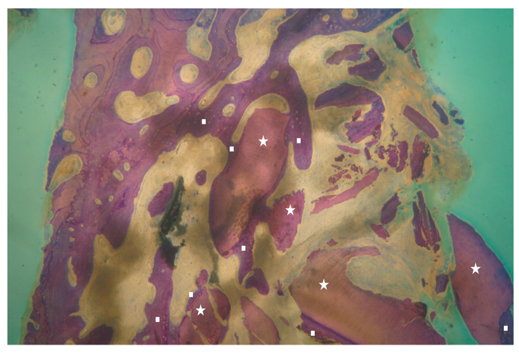
The vertical average defect was 9.16 mm, buccal lateral/palatal 7.0 mm, and mesio-distal 10.3 mm. The measurements were intraoperatively performed with a millimeter periodontal probe. The endodontic material mechanically cleaned before the tooth was placed inside the Tooth Transformer device. The endodontic material was removed using a drill bur with the attention to cut the canal in excess with the aim to remove all the cement. No surgical complications were registered. Pre and post-operative X-rays (section from CBCT) were collected from the same case (Figure 1, Figure 2, Figure 3 and Figure 4). After four months, 13 histological and histomorphometrical evaluations were performed (Figure 5). The Bone Volume/Total Volume average (BV%) was 41.47 (S.D. ± 11.51), the Residual Graft/Total Volume average (Graft%) was 16.60 (S.D. ± 7.09), and the Vital Bone/Total Bone average (VB%) was 21.89 (S.D. ± 9.72). No extraneous material (gutta-percha or cement) was detected in all samples (Table 3).
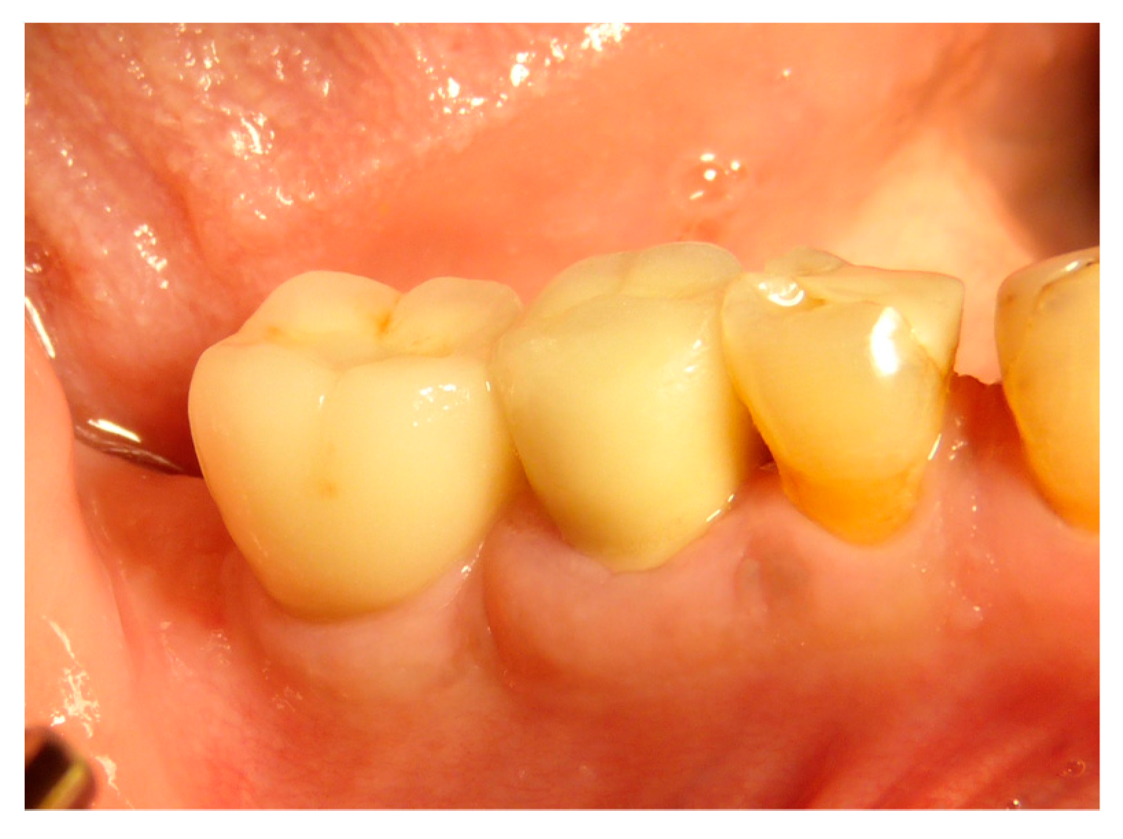
The faraway graft procedure follow up is 40 months and the near graft procedure follow up is 9 months (Average 24.5 months). The faraway implant follow up is 35 months and the near implant follow up is five months. The success rate of the tooth graft procedure is 99.1% (one site was infected and lost the regeneration and the implant) (Figure 6, Figure 7 and Figure 8). In all cases, after the all implants were inserted, complete osseointegration after proper healing period was achieved. After the healing period, hard and soft tissues were stable (Figure 9, Figure 10 and Figure 11). The healing of soft tissues after grafting procedures was particularly free of complications. The implant success rate was 98.94% (one implant failed).

4. Discussion

The endodontically treated tooth does not necessarily lose bone. To date, mainly bone of deproteinized bovine origin or alloplastic materials are used as graft [15]. These materials have the characteristic of maintaining space and are embedded in the bone matrix in formation. The newly formed bone tissue will consist mainly of residues of the graft material and a small percentage of new bone. The use of tooth particulates, even after root canal treatment, offers the advantage of possessing osteoinductive molecules, like BMP-2 [16], that are present in the dentin. The use of the tooth graft is evaluated as an autologous graft, but some doubt remains about the use of teeth with root canal therapies. This manuscript evaluates bone augmentation results, soft tissue healing, and implant survival rate after socket preservation using a new device able to shred and decontaminate dental elements and transform the teeth, also the teeth with root canal therapies, in graft material. Tooth granules were surrounded by newly formed bone. Some tooth granules were incorporated into the bone trabeculae, and they appeared to be partially resorbed. This fact testified that tooth graft underwent remodeling processes just like the native bone. A recent literature review reported an implant survival rate of 97.7% corresponding to our results [17] The present study shows a high clinical survival rate of the implants, but the most important result is the possibility to insert implants four months after a socket preservation using a tooth graft from teeth with endodontic treatment. In addition, 99.1% of the socket preservation made in this study has been successful. The 13 histological analyses of the present multicenter study showed bone regeneration and no inflammatory reactions around dentin granules. The graft, in all the cases analyzed, was subjected to the physiological bone remodeling phenomena, demonstrating an excellent integration with the host tissues. No variation, either clinical or histological, has been noted using undergoing endodontic treatments. No extraneous material (gutta-percha or cement) was detected in all samples. The 98.94% implant success rate and only one implant failure detected after four months of follow up are comparable to the reported data in [18,19,20,21,22,23].

5. Conclusions

Future studies with long follow up are needed in order to better evaluate the potential of demineralized dentin autografts. However, the data obtained confirmed that the endodontically treated tooth used as an autologous graft in post-extraction sockets promotes the production of vital bone of adequate volume. The lack of endodontic treatment residues in all the samples examined confirm the cleaning procedure used. The volumes of newly bone are adequate to support the implant insertion in implant-prosthetic rehabilitations of the jaws. These grafts open up new scenarios in clinical research for the tooth, no longer waste material but true scaffold with osteoinductive characteristics. This innovative device allowed for process and use, as bone graft, any patient’s tooth in a very short time, opening up a new research field. All decontamination, disinfection, and demineralization processes are totally electronically managed by the machine itself without any possibility of human error or injury.

6. Approval

The Ethical Comitee approval: request ID richhtnc4, protocol N°1869 12/12/2018, approved 17 verb 21.03.19 St.638 PI Perfetti.pdf.

Author Contributions

Conceptualization, E.M., C.M., P.T. and F.M.; Data curation, A.P., H.K.L.H., U.G., M.C. (Marcello Contessi), P.T. and F.M.; Formal analysis, F.F., S.N.D.H. and M.C. (Martin Celko); Investigation, A.P., J.H.S., H.K.L.H., S.N.D.H., E.G., U.G., M.C. (Marcello Contessi), M.C. (Martin Celko) and F.M.; Methodology, J.H.S., E.G. and C.M.; Project administration, E.M. and F.M.; Software, A.B.; Supervision, A.B. and P.T.; Writing—original draft, E.M., P.T. and F.M.; Writing—review & editing, F.F.

Funding

This research received no external funding.

Conflicts of Interest

The authors state that this article does not engender any conflicts of interest, whatsoever, with any organization or individual.

References

  1. Atieh, M.A.; Alsabeeha, N.H.; Payne, A.G.; Duncan, W.; Faggion, C.M.; Esposito, M. Interventions for replacing missing teeth: Alveolar ridge preservation techniques for dental implant site development. Cochrane Database Syst. Rev. 2015, 5, CD010176. [Google Scholar] [CrossRef] [PubMed]
  2. Avila-Ortiz, G.; Elangovan, S.; Kramer, K.W.; Blanchette, D.; Dawson, D.W. Effect of alveolare ridge preservation after tooth exytraction: A systematic review and meta-analysis. J. Dent. Res. 2014, 93, 950–958. [Google Scholar] [CrossRef] [PubMed]
  3. Vittorini Orgeas, G.; Clementini, M.; De Risi, V.; De Sanctis, M. Surgical techniques for alveolar socket preservation: A systematic review. Int. J. Oral Maxillofac. Implant. 2013, 28, 1049–1061. [Google Scholar] [CrossRef] [PubMed] [Green Version]
  4. Yeomans, J.D.; Urist, M.R. Bone induction by decalcified dentine implanted into oral, osseous and muscle tissues. Arch. Oral Biol. 1967, 12, 999–1008. [Google Scholar] [CrossRef]
  5. Bang, G.; Urist, M.R. Bone induction in excavation chambers in matrix of decalcified dentin. Arch. Surg. 1967, 94, 781–789. [Google Scholar] [CrossRef]
  6. Bessho, K.; Tanaka, N.; Matsumoto, J.; Tagawa, T.; Murata, M. Human dentin-matrix-derived bone morphogenetic protein. J. Dent. Res. 1991, 70, 171–175. [Google Scholar] [CrossRef] [PubMed]
  7. Nakashima, M. Bone Morphogenetic proteins in dentin regeneration for potential use in endodontic therapy. Cytokine Growth Factor Rev. 2005, 16, 369–376. [Google Scholar] [CrossRef]
  8. Rijal, G.; Shin, H.I. Human tooth-derived biomaterial as a graft substitute for hard tissue regeneration. Regen. Med. 2017, 12, 263–273. [Google Scholar] [CrossRef]
  9. Boyne, P.J. Experimental evaluation of the osteogenic potential of bone graft materials. Annu. Meet. Am. Inst. Oral Biol. 1969, 13–21. [Google Scholar] [PubMed]
  10. Mellonig, J.T.; Bowers, G.M.; Cotton, W.R. Comparison of bone graft materials. Part II. New bone formation with autografts and allografts: A histological evaluation. J. Periodontol. 1981, 52, 297–302. [Google Scholar] [CrossRef]
  11. Colnot, C.; Romero, D.M.; Huang, S.; Helms, J.A. Mechanisms of action of demineralized bone matrix in the repair of cortical bone defects. Clin. Orthop. Relat. Res. 2005, 435, 69–78. [Google Scholar] [CrossRef] [PubMed]
  12. Araújo, M.G.; Sonohara, M.; Hayacibara, R.; Cardaropoli, G.; Lindhe, J. Lateral ridge augmentation by the use of grafts comprised of autologous bone or a biomaterial. An experiment in the dog. J. Clin. Periodontol. 2002, 29, 1122–1131. [Google Scholar] [CrossRef] [PubMed]
  13. Minamizato, T.; Koga, T.; Nakatani, Y.; Umebayashi, M.; Sumita, Y.; Ikeda, T.; Asahina, I. Clinical application of autogenous partially demineralized dentin matrix prepared immediately after extraction for alveolar bone regeneration in implant dentistry: A pilot study. Int. J. Oral Maxillofac. Surg. 2018, 47, 125–132. [Google Scholar] [CrossRef] [PubMed]
  14. Kim, S.Y.; Kim, Y.K.; Park, Y.H.; Park, J.C.; Ku, J.K.; Um, I.W.; Kim, J.Y. Evaluation of the Healing Potential of Demineralized Dentin Matrix Fixed with Recombinant Human Bone Morphogenetic Protein-2 in Bone Grafts. Materials (Basel) 2017, 10, 1049. [Google Scholar] [CrossRef] [Green Version]
  15. Y Baena, R.R.; Lupi, S.M.; Pastorino, R.; Maiorana, C.; Lucchese, A.; Rizzo, S. Radiographic evaluation of regenerated bone following poly (lactic-co-glycolic) acid/hydroxyapatite and deproteinized bovine bone graft in sinus lifting. J. Craniofac. Surg. 2013, 24, 845–848. [Google Scholar] [CrossRef]
  16. Bono, N.; Tarsini, P.; Candiani, G. Demineralized dentin and enamel matrices as suitable substrates for tooth regeneration: Thorough characterization and in vitro. J. Appl. Biol. Func. Mater. 2017, 15, e236–e243. [Google Scholar]
  17. Rizzo, S.; Zampetti, P.; Rodriguez, Y.; Baena, R.; Svanosio, D.; Lupi, S.M. Retrospective analysis of 521 endosseous implants placed under antibiotic prophylaxis and review of literature. Minerva Stomatol. 2010, 59, 75–88. [Google Scholar]
  18. Minetti, E.; Berardini, M.; Trisi, P. A new tooth processing apparatous allowing to obtain dentin grafts for bone augmentation: The tooth transformer. Open Dent. J. 2019, 13, 6–14. [Google Scholar] [CrossRef]
  19. Eliasson, A.L.; Blomqvist, F.; Wennerberg, A.; Johansson, A. A retrospective analysis of early and delayed loading of full-arch mandibular prostheses using three different implant systems: Clinical results with up to 5 years of loading. Clin. Implant. Dent. Relat. Res. 2009, 11, 134–148. [Google Scholar] [CrossRef]
  20. Dierens, M.; Vandeweghe, S.; Kisch, J.; Nilner, K.; De Bruyn, H. Long-term follow-up of turned single implants placed in periodontally healthy patients after 16–22 years: Radiographic and peri-implant outcome. Clin. Oral Impl. Res. 2012, 23, 197–204. [Google Scholar] [CrossRef]
  21. Eghbali, A.; De Bruyn, H.; De Rouck, T.; Cleymaet, R.; Wyn, I.; Cosyn, J. Single implant treatment in healing versus healed sites of the anterior maxilla: A clinical and radiographic evaluation. Clin. Implant. Dent. Relat. Res. 2012, 14, 336–346. [Google Scholar] [CrossRef] [PubMed]
  22. Creugers, N.H.; Kreulen, C.M.; Snoek, P.A.; De Kanter, R.J. A systematic review of single-tooth restorations supported by implants. J. Dent. 2000, 28, 209–217. [Google Scholar] [CrossRef]
  23. Jung, R.E.; Pjetursson, B.E.; Glauser, R.; Zembic, A.; Zwahlen, M.; Lang, N.P. A systematic review of the 5-year survival and complication rate of implant-supported single crowns. Clin. Oral Implant. Res. 2008, 19, 119–130. [Google Scholar] [CrossRef] [PubMed]
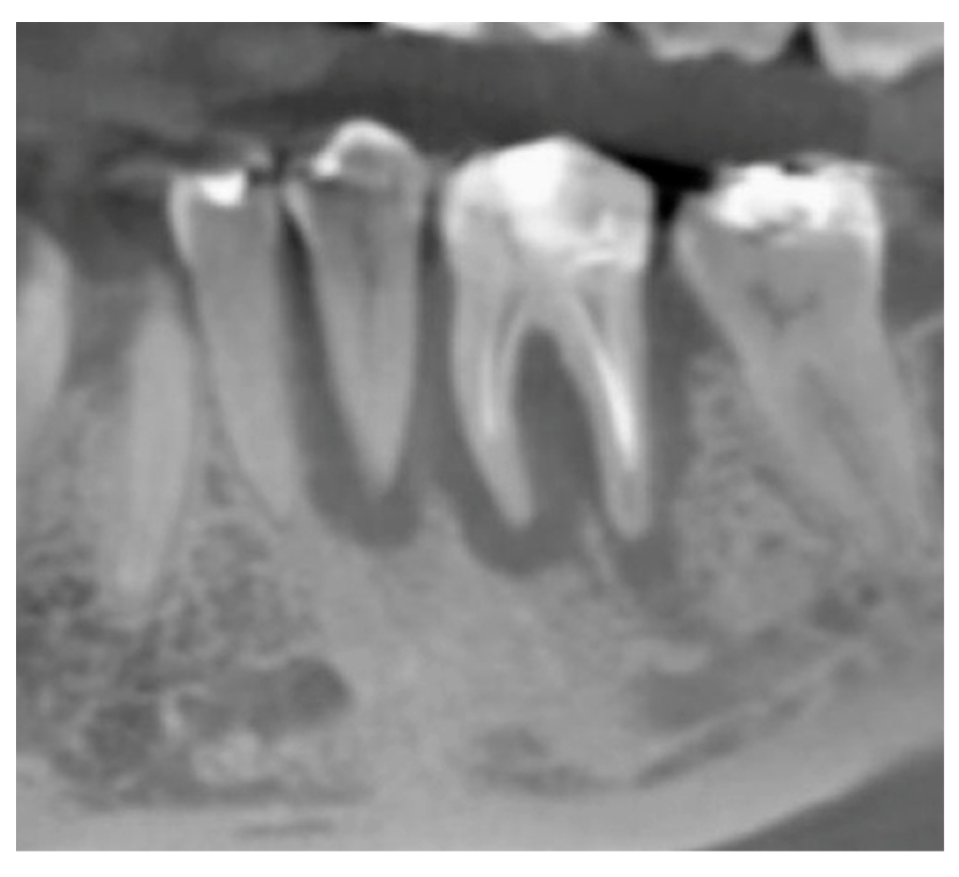
Figure 1. X-ray, elements 35 and 36 surrounded by a big bone defect.
Applsci 09 05396 g001
Figure 2. In the cbct section in zone 36, it is possible to note the bone loss dimension.
Applsci 09 05396 g002
Figure 3. The cbct section in zone 36, two months after the Guided Bone Regeneration (GBR). The defect was completely filled using the endodontical treated elements site 3.5 and 3.6 (Italian teeth numeration).
Applsci 09 05396 g003
Figure 4. The cbct section in zone 36 after the implants healing, six months after the GBR.
Applsci 09 05396 g004
Figure 5. X-ray after 30 months of follow up showing the implant with the prosthesis.
Applsci 09 05396 g005
Figure 6. Newly formed bone trabeculae and graft particles were observed. Is possible to observe with the dentin granules. All of the dentin granules are almost completely surrounded by newly formed bone. With the white square is indicated the newly formed bone (woven bone). No inflammatory or other adverse reaction are visible around the particles. (Magnification 30×—Toluidine Blue).
Applsci 09 05396 g006
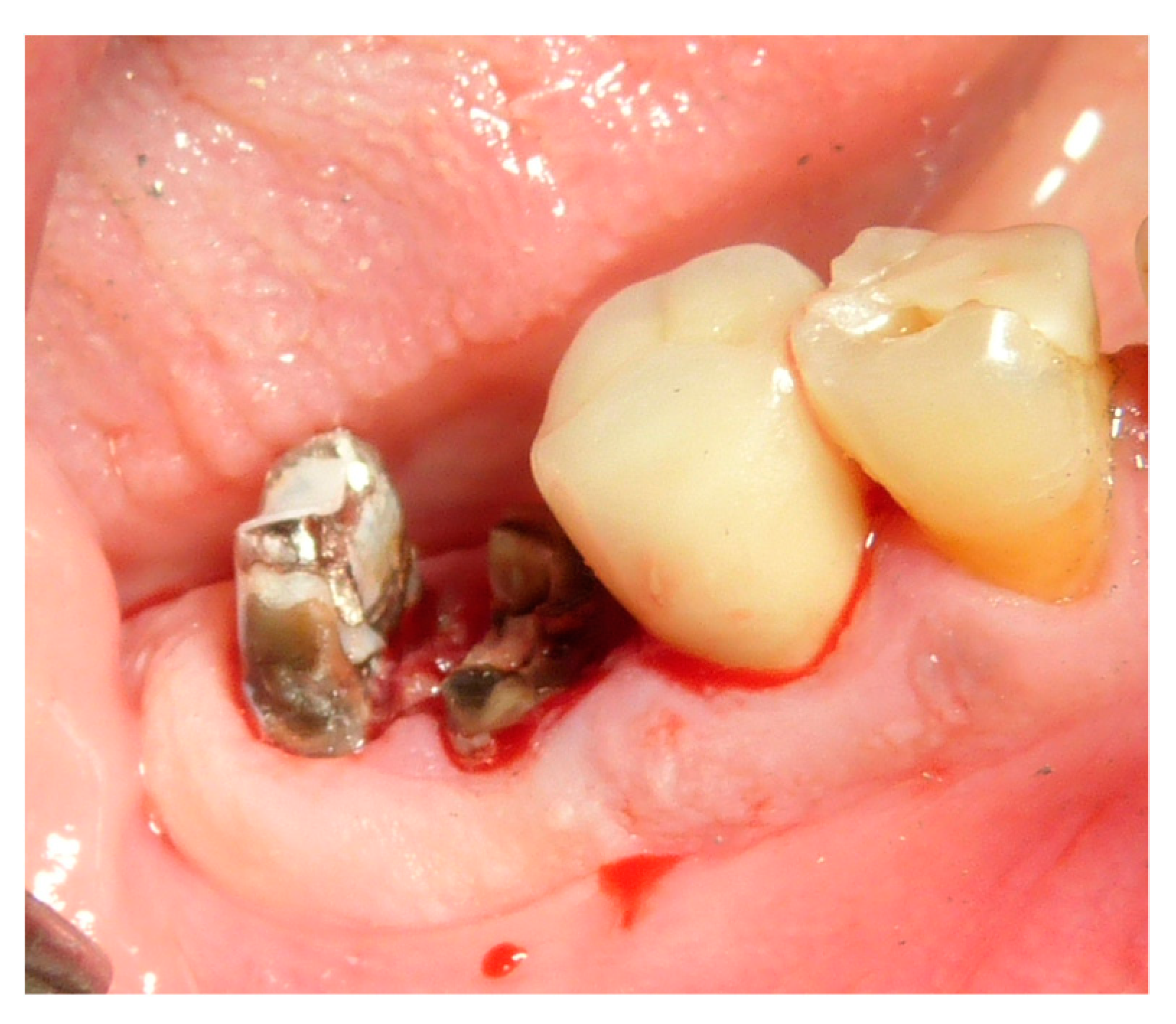
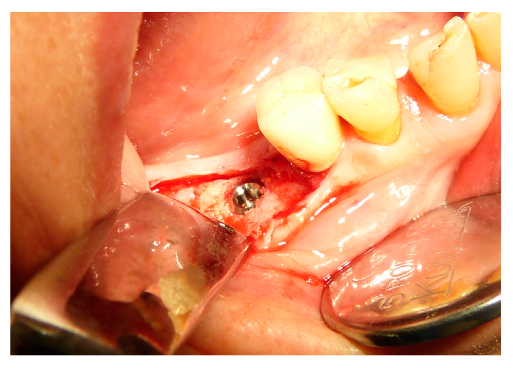
Figure 7. Dental element 46 to be extracted with the need for ridge maintenance due to an extensive interradicular defect.
Applsci 09 05396 g007
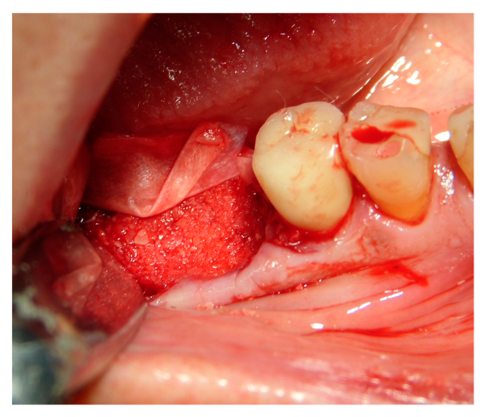
Figure 8. Ridge maintenance through the use of the extracted element and cover with a resorbable membrane.
Applsci 09 05396 g008
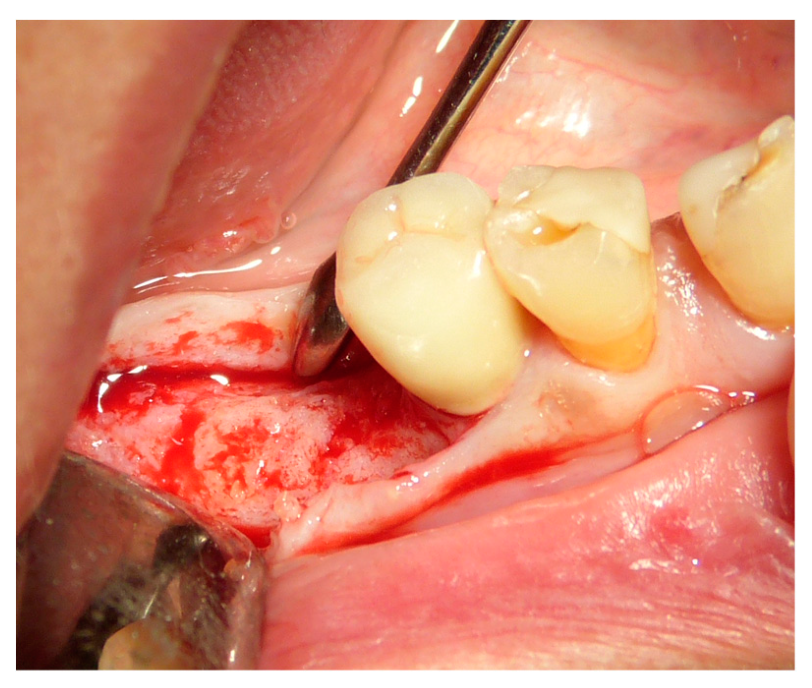
Figure 9. Reopening after three months.
Applsci 09 05396 g009
Figure 10. Implant placement in the regenerated bone after three months.
Applsci 09 05396 g010
Figure 11. Prosthesis and 19-months of follow-up.
Applsci 09 05396 g011
Table 1. Study design.
Surgeons10
Extracted teeth119
incisive 26
canine 8
premolars 36
molars 49
Extraction reasonsCrown trauma
external root resorption
Periodontitis
Missing tooth
Root fracture
Infected root
Socket sites treated106
mandible 60
maxillae 59
Implants106 (Certain—3i Biomet)
3,25–4–5 mm diameter
10–11,5–13 mm lenght
Implant failure1
Table 2. Types of defects of the alveolar sockets.
Percentage of alveolar socket defects1 walls
12%
2 walls
3%
3 walls
43%
4 walls
42%
Table 3. Histomorphometric analysis of the biopsies after 4 months of the alveolar sockets preservation procedures.
Histomorphometric AnalysisPercentageStandard Deviation
Vital Bone/Total Bone average (VB%)21.89±9.72
Residual Graft/Total Volume average
(Graft%)
16.60±7.09
Bone Volume/Total Volume average
(BV%)
41.47±11.51