3.1. Skin Anti-Aging and Structural Support
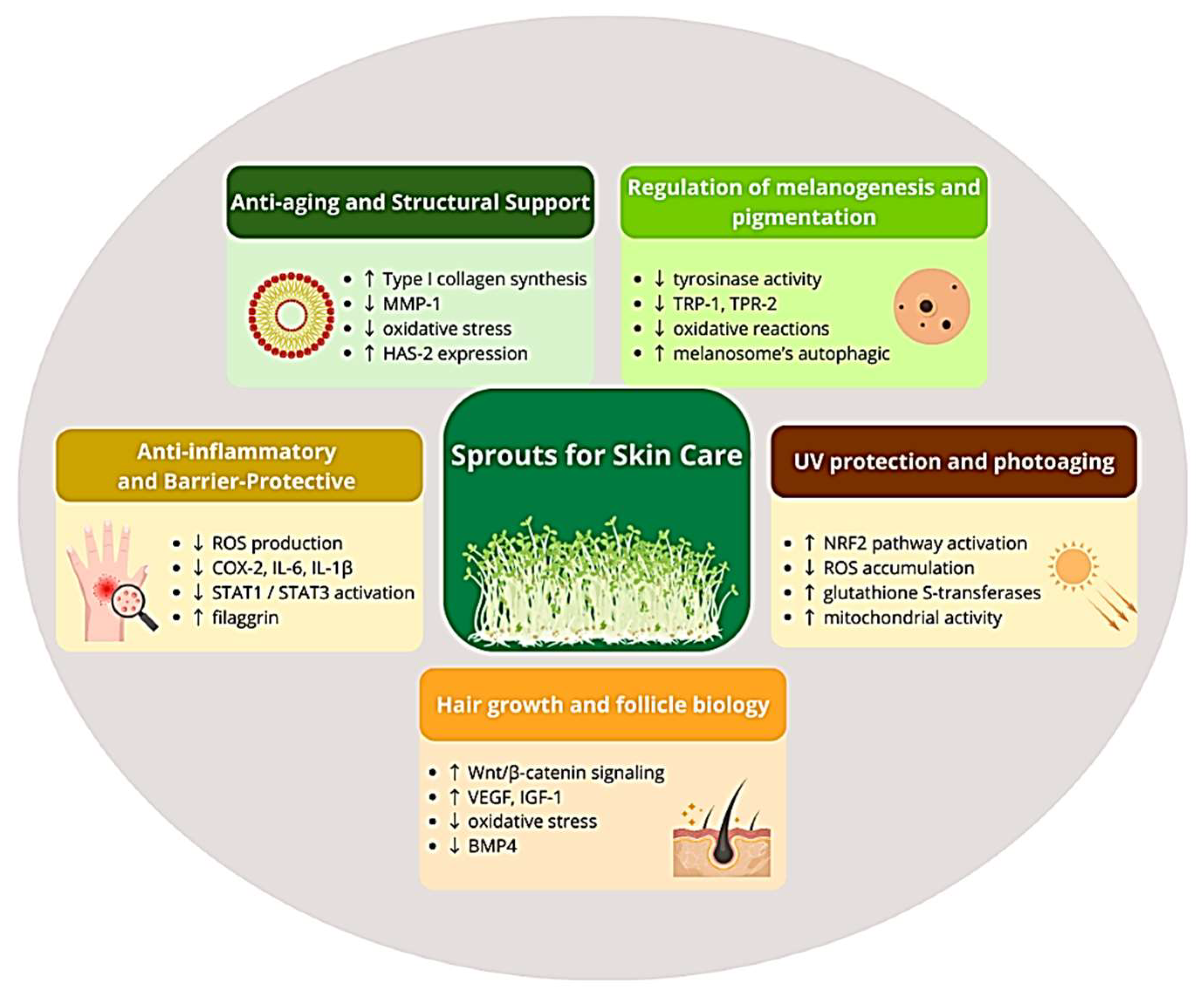
Sprout extracts are increasingly recognized as promising functional ingredients in anti-aging skin care due to their ability to modulate collagen homeostasis, enhance cellular defense mechanisms, and improve skin hydration. Collagen as the main structural protein of the dermis is responsible for its strength, elasticity and structural integrity, creating the scaffolding of the extracellular matrix necessary for the proper functioning and regeneration of the skin [
19]. Therefore, collagen plays a crucial role in skin firmness and elasticity, and its progressive degradation with age is one of the main mechanisms underlying wrinkle formation and visible signs of skin aging [
20]. Clinical studies show that oral collagen supplementation can improve skin elasticity and hydration, highlighting the importance of collagen homeostasis as a target for topical strategies [
21]. With age, collagen synthesis decreases and the activity of matrix metalloproteinases (MMPs) increases, which leads to the degradation of collagen fibers, loss of skin firmness and the formation of wrinkles [
22]. In the skin aging process, matrix metalloproteinase-1 (MMP-1/collagenase-1) in particular plays a key role in initiating the degradation of type I and III collagen, while MMP-3 (stromelysin-1) and MMP-9 (gelatinase-B) additionally degrade collagen fragments and other components of the extracellular matrix [
23]. Moreover, the combined actions of MMP-1, -3, and -9 can degrade most proteins in the skin ECM [
24]. In turn, preserving the ECM is a key target of anti-aging strategies, as excessive activity of matrix-degrading enzymes—especially collagenase and elastase—directly contributes to dermal thinning and wrinkle formation [
25]. In this context, the results of the existing studies indicate that sprout extracts may exert complementary effects by stimulating the synthesis of extracellular matrix components and limiting collagen degradation. These effects are attributed to bioactive compounds such as polyphenols, sulforaphane, amino acids (e.g., tryptophan), and vitamin C. They exhibit antioxidant and cytoprotective properties that help maintain skin integrity and slow aging [
26,
27].
Mongolian milkvetch (
Astragalus membranaceus L.) sprout ethanolic extract has attracted attention for its pro-regenerative properties. In vitro studies on human dermal fibroblasts demonstrated a dose-dependent increase in type I procollagen production to approximately 180–220% of control levels at the concentrations of 0.5–2% (
v/
v), without affecting cell viability (>90%). Phytochemical analysis identified tryptophan as the primary bioactive compound responsible for this effect, as isolated tryptophan independently elevated procollagen synthesis within a comparable range [
28]. This is particularly important information because type I collagen constitutes approximately 80–90% of the total collagen in the dermis and is essential for maintaining skin strength and structural integrity [
29].
The anti-aging potential of fermented ginseng sprouts (
Panax ginseng L.) was examined. The safety of the ethanol extract was assessed in the human dermal fibroblast cell line CCD-986SK at concentrations of 0.5, 1, 2, 5%, and 10%, and their viability did not drop below 80% even at the highest 10% concentration tested. Additionally, the extract significantly inhibited MMP-1 expression, to 51.85 ± 6.09% at the highest tested extract concentration. Since overexpression of MMP-1 leads to collagen degradation and, consequently, wrinkle formation, the obtained results indicate the potential of the extract as an anti-aging ingredient [
30].
Similar study on human skin fibroblasts exposed to the ethanolic extracts from sprouted barley (
Hordeum vulgare L.) indicated significantly increased type I collagen synthesis while simultaneously inhibiting MMP-1 secretion, and the effect was dose-dependent. The extracts were tested at 1, 5, and 10 µg/mL, maintaining >95% cell viability at 10 µg/mL. At this concentration, type I procollagen synthesis increased 44%, while MMP-1 levels decreased about 27%. Metabolomic analysis revealed significant differences in the secondary metabolite profile between the sprouts grown in deionized water and highly mineralized water. The increased content of oligomeric procyanidins and prodelphinidins in the sprouts grown in highly mineralized water was correlated with the anti-wrinkle effect of the extract [
31].
Another study evaluated the potential of wheat sprout extract (
Triticum aestivum L.) as an anti-aging cosmetic ingredient. The extract showed no toxicity to human dermal fibroblasts up to a concentration of 50 µg/mL maintaining >90% cell viability. In a photoaging model, the cells were exposed to UVB (100 mJ/cm
2), which induced an increase in the expression of MMP-1, an enzyme responsible for collagen degradation. Wheat sprout extract significantly inhibited MMP-1 secretion in a dose-dependent manner at concentrations of 6.25, 12.5, 25, and 50 mg/mL by 46.5, 50.0, 52.2, and 52.0%, respectively. This mechanism was primarily linked to the inhibition of ERK phosphorylation, indicating reduced downstream signaling [
32].
Similar anti-collagenase activity was observed for methanol extracts from water-pepper (
Polygonum hydropiper L.) sprouts. In vitro enzyme inhibition assays using purified collagenase and a synthetic peptide substrate showed concentration-dependent inhibition, with an IC
50 value of 156.7 µg/mL for the crude extract. To identify the active constituents, the extract was subjected to activity-guided fractionation, which significantly enhanced inhibitory potency. Among the obtained fractions, the 50% methanol eluate from the butanol fraction showed an IC
50 value of 23.5 µg/mL, indicating enrichment of bioactive compounds. Further phytochemical analysis of this active fraction led to the identification of hyperoside as the principal inhibitory compound. Hyperoside exhibited substantially higher anti-collagenase activity (IC
50 = 1.9 µg/mL) compared to the positive control EDTA (IC
50 = 110.6 µg/mL). Kinetic analysis revealed a noncompetitive inhibition mechanism (Ki = 0.3 µg/mL), suggesting preferential binding to the enzyme–substrate complex. Quantitative analysis showed that hyperoside constitutes approximately 6.9% (
w/
w) of the crude methanol extract, confirming its major contribution to the overall collagenase inhibitory activity. It should be noted, however, that methanol was used as an extraction solvent in this study to maximize the recovery of phenolic compounds and enable the extraction of the most active fractions and the identification of compounds, and not as a solvent intended for cosmetic applications. For cosmetic applications, the identified bioactive compounds would need to be obtained using cosmetically acceptable solvents (e.g., ethanol, water or mixtures thereof) in accordance with regulatory standards [
33].
Interesting cytoprotective effects were observed for aqueous extracts of black soybean (
Glycine max L.) sprouts under oxidative stress conditions. In human skin fibroblasts exposed to hydrogen peroxide (800 μmol/L H
2O
2 for 4 h), the extract (40 mg/mL) significantly increased cell survival compared to the cells exposed to oxidative stress alone, reaching 61.5 ± 5.26, 61.9 ± 4.11, and 63.8 ± 4.87% of control values depending on sprout length. Importantly, the protective effect was observed after 24 h pre-incubation of the cells with the extract, followed by exposure to H
2O
2 for 4 h, suggesting the involvement of adaptive or protective mechanisms of the cells, in addition to the direct free radicals scavenging. However, the extract used in the study was not chemically characterized beyond the determination of total protein (up to 18 mg/mL) and polysaccharide content (3.5–5 mg/mL). These results indicate the potential importance of black soybean sprout extracts in protecting skin fibroblasts from oxidative stress and maintaining skin homeostasis during the aging process [
34].
An ethanolic micellar extract from common bean (
Phaseolus vulgaris L.) sprouts demonstrated multifunctional anti-aging effects, including inhibition of collagenase, elastase, and tyrosinase. Furthermore, the extract supported the proliferation of human dermal fibroblasts and stimulated collagen synthesis in a dose-dependent manner, suggesting its potential to support extracellular matrix regeneration and maintain the structural integrity of the dermis. Phytochemical analysis revealed a high content of polyphenols (192.85 ± 10.24 mg GAE/g extract) and flavonoids (178.73 ± 1.59 mg QE/g extract), which may contribute to these effects. The presence of polyphenolic compounds, including isoflavones such as daidzein and glycitein, may further contribute to these effects through phytoestrogen-like effects, which are associated with improved collagen metabolism and the maintenance of skin structure. In the study, the term “micellar extract” refers to the formulation method and not to a separate extraction solvent system, indicating that the ethanol extract was incorporated into the micellar system to improve the solubility and bioavailability of the phenolic compounds. Therefore, the observed biological activity should be attributed primarily to the phytochemical composition of the ethanol extract, which contained a wide range of polyphenols, including flavonoids and isoflavones [
35].
Peanut sprout extracts (
Arachis hypogaea L.) have been studied for their effects on skin functions, including hydration, epidermal barrier, and regeneration in human HaCaT keratinocyte models. Phytochemical analysis showed that the 100% ethanol extract contained the highest total polyphenol (78.06 mg/g) and flavonoid content (6.54 mg/g), while the 50% ethanol extract contained the highest levels of resveratrol (4.71 μg/g) and polydatin (3.14 μg/g). Gene expression analysis showed that 50% and 100% ethanol extracts significantly increased the expression of hyaluronic acid synthase (HAS-2), which catalyzes the formation of high-molecular-weight hyaluronic acid responsible for maintaining skin hydration. In terms of epidermal barrier function, 50% ethanol extract most strongly increased the expression of filaggrin and involucrin—the proteins crucial for proper keratinocyte differentiation and stratum corneum integrity. In the scratch assay, the extracts significantly stimulated cell migration across the scratch. According to the authors, this effect was associated with an increase in HAS-2 expression and the presence of resveratrol and polydatin [
36].
Evening primrose (
Oenothera biennis L.) sprout ethanolic extract also demonstrated multifaceted activity relevant to skin aging conducted on human dermal fibroblasts in vitro. The extract (0.125–1 mg/mL) significantly inhibited the production of matrix metalloproteinases MMP-1 and MMP-2 in a dose-dependent manner. At the same time, the extract increased the synthesis of type I procollagen, which may suggest a dual mechanism promoting the integrity of the extracellular matrix. The moisturizing effect was assessed in vitro, using human HaCaT keratinocytes, where the extract increased hyaluronic acid synthesis in a dose-dependent manner, achieving a significant effect at concentrations of 50 and 100 µg/mL (by 75.4 and 165.0%, respectively, compared to control). This was further confirmed by the increased expression of the HAS2 and aquaporin-3 (AQP3) genes, which play a key role in hyaluronic acid synthesis and water transport in the epidermis, indicating a potential improvement in skin hydration and epidermal barrier function. Phytochemical analysis of the extract revealed the presence of high content of phenolic acids and flavonoid glycosides, including gallic acid (28.28 mg/g), ellagic acid (18.30 mg/g), quercetin glucuronide (16.09 mg/g) and luteolin glucuronide (7.75 mg/g), which are likely responsible for the observed anti-aging, antioxidant and moisturizing properties [
17].
A sole ex vivo study on human skin explants indicated that sunflower (
Helianthus annuus L.) sprout extract showed a significant effect on the expression of genes related to epidermal barrier integrity and extracellular matrix homeostasis. The skin explants were treated with the extract at a dose of 2 mg/cm
2 (equivalent to 1 mg/tissue) for 24 h, and gene expression changes were considered significant at a fold change more over two. The increased expression of the genes related to keratinocyte differentiation and barrier function, such as KRT2, LCE1B and LIPN, was observed, while the expression of pro-inflammatory genes (IL6, CXCL8) and the MMP2 gene—metalloproteinase involved in collagen degradation—was reduced. The extract was phytochemically characterized, with acetyl-L-carnitine, ATP, eicosapentaenoic acid (EPA), phospholipids, and triglycerides as predominant compounds, the presence of which may support mitochondrial function, skin barrier integrity, and regenerative processes [
18].
Two human studies also provided evidence of the potential of some sprouts in the context of their topical use. Five healthy adult volunteers (aged 37–71 years) participated in a randomized, placebo-controlled, single-blind, split-body clinical trial (where each participant applied the extract to one arm and vehicle alone to the contralateral arm, serving as their own control). Broccoli (
Brassica oleracea var.
italica Plenck) sprout’s ethyl acetate extract containing 500 nmol of sulforaphane/mL was applied topically (1 mL/day) for seven consecutive days as a solution prepared in jojoba oil at a concentration of 500 nmol of sulforaphane/mL. Participants applied 1 mL of the preparation daily on the inner side of one arm, while the opposite arm was treated with vehicle alone (placebo). Local application of broccoli sprout extract induced significant molecular changes in the epidermis without observed adverse effects or histological changes. Keratin 17 (KRT17) mRNA expression increased significantly in all participants, reaching an average of 3.6 ± 0.2-fold, while keratin 16 (KRT16) expression showed interindividual variability, reaching an increase of approximately 4–14-fold in some subjects. The NRF2 pathway was also activated, as confirmed by the increased expression of the antioxidant response gene NQO1 in three of the four participants. Importantly, the expression of structural keratins associated with the mechanical integrity of the epidermis, such as KRT5 and KRT14, was not changed. These results indicate that topical application of broccoli sprout extract can modulate gene expression in the epidermis and activate adaptive antioxidant defense mechanisms without compromising the structural integrity of the skin. However, it should be emphasized that the short duration of the intervention and the small sample size limit the ability to draw conclusions regarding long-term clinical effectiveness. This was a pilot study with a very small sample size (n = 5, with only 4 participants included in the final analysis), short duration (7 days) and evaluation limited mainly to molecular biomarkers and not to clinical endpoints. Therefore, although the results indicate biological activity, they do not allow conclusions to be drawn about clinical anti-aging effects. It should also be noted that ethyl acetate was used solely as an extraction solvent to isolate sulforaphane-rich fractions and was removed prior to formulation, meaning it does not reflect the composition of the final topically applied preparation [
37].
In a single-blind clinical trial involving 45 women aged 42–64, twice-daily application of a cosmetic formulation containing 4% micellar ethanolic extract of common bean (
Phaseolus vulgaris L.) sprouts for 12 weeks led to statistically significant improvements in skin hydration and elasticity compared to placebo. The formulation was applied as an o/w emulsion cream, specifically developed for the study, with a precisely defined concentration of 4% (
w/
w) of the extract. However, the authors provided only qualitative composition of the extract, with the presence of phenolic acids (caffeic, ferulic, and p-coumaric acids) and flavonoids (rutin, quercetin). Analysis of skin biomechanical parameters revealed a significant improvement in total elasticity after 12 weeks of use (+13.3%), while net elasticity showed significant improvement after just 8 weeks of use. Skin hydration increased by 22.31% (vs. 3.52% in placebo and 13.96% in the reference cream). At the same time, no significant changes were observed in the melanin index, erythema, or sebum secretion, indicating that the product’s primary effect is related to improving skin biomechanical properties and barrier function, rather than modulating pigmentation or sebaceous gland activity. The product was well tolerated by study participants, with no adverse effects reported. Importantly, the effectiveness of the cream containing the extract was comparable and, in some parameters, even superior to the reference cream containing active ingredients such as coenzyme Q10,
Argania spinosa kernel oil, and
Vitis vinifera seed oil, known for their anti-aging and moisturizing properties [
38].
Current evidence indicates that sprout extracts act through convergent biological pathways—including stimulation of collagen synthesis, inhibition of matrix degradation, enhancement of antioxidant defenses, and improvement of epidermal hydration. Despite encouraging mechanistic data and promising but scarce clinical results, larger, randomized studies with longer follow-up are needed to establish their long-term dermatological significance.
3.2. Anti-Inflammatory and Barrier-Protective Effects
Chronic low-grade inflammation is increasingly recognized as a fundamental factor of skin aging, contributing to extracellular matrix degradation, pigmentary alterations, impaired barrier function, and reduced regenerative capacity [
39,
40]. Accordingly, sprout extracts capable of attenuating oxidative stress and inflammatory signaling are gaining attention as multifunctional dermatological agents.
Peanut (
Arachis hypogaea L.) sprout ethanolic extracts represent an example of antioxidant-mediated anti-inflammatory activity. In an in vitro model using human keratinocytes (HaCaT), skin inflammation was induced by a synthetic secretagogue (compound denoted as 48/80) used to stimulate oxidative stress and inflammatory signaling. Cells were treated with extract at concentrations ranging from 0.01 to 1.0 mg/mL. Treatment with peanut sprout ethanol extract reduced reactive oxygen species (measured by [specify assay, e.g., DCFH-DA fluorescence]) and significantly reduced the expression of key inflammatory mediators, including cyclooxygenase-2 (COX-2) and nerve growth factor (NGF), at both mRNA and protein levels in a dose-dependent manner. Densitometric analysis indicated a reduction in COX-2 and NGF mRNA expression by approximately 40–80% and COX-2 protein levels by up to 70–90% at higher concentrations. Furthermore, the extract increased the expression of IκBα, an endogenous inhibitor of NF-κB, indicating suppression of NF-κB-dependent inflammatory signaling pathways. These results indicate that the anti-inflammatory effects of peanut sprout extract are primarily mediated through antioxidant mechanisms and modulation of redox-sensitive inflammatory pathways [
41].
A 70% ethanol extract of wheat (
Triticum aestivum L.) sprouts also demonstrated anti-inflammatory effects in cytokine-stimulated human keratinocytes (HaCaT), observed as a significant decrease in the expression of pro-inflammatory chemokines, including RANTES (CCL5), MDC (CCL22), and IP-10 (CXCL10), which play a key role in immune cell recruitment and chronic skin inflammation. Quantitatively, the extract reduced mRNA expression levels of these chemokines by approximately 50–80% compared to cytokine-stimulated controls, depending on the time point. Mechanistically, these effects were associated with the inhibition of STAT1 phosphorylation and concomitant regulation of SOCS1 expression, a negative regulator of cytokine signaling that suppresses STAT-dependent inflammatory pathways. These findings indicate that the extract may attenuate inflammatory signaling in keratinocytes and contribute to maintaining skin immune homeostasis during inflammatory conditions. However, no information was provided on chemical characterization of the extract, which limits direct attribution of the observed effects to specific phytocompounds [
42].
A study was performed on kamut (
Triticum turgidum ssp.
turanicum), another representative of Poaceae family. Ethanol extract from the fermented sprouts demonstrated improved safety and anti-inflammatory activity compared to its non-fermented counterpart in vitro. In human keratinocytes, the fermented extract maintained >80% cell viability even at high concentrations, indicating low cytotoxicity. After exposure to 600 µg/mL, COX-2 mRNA expression decreased to approximately 18.5% of LPS-stimulated levels, accompanied by decreased expression of IL-6 (26%) and IL-1β (32.8%), indicating a strong suppression of inflammatory signaling. Fermentation increased β-glucan content from 34.6 to 46.9 mg/g dry weight and improved radical scavenging capacity (from 57.8 to 71.7%). These compositional changes suggest that microbial fermentation can modulate the biological activity of kamut sprout extracts by increasing the content of bioactive compounds associated with antioxidant and anti-inflammatory effects [
43].
In addition to classical inflammatory pathways, sprout extracts may also regulate itch-related mediators such as IL-31. IL-31 plays a key role in skin inflammation by promoting pruritus. It disrupts epidermal barrier function and stimulates sensory neurons, contributing to chronic itch and inflammation in conditions such as atopic dermatitis. Ethanol extract from adlay (
Coix lacryma-jobi L.) sprouts significantly inhibited histamine release which increased from 29.2 to 215.1 pg/mL after stimulation with PMA and A23187. Extract also reduced IL-31 secretion in activated mast cells, indicating modulation of both histamine-dependent and -independent itch pathways. Coixol (6-methoxy-2-benzoxazolinone, MBOA)—present at a concentration of 41.30–95.46 mg/g dw—has been proposed as a potential bioactive compound, although confirmation using isolated molecules is still required [
44].
Environmental stressors are another important factor in skin inflammation. Broccoli (
Brassica oleracea var.
italica Plenck) sprout aqueous extract significantly attenuated particulate-induced oxidative stress and inflammatory responses in human HaCaT keratinocytes. Cells were pretreated with the extract for 1 h before exposure to particulate matter (PM, 40 µg/mL). Pretreatment with the extract significantly reduced the expression of key pro-inflammatory mediators, including MMP-1, COX-2, and IL-6, without the impact on cell viability. At concentrations of 12.5–50 µg/mL, MMP-1, COX-2, and IL-6 levels were reduced by approximately 40–70% compared to PM-treated controls. Mechanistic analyses showed that these effects were mediated by direct inhibition of p38α MAPK activity via allosteric, ATP-independent binding, leading to further inhibition of MSK1/2 phosphorylation and reduced activation of AP-1 and NF-κB signaling pathways. Moreover, the extract showed strong antioxidant activity and effectively reduced the accumulation of ROS induced by particulate matter [
45].
Two in vivo studies were also conducted, supporting anti-inflammatory potential of some sprouts. Oat (
Avena sativa L.) sprout extract revealed the ability to restore skin barrier integrity and modulate the immune response in the model of oxazolone-induced allergic contact dermatitis in mice (n = 24). Local treatment with 3% ethanolic extract and its combination with oral administration (100 mg/kg/day) significantly reduced transepidermal water loss and lowered plasma IgE levels, which were approximately threefold higher in untreated animals. Treatment also reduced inflammatory responses, as evidenced by reduced mast cell infiltration and decreased Th2-related cytokine levels, accompanied by a reduction in CD4
+ and CD8
+ T cells. Molecular analyses further revealed the increased expression of epidermal differentiation complex proteins, including filaggrin, loricrin, and involucrin, which are essential for the formation and function of the epidermal barrier. These effects were associated with activation of MAPK signaling pathways and suppression of STAT3 signaling, indicating a coordinated regulation of keratinocyte differentiation and inflammatory responses. These results support the role of oat sprout extract in restoring epidermal barrier function and alleviating inflammation in vivo [
46]. In a similar study, peanut (
Arachis hypogaea L.) sprout ethanolic extract was tested in an oxazolone-induced contact dermatitis model in mice (n = 16). Topical application of the 5% extract ointment applied to the dorsal skin three times per week for two weeks after oxazolone sensitization reduced epidermal desquamation, edema, and epidermal thickening and also reduced hyperkeratosis and inflammatory infiltrate. Histological analyses also showed reduced spongiosis, reduced vasodilation and inflammatory cell infiltration, and an overall reduction in skin thickness. Elevated COX-2 and NGF expression in inflamed tissue was also inhibited after treatment. This activity is largely attributed to trans-resveratrol and related polyphenols, although the contribution of individual components remains unresolved [
43].
3.3. UV-Protection and Regulation of Skin Pigmentation
Ultraviolet (UV) radiation causes oxidative stress, inflammation, pigmentation changes and extracellular matrix degradation, translating into premature skin aging. Recent studies suggest that sprout extracts may prevent such changes and improve skin immunity primarily by activating endogenous cytoprotective [
47,
48].
The antioxidant activity and melanogenesis-inhibiting potential of aqueous extracts of black soybean sprouts (
Glycine max L.) have been demonstrated. In the DPPH test, extracts at a concentration of 40 mg/mL demonstrated a free radical scavenging capacity of over 95%, comparable to vitamin C [
34]. They were also characterized by greater chemical stability under conditions of variable pH and temperature than the reference substance. Furthermore, a cell-free enzymatic assay demonstrated strong inhibition of tyrosinase activity, reaching up to 98%, exceeding the effect observed for arbutin used as a positive control. These results indicate the extract’s potential to directly inhibit enzymatic stages of melanogenesis, which is relevant in the context of UV-induced hyperpigmentation [
34]. However, it should be emphasized that the observed effects were demonstrated only in cell-free systems, and the extract was not phytochemically characterized beyond the determination of total protein and polysaccharide content [
34].
A complex, multifaceted study on sunflower (
Helianthus annuus L.) sprout extract was performed to demonstrate its protective effects in cellular, ex vivo, and clinical models. Primary human dermal fibroblasts, incubated with the extract at concentrations of 0.1–0.5% (
w/
v), showed a significant increase in intracellular ATP production (24–45%) under in vitro conditions of metabolic fatigue induced by glucose deprivation, indicating improved mitochondrial function and potential support for cellular metabolism. Simultaneously, the extract demonstrated antioxidant properties, reducing reactive oxygen species without inducing cytotoxicity. However, human HaCaT keratinocytes incubated with the extract for 24 h demonstrated a significant reduction in the accumulation of advanced glycation end products (AGEs) induced by UVA radiation (20 J/cm
2), reaching a reduction of 39.1% at a concentration of 0.01% (
w/
w), indicating a potential protective effect against photoaging-related damage to skin proteins and structures. Ex vivo studies using human skin explants also revealed significant changes in the expression of genes associated with epidermal barrier function and skin homeostasis. The phytochemical composition of the extract included a number of bioactive secondary metabolites, including acetyl-L-carnitine, ATP, eicosapentaenoic acid (EPA), phospholipids, and triglycerides, which may support mitochondrial function, skin barrier integrity, and antioxidant mechanisms. The extract was further tested in a randomized, double-blind clinical trial involving 28 women aged 30–60 with moderate symptoms of photoaging in the Griffiths scale (for example, fine lines and wrinkles, reduced firmness, dull appearance). Twice-daily application of the serum containing sunflower sprout extract for 7 days led to a statistically significant improvement in clinical parameters, including radiance, smoothness, firmness and reduction of fine wrinkles. The improvement in skin radiance was also confirmed instrumentally, demonstrating an approximately 19% increase in skin radiance after three days of use. The preparation was well tolerated and did not cause any significant side effects [
18].
The UV-protective properties of broccoli (
Brassica oleracea var.
italica Plenck) sprout aqueous extract have been confirmed in both animal models and limited human studies. In SKH-1 hairless mice (n = 12), topical application of the extract, containing 100–600 nmol sulforaphane per 2 cm diameter skin area, applied once daily for three consecutive days before UV exposure, significantly reduced UVB-induced inflammation and edema and decreased myeloperoxidase activity while increasing the activity of cytoprotective enzymes, including NQO1, indicating activation of endogenous skin defense mechanisms. In a subsequent human study involving six healthy volunteers, topical application of the extract containing 200 or 400 nmol sulforaphane per 2 cm diameter skin area, applied once daily for three days before exposure to UV radiation, led to a significant reduction in the severity of erythema, a marker of skin damage. The mean reduction in erythema was 37.7%, despite significant interindividual variability in response. The protective effect was dose-dependent and lasted for at least 48–72 h after the end of application, which indicates that the mechanism of action of the extract is the induction of long-lasting cytoprotective mechanisms and not the direct absorption of UV radiation, as is the case with classic sunscreens. Importantly, the extract used in the study was standardized for isothiocyanates, with approximately 90% sulforaphane content, a compound responsible for activation of the Keap1–Nrf2 pathway and induction of phase II detoxification enzymes including NQO1, glutathione S-transferases, and heme oxygenase-1 [
49].
Taken together, sprout-derived extracts appear to provide multilayered photoprotection by enhancing antioxidant defenses, suppressing inflammation, limiting melanogenesis.
3.4. Modulation of Melanogenesis
Dysregulated melanogenesis contributes to hyperpigmentation and uneven skin tone, both of which are major features of photoaged skin [
50]. Consequently, increasing attention has been directed toward sprout extracts capable of limiting melanin synthesis, enhancing antioxidant defenses, or promoting pigment turnover through alternative cellular pathways [
51].
Ethyl acetate fraction of the methanolic extract from mung bean (
Vigna radiata L.) sprouts demonstrated significant inhibition of tyrosinase by 62.9% in cell-free enzymatic assay. The authors suggested that the effect is largely attributed to polyphenols such as vitexin and isovitexin [
52]. In a follow-up study, the authors proved that the fraction also demonstrated the potential to inhibit melanogenesis in B16F1 mouse melanoma cells. The fraction from the sprouts germinated for 1 day demonstrated the strongest inhibitory effect on melanin synthesis, with a decrease in melanin content by 56.5% compared to the control. In comparison, arbutin showed only 16.3% inhibition. Sprouts cultured for 12 h and 2 days also demonstrated a stronger effect than arbutin. In an in vitro tyrosinase activity assay, the highest inhibition was observed for the ethyl acetate fraction from 2-day sprouts. Again, vitexin and isovitexin were indicated as the compounds responsible for the activity, with their highest content determined in the extract from 1-day-old sprouts (11.37 and 2.55 µg/g, respectively) [
53]. A different strategy was used to modulate moth bean (
Vigna aconitifolia Jacq.) sprout composition. Treatment of seeds with 0.05% Ascophyllum nodosum extract produced sprouts with enhanced tyrosinase inhibitory activity, reaching maximal efficacy after 24 h of exposure, whereas higher concentrations reduced activity. Although the responsible compounds were not identified, the results suggest that seaweed-derived bioactive constituents may potentiate enzyme inhibition. It should also be noted that ethyl acetate was used solely as an extraction solvent to isolate active fraction and was removed prior to formulation, meaning it does not reflect the composition of the final topically applied preparation [
54].
Antimelanogenic activity has also been demonstrated for methanolic extracts from safflower (
Carthamus tinctorius L.) sprouts. Studies were conducted in a cell model using murine-derived melanocytes, in which the extracts inhibited melanin synthesis in a dose-dependent manner. Importantly, the biological activity of the extracts depended on the sprout cultivation conditions—the extracts obtained from sprouts grown in the dark or under fluorescent light showed significant inhibitory effect on melanogenesis, while exposure to blue LED light (450 nm) resulted in a slight increase in melanin synthesis. Red LED light (660 nm) used in the study did not show a clear inhibitory pattern, and phytochemical analysis identified lignan 8-hydroxyarctigenin and the phenolic alcohol tyrosol as the main bioactive compounds. The 8-hydroxyarctigenin demonstrated a pronounced antimelanogenic effect, reducing melanin synthesis by approximately 30% at a concentration of 5 µg/mL and by 42% at 10 µg/mL, while exhibiting low cytotoxicity (<10%). This compound was present at the highest concentrations in extracts from sprouts grown in the dark (1.03 ± 0.08 mg/g of extract) and under fluorescent light (0.83 ± 0.02 mg/g of extract). On the contrary, tyrosol did not demonstrate significant melanogenesis-inhibiting or cytotoxic effects, indicating that it is not the primary compound responsible for the observed depigmenting effect. It should be noted that methanol was used in this study as an analytical extraction solvent to efficiently isolate phenolic compounds, rather than for cosmetic application; therefore, the results reflect the activity of the identified bioactive constituents, which may be obtained using cosmetically acceptable solvents (e.g., ethanol or water) [
55].
Barley sprouts (
Hordeum vulgare L.) were shown to influence melanogenesis in B16F10 mouse melanoma cells, a standard cell model for skin pigmentation studies. The aqueous extract was shown to significantly reduce melanin synthesis, stimulated in the cells by α-MSH. At a concentration of 250 μg/mL, melanin content decreased by approximately 40% compared to the control. Interestingly, this effect was stronger than that of arbutin at a dose of 150 ppm, which reduced melanogenesis by approximately 20%. Moreover, the extract at concentrations of 125 and 250 μg/mL reduced tyrosinase activity by approximately 40%, with the effect comparable to arbutin. The expression of proteins involved in melanogenesis also reduced tyrosinase, as well as the transcription factor MITF, which regulates its expression. The extract contained phenolic compounds such as p-coumaric acid (0.089%), ferulic acid (0.094%) and vanillic acid (0.055%), which may contribute to the observed activity [
56]. A similar study also indicated that the aqueous extract of barley sprouts, rich in simple sugars (glucose and fructose), amino acids, and phenolic compounds (reported as total phenolic content), demonstrated approximately 80% inhibition of tyrosinase activity, indicating a strong depigmentation potential in vitro. This mechanism is likely due to the presence of phenolic compounds, which exhibit antioxidant activity, limiting oxidative reactions in the melanin synthesis pathway [
57]. In contrast, ethanolic extract of wheat (
Triticum aestivum L.) sprouts, another species from the
Poaceae family, demonstrated only a weak inhibitory effect on melanin synthesis (approximately 20% at the highest concentration) in α-MSH-stimulated B16F10 mouse melanoma cells. Additional analysis of MITF and tyrosinase protein expression did not reveal any suppression of these markers, confirming the extract’s limited depigmenting effect [
32].
On the other hand, some biotechnological processing may amplify depigmenting potential, as observed for oat (
Avena sativa L.) sprouts, fermented with
Lacticaseibacillus casei. The sprout aqueous extracts significantly reduced melanin content in murine B16F10 melanoma cells while inhibiting tyrosinase activity and downregulating key melanogenic regulators, including MITF and TRP-2. Fermentation increased polyphenol and flavonoid content in the sprouts and produced an approximately 11-fold increase in avenanthramide A, an oat-specific compound known for antioxidant and anti-inflammatory activity. The extracts also enhanced free radical scavenging, exhibited superoxide dismutase–like activity, and reduced intracellular reactive oxygen species, suggesting a dual mechanism combining melanogenesis suppression with oxidative stress mitigation [
58]. Similarly, bioconversion of broccoli (
Brassica oleracea L.) sprouts with
Bacillus amyloliquefaciens resulted in changes in the qualitative chemical profile of their aqueous extract, but also in a strong, dose-dependent reduction in melanin content in α-MSH-stimulated B16 cells, and a decrease in tyrosinase activity. The unbioconverted sprout extract revealed only weak impact on melanin content and tyrosinase activity. Similarly, strong, dose-dependent inhibition of the expression of key regulators of melanogenesis, such as MITF, tyrosinase, TRP-1, and TRP-2, was observed only for the bioconverted sprouts [
59].
Another method to improve the effectiveness of the extract is to increase its penetration into the skin by using special formulations, as described for radish (
Raphanus sativus var.
Longipinnatus) sprout aqueous extract, encapsulated in transferosomes. In an enzymatic model using fungal tyrosinase, transferosomes showed dose-dependent inhibition of the enzyme compared with a negative control and kojic acid used as the positive reference inhibitor, with an IC
50 of 1.40 mg/mL. Although melanin content was significantly reduced at concentrations ≥ 100 μg/mL in α-MSH-stimulated B16-F10 cells, this effect should be interpreted cautiously because B16-F10 viability was already significantly decreased at concentrations ≥ 50 μg/mL [
60].
The ethyl acetate fraction of the sprout extract from Japanese lady bell (
Adenophora triphylla var.
japonica) was characterized by strong antioxidant activity in the ABTS, DPPH and FRAP tests and significantly inhibited lipid peroxidation. Enzymatic studies showed that the fraction inhibited tyrosinase activity while strongly inhibiting α-glucosidase activity, suggesting an additional mechanism involving disruption of tyrosinase glycosylation and limitation of its biological activity. In B16/F10 murine melanoma cells stimulated with α-MSH, the fraction significantly reduced melanin levels (approximately 40% reduction at 200 µg/mL), confirming its depigmentation potential. The main compounds of the fraction were chlorogenic acid and rutin, the presence of which was associated with antioxidant and whitening effects. It should be noted that ethyl acetate was used as a fractionation solvent to isolate semi-polar bioactive compounds, and the observed activity is attributed to these constituents, which can be obtained using cosmetically acceptable extraction systems [
61].
Importantly, not all depigmentation mechanisms rely on inhibiting melanin synthesis. Lotus (
Nelumbo nucifera Gaertn.) sprout aqueous–glycolic extract (LSE) provides a mechanistically distinct example, stimulating melanosome degradation. The extract was prepared using an optimized method that increased the content of naturally occurring bisbenzylisoquinoline alkaloids, specifically neferine and liensinin, which were identified as its main bioactive components. The LSE treatment significantly reduced melanin accumulation in MNT-1 human melanoma cells, primary human melanocytes, and a reconstructed human skin analogue (MelanoDerm), confirming its depigmentation effect in multiple biologically relevant models. Studies using purified neferine and liensinin demonstrated a dose-dependent reduction in melanin content, with neferine at a concentration of 5 µM significantly reducing pigmentation without inhibiting tyrosinase activity or downregulating melanogenesis-related gene expression. These findings indicate that the pigmentation reduction was not due to suppression of melanin synthesis. Instead, both the extract and its active alkaloids increased autophagic flux, as evidenced by increased LC3-II levels and Beclin-1 phosphorylation, consistent with activation of the AMPK-dependent autophagy pathway. Transmission electron microscopy of treated human skin analogs revealed clusters of melanosomes in autophagosome-like structures in approximately 90% of melanocytes, indicating selective autophagic degradation of melanosomes [
62].
Apart from mechanistic in vitro studies, some human studies also support the relevance of the use of sprout extracts in photoaged and hyperpigmented skin.
A randomized, blinded, split-body study of women aged 52–77 years with photoaged skin showed that once-daily topical application of broccoli (
Brassica oleracea var
. italica Plenck) sprout extract containing 5 nmol of sulforaphane for seven days activated the NRF2 pathway in four of five participants. This molecular response correlated with a mean improvement in uneven pigmentation of 2.8 ± 0.4 points and an approximately 50% reduction in melanin deposition. Importantly, NRF2 activation was accompanied by the inhibition of MMP-1 expression, which decreased from elevated baseline levels to undetectable values after treatment. Despite the small sample size, these results suggest that even short-term NRF2 induction may contribute to measurable improvement in photoaged skin [
63].
A randomized, double-blind, biface study evaluated a multimodal, hyperpigmentation-correcting serum containing lotus (
Nelumbo nucifera Gaertn.) sprout extract as part of a proprietary formula. The study included 113 women aged 30–65 years with moderate to severe facial hyperpigmentation, including a subgroup of 44 patients with melasma. Twice-daily use for 12 weeks resulted in an 11.7% reduction in overall hyperpigmentation, comparable to the 13.1% reduction observed with the reference treatment containing 4% hydroquinone, with no statistically significant difference between treatments. Skin tone uniformity improved by 8.8%, while in the melasma subgroup, mMASI scores decreased by 50.6%, from 4.5 to 2.2, similar to the 53.7% reduction observed with hydroquinone. Quality of life scores also improved significantly, with the MelasQoL index decreasing from 46.6 to 33.0. Both treatments were well tolerated, with no serious adverse events reported. Importantly, lotus sprout extract was not administered as a standalone intervention but was incorporated into a multi-ingredient formulation containing additional active ingredients, including tranexamic acid and niacinamide, which target complementary pathways involved in melanocyte activation, melanin distribution, and inflammation. Moreover, the extract itself has not been chemically characterized for specific phytochemical composition or standardized to defined bioactive markers. Therefore, although the preparation demonstrated clinically significant depigmenting efficacy comparable to hydroquinone, the observed effects cannot be attributed solely to the lotus sprout extract but rather reflect the potentially synergistic effects of the multiple active ingredients, even drugs, contained in the preparation [
64].
In summary, sprout extracts appear to regulate skin pigmentation through multiple complementary mechanisms, including tyrosinase inhibition, melanogenic signal suppression, enhancement of antioxidant defenses, and activation of melanosome turnover. Furthermore, early clinical studies suggest that sprout extracts may contribute to improving hyperpigmentation, even skin tone, and matrix remodeling, primarily through NRF2-mediated cytoprotection and modulation of melanogenic pathways.
3.5. Effects on Hair Growth and Follicle Biology
Hair follicle aging is caused by complex interactions involving impaired growth factor signaling, androgen-dependent follicle miniaturization, oxidative stress, and chronic microinflammation [
65,
66]. Therefore, sprout extracts that can modulate these pathways are increasingly being investigated as multifunctional agents to support hair regeneration and scalp health.
An aqueous extract of pea (
Pisum sativum L.) sprouts was evaluated for its effects on hair cycle regulation through gene expression analysis of plucked human hair follicles following in vivo treatment. In a study involving 22 volunteers (11 women and 11 men) aged 46–61 years, participants applied scalp formulations containing 2% plant extracts twice daily for two weeks. Gene expression analysis revealed a significant increase in fibroblast growth factor 7 (FGF7) and noggin expression by 56% and 85%, respectively. Both proteins are key regulators synthesized in dermal papilla cells: noggin promotes the transition from telogen to anagen by inhibiting BMP4, while FGF7 stimulates proliferation of hair germ cells and supports initiation of a new growth phase. Although pea sprouts are rich in isoflavones and other polyphenols that may contribute to these effects, the specific bioactive compounds responsible were not identified [
67]. Consistent results were later obtained in a smaller human study of 10 volunteers, in which topical application of a preparation containing 2% pea sprout extract for two weeks similarly increased the expression of FGF7 and noggin. In the same research program, a pilot study of oral supplementation (100 mg/day for 8 weeks) was conducted, resulting in a 33.9% reduction in hair loss after 28 days, with 95% of participants reporting reduced hair loss and no side effects. These observations suggest a potential role of pea sprout extract in modulating pathways related to the hair growth cycle, possibly through direct regulation of gene expression combined with indirect antioxidant and anti-inflammatory effects. However, the extract used in these studies has not been phytochemically characterized, and larger placebo-controlled studies are needed to confirm its clinical efficacy [
68]. Similar in vitro data indicate that an aqueous extract of pea (
Pisum sativum L.) sprouts stimulates proliferation and modulates growth factor expression in human dermal papilla cells. Although cells were exposed to a broad concentration range (1–100 µg/mL), significant proliferative and molecular effects were observed primarily at 10 and 50 µg/mL after 24 h of treatment. At these concentrations, the extract upregulated β-catenin, IGF-1, VEGF, and FGF7 while downregulating BMP4 and TGF-β2, indicating activation of proanagen signaling pathways. Under oxidative stress conditions, co-treatment with the extract (10–50 µg/mL) increased the expression of antioxidant enzymes, including catalase and superoxide dismutase. In parallel, in lipopolysaccharide-stimulated RAW 264.7 macrophages, the extract reduced IL-6 and TNF-α expression in a concentration-dependent manner, demonstrating antioxidant and anti-inflammatory effects. However, the extract was not phytochemically characterized [
69].
Perilla (
Perilla frutescens (L.) Britt.) sprout supercritical CO
2 extracts were evaluated for their regenerative effects. The extracts significantly increased dermal papilla cell proliferation by approximately 132–147% compared to the control group and accelerated cell migration, achieving complete wound closure within 48 h at a concentration of 0.125 mg/mL. These effects were superior to those of minoxidil in migration assays. Restoration of cell viability after potassium channel blockade further suggested activation of KATP channels, a pathway associated with enhanced hair follicle proliferation. Simultaneous inhibition of nitric oxide production and lipid peroxidation indicated protection against inflammatory and oxidative damage. Phytochemical analysis confirmed the presence of multiple bioactive components, including polyphenols (1.17–11.31 mg GAE/g extract), caffeic acid (12 µg/g extract), tocopherols, and high levels of polyunsaturated fatty acids—particularly α-linolenic acid (59%) and linoleic acid (17%). These compounds modulate oxidative stress, inflammatory signaling, and androgen-related pathways, supporting their mechanistic involvement in the observed hair growth-promoting effects [
70].
Current evidence indicates that sprout-derived extracts promote hair growth through convergent mechanisms, including activation of growth factor signaling, stimulation of Wnt/β-catenin pathways, modulation of androgen metabolism, and attenuation of oxidative and inflammatory stress.