Article

Protective Effects of Ginseng on the Blood–Brain Barrier in Rats Exposed to 2600 MHz Radiofrequency Radiation


1
Department of Bioengineering, Institute of Science and Technology, Süleyman Demirel University, Isparta 32260, Turkey
2
Department of Electrical and Electronics Engineering, Faculty of Engineering and Natural Sciences, Süleyman Demirel University, Isparta 32260, Turkey
3
Department of Nutrition and Dietetics, Faculty of Health Sciences, Süleyman Demirel University, Isparta 32260, Turkey
4
Genetic Research Unit, Innovative Technologies Application and Research Center, Süleyman Demirel University, Isparta 32260, Turkey
*
Author to whom correspondence should be addressed.
Appl. Sci. 2026, 16(8), 4012; https://doi.org/10.3390/app16084012
Submission received: 12 March 2026 / Revised: 15 April 2026 / Accepted: 16 April 2026 / Published: 21 April 2026

Abstract

Radiofrequency radiation (RFR) exposure is higher in the structures surrounding the ears and head. Moreover, the brain is the organ most vulnerable to microwave radiation, exhibiting earlier and more severe mitochondrial damage compared to other tissues. The present study investigated the impact of 2600 MHz RFR on the blood–brain barrier (BBB) through immunohistochemical, genetic, and biochemical analyses and explored the potential protective role of ginseng. The animals in the study were randomly assigned to five groups (n = 8 per group): control, sham, ginseng, 2600 MHz RFR, and 2600 MHz RFR + ginseng. The RFR groups were exposed to RFR for 1 h day−1 for 30 days. Ginseng was applied every day (150 mg/kg/day) by gavage for 30 days. Histopathological examination of the 2600 MHz RFR group revealed degenerative changes, vacuolization, vascular dilatation, and mild edema, particularly in cortical neurons. These alterations appeared reduced in the sham and 2600 MHz RFR + ginseng groups. The immunohistochemical findings indicated moderate damage in the RFR group, whereas a statistically significant decrease was observed in the ginseng-treated groups (p < 0.05). Overall, the findings suggest that 2600 MHz RFR may be associated with increased apoptotic activity and ginseng may exert a protective effect.

1. Introduction

Microwaves are considered non-ionizing radiation with a wide frequency spectrum ranging from 300 MHz to 300 GHz [1]. The central nervous system has been shown to be highly vulnerable to microwave radiation. Microwaves have the potential to harm the brain and disrupt neurotransmitters, which are crucial for signal transmission throughout the body. Microwave radiation may negatively affect the body by interfering with normal signaling processes. However, it is also widely applied in medical fields, particularly for the early detection and diagnosis of tumors. Overall, microwaves can produce beneficial, negligible, or harmful effects on biological systems depending on the exposure conditions [2].
Electromagnetic waves transmit energy, and when absorbed by the human body, they can raise the temperature of biological tissues. Research indicates that long-term exposure to electromagnetic radiation may contribute to a range of health effects, including tumor formation, cancer, reproductive disorders, cellular and DNA damage, cardiac rhythm disturbances, as well as harm to the brain, eyes, and skeletal system [3]. The brain is one of the organs most affected by cell phone use as the device is usually held close to the head. RFR exposure is higher in the structures surrounding the ears and head. In addition, the brain is the organ most susceptible to microwave radiation-induced mitochondrial damage [4,5].
The BBB controls bidirectional exchange between blood and brain, providing nutrition, maintaining central nervous system (CNS) homeostasis, and enabling communication between the CNS and peripheral tissues. The cells that make up the BBB communicate with cells in the brain and the surrounding area [6]. When brain tissue is damaged, the structure of the BBB is disrupted, leading to abnormal permeability. The best antibody for detecting astrocytes is anti-glial fibrillary acidic protein (GFAP), and the most widely used marker for detecting astrocytes, primarily reactive forms, is GFAP. GFAP is found in the cytoplasm of BBB astrocyte cells. In various CNS injuries, reactive astrocytes hypertrophy, extend their processes, and regulate GFAP [7].
Studies have shown that ginseng has antioxidant, anti-inflammatory, immunomodulatory, and anticancer properties, and plays a protective role in BBB integrity [8,9,10,11,12]. There is a lack of information in the literature on the effects of 2600 MHz RFR on the BBB and on the application of treatments that can prevent potential harmful effects, such as ginseng. This study aimed to determine whether ginseng is protective against BBB damage caused by RFR exposure. The use of a rat model, as well as extensive immunohistochemistry, genetics, and biochemistry analyses, can enhance our knowledge on this subject.
In our previous study, the effects of 2100 MHz and 2450 MHz electromagnetic field exposure on the blood–brain barrier were investigated [13]. In the present study, we focused on the biological effects of 2600 MHz radiofrequency radiation and evaluated whether similar apoptotic mechanisms occur at this frequency.

2. Materials and Methods

2.1. Animals and Experimental Design

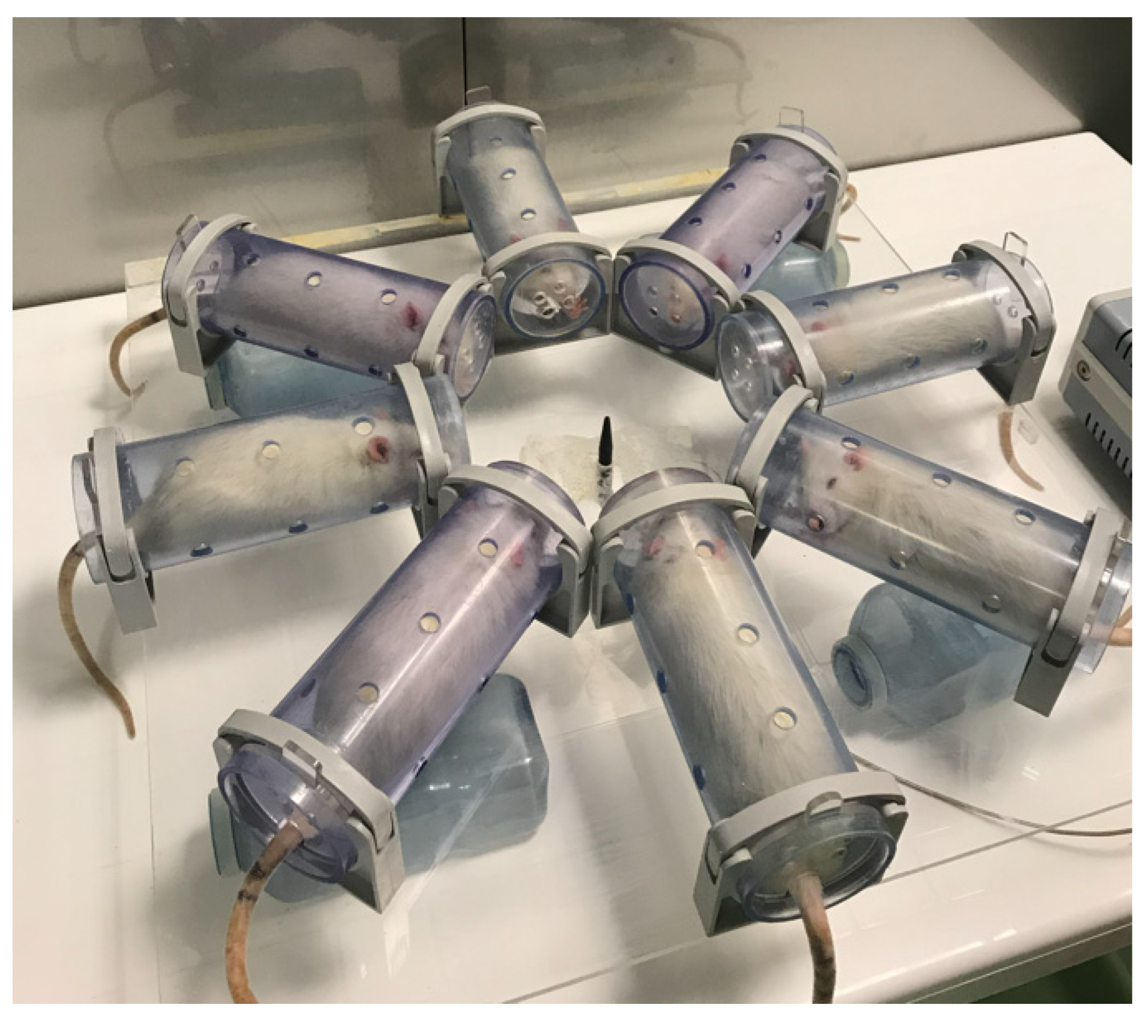
This study was conducted using 40 young Wistar albino male adult rats. Ethical approval for the experimental procedures was granted by the Experimental Animals Research Unit of Süleyman Demirel University (approval date: 19 July 2019; decision No. 8). During the experimental period, the animals were housed in the Experimental Animal Laboratory under standard environmental conditions, including a 12 h light/12 h dark cycle, a room temperature of 21–23 °C, and relative humidity of 45–60%. All animals were maintained under identical conditions and had unrestricted access to standard laboratory feed and drinking water throughout the experimental period. The animals were randomly assigned to five experimental groups (n = 8 per group) (Figure 1).
  • Control group: The rats were not exposed to RFR and were given 1 mL of water using the gavage method.
  • Sham exposure group: The rats were kept in the RFR apparatus without RFR application and were given 1 mL of water using the gavage method.
  • Ginseng group: The rats were administered 150 mg/kg/day antioxidant (Panax Ginseng Root Extract Powder, Sepe Natural Company, İzmir, Turkey) by the gavage method [14].
  • 2600 MHz RFR group: The rats were exposed to 2600 MHz RFR (1 h/day for 30 days) and were given 1 mL of water using the gavage method.
  • 2600 MHz RFR + ginseng group: The rats were exposed to 2600 MHz RFR (1 h/day for 30 days) and administered 150 mg/kg/day ginseng by the gavage method.
All experimental groups were treated during the same 30-day experimental period and consisted of animals from the same batch that were housed under identical environmental and laboratory conditions. In accordance with the principle of reduction in animal research and to prevent unnecessary sacrifice of additional animals, the control, sham, and ginseng groups were used in both this study and our previous study examining the effects of 2100 MHz and 2450 MHz electromagnetic field exposure on the blood–brain barrier [13]. All groups were maintained under identical experimental conditions. After the 30-day exposure period, the rats were euthanized under ketamine–xylazine anesthesia. Brain tissues were collected; samples for histopathological and immunohistochemical analyses were fixed in 10% buffered formaldehyde, while those for biochemical and genetic analyses were stored at −80 °C.
The control, sham, and ginseng groups were also used in our previous study [13] in accordance with the 3Rs principle (Reduction) to avoid the unnecessary use of experimental animals. All experimental groups were treated during the same 30-day experimental period under identical conditions. The control, sham, and ginseng groups were jointly evaluated across experimental conditions. The RFR exposure groups were exposed to different frequencies (2100 MHz, 2450 MHz, and 2600 MHz) within the same experimental framework. Each frequency exposure was applied for 1 h per day over the 30-day period. However, these frequency exposures were not administered simultaneously; instead, they were applied at different times of the day using the same experimental setup and identical exposure durations. This allowed for individual comparisons. All experiments were conducted under identical environmental and laboratory conditions to ensure standardization and minimize variability. This design enabled independent evaluation of each frequency while maintaining comparable experimental conditions. Statistical analyses and comparisons were made accordingly.

2.2. Exposure System and SAR Calculation

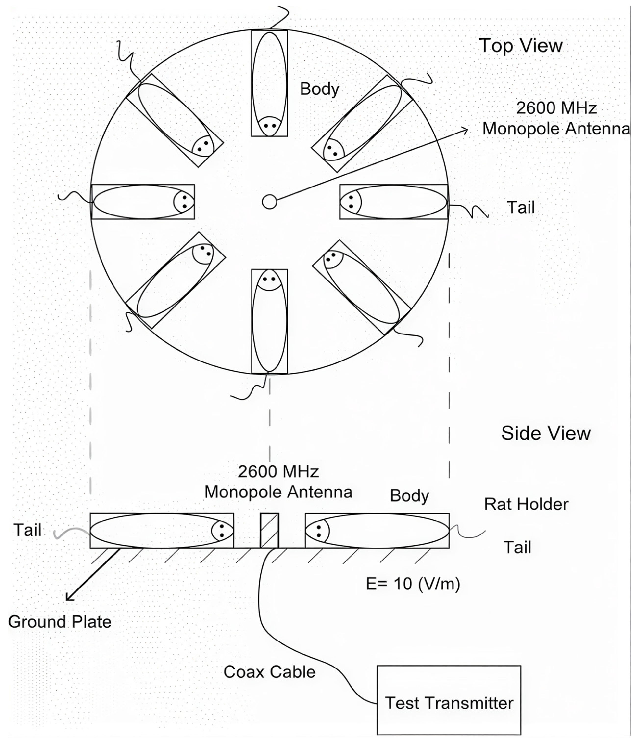
The experiments were conducted in the Experimental Animals Laboratory of Süleyman Demirel University (in an electromagnetically isolated room with a shielding efficiency of 80 dB, without any mobile phones or any other devices that could affect the environment). The carousel exposure system consisted of an RF generator with a center frequency of 2600 MHz (tuning range: 2550–2650 MHz; Set Elektronik A.Ş., Sakarya, Türkiye) that was connected to a monopole antenna with a characteristic impedance of 50 Ω. Each animal was individually restrained in cylindrical plastic tubes (7 cm diameter, 22 cm length) and placed at equal distances from the antenna during exposure. The RF generator output was carefully tuned in a stepwise manner to obtain an electric field strength of 10 V/m at the brain region, with measurements performed using an electromagnetic field meter produced by EXTECH Instruments Corporation (Nashua, NH, USA) [15]. At this stage, energy absorption within the brain tissue was quantified in terms of specific absorption rate (SAR). A visual overview of the study configuration is presented in Figure 2.
Using the formulas from the book published by Polk and Postow, and based on the calculations we previously provided, the conductivity (S/m) and relative dielectric constant of rat brain tissue at 2600 MHz were calculated [15,16]. The calculated values are given in Table 1. Subsequently, the average SAR value generated in the tissue was automatically calculated in MATLAB R2021b (MathWorks, Natick, MA, USA) and was found to be 61.76 mW/kg.

2.3. Histopathological Analysis

Brain tissues were sectioned into 5 μm thick sections, which were subjected to routine deparaffinization and rehydration through a xylene–graded alcohol series. Following rinsing in distilled water, the sections were stained with Luxol Fast Blue at 60 °C for 2 h. Differentiation was carried out in 95% alcohol, after which, the samples were washed and treated with a lithium carbonate solution at room temperature for 20 min to enhance contrast. Subsequently, the sections were passed through 70% alcohol and distilled water in a stepwise manner. After staining in a Cresyl Violet solution at room temperature for 2–5 min, the preparations were briefly rinsed with distilled water, dehydrated through a graded alcohol series, cleared in xylol, and finally mounted using entellan [17,18].

2.4. Immunohistochemistry Analysis

Endothelial junction integrity within the blood–brain barrier was analyzed using a streptavidin–peroxidase-based immunohistochemical approach [18] targeting ZO-1 and occludin, while astrocytic alterations were evaluated by measuring GFAP and aquaporin-4 (AQP-4) expression in astrocyte endfeet. Paraffin-embedded sections mounted on poly-L-lysine-coated slides were subjected to deparaffinization, followed by washing in phosphate-buffered saline (PBS; Sigma, P4417, Steinheim, Germany). Endogenous peroxidase activity was quenched by briefly incubating the sections in hydrogen peroxide for 5 min, which were subsequently rinsed with PBS (3 × 5 min). Antigen retrieval was performed by heat-induced epitope retrieval in a 1 M sodium citrate buffer (pH 6.0) using microwave heating for 12 min. After washing, non-specific binding sites were blocked using an Ultra V Block solution (ScyTek, AAA125 69144, Logan, UT, USA) for 5 min. The sections were then incubated with primary antibodies against ZO-1, occludin, GFAP, and AQP-4 for 1 h at room temperature under humidified conditions. Following PBS washes, the samples were treated with a biotinylated goat anti-polyvalent secondary antibody (ScyTek, ABF125 68813, Logan, UT, USA) for 30 min. After additional washing, streptavidin–peroxidase (ScyTek, ABG125 69365, Logan, UT, USA) was applied for 30 min. Immunoreactivity was visualized using a diaminobenzidine (DAB) substrate system (ScyTek, ACH500, Logan, UT, USA), and the reaction was terminated by rinsing with PBS upon achieving optimal staining, observed using light microscopy. The sections were examined using a Leica DM 500 light microscope (Wetzlar, Germany), and representative micrographs were obtained. Staining intensity and distribution were evaluated semi-quantitatively using a four-tier scoring scale (0: none; +1: slight; +2: moderate; +3: severe).

2.5. TUNEL Method

Apoptotic cell death associated with DNA fragmentation was examined using the TUNEL (terminal deoxynucleotidyl transferase-mediated dUTP nick end labeling) technique. Brain samples obtained after decapitation were fixed in 10% formalin, processed according to routine histological procedures, and embedded in paraffin. Sections (5 μm) were cut and mounted on poly-L-lysine-coated slides. Deparaffinization was performed with xylene, followed by rehydration through graded alcohol solutions and rinsing with phosphate-buffered saline (PBS). Proteolytic treatment was carried out using 5% proteinase K for 10 min, and endogenous peroxidase activity was blocked by incubating in 3% hydrogen peroxide for 5 min. After washing, the sections were incubated with equilibration buffer for 20 min at 37 °C in a humidified chamber. Terminal deoxynucleotidyl transferase (TdT) enzyme was then applied, and incubation was continued for 90 min under the same conditions. The reaction was terminated by sequential incubation in block buffer and stop/wash buffer for 10 min each. Subsequently, the sections were incubated with anti-digoxigenin–peroxidase for 30 min, and apoptotic cells were visualized using a diaminobenzidine (DAB) substrate. Cells exhibiting brown nuclear staining were considered TUNEL-positive.

2.6. Gene Expression Analysis

Analysis of gene expression was performed based on the methodology reported previously [13]. Total RNA was obtained from brain tissues of rats using a tissue-specific RNA extraction kit (MG-DRNA-01, Hibrigen Biotechnology, Kocaeli, Turkey). The extracted RNA was reverse-transcribed into complementary DNA (cDNA) using a High-Capacity cDNA Synthesis Kit (Wizbio, WizScript™, Seongnam, Republic of Korea). Quantitative real-time PCR (qPCR) was conducted using WizPure™ qPCR Master Mix (SYBR) on an Applied Biosystems™ 7500 Fast Real-Time PCR System (Thermo Fisher Scientific, Waltham, MA, USA). The expression levels of COX-2 were quantified according to the comparative CT (ΔΔCT) approach, using GAPDH as the housekeeping control.

2.7. Enzyme Linked Immunosorbent Assay (ELISA)

ELISA analysis was performed as described in our earlier study [13] using a “RAT apoptosis regulator BAX ELISA kit” (Catalog No: E1869Ra; Bioassay Technology Laboratory (BT-Lab), Shanghai, China).

2.8. Statistical Analysis

Statistical analyses were performed using IBM SPSS Statistics 27.0 (IBM Corp., Armonk, NY, USA). Since the data were not normally distributed according to the Shapiro–Wilk test, group comparisons were conducted using the Kruskal–Wallis test followed by the Mann–Whitney U test. p < 0.05 was considered statistically significant.
Exact p-values were reported where applicable to improve transparency. Pairwise comparisons were performed using the Mann–Whitney U test without applying a formal correction for multiple comparisons, and the results were interpreted in conjunction with overall group differences.

3. Results

3.1. Histopathological Findings

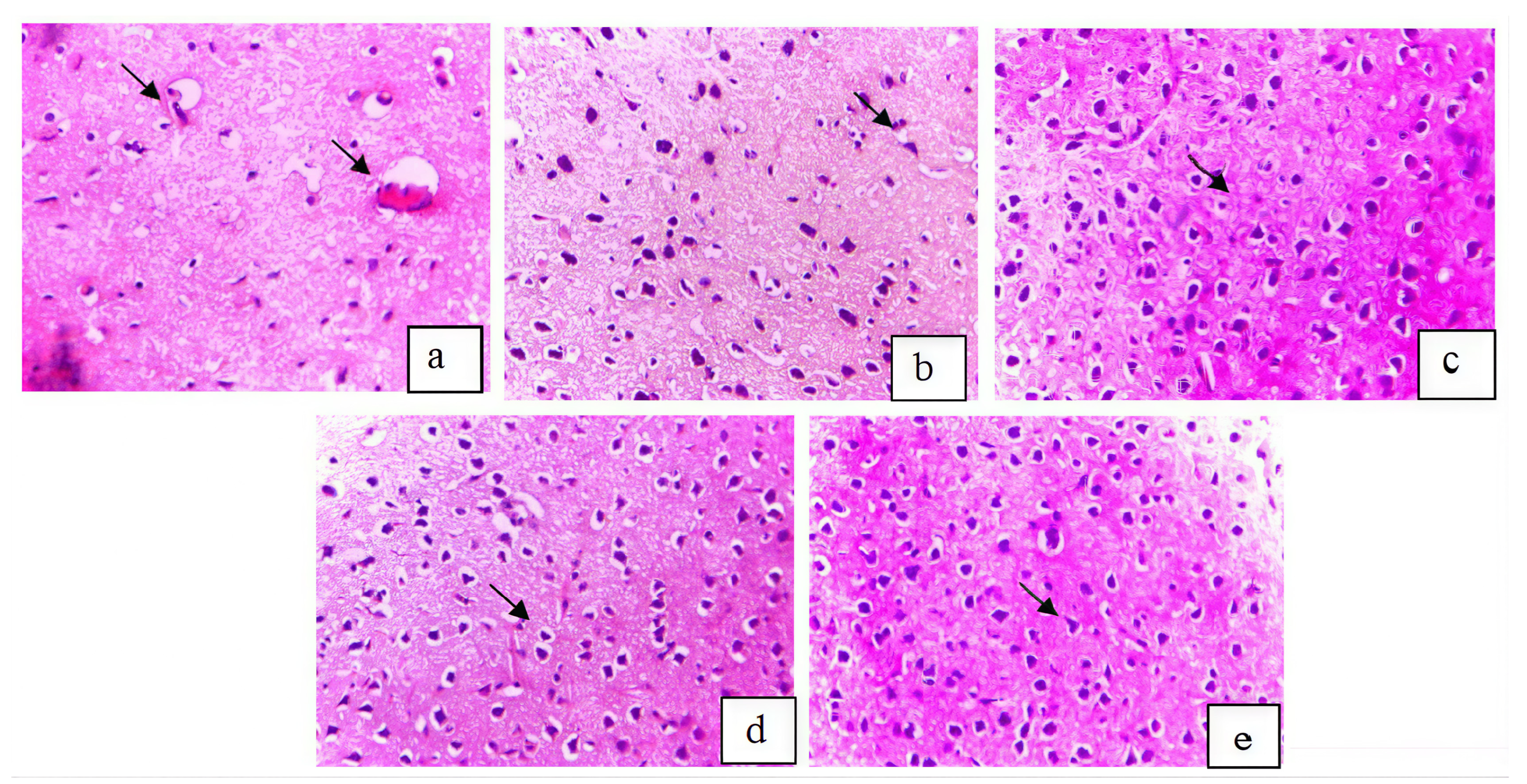
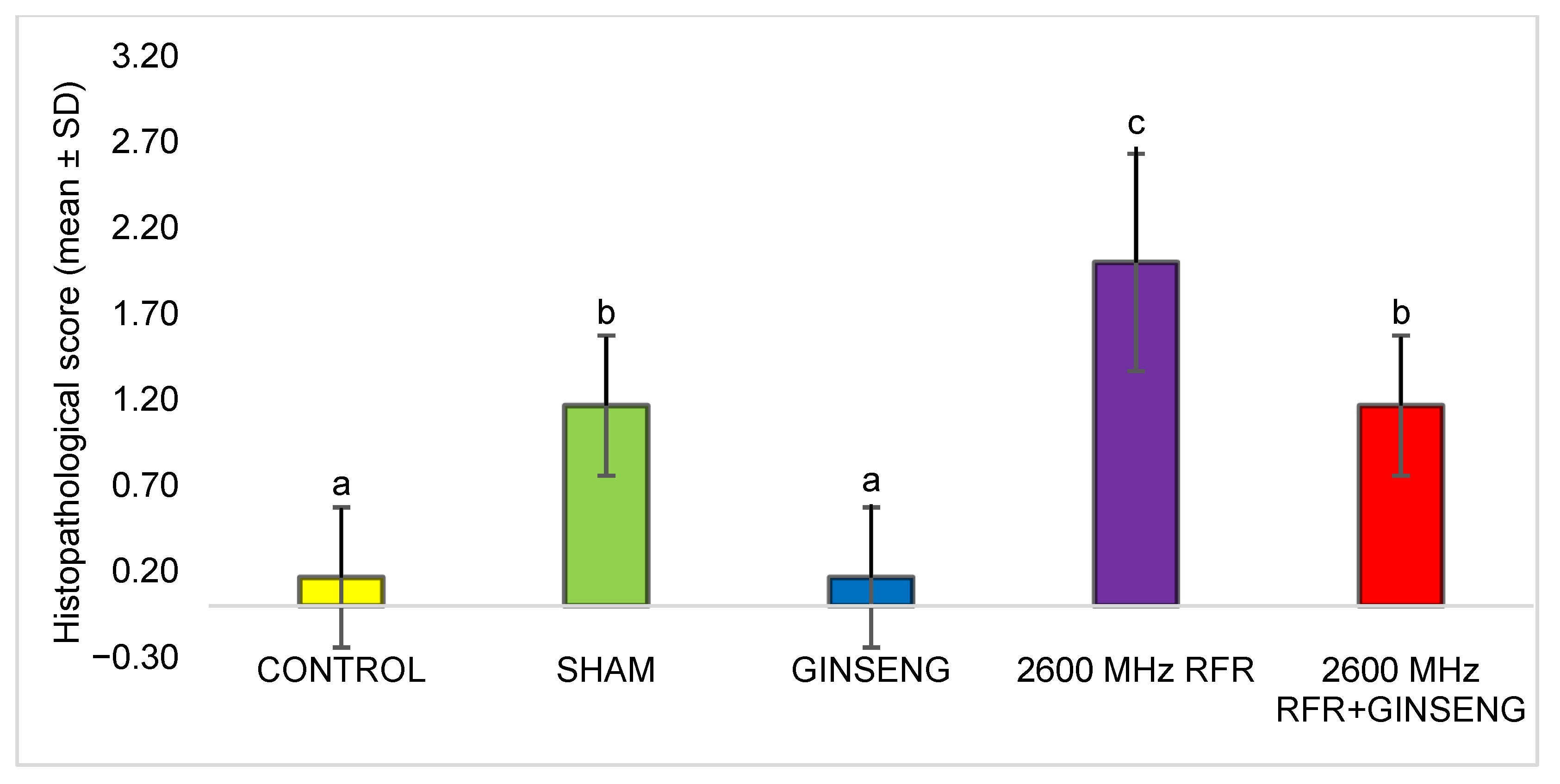
Semi-quantitative histopathological evaluation of brain tissue in the experimental groups was performed. Brain damage was scored on a scale from 0 to +3 (0: none; +1: slight; +2: moderate; +3: severe). The semi-quantitative scores and corresponding histopathological findings are presented in Table 2. Light microscopy images of the groups are shown in Figure 3.
According to the Kruskal–Wallis test, there were statistically significant differences between the groups (p ≤ 0.001). Pairwise comparisons were performed using the Mann–Whitney U test, and significant differences between groups are indicated by different superscript letters in Figure 4. The corresponding p-values were as follows: sham vs. control (p = 0.006), control vs. 2600 MHz RFR (p = 0.003), sham vs. 2600 MHz RFR (p = 0.026), and 2600 MHz RFR vs. 2600 MHz RFR + ginseng (p = 0.026).

3.2. Immunohistochemical Findings

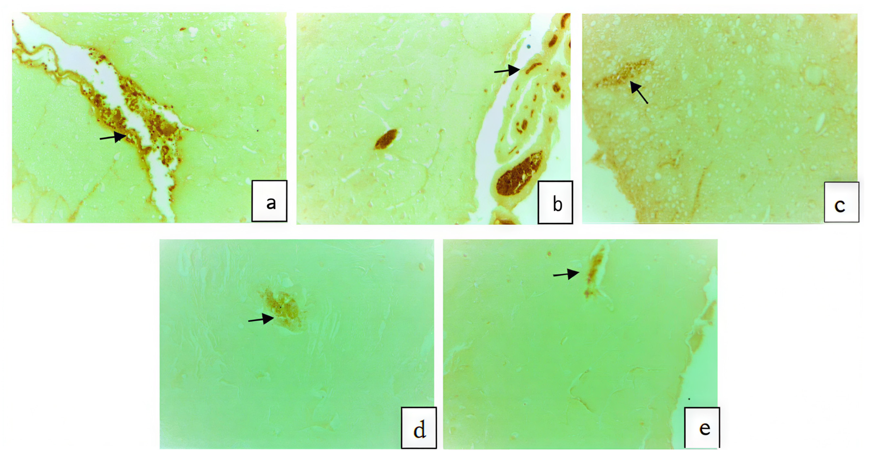
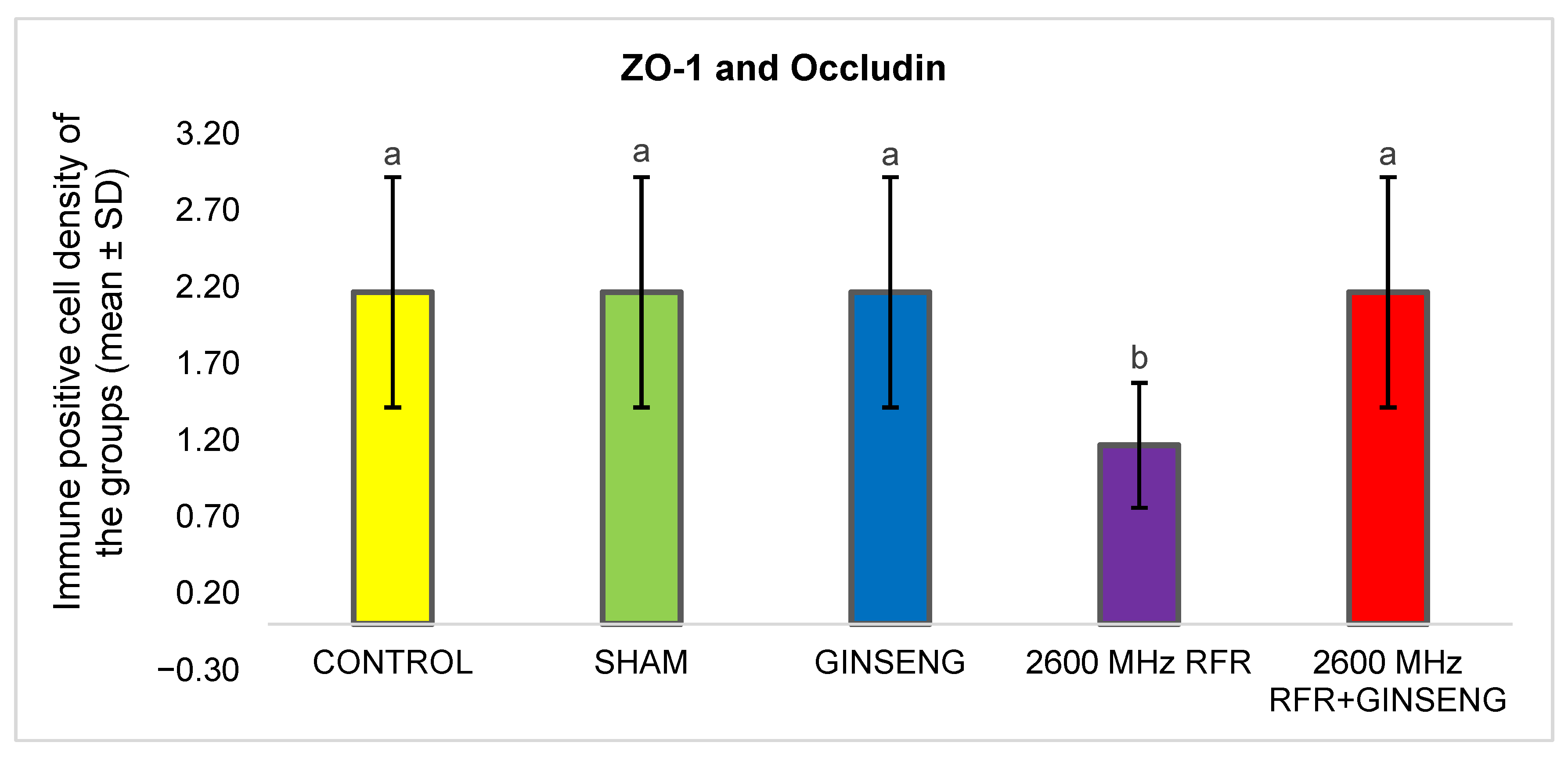
The staining intensity of ZO-1 and occludin within endothelial cells of the blood–brain barrier was assessed on a semi-quantitative scale from 0 (none) to 3 (severe). Group comparisons performed using the Kruskal–Wallis test indicated a statistically significant variation (p = 0.007). Pairwise comparisons were performed using the Mann–Whitney U test. No significant differences were found between the control, sham, and ginseng groups (p = 1.00). The corresponding p-values were as follows: control vs. 2600 MHz RFR (p = 0.023) and 2600 MHz RFR vs. 2600 MHz RFR + ginseng (p = 0.023). Significant differences between groups are indicated by different superscript letters in Figure 5. A cytoplasmic immunostaining score graph for ZO-1 and occludin expression in the groups is given in Figure 5. Light microscopic images of the experimental groups are shown in Figure 6.
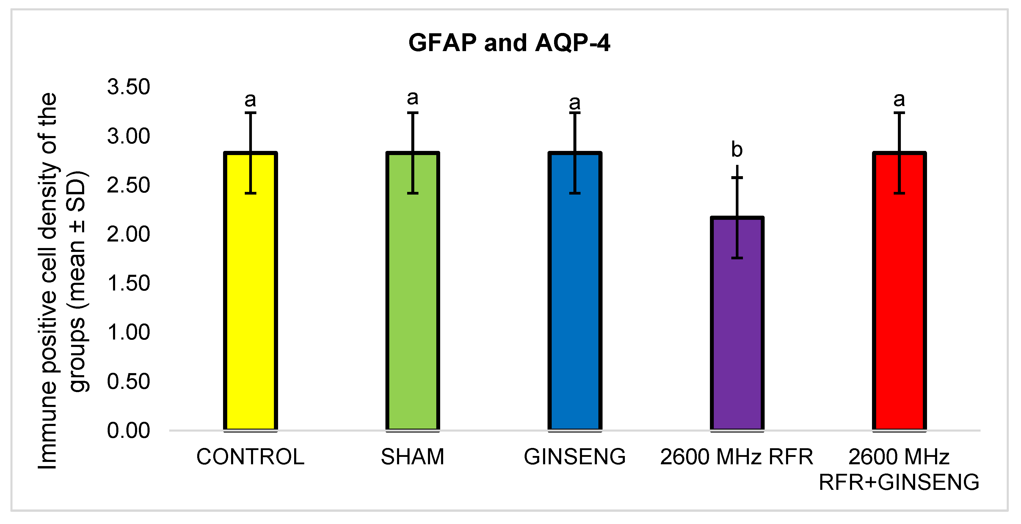
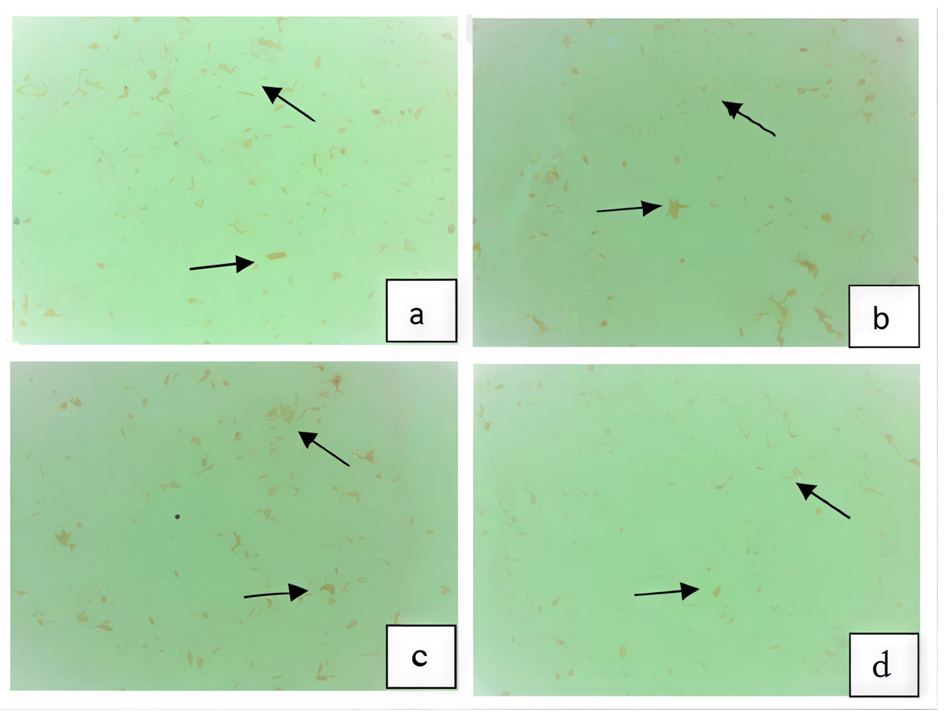
GFAP staining in astrocytes associated with the blood–brain barrier, together with AQP-4 levels in astrocytic endfeet, was quantified using a semi-quantitative grading approach ranging from 0 (none) to 3 (severe). Statistical analysis performed using the Kruskal–Wallis test indicated a significant variation between the groups (p = 0.005). Subsequent pairwise analyses were conducted using the Mann–Whitney U test. No significant differences were observed among the control, sham, and ginseng groups (p = 1.00). The corresponding p-values were as follows: control vs. 2600 MHz RFR (p = 0.027), sham vs. 2600 MHz RFR (p = 0.027), and 2600 MHz RFR vs. 2600 MHz RFR + ginseng (p = 0.027). Significant differences between groups are indicated by different superscript letters in Figure 7. A cytoplasmic immunostaining score graph for GFAP and AQP-4 expression in the groups is given in Figure 7. Light microscope images of the groups are given in Figure 8.
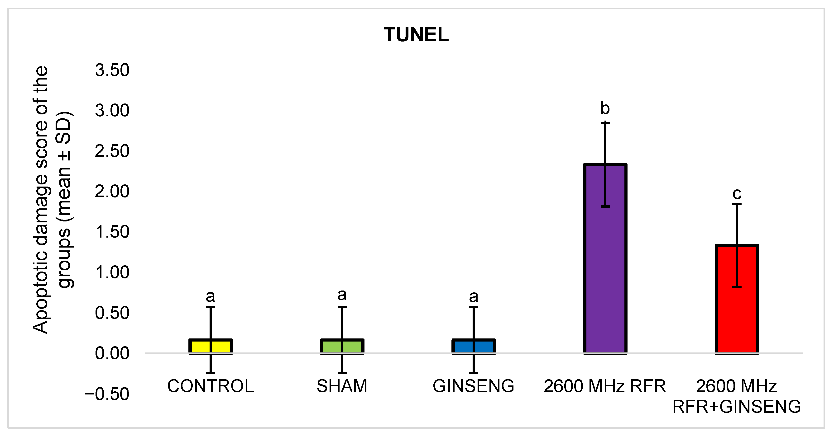
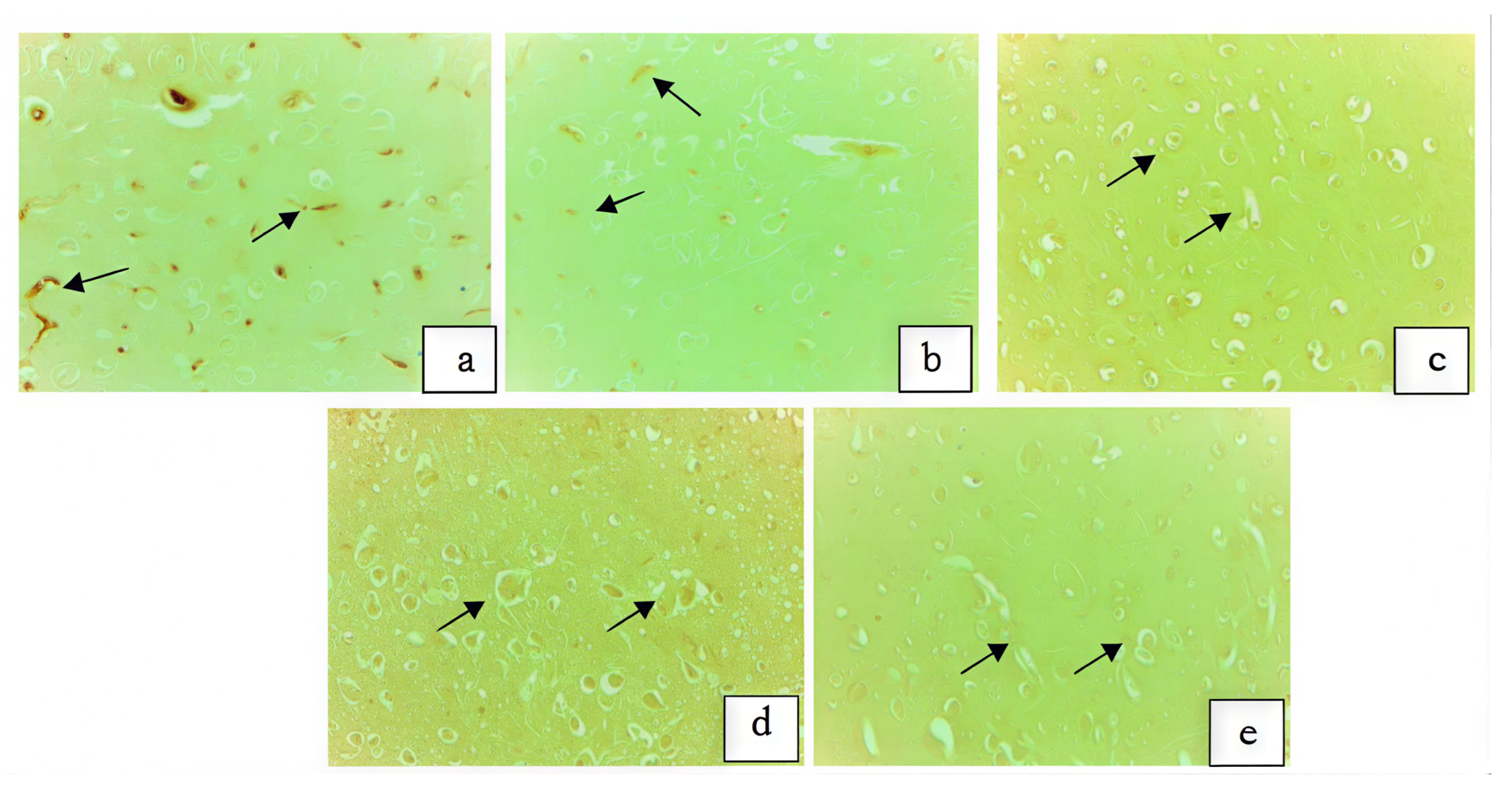
Apoptotic cells detected by the TUNEL method were evaluated using a semi-quantitative scoring system ranging from 0 to +3. The 2600 MHz RFR group exhibited the highest level of apoptotic immunopositivity, whereas the 2600 MHz RFR + ginseng group showed reduced staining intensity. Apoptotic cells were rarely observed in the control, sham, and ginseng groups. Statistical analysis demonstrated a significant difference among the groups (p < 0.001). Pairwise comparisons were performed using the Mann–Whitney U test. No significant differences were observed among the control, sham, and ginseng groups (p = 1.00). The corresponding p-values were as follows: control vs. 2600 MHz RFR (p = 0.002), sham vs. 2600 MHz RFR (p = 0.002), and 2600 MHz RFR vs. 2600 MHz RFR + ginseng (p = 0.014). Differences between groups are indicated by superscript letters in Figure 9. A graph of the immune-positive apoptotic cell scores of the groups is given in Figure 9. Representative light microscopy images corresponding to the TUNEL assay are presented in Figure 10.

3.3. Gene Expression Findings

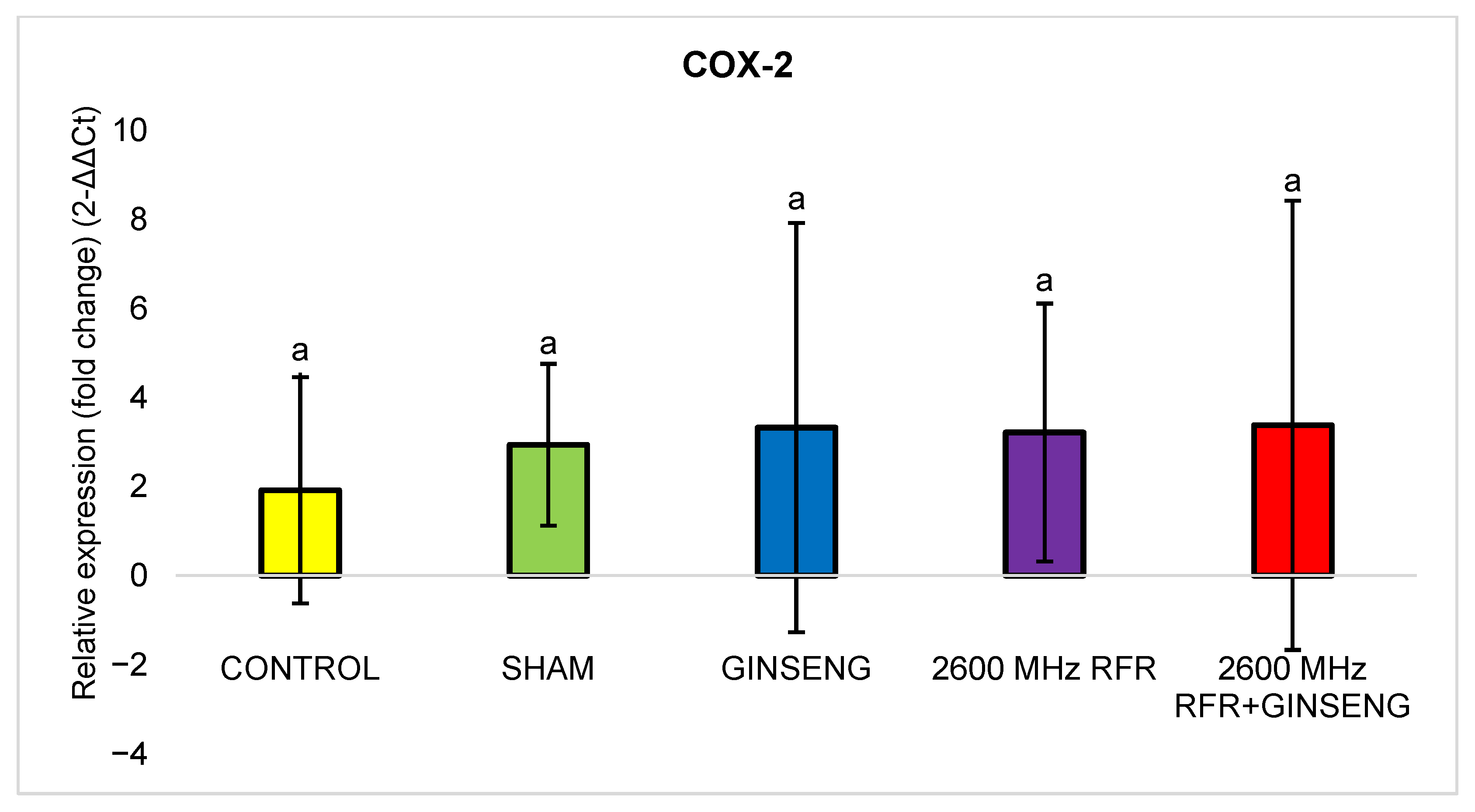
The relative COX-2 gene expression levels in rat brain tissues were evaluated by qPCR and are expressed as the mean ± SD. There were no statistically significant differences among the experimental groups according to the Kruskal–Wallis test (p = 0.640). Pairwise comparisons were performed using the Mann–Whitney U test. No significant differences were observed among the control, sham, and ginseng groups (control vs. sham: p = 0.275; sham vs. ginseng: p = 0.513). The corresponding p-values were as follows: sham vs. 2600 MHz RFR (p = 0.827) and 2600 MHz RFR vs. 2600 MHz RFR + ginseng (p = 0.827). The relative expression levels of COX-2 in the groups are presented in Figure 11.

3.4. Biochemical Findings

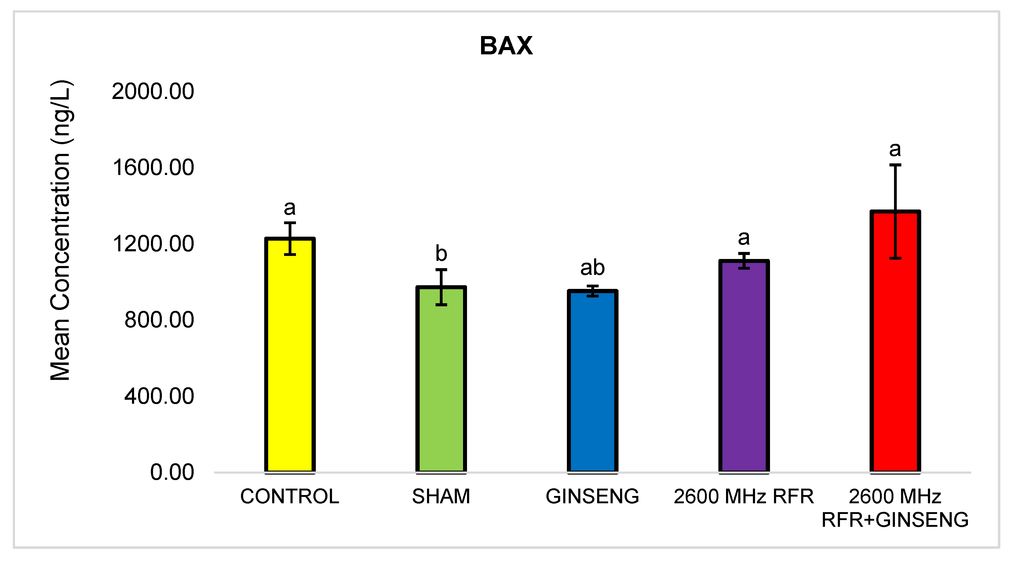
The BAX protein concentrations in rat brain tissues were determined by ELISA and are expressed as the mean ± SD. There were statistically significant differences among the groups according to the Kruskal–Wallis test (p = 0.021). Pairwise comparisons were performed using the Mann–Whitney U test. The corresponding p-values were as follows: control vs. sham (p = 0.05), sham vs. ginseng (p = 0.827), sham vs. 2600 MHz RFR (p = 0.05), and 2600 MHz RFR vs. 2600 MHz RFR + ginseng (p = 0.127). Significant differences between groups are indicated by different superscript letters in Figure 12.

4. Discussion

The increased use of mobile phones in recent years has heightened interest in the effects of radiofrequency electromagnetic radiation (RF-EMR) on the brain, particularly because these devices are held close to the head. RF-EMR can induce mild heating, especially in superficial tissues. However, studies investigating its effects on brain tissue and BBB structures have yielded inconsistent results; while some report increased BBB permeability, neuronal damage, DNA mutations, and tumor development, others have found no such effects [19,20].
BBB integrity is maintained by tight junction (TJ) proteins, including occludin, claudin-5, and ZO-1. These proteins regulate the permeability of endothelial cells in cerebral vessels, thereby forming a selective barrier. Inflammatory factors can increase BBB permeability by disrupting the structure of tight junction proteins, leading to cellular damage such as degeneration, apoptosis, and necrosis of neurons. Among these, ZO-1 plays a critical role in maintaining BBB integrity. Decreased ZO-1 levels may result in disruption of tight junctions and the loss of barrier function, whereas increased ZO-1 expression has been associated with enhanced BBB stability [21,22,23].
AQP-4 is an essential membrane protein that regulates water exchange at the BBB and the cerebrospinal fluid interface. This water movement is critical for maintaining brain volume and osmotic balance. Aquaporins are channel proteins that facilitate bidirectional water transport across cell membranes, and AQP-4 is the most abundant subtype in astrocyte endfeet, particularly in the perivascular and subpial regions [24].
GFAP is an important structural protein involved in maintaining central nervous system (CNS) homeostasis and mediating the astrocytic response to brain injury. Reactive gliosis develops in various conditions, which leads to CNS damage. Increased GFAP expression is associated with cellular hypertrophy and hyperplasia in astrocytes. Increased GFAP levels indicate reactive astrocytes and brain injury. Furthermore, the number of GFAP-positive astrocytes has been reported to increase as the severity of trauma increases [25,26].
Studies have shown that microwave radiation may cause a decrease in ZO-1 and occludin expression, and an increase in occludin tyrosine phosphorylation and GFAP expression in the brain. It has been reported that BBB permeability increases after exposure to microwave radiation [27,28,29]. In this study, degeneration, vacuolization, vascular dilatation, and low-level edema were observed in the 2600 MHz RFR group, particularly in cortical neurons. A significant reduction in ZO-1 and GFAP expression was identified in the 2600 MHz RFR group relative to the control and sham groups (p = 0.023 for ZO-1; p = 0.027 for GFAP). There was a greater increase in the RFR + ginseng group than in the RFR group (p = 0.023). It was concluded that ginseng mitigated the effects of RFR on BBB.
Saikhedkar et al. [30] detected neurodegenerative cells in the brain tissue of rats exposed to 900 MHz radiofrequency radiation for 4 h per day for 15 days, based on histological examination of the brain tissue. They concluded that radio wave exposure causes neurodegeneration. Also, Alkis et al. [31] concluded that 900, 1800 and 2100 MHz radiofrequency radiation can cause oxidative damage in rat brain tissues. Research exploring the impact of radiofrequency radiation on brain tissue has yielded inconsistent results. For instance, Dogan et al. [20] indicated that electromagnetic exposure from 3G mobile devices did not produce a significant change in apoptotic cell counts in rat brain tissue. Conversely, exposure to 900 MHz RFR has been linked to structural alterations in multiple brain regions, including the frontal cortex, basal ganglia, hippocampus, and cerebellum [32]. Moreover, repeated daily application of 2100 MHz RFR for 30 min over a 90-day period has been associated with an increase in apoptotic activity in brain tissue [33]. Parasuraman et al. [34] identified apoptosis-related alterations in brain tissue in association with 2450 MHz RFR application, as determined by histopathological assessment. Zheng et al. [35] used the TUNEL method to assess whether exposure to 2650 MHz RFR affected apoptosis in the hippocampus. They concluded that there was no significant apoptosis in hippocampal cells in the RFR group. Delen et al. [36] demonstrated the protective role of melatonin on apoptosis in rat brain tissue induced by 2600 MHz RFR exposure (30 min per day, 5 days per week, for 1 month). Our findings demonstrated that the 2600 MHz RFR group exhibited significantly greater apoptotic activity than the control and sham groups (p = 0.002). However, treatment with ginseng significantly reduced apoptosis compared with the RFR group (p = 0.014). It was observed that RFR induced apoptosis in brain tissue, and daily administration of 150 mg/kg ginseng had a protective effect against apoptosis.
Long-term interaction with electromagnetic radiation has been suggested to impair mitochondrial function and initiate apoptotic mechanisms in neuronal cells [37]. Gupta et al. [37] also demonstrated that application of 2450 MHz RFR led to elevated expression levels of cytochrome-c and caspase-9, contributing to the activation of the mitochondrial apoptotic cascade. In contrast, no statistically meaningful changes were detected in apoptotic marker expression in rats subjected to 900 MHz and 1800 MHz RFR. Since the BCL-2 protein family plays an important role in determining cellular sensitivity and resistance to various apoptosis-inducing stimuli, the effects of RFR and ginseng on gene expression were examined to better understand the protective mechanisms of ginseng against RFR-induced effects on the BBB.
Apoptosis is a programmed cell death process that can be triggered by specific stimuli through intrinsic or extrinsic pathways under both physiological and pathological conditions. A frequently described mechanism underlying chemotherapeutic agent-induced apoptosis involves increased permeability of the mitochondrial membrane through a mitochondria-mediated pathway controlled by members of the BCL-2 protein family. In this context, the relative levels of BCL-2 and BAX play a pivotal role as the BCL-2/BAX ratio governs the susceptibility of cells to apoptotic death. In studies investigating microwave radiation, apoptosis is considered a key indicator of a cellular response. These studies typically examine BCL-2 family proteins (anti-apoptotic BCL-2 and pro-apoptotic BAX), apoptosis-related factors such as cytochrome c, and regulatory genes including p53. Several studies have reported that EMR exposure may increase apoptosis, accompanied by elevated p53, BAX, and caspase-3 expression, with mitochondrial dysfunction being a major contributing factor [38,39,40]. Zhu et al. [41] reported that 900 MHz microwave radiation increased the number of BAX-positive cells in the brain; BAX is a pro-apoptotic protein that induces cell death, which can be assessed using the TUNEL method. Continuous exposure to 1950 MHz EMR has been shown to cause a significant increase in BAX levels in astrocyte mitochondria, a decrease in BCL-2 levels, and an increase in apoptosis [42]. A different study assessed gene expression changes in cortical neurons under 1800 MHz mobile signal exposure and reported that BAX inhibited BCL-2 while enhancing apoptotic activity [43]. Similarly, research conducted on mouse brain tissue indicated that RF/microwave radiation resulted in lower BCL-2 expression compared with the control group [44]. These results suggest that BAX is involved in microwave exposure-induced cellular apoptosis. Similarly, in this study, using the TUNEL method and immunohistochemistry, apoptotic changes were evident in the brain tissue of the RFR-exposed groups. Gene expression and ELISA methods were applied to determine the pathways responsible for this effect. The 2600 MHz RFR group showed higher BAX protein concentrations compared with the sham group (p = 0.05). In contrast, treatment with 150 mg/kg ginseng did not significantly alter BAX protein levels as no difference was detected between the RFR + ginseng and RFR-only groups.
Previous studies suggest that exposure to extremely high-frequency EMR may suppress COX-2 induction during inflammatory processes [45]. It has been shown that 1800 MHz EMR with a SAR value of 0.6 W/kg applied for 2 h daily for 3 months caused significant DNA fragmentation and overexpression of the COX-2 apoptotic gene in the brain [19]. In our study, the SAR value was calculated as 61.76 mW/kg. This value is substantially lower than the 2 W/kg limit recommended by the International Commission on Non-Ionizing Radiation Protection (ICNIRP) for localized exposure for the general public [46]. Contrary to what has been shown in these studies, Choi et al. [47] showed that p53 enhances COX-2 expression, while COX-2 suppresses apoptosis associated with p53 or genotoxic stress and modulates the transcriptional activity of p53. Sun et al. [48] demonstrated that COX-2 overexpression can upregulate BCL-2. Similarly, De Vries [49] demonstrated that COX-2 overexpression inhibits apoptosis. As shown in the reviewed studies, conflicting results have been reported regarding the relationships between COX-2, apoptosis, and EMR. In this study, to clarify this contradiction based on our own analyses, we examined COX-2 gene expression and BAX protein concentrations to determine which pathways are activated along with the immunohistochemically observed apoptosis. Similar to the studies discussed above, COX-2 expression in the EMR groups tended to be lower than in the sham group; however, this difference did not reach statistical significance (p = 0.127). At the same time, an increase in BAX protein concentration was observed in groups with decreased COX-2 expression, suggesting that decreased COX-2 expression may be associated with increased BAX levels and apoptotic processes. In groups with increased COX-2 and decreased BAX levels, COX-2 expression may be involved in modulating apoptotic processes. This observation is consistent with our immunohistochemical findings. Furthermore, COX-2 expression levels were higher in the RFR + ginseng group than in the RFR group, suggesting that ginseng may exert a protective effect against RFR-induced apoptosis, potentially through modulation of COX-2 gene expression.
Ginseng (Panax ginseng C.A. Meyer), known as the “king of medicinal herbs,” has been used for thousands of years in Asia, particularly in Korea and China, to treat various diseases. The word “Panax” means “all-healing” in Greek, and ginseng is described in traditional Chinese medicine for its vital energy-boosting, regenerative, and longevity-enhancing effects [11,12]. Ginsenoside Rg1 can slow neurodegeneration, reduce cell apoptosis, and increase the number of glial fibrillary acidic protein-positive cells. One study showed that ginsenoside Rg1 markedly enhanced BCL-2 protein expression while concurrently reducing BAX protein levels. Ginsenoside Rg1 promoted brain tissue repair by enhancing its anti-apoptosis effect [50]. In addition, ginsenoside Rg1 ameliorated neurological damage by downregulating AQP-4 expression [51]. Studies have also suggested that ginseng may exert protective effects against RFR-induced damage [52,53]. To assess the influence of KRG (Korean red ginseng) on inflammation, COX-2 mRNA levels were measured in rat brain tissue. KRG was shown to significantly reduce the expression of this gene and may exert anti-inflammatory effects in vivo [54]. In this study, the protective effect of ginseng on the brain was demonstrated through histopathological evaluation; immunohistochemical evaluation of ZO-1, occludin, GFAP and AQP-4; and measurement of COX-2 gene expression and BAX protein concentrations. There were other studies where ginseng administration did not have statistically significant effects. It was thought that the daily dose of 150 mg/kg ginseng might have been insufficient. Applying a higher dose might make the results more significant.

5. Conclusions

According to previous studies, increased BAX protein levels are generally associated with enhanced apoptotic activity. In this study, the immunohistochemical findings demonstrated increased apoptosis in the RFR groups. Although some variation in COX-2 expression was observed, these differences were not statistically significant. Therefore, any relationship between COX-2 expression and apoptosis should be interpreted with caution and cannot be considered a definitive mechanistic link. The mechanisms underlying the protective effects of ginseng remain unclear. Although changes in COX-2 expression were observed, these findings do not support a direct mechanistic role due to the lack of statistical significance. Therefore, factors other than COX-2 may contribute to the observed protective effects.
Since RFR effects were observed histologically, genetic and biochemical studies were conducted to investigate these processes further. The effects of RFR on brain tissue were clearly demonstrated both histologically and immunohistochemically.
The potential protective role of ginseng against RFR-induced damage was also evaluated; however, the precise mechanisms underlying these effects remain unclear and require further investigation. Although ginseng is known to possess antioxidant properties, oxidative stress markers were not evaluated in the present study. In particular, parameters such as reactive oxygen species (ROS) levels were not measured. Therefore, the involvement of oxidative processes in RFR-induced effects and the protective role of ginseng remains unclear and requires further investigation in future studies.
BAX is a widely used marker of apoptosis and was included in this study to provide biochemical support for the histological findings. The results demonstrated increased apoptotic activity in the RFR groups, consistent with the immunohistochemical observations. Although there were some discrepancies between the molecular and histological findings, these differences highlight the complexity of apoptotic processes and suggest that multiple factors may be involved.
In this study, variations in COX-2 expression were observed; however, these differences were not statistically significant. Therefore, its potential relationship with apoptosis should be interpreted with caution. Overall, these findings emphasize the importance of integrating histological, immunohistochemical, and molecular analyses to better understand the cellular responses to RFR exposure.

Author Contributions

Experiment, İ.P.K.; collection of tissues at the end of the experiment, İ.P.K. and N.Ş.; electromagnetic field setup, electromagnetic measurements and calculations, İ.P.K., Ö.C., and S.Ç.; histopathological and immunohistochemical examinations, N.Ş.; genetics and biochemical analysis, U.Ş.; writing—original draft preparation, İ.P.K. All authors have read and agreed to the published version of the manuscript.

Funding

This research was supported by The Scientific Research Projects Coordination Unit of Süleyman Demirel University (Project Code: FDK-2020-7451).

Institutional Review Board Statement

The study was conducted in accordance with the Declaration of Helsinki, and approved by the Institutional Ethics Committee of Süleyman Demirel University Animal Experiments Local Ethics Committee, Turkey (date of registration: 19 July 2019; issue: 8).

Informed Consent Statement

Not applicable.

Data Availability Statement

All the research data related to this manuscript will be available upon reasonable request to the corresponding author.

Conflicts of Interest

The authors declare no conflicts of interest.

References

  1. Liu, S.; Cai, W.; Luo, Y.; Dou, J.; Wu, J.; Wu, H.; Han, Z.; Yu, J.; Liang, P. CEUS versus MRI in evaluation of the Effect of Microwave Ablation of Breast Cancer. Ultrasound Med. Biol. 2022, 48, 617–625. [Google Scholar] [CrossRef]
  2. Lin, J.C. The Microwave Auditory Effect. In Auditory Effects of Microwave Radiation; Springer: Cham, Switzerland, 2021; pp. 127–173. [Google Scholar]
  3. Mahboubi, A. The Side Effects of Electromagnetic Waves on the Human Brain: A review study. In Proceedings of the 11th Majlesi Conference on Electrical Engineering, Majlesi, Iran, 6 August 2022; Available online: https://hal.science/hal-03762538/ (accessed on 25 February 2026).
  4. Kaplan, S.; Deniz, O.G.; Önger, M.E.; Türkmen, A.P.; Yurt, K.K.; Aydın, I.; Altunkaynak, B.Z.; Davis, D. Electromagnetic field and brain development. J. Chem. Neuroanat. 2016, 75, 52–61. [Google Scholar] [CrossRef]
  5. Varghese, R.; Majumdar, A.; Kumar, G.; Shukla, A. Rats exposed to 2.45 GHz of non-ionizing radiation exhibit behavioral changes with increased brain expression of apoptotic caspase 3. Pathophysiology 2018, 25, 19–30. [Google Scholar] [CrossRef]
  6. Banks, W.A.; Reed, M.J.; Logsdon, A.F.; Rhea, E.M.; Erickson, M.A. Healthy aging and the blood-brain barrier. Nat. Aging 2021, 1, 243–254. [Google Scholar] [CrossRef]
  7. Kovacs, G.G. Cellular reactions of the central nervous system. Handb. Clin. Neurol. 2017, 145, 13–23. [Google Scholar] [PubMed]
  8. Chen, W.; Guo, Y.; Yang, W.; Zheng, P.; Zeng, J.; Tong, W. Protective effect of ginsenoside Rb1 on integrity of blood-brain barrier following cerebral ischemia. Exp. Brain Res. 2015, 233, 2823–2831. [Google Scholar] [CrossRef]
  9. Liu, D.; Zhang, T.; Chen, Z.; Wang, Y.; Ma, S.; Liu, J.; Liu, J. The beneficial effect of ginsenosides extracted by pulsed electric field against hydrogen peroxide-induced oxidative stress in HEK-293 cells. J. Ginseng Res. 2017, 41, 169–179. [Google Scholar] [CrossRef]
  10. Yu, T.; Yang, Y.; Kwak, Y.S.; Song, G.G.; Kim, M.Y.; Rhee, M.H.; Cho, J.Y. Ginsenoside Rc from Panax ginseng exerts anti-inflammatory activity by targeting TANK-binding kinase 1/interferon regulatory factor-3 and p38/ATF-2. J. Ginseng Res. 2017, 41, 127–133. [Google Scholar] [CrossRef] [PubMed]
  11. Im, D.S. Pro-Resolving Effect of Ginsenosides as an Anti-Inflammatory Mechanism of Panax ginseng. Biomolecules 2020, 10, 444. [Google Scholar] [CrossRef] [PubMed]
  12. Kim, M.; Mok, H.; Yeo, W.S.; Ahn, J.H.; Choi, Y.K. Role of ginseng in the neurovascular unit of neuroinflammatory diseases focused on the blood-brain barrier. J. Ginseng Res. 2021, 45, 599–609. [Google Scholar] [CrossRef]
  13. Postacı Karaman, İ.; Coşkun, Ö.; Şenol, N.; Şahin, U.; Çömlekçi, S. Immunohistochemical, Biochemical and Genetic Evaluation of the Effects of Ginseng Administration on Blood–Brain Barrier in Rats Exposed to 2100 MHz and 2450 MHz Electromagnetic Radiation. Appl. Sci. 2026, 16, 2376. [Google Scholar] [CrossRef]
  14. Choi, J.H.; Jang, M.; Nah, S.Y.; Oh, S.; Cho, I.H. Multitarget effects of Korean red ginseng in animal model of Parkinson’s disease: Antiapoptosis, antioxidant, antiinflammation, and maintenance of blood-brain barrier integrity. J. Ginseng Res. 2018, 42, 379–388. [Google Scholar] [CrossRef] [PubMed]
  15. Karaman, I.P.; Coskun, O.; Senol, N.; Sahin, M.; Comlekci, S. Alleviative effect of quercetin on rat testicular against 2600 MHz electromagnetic field. Int. J. Radiat. Res. 2024, 22, 537–543. [Google Scholar] [CrossRef]
  16. Polk, C.; Postow, E. Biological Effects of Electromagnetic Fields, 1st ed.; CRC Press: Boca Raton, FL, USA, 1996. [Google Scholar]
  17. Bancroft, J.D.; Stevens, A.; Turner, D.R. Theory and Practice of Histological Techniques; Churchill Livingstone: New York, NY, USA; Edinburg, UK, 1996; pp. 126–129. [Google Scholar]
  18. Şenol, N.; Kaya, E.; Coşkun, Ö.; Aslankoç, R.; Çömlekçi, S. Evaluation of the Effects of a 50 Hz Electric Field on Brain Tissue by Immunohistochemical Method, and on Blood Tissue by Biochemical, Physiological and Comet Method. Appl. Sci. 2023, 13, 3276. [Google Scholar] [CrossRef]
  19. Hussein, S.; El-Saba, A.A.; Galal, M.K. Biochemical and histological studies on adverse effects of mobile phone radiation on rat’s brain. J. Chem. Neuroanat. 2016, 78, 10–19. [Google Scholar] [CrossRef]
  20. Dogan, M.; Turtay, M.G.; Oguzturk, H.; Samdanci, E.; Turkoz, Y.; Tasdemir, S.; Bakir, S. Effects of Electromagnetic Radiation Produced by 3G Mobile Phones on Rat Brains: Magnetic Resonance Spectroscopy, Biochemical and Histopathological Evaluation. Hum. Exp. Toxicol. 2012, 31, 557–564. [Google Scholar] [CrossRef]
  21. Luissint, A.C.; Artus, C.; Glacial, F.; Ganeshamoorthy, K.; Couraud, P.O. Tight junctions at the blood brain barrier: Physiological architecture and disease-associated dysregulation. Fluids Barriers CNS 2012, 9, 23. [Google Scholar] [CrossRef] [PubMed]
  22. Lee, J.Y.; Park, C.S.; Choi, H.Y.; Yune, T.Y. Ginseng Extracts, GS-KG9 and GS-E3D, Prevent Blood–Brain Barrier Disruption and Thereby Inhibit Apoptotic Cell Death of Hippocampal Neurons in Streptozotocin-Induced Diabetic Rats. Nutrients 2020, 12, 2383. [Google Scholar] [CrossRef] [PubMed]
  23. Chen, W.; Ju, X.Z.; Lu, Y.; Ding, X.W.; Miao, C.H.; Chen, J.W. Propofol improved hypoxia-impaired integrity of blood-brain barrier via modulating the expression and phosphorylation of zonula occludens-1. CNS Neurosci. Ther. 2019, 25, 704–713. [Google Scholar] [CrossRef] [PubMed]
  24. Zhang, Y.; Xu, K.; Liu, Y.; Erokwu, B.O.; Zhao, P.; Flask, C.A.; Estebanez, C.R.; Farr, G.W.; LaManna, J.C.; Boron, W.F.; et al. Increased cerebral vascularization and decreased water exchange across the blood-brain barrier in aquaporin-4 knockout mice. PLoS ONE 2019, 14, e0218415. [Google Scholar] [CrossRef] [PubMed]
  25. Cikriklar, H.I.; Uysal, O.; Ekici, M.A.; Ozbek, Z.; Cosan, D.T.; Yucel, M.; Yurumez, Y.; Baydemir, C. Effectiveness of GFAP in Determining Neuronal Damage in Rats with Induced Head Trauma. Turk. Neurosurg. 2016, 26, 878–889. [Google Scholar]
  26. Lagos-Cabré, R.; Burgos-Bravo, F.; Avalos, A.M.; Leyton, L. Connexins in Astrocyte Migration. Front. Pharmacol. 2020, 10, 1546. [Google Scholar] [CrossRef] [PubMed]
  27. Ammari, M.; Brillaud, E.; Gamez, C.; Lecomte, A.; Sakly, M.; Abdelmelek, H.; De Seze, R. Effect of a chronic GSM 900 MHz exposure on glia in the rat brain. Biomed. Pharmacother. 2008, 62, 273–281. [Google Scholar] [CrossRef]
  28. Wang, L.F.; Li, X.; Gao, Y.B.; Wang, S.M.; Zhao, L.; Dong, J.; Yao, B.W.; Xu, X.P.; Chang, G.M.; Zhou, H.M.; et al. Activation of VEGF/Flk-1-ERK Pathway Induced Blood–Brain Barrier Injury After Microwave Exposure. Mol. Neurobiol. 2015, 52, 478–491. [Google Scholar] [CrossRef]
  29. Eberhardt, J.L.; Persson, B.R.; Brun, A.E.; Salford, L.G.; Malmgren, L.O. Blood-brain barrier permeability and nerve cell damage in rat brain 14 and 28 days after exposure to microwaves from GSM mobile phones. Electromagn. Biol. Med. 2008, 27, 215–229. [Google Scholar] [CrossRef]
  30. Saikhedkar, N.; Bhatnagar, M.; Jain, A.; Sukhwal, P.; Sharma, C.; Jaiswal, N. Effects of mobile phone radiation (900 MHz radiofrequency) on structure and functions of rat brain. Neurol. Res. 2014, 36, 1072–1079. [Google Scholar] [CrossRef]
  31. Alkis, M.E.; Bilgin, H.M.; Akpolat, V.; Dasdag, S.; Yegin, K.; Yavas, M.C.; Akdag, M.Z. Effect of 900-, 1800-, and 2100-MHz radiofrequency radiation on DNA and oxidative stress in brain. Electromagn. Biol. Med. 2019, 38, 32–47. [Google Scholar] [CrossRef]
  32. Celiközlü, S.D.; Ozyurt, M.S.; Cimbiz, A.; Yardimoglu, M.Y.; Cayci, M.K.; Ozay, Y. The Effects of Long-Term Exposure of Magnetic Field via 900-Mhz GSM Radiation on Some Biochemical Parameters and Brain Histology in Rats. Electromagn. Biol. Med. 2012, 31, 344–355. [Google Scholar] [CrossRef] [PubMed]
  33. Seymen, C.M.; Ilgaz, C.; Erdogan, D.; Elmas, C.; Saglam, A.; Elmazoglu, Z.; Aral, B.S.; Kaplanoglu, G.T. Melatonin Modulates NMDA-Receptor 2B/Calpain-1/ Caspase-12 Pathways in Rat Brain After Long Time Exposure to GSM Radiation. Turk. Neurosurg. 2019, 29, 887–900. [Google Scholar] [CrossRef] [PubMed]
  34. Parasuraman, S.; Yee, S.W.K.; Chuon, B.L.C.; Ren, L.Y. Behavioral, biochemical, and pathological alterations induced by electromagnetic radiation in Sprague-Dawley rats. BLDE Univ. J. Health Sci. 2016, 1, 61–63. [Google Scholar] [CrossRef]
  35. Zheng, R.; Zhang, X.; Gao, Y.; Gao, D.; Gong, W.; Zhang, C.; Dong, G.; Li, Z. Biological effects of exposure to 2650 MHz electromagnetic radiation on the behavior, learning, and memory of mice. Brain Behav. 2023, 13, e3004. [Google Scholar] [CrossRef]
  36. Delen, K.; Sırav, B.; Oruç, S.; Seymen, C.M.; Kuzay, D.; Yeğin, K.; Take Kaplanoğlu, G. Effects of 2600 MHz Radiofrequency Radiation in Brain Tissue of Male Wistar Rats and Neuroprotective Effects of Melatonin. Bioelectromagnetics 2021, 42, 159–172. [Google Scholar] [CrossRef] [PubMed]
  37. Gupta, S.K.; Mesharam, M.K.; Krishnamurthy, S. Electromagnetic radiation 2450 MHz exposure causes cognition deficit with mitochondrial dysfunction and activation of intrinsic pathway of apoptosis in rats. J. Biosci. 2018, 43, 263–276. [Google Scholar] [CrossRef] [PubMed]
  38. Li, D.Y.; Song, J.D.; Liang, Z.Y.; Oskouei, K.; Xiao, X.Q.; Hou, W.Z.; Li, J.T.; Yang, Y.S.; Wang, M.L.; Murbach, M. Apoptotic Effect of 1800 MHz Electromagnetic Radiation on NIH/3T3 Cells. Int. J. Environ. Res. Public Health 2020, 17, 819. [Google Scholar] [CrossRef]
  39. An, Y.; Li, J.; Liu, Y.; Fan, M. Neuroprotective effect of novel celecoxib derivatives against spinal cord injury via attenuation of COX-2, oxidative stress, apoptosis and inflammation. Bioorg. Chem. 2020, 101, 104044. [Google Scholar] [CrossRef] [PubMed]
  40. Barati, M.; Darvishi, B.; Javidi, M.A.; Mohammadian, A.; Shariatpanahi, S.P.; Eisavand, M.R.; Madjid Ansari, A. Cellular stress response to extremely low-frequency electromagnetic fields (ELF-EMF): An explanation for controversial effects of ELF-EMF on apoptosis. Cell Prolif. 2021, 54, e13154. [Google Scholar] [CrossRef]
  41. Zhu, Y.; Gao, F.; Yang, X.; Shen, H.; Liu, W.; Chen, H.; Jiang, X. The Effect of Microwave Emmision from Mobile Phones on Neuron Survival in Rat Central Nervous System. Prog. Electromagn. Res. 2008, 82, 287–298. [Google Scholar] [CrossRef][Green Version]
  42. Liu, Y.X.; Tai, J.L.; Li, G.Q.; Zhang, Z.W.; Xue, J.H.; Liu, H.S.; Zhu, H.; Cheng, J.D.; Liu, Y.L.; Li, A.M.; et al. Exposure to 1950-MHz TD-SCDMA electromagnetic fields affects the apoptosis of astrocytes via caspase-3-dependent pathway. PLoS ONE 2012, 7, e42332. [Google Scholar] [CrossRef]
  43. Liu, M.L.; Wen, J.Q.; Fan, Y.B. Potential protection of green tea polyphenols against 1800 MHz electromagnetic radiation-induced injury on rat cortical neurons. Neurotox. Res. 2011, 20, 270–276. [Google Scholar] [CrossRef]
  44. Karaca, E.; Durmaz, B.; Altug, H.; Yildiz, T.; Guducu, C.; Irgi, M.; Gulcihan, M.; Koksal, C.; Ozkinay, F.; Gunduz, C.; et al. The genotoxic effect of radiofrequency waves on mouse brain. J. Neurooncol. 2012, 106, 53–58. [Google Scholar] [CrossRef]
  45. Gapeyev, A.B.; Mikhailik, E.N.; Chemeris, N.K. Anti-inflammatory effects of low-intensity extremely high-frequency electromagnetic radiation: Frequency and power dependence. Bioelectromagnetics 2008, 29, 197–206. [Google Scholar] [CrossRef]
  46. International Commission on Non-Ionizing Radiation Protection (ICNIRP). Guidelines for Limiting Exposure to Electromagnetic Fields (100 kHz to 300 GHz). Health Phys. 2020, 118, 483–524. [Google Scholar] [CrossRef]
  47. Choi, E.M.; Heo, J.I.; Oh, J.Y.; Kim, Y.M.; Ha, K.S.; Kim, J.I.; Han, J.A. COX-2 regulates p53 activity and inhibits DNA damage-induced apoptosis. Biochem. Biophys. Res. Commun. 2005, 328, 1107–1112. [Google Scholar] [CrossRef]
  48. Sun, Y.; Tang, X.M.; Half, E.; Kuo, M.T.; Sinicrope, F.A. Cyclooxygenase-2 overexpression reduces apoptotic susceptibility by inhibiting the cytochrome c-dependent apoptotic pathway in human colon cancer cells. Cancer Res. 2002, 62, 6323–6328. [Google Scholar]
  49. De Vries, E.F.J. Imaging of cyclooxygenase-2 (COX-2) expression: Potential use in diagnosis and drug evaluation. Curr. Pharm. Des. 2006, 12, 3847–3856. [Google Scholar] [CrossRef]
  50. Bao, C.; Wang, Y.; Min, H.; Zhang, M.; Du, X.; Han, R.; Liu, X. Combination of ginsenoside Rg1 and bone marrow mesenchymal stem cell transplantation in the treatment of cerebral ischemia reperfusion injury in rats. Cell. Physiol. Biochem. 2015, 37, 901–910. [Google Scholar] [CrossRef]
  51. Zhou, Y.; Li, H.Q.; Lu, L.; Fu, D.L.; Liu, A.J.; Li, J.H.; Zheng, G.Q. Ginsenoside Rg1 provides neuroprotection against blood brain barrier disruption and neurological injury in a rat model of cerebral ischemia/reperfusion through downregulation of aquaporin 4 expression. Phytomedicine 2014, 21, 998–1003. [Google Scholar] [CrossRef]
  52. Maskey, D.; Lee, J.K.; Kim, H.R.; Kim, H.G. Neuroprotective effect of ginseng against alteration of calcium binding proteins immunoreactivity in the mice hippocampus after radiofrequency exposure. BioMed Res. Int. 2013, 2013, 812641. [Google Scholar] [CrossRef] [PubMed]
  53. Aryal, B.; Maskey, D.; Kim, M.J.; Yang, J.W.; Kim, H.G. Effect of Ginseng on Calretinin Expression in Mouse Hippocampus Following Exposure to 835 MHz Radiofrequency. J. Ginseng Res. 2011, 35, 138–148. [Google Scholar] [CrossRef] [PubMed]
  54. Iqbal, H.; Kim, S.K.; Cha, K.M.; Jeong, M.S.; Ghosh, P.; Rhee, D.K. Korean Red Ginseng alleviates neuroinflammation and promotes cell survival in the intermittent heat stress-induced rat brain by suppressing oxidative stress via estrogen receptor beta and brain-derived neurotrophic factor upregulation. J. Ginseng Res. 2020, 44, 593–602. [Google Scholar] [CrossRef] [PubMed]
Figure 1. Exposure setup.
Applsci 16 04012 g001
Figure 2. Experimental design.
Applsci 16 04012 g002
Figure 3. (a) Degeneration and vacuolization in neurons in 2600 MHz RFR group (score: +2 (moderate)) (arrows); H&E stained, ×40 magnification. (b) Degeneration and vacuolization in 2600 MHz RFR + ginseng group neurons (score: +1 (slight)) (arrow); H&E, ×40. (c) Normal-looking neurons in ginseng group (arrow); H&E, ×40. (d) Normal-looking neurons in sham group (arrow); H&E, ×40. (e) Normal-looking neurons in control group (arrow); H&E, ×40.
Applsci 16 04012 g003
Figure 4. Histopathological damage in the groups. Different superscript letters (a–c) indicate statistically significant differences between groups (p < 0.05).
Applsci 16 04012 g004
Figure 5. Cytoplasmic immunostaining score graph for ZO-1 and occludin expression in the groups. Different superscript letters (a, b) indicate statistically significant differences between groups (p < 0.05).
Applsci 16 04012 g005
Figure 6. (a) Control group BBB ZO-1 immune-positive cells (+2) (arrow); ×40 magnification. (b) Control group BBB occludin immune-positive cells (+2) (arrow); ×40. (c) 2600 MHz RFR + ginseng group ZO-1 immune-positive cells (+2) (arrow); ×40. (d) 2600 MHz RFR + ginseng group occludin immune-positive cells (+2) (arrow); ×40. (e) 2600 MHz RFR group ZO-1 immune-positive cells (+1) (arrow); ×40.
Applsci 16 04012 g006
Figure 7. Cytoplasmic immunostaining score graph for GFAP and AQP-4 expression in the groups. Different superscript letters (a, b) indicate statistically significant differences between groups (p < 0.05).
Applsci 16 04012 g007
Figure 8. (a) Control group astrocyte GFAP immune-positive cells (+3) (arrows); ×40 magnification. (b) Control group astrocyte AQP-4 immune-positive cells (+3) (arrows); ×40 magnification. (c) 2600 MHz RFR + ginseng group astrocyte AQP-4 immune-positive cells (+3) (arrows); ×40 magnification. (d) 2600 MHz RFR group astrocyte AQP-4 immune-positive cells (+2) (arrows); ×40 magnification.
Applsci 16 04012 g008
Figure 9. Immune-positive apoptotic cell scores of the groups. Different superscript letters (a–c) indicate statistically significant differences between groups (p < 0.05).
Applsci 16 04012 g009
Figure 10. (a) 2600 MHz RFR group TUNEL-positive cells (+2) (arrows); ×40 magnification. (b) 2600 MHz RFR + ginseng group TUNEL-positive cells (+1) (arrows); ×40 magnification. (c) Ginseng group TUNEL-negative cells; (arrows); ×40 magnification. (d) Sham group TUNEL-negative cells; (arrows); ×40 magnification. (e) Control group TUNEL-negative cells; (arrows); ×40 magnification.
Applsci 16 04012 g010
Figure 11. Average change in COX-2 gene expression in groups. Different letters indicate statistically significant differences between groups; groups sharing the same letter (e.g., “a”) are not significantly different.
Applsci 16 04012 g011
Figure 12. BAX protein concentrations in the groups. Different letters indicate statistically significant differences between groups; groups sharing at least one letter (e.g., “a” and “ab”) are not significantly different.
Applsci 16 04012 g012
Table 1. Electrical properties of rat brain tissue at 2600 MHz.
FrequencyConductivity (S/m)Relative Dielectric Constant
2600 MHz1.1411
Table 2. Semi-quantitative histopathological findings in rat brain tissues of the experimental groups.
GroupScoreHistopathological Findings
Control0 (None)Cortical neurons appeared morphologically normal; no degeneration, vacuolization, edema, or vascular alterations were observed
Sham+1 (Slight)Mild neuronal degeneration, vacuolization, and slight vascular dilatation were observed in the cortical region
Ginseng0 (None)Brain tissue morphology was normal and no pathological alterations were detected
2600 MHz RFR+2 (Moderate)Neuronal degeneration and vacuolization were observed together with vascular dilatation, congestion, and mild edema, particularly in cortical neurons
2600 MHz RFR + Ginseng+1 (Slight)Mild degeneration and vacuolization were observed; vascular alterations were less pronounced and edema or congestion were not detected
Disclaimer/Publisher’s Note: The statements, opinions and data contained in all publications are solely those of the individual author(s) and contributor(s) and not of MDPI and/or the editor(s). MDPI and/or the editor(s) disclaim responsibility for any injury to people or property resulting from any ideas, methods, instructions or products referred to in the content.