Article

A Stimuli-Responsive Hybrid Platform for the On-Demand Delivery of Vitamin B12

by
Sara Huerta-Cebollada
and
Jesús Antonio Fuentes-García
*
Instituto de Nanociencia y Materiales de Aragón (INMA), CSIC, Universidad de Zaragoza, 50009 Zaragoza, Spain
*
Author to whom correspondence should be addressed.
Appl. Sci. 2026, 16(4), 1997; https://doi.org/10.3390/app16041997
Submission received: 16 December 2025 / Revised: 20 January 2026 / Accepted: 23 January 2026 / Published: 17 February 2026
(This article belongs to the Special Issue Magnetic Nanoparticles in Theranostic and Health Applications)

Featured Application

A hybrid smart platform was designed for the magnetically triggered release of B12 as a model molecule. This proof-of-concept study demonstrates a heat-responsive nanocomposite capable of modulating the delivery of molecules in response to an externally applied magnetic field and the resulting thermal stimulus.

Abstract

Physically triggered drug release is an emerging field focused on developing materials capable of modulating release kinetics in response to external stimuli. In this work, we present a strategy for the fabrication and evaluation of heat-mediated drug release from electrospun fibers composed of a polyacrylonitrile (PAN) and poly (methyl vinyl ether-alt-maleic acid) (PMA-MVE) blend, encapsulating vitamin B12 (B12-NFs) as a model. Following thermal treatments at 90, 120, and 180 °C, results from SEM, TGA, DSC, and FTIR confirmed that increasing the crosslinking temperature promoted the formation of a more hydrophobic matrix (contact angle > 150°), which effectively reduced spontaneous drug leakage. As a proof-of-concept, we evaluated the sensitivity of the elaborated B12 to heating in aqueous media using UV-Vis spectrometry. The results indicate that the release kinetics followed a sigmoidal profile governed by the dissolution Gompertz model. This laboratory-scale evaluation establishes the fundamental mechanisms for magnetically triggered platforms based on polymeric blends, providing a robust framework for the design of remotely activated, non-invasive drug delivery platforms.

1. Introduction

Innovative drug dosage forms and delivery systems have been developed in recent years to enhance the therapeutic efficacy of active agents for treating a wide range of high-impact conditions, including metabolic, autoimmune, degenerative, and infectious diseases [1,2,3,4]. From colloidal suspensions to three-dimensional scaffolds, both organic and inorganic nanomaterials have served as essential building blocks for the implementation of such technologies [5,6,7,8,9,10].
For the development of robust systemic delivery strategies, nanostructures in colloidal dosage forms have been developed to facilitate active and selective transport. Specific organs or damaged cells can be targeted through selective functionalization, improving therapeutic precision [11,12,13,14,15]. In parallel, localized delivery approaches based on polymeric nanofibers have gained increasing attention due to their ability to cover large surface areas while offering compositional versatility [16,17,18,19].
The scaffolds based on electrospun polymeric nanofiber (EPN) are particularly well-suited for applications in tissue engineering and localized drug delivery due to their ability to mimic the extracellular matrix. These scaffolds support biocompatibility, promote cell adhesion, and allow for precise modulation of dosage, enabling applications ranging from bone repair to skin regeneration [20,21,22,23,24,25]. The intrinsic properties of EPNs can be tailored through polymer chemistry to meet the specific requirements of localized release, including responsiveness to external or physiological stimuli at targeted stages of treatment [26,27,28,29,30,31].
Blends of synthetic copolymers have been engineered to serve as the basis for multifunctional smart platforms [32,33,34]. Such polymeric blends offer the advantage of accommodating multiple components within a matrix governed by viscoelastic, physical, and chemical compatibility. Furthermore, drug molecules can be co-loaded with imaging agents, such as fluorescent or radioactive tracers, facilitating real-time monitoring of biodistribution and pharmacokinetics in preclinical or clinical settings [35,36,37,38]. Additionally, incorporation of sensitizing agents (such as photosensitizers for photodynamic therapy or radiosensitizers for radiotherapy) can synergistically enhance treatment efficacy by increasing the sensitivity of pathological tissues to external therapeutic stimuli [39,40,41,42].
In this context, poly (maleic acid-alt-methyl vinyl ether) (PMA-MVE) presents a promising candidate for the development of versatile drug delivery matrices. This alternating copolymer, consisting of maleic acid (or maleic anhydride) and methyl vinyl ether units, has attracted significant attention due to its physicochemical properties and potential applications in stimuli-responsive delivery systems [43,44,45,46,47]. Its solubility in both aqueous and organic media, together with its biocompatibility and degradability into non-toxic byproducts, make PMA-MVE a valuable component for formulating environmentally friendly and safe drug carriers. Moreover, its chemical structure allows for further functionalization, including the attachment of targeting ligands, enabling the design of complex, entangled architectures for controlled drug delivery [48,49,50,51,52].
Upon contact with aqueous environments, PMA-MVE hydrolyzes into poly (methyl vinyl ether-alt-maleic acid), a water-soluble derivative that is readily cleared and has been shown to be non-toxic in both in vitro [53] and in vivo models [54]. PMA-MVE nanoparticles exhibit no significant in vivo toxicity or inflammatory response, improving intestinal absorption [54]. Furthermore, its biocompatibility as a matrix for drug delivery has been extensively documented in the development of materials for theranostics of Diabetes, evaluated on INS-1 cells delivery systems [55].
In this study, by incorporating vitamin B12 as a model into PMA-MVE -based electrospun scaffolds, the suitability for a drug release platform was evaluated. The design leverages the chemical interaction between the carboxylic acid groups of the polymer and the amine functionalities of the cargo, integrated within a thermally crosslinked polyacrylonitrile (PAN) matrix. By modulating the crosslinking temperature, the molecular architecture and aqueous solubility of the nanofibers were modified towards drug release modifications.
The performed characterization confirmed that these structural modifications enable both drug retention and triggered release under specific stimuli. As a proof-of-concept of the drug release platform, we demonstrate that the release profiles are sensitive to heat-mediated induction using water as a liquid carrier. Furthermore, the ability to modulate the thermal response through magnetic field parameters (frequency and intensity) establishes these hybrid materials as promising candidates for smart, on-demand drug delivery platforms capable of remote, non-invasive activation towards fine control of the drug dosage.

2. Materials and Methods

2.1. Reagents

Poly (maleic acid-alt-methyl vinyl ether) (PMA-MVE) (anhydrous, Mw ~1,080,000, Mn ~311,000 (average)), PAN (average Mw 150,000 (Typical)), N,N-Dimethylformamide (DMF, ReagentPlus®, ≥99%), and vitamin B12 (cyanocobalamin ≥ 98%) were purchased from Merck (KGaA, Darmstadt, Germany) and used as received.

2.2. Materials Elaboration

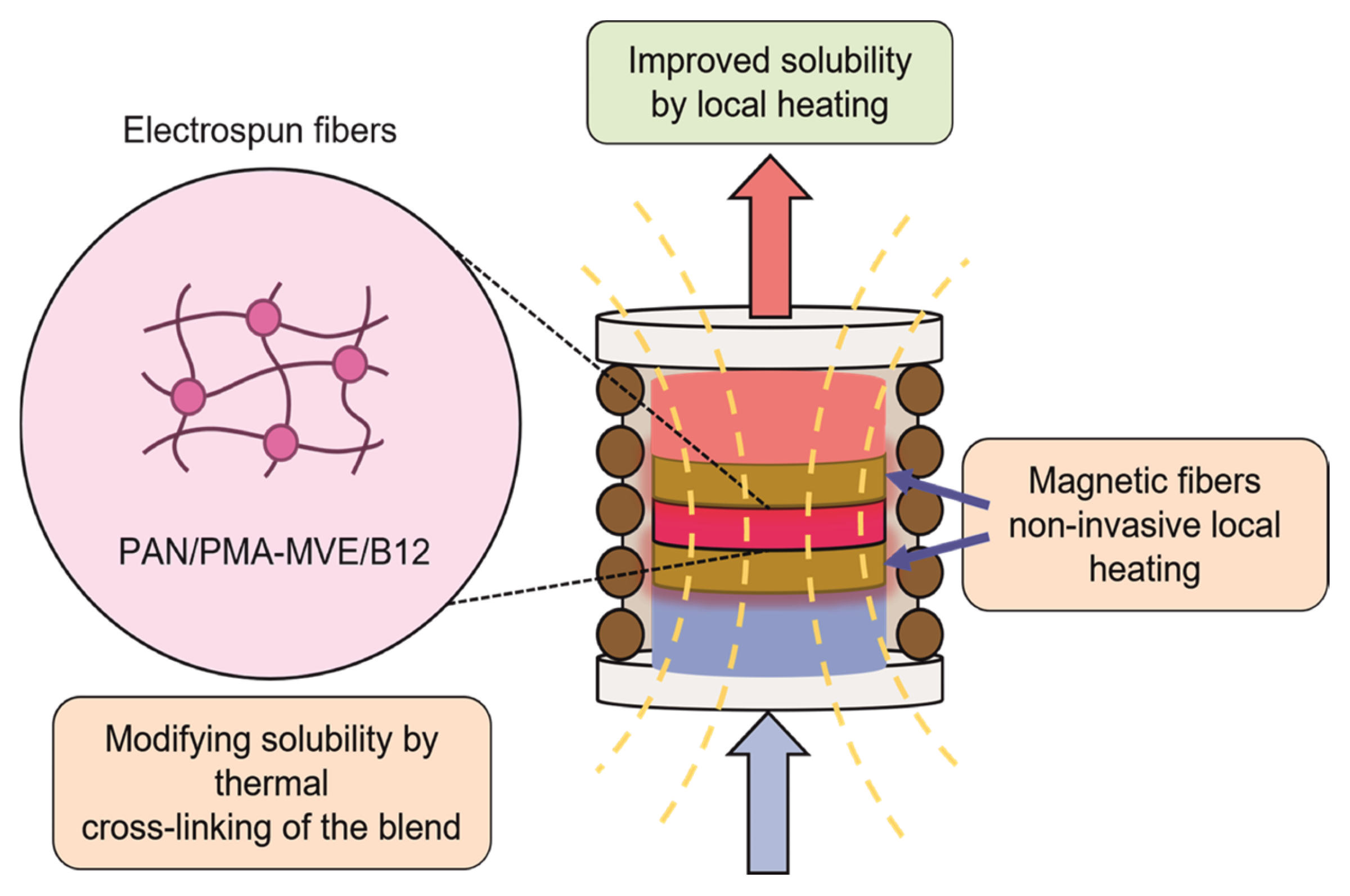
Figure 1 schematically illustrates the strategy for the fabrication and evaluation of the stimuli-responsive hybrid platform developed in this study. The polymer blend contains water-soluble PVM/MA and water-insoluble PAN, leveraging their capacity for thermal cross-linking to modulate the solubility and structural integrity of the matrix. This smart delivery platform is designed for the controlled release of vitamin B12 in aqueous media, utilizing alternating magnetic fields (AMF) to provide non-invasive inductive heating. The inductive heating activation facilitates the dissolution and diffusion of the encapsulated vitamin B12, enabling remote-controlled, on-demand drug release.

2.2.1. Polymer Blend Elaboration

For polymeric blend preparation, 0.4 g of PMA-MVE was dissolved in DMF (10 mL). Then, vitamin B12 (10 mg) was added, and the suspension was heated to 60 °C. Later, PAN (1 g) was dissolved in the solution and stirred for 2 h, after which the heating was turned off. The blend was continuously stirred for 12 h to promote entanglement of the PAN polymeric chains and electrostatic interactions between carboxylate anions from PMA-MVE and amine groups from vitamin B12.

2.2.2. Electrospinning

The as-prepared solution was loaded into a 10 mL syringe and fed (0.7 mL/h) to the electrospinning setup using a rotative collector, working distance of 15 cm, and an applied high voltage of 8 kV. The obtained B12-NFs mat was dried (60 °C, 12 h) and prepared for further characterization and evaluation.

2.2.3. Thermal Treatment

Thermal crosslinking was studied at 90, 120, 160, and 180 °C, considering the well-known transition temperatures of PAN where the nitrile groups are available to interact (less than 220 °C) with the carboxylic acid groups (-COOH) in PMA-MVE through hydrogen bonding to entrap amine groups. The samples were labeled as B12-NFs-90, B12-NFs-120, B12-NFs-160, and B12-NFs-180, respectively.

2.3. Materials Characterization

2.3.1. Electron Microscopy

To determine the physical properties of the polymeric mats, electron microscopy was performed using a Thermo Fisher Scientific INSPECT-F50, Waltham, MA, USA Scanning Electron Microscope for imaging, along with energy-dispersive X-ray spectroscopy (EDS) for compositional analysis. Samples were coated with carbon after the analysis to ensure surface conductivity.

2.3.2. Thermal Analysis

Thermal analysis was performed using Differential Scanning Calorimetry (DSC) with a DSC822e Module (Mettler Toledo, Greifensee, Switzerland) from 50 to 500 °C at a heating rate of 1 °C/minute under N2 atmosphere. Thermo Gravimetric Analysis (TGA) was conducted using a Mettler Toledo TGA SDTA851 analyzer from 50 to 800 °C at a heating rate of 10 °C min−1 and an N2 purge of 60 mL min−1 in a ceramic pan.

2.3.3. FTIR Spectroscopy

Normal vibration modes of the functional groups in the samples were detected using Attenuated Total Reflectance Fourier Transform Infrared spectroscopy (ATR FT-IR); the spectra from 4000 to 600 cm−1 were acquired using a Bruker VERTEX 70v FT-IR Spectrometer (Bruker, Billerica, MA, USA).

2.3.4. Contact Angle Measurement

After the deposition of a drop (20 µL), digital images were taken (Dino-Lite AM3111 digital microscope, AnMo Electronics Co., Taipei, Taiwan), and the complementary angles between the drops and the surface of B12-NFs mats were measured using DINOCAPTURE 2.0 software.

2.4. Drug Release Platform Evaluation

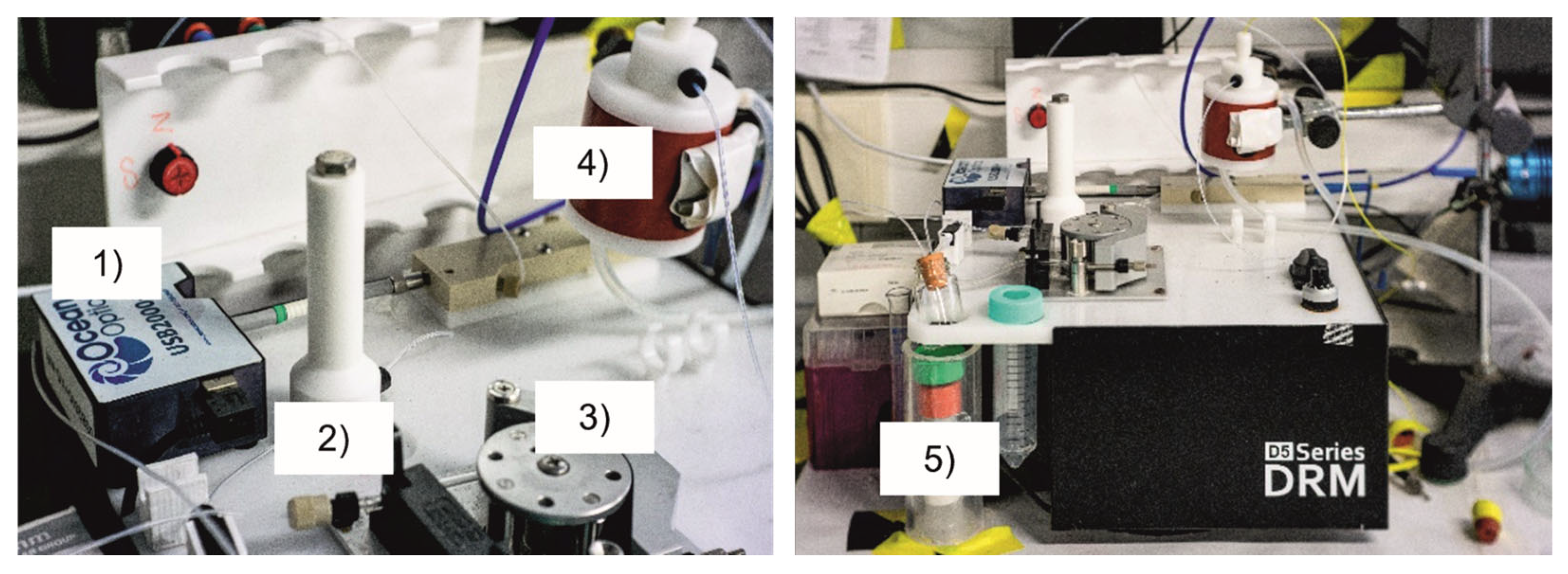
For the determination of the modifications in the drug release profiles promoted by the materials modifications using thermal treatment, the experimental setup presented in Figure 2 was employed to monitor the amount of released B12, using as a reference the characteristic 510 nm absorbance band over time, using the continuous system reported in [56] with Milli-Q water flowing in the system in all experiments.
To evaluate spontaneous release kinetics from the different B12-NFs samples, the profiles were evaluated at room temperature (20 °C) and 30 °C, controlling the temperature in the sample chamber using recirculating water flow in a thermalized bath. Three disks of 10 mm diameter, trimmed before thermal treatment, were used. The disks were placed in a sample holder for the evaluation of the release profiles. Magnetically triggered release kinetics of samples B12-NFs-90 to B12-NFs-180 were evaluated under stimulation by an alternating magnetic field (AMF, f = 366 kHz, 350 G), where the three disks of B12-NFs were placed between two inductive heating electrospun magnetic nanofibers, elaborated as reported in [56]. After 10 min of spontaneous release, the samples were stimulated with an AMF and evaluated. Once the steady state was reached, the AMF was applied again, and the release profile curves were analyzed. This experiment was conceived to demonstrate the capacity of the B12-NFs to modify the release profile as a consequence of localized inductive heating.
The experimental release profiles were fitted using the Gompertz dissolution model (Equation 1), and the temperature impact on the delivery kinetics was determined by comparing the release rate (k), according to Equation (1) [57,58]:
y = A e e k ( t t c )
where the parameters are defined as follows:
  • y = concentration reached at time t [mg/mL].
  • A = maximum released concentration [mg/mL].
  • k = release rate constant [s1].
  • t = time [s].
  • tc = time to reach 63% of the release [s].

3. Results

3.1. B12-NFs Properties

The physical and chemical properties obtained from the B12-NFs characterization confirm the successful fabrication of drug-loaded fibers with controlled size and morphology. Visually, the electrospun fibers appeared as a homogeneous pink mat, resulting from the combination of the characteristic red color of vitamin B12 and the white hue of the polymeric blend.
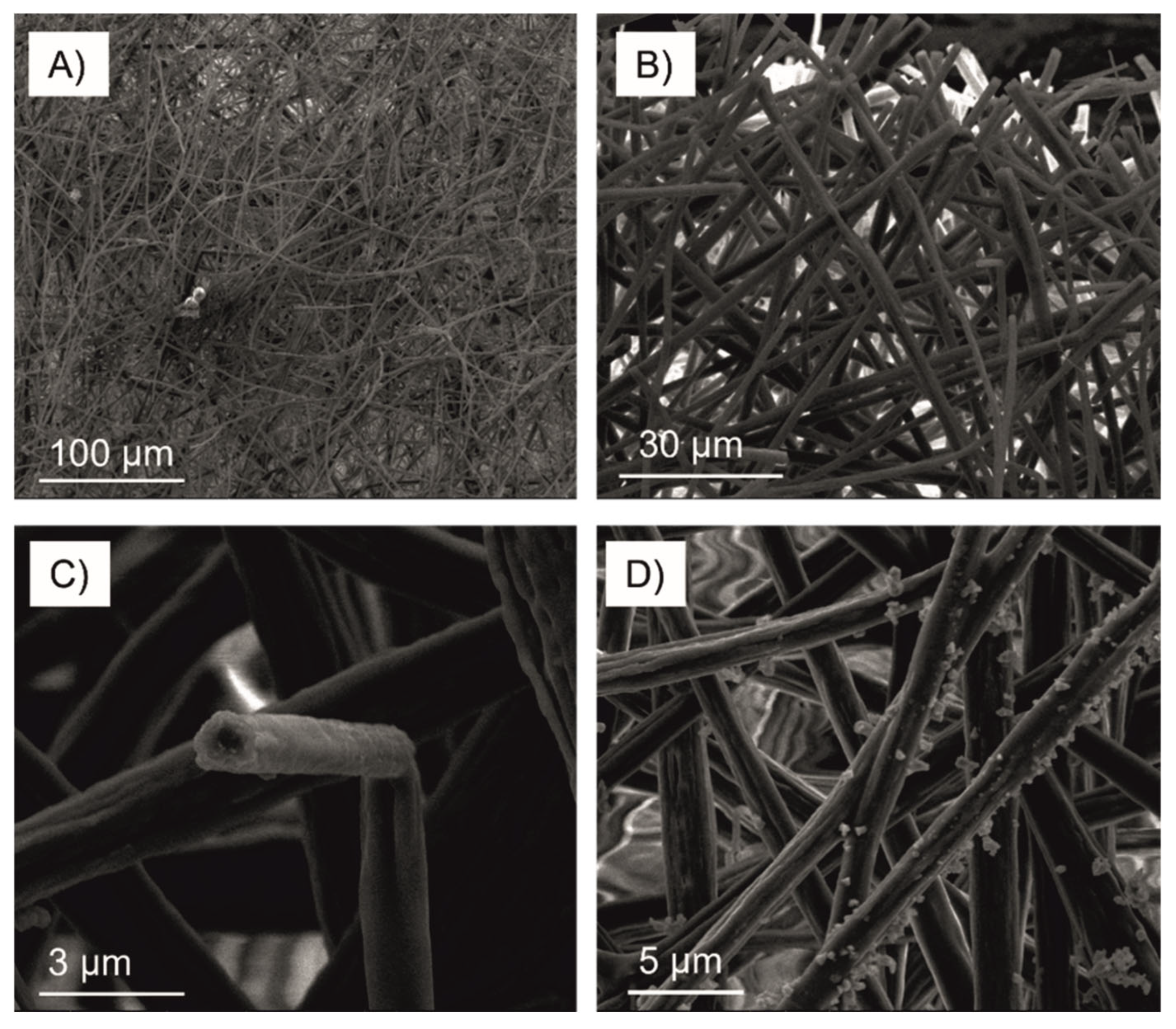
As shown in Figure 3A–C, the fibers were continuous, solid, and rough, with an average diameter of approximately 1.50 ± 0.35 µm. The electron microscopy images (Figure 3D) revealed compact, randomly oriented B12-NFs with a textured surface, suggesting that vitamin B12 was distributed both within and on the surface of the fibers. This highlights the successful integration of the drug into the nanofiber matrix.
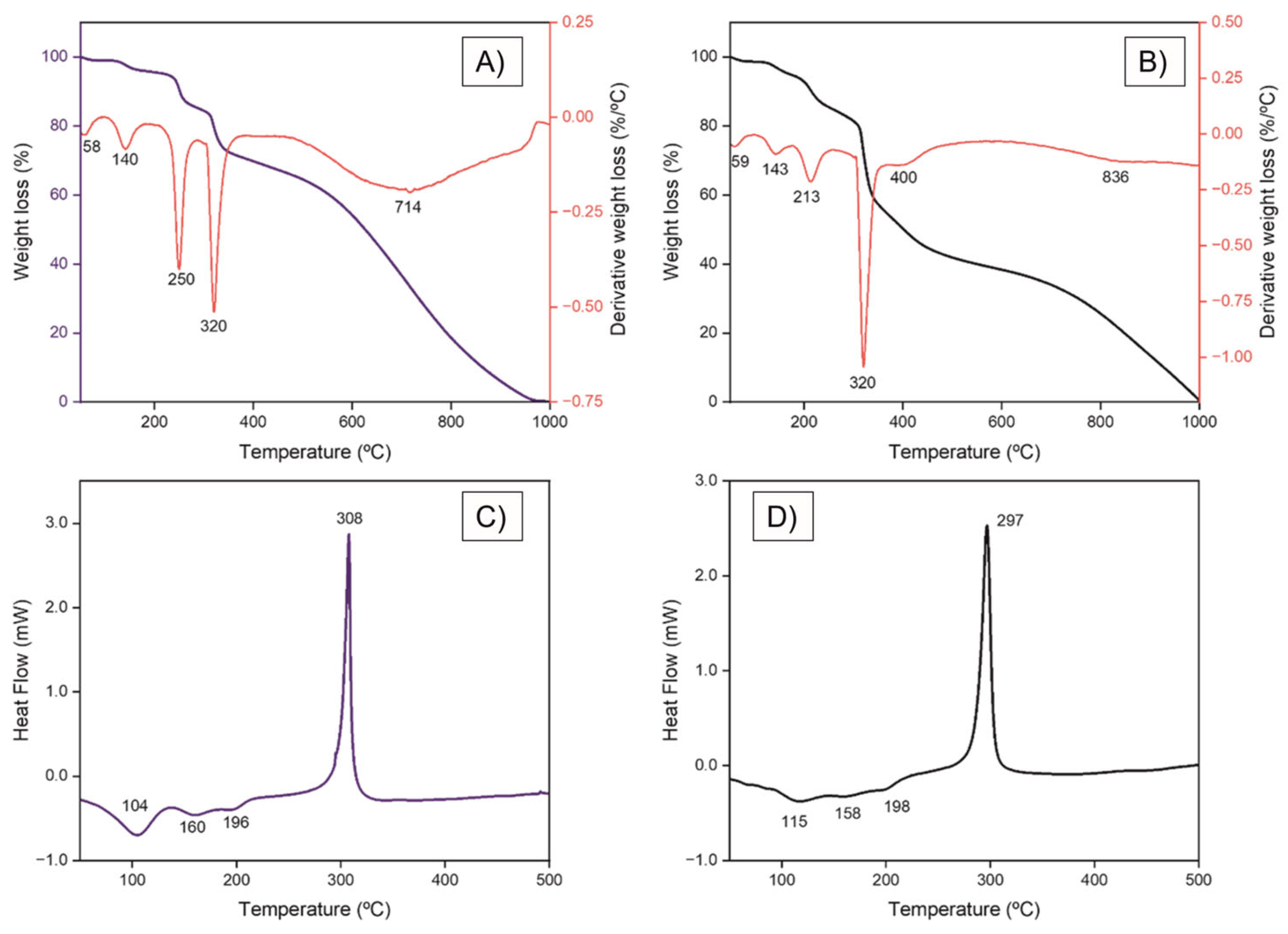
Thermal analysis results were summarized in Figure 4. TGA thermograms revealed slight differences between blank electrospun fibers (PMA-MVE/PAN) and the B12-NFs (Figure 4A,C). Figure 4B,D show the DSC thermograms, where the thermal transitions were slightly shifted. The electrospun PMA-MVE/PAN/B12 fibers thermograms show weight loss ( 10%) at 213 °C and a higher glass transition temperature (Tg) compared with the blank sample, suggesting interactions among the components as temperature increases, but associated with B12 degradation, which was not found up to 200 °C. These results allow us to conclude that below 200 °C, the samples exhibited modifications related to the polymeric blend, ensuring that thermal treatment within the working interval (90–180 °C) does not degrade the B12 molecule.
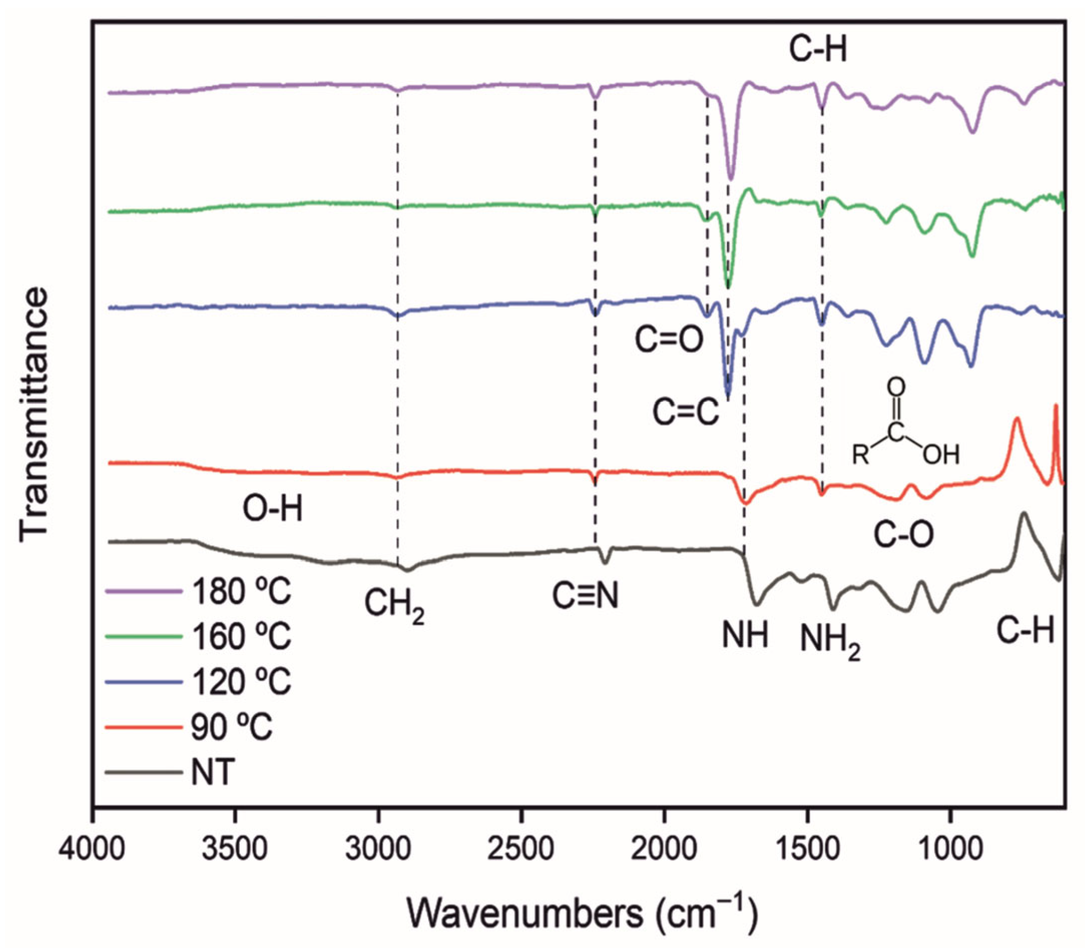
The normal vibration modes of the molecules that compose the fabricated materials were analyzed after the thermal treatment at 90, 120, 160, and 180 °C, and compared to non-treated samples (NT). As shown in Figure 5, the FTIR spectra revealed interactions of the main functional groups of the different samples. The spectral modifications are associated with conformational modifications of the polymeric chains, mainly attributed to chain reorganization produced by the nitrile group volatilization [59].
Table 1 summarizes the characteristic vibrational modes identified in the FTIR spectra of the analyzed samples. The bands corresponding to the amine functional groups of the B12 molecule (at ~1700 and 1550 cm−1) appeared more intense and better resolved compared to those observed in the composite samples (Figure 5). This difference may be attributed to modifications in the polymeric matrix, which can restrict the mobility and accessibility of the amine groups at the surface. Such constraints likely alter the vibrational degrees of freedom of the B12 molecule, resulting in band shifts or intensity changes within the complex system.
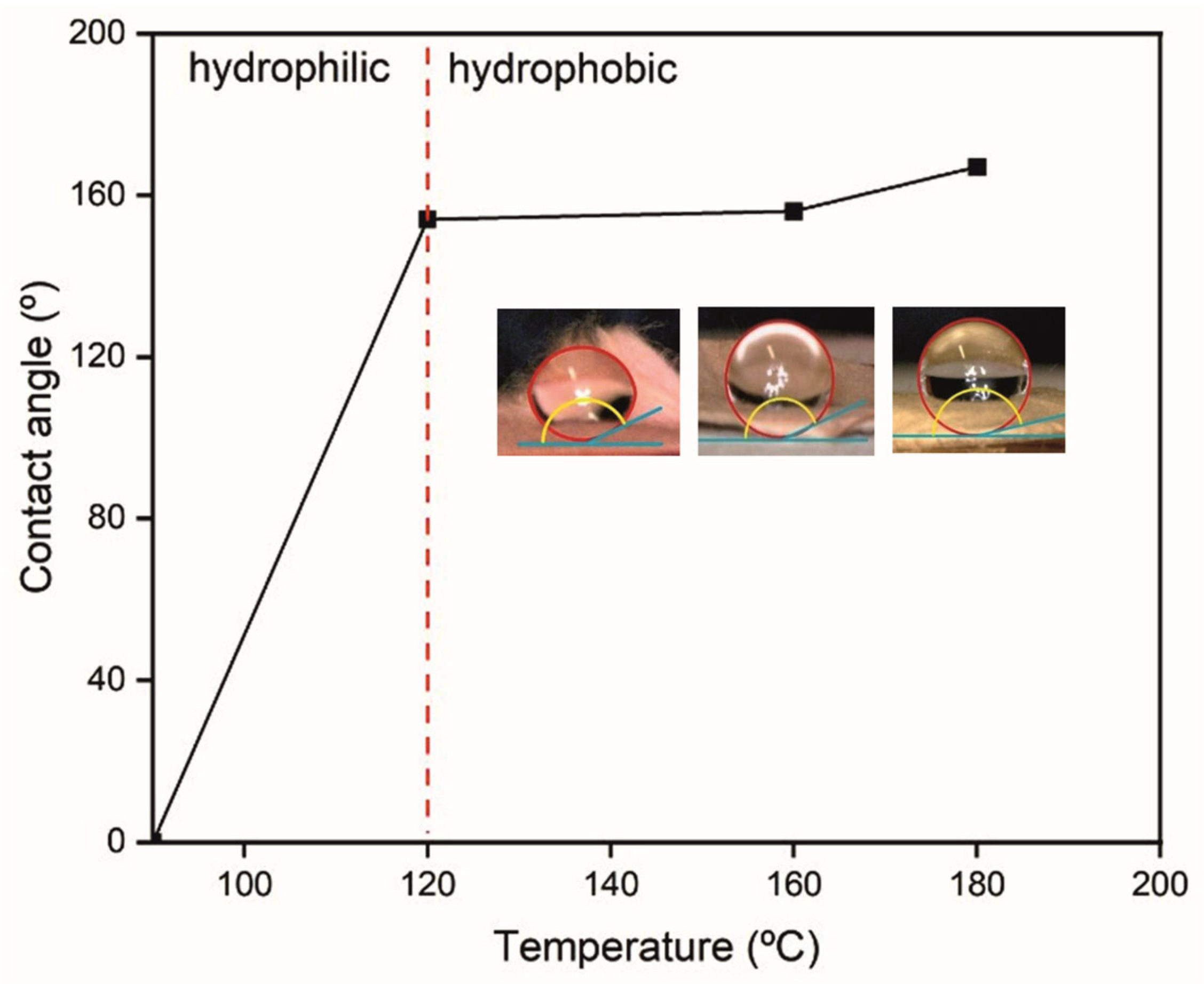
The effects of thermal treatment on the wettability of the produced B12-NFs were evaluated by analyzing the contact angle. The evolution of the contact angle as a function of increasing thermal treatment temperature is shown in Figure 6. Superhydrophobic behavior (contact angle > 150°) was observed in samples treated above 120 °C; in contrast, for the samples treated at 90 °C, it was not possible to register the contact angle due to immediate wetting.

3.2. Drug Release Response

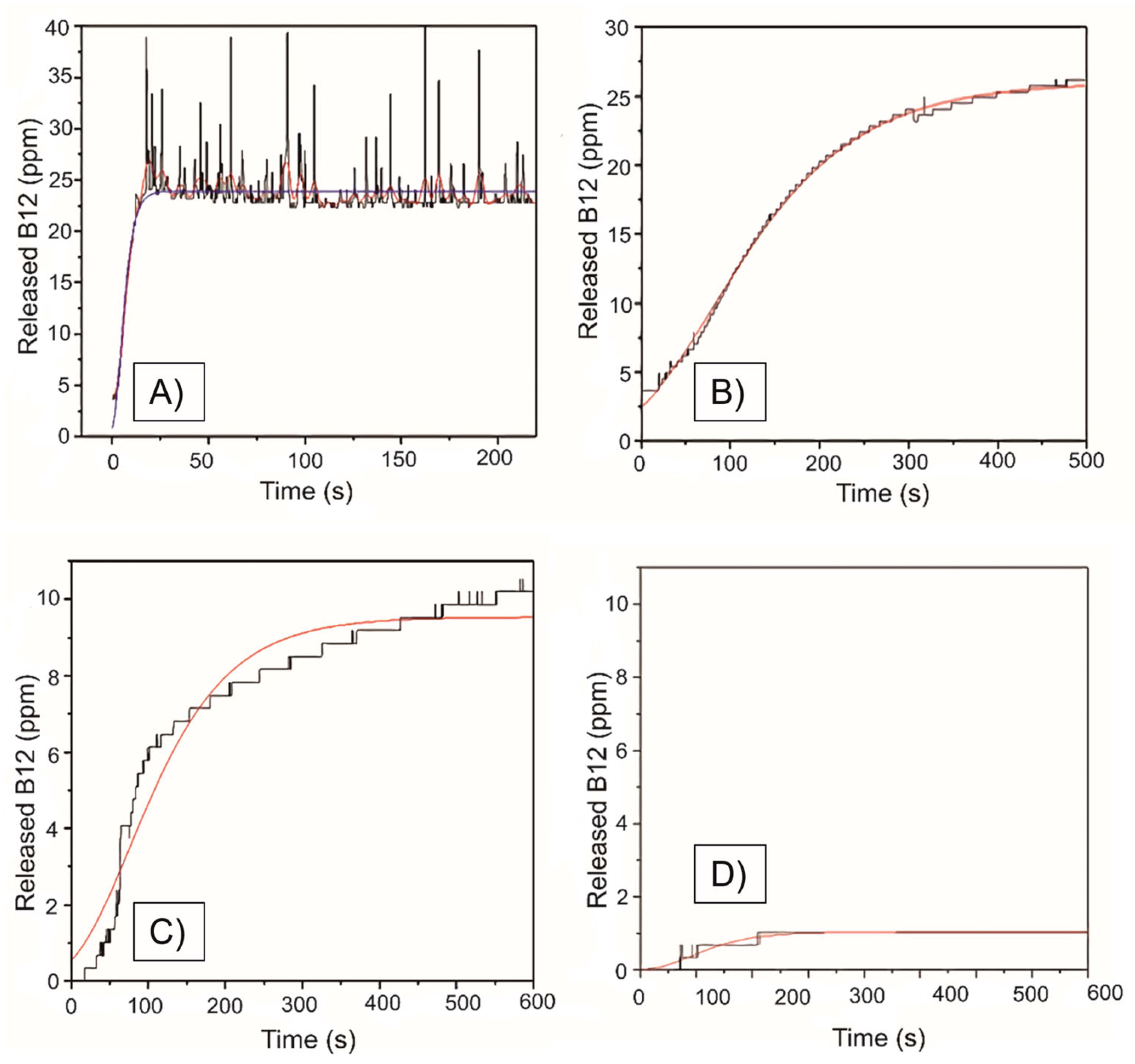
Figure 7 shows representative release profiles from the evaluated samples at room temperature (20 °C). The profile from the sample treated at 90 °C (Figure 7A) shows a steady state after 25 s, while the hydrophobic samples (Figure 7B–D) released less B12 and increased time for the steady state. Reduced wettability may limit interactions between the liquid carrier (Milli-Q water) and the B12-NFs, thereby decreasing the system’s ability to release the molecule. This suggests that the dissolution profile of the samples is modified as a function of thermal treatment.
The observed spontaneous release profiles were adjusted to the Gompertz dissolution model (Figure 7, red lines). The triplicated measurements results showed good fitting with the selected model, and the summary of parameters as a result of the fitting can be observed in Table 2. As can be observed, the experiment for the 180 °C samples was performed only once as a reference for this thermal treatment temperature. This is because the release was near the detection limits of the equipment, and the temperature was close to the degradation of B12. However, the release profile from 180 °C samples still has a good fit with the Gompertz profile (R2 = 0.89397).
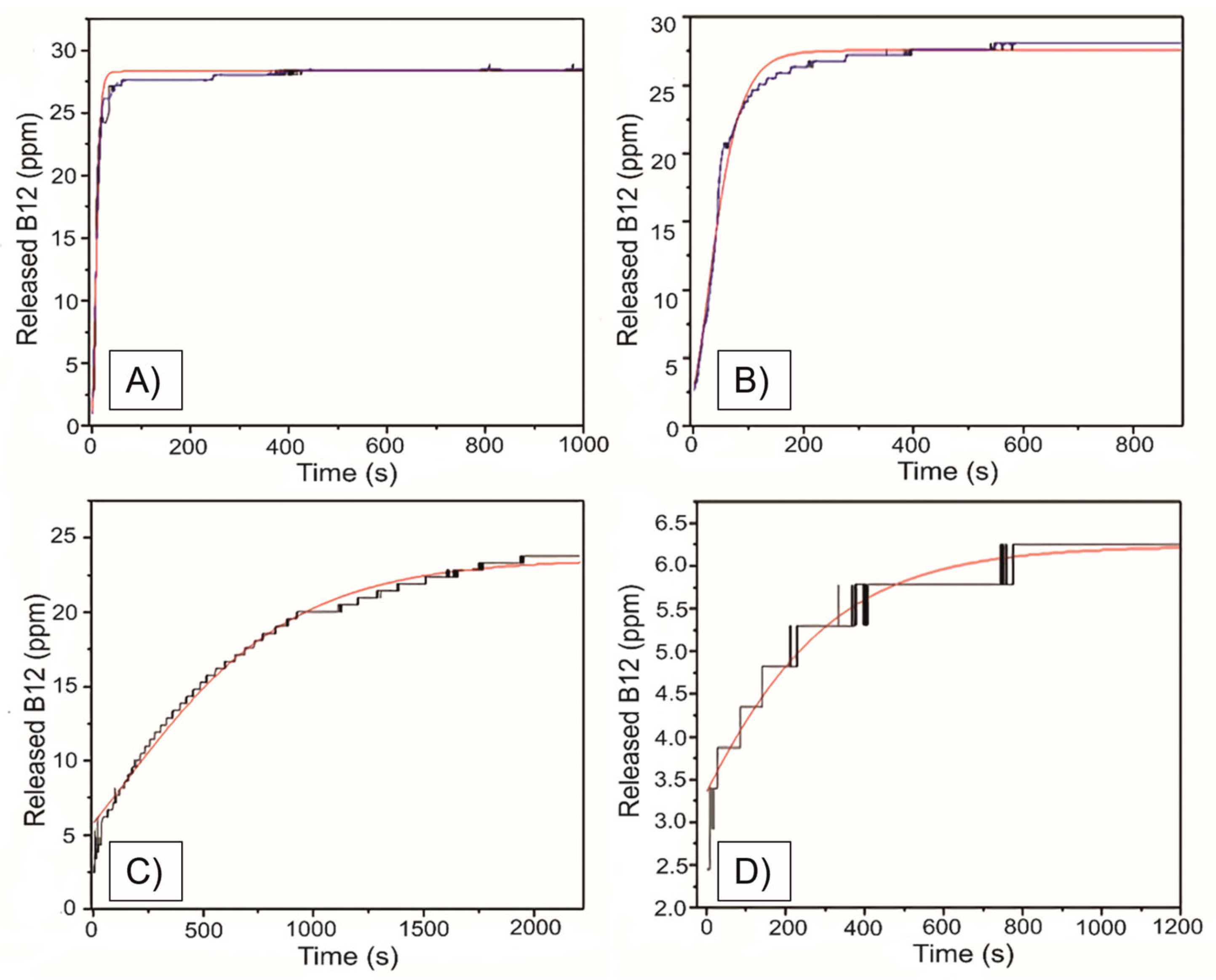
The water bath temperature was subsequently raised to maintain 30 °C within the sample holder (Figure 2 and Figure 4), allowing for the evaluation of the thermal sensitivity of the spontaneous release. Representative release profiles from the B12-NFs (90, 120, 160, and 180 °C) samples are presented in Figure 8A–D, and the Gompertz fitting is represented by red lines. It can be observed that all the profiles showed superior release capacity from B12-NFs evaluated at 30 °C, compared with 20 °C (Figure 7). These results suggest a dependency of the capacity to deliver B12 on the temperature of the surrounding media.
The fitting parameters for the Gompertz dissolution model for the obtained release profiles are summarized in Table 3. Variability of the results can be attributed to the changes in the mass of the samples evaluated. The three disks employed can have variability in weight and B12 loading. On the other hand, the dependence of the release rate on the increase in temperature can be observed in the differences in the release rate (k) between the samples evaluated at 30 °C and those evaluated at 20 °C. The obtained values of R2 for fitting are superior as well, supporting the hypothesis of dissolution mechanism in the release response of the elaborated materials.
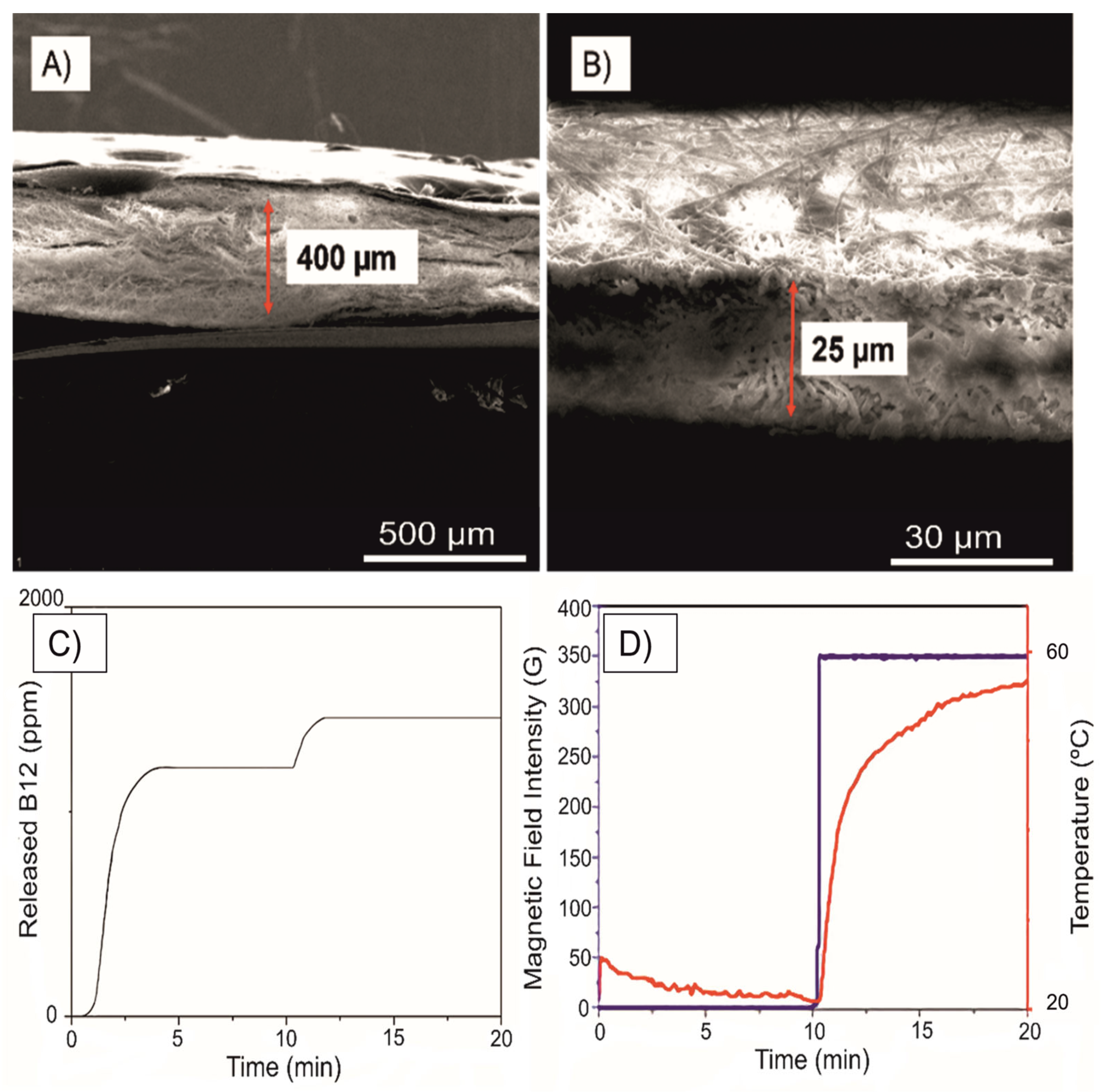
SEM images of the membranes used to evaluate the magnetically triggered drug release platform are shown in Figure 9. B12-NFs (Figure 9A) were placed between compacted magnetic fibers (PAN/FeMnO), which served as an inductive heating source, and were stimulated using an alternating magnetic field (AMF) at 28 kA/m (≈350 G). The representative drug release response from the evaluated B12-NFs is presented in Figure 9C, where the influence of the magnetic stimulus on the release profile can be observed. AMF was activated after 10 min of spontaneous release, upon reaching steady state. Figure 9D displays the temperature increase induced by AMF activation, confirming the localized heating effect of the magnetic platform.
The magnetically triggered response of the materials is summarized in Table 4. The obtained results correspond to the release after the steady state was reached after 10 min (Figure 9C). Once the spontaneous release was constant, the AMF was switched on, and the modifications to the release profile were recorded using the continuous flow UV-Vis monitor. The platform showed improved release rates compared with 30 °C and 20 °C. The maximum release concentration (A) and the release rate (k) increase significantly.

4. Discussion

The observed surface features suggest that the critical solubility point of vitamin B12 in the polymeric precursor solution was not reached during the fabrication. The solubility of vitamin B12 in the precursor solution plays a crucial role in determining its homogeneity, drug loading efficiency, and the final morphology of the nanofibers. Exceeding the solubility threshold could result in phase separation or surface aggregation, potentially contributing to the decorated-like texture observed in the microscopy images.
For the drug-loading process in the B12-NFs system, both maleic acid and methyl vinyl ether units in PMA-MVE can engage in hydrogen bonding interactions with DMF molecules. This interaction can further enhance the solubility of the polymer and the potential interaction with amine-based drugs. When PMA-MVE is dissolved in a basic solvent, the protonation state of its carboxylic acid groups can change, leading to alterations in solubility, conformation, and potentially, chemical reactivity.
Additionally, carboxylate anions (COO) and amine groups (NH2) can form hydrogen bonds with each other. The oxygen atom in the carboxylate group carries a partial negative charge, while the nitrogen atom in the amine group carries a partial positive charge. This charge separation allows for hydrogen bonding between the two functional groups. The hydrogen bond formation enhances the stability of the interaction between carboxylate anions and amine groups.
Carboxylate anions and amine groups may interact through ion–dipole interactions. The ionic nature of the carboxylate and the polarity of the amine nitrogen create a strong electrostatic attraction between them. This interaction is like the attraction between ions and polar molecules in solution, ensuring the B12 encapsulation and integration on the fibers.
Thermal treatment is a straightforward strategy to control the drug release capacities of B12-NFs. By controlling the crosslinking temperature, material properties such as hydrophilicity, swelling behavior, and degradation rate can be tailored to suit specific applications. Temperature affects the reaction rate of the crosslinking reaction, influences the degree and uniformity of crosslinking, which determines the properties of the final polymer network. Low temperature may lead to incomplete crosslinking or irregular network formation, affecting the mechanical strength and stability of the B12-NFs. However, excessive temperature can cause over-crosslinking, leading to brittleness or degradation of the B12-NFs.
Selecting the right temperature prevents thermal degradation of the polymer or drug embedded in the fibers. It also ensures the formation of a well-organized and homogeneous crosslinked network, balancing flexibility and rigidity. Avoiding elevated temperatures helps to remove residual solvents or byproducts from the crosslinking reaction, ensuring a clean and stable network. According to the FT-IR spectra (Figure 3), the band at 1450 cm−1 is associated with CH2 bending (scissoring), and at 1170 cm−1, it is associated with the unsaturated esters and carboxylic acids [60,61]. It is observed that the vibrations corresponding to the -OH groups only appear in the membranes with a temperature treatment lower than the boiling temperature of water. The vibrational modes of the functional groups listed in Table 1 represent the main interactions of the polymer chain. In the sharp peak centered at 2250 cm−1 corresponding to the nitrile group (C≡N), there are no significant changes, but the rest of the band loses intensity as the treatment temperature increases. As the temperature increases in the heat treatment, the nitrile groups begin to dissociate, which initiates the cross-linking process.
Almost all the bands that appear within the 1800–600 cm−1 interval are due to the functional groups provided by vitamin B12, being able to identify both primary and secondary amines [62] (Table 1). However, it is important to highlight the C=C interactions that are observed centered at 1750 and 1725 cm−1, which is proof that the polymer chains undergo conformational changes following treatment at 120 °C. These conformational changes directly impact the physicochemical properties of the material. With this characterization, it is possible to predict more evident changes in the behavior of the material after 120 °C of heat treatment.
The samples subjected to the highest temperature (B12-NFs180) were the most hydrophobic, with a contact angle of 167 °C. All of them can be considered superhydrophobic, surpassing the superhydrophobic threshold (150°) with 154° for B12-NFs120 and 157° for B12-NFs160, respectively. In the case of the membrane being treated at 90 °C, since the water drop was expanded instantly and wetted all the mat, it was classified as hydrophilic. The thermal treatment of B12-NFs leads to cross-linking via inter-macromolecular esterification and anhydride formation controlled by temperature and thermal treatment time.
The cross-linked B12-NFs materials are able to deliver vitamin B12 in water, and the release rate can be modified by temperature and magnetic field stimulation using magnetic fibers. The thermal treatment of the prepared B12-NFs determines the fiber’s mat properties, while the heating stimulus on the evaluated B12-NFs in water promotes release enhancement. The elaborated platform showed evidence of modified release profiles using the intrinsic properties (cross-linking) of the materials and the magnetic stimulus (frequency and intensity) as control parameters.
Once a steady state of the spontaneous release was ensured (10 min), the magnetic stimulus was applied to increase the local temperature. The observed behavior described in Figure 9 is associated with an increment in the release rate because of the enhanced dissolution of the B12-NFs once the magnetic stimulation is applied.
From the comparison of Table 4, it is possible to establish that the involved mechanism is the dissolution of the fibers, supported by the observed Gompertz response, associated with this phenomenon. Higher cross-link temperature promotes stronger chemical interactions that impact the degree of solubility of the B12-NFs and the observed spontaneous release. The AMF, as a non-invasive stimulus, is capable of modifying the release profiles from the B12-NFs, modifying the solubility based on the local heating.
While the current study successfully establishes the fundamental release mechanisms of the [PMA-MVE/PAN] matrix under controlled laboratory conditions, it is important to acknowledge the limitations of using Milli-Q water as the release medium. In a biological environment, physiological factors such as ionic strength and pH fluctuations (e.g., in Phosphate-Buffered Saline, pH 7.4) are expected to influence the electrostatic interactions between the polymer network and the therapeutic cargo. Furthermore, the inevitable formation of protein corona upon exposure to biological fluids may alter the surface porosity and diffusion pathways of the nanofibers, potentially modulating the release kinetics in vivo. Consequently, the results presented here serve as a necessary physicochemical baseline, providing the structural and thermal evidence required to advance toward future evaluations in simulated physiological media and complex biological environments.
The future outlook for magnetically triggered electrospun platforms lies in the transition from proof-of-concept to in vitro and in vivo evaluation within living systems. While this study establishes the fundamental sensitivity of B12-NFs to inductive heating, future research should focus on the implementation of Curie-limited nanoparticles to ensure intrinsic thermal safety at physiological thresholds (41–45 °C). Additionally, the AMF conditions (intensity and frequency) play an important role in the control of the temperature, opening the gates for multiparametric control of the spatiotemporal molecule release. Furthermore, the integration of these hybrid fibers into theranostic frameworks could revolutionize personalized medicine, as the magnetic signal serves as both a release trigger and a contrast agent for real-time imaging. Addressing the long-term biocompatibility and the influence of the protein corona in complex media will be the final steps in translating these non-invasive, remote-controlled scaffolds from the laboratory to clinical implementation.

5. Conclusions

This study successfully demonstrates a hybrid electrospun platform capable of precisely modulating the release of a model drug through thermal crosslinking and remote magnetic induction. The thermal processing temperature was identified as a critical parameter in governing the release kinetics; higher temperatures fostered a more compact and hydrophobic matrix, effectively suppressing spontaneous drug leakage. Under magnetic actuation, the system exhibited a remarkable transition from passive to active delivery. Notably, for the B12-NFs treated at 90 °C, the application of an alternating magnetic field (AMF) increased the release rate constant from 0.346 s−1 to 1.283 s−1, while the maximum concentration reached 1.875 mg/mL, representing more than a 70-fold enhancement compared to passive diffusion, 0.026 mg/mL. These results confirm that inductive heating can effectively overcome diffusion barriers within the nanofiber matrix, restoring high release rates on demand. These results establish a robust physicochemical framework for the development of smart scaffolds. The ability to tune the thermal response via magnetic field parameters offers a promising strategy for future therapeutic systems requiring high spatial and temporal control over dosage. Further studies in physiological media will be essential to translate this proof-of-concept into clinical applications.

Author Contributions

Conceptualization: J.A.F.-G.; methodology: S.H.-C.; software: S.H.-C.; validation: J.A.F.-G.; formal analysis: S.H.-C. and J.A.F.-G.; investigation: S.H.-C.; resources: S.H.-C.; data curation: S.H.-C. and J.A.F.-G.; writing—original draft preparation: S.H.-C. and J.A.F.-G.; writing—review and editing: J.A.F.-G.; visualization: J.A.F.-G.; supervision: J.A.F.-G. All authors have read and agreed to the published version of the manuscript.

Funding

This research received no external funding.

Data Availability Statement

The raw data supporting the conclusions of this article will be made available by the authors on request.

Acknowledgments

The authors would like to acknowledge the use of Servicio General de Apoyo a la Investigación-SAI, Universidad de Zaragoza, and Gerardo Goya for the support in the development of the experimental part of this report.

Conflicts of Interest

The authors declare no conflicts of interest.

References

  1. Placha, D.; Jampilek, J. Chronic inflammatory diseases, anti-inflammatory agents and their delivery nanosystems. Pharmaceutics 2021, 13, 64. [Google Scholar] [CrossRef]
  2. Liu, J.; Ting, J.P.; Al-Azzam, S.; Ding, Y.; Afshar, S. Therapeutic advances in diabetes, autoimmune, and neurological diseases. Int. J. Mol. Sci. 2021, 22, 2805. [Google Scholar] [CrossRef] [PubMed]
  3. Morgan, B.P.; Harris, C.L. Complement, a target for therapy in inflammatory and degenerative diseases. Nat. Rev. Drug Discov. 2015, 14, 857–877. [Google Scholar] [CrossRef] [PubMed]
  4. Chakraborty, P.; Aravindhan, V.; Mukherjee, S. Helminth-derived biomacromolecules as therapeutic agents for treating inflammatory and infectious diseases: What lessons do we get from recent findings? Int. J. Biol. Macromol. 2023, 241, 124649. [Google Scholar] [CrossRef] [PubMed]
  5. Osman, N.; Devnarain, N.; Omolo, C.A.; Fasiku, V.; Jaglal, Y.; Govender, T. Surface modification of nano-drug delivery systems for enhancing antibiotic delivery and activity. Wiley Interdiscip. Rev. Nanomed. Nanobiotechnol. 2022, 14, e1758. [Google Scholar] [CrossRef]
  6. Sultana, A.; Zare, M.; Thomas, V.; Kumar, T.S.; Ramakrishna, S. Nano-based drug delivery systems: Conventional drug delivery routes, recent developments and future prospects. Med. Drug Discov. 2022, 15, 100134. [Google Scholar] [CrossRef]
  7. Cheng, X.; Xie, Q.; Sun, Y. Advances in nanomaterial-based targeted drug delivery systems. Front. Bioeng. Biotechnol. 2023, 11, 1177151. [Google Scholar] [CrossRef]
  8. Chandrakala, V.; Aruna, V.; Angajala, G. Review on metal nanoparticles as nanocarriers: Current challenges and perspectives in drug delivery systems. Emerg. Mater. 2022, 5, 1593–1615. [Google Scholar] [CrossRef]
  9. Ussia, M.; Privitera, V.; Scalese, S. Unlocking the potential and versatility of quantum dots: From biomedical to environmental applications and smart micro/nanorobots. Adv. Mater. Interfaces 2024, 11, 2300970. [Google Scholar] [CrossRef]
  10. Alshammari, B.H.; Lashin, M.M.; Mahmood, M.A.; Al-Mubaddel, F.S.; Ilyas, N.; Rahman, N.; Sohail, M.; Khan, A.; Abdullaev, S.S.; Khan, R. Organic and inorganic nanomaterials: Fabrication, properties and applications. RSC Adv. 2023, 13, 13735–13785. [Google Scholar] [CrossRef]
  11. Zou, Y.; Huang, B.; Cao, L.; Deng, Y.; Su, J. Tailored mesoporous inorganic biomaterials: Assembly, functionalization, and drug delivery engineering. Adv. Mater. 2021, 33, 2005215. [Google Scholar] [CrossRef]
  12. Wang, X.; Zhong, X.; Li, J.; Liu, Z.; Cheng, L. Inorganic nanomaterials with rapid clearance for biomedical applications. Chem. Soc. Rev. 2021, 50, 8669–8742. [Google Scholar] [CrossRef] [PubMed]
  13. Tören, E.; Buzgo, M.; Mazari, A.A.; Khan, M.Z. Recent advances in biopolymer based electrospun nanomaterials for drug delivery systems. Polym. Adv. Technol. 2024, 35, e6309. [Google Scholar] [CrossRef]
  14. Jadhav, V.; Roy, A.; Kaur, K.; Rai, A.K.; Rustagi, S. Recent advances in nanomaterial-based drug delivery systems. Nano-Struct. Nano-Objects 2024, 37, 101103. [Google Scholar] [CrossRef]
  15. Sahu, T.; Ratre, Y.K.; Chauhan, S.; Bhaskar, L.V.K.S.; Nair, M.P.; Verma, H.K. Nanotechnology based drug delivery system: Current strategies and emerging therapeutic potential for medical science. J. Drug Deliv. Sci. Technol. 2021, 63, 102487. [Google Scholar] [CrossRef]
  16. Zhang, M.; Hu, W.; Cai, C.; Wu, Y.; Li, J.; Dong, S. Advanced application of stimuli-responsive drug delivery system for inflammatory arthritis treatment. Mater. Today Bio 2022, 14, 100223. [Google Scholar] [CrossRef]
  17. Liu, X.; Wu, Z.; Guo, C.; Guo, H.; Su, Y.; Chen, Q.; Sun, C.; Liu, Q.; Chen, D.; Mu, H. Hypoxia responsive nano-drug delivery system based on angelica polysaccharide for liver cancer therapy. Drug Deliv. 2022, 29, 138–148. [Google Scholar] [CrossRef]
  18. Ghalkhani, M.; Kaya, S.I.; Bakirhan, N.K.; Ozkan, Y.; Ozkan, S.A. Application of nanomaterials in development of electrochemical sensors and drug delivery systems for anticancer drugs and cancer biomarkers. Crit. Rev. Anal. Chem. 2022, 52, 481–503. [Google Scholar] [CrossRef]
  19. Lv, Y.; Li, W.; Liao, W.; Jiang, H.; Liu, Y.; Cao, J.; Lu, W.; Feng, Y. Nano-drug delivery systems based on natural products. Int. J. Nanomed. 2024, 19, 541–569. [Google Scholar] [CrossRef]
  20. Patel, P.R.; Gundloori, R.V.N. A review on electrospun nanofibers for multiple biomedical applications. Polym. Adv. Technol. 2023, 34, 44–63. [Google Scholar] [CrossRef]
  21. El-Seedi, H.R.; Said, N.S.; Yosri, N.; Hawash, H.B.; El-Sherif, D.M.; Abouzid, M.; Abdel-Daim, M.M.; Yaseen, M.; Omar, H.; Shou, Q.; et al. Gelatin nanofibers: Recent insights in synthesis, bio-medical applications and limitations. Heliyon 2023, 9, e16228. [Google Scholar] [CrossRef] [PubMed]
  22. Fu, L.; Feng, Q.; Chen, Y.; Fu, J.; Zhou, X.; He, C. Nanofibers for the immunoregulation in biomedical applications. Adv. Fiber Mater. 2022, 4, 1334–1356. [Google Scholar] [CrossRef]
  23. Yan, B.; Zhang, Y.; Li, Z.; Zhou, P.; Mao, Y. Electrospun nanofibrous membrane for biomedical application. SN Appl. Sci. 2022, 4, 172. [Google Scholar] [CrossRef] [PubMed]
  24. Nirwan, V.P.; Kowalczyk, T.; Bar, J.; Buzgo, M.; Filová, E.; Fahmi, A. Advances in electrospun hybrid nanofibers for biomedical applications. Nanomaterials 2022, 12, 1829. [Google Scholar] [CrossRef]
  25. Gavande, V.; Nagappan, S.; Seo, B.; Lee, W.K. A systematic review on green and natural polymeric nanofibers for biomedical applications. Int. J. Biol. Macromol. 2024, 262, 130135. [Google Scholar] [CrossRef]
  26. Min, H.Y.; Lee, H.Y. Molecular targeted therapy for anticancer treatment. Exp. Mol. Med. 2022, 54, 1670–1694. [Google Scholar] [CrossRef]
  27. Zhong, L.; Li, Y.; Xiong, L.; Wang, W.; Wu, M.; Yuan, T.; Yang, W.; Tian, C.; Miao, Z.; Wang, T.; et al. Small molecules in targeted cancer therapy: Advances, challenges, and future perspectives. Signal Transduct. Target. Ther. 2021, 6, 201. [Google Scholar] [CrossRef]
  28. Janczura, M.; Sip, S.; Cielecka-Piontek, J. The development of innovative dosage forms of the fixed-dose combination of active pharmaceutical ingredients. Pharmaceutics 2022, 14, 834. [Google Scholar] [CrossRef]
  29. Xu, Z.; Dong, Q.; Li, W. Architectural design of block copolymers. Macromolecules 2024, 57, 1869–1884. [Google Scholar] [CrossRef]
  30. Liao, S.; Li, B.; Ma, Z.; Wei, H.; Chan, C.; Ramakrishna, S. Biomimetic electrospun nanofibers for tissue regeneration. Biomed. Mater. 2006, 1, R45. [Google Scholar] [CrossRef]
  31. Jiang, T.; Carbone, E.J.; Lo, K.W.H.; Laurencin, C.T. Electrospinning of polymer nanofibers for tissue regeneration. Prog. Polym. Sci. 2015, 46, 1–24. [Google Scholar] [CrossRef]
  32. Kong, B.; Liu, R.; Guo, J.; Lu, L.; Zhou, Q.; Zhao, Y. Tailoring micro/nano-fibers for biomedical applications. Bioact. Mater. 2023, 19, 328–347. [Google Scholar] [CrossRef] [PubMed]
  33. Bognitzki, M.; Frese, T.; Steinhart, M.; Greiner, A.; Wendorff, J.H.; Schaper, A.; Hellwig, M. Preparation of fibers with nanoscaled morphologies: Electrospinning of polymer blends. Polym. Eng. Sci. 2001, 41, 982–989. [Google Scholar] [CrossRef]
  34. Nagam Hanumantharao, S.; Rao, S. Multi-functional electrospun nanofibers from polymer blends for scaffold tissue engineering. Fibers 2019, 7, 66. [Google Scholar] [CrossRef]
  35. Luraghi, A.; Peri, F.; Moroni, L. Electrospinning for drug delivery applications: A review. J. Control. Release 2021, 334, 463–484. [Google Scholar] [CrossRef]
  36. Martínez-Pérez, C.A. Electrospinning: A promising technique for drug delivery systems. Rev. Adv. Mater. Sci. 2020, 59, 441–454. [Google Scholar] [CrossRef]
  37. Yoo, H.S.; Kim, T.G.; Park, T.G. Surface-functionalized electrospun nanofibers for tissue engineering and drug delivery. Adv. Drug Deliv. Rev. 2009, 61, 1033–1042. [Google Scholar] [CrossRef]
  38. Duan, X.; Chen, H.L.; Guo, C. Polymeric nanofibers for drug delivery applications: A recent review. J. Mater. Sci. Mater. Med. 2022, 33, 78. [Google Scholar] [CrossRef]
  39. Zheng, X.; Jin, Y.; Liu, X.; Liu, T.; Wang, W.; Yu, H. Photoactivatable nanogenerators of reactive species for cancer therapy. Bioact. Mater. 2021, 6, 4301–4318. [Google Scholar] [CrossRef]
  40. Clement, S.; Campbell, J.M.; Deng, W.; Guller, A.; Nisar, S.; Liu, G.; Wilson, B.C.; Goldys, E.M. Mechanisms for tuning engineered nanomaterials to enhance radiation therapy of cancer. Adv. Sci. 2020, 7, 2003584. [Google Scholar] [CrossRef]
  41. Cao, Z.; Li, D.; Wang, J.; Yang, X. Reactive oxygen species-sensitive polymeric nanocarriers for synergistic cancer therapy. Acta Biomater. 2021, 130, 17–31. [Google Scholar] [CrossRef] [PubMed]
  42. Khan, M.I.; Hossain, M.I.; Hossain, M.K.; Rubel, M.H.K.; Hossain, K.M.; Mahfuz, A.M.U.B.; Anik, M.I. Recent progress in nanostructured smart drug delivery systems for cancer therapy: A review. ACS Appl. Bio Mater. 2022, 5, 971–1012. [Google Scholar] [CrossRef] [PubMed]
  43. Orienti, I.; Gentilomi, G.; Bigucci, F.; Luppi, B.; Zecchi, V. Substituted Poly (Methyl Vinyl Ether-alt-Maleic Anhydride) for the Release Control and Targeting of Methotrexate. Arch. Pharm. Int. J. Pharm. Med. Chem. 1998, 331, 347–351. [Google Scholar] [CrossRef]
  44. Shahbazi, M.A.; Almeida, P.V.; Mäkilä, E.; Correia, A.; Ferreira, M.P.; Kaasalainen, M.; Salonen, J.; Hirvonen, J.; Santos, H.A. Poly (methyl vinyl ether-alt-maleic acid)-functionalized porous silicon nanoparticles for enhanced stability and cellular internalization. Macromol. Rapid Commun. 2014, 35, 624–629. [Google Scholar] [CrossRef]
  45. Castañeda, P.S.; Domínguez Delgado, C.L.; Cruz, I.M.; Contreras, L.M.; Trinidad, E.M.; Cervantes, M.L.; Escobar-Chávez, J.J. Development of Poly (Methyl vinyl ether-alt-maleic acid) Microneedles for Transdermal Delivery of Atorvastatin Calcium. Curr. Pharm. Biotechnol. 2020, 21, 852–861. [Google Scholar] [CrossRef]
  46. Varshosaz, J.; Jahanian, A.; Maktoobian, M. Optimization of Poly (methyl vinyl ether-co-maleic acid) Electrospun Nanofibers as a Fast-Dissolving Drug Delivery System. Adv. Biomed. Res. 2018, 7, 84. [Google Scholar] [CrossRef]
  47. Mira, A.; Mateo, C.R.; Mallavia, R.; Falco, A. Poly (methyl vinyl ether-alt-maleic acid) and ethyl monoester as building polymers for drug-loadable electrospun nanofibers. Sci. Rep. 2017, 7, 17205. [Google Scholar] [CrossRef]
  48. Khutoryanskaya, O.V.; Khutoryanskiy, V.V.; Pethrick, R.A. Characterisation of Blends Based on Hydroxyethylcellulose and Maleic Acid-alt-Methyl Vinyl Ether. Macromol. Chem. Phys. 2005, 206, 1497–1510. [Google Scholar] [CrossRef]
  49. Lee, H.; Kim, J.; Lee, M.; Kang, J. Dynamic Bond Chemistry in Soft Materials: Bridging Adaptability and Mechanical Robustness. Chem. Rev. 2025, 125, 11379–11425. [Google Scholar] [CrossRef]
  50. García-Verdugo, K.F.; Ramírez-Irigoyen, A.J.; Castillo-Ortega, M.; Rodríguez-Félix, D.E.; Quiroz-Castillo, J.M.; Tánori-Córdova, J.; Rodríguez-Félix, F.; Ledezma-Pérez, A.; del Castillo-Castro, T. A pH/Temperature-Sensitive s-IPN Based on Poly (vinyl alcohol), Poly (vinyl methyl ether-alt-maleic acid) and Poly (vinyl methyl ether) Prepared by Autoclaving. Macromol. Res. 2022, 30, 353–364. [Google Scholar] [CrossRef]
  51. Caló, E.; de Barros, J.M.; Fernández-Gutiérrez, M.; San Román, J.; Ballamy, L.; Khutoryanskiy, V.V. Antimicrobial hydrogels based on autoclaved poly (vinyl alcohol) and poly (methyl vinyl ether-alt-maleic anhydride) mixtures for wound care applications. RSC Adv. 2016, 6, 55211–55219. [Google Scholar] [CrossRef]
  52. Pawlaczyk, M.; Schroeder, G. Dual-Polymeric Resin Based on Poly (methyl vinyl ether-alt-maleic anhydride) and PAMAM Dendrimer as a Versatile Supramolecular Adsorbent. ACS Appl. Polym. Mater. 2021, 3, 956–967. [Google Scholar] [CrossRef]
  53. Arbos, P.; Arangoa, M.A.; Campanero, M.A.; Irache, J.M. Quantification of the bioadhesive properties of protein-coated PVM/MA nanoparticles. Int. J. Pharm. 2002, 242, 129–136. [Google Scholar] [CrossRef] [PubMed]
  54. Arbos, P.; Campanero, M.A.; Arangoa, M.A.; Renedo, M.J.; Irache, J.M. Influence of the surface characteristics of PVM/MA nanoparticles on their bioadhesive properties. J. Control. Release 2003, 89, 19–30. [Google Scholar] [CrossRef] [PubMed]
  55. Ren, T.; Zheng, X.; Bai, R.; Yang, Y.; Jian, L. Bioadhesive poly (methyl vinyl ether-co-maleic anhydride)-TPGS copolymer modified PLGA/lipid hybrid nanoparticles for improving intestinal absorption of cabazitaxel. Int. J. Pharm. 2022, 611, 121301. [Google Scholar] [CrossRef]
  56. Fuentes-García, J.A.; Sanz, B.; Mallada, R.; Ibarra, M.R.; Goya, G.F. Magnetic nanofibers for remotely triggered catalytic activity applied to the degradation of organic pollutants. Mater. Des. 2023, 226, 111615. [Google Scholar] [CrossRef]
  57. Costa, P.; Lobo, J.M.S. Modeling and comparison of dissolution profiles. Eur. J. Pharm. Sci. 2001, 13, 123–133. [Google Scholar] [CrossRef]
  58. Polli, J.E.; Rekhi, G.S.; Augsburger, L.L.; Shah, V.P. Methods to compare dissolution profiles and a rationale for wide dissolution specifications for metoprolol tartrate tablets. J. Pharm. Sci. 1997, 86, 690–700. [Google Scholar] [CrossRef]
  59. Ruhland, K.; Frenzel, R.; Horny, R.; Nizamutdinova, A.; van Wüllen, L.; Moosburger-Will, J.; Horn, S. Investigation of the chemical changes during thermal treatment of polyacrylonitrile and 15N-labelled polyacrylonitrile by means of in-situ FTIR and 15N NMR spectroscopy. Polym. Degrad. Stab. 2017, 146, 298–316. [Google Scholar] [CrossRef]
  60. Rohatgi, C.V.; Dutta, N.K.; Choudhury, N.R. Separator membrane from crosslinked poly (vinyl alcohol) and poly (methyl vinyl ether-alt-maleic anhydride). Nanomaterials 2015, 5, 398–414. [Google Scholar] [CrossRef]
  61. Semblante, G.U.; You, S.J.; Wu, G.H.; Chang, T.C.; Yen, F.C. Pore size and flux behavior of polyvinylidene fluoride and polymethyl vinyl ether-alt-maleic anhydride with TiO2. Chem. Eng. J. 2014, 241, 513–520. [Google Scholar] [CrossRef]
  62. Song, Z.; Baker, W.E. Chemical reactions and reactivity of primary, secondary, and tertiary diamines with acid functionalized polymers. J. Polym. Sci. Part A Polym. Chem. 1992, 30, 1589–1600. [Google Scholar] [CrossRef]
Figure 1. Design of the experimental strategy for the elaboration of the drug release platform for on-demand delivery of vitamin B12 based on the inductive heating produced by the magnetic fibers under a non-invasive alternating magnetic field, modifying the solubility of the system and improving the release from the polymeric matrix. Arrows represent the direction of the flux, blue for the inlet and red for the outlet of the liquid carrier (water).
Applsci 16 01997 g001
Figure 2. Drug release monitor in continuous flow. (1) UV-Vis spectrometer, (2) bubble trap, (3) peristaltic pump, (4) sample holder with calefaction and magnetic coil for AMF stimulation, and (5) reservoir with stirring.
Applsci 16 01997 g002
Figure 3. Scanning electron microscopy images from obtained B12-NFs fibers. The images at different magnifications show drug-loaded homogeneous polymeric fibers, (A) panoramic view, (B) randomly oriented fibers, (C) close-up view for surface and cross-section detail, and (D) the surface of fibers is decorated in some areas.
Applsci 16 01997 g003
Figure 4. Thermograms of evaluated samples. Thermogravimetric analysis of PMA-MVE/PAN as blank (A) and PMA-MVE/PAN/B12 (B). Differential scanning calorimetry of PMA-MVE/PAN (C) and PMA-MVE/PAN/B12 (D).
Applsci 16 01997 g004
Figure 5. FTIR spectra of B12-NFs treated at different temperatures. Interactions from amine groups were reduced compared to the modified B12-NFs with non-treated (NT) by the effect of polymer chain reorganization during the heating treatment.
Applsci 16 01997 g005
Figure 6. Evolution of the contact angle on the surface of B12-NFs treated at different temperatures, namely 90, 120, 160, and 180 °C. The contact angle reveals superhydrophobic behavior (contact angle > 150°) from 120 °C thermal treatment.
Applsci 16 01997 g006
Figure 7. Representative spontaneous release profiles in water from B12-NFs treated at different temperatures: 90 °C (A), 120 °C (B), 160 °C (C), and 180 °C (D). The evaluation was performed at room temperature (20 °C). Experimental recorded values in black, and the Gompertz fitting is represented by the red line.
Applsci 16 01997 g007
Figure 8. Spontaneous release profiles of B12-NFs at a constant thermalized temperature of 30 °C. Samples were previously treated at different crosslinking temperatures: (A) 90 °C, (B) 120 °C, (C) 160 °C, and (D) 180 °C. Experimental recorded values in black, and the Gompertz fitting is represented by the red line.
Applsci 16 01997 g008
Figure 9. A hybrid smart platform for the release of vitamin B12. B12-NFs (A) and magnetic fibers (B) were evaluated as a magnetically triggered drug release platform. The typical release profile is presented in (C), where it is possible to observe the effect of the AMF activation at 10 min. The temperature profile with magnetic field intensity is shown in (D), indicating up to 60 °C achieved through inductive heating.
Applsci 16 01997 g009
Table 1. Main vibrational normal modes detected in the samples. The labeled frequencies ( ν ¯ ) are associated with the corresponding functional group.
ν ¯ (cm−1) Functional Group
3400O-H
2900CH2
2400–2200C≡N
1700, 1550NH2
1550NH
1750C=O
1725C=C
1425C-H
1240, 1100C-O
780–580C-H
Table 2. Parameters from the Gompertz model adjusted release kinetics from thermally treated B12-NFs (90, 120,160, and180 °C). The mass of the B12-NFs (mg) is reported. Maximum release concentration (A [mg/mL]), release rate constant (k [s−1]), and time to reach 63% of the release (tc [s]) from the release profiles obtained at room temperature (20 °C). R2 of each release profile adjustment is provided. Average and standard deviation (σ) were calculated for triplicated measurements.
SamplesB12-NFs (mg)MeasureA (mg/mL)k (s−1)tc (s)R2
90 °C2.210.0240.2734.40.89745
2.720.0290.3784.30.89531
2.730.0270.3602.60.80021
Average0.0270.3373.8
σ0.0030.0561.0
120 °C2.710.0290.009104.30.99763
2.120.0240.2714.90.96525
2.430.0260.01178.60.99793
Average0.0260.09762.6
σ0.0020.15151.6
160 °C2.310.0100.01475.50.94767
1.420.0060.0198.20.88721
2.030.0080.04315.50.86573
Average0.0080.02533.1
σ0.0020.01536.9
180 °C0.810.0010.016390.10.89397
Table 3. Parameters from the Gompertz model adjusted release kinetics from thermally treated B12-NFs (90, 120, 160, and 180 °C). The mass of the B12-NFs (mg) is reported. Maximum release concentration (A [mg/mL]), release rate constant (k [s−1]), and time to reach 63% of the release (tc [s]) from the release profiles obtained at room temperature (30 °C). R2 of each release profile adjustment is provided. Average and standard deviation (σ) were calculated for triplicated measurements.
SamplesB12-NFs (mg)MeasureA (mg/mL)k (s−1)tc (s)R2
90 °C2.810.0280.1836.00.93309
2.620.0240.49318.80.93093
2.430.0240.3615.50.92125
Average0.0260.34610.1
σ0.0020.1557.5
120 °C2.810.0280.03026.30.97767
2.220.0250.46618.20.81065
2.330.0260.19416.60.97527
Average0.0260.23020.4
σ0.0010.2205.2
160 °C1.610.0140.00624.40.97156
2.520.0240.002150.00.98988
2.030.0200.004107.90.98872
Average0.0190.00494.1
σ0.0050.00263.9
180 °C1.010.0060.004−109.70.95152
Table 4. Parameters from the Gompertz model adjusted release kinetics from thermally treated B12-NFs (90, 120, 160, and 180 °C). Maximum release concentration (A [mg/mL]), release rate constant (k [s−1]), and time to reach 63% of the release (tc [s]) from the release profiles obtained using magnetic stimulus (up to 60 °C). The mass of the B12-NFs (mg) and magnetic fibers is reported. R2 of each release profile adjustment is provided. Average and standard deviation (σ) were calculated for triplicated measurements.
Fiber’s Mass (mg)
SamplesMeasureA (mg/mL)k (s−1)tc (s)B12-NFsMagneticR2
90 °C11.7191.8199.01.480.06445
22.4420.7197.02.210.40.87578
31.4631.3128.919.50.95898
Average1.8751.2838.3
σ0.5080.5511.1
120 °C11.5480.8138.51.18.60.90973
22.8660.9528.12.180.94088
32.0560.9537.91.39.20.90484
Average2.1570.9068.2
σ0.6650.0810.3
160 °C11.5521.0108.42.68.10.97793
21.2631.2078.92.410.30.87452
31.7761.2078.71.89.80.90015
Average1.5301.1418.7
σ0.2570.1140.3
180 °C10.7810.7579.02.19.50.88692
Disclaimer/Publisher’s Note: The statements, opinions and data contained in all publications are solely those of the individual author(s) and contributor(s) and not of MDPI and/or the editor(s). MDPI and/or the editor(s) disclaim responsibility for any injury to people or property resulting from any ideas, methods, instructions or products referred to in the content.