Systematic Review

AI-Enhanced CBCT for Quantifying Orthodontic Root Resorption: Evidence from a Systematic Review and a Clinical Case of Severe Bilateral Canine Impaction


by
Teresa Pinho
1,2,*,†,
Letícia Costa
1,† and
João Pedro Carvalho
1
1
UNIPRO—Oral Pathology and Rehabilitation Research Unit, University Institute of Health Science (IUCS), Cooperative of Polytechnic and University Education (CESPU), 4585-116 Gandra, Portugal
2
UMIB—Multidisciplinary Biomedical Research Unit, Abel Salazar Institute of Biomedical Sciences (ICBAS), University of Porto, 4050-313 Porto, Portugal
*
Author to whom correspondence should be addressed.
These authors contributed equally to this work.
Appl. Sci. 2026, 16(2), 771; https://doi.org/10.3390/app16020771
Submission received: 13 December 2025 / Revised: 3 January 2026 / Accepted: 9 January 2026 / Published: 12 January 2026
(This article belongs to the Special Issue Advancements and Updates in Digital Dentistry)

Abstract

Background: Artificial intelligence (AI) integrated with cone-beam computed tomography (CBCT) has rapidly advanced the diagnostic capability of orthodontics, particularly for quantifying external root resorption (ERR). High-risk scenarios such as bilateral maxillary canine impaction require objective tools to guide treatment decisions and prevent irreversible damage. Objectives: To evaluate the diagnostic accuracy and clinical applicability of AI-assisted CBCT for orthodontically induced ERR, and to demonstrate its value in a complex clinical case where decision-making regarding canine traction versus extraction required precise risk quantification and definition of biological limits. Methods: A systematic review following PRISMA 2020 guidelines was conducted in PubMed, ScienceDirect, and Cochrane Library (2015–September 2025). Eligible studies applied AI-enhanced CBCT to assess ERR in orthodontic patients. Additionally, a clinical case with bilaterally impacted maxillary canines was evaluated using CBCT with automated AI segmentation and manual refinement to quantify root volume changes and determine prognostic thresholds for treatment modification. Results: Nine studies met the inclusion criteria. AI-based imaging, predominantly convolutional neural networks, showed high diagnostic accuracy (up to 94%), improving reproducibility and reducing operator dependency. In the clinical case, volumetric monitoring showed rapid progression of ERR in the lateral incisors (LI) associated with a persistent unfavorable 3D spatial relationship between the canines and incisor roots, despite controlled distal traction with skeletal anchorage, leading to a timely change in the treatment plan and extraction of the severely compromised incisors with substitution by the canines. AI-generated data provided objective evidence supporting safer decision-making and prevented further structural deterioration. Conclusions: AI-enhanced CBCT enables early, objective, and quantifiable ERR assessment, strengthening prognosis-based decisions in orthodontics. Findings of this review and the clinical case highlight the translational relevance of AI for managing high-risk cases, such as maxillary canine impaction with extensive LI resorption, supporting future predictive AI models for safer canine traction.

1. Introduction

Root resorption (RR) is defined as the undesirable loss of dental hard tissue, resulting in reduced root length and volume [1]. This phenomenon is usually linked to localized inflammatory processes in the periodontium triggered by orthodontic forces [2,3]. Even mild RR can compromise long-term tooth stability, while severe cases may significantly alter the crown-to-root ratio, increasing the risk of mobility, occlusal disturbances, and ultimately tooth loss [4,5,6].
Maxillary canine impaction has an estimated prevalence of approximately 1–3% in the general population, positioning maxillary canines as the most frequently impacted teeth after third molars [7,8]. It occurs more commonly unilaterally and shows a higher prevalence in women than in men [9,10].
Importantly, up to 67% of LI adjacent to displaced palatal canines exhibit some degree of RR, while approximately 11% of central incisors are also affected, mainly due to the ectopic eruption path of the canines and their intimate contact with the incisor roots [11]. Early detection is crucial, as therapeutic decisions, such as whether to begin canine traction or extract compromised incisors, may have irreversible consequences for both function and esthetics [12,13].
Several imaging methods are available for detecting RR. Traditional two-dimensional techniques, such as periapical and panoramic radiographs, are widely used due to their low cost and ease of execution, but they have limitations, particularly for detecting early lesions or those in areas of anatomical overlap [14,15,16].
CBCT provides a detailed three-dimensional evaluation with higher sensitivity and diagnostic accuracy, complementing two-dimensional imaging when needed [11,17,18,19]. Due to its high cost and radiation exposure, particularly concerning children, CBCT should be used only when clinically justified, in accordance with the ALARA principle [20,21,22]. However, manual CBCT interpretation can be time-consuming and subject to interobserver variability, which may limit its routine clinical application for volumetric quantification.
In recent years, deep learning-based approaches, particularly convolutional neural network (CNN) architectures, have been applied to CBCT analysis, enabling automated segmentation of anatomical structures with high accuracy. This facilitates the identification and assessment of root alterations, optimizing orthodontic planning and making the analysis more objective and reproducible [23,24,25,26,27]. Most existing studies have primarily focused on the diagnostic accuracy of deep learning-based methods for detecting and quantifying RR, demonstrating high reproducibility and performance, particularly with convolutional neural networks [28], offering strong potential support for decision-making in high-risk clinical cases.
Thus, despite the growing body of evidence supporting the diagnostic capabilities of AI-assisted CBCT analysis, a clear gap remains regarding its role as a decision-support tool in orthodontic treatment planning.
Therefore, the aim of this study is twofold: (1) To systematically evaluate the diagnostic performance of deep learning-assisted CBCT in detecting orthodontically induced RR, and (2) To demonstrate its clinical relevance through a high-risk case involving bilaterally impacted maxillary canines (IMCs) and severe LI resorption, in which AI-guided volumetric analysis contributed decisively to treatment planning.

2. Materials and Methods

2.1. Review Guidelines

This systematic review was conducted in accordance with the PRISMA 2020 (Preferred Reporting Items for Systematic Reviews and Meta-Analyses) guidelines [29]. The study protocol is registered on the PROSPERO platform under the registration number CRD420251157228.

2.2. Selection Criteria

Based on research question, the following eligibility criteria were established:
Inclusion criteria:
-
Articles published in English between 2015 and September 2025.
-
Studies involving patients undergoing orthodontic treatment.
Articles assessing root resorption through CBCT with the aid of deep learning-based methods.
Exclusion criteria:
-
Studies involving other types of treatment.
-
Systematic review, case reports, theses, and dissertations.

2.3. Eligibility Criteria

The research question was formulated using the PICOS (Population, Intervention, Comparison, Outcomes, and Study design) strategy: “What is the accuracy of deep learning-based methods in detecting root resorption in CBCT images compared to conventional clinical radiographic assessment methods?” (Table 1).

2.4. Search Strategy

A literature search was conducted in PubMed, ScienceDirect, and the Cochrane Library. Articles published between 2015 and September 2025 were selected using the keywords specified in Table 2, as this time window corresponds to the period during which deep learning-based approaches, particularly CNN-driven automated CBCT segmentation and volumetric quantification, became clinically applicable and methodologically robust in orthodontic research.

2.5. Selection of Articles and Data Collection

The advanced search was conducted using predefined terms. Records were independently screened by L.C. and verified by a second reviewer (J.P.C.). Disagreements were resolved through discussion, and unresolved cases were adjudicated by a third reviewer (T.P.). All records were imported into Zotero® (version 7.0.30), where duplicates were automatically removed. Reference lists of the included studies were also examined to identify additional sources. Data extracted from the selected articles were organized chronologically in Table 3.

2.6. Quality Assessment and Risk of Bias

The methodological quality of the included studies was assessed using tools appropriate to each study design. The risk of bias assessment was independently performed by two reviewers (L.C. and J.P.C.), and any disagreements were resolved through discussion and consensus with a third reviewer (T.P.).
PROBAST assesses four key domains (participants, predictors, outcomes, and analysis), allowing for a detailed identification of potential sources of bias, particularly those related to model validation. This approach is more suitable than tools such as QUADAS-2, designed for diagnostic accuracy studies, or RoB 2.0, intended for randomized clinical trials, as it more accurately captures critical issues in prediction models, such as lack of external validation, overfitting, and limited sample size. Consequently, it enables a more reliable and robust assessment of the risk of bias in the evaluated models.

3. Results

3.1. Selection of Articles

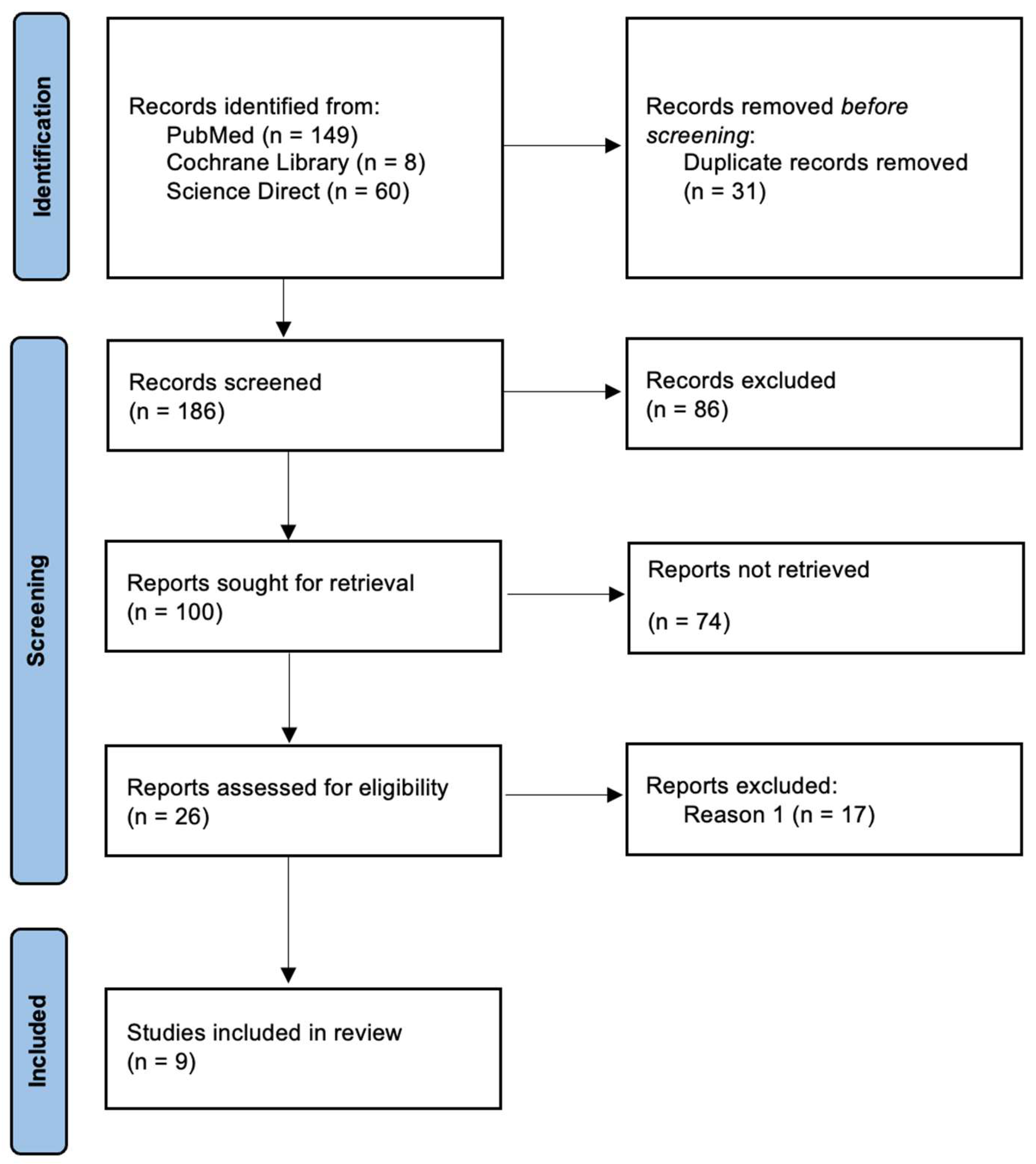
The literature search identified 217 articles. After a thorough screening of titles and abstracts, 100 articles were selected for full-text review. Of these, 74 were excluded because they did not provide relevant information for the study objectives. A total of 26 articles were fully assessed, and 9 were included in this review, as shown in Figure 1.

3.2. Sample Characteristics for Study Quality

The risk of bias assessment in the included studies, all applying AI in CBCT, indicated that the predictors and outcomes domains predominantly presented a moderate risk of bias across all analyzed studies. Specifically, 3 studies had a low risk of bias, while 6 studies had a moderate risk of bias. Some experimental and observational studies showed a moderate risk in the validation domain, mainly due to the lack of external validation or limited sample size. These studies include “Alqahtani et al. [30], 2023”, “Reduwan et al. [31], 2024”, “Huang et al. [32], 2025”, “Lin et al. [33], 2025”, “ElShebiny et al. [34], 2025”, and “Estrella et al. [35], 2025. On the other hand, “Xu et al. [28], 2024”, “Zheng et al. [36], 2025”, and “Mahdavifar et al. [37], 2025” presented a low risk of bias also in the validation domain, resulting in an overall low risk. In the remaining studies, the overall risk was considered moderate. A complete overview is provided in Table 3.
Table 3. Methodological quality assessment of included AI-based diagnostic studies on RR (PROBAST).
Authors/Year of PublicationParticipantsPredictorsOutcomesValidationOverall Risk
Alqahtani et al. (2023) [30]LLLMM
Reduwan et al. (2024) [31]LLLMM
Xu et al. (2024) [28]LLLLL
Zheng et al. (2025) [36]LLLLL
Huang et al. (2025) [32]LLLMM
Lin et al. (2025) [33]LLLMM
ElShebiny et al. (2025) [34]LLLMM
Estrella et al. (2025) [35]LLLMM
Mahdavifar et al. (2025) [37]LLLLL
L: Low; M: Moderate; H: High.

3.3. Characteristics of the Included Studies

For each study included in this systematic review, data were extracted on general characteristics such as authors, year of publication, study design, study goals, population, and interventions. Outcomes, including RR quantification, volumetric changes, AI model performance metrics, and clinical determinants, were also collected and analyzed, as summarized in Table 4.

3.4. AI Approaches and Study Characteristics in Root Resorption Research

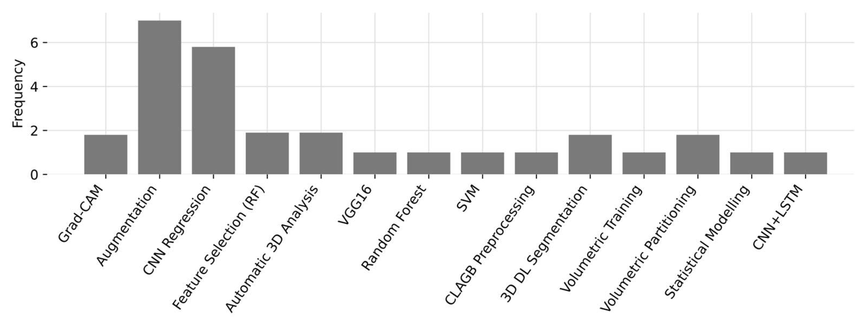
The analysis of AI applications in studies on RR revealed that CNN is the predominant technique, appearing in 7 out of the 9 reviewed studies. Automatic 3D segmentation is also widely used, appearing in 4 studies, highlighting its role in the three-dimensional quantification of roots. Hybrid approaches, such as CNN combined with Random Forest and SVM, were applied in only one study, while CNN-LSTM appeared in another, specifically aimed at classifying lesion severity. Overall, CNN-based approaches and automatic 3D segmentation were the most frequently reported methods among the included studies, as illustrated in Scheme 1.
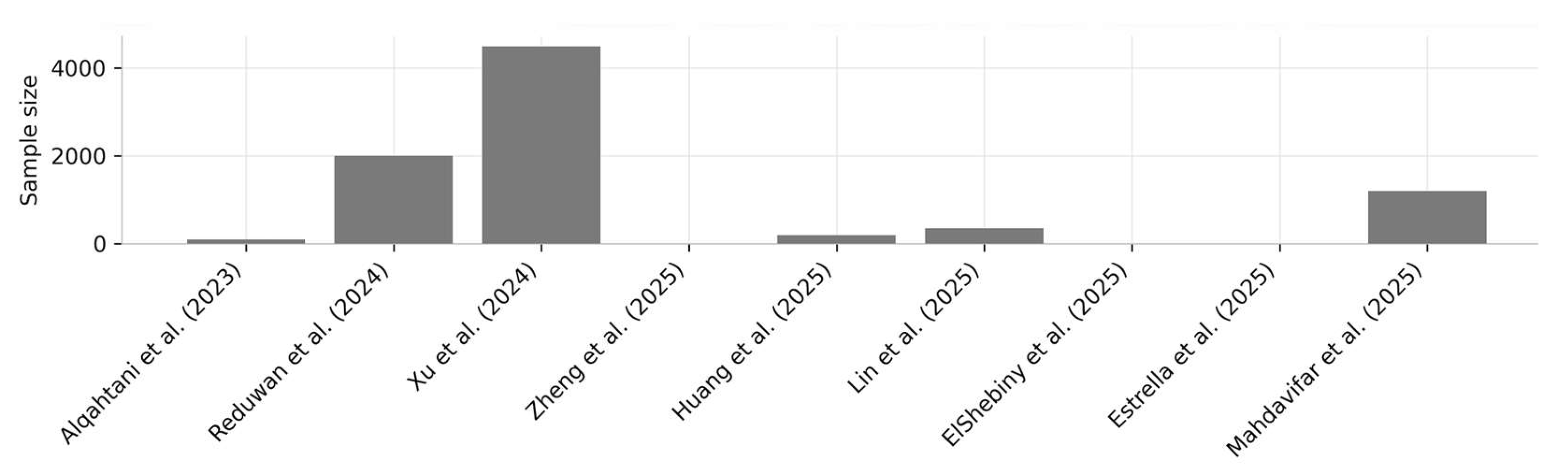
The study populations varied considerably, ranging from small cohorts with only 20 participants (“Alqahtani et al. [30], 2023”), to large samples exceeding 4500 patients (“Zheng et al. [36], 2025”). This wide heterogeneity reflects not only methodological diversity but also the application of AI across different research scales. Smaller studies generally focused on protocol validation or automated segmentation, whereas larger studies explored automatic classification of RR and detailed volumetric assessments using CBCT, as illustrated in Scheme 2.
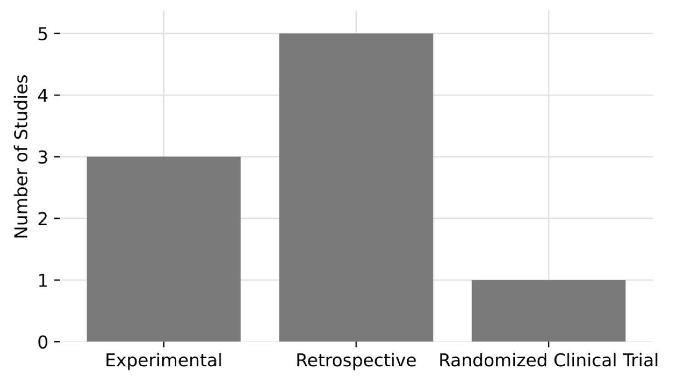
The analysis encompassed nine studies published between 2023 and 2025. The majority were retrospective (5/9), followed by experimental studies (3/9), and one randomized clinical trial (1/9). This distribution highlights a predominance of retrospective and experimental designs in the evaluation of RR using AI techniques, as illustrated in Scheme 3.

3.5. From Diagnostic Accuracy to Clinical Decision-Making

The evidence gathered in this review demonstrates that AI-enhanced CBCT techniques allow early identification and volumetric quantification of external RR with high diagnostic accuracy and reproducibility. In clinical decision-making, these advantages are particularly important in high-risk scenarios, such as IMC with intimate root proximity to LI. In such cases, the ability to detect small volumetric losses and evaluate their progression can prevent irreversible damage and help determine whether canine traction or alternative treatment approaches should be selected. These findings form the scientific basis for the clinical case presented below, in which AI-assisted volumetric monitoring helped guide the treatment strategy and protect long-term dental prognosis.

3.6. Clinical Case

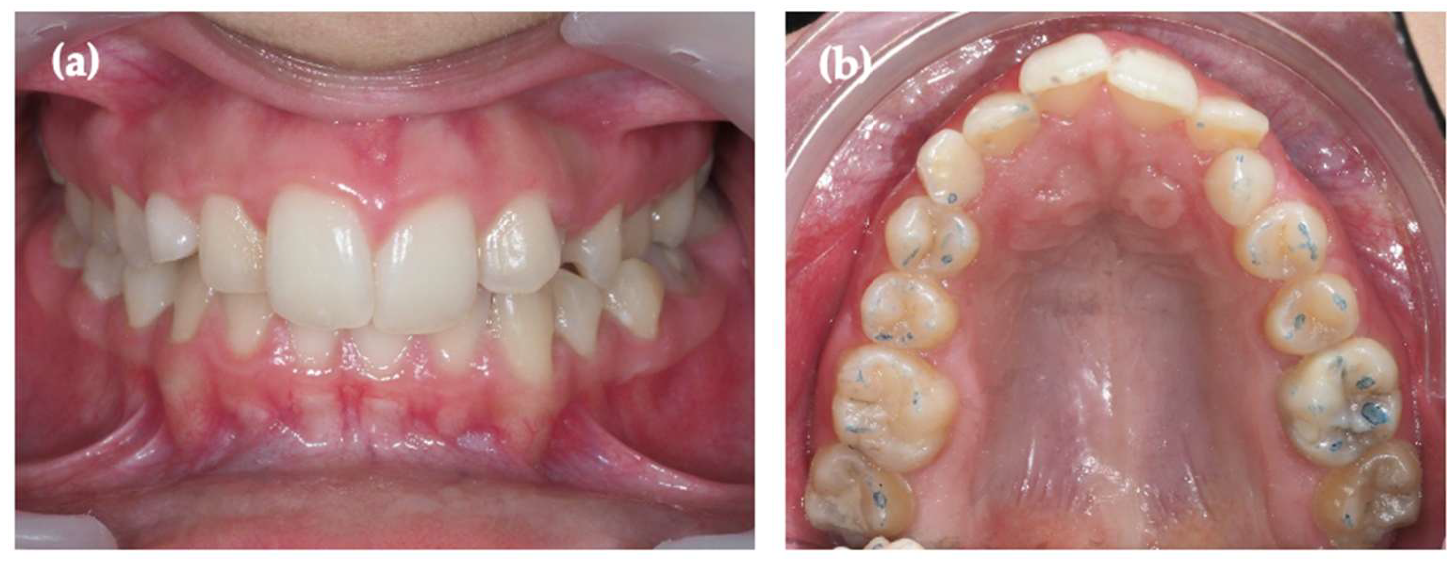
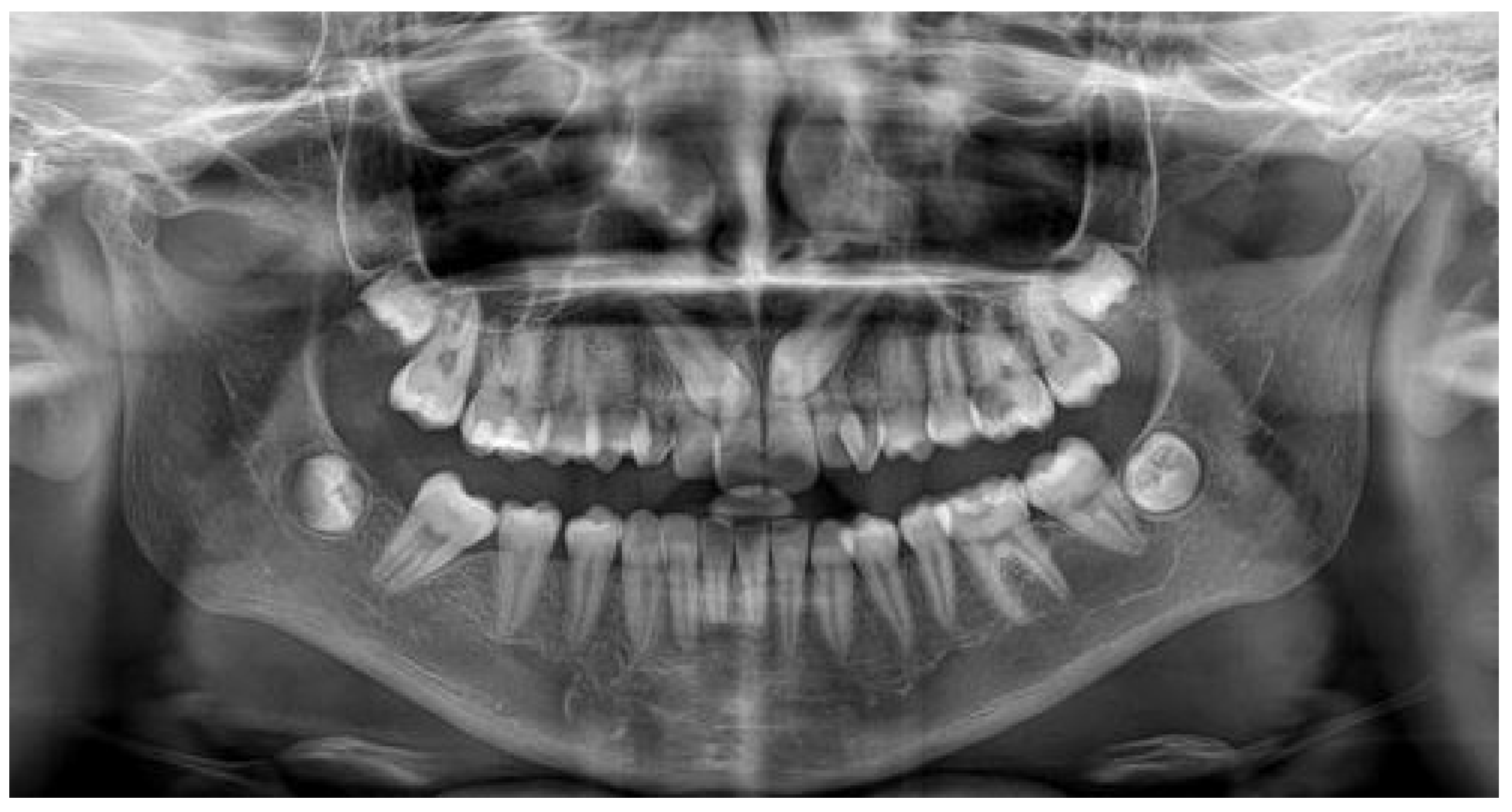
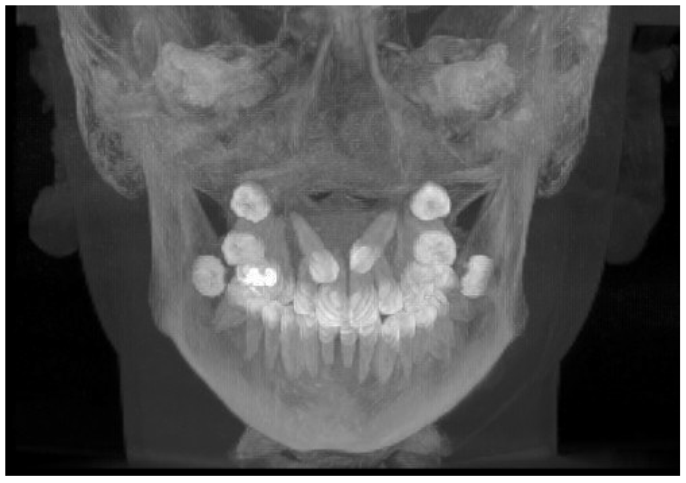
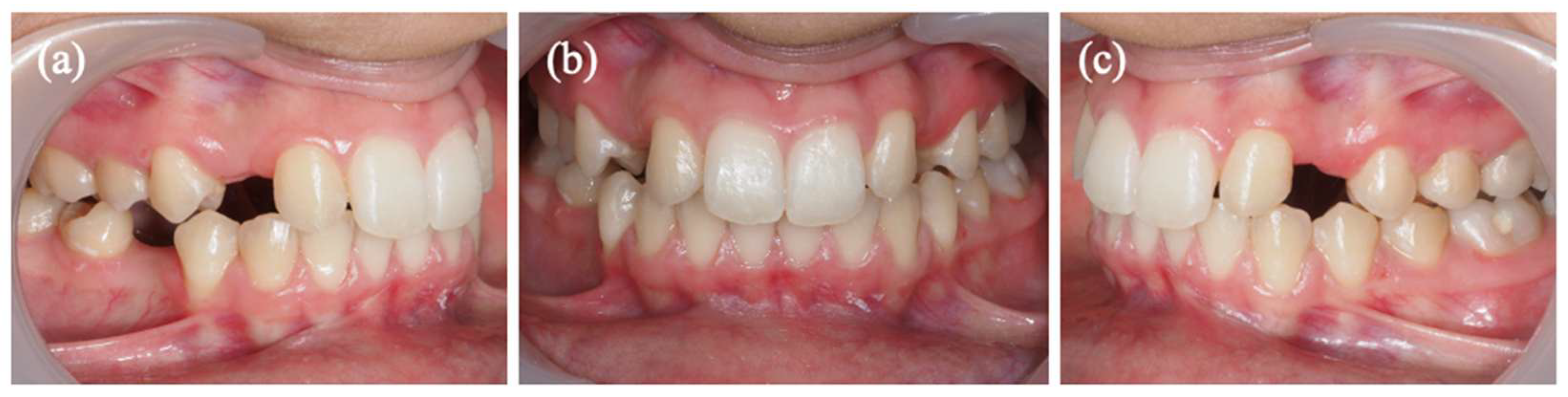
A 14-year-old female presented with bilateral impaction of the maxillary canines (#13 and #23) and retained deciduous canines (#53 and #63), as demonstrated in Figure 2a,b. Initial panoramic radiography and CBCT confirmed bilateral palatal impaction with severe mesial displacement, causing complete overlap of the canine crowns over the roots of the LIs (#12 and #22) (Figure 3 and Figure 4).
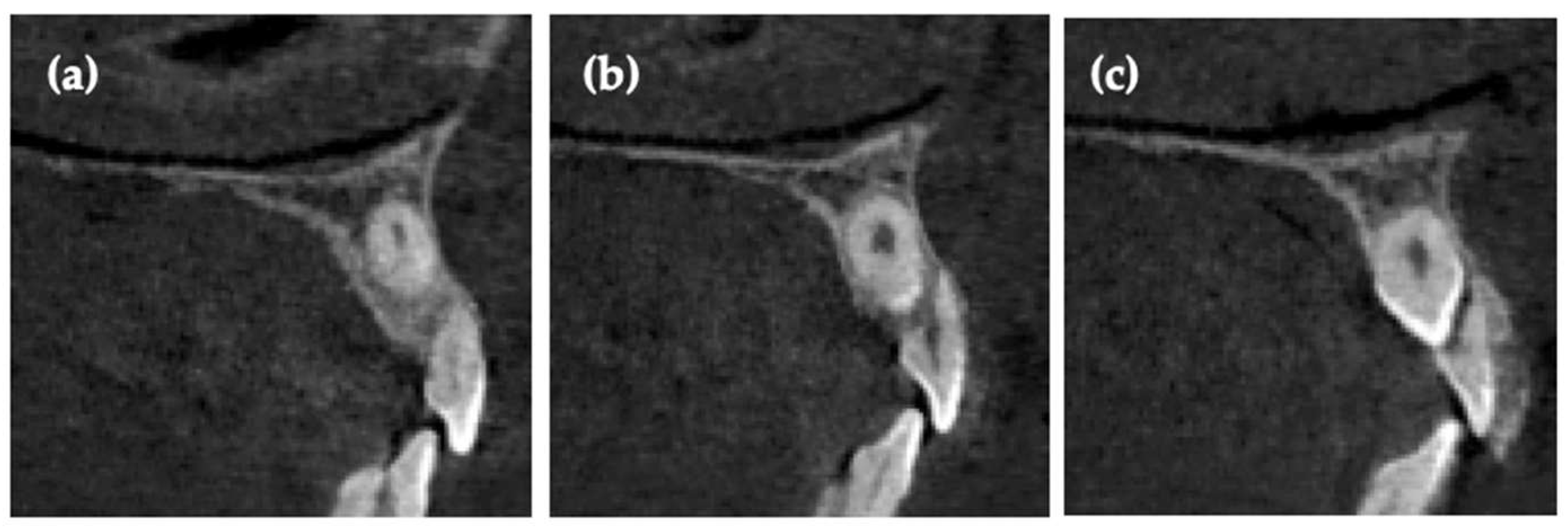
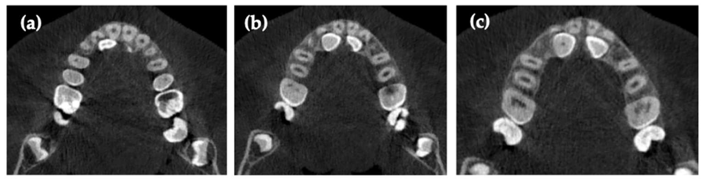
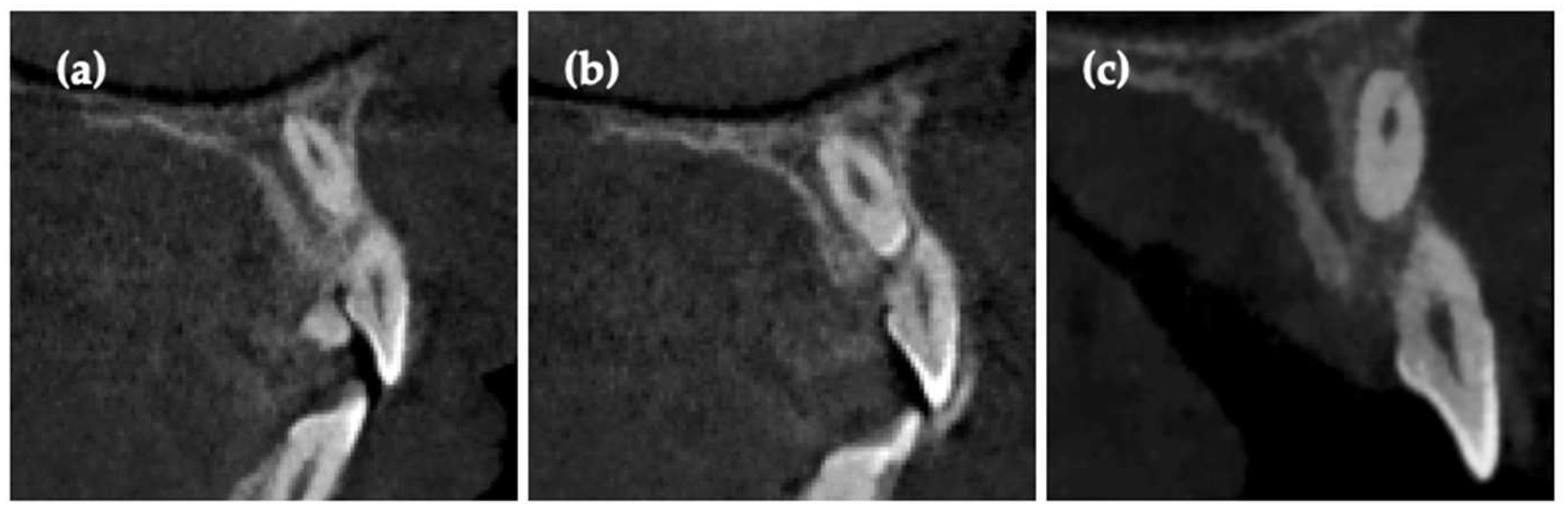
The impacted canines were positioned high in the alveolar bone, with the crowns lying close to the apical and middle thirds of the adjacent incisor roots. Initial signs of external RR were evident on the LIs, raising concern about the prognosis of these teeth (Figure 5, Figure 6 and Figure 7).
According to the CBCT-based difficulty index proposed by Chauhan et al. [38], both IMCs were classified as having maximum difficulty (score 11–15), as shown in Table 5.
CBCT confirmed direct contact between the crowns of the impacted canines and the middle and apical thirds of the LI roots, resulting in severe external RR in tooth #12 and #22 (Grade 3, with pulp involvement), according to “Ericson et al. [7], 2000”, as shown in Table 6. The central incisors exhibited no signs of RR (Grade 0), indicating that the effects of the impacted canines were limited to the LIs. Classification based on orthopantomography (OPG), presented in Table 7 and following “Ericson et al. [15] 1988”, revealed Grade 4 cusp-root overlap for both LIs, representing the highest risk for resorption.
The severity of RR was classified according to “Ericson et al. [7], 2000”, Grade 0 = no resorption; Grade 1 = mild resorption, limited to cementum or superficial dentin; Grade 2 = moderate resorption, extending into dentin with more than half of the thickness affected but without pulp involvement; Grade 3 = severe resorption, with exposure or involvement of the pulp chamber.
The OPG classification system proposed by “Ericson et al. [15] 1988”, includes: Grade 1 = no overlap; Grade 2 = overlap of less than half the root width; Grade 3 = overlap of more than half the root width; Grade 4 = complete overlap or extension beyond the LI root.
This treatment stage, considering the extensive mesial contact of the impacted canines with the roots of the maxillary central incisors, the initial therapeutic strategy consisted of distal canine traction to eliminate this direct interaction. The subsequent decision, either (1) preservation of the LIs with definitive canine alignment or (2) their extraction followed by canine substitution, would depend on both the structural response of the LIs and the predictability of canine traction.
Bilateral open surgical exposure was performed to access the crown of the impacted canines. Palatal buttons with ligature wire were bonded and sealed with two layers of flowable composite resin, providing controlled traction while minimizing debonding risk and preventing soft-tissue closure (sandwiched technique) [39,40].
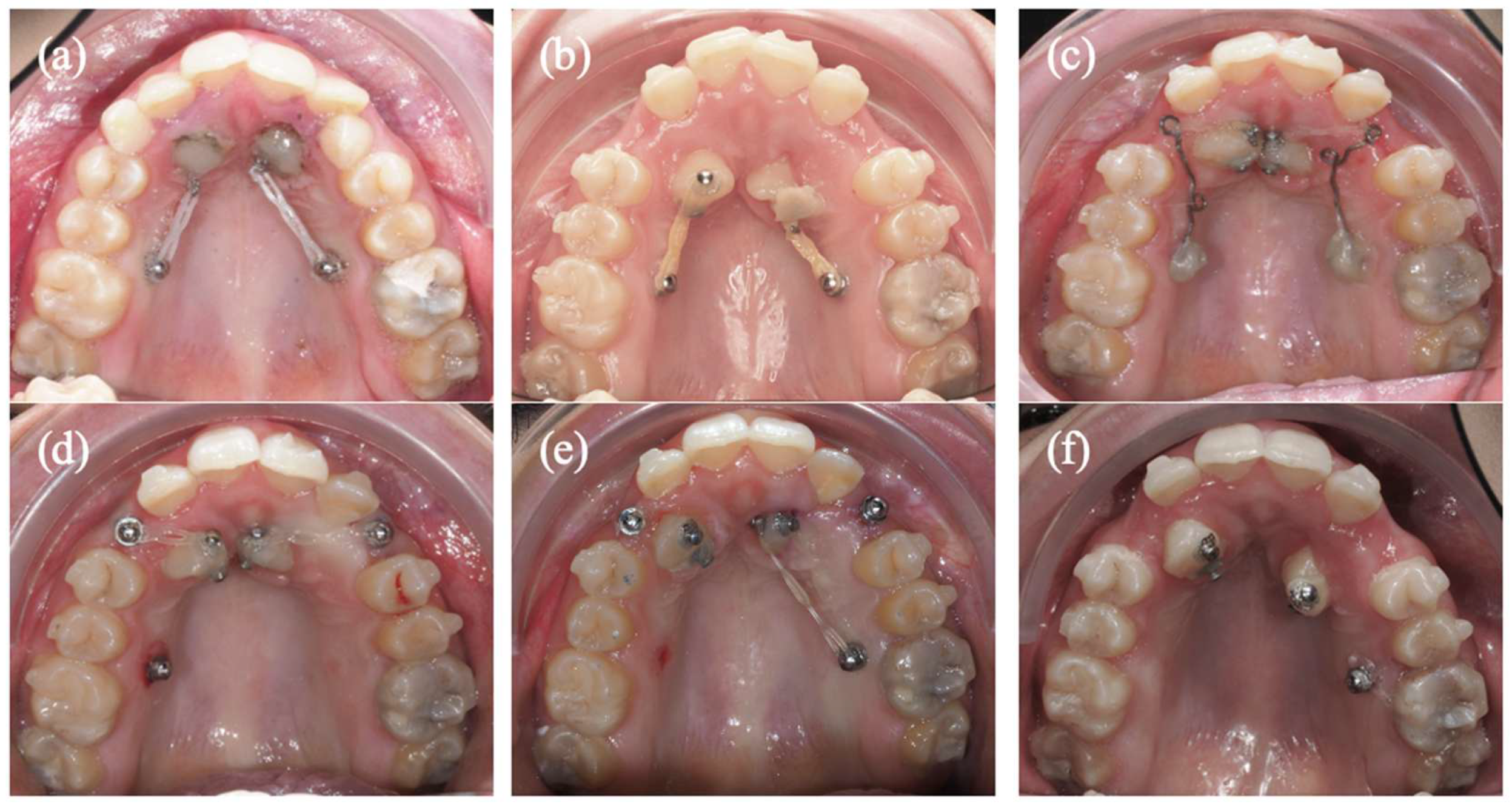
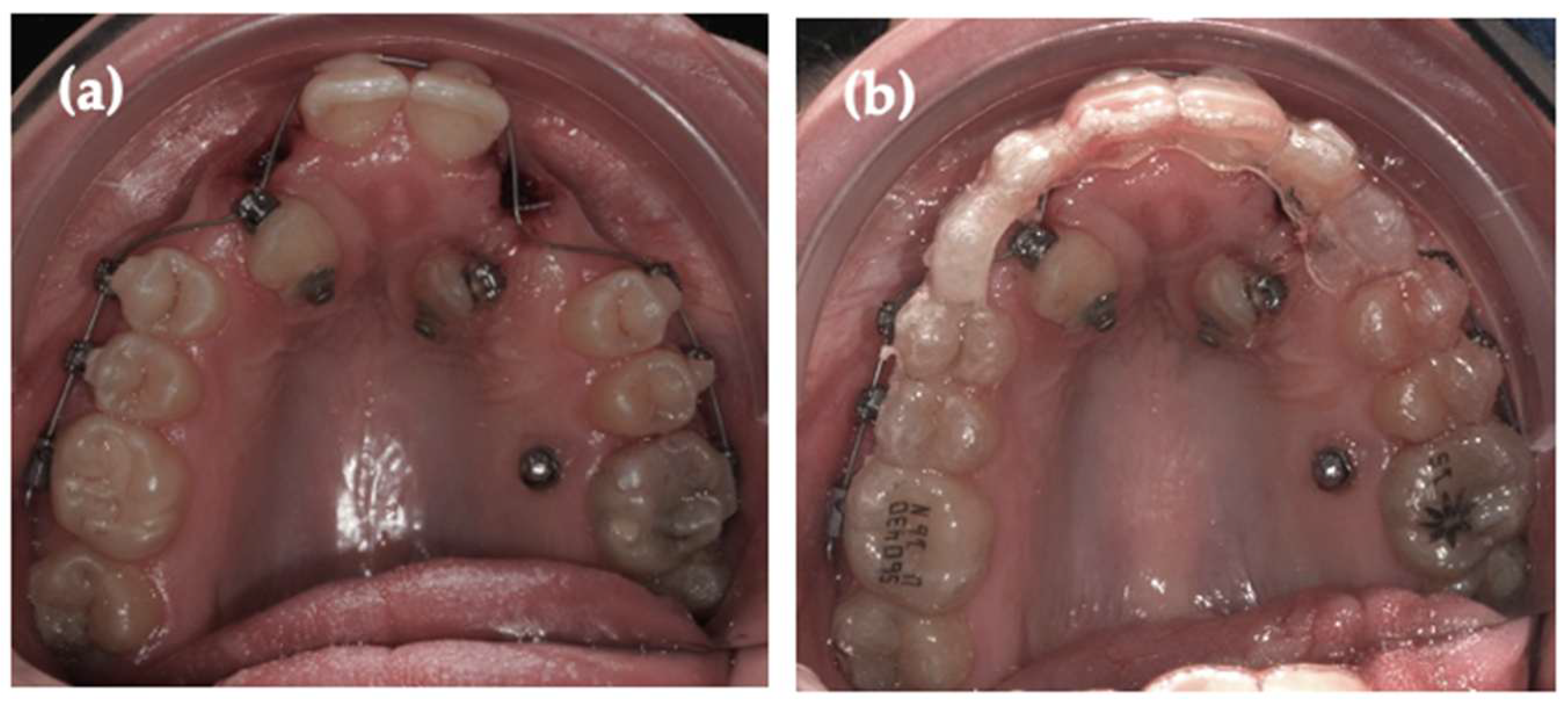
Two palatal mini-implants were initially placed between the maxillary second premolars and first molars to provide skeletal anchorage for distal traction of the impacted canines and to disengage them from direct contact with the incisor roots (Figure 8a,b). However, due to the complex root morphology and the palatal eruption pathway close to the palatal raphe, the canine crowns exhibited progressive convergence. To improve lateral redirection of the traction forces, two auxiliary cantilevers supported by the palatal mini-implants were subsequently added (Figure 8c).
Only slight improvement in the position of both canines (#13 and #23) was observed, as they remained close to the palatal midline and in proximity to the roots of the LIs. Given the persistent unfavorable positioning and limited biomechanical efficiency of this approach, two additional vertical mini-implants were placed in the alveolar crest of the first and second quadrants to redirect the canines toward the alveolar ridge after distalization of their crowns (Figure 8d). At this stage, tooth #13 showed a more favorable response compared to tooth #23 (Figure 8e).
A subsequent retrospective CBCT evaluation with AI-assisted volumetric analysis confirmed that external RR had continued to progress during treatment. Tooth #12 showed a volumetric reduction of 2.916 mm3 and tooth #22 a reduction of 2.082 mm3, indicating ongoing structural deterioration with imminent pulpal involvement, particularly in tooth #12. At the time, based on clinical and radiographic assessment, further canine traction was considered biologically unsafe, and the decision was made to extract both maxillary LIs (#12 and #22) (Figure 8f). The AI-based analysis subsequently corroborated the biological severity that had clinically justified this decision.
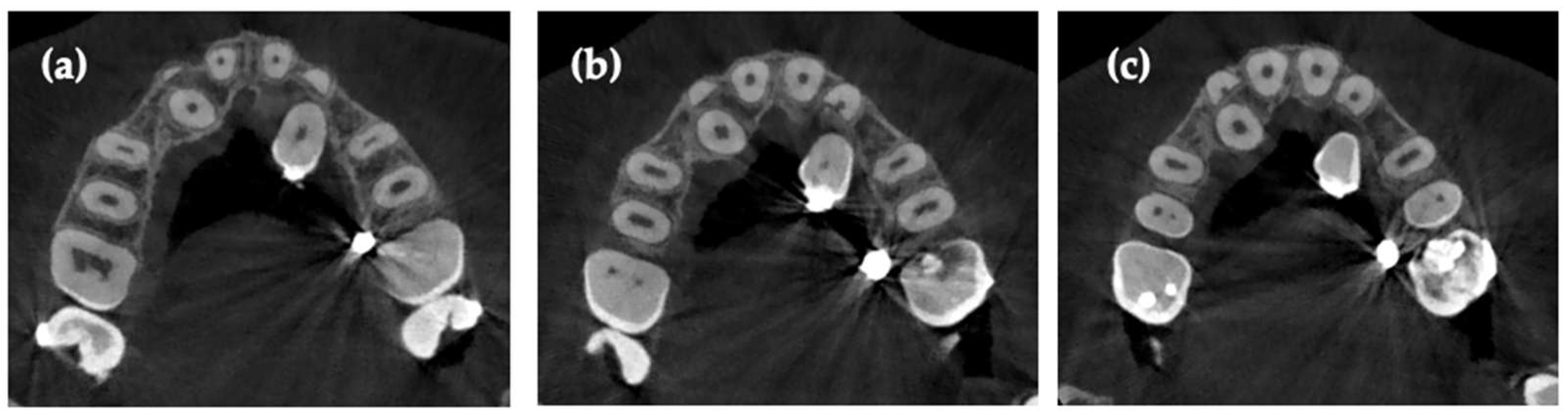
Axial CBCT evaluation demonstrated that, although external RR was more advanced in tooth #12, both maxillary LIs (#12 and #22) exhibited extensive resorptive predominantly affecting the apical and middle thirds, with additional extension toward the cervical level (Figure 9a–c). In both teeth, persistent close proximity between the impacted canines and the resorbed root surfaces was observed across multiple root levels. Although the degree of volumetric loss was greater in tooth #12, the similar distribution pattern and ongoing progression of resorption in tooth #22 indicated an equally unfavorable structural prognosis for both LIs.
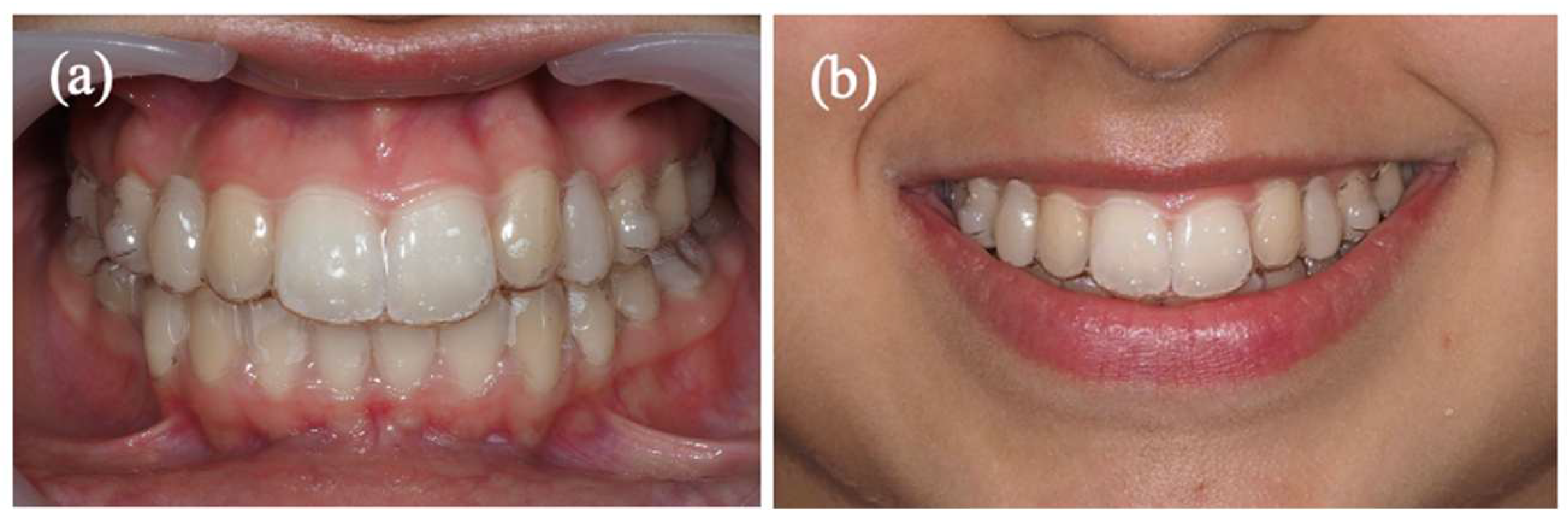
Following the bilateral extraction of the maxillary LIs (Figure 10a,b and Figure 11), the orthodontic treatment strategy was redirected toward canine substitution. Both maxillary canines were orthodontically tractioned and progressively repositioned into the LI spaces (Figure 12a–c). Simultaneously, space was deliberately preserved in the original canine sites by placing composite resin within the aligners, ensuring space maintenance and esthetic continuity. These sites are planned for future rehabilitation with implant-supported restorations at the appropriate chronological and skeletal age (Figure 13a,b).

3.7. AI Application in the Clinical Case

In this clinical case, deep learning -assisted analysis of sequential CBCT datasets was applied to objectively quantify RR and to support clinical decision-making in a high-risk scenario involving a bilateral IMC. Volumetric assessment was performed at three clinically relevant time points: pre-treatment (T0), immediately before the decision to extract the maxillary LIs (T0′), and post-treatment (T1).
Two analytical approaches were adopted: (1) a focused evaluation of the maxillary LIs, which exhibited severe resorption related to direct canine contact, and (2) a global volumetric assessment of the remaining maxillary teeth to quantify orthodontically induced RR.
All CBCT datasets were imported into 3D Slicer® (version 5.8.1). To ensure volumetric consistency, scans were resampled to identical voxel spacing, and rigid registration was performed to align follow-up scans to the baseline (T0), allowing reliable comparison of root volumes over time.

3.7.1. Volumetric Evaluation of Maxillary LI Affected by RR

The maxillary LI were analyzed separately due to the presence of severe external RR caused by palatally displaced impacted canines, resulting in a distinct and clinically critical resorption pattern. The extent of structural damage ultimately required extraction of both LIs prior to definitive orthodontic alignment.
Tooth segmentation was initially performed using the DentalSegmentator module, based on the nnU-Net deep learning framework for three-dimensional CBCT segmentation. Manual refinement was subsequently applied to correct minor inaccuracies related to anatomical variability and imaging artifacts. Crowns were separated at the cement–enamel junction (CEJ) to ensure that volumetric measurements reflected only root anatomy.
Volumetric measurements, summarized in Table 8, indicated a substantial reduction in root structure for both LIs. Differences observed between the segmentation approaches at T0 and T0′ reflected ongoing resorption during attempted canine traction, ranging from 1.56% to 2.64%, which corresponded to absolute volume discrepancies of 2.082–2.916 mm3.

3.7.2. Reconstruction of Maxillary LI Root Morphology

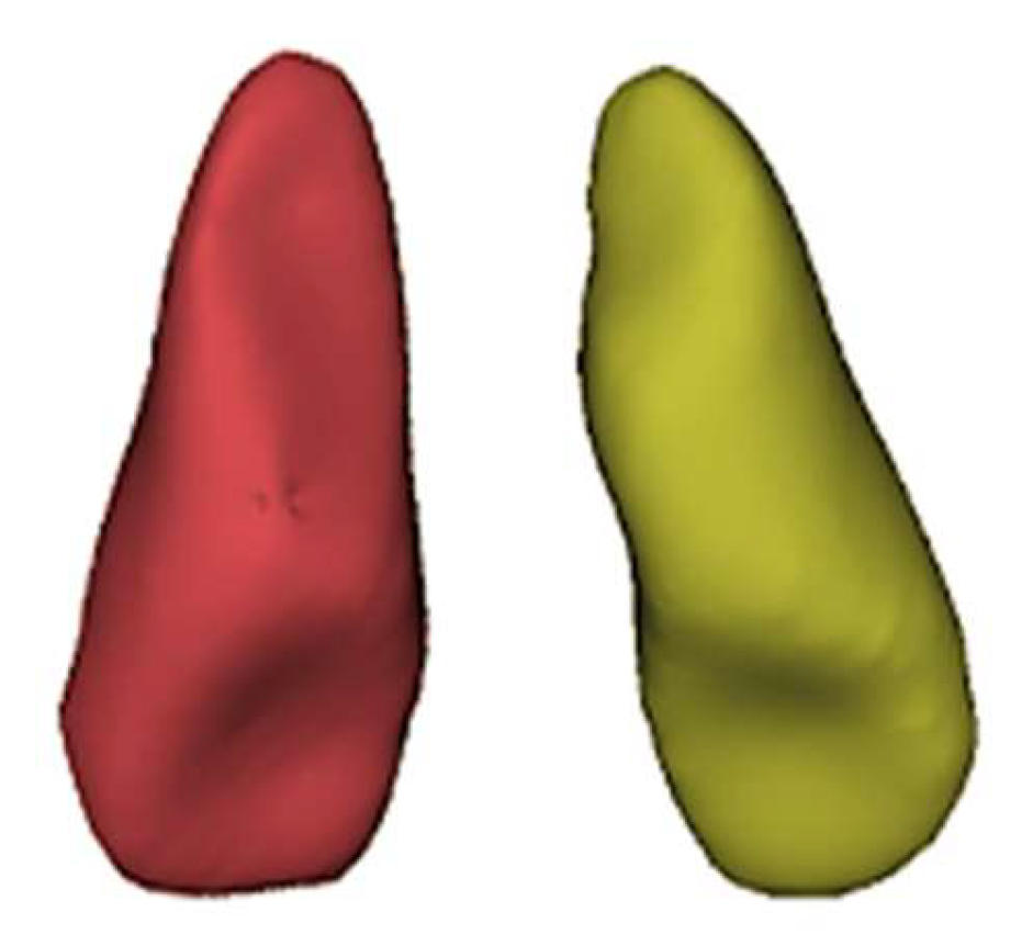
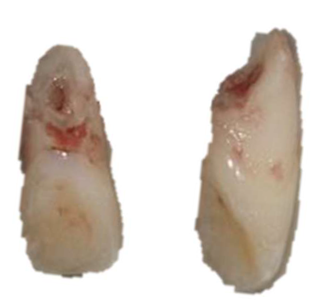
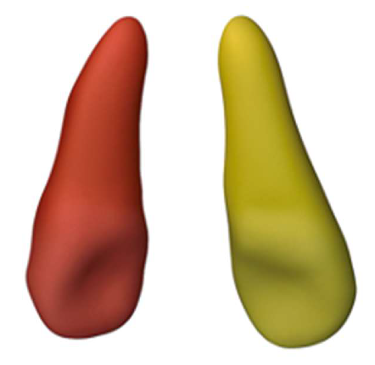
As no CBCT dataset captured intact maxillary LIs prior to the onset of resorption, a presumptive reconstruction of the original root morphology was performed. Importantly, clinical decision-making was guided by the progression of real, observed volumetric changes between sequential CBCT scans, rather than by the reconstructed values. This reconstruction was based on the remaining root fragments visible at T0, following their anatomical trajectory and curvature, and supported by published root–crown ratio (RCR) data. This reconstruction was not intended to reproduce the exact original anatomy nor to serve as a validated anatomical reference, but rather to provide a descriptive and contextual framework to estimate the magnitude of irreversible root loss in the absence of pre-resorption imaging. Although this reconstructed morphology does not represent the exact original anatomy, it enabled a consistent volumetric approach to contextualize the severity of tissue loss. Figure 14 and Figure 15 illustrate the extent of ERR observed in the extracted maxillary LIs and the presumptive reconstruction of their original root morphology prior to canine-induced resorption.
Root length estimation for the maxillary LI was based on clinical crown measurements obtained from CBCT (8.356 mm for tooth 12 and 8.506 mm for tooth 22). Two sources supported this estimation: Alhaidary et al. [41], who reported a mean root-to-crown ratio (RCR = 1.35) for maxillary LI using CBCT, and Madukwe et al. [42], who proposed a direct multiplier (1.375) to derive root length from crown length in extracted teeth.
Using the RCR of 1.35, the estimated root lengths were 11.28 mm for tooth 12 and 11.48 mm for tooth 22, yielding total tooth lengths of 19.64 mm and 19.99 mm, respectively. The discrepancy between the two methods (~0.2 mm) remains within normal anatomical variability. These values allow accurate three-dimensional reconstruction and volumetric approximation of the maxillary LI roots. The estimated root lengths (11.28–11.48 mm) and total tooth lengths (19.64–19.99 mm) provide reliable parameters for endodontic planning, orthodontic assessment, and digital modeling, while acknowledging expected individual variation.
Based on the mean RCR by Alhaidary et al. [41] the estimated values were calculated as follows:
Tooth 12:
Root = 8.356 × 1.35 ≈ 11.28 mm
Total tooth length = 8.356 + 11.28 ≈ 19.64 mm
Tooth 22:
Root = 8.506 × 1.35 ≈ 11.48 mm
Total tooth length = 8.506 + 11.48 ≈ 19.99 mm
Table 9 presents the presumptive pre-resorption root volumes of the maxillary LI (208.754 mm3 for tooth 12 and 224.428 mm3 for tooth 22). Unlike the previously calculated real resorption volumes between T0 and T0′, these values were estimated using a presumptive reconstruction, approximating the original tooth morphology as closely as possible. The differences between the presumptive volumes and the real volumes at T0′ (101.267 mm3 and 92.938 mm3) illustrate the substantial radicular loss resulting from the resorptive process.

3.7.3. AI Segmentation vs. Manual Segmentation of Maxillary LI

Despite the increasing reliability of deep learning -based automatic segmentation tools, manual segmentation remains an important reference in volumetric studies, serving as a benchmark to evaluate the accuracy and agreement of the automated method. To assess the reliability of AI-based segmentation, root volumes obtained automatically were compared with manual segmentation at T0 and T0′ (Table 10 and Table 11).
Differences between the methods ranged from 1.7% to 15.4%, showing good agreement across the segmentation approaches despite the challenge posed by the complex anatomy and advanced resorptive defects. Because the sample size was very small (n = 2 teeth), the findings were described only qualitatively rather than statistically. Overall, AI-based segmentation exhibited acceptable accuracy and reproducibility, and it offered a faster, more consistent workflow, supporting its utility for volumetric assessment in complex clinical cases, with manual segmentation maintained as the methodological reference standard.

3.7.4. Remaining Maxillary Teeth

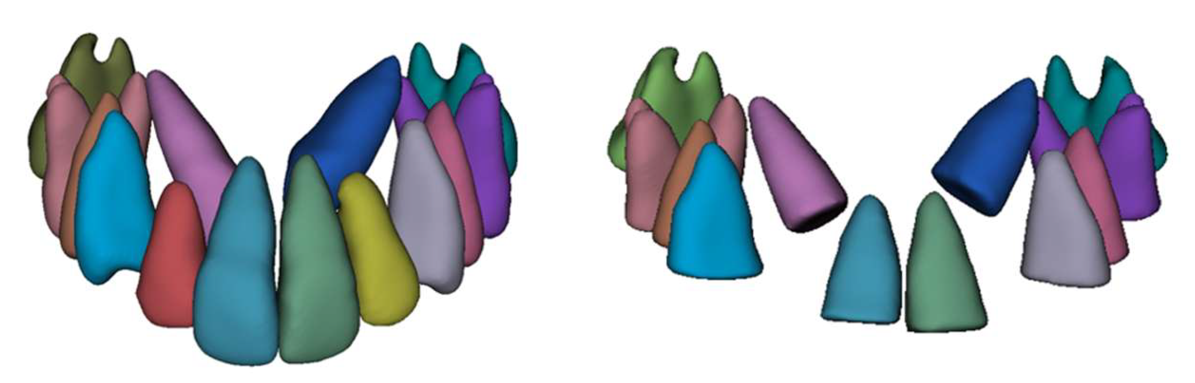
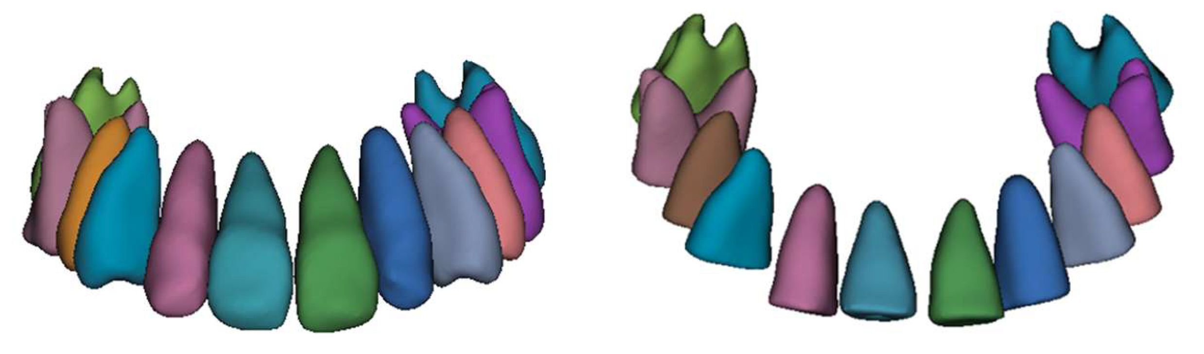
Segmentation of the remaining maxillary teeth was performed using the same AI-assisted workflow, with manual refinement as needed. Crowns were removed at the CEJ, and root volumes were measured at T0 and T1 to quantify orthodontically induced RR. Representative three-dimensional segmentations at baseline and post-treatment are illustrated in Figure 16 and Figure 17. Absolute root volumes and volumetric changes are presented in Table 12.
Before Treatment (T0)
Figure 16. Frontal views illustrating tooth segmentation at T0 and the corresponding root segmentation without the illustration of the LIs.
Applsci 16 00771 g016
After Treatment (T1)
Figure 17. Frontal views showing tooth segmentation at T1 and root segmentation after the canines were repositioned into the LI sites.
Applsci 16 00771 g017
The analysis demonstrated a consistent reduction in root volume across all evaluated teeth, confirming the occurrence of orthodontically induced RR. The magnitude of resorption varied among tooth groups. The maxillary central incisors exhibited the highest relative volumetric loss (up to 16.9%), while most premolars and molars generally showed smaller reductions, typically ranging from 3% to 5%. Notably, some posterior teeth also demonstrated higher-than-expected volumetric loss, highlighting that RR was not limited to the anterior region in this complex treatment.

4. Discussion

IMC frequently poses a substantial threat to adjacent incisors due to their ectopic eruption pathway and close proximity to the roots [12]. As demonstrated in the recent literature [28,32,36] deep learning—assisted CBCT enhances the detection and monitoring of RR by providing objective, quantitative, and early identification of structural changes. In the present case, this technology yielded critical evidence that continued traction of the impacted canine would further compromise the LIs, thereby supporting a timely and biologically safer modification of the treatment plan.
This study was conducted to determine whether deep learning -based volumetric quantification could facilitate earlier and more confident recognition of biological limits when compared with conventional manual assessment, potentially preventing the continuation of harmful forces beyond safe thresholds.
By enabling earlier and more confident recognition of biological limits, deep learning-based volumetric quantification also improves operational efficiency, standardizes measurements, and reduces evaluation times [34], all of which are critical in rapidly evolving clinical situations such as severe canine-induced RR.
Our review demonstrates that deep learning -based segmentation, particularly using CNNs and automatic 3D volumetric analysis, provides high reproducibility and accuracy in detecting EARR [28,30,31,32,33,34,35,36,37]. Across the included studies, deep learning approaches achieved strong correlations with manual measurements (ICC > 0.95) and showed excellent sensitivity for volumetric loss quantification [30,32,35,36]. In our study, the deep learning -assisted and manual segmentation of the LIs at T0 and T0′ differed by only 1–15%, indicating good concordance between methods and reinforcing the reliability of the automated volumetric analysis. While manual CBCT-based volumetric assessment remains a valid reference standard, it is inherently time-consuming and more susceptible to operator-dependent variability, particularly in longitudinal follow-up, whereas AI-assisted segmentation enables more standardized and repeatable volumetric comparisons across sequential time points [30,34,36].
As summarized in Table 3, most available studies focus on the technical validation of deep learning -based CBCT segmentation and quantification, while evidence regarding its direct impact on active orthodontic decision-making remains limited. This gap provided the rationale for the clinical case presented in this study
The findings of the present clinical case directly reflect the evidence synthesized in the systematic review. The application of deep learning -based volumetric analysis allowed objective monitoring of resorption progression during active canine traction, demonstrating continuous structural deterioration despite multiple biomechanical strategies.
In addition to the documented progression of resorption, the three-dimensional CBCT analysis consistently demonstrated an unfavorable spatial relationship between the canine roots and the remaining LI roots, which persisted despite sequential attempts at force redirection using skeletal anchorage, cantilevers, and sectional mechanics. This anatomical constraint rendered further traction of the canines biologically unsafe and mechanically unpredictable, independently of force optimization.
Therefore, the indication for extraction was not based solely on the increase in resorption rates, but also on the proven impossibility of establishing a safe eruptive corridor for the canines without inducing additional irreversible damage to the LIs. Importantly, treatment decisions in this case were guided by the progression of the real volumetric changes observed between sequential CBCT scans, while the presumptive root reconstruction served exclusively as a contextual tool to illustrate the magnitude of irreversible tissue loss.
This objective quantification was decisive in recognizing that biological limits had been exceeded and that further attempts at LI preservation were no longer predictable, exemplifying the translational value of deep learning enhanced CBCT in complex orthodontic decision-making.
Despite the promising progress, the existing literature still requires broader real-world validation. Overall, PROBAST evaluation indicated low risk of bias in predictor and outcome domains; however, moderate risk persists in validation due to limited sample sizes and insufficient external confirmation (“Alqahtani et al. [30], 2023”, “Reduwan et al. [31], 2024”, “Huang et al. [32], 2025”, “Lin et al. [33], 2025”, “ElShebiny et al. [34], 2025”, and “Estrella et al. [35], 2025). Conversely, some studies, such as “Xu et al. [28], 2024”, “Zheng et al. [36], 2025”, and “Mahdavir et al. [37], 2025”, incorporated more robust validation procedures.
It is noteworthy that most previous studies evaluated deep learning performance strictly as a diagnostic tool and not within the context of active orthodontic decision-making. Thus, the present clinical case contributes meaningful translational evidence demonstrating how AI may influence both the timing and direction of treatment strategies.
Across the reviewed literature, automated segmentation, three-dimensional volumetric quantification, and severity classification have been shown to enable precise and reproducible measurements of root morphology, particularly in anterior teeth, which are inherently more susceptible to RR [32,33].
From a biomechanical perspective, the present case also highlights the role of skeletal anchorage in attempting to control eruption vectors in high-risk impaction scenarios. Although skeletal anchorage has been shown to improve force control and reduce iatrogenic side effects in complex movements [43,44], the persistent intimate relationship between the canines and LI roots in this case demonstrates that even optimized force systems cannot always overcome unfavorable anatomical constraints.
Regarding orthodontic mechanics, several studies suggest that clear aligner therapy may be associated with lower or comparable levels of orthodontically induced RR when compared with conventional fixed appliances [45,46]. The combined use of aligners with sectional fixed appliances and skeletal anchorage in the present case may therefore have contributed to limiting the extent of RR observed in the remaining dentition. Nevertheless, the repeated attempts at canine distalization, despite biomechanical optimization, likely acted as a cumulative risk factor for the progression of resorption in the already compromised LIs.
Furthermore, monitoring and follow-up of RR is feasible; however, when canine substitution following the extraction of compromised LIs is required, it represents a well-supported alternative for long-term stability, esthetics, and periodontal health in cases of severe RR and poor prognosis [47].
Despite these advantages, current AI models still require enhanced generalization through multicenter datasets. A notable limitation of the present study is that AI was applied retrospectively. Although the clinical decision was ultimately appropriate, AI-based monitoring could have accelerated this decision and reduced the period of risk exposure if implemented prospectively.
In the future, deep learning-enhanced CBCT analysis may evolve from a retrospective and monitoring tool to a true predictive instrument. By integrating initial three-dimensional anatomical data, root proximity metrics, angulation vectors, and bone density parameters, deep learning could potentially estimate at baseline, the probability of successful canine traction without biologically unacceptable root damage. Such predictive modeling would fundamentally change treatment planning in complex impaction cases, allowing clinicians to anticipate unfavorable outcomes and avoid prolonged periods of biologically risky mechanics.
In conclusion, this clinical case illustrates that deep learning -assisted CBCT can provide clinically actionable insights in severe canine impaction scenarios, enabling precise assessment of root integrity and promoting biologically safer treatment modifications. The combined use of CBCT and automated algorithms facilitates early detection and timely intervention, contributing to long-term tooth preservation. Future prospective studies incorporating automated volumetric alerts into active orthodontic monitoring are recommended to validate the full clinical benefits suggested by this retrospective analysis.

5. Conclusions

The application of deep learning in conjunction with CBCT represents a transformative advancement in orthodontic diagnostics, offering increased precision, reproducibility, and objectivity compared with conventional manual or 2D methods.
In this study, deep learning-based volumetric analysis confirmed the accuracy of the clinical decision to extract the compromised LIs. Importantly, treatment decisions were driven by the progression of real volumetric changes observed in sequential CBCT scans, while estimated or reconstructed values were used exclusively for contextual interpretation of tissue loss severity.
Despite methodological limitations in the current literature, including restricted sample sizes and limited real-world validation, a consistently strong performance of deep learning -assisted CBCT has been demonstrated for the detection and quantification of apical RR.
The present clinical case adds translational evidence the deep learning-assisted CBCT can meaningfully support clinical judgment in high-risk orthodontic scenarios, particularly when treatment decisions may have irreversible consequences for dental prognosis.
As these technologies continue to mature, prospective studies incorporating automated volumetric alerts during active orthodontic therapy are essential to establish their true clinical impact. Future developments should focus on predictive AI models capable of estimating, at baseline, the probability of successful canine traction without biologically unacceptable root damage. While AI may substantially enhance diagnosis and treatment planning, it should be regarded as a decision-support tool rather than a substitute for the clinician’s biological judgment and responsibility. This case highlights that, even with expert management and advanced biomechanics, certain anatomical and biological limitations are insurmountable, reinforcing the need for timely, biologically guided treatment decisions supported but not replaced by AI.

Author Contributions

T.P. conceived and designed the work, revised and finalized the manuscript, and performed the clinical case treatment. L.C. designed and conceived the review, acquired, analyzed, and interpreted the data, and drafted the initial version of the review. J.P.C. designed and conceived the review, acquired, analyzed, and interpreted the data, and drafted the initial version of the review. All authors have read and agreed to the published version of the manuscript.

Funding

This research received no external funding.

Institutional Review Board Statement

This study was conducted in accordance with the Declaration of Helsinki and approved by the Ethics Committee of Cooperativa de Ensino Superior Politécnico e Universitário (CESPU), under reference 38/CE-IUCS/2024, approval granted on 7 March 2025.

Informed Consent Statement

Informed consent was obtained from the subject involved in the study.

Data Availability Statement

Data that support this study’s findings are available from the corresponding author upon request.

Conflicts of Interest

The authors declare no conflicts of interest.

Abbreviations

The following abbreviations are used in this manuscript:
AccAccuracy
AIArtificial Intelligence
AUCArea Under the Curve
BorutaBoruta Feature Selection Algorithm
CBCTCone-beam computed tomography
CLAHEContrast Limited Adaptive Histogram Equalization
CNNConvolutional Neural Network
EARRExternal Apical Radicular Resorption
ERRExternal Root Resorption
FSTFeature Selection Technique
Grad-CAMGradient-weighted Class Activation Mapping
IMCImpacted Maxillary Canine
LILateral Incisor
LSTMLong Short-Term Memory
OPGOrthopantomography
PICOSPopulation, Intervention, Comparison, Outcome, Study Design
PRISMAPreferred Reporting Items for Systematic Reviews and Meta-Analyses
RFRandom Forest
RFERecursive Feature Elimination
RRRadicular Reabsorption
SVMSupport Vector Machine
VGG16Visual Geometry Group 16
WAWeighted accuracy

References

  1. Sharab, L.; Morford, L.; Dempsey, J.; Falcão-Alencar, G.; Mason, A.; Jacobson, E.; Kluemper, G.T.; Macri, J.V.; Hartsfield, J.K. Genetic and Treatment-Related Risk Factors Associated with External Apical Root Resorption Concurrent with Orthodontia. Orthod. Craniofac. Res. 2015, 18, 71–82. [Google Scholar] [CrossRef]
  2. Khalaf, K.; Mustafa, S.; Maarouf, T. Is Orthodontic Treatment a Risk Factor of Cervical Root Resorption? A Systematic Review. Eur. J. Dent. 2022, 16, 729–736. [Google Scholar] [CrossRef] [PubMed]
  3. Iglesias-Linares, A.; Hartsfield, J.K. Cellular and Molecular Pathways Leading to External Root Resorption. J. Dent. 2017, 96, 145–152. [Google Scholar] [CrossRef] [PubMed]
  4. Sondeijker, C.F.; Lamberts, A.; Beckmann, S.; Kuitert, R.B.; van Westing, K.; Persoon, S.; Kuijpers-Jagtman, A.M. Development of a Clinical Practice Guideline for Orthodontically Induced External Apical Root Resorption. Eur. J. Orthod. 2020, 42, 115–124. [Google Scholar] [CrossRef] [PubMed]
  5. Maués, C.P.; do Nascimento, R.R.; Vilella, O.V. Severe Root Resorption Resulting from Orthodontic Treatment: Prevalence and Risk Factors. Dent. Press J. Orthod. 2015, 20, 52–58. [Google Scholar] [CrossRef]
  6. Deng, Y.; Sun, Y.; Xu, T. Evaluation of Root Resorption after Comprehensive Orthodontic Treatment Using Cone Beam Computed Tomography (CBCT): A Meta-Analysis. BMC Oral Health 2018, 18, 116. [Google Scholar] [CrossRef]
  7. Ericson, S.; Kurol, P. Resorption of Incisors After Ectopic Eruption of Maxillary Canines: A CT Study. Angle Orthod. 2000, 70, 415–423. [Google Scholar]
  8. Dachi, S.F.; Howell, F.V. A Survey of 3874 Routine Full-Mouth Radiographs. I. A Study of Retained Roots and Teeth. Oral Surg. Oral Med. Oral Pathol. 1961, 14, 916–924. [Google Scholar] [CrossRef]
  9. Bishara, S.E.; Ortho, D. Impacted Maxillary Canines: A Review. Am. J. Orthod. Dentofac. Orthop. 1992, 101, 159–171. [Google Scholar] [CrossRef]
  10. Miresmaeili, A.; Shokri, A.; Salemi, F.; Dehghani, F.; Shahidi-Zandi, V.; Rad, R.; Shahdoost, M. Morphology of Maxilla in Patients with Palatally Displaced Canines. Int. Orthod. 2019, 17, 130–135. [Google Scholar] [CrossRef]
  11. Walker, L.; Enciso, R.; Mah, J. Three-Dimensional Localization of Maxillary Canines with Cone-Beam Computed Tomography. Am. J. Orthod. Dentofac. Orthop. 2005, 128, 418–423. [Google Scholar] [CrossRef] [PubMed]
  12. Türker, N.; Yıldırım, E.A.; Bulut, D.G.; Ustaoğlu, G. Effect of Maxillary Impacted Canine Teeth on Root Resorption of Adjacent Teeth: A CBCT-Based Observational Study. BMC Oral Health 2025, 25, 1793. [Google Scholar] [CrossRef] [PubMed]
  13. Litsas, G.; Acar, A. A Review of Early Displaced Maxillary Canines: Etiology, Diagnosis and Interceptive Treatment. Open Dent. J. 2011, 5, 39–47. [Google Scholar] [CrossRef] [PubMed]
  14. Alqerban, A.; Jacobs, R.; Fieuws, S.; Willems, G. Comparison of two cone beam computed tomographic systems versus panoramic imaging for localization of impacted maxillary canines and detection of root resorption. Eur. J. Orthod. 2011, 33, 93–102. [Google Scholar] [CrossRef]
  15. Ericson, S.; Kurol, J. Resorption of Maxillary Lateral Incisors Caused by Ectopic Eruption of the Maxillary Canines: A Clinical and Radiographic Analysis of Predisposing Factors. Am. J. Orthod. Dentofac. Orthop. 1988, 94, 503–513. [Google Scholar] [CrossRef]
  16. Ericson, S.; Kurol, J. Radiographic Examination of Ectopically Erupting Maxillary Canines. Am. J. Orthod. Dentofac. Orthop. 1987, 91, 483–492. [Google Scholar] [CrossRef]
  17. Bellini-Pereira, S.A.; Almeida, J.; Aliaga-Del Castillo, A.; Dos Santos, C.C.O.; Henriques, J.F.C.; Janson, G. Evaluation of Root Resorption Following Orthodontic Intrusion: A Systematic Review and Meta-Analysis. Eur. J. Orthod. 2021, 43, 432–441. [Google Scholar] [CrossRef]
  18. Polizzi, A.; Serra, S.; Leonardi, R. Use of CBCT in Orthodontics: A Scoping Review. J. Clin. Med. 2024, 13, 6941. [Google Scholar] [CrossRef]
  19. Baena-de la Iglesia, T.; Yanez-Vico, R.M.; Iglesias-Linares, A. Diagnostic Performance of Cone Beam Computed Tomography to Diagnose In Vivo/In Vitro Root Resorption: A Systematic Review and Meta-Analysis. J. Evid.-Based Dent. Pract. 2023, 23, 101803. [Google Scholar] [CrossRef]
  20. Abdelkarim, A. Cone-Beam Computed Tomography in Orthodontics. Dent. J. 2019, 7, 89. [Google Scholar] [CrossRef]
  21. Kleinerman, R.A. Cancer Risks Following Diagnostic and Therapeutic Radiation Exposure in Children. Pediatr. Radiol. 2006, 36, 121–125. [Google Scholar] [CrossRef] [PubMed]
  22. Frane, N.; Bitterman, A. Radiation Safety and Protection; StatPearls Publishing: Treasure Island, FL, USA, 2023. [Google Scholar]
  23. Duan, W.; Chen, Y.; Zhang, Q.; Lin, X.; Yang, X. Refined Tooth and Pulp Segmentation Using U-Net in CBCT Images. Dentomaxillofac. Radiol. 2021, 50, 20200251. [Google Scholar] [CrossRef] [PubMed]
  24. Chung, M.; Lee, M.; Hong, J.; Park, S.; Lee, J.; Lee, J.; Yang, I.-H.; Lee, J.; Shin, Y.-G. Pose-Aware Instance Segmentation Framework from Cone Beam CT Images for Tooth Segmentation. Comput. Biol. Med. 2020, 120, 103720. [Google Scholar] [CrossRef] [PubMed]
  25. Fontenele, R.C.; Gerhardt, M.D.N.; Pinto, J.C.; Park, S.; Lee, J.; Lee, J.; Yang, I.-H.; Lee, J.; Shin, Y.-G. Influence of Dental Fillings and Tooth Type on the Performance of an Artificial Intelligence-Driven Tool for Automatic Tooth Segmentation on CBCT Images—A Validation Study. J. Dent. 2022, 119, 104069. [Google Scholar] [CrossRef]
  26. Wang, H.; Minnema, J.; Batenburg, K.J.; Forouzanfar, T.; Hu, F.; Wu, G. Multiclass CBCT Image Segmentation for Orthodontics with Deep Learning. J. Dent. Res. 2021, 100, 943–949. [Google Scholar] [CrossRef]
  27. Li, Q.; Chen, K.; Han, L.; Zhuang, Y.; Li, J.; Lin, J. Automatic Tooth Roots Segmentation of Cone Beam Computed Tomography Image Sequences Using U-Net and RNN. J. X-Ray Sci. Technol. 2020, 28, 905–922. [Google Scholar] [CrossRef]
  28. Xu, S.; Peng, H.; Yang, L.; Zhong, W.; Gao, X.; Song, J. An Automatic Grading System for Orthodontically Induced External Root Resorption Based on Deep Convolutional Neural Network. J. Imaging Inf. Med. 2024, 37, 1800–1811. [Google Scholar] [CrossRef]
  29. Page, M.J.; McKenzie, J.E.; Bossuyt, P.M.; Boutron, I.; Hoffmann, T.C.; Mulrow, C.D.; Shamseer, L.; Tetzlaff, J.M.; Akl, E.A.; Brennan, S.E. The PRISMA 2020 Statement: An Updated Guideline for Reporting Systematic Reviews. BMJ 2021, 372, 71. [Google Scholar] [CrossRef]
  30. Alqahtani, K.A.; Jacobs, R.; Shujaat, S.; Politis, C.; Shaheen, E. Automated Three-Dimensional Quantification of External Root Resorption Following Combined Orthodontic–Orthognathic Surgical Treatment: A Validation Study. J. Stomatol. Oral Maxillofac. Surg. 2024, 124, 101289. [Google Scholar] [CrossRef]
  31. Reduwan, N.H.; Aziz, A.A.; Mohd Razi, R.; Abdullah, E.R.M.F.; Nezhad, S.M.M.; Gohain, M.; Ibrahim, N. Application of Deep Learning and Feature Selection Technique on External Root Resorption Identification on CBCT Images. BMC Oral Health 2024, 24, 252. [Google Scholar] [CrossRef]
  32. Huang, J.; Wang, M.; Tang, X.; Zheng, L.; Yang, C. Three-Dimensional Partitioning and Quantification of Orthodontic Root Resorption via Automatic Root Extraction from Cone Beam Computed Tomography. BMC Oral Health 2025, 25, 348. [Google Scholar] [CrossRef] [PubMed]
  33. Lin, J.; Zheng, Q.; Wu, Y.; Zhou, M.; Chen, J.; Wang, X.; Kang, T.; Zhang, W.; Chen, X. Quantitative Analysis and Clinical Determinants of Orthodontically Induced Root Resorption Using Automated Tooth Segmentation from CBCT Imaging. BMC Oral Health 2025, 25, 694. [Google Scholar] [CrossRef]
  34. ElShebiny, T.; Abdelrauof, D.; Elattar, M.; Motro, M.; Retrouvey, J.M.; El-Dawlatly, M.; Mostafa, Y.A.; AlHazmi, A.; Palomo, J.M. A Deep Learning Model for Multiclass Tooth Segmentation on Cone Beam Computed Tomography Scans. Am. J. Orthod. Dentofac. Orthop. 2025, 168, 191–198. [Google Scholar] [CrossRef] [PubMed]
  35. Estrella, N.F.; Alexandra, D.S.; Yun, C.; Palma-Fernández, J.C.; Alejandro, I.-L. AI-Aided Volumetric Root Resorption Assessment Following Personalized Forces in Orthodontics: Preliminary Results of a Randomized Clinical Trial. J. Evid.-Based Dent. Pract. 2025, 25, 102095. [Google Scholar] [CrossRef] [PubMed]
  36. Zheng, Q.; Ma, L.; Wu, Y.; Gao, Y.; Li, H.; Lin, J.; Qing, S.; Long, D.; Chen, X.; Zhang, W. Automatic 3-Dimensional Quantification of Orthodontically Induced Root Resorption in Cone Beam Computed Tomography Images Based on Deep Learning. Am. J. Orthod. Dentofac. Orthop. 2025, 167, 188–201. [Google Scholar] [CrossRef]
  37. Mahdavifar, S.; Fakhrahmad, S.M.; Ansarifard, E. Estimating the Severity of Oral Lesions via Analysis of Cone Beam Computed Tomography Reports: A Proposed Deep Learning Model. Int. Dent. J. 2025, 75, 135–143. [Google Scholar] [CrossRef]
  38. Chauhan, D.; Datana, S.; Agarwal, S.S.; Vishvaroop; Varun, G. Development of Difficulty Index for Management of Impacted Maxillary Canine: A CBCT-Based Study. Med. J. Armed Forces India 2022, 78, 61–67. [Google Scholar] [CrossRef]
  39. Pinho, T.; Carvalho, J.P. Three-Dimensional Comparison of CBCT and Intraoral Scans for Assessing Orthodontic Traction of Impacted Canines with Clear Aligners. Dent. J. 2025, 13, 286. [Google Scholar] [CrossRef]
  40. Pinho, T.; Amaral, R. Transposed Maxillary Canines: Narrative Review with Clinical Case Report. Dent. J. 2025, 13, 251. [Google Scholar] [CrossRef]
  41. Alhaidary, S.; Al-Haddad, K.; Al-Harazi, G.; Mashyakhy, M.; Adlan, S.; Abu-Melha, A. Root-Crown Ratio of Maxillary and Mandibular Anterior Permanent Teeth in Yemeni Adults Using CBCT. J. Contemp. Dent. Pract. 2024, 25, 1118–1126. [Google Scholar] [CrossRef]
  42. Madukwe, I. Anatomical Crown-Root Ratio: A Proxy to Root Length Determination in Rural Endodontic Practice. Ibom Med. J. 2016, 9, 15–18. [Google Scholar] [CrossRef]
  43. Heravi, F.; Shafaee, H.; Forouzanfar, A.; Zarch, S.; Merati, M. The Effect of Canine Disimpaction Performed with Temporary Anchorage Devices Before Comprehensive Orthodontic Treatment to Avoid Root Resorption of Adjacent Teeth. Dent. Press J. Orthod. 2016, 21, 65–72. [Google Scholar] [CrossRef]
  44. Greco, M.; Machoy, M. Impacted Canine Management Using Aligners Supported by Orthodontic Temporary Anchorage Devices. Int. J. Environ. Res. Public Health 2023, 20, 131. [Google Scholar] [CrossRef]
  45. Fang, X.; Qi, R.; Liu, C. Root Resorption in Orthodontic Treatment with Clear Aligners: A Systematic Review and Meta-Analysis. Orthod. Craniofac Res. 2019, 22, 259–269. [Google Scholar] [CrossRef]
  46. Li, Y.; Deng, S.; Mei, L.; Li, Z.; Zhang, X.; Yang, C.; Li, Y. Prevalence and Severity of Apical Root Resorption During Orthodontic Treatment with Clear Aligners and Fixed Appliances: A Cone-Beam Computed Tomography Study. Prog. Orthod. 2020, 21, 1. [Google Scholar] [CrossRef]
  47. Li, N.; Yang, L.; Yang, Q.; Wang, H.; Xu, X.; Wang, T. Long-Term Follow-Up After the Treatment of Impacted Canines in the Maxilla Causing Severe Root Resorption of the Lateral Incisors: Two Case Reports. BMC Oral Health 2024, 24, 482. [Google Scholar] [CrossRef]
Figure 1. PRISMA Flow diagram. Reason 1: Irrelevant to the topic.
Applsci 16 00771 g001
Scheme 1. Frequency of AI Techniques used in RR studies.
Applsci 16 00771 sch001
Scheme 2. Sample size per study [28,30,31,32,33,34,35,36,37].
Applsci 16 00771 sch002
Scheme 3. Distribution of study designs in RR research.
Applsci 16 00771 sch003
Figure 2. (a,b) Initial intraoral frontal and occlusal photos.
Applsci 16 00771 g002
Figure 3. Panoramic radiograph showing IMC with cusp overlap on the roots of the lateral and central incisors.
Applsci 16 00771 g003
Figure 4. Initial frontal CBCT reconstruction.
Applsci 16 00771 g004
Figure 5. (ac) CBCT axial sections showing IMC in proximity to the roots of the lateral and central incisors, with varying degrees of contact and RR.
Applsci 16 00771 g005
Figure 6. (ac) Sagittal CBCT sections showing the impacted canine (#13) in contact with the LI root, with extensive external RR.
Applsci 16 00771 g006
Figure 7. (ac) Sagittal CBCT sections showing the impacted canine (#23) in contact with the LI root, with extensive external RR.
Applsci 16 00771 g007
Figure 8. (af) Sequential occlusal intraoral views illustrating the biomechanics of impacted canine traction supported by skeletal anchorage. (a,b) Initial placement of palatal mini-implants between the maxillary second premolars and first molars and distal traction of the impacted canines to disengage them from the incisor roots. (c) Addition of auxiliary cantilevers to redirect the traction forces laterally. (d) Placement of additional vertical mini-implants in the alveolar crest to guide the canines toward the alveolar ridge. (e) Active traction from the alveolar crest, showing a more favorable response of tooth #13 than #23. (f) Final stage before treatment modification, preceding the decision for bilateral extraction of the maxillary LIs.
Applsci 16 00771 g008
Figure 9. (ac) Axial CBCT slices obtained after 15 months of traction attempts demonstrating persistent canine–lateral incisor contact, progressive external RR, and absence of a safe eruptive corridor, which ultimately contraindicated further traction and justified bilateral LI extraction.
Applsci 16 00771 g009
Figure 10. (a,b) Intraoral views after extraction of the maxillary LIs, illustrating sectional fixed appliance in conjunction with aligner anchorage.
Applsci 16 00771 g010
Figure 11. Extracted maxillary LIs showing extensive external RR: tooth #12 and tooth #22, with resorption extending to the cervical third of the roots, predominantly affecting the palatal surface and more severe in tooth #12, although both teeth exhibited extensive structural damage.
Applsci 16 00771 g011
Figure 12. (ac) Intraoral photographs during aligner therapy, showing canines repositioned into the LI sites, with space preserved for future implant-supported crowns in the canine positions.
Applsci 16 00771 g012
Figure 13. (a,b) Intraoral and smile photographs during aligner therapy, showing composite resin build-ups in the canine areas to improve esthetics and mask the absence of the canines, until implant placement in the canine regions.
Applsci 16 00771 g013
Figure 14. Extracted maxillary LI showing extensive external RR involving the middle and apical thirds in AI segmentation: tooth #12 and tooth #22.
Applsci 16 00771 g014
Figure 15. Presumptive reconstruction of the intact roots of the maxillary LI, considering the length the tooth would have had if it were intact, prior to the resorption caused by the canines.
Applsci 16 00771 g015
Table 1. PICOS Strategy.
P (Population)Patients undergoing orthodontic treatment.
I (Intervention)Assessment of apical ERR induced by orthodontic treatment using CBCT assisted by AI.
C (Comparison)Conventional imaging techniques (2D radiographs) or CBCT without AI assistance.
O (Outcomes)Accuracy and sensitivity in detecting RR and diagnostic efficiency with the aid of AI.
S (Study Design)Retrospective observational studies, deep learning-based diagnostic experimental studies, and randomized clinical trials.
Table 2. Databases and research strategy.
DatabaseSearch StrategyArticles Found
PubMed“Cone-Beam Computed Tomography” [Mesh] AND “Root Resorption” [Mesh] AND (“orthodontics” OR “orthodontic treatment”) OR (“Cone-Beam Computed Tomography” [Mesh] AND “Root Resorption” [Mesh] AND (“orthodontics” OR “orthodontic treatment”) AND (“deep learning”) [Mesh])149
Science Direct(“Root Resorption”) AND (“orthodontics”) AND (“Cone-Beam Computed Tomography” OR “CBCT”) AND (“Artificial Intelligence”)60
Cochrane
Library
(“root resorption” OR “tooth resorption” OR “external root resorption”) AND (orthodontic OR “orthodontic treatment” OR orthodontics) AND (“artificial intelligence” OR AI OR “machine learning” OR “deep learning” OR “neural network” OR “computer-assisted” OR “automated”)8
Table 4. Data and outcomes from articles.
Authors and Year of the PublicationStudy DesignGoalsPopulationInterventionsOutcomes
Alqahtani et al. (2023)
[30]
Retrospective studyTo validate an automated 3D protocol for quantifying RR after orthodontics and orthognathic surgery using CBCT.n = 20
  • Automatic segmentation via Convolutional Neural Network (CNN).
  • Automated 3D analysis to measure changes in root volume and length.
  • Intra-observer intra-class correlation coefficient (ICC) was excellent (1.0).
  • Average error of 0 mm for root length and 0 mm3 for root volume.
  • Processing time: 56.8 ± 7 s to quantify ERR.
  • Both patient groups showed negligible changes in root length and volumetric ratio between T0 and T1.
  • At T0-T2, Group A had a lower ERR ratio, with decreased root volume and length compared to Group B.
Reduwan et al. (2024)
[31]
Experimental
study
Evaluate the performance of AI and feature selection in detection RR.n = 88
  • Image preprocessing using CLAHE filter.
  • Four pre-trained CNN models (VGG16, EfficienteNetB4) combined with RF and SVM.
  • Optimization with Boruta (FST) and RFE.
  • RF + VGG16 showed the best performance in identifying RR.
  • RF + FST + VGG achieved Acc 81.9%, WA 83% and AUC 96%.
  • A significant difference was found among the 8 DLMs (p = 0.008).
Xu et al. (2024)
[28]
Retrospective studyDevelop and evaluate an automatic AI system to classify orthodontically induced RR.n = 2146
  • Six pre-trained CNNs: EfficientNet-B1 to B5, MobileNet-V3.
  • Training and validation via cross-validation.
  • Application of Grad-CAM for interpretation.
  • Average model accuracy ≈ 0.92.
  • EfficientNet-B4 achieved ≈ 0.94 accuracy.
  • The apical region was identified as the main decision area by Grad-CAM.
Zheng et al. (2025)
[36]
Retrospective studyTo develop and validate an automatic 3D model for quantifying RR using CBCT.n = 4534
  • Automatic root segmentation in CBCT with 3D deep learning.
  • Innovative volumetric partitioning.
  • The proposed method showed a strong correlation with manual measurements for root volume and OIRR severity assessment.
  • Intraclass correlation coefficient (ICC) values for average volume measurements at each tooth position exceeded 0.95 (p < 0.001).
  • Accuracy for different OIRR severity classifications surpassed 0.8.
Huang et al. (2025)
[32]
Retrospective studyTo quantify RR in 3D through automatic root extraction from CBCT.n = 36
  • Automatic root segmentation using AI in CBCT.
  • Integration with intraoral scanners.
  • There was a significant reduction in root volume in both groups (p < 0.001).
  • Group II (extraction) showed greater ERR in the upper anterior teeth and in the apical third (p < 0.05).
  • Group I presented less ERR in the posterior maxillary teeth but more in the cervical third (p < 0.05).
  • ER was predominantly located in the apical third of the root (p < 0.001).
Lin et al. (2025)
[33]
Retrospective studyTo quantify orthodontically induced RR and analyze clinical determinants.n = 108
  • Automatic tooth segmentation using AI from CBCT images.
  • Multivariable linear regression to identify risk factors.
  • Root volume significantly decreased after orthodontic treatment (p < 0.001).
  • Age, gender, open/deep bite, severe crowding, and other factors significantly influenced RR at different tooth positions.
  • Multivariable regression analysis showed these factors predict RR, explaining 3% to 15.4% of the variance.
ElShebiny et al. (2025)
[34]
Experimental
study
To develop and validate an AI algorithm for multiclass segmentation in CBCT.n = 210
  • CNN for automatic multiclass tooth segmentation.
  • Accurate 3D segmentation.
  • Applicable for volumetric analysis and integration into digital workflows.
  • Promising model for automatic use, supporting faster and more consistent analyses compared to traditional segmentation methods.
Estrella et al. (2025)
[35]
Randomized clinical trialTo quantify the impact of specialized forces versus standard forces on RR and compare 3D versus AI-based quantification.n = 43
  • AI for automatic root segmentation in CBCT to quantify root volume and length loss.
  • Integration with intraoral scanners.
  • Manual segmentations detected overall volumetric loss; AI showed similar or even greater loss near the apex.
  • AI is useful but may underestimate loss outside the apex compared to non-AI techniques.
Mahdavifar et al. (2025)
[37]
Experimental
study
To develop and validate an AI model capable of classifying the severity of oral lesions from CBCT reports.n = 1134
  • CNN-LSTM model to classify lesion severity in CBCT reports.
  • Feature selection with Boruta and RFE.
  • Interpretation using Grad-CAM.
  • The CNN-LSTM model outperformed competing models and demonstrated strong capability to distinguish between high- and low-risk lesions.
  • Potential use for early detection of RR.
AI (Artificial Intelligence); Acc (Accuracy); AUC (Area Under the Curve); Boruta (Boruta Feature Selection Algorithm); CLAHE (Contrast Limited Adaptive Histogram Equalization); CNN (Convolutional Neural Network); ERR (External Root Resorption); FST (Feature Selection Technique); Grad-CAM (Gradient-weighted Class Activation Mapping); LSTM (Long Short-Term Memory); RF (Random Forest); RFE (Recursive Feature Elimination); RR (Radicular reabsorption); SVM (Support Vector Machine); VGG16 (Visual Geometry Group 16); WA (Weighted accuracy).
Table 5. Difficulty level of IMCs according to the CBCT-based index by Chauhan et al.
IMCAngulationVertical PositionBucco-Palatal
Position
Horizontal
Position
RotationTotal
132224212
233324214
Table 6. Three-dimensional Evaluation of RR Severity in LI Adjacent to IMC.
IMCAdjacent ToothDescriptionDegree of Resorption
1312 (UR lateral incisor)Root resorption reaches the pulp chamber (pulp involvement evident)3
(Severe)
2322 (UL lateral incisor)Root resorption reaches the pulp chamber (pulp involvement evident)3
(Severe)
Table 7. OPG classification of overlap between IMCs and adjacent IL.
IMCAdjacent ToothOverlap (OPG)Grade
1312 (UR lateral incisor)Cusp completely overlaps the root and extends beyondGrade 4
2322 (UL lateral incisor)Cusp completely overlaps the root and extends beyondGrade 4
Table 8. Absolute root volume and percentage difference in the LIs.
RootReal Volume in the Initial CBCT, T0 (mm3)Real Volume Before the Extraction of the LI, T0′ (mm3)ΔV Volume T0-T0′
(mm3)
12110.403107.4872.916
22133.572131.4902.082
Table 9. Presumptive Root Volume of Maxillary LI Before Resorption and Volume Difference Relative to T0′.
RootPresumptive Volume of the LIs Before Resorption (mm3)ΔV Presumptive vs. Real Volume—T0′
(mm3)
12208.754101.267
22224.42892.938
Table 10. Comparison of LI root volumes at T0 measured by AI and manual segmentation.
RootVolume at T0 AI (mm3)Volume at T0 Manual (mm3)ΔV (mm3)ΔV (%)
12110.403127.45317.0515.44
22133.572130.2832.2891.712
Table 11. Comparison of LI root volumes at T0′ measured by AI and manual segmentation.
RootVolume at T0′ AI (mm3)Volume at T0′ Manual (mm3)ΔV (mm3)ΔV (%)
12107.487123.63316.14615.021
22131.490127.2514.2393.223
Table 12. Absolute root volume at T0 and T1 and percentage difference.
RootVolume T0 (mm3)Volume T1 (mm3)ΔV T0-T1 (mm3)ΔV T0-T1 (%)
11281.998234.32647.67216.90
13249.912230.98518.9277.57
14215.109208.4136.6963.11
15243.459197.42446.03518.90
16467.802453.60014.2023.04
17478.278457.29920.9794.38
21260.766237.57323.1938.89
23251.235243.6227.6133.03
24201.366194.2927.0743.51
25239.490226.96212.5285.23
26468.666442.93525.7315.49
27473.877467.9375.941.25
Disclaimer/Publisher’s Note: The statements, opinions and data contained in all publications are solely those of the individual author(s) and contributor(s) and not of MDPI and/or the editor(s). MDPI and/or the editor(s) disclaim responsibility for any injury to people or property resulting from any ideas, methods, instructions or products referred to in the content.