


Featured Application
Radiation therapy, often used in combination with chemotherapy, remains a cornerstone in the treatment of brain cancers such as gliomas. However, the blood–brain barrier (BBB) presents a major obstacle by limiting drug penetration and is itself susceptible to radiation-induced damage. In this study, we investigated the impact of X-ray irradiation on two BBB-representative endothelial cell models—murine and human. We assessed various cellular responses, including viability, proliferation, migration, cytoskeletal organization, and oxidative stress. The two models exhibited distinct responses to radiation, providing novel insights into BBB dynamics following exposure. These findings contribute to the development of more targeted and less harmful therapeutic strategies for brain cancer treatment, with potential improvements in drug delivery and reduced adverse effects.
Abstract
Ionizing radiation remains the primary approach for treating brain cancer and is frequently used in combination with chemotherapy. However, when it comes to gliomas, the effective delivery of therapeutic agents is hindered by the limited permeability of the blood–brain barrier (BBB). Consequently, selecting the most suitable and least harmful type of ionizing radiation is essential, given its potential side effects on healthy cells within the tumor microenvironment. In this study, we explored the impact of X-ray exposure on two in vitro BBB endothelial cell models—murine and human. Post-irradiation, we evaluated cell viability, clonogenic capacity, cell cycle progression, reactive oxygen species (ROS) levels, formation of micronuclei and γ-H2AX foci, as well as alterations in cytoskeletal organization, cell migration, and intracellular calcium dynamics. The results demonstrate notable differences between the two endothelial cell lines, suggesting the human cell line is more sensitive to X-rays. In conclusion, our study provides valuable insights into the brain microvascular endothelial cells’ response to radiation, laying the groundwork for strategies to protect healthy brain tissue.
1. Introduction
Cancer continues to rank among the leading causes of mortality worldwide, and radiotherapy remains one of the most extensively employed treatment modalities. However, despite its primary objective of selectively targeting malignant cells, ionizing radiation frequently induces collateral damage to surrounding healthy tissues. In the context of brain tumor treatment, for example, endothelial cells comprising the blood–brain barrier (BBB) may be inadvertently exposed to radiation, potentially compromising the barrier’s structural and functional integrity [1].
The BBB plays a vital role in maintaining brain homeostasis by regulating molecular exchange between the bloodstream and the central nervous system (CNS). Its highly selective permeability is essential for shielding neural tissue from potentially harmful agents. Consequently, radiation-induced damage to the BBB has emerged as a critical concern in neuro-oncology. Numerous studies have demonstrated that microvascular endothelial cells, the primary cellular component of the BBB, are particularly sensitive to radiation-induced injury [2,3]. These effects include apoptosis, senescence, cytoskeletal disruption, endothelial activation, increased permeability, and the release of extracellular vesicles [4]. Such changes not only impair barrier function but may also exacerbate neuroinflammatory and neurodegenerative processes [5].
One of the key mechanisms underlying radiation-induced endothelial dysfunction is the generation of reactive oxygen species (ROS), which disturb intracellular signaling pathways and promote oxidative stress [3,6]. This oxidative environment triggers a proinflammatory response marked by the upregulation of cytokines such as TNF-α, IL-8, and IL-1, as well as the release of damage-associated molecular patterns (DAMPs). These DAMPs activate toll-like and purinergic receptors, leading to increased expression of adhesion molecules such as ICAM-1, VCAM-1, and E-selectin [6].
A growing body of research has examined how different types of endothelial cells respond to X-ray radiation, highlighting the importance of both dose-dependent and cell-specific effects. In brain radiotherapy, the clinically relevant single-fraction doses delivered to normal tissue surrounding the tumor typically range from 1 to 10 Gy, depending on the treatment plan [7]. By contrast, experimental studies frequently explore a broader dose range to probe molecular pathways and adaptive responses. Collectively, this body of work shows that endothelial responses to radiation span a dynamic continuum, from subtle regulatory adaptations at low doses to profound structural and functional disruption at high doses.
At low doses (<1 Gy), radiation can induce protective or regulatory effects rather than overt injury. Eckert et al. (2022) demonstrated anti-inflammatory responses in human primary microvascular endothelial cells exposed to 0.1–2 Gy, indicating a finely tuned, dose-dependent adaptation [8]. As the dose increases to the moderate range (1–10 Gy)—a range particularly relevant for replicating blood–brain barrier (BBB) exposure during therapeutic brain irradiation in in vitro systems—the cellular response gradually shifts toward damage and remodeling. In human umbilical vein endothelial cells (HUVECs), doses between 0.5 and 4 Gy reduce cell viability, induce micronuclei formation, and increase apoptotic cell death [9], while exposures of 2–4 Gy disrupt VE-cadherin and enhance vascular permeability [10]. Similar patterns are observed in brain-specific models, such as primary rat brain microvascular endothelial cells, where irradiation induces transcriptomic alterations involving cell cycle regulation, apoptosis, migration, and intercellular junction organization [11]. Supporting these observations, Xu and colleagues found that exposure to 5 and 10 Gy upregulates Orai2 expression and enhances store-operated calcium entry (SOCE), resulting in intracellular calcium accumulation [12].
Murine endothelial cell lines, particularly bEnd.3, have further advanced understanding of BBB-specific responses to moderate radiation doses. Zhou and colleagues showed that 10 Gy exposure increases permeability and downregulates the tight junction proteins ZO-1 and claudin-5 [13], while doses between 1 and 8 Gy reduce survival, induce G2/M arrest, increase apoptosis, and promote micronuclei formation [14]. In addition, Banaz-Yaşar et al. (2005) observed elevated CX43 transcript levels following 5 Gy exposure in both bEnd.3 and Ea.hy926 cells, pointing to impaired gap junction communication [15].
At higher doses (>10 Gy), these effects intensify, leading to profound endothelial dysfunction. At 20 Gy, cells exhibit marked intracellular calcium dysregulation [11], while at 25 Gy, bEnd.3 cells display pronounced morphological alterations and reduced E-selectin expression [16]. Altogether, these findings underscore a clear dose–response relationship in endothelial radiation biology, in which low doses can trigger adaptive signaling, moderate doses induce controlled injury and remodeling, and high doses result in overt structural and functional breakdown of the endothelial barrier.
Further evidence of radiation-induced endothelial damage comes from studies on DNA damage and oxidative stress markers. Hoorelbeke and his colleagues found increased γ-H2AX foci, calcium oscillations, and ROS production in RBE4 and primary mouse brain microvascular endothelial cells after exposure to 1 and 20 Gy. Moreover, they identified a bystander effect mediated by connexin-based gap junctions and hemichannels, purinergic receptor signaling (involving ATP and IP3), and reactive oxygen/nitrogen species, which propagated damage to non-irradiated neighboring cells [3].
The relative biological effectiveness (RBE) of X-rays, traditionally assumed to be 1 for photons between 0.1 and 3 MeV [17], has also been reevaluated. Ben Kacem et al. (2022) demonstrated that dose rate and energy can influence RBE, reporting a value of 2.86 in HUVECs based on long-term viability after fractionated exposure [18].
Taken together, these findings illustrate the complex and multifactorial responses of endothelial cells to ionizing radiation. The spectrum of effects—ranging from cell death and barrier disruption to calcium signaling perturbations and inflammatory activation—varies with cell type, dose, and irradiation conditions. This complexity underscores the importance of further mechanistic studies, particularly on brain-specific endothelial models that more closely replicate the human BBB.
In light of these considerations, the present study aims to characterize the radiation response of two brain microvascular endothelial cell lines—murine bEnd.3 and human HBEC-5i—following exposure to clinically relevant doses of X-rays. While bEnd.3 cell line is largely used in such types of studies (it is used here rather as a reference model), to the best of our knowledge, this is the first investigation to specifically examine the HBEC-5i line in this context. In addition to standard markers of radiation-induced damage, this study explores novel functional endpoints, including ATP-mediated calcium ion signaling, which has not yet been characterized in relation to radiation exposure. By providing a detailed analysis of cellular and molecular changes in these BBB-relevant models, this research seeks to contribute to the development of safer, more targeted radiotherapy approaches for treating CNS malignancies.
2. Materials and Methods
2.1. Brain Endothelial Cells Culture
The experiments described in this study were conducted using two endothelial cell lines: mouse brain microvascular endothelial cells (bEnd.3, ATCC, Manassas, VA, USA, CRL-2299) and human brain microvascular endothelial cells (HBEC-5i, ATCC, USA, CRL-3245). The bEnd.3 cells were maintained in Advanced DMEM (Gibco, Thermo Fisher Scientific, Waltham, MA, USA, Cat. No. 12491023) supplemented with 10% fetal bovine serum, 1% L-glutamine (Biowest, X0550, Nuaille, France), and antibiotics (100 U/mL penicillin and 100 μg/mL streptomycin). HBEC-5i cells were cultured in DMEM/F12 medium (PAN-Biotech, P04-41150, Aidenbach, Germany), enriched with 10% fetal bovine serum and 40 μg/mL endothelial cell growth factor (ECGF; Sigma Aldrich, E2759, Darmstadt, Germany), and grown in culture flasks pre-coated with a 0.1% gelatin solution to enhance cell adherence.
2.2. Irradiation Protocol Using X-Rays
Cell irradiation was performed under ISO 17025 Quality Assurance conditions at the Ionizing Radiation Metrology Laboratory of IFIN-HH, Romania’s Secondary Standard Dosimetry Laboratory within the WHO/IAEA SSDL Network. Cells were irradiated in well plates using an X-Strahl XRC-160-ST continuous-current X-ray generator. To prevent contamination, each plate was sealed with a fine breathable membrane placed beneath the lid. The X-ray unit operated at 150 kV and 20 mA. Beam hardening was achieved with a 4 mm Al filter (plus the 0.8 mm Be inherent filtration). The half-value layer (HVL), experimentally determined, was (5.41 ± 0.11) mm Al (k = 2). Well plates with cells were positioned in a vertical position (the 2D cell cultures were positioned in a vertical plane) on a 2 cm-thick polystyrene platform, 147.7 cm from the x-ray tube, in a horizontal-beam geometry with negligible backscatter. The well plates were oriented with the bottom toward the X-ray tube. Alignment was ensured using calibrated high-precision laser distance and leveling instruments. The collimator’s 20° opening produced an isodose field of ~8″ × 9″, fully covering the plates. Mean values of beam flatness and symmetry, assessed with 8″ × 10″ GafChromic EB3 films, were found to be <5% and <3%, respectively, ensuring dose uniformity within ± 5% across the entire film area. The 4 mm Al filtration removed low-energy photons and stabilized the spectrum, which ranged from ~20 keV to 150 keV with an average energy of ~64 keV, as characterized using a Timepix3 spectrometer (AdvaPIX, Advacam, Prague, Czech Republic). Dose measurements were obtained with a calibrated PTW Nomex T11049 absorbed-dose multimeter, traceable to the Barracuda RTI Electronics standard (directly traceable to Germany’s primary standard). Under the same geometric setup used for cell exposure, the measured dose rate was (0.125 ± 0.004) Gy/min (k = 2). To quantify the scatter effects of well plates and culture medium on the absorbed dose, Monte Carlo simulations (MCNP) were performed [19]. In this way, the uncertainty associated with dose measurement was reevaluated, leading to an actual absorbed dose rate value of (0.125 ± 0.021) Gy/min (k = 2). Particularly, the uncertainty due to scatter effects in case of each type of plate used in the various assays, estimated by MCNP simulations, is: 16% for the 24-well plate, 12.2% for the 12-well plate, and 11% for the 6-well plate.
Three distinct cellular passages have been used to prepare the plates (for each irradiation conditions at least three wells were included for each passage). The plates with different passages were independently irradiated. To keep the proper growing conditions for cells during transport and irradiation, the wells were sealed using a breathable tape (Corning, 3345, New York, NY, USA). The control cells were subjected to the same protocol, except for the irradiation.
2.3. Assessment of Cell Viability via MTT Assay
Cells were seeded in 24-well plates at a density of 15,000 cells per well. Following a 24 h incubation period, they were subjected to X-ray irradiation at doses ranging from 1 to 10 Gy. Five days post-irradiation, cell viability was assessed using the MTT assay. Cells were incubated for 3 h with a yellow tetrazolium salt solution, which is metabolically reduced by viable cells to form insoluble purple formazan crystals. These crystals were subsequently solubilized using dimethyl sulfoxide (DMSO), and the absorbance of each sample was measured at 570 nm using a Mithras microplate reader (Berthold Technologies, Bad Wildbad, Germany). Cell viability was quantified by normalizing the absorbance values of irradiated samples to those of the non-irradiated control group after the background (absorbance of the PBS-filled wells) subtraction.
2.4. Clonogenic Assay for Cell Survival
Cells were seeded in 6-well plates and allowed to adhere and grow for 24 h prior to irradiation. The initial number of cells varied between 750 and 3100 depending on the intended radiation dose, which ranged from 1 Gy to 10 Gy. Following irradiation, the cultures were maintained in an incubator for 11 days to allow colony formation. At the end of this incubation period, wells were gently washed once with PBS, then fixed and stained with a Crystal Violet solution (C0775, Merck Millipore, Burlington, MA, USA) for a minimum of 30 min. After staining, samples were rinsed with water and air-dried at room temperature. Colonies (number of cells/colony > 50) were subsequently counted using a stereomicroscope.
The surviving fraction for each condition was calculated as the ratio between the plating efficiency of irradiated cells (defined as the number of colonies formed divided by the number of cells initially plated) and the plating efficiency of the non-irradiated control. The experimental data were fitted using the Linear-Quadratic (LQ) Model [20], expressed by the following equation:
where alpha and beta are parameters that describe the cell radiosensitivity, and x represents the dose.
y = exp(−alpha × x − beta × x2)
2.5. Cell Cycle Analysis
For this experiment, cells were seeded in 24-well plates and irradiated with X-ray doses ranging from 1 to 10 Gy. At 24 h post-irradiation, cells were harvested by trypsinization, fixed in 70% ethanol pre-cooled to −20 °C, and stored at −20 °C until analysis. After a minimum of 24 h, the cells were stained with a solution containing PBS, 0.1% Triton X-100, 0.2 mg/mL RNase, and 20 μg/mL propidium iodide. Staining was performed in the dark at 37 °C for 30 min. Cell cycle distribution was subsequently assessed using a CytoFLEX flow cytometer (Beckman Coulter, Indianapolis, IN, USA). Percentage of cells found in each cycle phase was extracted using supplementary gates.
2.6. Reactive Oxygen Species Production
Cells were cultured on 12 mm diameter glass coverslips 24 h prior to X-ray irradiation at doses of 1, 2, 4, and 6 Gy. Twenty-four hours after irradiation, intracellular ROS levels were assessed using the CellROX™ reagent (C10444, Thermo Fisher Scientific, Waltham, MA, USA). Cells were incubated with the staining solution for 30 min at 37 °C, then fixed with 4% paraformaldehyde, permeabilized with 0.5% Triton X-100, and counterstained with Hoechst 33342 for nuclear visualization. Fluorescence images were captured for each sample, and ROS levels were quantified as the average fluorescence intensity per cell, calculated by dividing the total fluorescence signal by the number of nuclei. At least 30 images were analysed for each condition.
2.7. Cytoskeletal Staining and Quantification of Fiber Parameters
Endothelial cells were cultured on 12 mm diameter glass coverslips and irradiated with doses of 1, 5, and 10 Gy. After 24 h, the cells were fixed with 3.7% paraformaldehyde, stained with phalloidin (F432, Invitrogen, Waltham, MA, USA) for 2 h at room temperature to visualize F-actin, and counterstained with Hoechst 33342 to label nuclei. Fluorescence images were acquired using an Olympus BX51 epifluorescence microscope (Olympus Corporation, Tokyo, Japan). Quantitative analysis of actin fibers was performed using a previously established MATLAB R2020a script [21,22], which evaluated key structural parameters including fiber polarity, total number of fibers, average fiber length, and cumulative fiber length.
2.8. Nuclear Division Index and Micronuclei Quantification Protocol
To assess the genotoxic effects of X-ray irradiation, a micronuclei assay was performed. Micronuclei formation in bEnd.3 cells were evaluated at 5 days post-irradiation for doses ranging from 1 to 10 Gy. Prior to staining with Acridine Orange, cells were fixed using a cold methanol–acetic acid solution (stored at 4 °C) for a minimum of 20 min. Micronuclei were visualized and counted using an Olympus BX51 epifluorescence microscope. The results were expressed as the weighted average number of micronuclei per sample.
where the variable pᵢ denotes the percentage of binucleated cells containing i micronuclei. By summing the products of pᵢ and i across the population and dividing the total by 100, the average number of micronuclei per binucleated cell was determined. Additionally, the nuclear division index (NDI) was assessed as an indicator of cell proliferation, following the methodology described previously [23], particularly for cells analyzed five days post-irradiation.
Average number MN = (1 × p1 + 2 × p2 + … + 10 × p10)/100
2.9. γ-H2AX Staining for DNA Damage Assessment
Ionizing radiation is well known to induce DNA double-strand breaks (DSBs), which can be detected through immunolabeling of phosphorylated histone H2AX (γ-H2AX), a well-established marker of DSBs. To visualize these damage sites, cells were irradiated with X-rays at doses of 1 and 5 Gy. At 30 min, 4 h, 10 h, and 24 h post-irradiation, cells were fixed in 3.7% paraformaldehyde and stored at 4 °C until fluorescent staining was performed. The staining protocol included permeabilization, blocking, incubation with the γ-H2AX primary antibody (12-9865-42, Invitrogen, Waltham, MA, USA), and Hoechst 33342 nuclear staining for 1 h at room temperature. After several washing steps, the samples were resuspended in 100 µL phosphate-buffered saline and analyzed by flow cytometry using a CytoFLEX instrument (Beckman Coulter, Indianapolis, IN, USA). Data acquisition and analysis were performed with the CytoFLEX software, CytExpert 2.4. After gating to exclude debris and doublets, the median γ-H2AX fluorescence intensity was extracted for each sample. Results are presented as relative median values normalized to the control condition.
2.10. Ratiometric Analysis of Intracellular Calcium Levels
Intracellular calcium dynamics were evaluated 24 h after X-ray exposure at doses of 1 and 6 Gy. One day post-irradiation, cells were incubated in the dark at room temperature for 45 min with 1 μM FURA-2 AM (F1221, Thermo Fisher Scientific, Waltham, MA, USA), a ratiometric calcium-sensitive fluorophore, in the presence of 0.1% pluronic acid (F-127, Life Technologies, Carlsbad, CA, USA) to facilitate dye loading. Calcium imaging was performed using a CCD camera (Andor iXon EM, Oxford Instruments, Abingdon, United Kingdom) and a Polychrome V monochromator (Till Photonics GmbH, Grafelfing, Germany), both integrated into an Olympus IX71 inverted fluorescence microscope. The experimental protocol included a 2 min pre-stimulation wash with Ringer’s solution, followed by a 30 s pulse of 30 μM extracellular ATP, and a subsequent 2 min wash. Solution delivery was controlled via a 100 μm quartz perfusion needle connected to an 8-valve pressurized system (ALA Scientific Instruments, New York, NY, USA). Calcium transients were analyzed using a custom MATLAB program [24], extracting key parameters such as duration, latency, asymmetry, rise velocity, area under the curve, and amplitude.
2.11. Assessment of Cell Migration via Wound Healing Assay
To evaluate the migratory capacity of endothelial cells 24 h after exposure to X-ray doses of 1, 4, and 10 Gy, a wound healing assay was performed. Cells were cultured to confluence in 24-well plates and irradiated accordingly. After 24 h, a linear scratch was introduced into the cell monolayer using a pipette tip to simulate a wound. Phase-contrast images were acquired at multiple time points using an Olympus CKX53 inverted microscope. Wound width was quantified using a MATLAB-based image analysis algorithm, as previously described [25]. The cell migration rate was calculated following the method outlined by Grada et al. (2017) [26], and results were expressed as normalized migration rates, with each experimental condition compared to the corresponding non-irradiated control.
2.12. Statistical Analysis
All experiments were performed in triplicate and independently repeated at least three times for each assay. Data visualization was carried out using OriginPro 8 software (OriginLab Corporation, Northampton, MA, USA), and results are expressed as mean ± standard deviation (SD) or as medians and mentioned box parameters in case of box plots. Statistical comparisons between irradiated groups and the control were conducted using Kruskal–Wallis test with Mann–Whitney as post hoc. Statistical significance was indicated as follows: * p < 0.05, ** p < 0.01, *** p < 0.001.
3. Results
3.1. X-Ray Irradiation Impairs Viability and Triggers G2/M Arrest in Brain Endothelial Cells
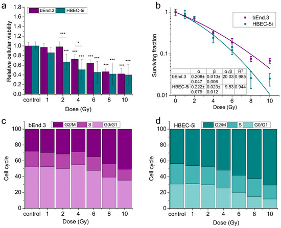
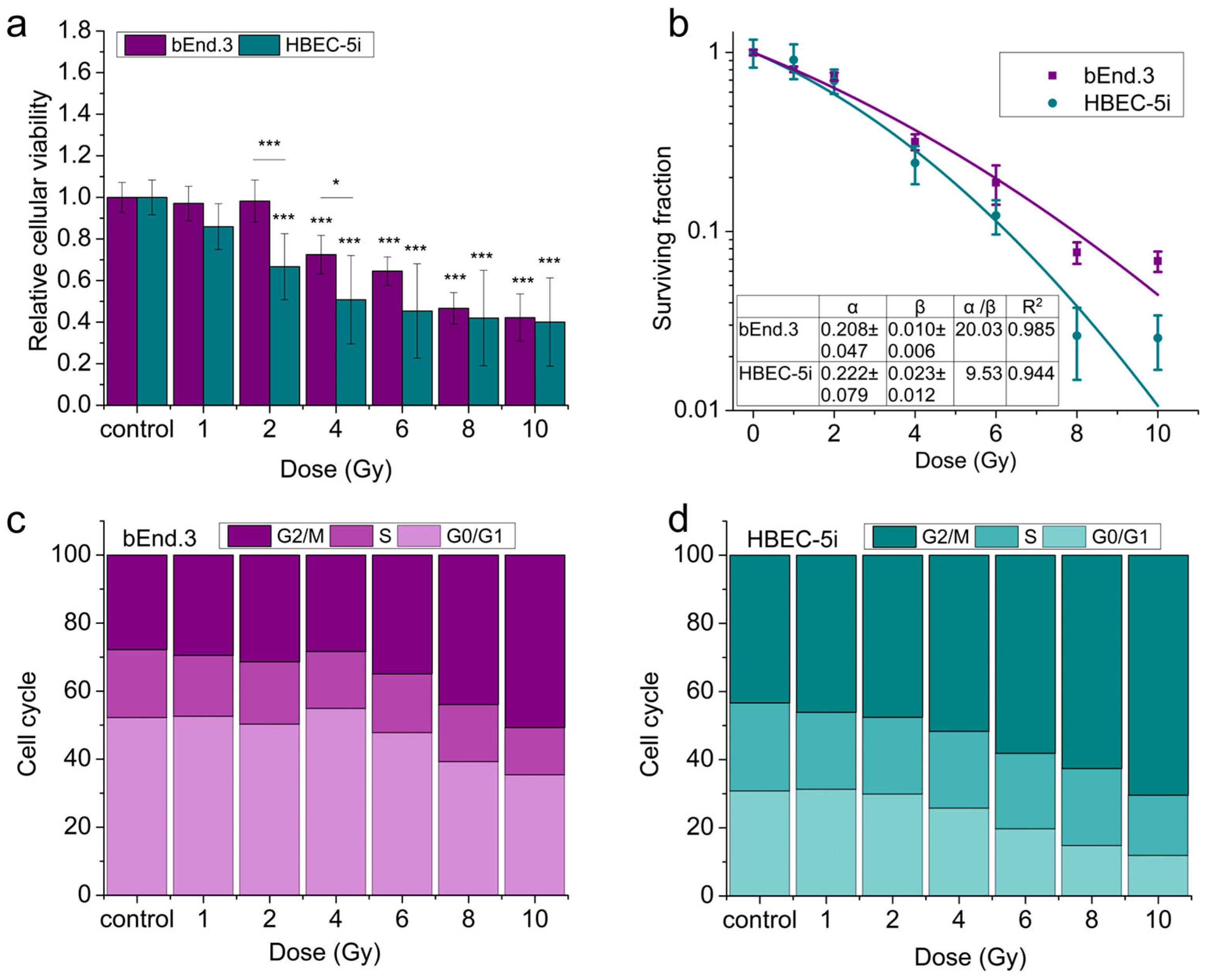
It is well established that exposure to ionizing radiation impacts cell viability and proliferation, and often induces cell cycle arrest in the G2/M phase [14,27]. In this context, we evaluated these cellular responses in two endothelial cell lines following X-ray exposure at doses ranging from 1 to 10 Gy (Figure 1). In mouse bEnd.3 cells, cell viability assessed five days post-irradiation decreased progressively with increasing radiation dose (Figure 1a). Notably, doses exceeding 4 Gy resulted in a statistically significant reduction in viability below 80%. A similar trend was observed in human HBEC-5i cells, where viability dropped below 80% at doses above 2 Gy. When comparing the two cell lines, HBEC-5i cells appeared more radiosensitive than bEnd.3 cells, particularly within the 1 to 6 Gy dose range. At higher doses, both cell types exhibited comparable reductions in viability.
Figure 1.
Effects of X-ray exposure on: (a) cell viability; (b) clonogenic survival; (c) cell cycle distribution in bEnd.3; (d) cell cycle distribution in HBEC-5i endothelial cells. Data in all panels represent mean ± SD from n = 3 independent experiments, each performed in triplicate. Statistical analysis was performed using Kruskal–Wallis test with Mann–Whitney as post hoc test. Statistical significance is indicated as follows: * p < 0.05 and *** p < 0.001.
As anticipated, similar trends were observed in the clonogenic assay results (Figure 1b). The clonogenic assay is widely regarded as a standard method for evaluating radiation-induced biological damage [28]. With increasing radiation dose, the ability of cells to form colonies from single progenitor cells was markedly diminished. Consistent with the viability data, the bEnd.3 cell line demonstrated greater resistance to radiation, maintaining a higher clonogenic potential compared to HBEC-5i cells. In addition, X-ray exposure led to notable alterations in cell cycle distribution. A dose-dependent accumulation of cells in the G2/M phase was observed (Figure 1c,d), indicating radiation-induced cell cycle arrest, with bEnd.3 cells again showing a more resistant phenotype.
3.2. X-Ray Irradiation Induces ROS Generation and DNA Damage
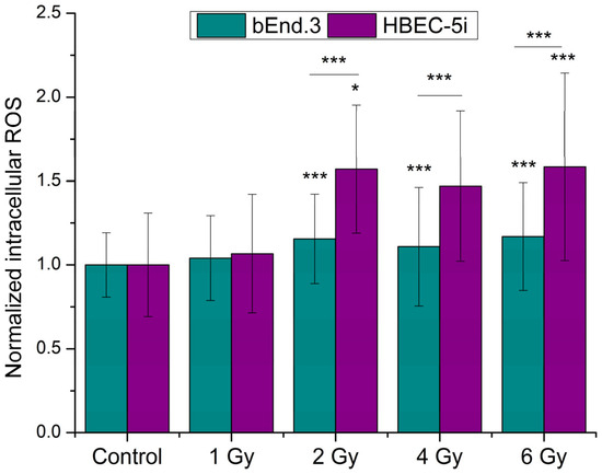
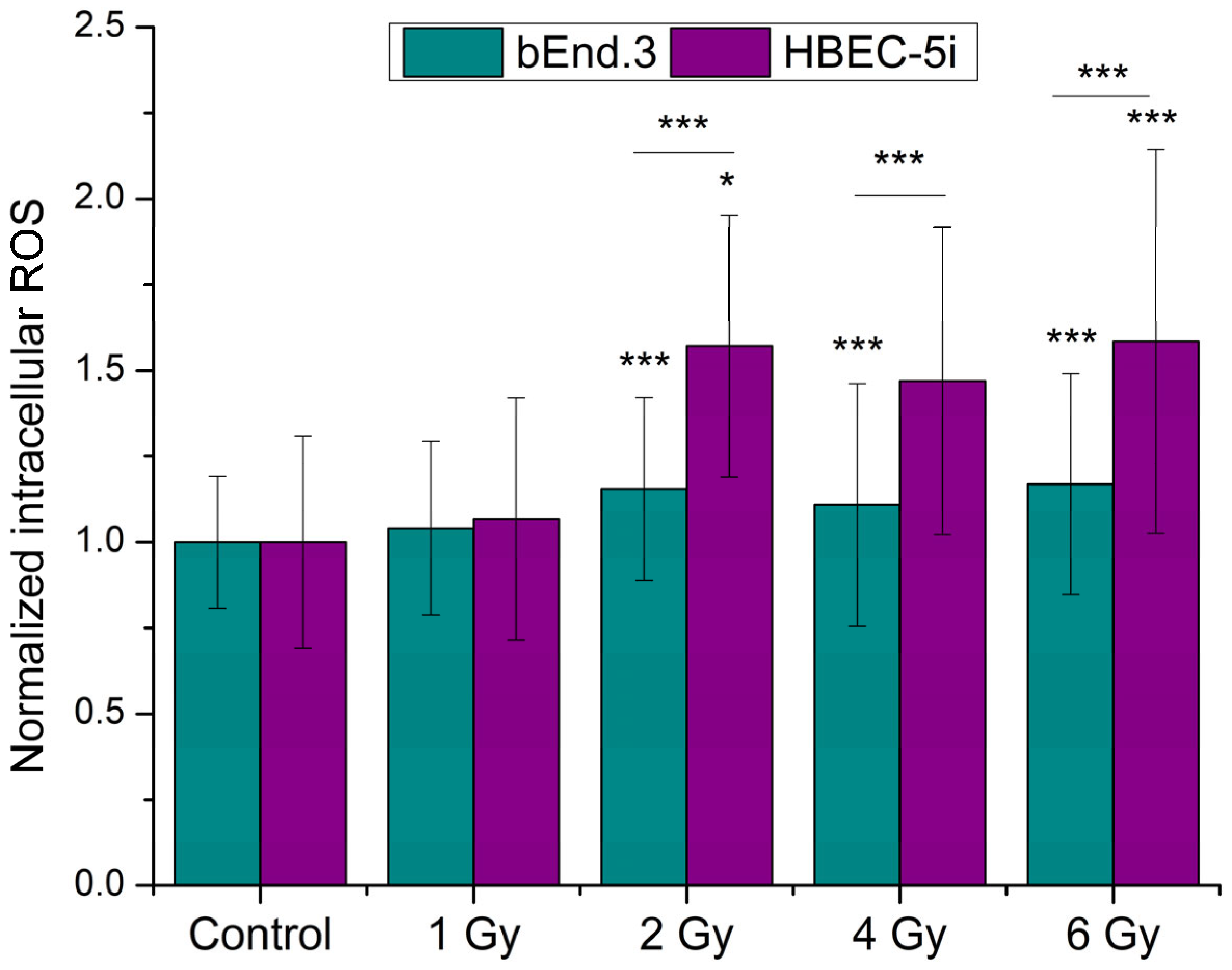
Regarding ROS production following X-ray exposure, a clear dose-dependent increase in oxidative stress markers was observed at 24 h post-irradiation. Notably, at doses exceeding 2 Gy, the difference between the two cell lines became statistically significant, with HBEC-5i cells exhibiting higher ROS levels compared to bEnd.3 cells. These findings are consistent with the cytotoxicity assay results, further indicating that human brain endothelial cells are more sensitive to radiation-induced oxidative stress than their murine counterparts (Figure 2).
Figure 2.
ROS generation 24 h after X-ray exposure at doses of 1–10 Gy. Data are represented as mean ± SD from n = 3 independent experiments, each performed in triplicate. Statistical analysis was performed using Kruskal–Wallis test with Mann–Whitney as post hoc test. Statistical significance is indicated as follows: * p < 0.05 and *** p < 0.001.
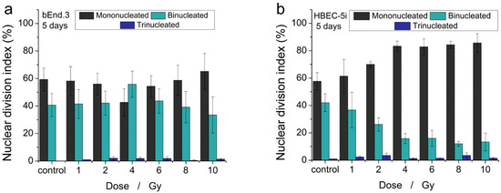
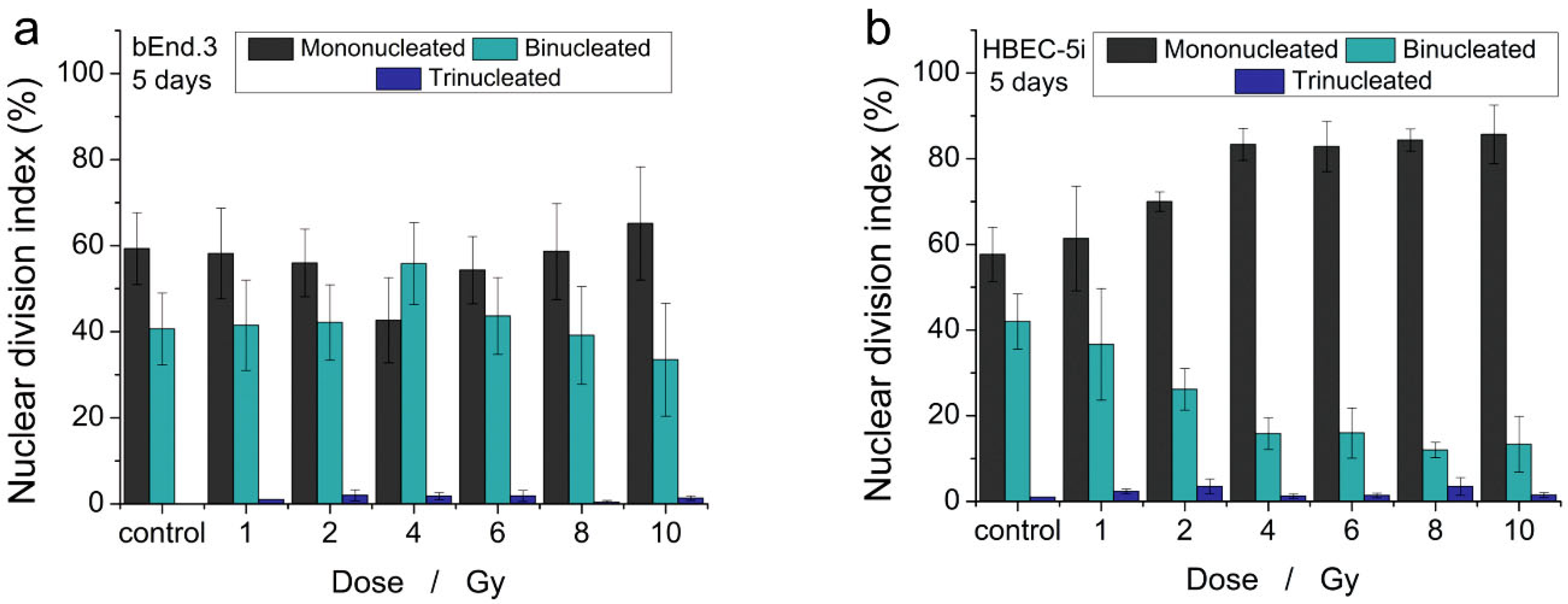
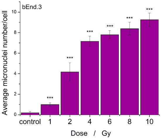
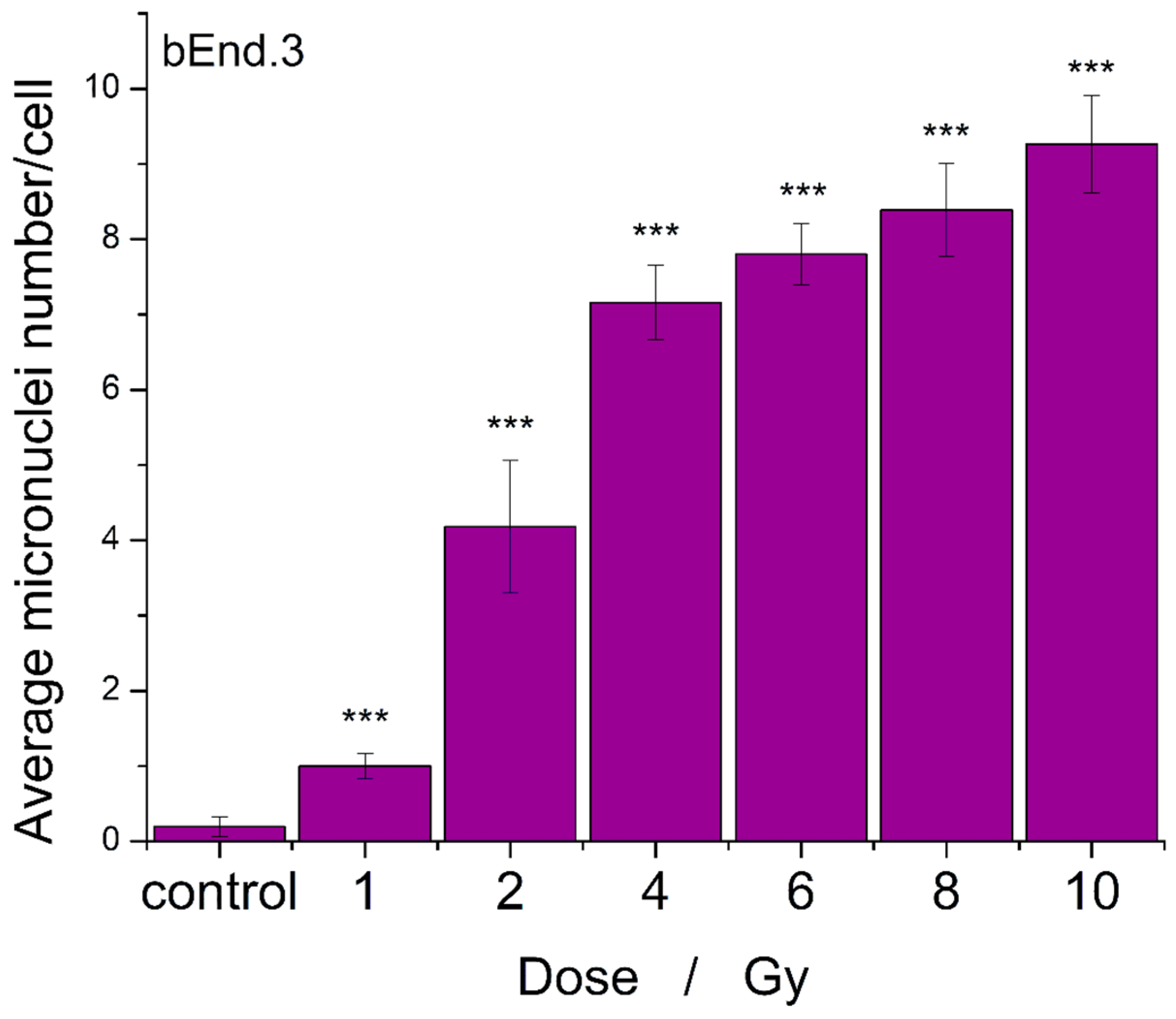
One of the most well-documented consequences of radiation exposure is the induction of genetic alterations, particularly DNA strand breaks. In this study, we assessed the effects of X-ray irradiation on the nuclear division index (NDI) five days post-exposure (Figure 3a,b). A dose-dependent decrease in the proportion of binucleated cells was observed, most notably in the HBEC-5i cell population. While bEnd.3 cells maintained a relatively stable percentage of binucleated cells across the examined dose range—with only a slight decline observed above 8 Gy—the HBEC-5i cultures exhibited a clear, monotonic decrease in this index starting from doses as low as 1 Gy. The average number of micronuclei in brain endothelial cells increased in a dose- and time-dependent manner, particularly in the bEnd.3 cell line, where elevated levels of micronuclei were still detectable five days after irradiation (Figure 4). Due to a dose-dependent decrease in the percentage of binucleated cells was observed in HBEC-5i population after 5 days of radiation exposure, the quantification of micronuclei number was not possible to achieve.
Figure 3.
Effects of X-ray on nuclear division index. (a) nuclear division index after 5 days post-irradiation in bEnd.3 cells; (b) nuclear division index after 5 days post-irradiation in HBEC-5i cells. Data are presented as mean ± SD from n = 3 independent experiments, each performed in triplicate.
Figure 4.
Effects of X-ray on micronuclei number in bEnd.3 cells after 5 days post-irradiation. Data are presented as mean ± SD from n = 3 independent experiments, each performed in triplicate. Statistical analysis was performed using Kruskal–Wallis test with Mann–Whitney as post hoc. Statistical significance is indicated as follows: *** p < 0.001.
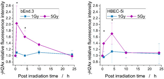
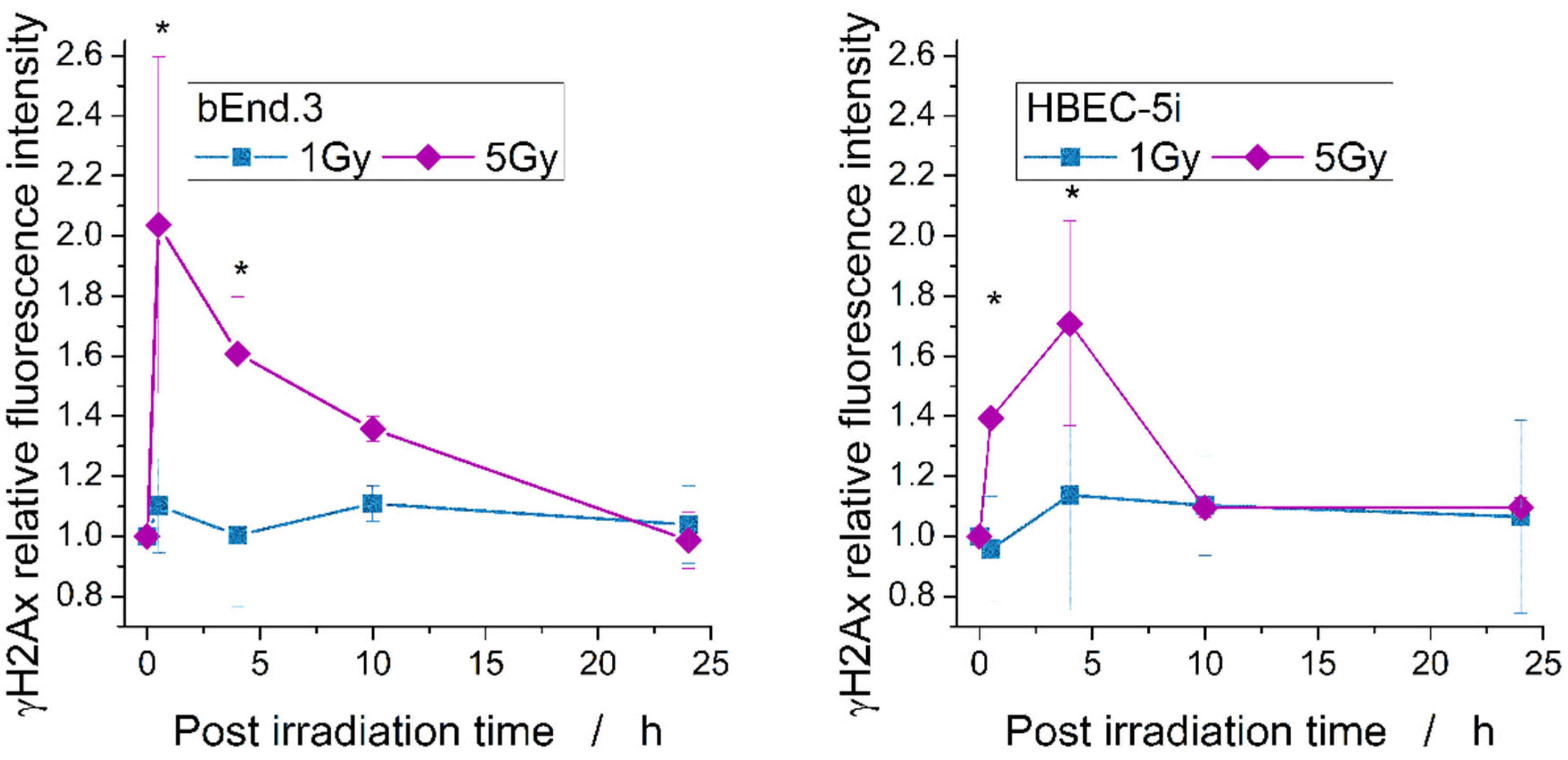
To evaluate DNA double-strand breaks, we quantified γ-H2AX foci formation at 30 min, 4 h, 10 h, and 24 h post-irradiation (Figure 5) using flow cytometry, providing a comprehensive view of the temporal dynamics of the cellular response to different radiation doses. As expected, γ-H2AX expression increased markedly in both endothelial cell lines at 30 min and, in the human cell line, at 4 h after exposure, indicating rapid induction of DNA damage. This was followed by a progressive decline at 10 and 24 h, particularly pronounced at the 5 Gy dose. Notably, mouse endothelial cells exhibited higher levels of DNA double-strand breaks at 5 Gy compared to human cells, but showed efficient repair, with damage levels returning close to baseline by 24 h.
Figure 5.
Expression of the DNA double-strand break marker γ-H2AX at 30 min and 4 h after irradiation. Data are presented as mean ± SD from at least n = 3 independent experiments, each performed in triplicate. Statistical analysis was performed using Kruskal–Wallis test with Mann–Whitney as post hoc. Statistical significance (compared to control) is indicated as follows: * p < 0.05.
3.3. X-Ray Exposure Modulates Intracellular Calcium Signaling
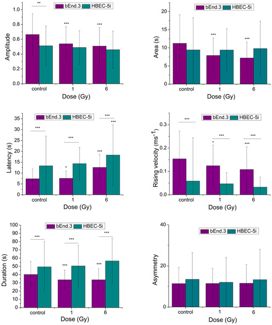
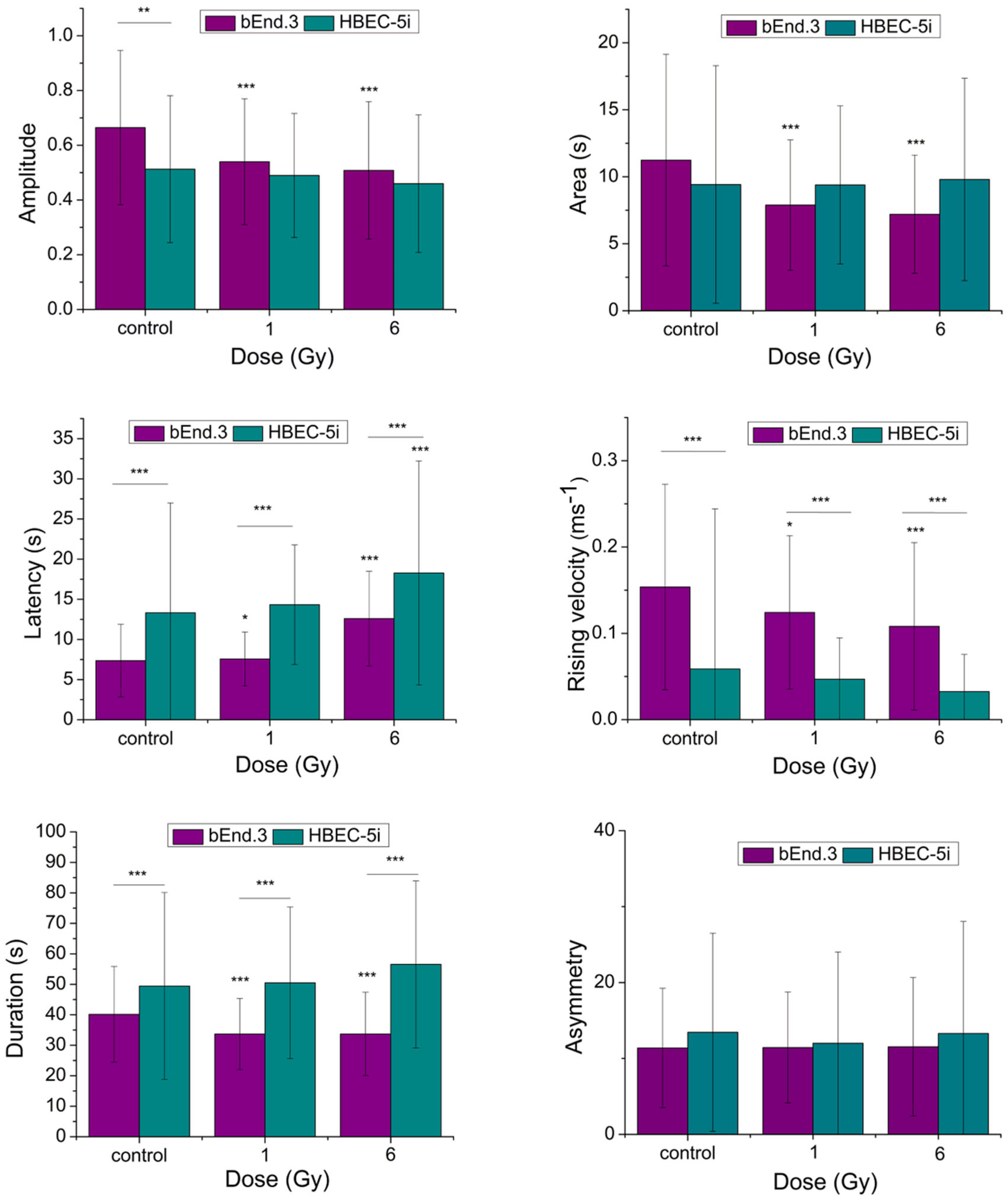
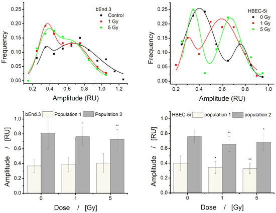
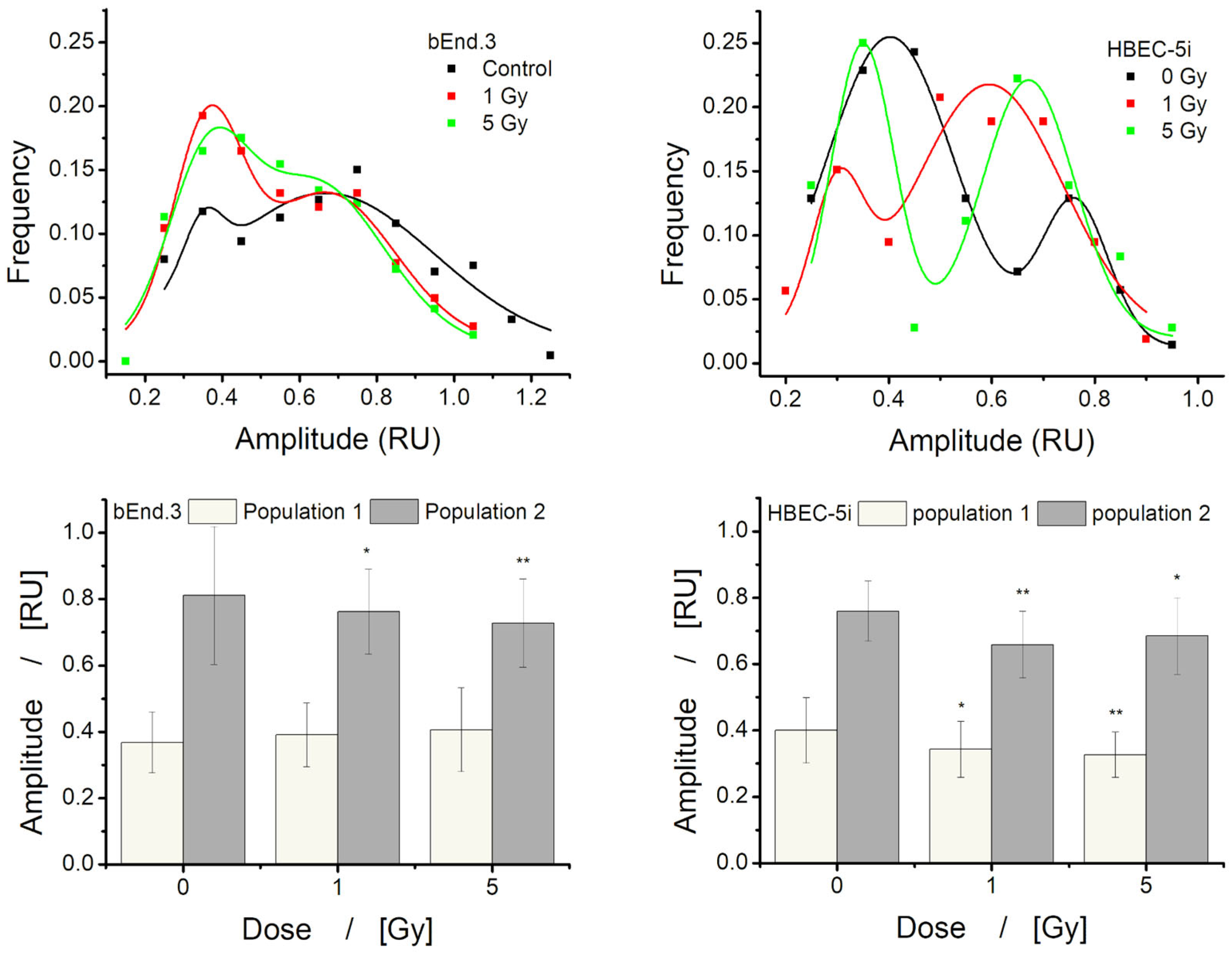
We also investigated the effects of X-ray irradiation on ATP-induced intracellular calcium transients (Figure 6). Analysis was performed using a previously developed MATLAB script [24], which calculates key parameters characterizing calcium signaling dynamics. In bEnd.3 cells, irradiation led to a dose-dependent decrease in transient duration, amplitude, area, and rising velocity, while latency increased and asymmetry remained unchanged. On the other hand, HBEC-5i cells exhibited increased duration and latency, area and asymmetry remained almost constant with increasing dose, while amplitude and rising velocity decreased with increasing dose. Comparatively, the calcium response in human cells was slower and more prolonged, with a delayed onset and reduced intensity following extracellular ATP stimulation. One of the main limitations of these data is the high standard deviation, which resulted in only a few conditions showing statistically significant differences compared with the control. Examination of the amplitude histograms revealed a bimodal distribution with two overlapping peaks for both cell lines (Figure 7, top). By isolating the values corresponding to each subpopulation indicated by the histograms and calculating the mean for each, the standard deviation was substantially reduced, leading to statistically significant differences from the control in most conditions. These findings are consistent with the overall decrease in mean amplitude observed when analyzing the entire dataset (Figure 6, top).
Figure 6.
Changes in calcium transient parameters following X-ray exposure at 1 and 6 Gy. Data are presented as mean ± SD from n = 3 independent experiments, each performed in triplicate. For each condition, at least 75 cells were analyzed. Statistical analysis was performed using Kruskal–Wallis test with Mann–Whitney as post hoc. Statistical significance (compared to control) is indicated as follows: * p < 0.05, ** p < 0.01, and *** p < 0.001.
Figure 7.
(up) Amplitude values frequency vs. amplitude level. (down) Mean ± SD of the amplitude values for each population of responses indicated by the frequency graphs. Statistical analysis was performed using Kruskal–Wallis test with Mann–Whitney as post hoc. Statistical significance (compared to control) is indicated as follows: * p < 0.05 and ** p < 0.01.
3.4. X-Ray Exposure Induces Cytoskeletal Remodeling, Alters Migration Rate, and Modulates Intracellular Calcium Signaling
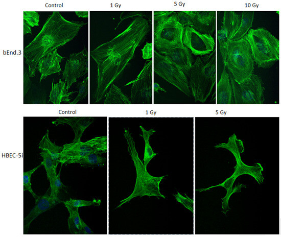
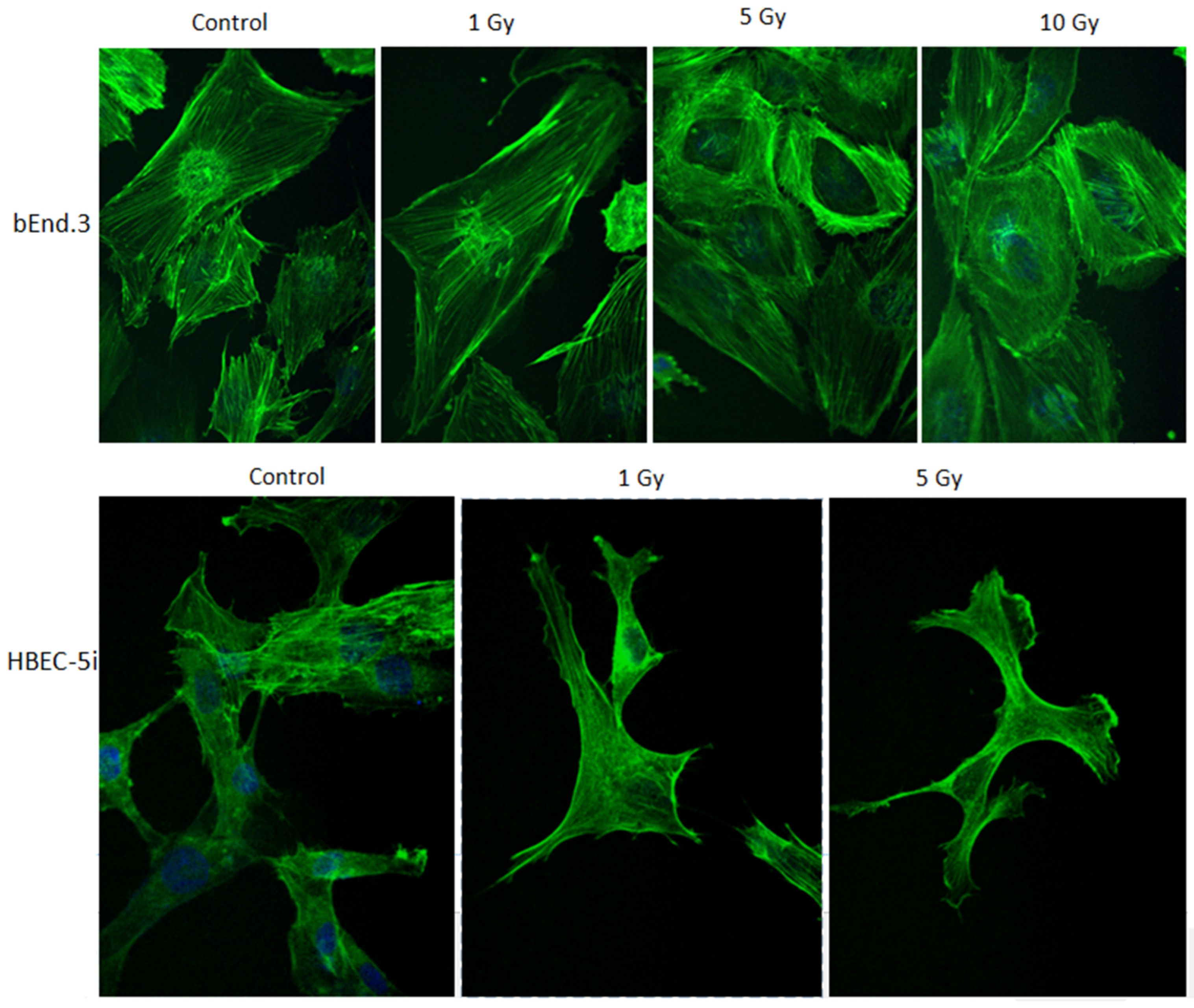
One of the mechanisms contributing to increased blood–brain barrier (BBB) permeability following irradiation is radiation-induced cytoskeletal remodeling. In our in vitro BBB models, we observed dose-dependent alterations in cytoskeletal organization. As the irradiation dose increased, notable morphological changes occurred in actin filament organization and overall cell shape. Qualitative assessment revealed that, compared with control conditions, bEnd.3 cells exhibited a shift from a fusiform to a more rounded morphology, whereas HBEC-5i cells showed no substantial morphological alterations (Figure 8). At 10 Gy, however, HBEC-5i cell viability was markedly reduced, resulting in poor image quality and preventing reliable morphological analysis.
Figure 8.
Cytoskeleton remodeling at 24 h after irradiation. Fluorescence images were acquired using a 40× objective magnification with two filters: one for the actin filament marked with Phalloidin (green) and the other for nucleus marked with Hoechst 33342 (blue).
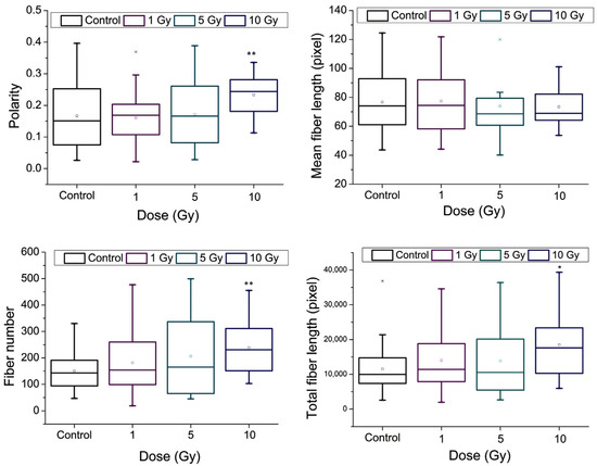
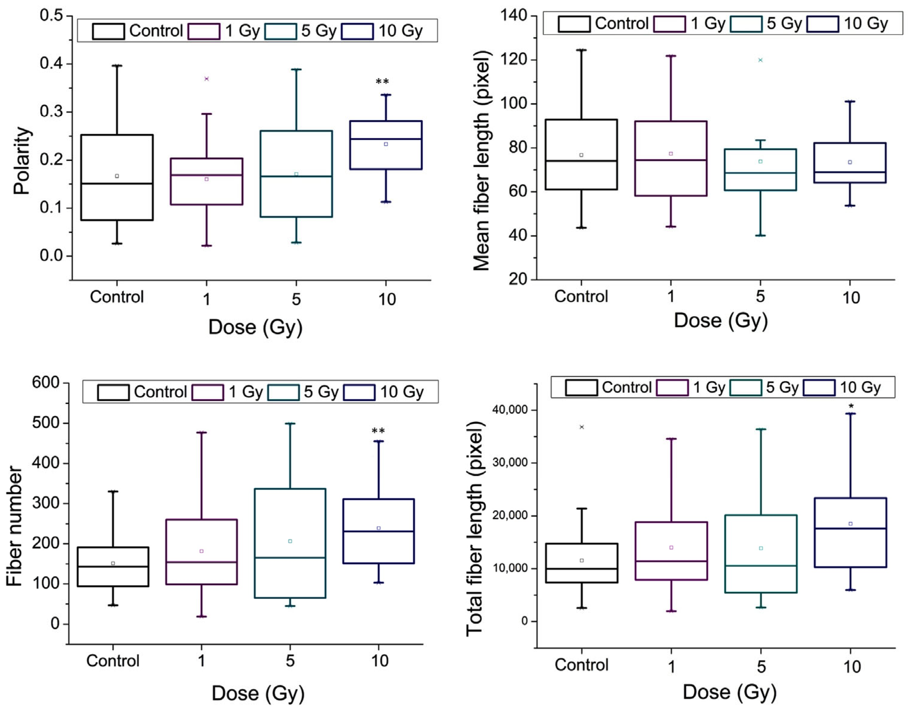
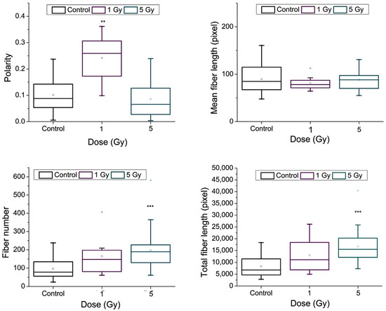
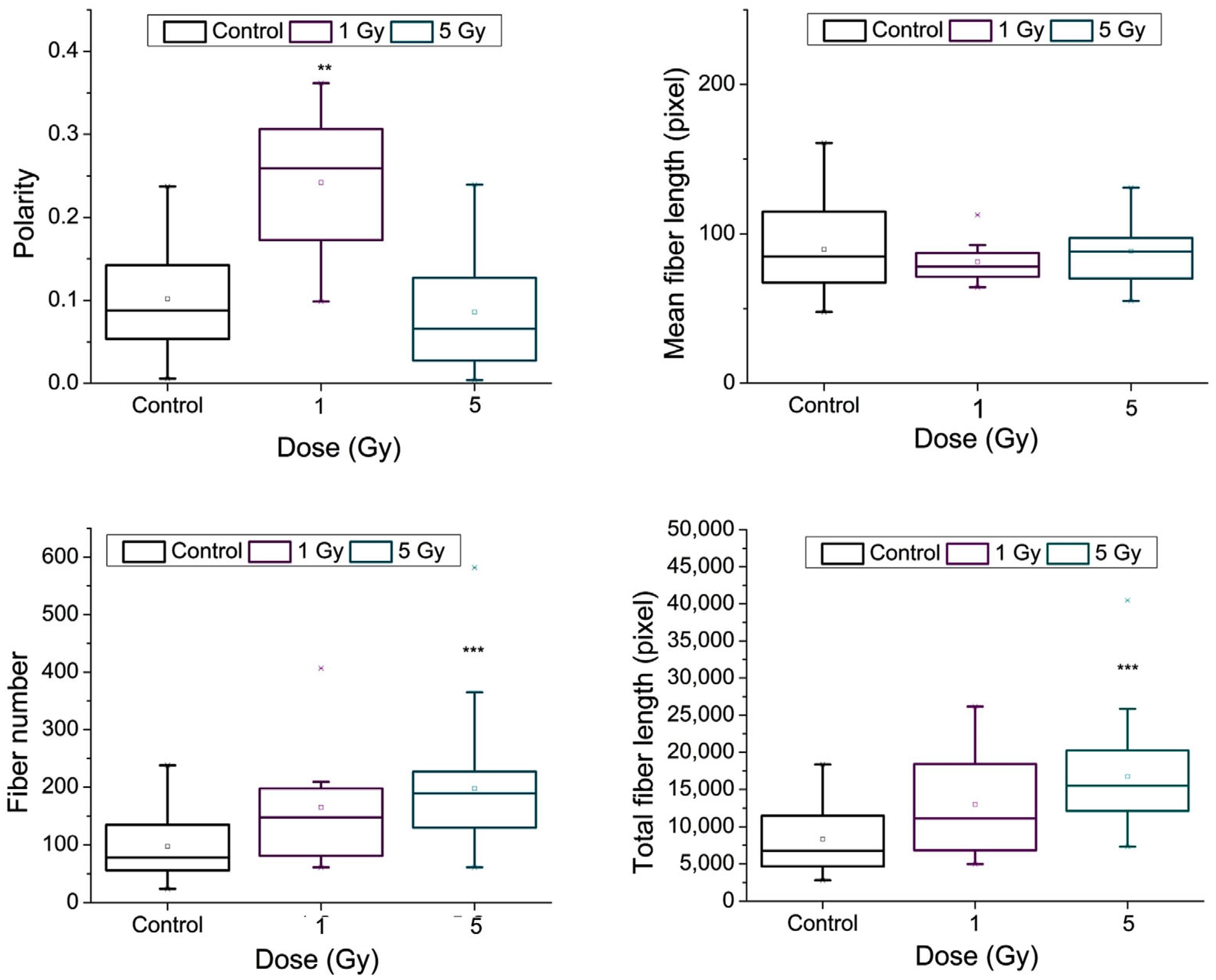
However, quantitative analysis of actin architecture, performed using the FiberScore algorithm, revealed statistically significant changes in actin fiber distribution in both endothelial cell lines (Figure 9 and Figure 10). Specifically, the number of actin fibers increased significantly with rising radiation dose, whereas the average fiber length remained largely unchanged. This resulted in an overall increase in total fiber length—an effect more pronounced in HBEC-5i cells, which exhibited approximately a two-fold increase at 5 Gy (the images at 10 Gy of HBEC-5i cell cultures were qualitatively not appropriate for the FiberScore analysis). In contrast, a similar degree of change was observed in bEnd.3 cells only at 10 Gy. Actin fiber polarity appeared to remain stable across most conditions, except in HBEC-5i cells exposed to 1 Gy, where a significant increase in polarity was detected.
Figure 9.
Quantitative analysis of actin fiber parameters in bEnd.3 cells 24 h post-irradiation using the Fiberscore algorithm. Data (n = 3 independent experiments, each performed in triplicate) are presented as box plots that include the following parameters: median (−), limits of the box 25–75 percentiles, maximum and minimum (-). For each condition, at least 50 cells were analyzed. Statistical analysis was performed using Kruskal–Wallis test with Mann–Whitney as post hoc. Statistical significance (compared to control) is indicated as follows: * p < 0.05, ** p < 0.01.
Figure 10.
Quantitative analysis of actin fiber parameters in HBEC-5i cells 24 h post-irradiation using the Fiberscore algorithm. Data (n = 3 independent experiments, each performed in triplicate) are presented as box plots that include the following parameters: median (−), limits of the box 25–75 percentiles, maximum and minimum (-). For each condition, at least 50 cells were analyzed. Statistical analysis was performed using Kruskal–Wallis test with Mann–Whitney as post hoc. Statistical significance (compared to control) is indicated as follows: ** p < 0.01, and *** p < 0.001.
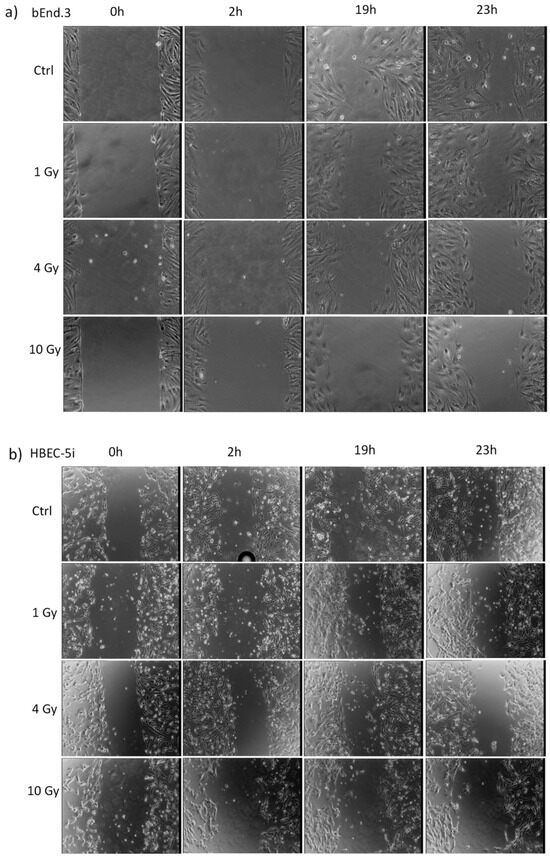
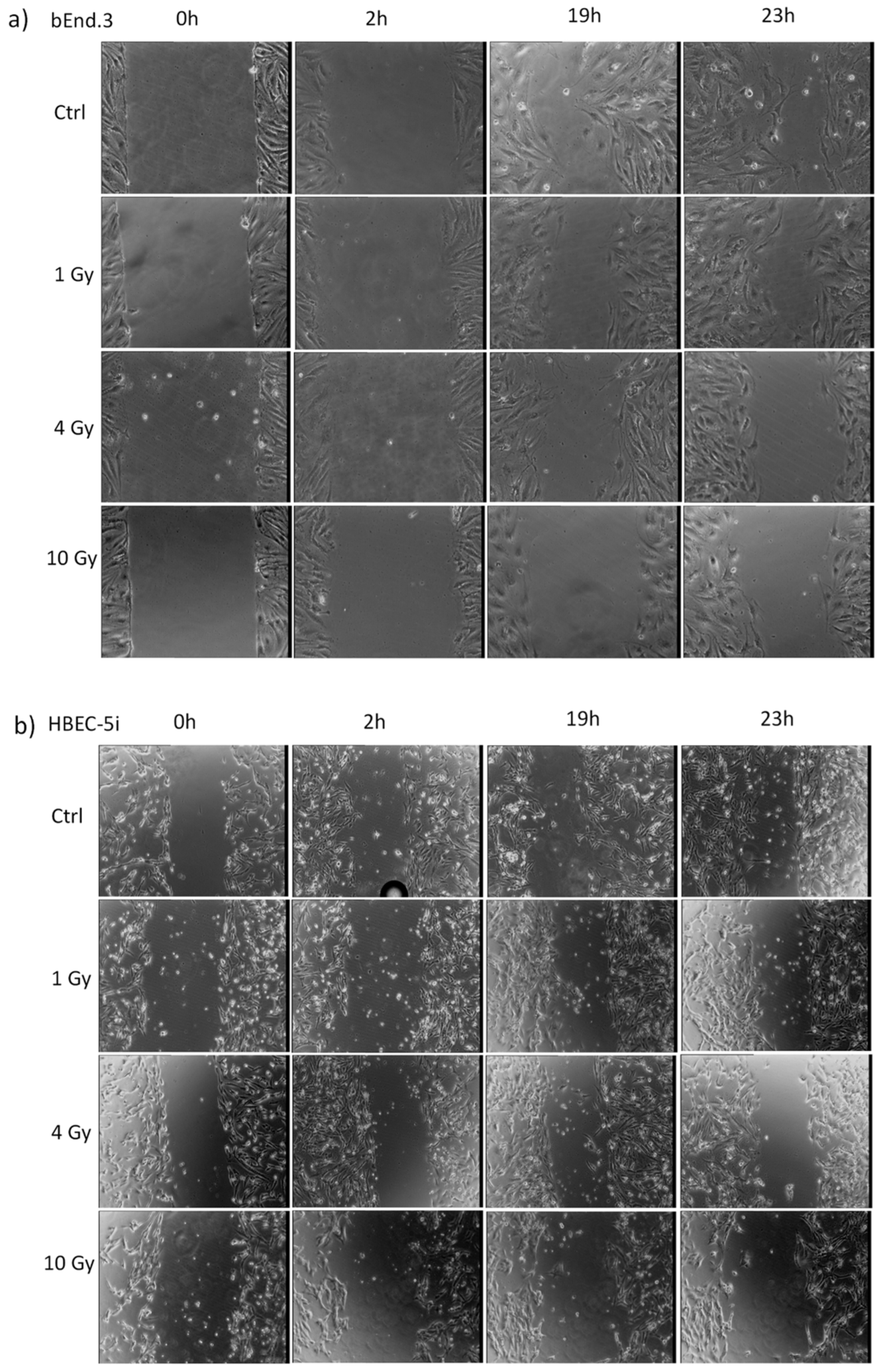
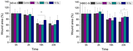
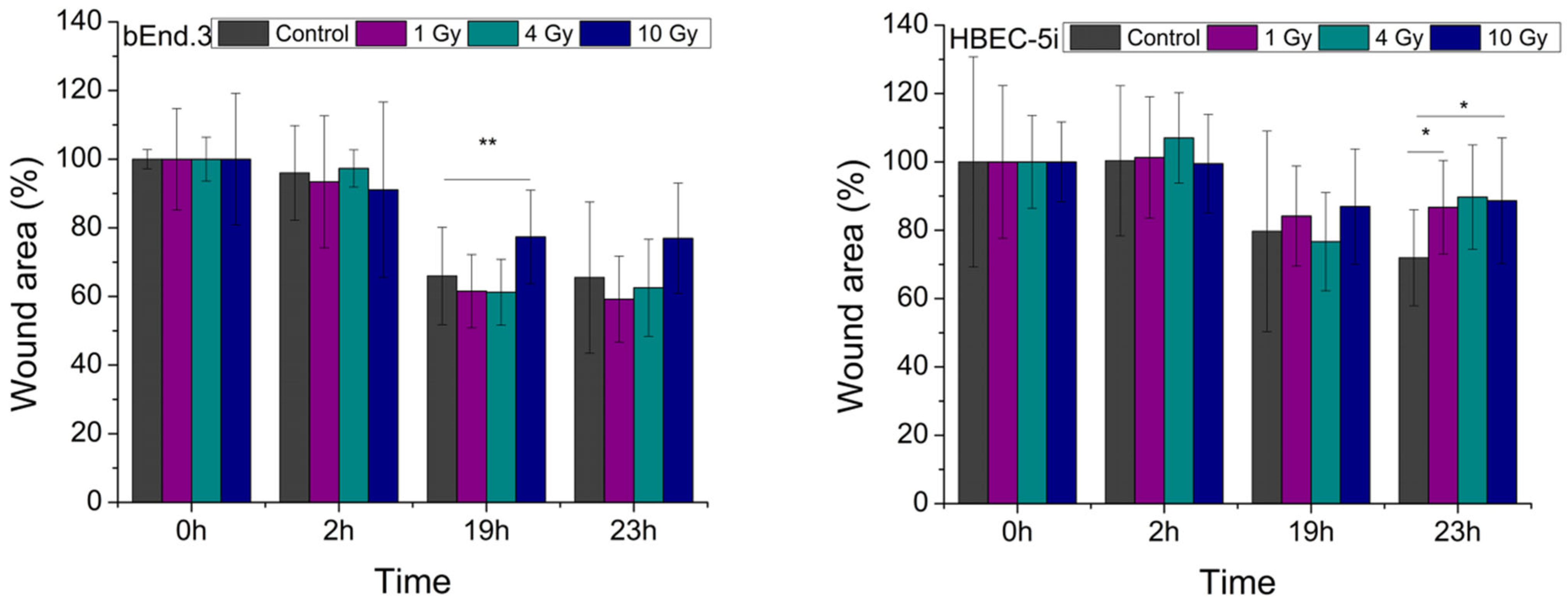
Regarding endothelial cell migration, a noticeable reduction in the migration rate was observed in irradiated cells compared to controls (Figure 11 and Figure 12). For the mouse endothelial cell line (bEnd.3), 19 h after the scratch was introduced, cells exposed to 10 Gy displayed a significantly slower wound closure rate. In contrast, human HBEC-5i cells showed a statistically significant reduction in migration across all irradiated groups as early as 23 h post-scratch.
Figure 11.
The time evolution of cell migration in in vitro wound healing test (representative images): (a) bEnd3 cells and (b) HBEC-5i cells.
Figure 12.
Evaluation of the wound area covered by irradiated endothelial cells at various time intervals. Data are presented as mean ± SD from n = 3 independent experiments, each performed in triplicate. Statistical analysis was performed using Kruskal–Wallis test with Mann–Whitney as post hoc. Statistical significance is indicated as follows: * p < 0.05, ** p < 0.01.
4. Discussion
Recent advances in radiotherapy research emphasize the need to consider not only tumor cells but also the surrounding healthy tissues within the tumor microenvironment. Exposure of healthy cells to X-rays can result in functional impairment and, ultimately, cell death—effects that become more pronounced at higher radiation doses [29]. In the treatment of brain tumors, particularly gliomas, a combined approach involving radiotherapy and chemotherapy is commonly employed [11,30]. Gliomas are known for their infiltrative nature, often spreading between and integrating with blood vessels, which leads to the unintended irradiation of the endothelial cells that form these vessels. These endothelial cells, integral to the structure and function of the BBB, become collateral targets during treatment. Although the BBB plays a crucial role in protecting neural tissue by selectively regulating the passage of substances from the bloodstream, this very selectivity poses a significant obstacle for drug delivery in the treatment of central nervous system disorders [2].
Irradiation of endothelial cells leads to a range of biological effects, including the production of ROS, DNA double-strand breaks, apoptosis, senescence, cell death, cytoskeletal reorganization, prolonged cell cycle phases, reduced expression of tight junction proteins, and increased endothelial permeability [6]. In the context of understanding radiation-induced effects on healthy cells within the tumor microenvironment, numerous studies have focused on the behavior of brain microvascular endothelial cells [31,32,33,34]. However, to date, no study has provided a detailed characterization of the human brain endothelial cell line HBEC-5i or directly compared it with bEnd.3 cells, a widely accepted murine BBB model. Actually, even the response of this cell line (bEnd.3) to X-ray irradiation remains poorly characterized, with only a limited number of studies available in the literature [6,14,15,35]. Such a comparison is particularly relevant, as HBEC-5i cells offer a more representative model for understanding how human brain endothelium responds to radiotherapy. In this study, we investigated both fundamental cellular functions—such as viability, cell cycle distribution, and genotoxic effects—using well-established methods, as well as functional properties, including cell migration and intracellular calcium signaling. To our knowledge, this is the first report to examine calcium signaling responses to irradiation in brain endothelial cells, providing new insights into how radiation affects purinergic signaling in the human cerebral microvasculature.
All methods employed to evaluate the impact of X-ray irradiation on cellular proliferative capacity—namely cell viability, survival fraction, and cell cycle progression—support existing findings in the literature regarding the detrimental effects of ionizing radiation on normal cell culture dynamics, with a clear dose-dependent relationship [9,36]. Specifically, the survival fraction followed the linear-quadratic model, with fitting parameters consistent with previous studies on various endothelial cell types, including bEnd.3 [14], human dermal microvascular endothelial cells [37] and human umbilical vein endothelial cells (HUVECs) [38].
The genotoxic effects observed in our study are consistent with findings reported in the previous literature. Notably, the dose-dependent increase in micronuclei formation in bEnd.3 cells closely mirrors the results presented in the only study we identified that specifically examines the effects of X-ray irradiation on bEnd.3 cells across a similar dose range [14], in which the authors conclude that bEnd.3 cells exhibit radiosensitivity. Comparable outcomes have also been reported in studies involving other types of endothelial cells [9,14,29]. An interesting distinction emerged in the comparison between the two cell lines: the percentage of binucleated cells in HBEC-5i cultures showed a marked decline at doses above 1 Gy, whereas in bEnd.3 cultures, a noticeable reduction occurred only at doses exceeding 6 Gy. This contrast further supports the conclusion that HBEC-5i cells are more sensitive to ionizing radiation than bEnd.3 cells.
Intracellular calcium ions play a crucial role in regulating endothelial cell metabolism, acting as key mediators in multiple intracellular signaling pathways [39]. Calcium ions have a dual role in the regulation of reactive oxygen species (ROS) signaling, acting in opposing directions. On one hand, elevated intracellular calcium can enhance ROS production by stimulating mitochondrial oxidative phosphorylation; on the other, it can promote antioxidant responses that reduce ROS levels [3]. Moreover, it has been proposed that ionizing radiation activates a signaling triad involving ATP, ROS, and calcium ions. ROS are among the first molecules generated following radiation exposure; however, due to their short half-life, the sustained propagation of radiation-induced effects relies on secondary messengers. In this context, extracellular ATP serves as a signaling support, while intracellular calcium ions function as key effectors, activating various metabolic pathways influenced by radiation [3,40]. In our study, exposure of endothelial cells to X-ray radiation was found to modulate the purinergic signaling pathway, as reflected by changes in the dynamics of calcium transients. These alterations were cell line-specific, suggesting distinct regulatory mechanisms in HBEC-5i and bEnd.3 cells. The complexity of radiation-induced calcium signaling modulation is further supported by transcriptomic analyses, which have identified both upregulation and downregulation of genes involved in calcium signaling pathways in brain microvascular endothelial cells following radiation exposure [11]. To the best of our knowledge, this is the first study to report the modulation of ATP-induced calcium transients by X-ray irradiation in endothelial cells. These functional changes indicate that radiation may impact key components of the purinergic signaling pathway. Ongoing and future work will integrate molecular biology approaches to further delineate the specific elements of this signaling cascade that are altered in response to radiation exposure.
Exposure to ionizing radiation is known to induce cytoskeletal remodeling, which can impact key cellular functions such as migration, adhesion, intercellular connectivity, and barrier permeability [4,41]. Specifically, qualitative increases in actin filament density have been reported in HUVEC cultures following X-ray irradiation at doses of 5 and 10 Gy [2], and across a broader dose range of 0–8 Gy [42]. Similar findings were observed in osteoblast cultures irradiated at 0.5 and 5 Gy, where an increase in actin filament formation at 24 h post-irradiation was correlated with the activation of the RhoA/Rho-associated kinase signaling pathway [43,44]. Our findings are consistent with these previous reports, demonstrating a radiation-induced increase in actin filament density in endothelial cells. In addition, our study provides a quantitative assessment of actin filament organization, including measurements of filament length and polarity, using advanced fluorescence image analysis through the Fiberscore algorithm. This highlights the value of computational image analysis tools in characterizing subtle cytoskeletal alterations induced by ionizing radiation.
As anticipated, cytoskeletal remodeling induced by ionizing radiation impacted the migratory capacity of endothelial cells, as assessed by the wound healing assay. Our results showed a dose-dependent reduction in migration rate, aligning with findings from previous studies. For instance, experiments conducted on primary bovine aortic endothelial cells and human aortic endothelial cells demonstrated a similar decrease in migration following γ-ray irradiation at doses of 2 and 10 Gy, with the effect being clearly dose-related [45]. However, it has to be mentioned a possible influence of proliferation rate on the wound closure. Consistent with other endpoints in our study, HBEC-5i cells appeared more radiosensitive than bEnd.3 cells. In HBEC-5i cultures, reduced migration was observed under all irradiation conditions, whereas in bEnd.3 cells, a significant decline in migration rate was evident only at the highest dose of 10 Gy.
5. Conclusions
In this study, we characterized the effects induced by X-rays in human endothelial cell model, HBEC-5i, and compared it with the widely used murine cell line bEnd.3. Our findings suggest that HBEC-5i offers a more relevant model for investigating the side effects of X-ray irradiation on the human brain. Given that X-rays are the most cost-effective and commonly used modality in radiotherapy, understanding their unintended effects on healthy brain tissue is of critical importance. Under our experimental conditions, the well-documented cytotoxic and genotoxic effects of ionizing radiation were confirmed, with HBEC-5i cells displaying greater radiosensitivity than bEnd.3 cells. Additionally, our data indicate that radiation-induced modulation of functional processes such as cell migration and calcium signaling may be linked to earlier events like cytoskeletal remodeling and ROS generation. While further studies are necessary to fully elucidate the underlying molecular mechanisms, our findings contribute to the growing body of evidence supporting the value of in vitro 2D cell culture models as foundational tools in radiobiological research—models that can pave the way for more advanced and physiologically relevant systems.
Author Contributions
Conceptualization, M.R. and B.M.R.; methodology, R.M., S.R.L.P., C.M.R., M.R.I.; software, R.M. and C.M.R.; formal analysis, R.M. and C.M.R.; investigation, R.M.; writing—R.M., B.M.R. and M.R.; writing—review and editing, M.R. and B.M.R.; supervision, M.R. and B.M.R.; funding acquisition, M.R. All authors have read and agreed to the published version of the manuscript.
Funding
This research was funded by the Romanian Ministry of Education and Research, project numbers PN 23210202/2023 and PN 23210203/2023.
Institutional Review Board Statement
Not applicable.
Informed Consent Statement
Not applicable.
Data Availability Statement
The original contributions presented in this study are included in the article; further inquiries can be directed to the corresponding author.
Conflicts of Interest
The authors declare that they have no competing interests. The funders had no role in the design of the study; the collection, analysis, and interpretation of data; or in writing the manuscript.
Abbreviations
The following abbreviations are used in this manuscript:
| BBB | Blood–brain barrier |
| CNS | Central nervous system |
| ECGF | Endothelial cell growth factor |
| DAMPs | Damage-associated molecular patterns |
| DMEM | Dulbecco’s Modified Eagle Medium |
| DMSO | Dimethyl sulfoxide |
| DNA | Deoxyribonucleic acid |
| DSB | Double-strand break |
| FBS | Fetal bovine serum |
| HUVEC | Human umbilical vein endothelial cell |
| HBEC-5i | Human brain endothelial |
| LQ Model | Linear Quadratic model |
| MTT | 3-(4,5-dimethylthiazol-2-yl)-2,5-diphenyltetrazolium bromide |
| NDI | Nuclear division index |
| PBS | Phosphate-buffered saline |
| PI | Propidium iodide |
| RBE | Relative biological effectiveness |
| ROS | Reactive oxygen species |
| RPM | Revolutions per minute |
| SOCE | Store-operated calcium entry |
| γ-H2AX | Phosphorylated histone H2AX |
References
- Hart, E.; Odé, Z.; Derieppe, M.P.P.; Groenink, L.; Heymans, M.W.; Otten, R.; Lequin, M.H.; Janssens, G.O.R.; Hoving, E.W.; Van Vuurden, D.G. Blood-Brain Barrier Permeability Following Conventional Photon Radiotherapy—A Systematic Review and Meta-Analysis of Clinical and Preclinical Studies. Clin. Transl. Radiat. Oncol. 2022, 35, 44–55. [Google Scholar] [CrossRef] [Scilit] [PubMed]
- Gabryś, D.; Greco, O.; Patel, G.; Prise, K.M.; Tozer, G.M.; Kanthou, C. Radiation Effects on the Cytoskeleton of Endothelial Cells and Endothelial Monolayer Permeability. Int. J. Radiat. Oncol. Biol. Phys. 2007, 69, 1553–1562. [Google Scholar] [CrossRef] [Scilit] [PubMed]
- Hoorelbeke, D.; Decrock, E.; De Smet, M.; De Bock, M.; Descamps, B.; Van Haver, V.; Delvaeye, T.; Krysko, D.V.; Vanhove, C.; Bultynck, G.; et al. Cx43 Channels and Signaling via IP3/Ca2+, ATP, and ROS/NO Propagate Radiation-Induced DNA Damage to Non-Irradiated Brain Microvascular Endothelial Cells. Cell Death Dis. 2020, 11, 194. [Google Scholar] [CrossRef] [Scilit] [PubMed]
- Guipaud, O.; Jaillet, C.; Clément-Colmou, K.; François, A.; Supiot, S.; Milliat, F. The Importance of the Vascular Endothelial Barrier in the Immune-Inflammatory Response Induced by Radiotherapy. BJR 2018, 91, 20170762. [Google Scholar] [CrossRef] [Scilit]
- Sharma, P.; Templin, T.; Grabham, P. Short Term Effects of Gamma Radiation on Endothelial Barrier Function: Uncoupling of PECAM-1. Microvasc. Res. 2013, 86, 11–20. [Google Scholar] [CrossRef] [Scilit]
- Baselet, B.; Sonveaux, P.; Baatout, S.; Aerts, A. Pathological Effects of Ionizing Radiation: Endothelial Activation and Dysfunction. Cell. Mol. Life Sci. 2019, 76, 699–728. [Google Scholar] [CrossRef] [Scilit]
- Central Nervous System (CNS) Tumours. Radiotherapy Dose Fractionation; The Royal College of Radiologists: London, UK, 2024.
- Eckert, D.; Rapp, F.; Tsedeke, A.T.; Molendowska, J.; Lehn, R.; Langhans, M.; Fournier, C.; Rödel, F.; Hehlgans, S. ROS- and Radiation Source-Dependent Modulation of Leukocyte Adhesion to Primary Microvascular Endothelial Cells. Cells 2022, 11, 72. [Google Scholar] [CrossRef] [Scilit]
- Helm, A.; Lee, R.; Durante, M.; Ritter, S. The Influence of C-Ions and X-Rays on Human Umbilical Vein Endothelial Cells. Front. Oncol. 2016, 6, 5. [Google Scholar] [CrossRef] [Scilit]
- Kouam, P.N.; Rezniczek, G.A.; Adamietz, I.A.; Bühler, H. Ionizing Radiation Increases the Endothelial Permeability and the Transendothelial Migration of Tumor Cells Through ADAM10-Activation and Subsequent Degradation of VE-Cadherin. BMC Cancer 2019, 19, 958. [Google Scholar] [CrossRef] [Scilit]
- Wu, Q.; Fang, Y.; Zhang, X.; Song, F.; Wang, Y.; Chen, H.; Du, J.; Zheng, C.-B.; Shen, B. Effect of X-Rays on Transcript Expression of Rat Brain Microvascular Endothelial Cells: Role of Calcium Signaling in X-Ray-Induced Endothelium Damage. Biosci. Rep. 2020, 40, BSR20193760. [Google Scholar] [CrossRef] [Scilit]
- Xu, F.; Wang, Y.; Gao, H.; Zhang, X.; Hu, Y.; Han, T.; Shen, B.; Zhang, L.; Wu, Q. X-Ray Causes mRNA Transcripts Change to Enhance Orai2-Mediated Ca2+ Influx in Rat Brain Microvascular Endothelial Cells. Front. Mol. Biosci. 2021, 8, 646730. [Google Scholar] [CrossRef] [Scilit] [PubMed]
- Zhou, H.; Jin, C.; Cui, L.; Xing, H.; Liu, J.; Liao, W.; Liao, H.; Yu, Y. HMGB1 Contributes to the Irradiation-induced Endothelial Barrier Injury through Receptor for Advanced Glycation Endproducts (RAGE). J. Cell. Physiol. 2018, 233, 6714–6721. [Google Scholar] [CrossRef] [Scilit] [PubMed]
- Ma, L.; Ye, Z.; Zhang, Y.; Shi, W.; Wang, J.; Yang, H. Irradiated Microvascular Endothelial Cells May Induce Bystander Effects in Neural Stem Cells Leading to Neurogenesis Inhibition. J. Radiat. Res. 2022, 63, 192–201. [Google Scholar] [CrossRef] [Scilit] [PubMed]
- Banaz-Yaşar, F.; Tischka, R.; Iliakis, G.; Winterhager, E.; Gellhaus, A. Cell Line Specific Modulation of Connexin43 Expression after Exposure to Ionizing Radiation. Cell Commun. Adhes. 2005, 12, 249–259. [Google Scholar] [CrossRef] [Scilit]
- Liu, S.; Sammons, V.; Fairhall, J.; Reddy, R.; Tu, J.; Hong Duong, T.T.; Stoodley, M. Molecular Responses of Brain Endothelial Cells to Radiation in a Mouse Model. J. Clin. Neurosci. 2012, 19, 1154–1158. [Google Scholar] [CrossRef] [Scilit]
- Valentin, J. Relative Biological Effectiveness (RBE), Quality Factor (Q), and Radiation Weighting Factor (wR): ICRP Publication 92: Approved by the Commission in January 2003. Ann. ICRP 2003, 33, 1–121. [Google Scholar] [CrossRef] [Scilit]
- Ben Kacem, M.; Benadjaoud, M.A.; Dos Santos, M.; Buard, V.; Tarlet, G.; Le Guen, B.; François, A.; Guipaud, O.; Milliat, F.; Paget, V. Variation of 4 MV X-Ray Dose Rate in Fractionated Irradiation Strongly Impacts Biological Endothelial Cell Response In Vitro. Int. J. Radiat. Biol. 2022, 98, 50–59. [Google Scholar] [CrossRef] [Scilit]
- Hill, M.A.; Patllo, I.S.; Aitkenhead, A.H.; Bazalova-Carter, M.; Carter, R.; Nill, S.; Nisbet, A.; Ghita-Pettigrew, M.; Poirier, Y.; Prise, K.M.; et al. The importance of standardization and challenges of dosimetry in conventional preclinical radiation biology research. Br. J. Radiol. 2025, 98, 993–1004. [Google Scholar] [CrossRef] [Scilit]
- McMahon, S.J. The Linear Quadratic Model: Usage, Interpretation and Challenges. Phys. Med. Biol. 2018, 64, 01TR01. [Google Scholar] [CrossRef] [Scilit]
- Rusu, C.M.; Bacalum, M.; Deftu, A.T.; Radu, B.M.; Radu, M. Digital Analysis of Actin FIlaments Images Obtained by Fluorescence Microscopy. Rom. J. Biophys. 2018, 28, 147–157. [Google Scholar]
- Lichtenstein, N.; Geiger, B.; Kam, Z. Quantitative Analysis of Cytoskeletal Organization by Digital Fluorescent Microscopy. Cytom. Part A 2003, 54A, 8–18. [Google Scholar] [CrossRef] [Scilit] [PubMed]
- Ipek, E. The Relationship of Micronucleus Frequency and Nuclear Division Index with Coronary Artery Disease SYNTAX and Gensini Scores. Anatol. J. Cardiol. 2017, 17, 483–489. [Google Scholar] [CrossRef] [Scilit] [PubMed]
- Radu, B.M.; Radu, M.; Tognoli, C.; Benati, D.; Merigo, F.; Assfalg, M.; Solani, E.; Stranieri, C.; Ceccon, A.; Fratta Pasini, A.M.; et al. Are They in or out? The Elusive Interaction between Qtracker® 800 Vascular Labels and Brain Endothelial Cells. Nanomedicine 2015, 10, 3329–3342. [Google Scholar] [CrossRef] [Scilit] [PubMed]
- Reyes-Aldasoro, C.C.; Wilson, I.; Prise, V.E.; Barber, P.R.; Ameer-Beg, M.; Vojnovic, B.; Cunningham, V.J.; Tozer, G.M. Estimation of Apparent Tumor Vascular Permeability from Multiphoton Fluorescence Microscopic Images of P22 Rat Sarcomas In Vivo. Microcirculation 2008, 15, 65–79. [Google Scholar] [CrossRef] [Scilit]
- Grada, A.; Otero-Vinas, M.; Prieto-Castrillo, F.; Obagi, Z.; Falanga, V. Research Techniques Made Simple: Analysis of Collective Cell Migration Using the Wound Healing Assay. J. Investig. Dermatol. 2017, 137, e11–e16. [Google Scholar] [CrossRef] [Scilit]
- Wang, Q.-Q.; Yin, G.; Huang, J.-R.; Xi, S.-J.; Qian, F.; Lee, R.-X.; Peng, X.-C.; Tang, F.-R. Ionizing Radiation-Induced Brain Cell Aging and the Potential Underlying Molecular Mechanisms. Cells 2021, 10, 3570. [Google Scholar] [CrossRef] [Scilit]
- Serrano-Mendioroz, I.; Garate-Soraluze, E.; Rodriguez-Ruiz, M.E. A Simple Method to Assess Clonogenic Survival of Irradiated Cancer Cells. In Methods in Cell Biology; Elsevier Science & Technology: San Diego, CA, USA, 2023; Volume 174, ISBN 978-0-323-89947-5. [Google Scholar]
- Possenti, L.; Mecchi, L.; Rossoni, A.; Sangalli, V.; Bersini, S.; Cicchetti, A.; Costantino, M.L.; Candrian, C.; Arrigoni, C.; Rancati, T.; et al. Radiobiological Studies of Microvascular Damage through In Vitro Models: A Methodological Perspective. Cancers 2021, 13, 1182. [Google Scholar] [CrossRef] [Scilit]
- Lerner, A.; Palmer, K.; Campion, T.; Millner, T.O.; Scott, E.; Lorimer, C.; Paraskevopoulos, D.; McKenna, G.; Marino, S.; Lewis, R.; et al. Gliomas in Adults: Guidance on Investigations, Diagnosis, Treatment and Surveillance. Clin. Med. 2024, 24, 100240. [Google Scholar] [CrossRef] [Scilit]
- Yoshida, Y.; Sejimo, Y.; Kurachi, M.; Ishizaki, Y.; Nakano, T.; Takahashi, A. X-Ray Irradiation Induces Disruption of the Blood–Brain Barrier with Localized Changes in Claudin-5 and Activation of Microglia in the Mouse Brain. Neurochem. Int. 2018, 119, 199–206. [Google Scholar] [CrossRef] [Scilit]
- Sándor, N.; Walter, F.R.; Bocsik, A.; Sántha, P.; Schilling-Tóth, B.; Léner, V.; Varga, Z.; Kahán, Z.; Deli, M.A.; Sáfrány, G.; et al. Low Dose Cranial Irradiation-Induced Cerebrovascular Damage Is Reversible in Mice. PLoS ONE 2014, 9, e112397. [Google Scholar] [CrossRef] [Scilit]
- Li, Y.-Q.; Chen, P.; Jain, V.; Reilly, R.M.; Wong, C.S. Early Radiation-Induced Endothelial Cell Loss and Blood–Spinal Cord Barrier Breakdown in the Rat Spinal Cord. Radiat. Res. 2004, 161, 143–152. [Google Scholar] [CrossRef] [Scilit] [PubMed]
- Grabham, P.; Sharma, P. Acute Effects of Ionizing Radiation on Human Endothelial Barrier Function. J. Radiat. Res. 2014, 55, i97–i98. [Google Scholar] [CrossRef] [Scilit]
- Schröder, S.; Kriesen, S.; Paape, D.; Hildebrandt, G.; Manda, K. Modulation of Inflammatory Reactions by Low-Dose Ionizing Radiation: Cytokine Release of Murine Endothelial Cells Is Dependent on Culture Conditions. J. Immunol. Res. 2018, 2018, 2856518. [Google Scholar] [CrossRef] [Scilit] [PubMed]
- Panganiban, R.A.M.; Mungunsukh, O.; Day, R.M. X-Irradiation Induces ER Stress, Apoptosis, and Senescence in Pulmonary Artery Endothelial Cells. Int. J. Radiat. Biol. 2013, 89, 656–667. [Google Scholar] [CrossRef] [Scilit]
- Bernardo, T.; Kuntze, A.; Klein, D.; Heinzelmann, F.; Timmermann, B.; Von Neubeck, C. Endothelial Cell Response to Combined Photon or Proton Irradiation with Doxorubicin. IJMS 2023, 24, 12833. [Google Scholar] [CrossRef] [Scilit]
- Kim, E.H.; Kim, M.-S.; Jeong, Y.K.; Cho, I.; You, S.H.; Cho, S.H.; Lee, H.; Jung, W.-G.; Kim, H.D.; Kim, J. Mechanisms for SU5416 as a Radiosensitizer of Endothelial Cells. Int. J. Oncol. 2015, 47, 1440–1450. [Google Scholar] [CrossRef] [Scilit]
- Stoica, R.; Rusu, C.M.; Staicu, C.; Burlacu, A.; Radu, M.; Radu, B.M. Ca2+ Homeostasis in Brain Microvascular Endothelial Cells. In International Review of Cell and Molecular Biology; Elsevier: Amsterdam, The Netherlands, 2021; Volume 362, pp. 55–110. ISBN 978-0-12-824034-2. [Google Scholar]
- Voos, P.; Fuck, S.; Weipert, F.; Babel, L.; Tandl, D.; Meckel, T.; Hehlgans, S.; Fournier, C.; Moroni, A.; Rödel, F.; et al. Ionizing Radiation Induces Morphological Changes and Immunological Modulation of Jurkat Cells. Front. Immunol. 2018, 9, 922. [Google Scholar] [CrossRef] [Scilit]
- La Verde, G.; Artiola, V.; Panzetta, V.; Pugliese, M.; Netti, P.A.; Fusco, S. Cytoskeleton Response to Ionizing Radiation: A Brief Review on Adhesion and Migration Effects. Biomedicines 2021, 9, 1102. [Google Scholar] [CrossRef] [Scilit]
- Mohammadkarim, A.; Tabatabaei, M.; Parandakh, A.; Mokhtari-Dizaji, M.; Tafazzoli-Shadpour, M.; Khani, M.-M. Radiation Therapy Affects the Mechanical Behavior of Human Umbilical Vein Endothelial Cells. J. Mech. Behav. Biomed. Mater. 2018, 85, 188–193. [Google Scholar] [CrossRef] [Scilit]
- Huang, Q.; Chai, H.; Wang, S.; Sun, Y.; Xu, W. 0.5-Gy X-ray Irradiation Induces Reorganization of Cytoskeleton and Differentiation of Osteoblasts. Mol. Med. Rep. 2021, 23, 379. [Google Scholar] [CrossRef] [Scilit]
- Huang, Q.; Zhou, Z.; Yan, F.; Dong, Q.; Wang, L.; Sha, W.; Xu, Q.; Zhu, X.; Zhao, L. Low-dose X-ray Irradiation Induces Morphological Changes and Cytoskeleton Reorganization in Osteoblasts. Exp. Ther. Med. 2020, 20, 283. [Google Scholar] [CrossRef] [Scilit]
- Zheng, X.; Mohan, S.; Otto, R.A.; Natarajan, M. Endothelial Cell Migration Was Impaired by Irradiation-Induced Inhibition of SHP-2 in Radiotherapy: An In Vitro Study. JRR 2011, 52, 320–328. [Google Scholar] [CrossRef] [Scilit]
Disclaimer/Publisher’s Note: The statements, opinions and data contained in all publications are solely those of the individual author(s) and contributor(s) and not of MDPI and/or the editor(s). MDPI and/or the editor(s) disclaim responsibility for any injury to people or property resulting from any ideas, methods, instructions or products referred to in the content. |
© 2026 by the authors. Licensee MDPI, Basel, Switzerland. This article is an open access article distributed under the terms and conditions of the Creative Commons Attribution (CC BY) license.