Intraoperative X-Ray Guidance for Endourological Stone Surgery: Fluoroscopic Workflow, Radiation Dose Optimization, and Emerging Image-Guided Technologies

 , , and
Abstract
1. Introduction
2. Review Scope, Literature Search, and Study Selection
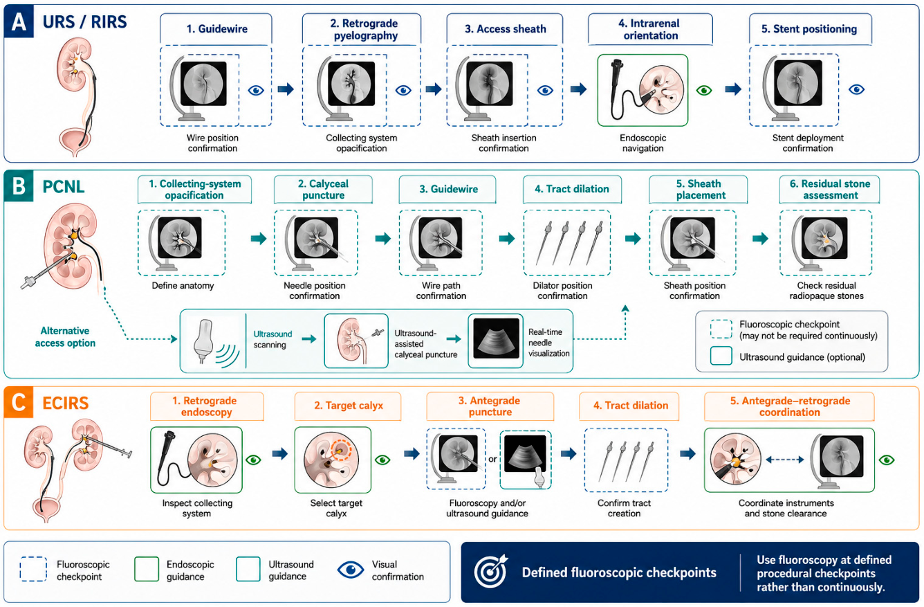
3. Clinical Role of Intraoperative X-Ray Imaging
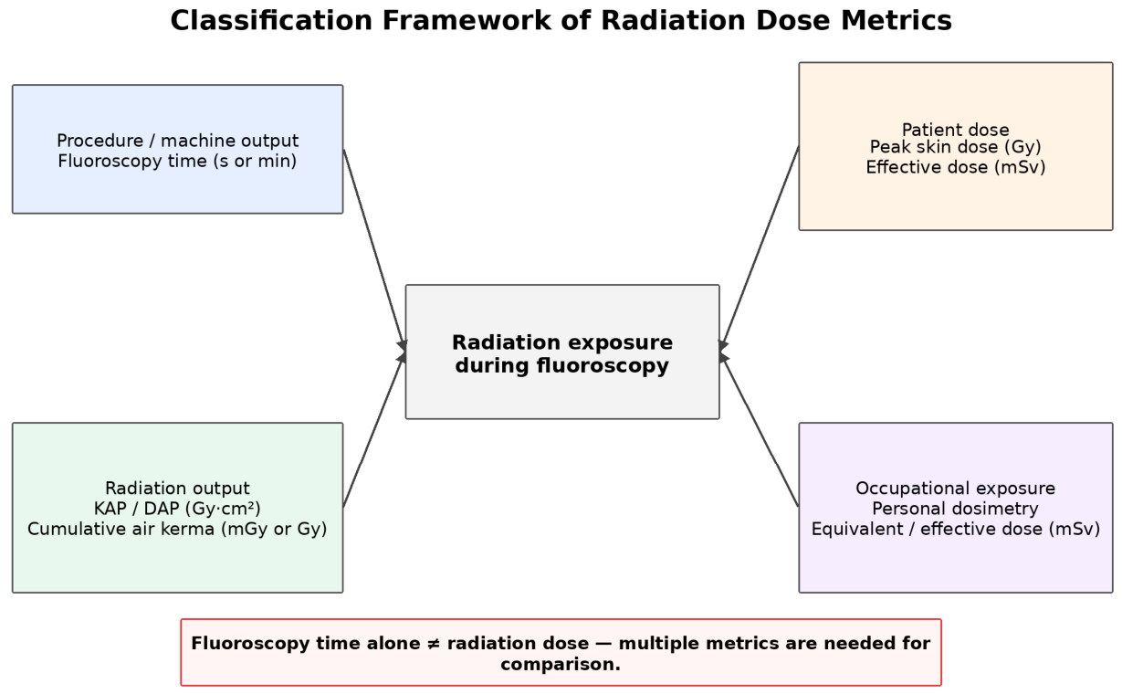
4. Radiation Dose Metrics and Determinants
5. Patient Radiation Exposure
6. Occupational Radiation Exposure
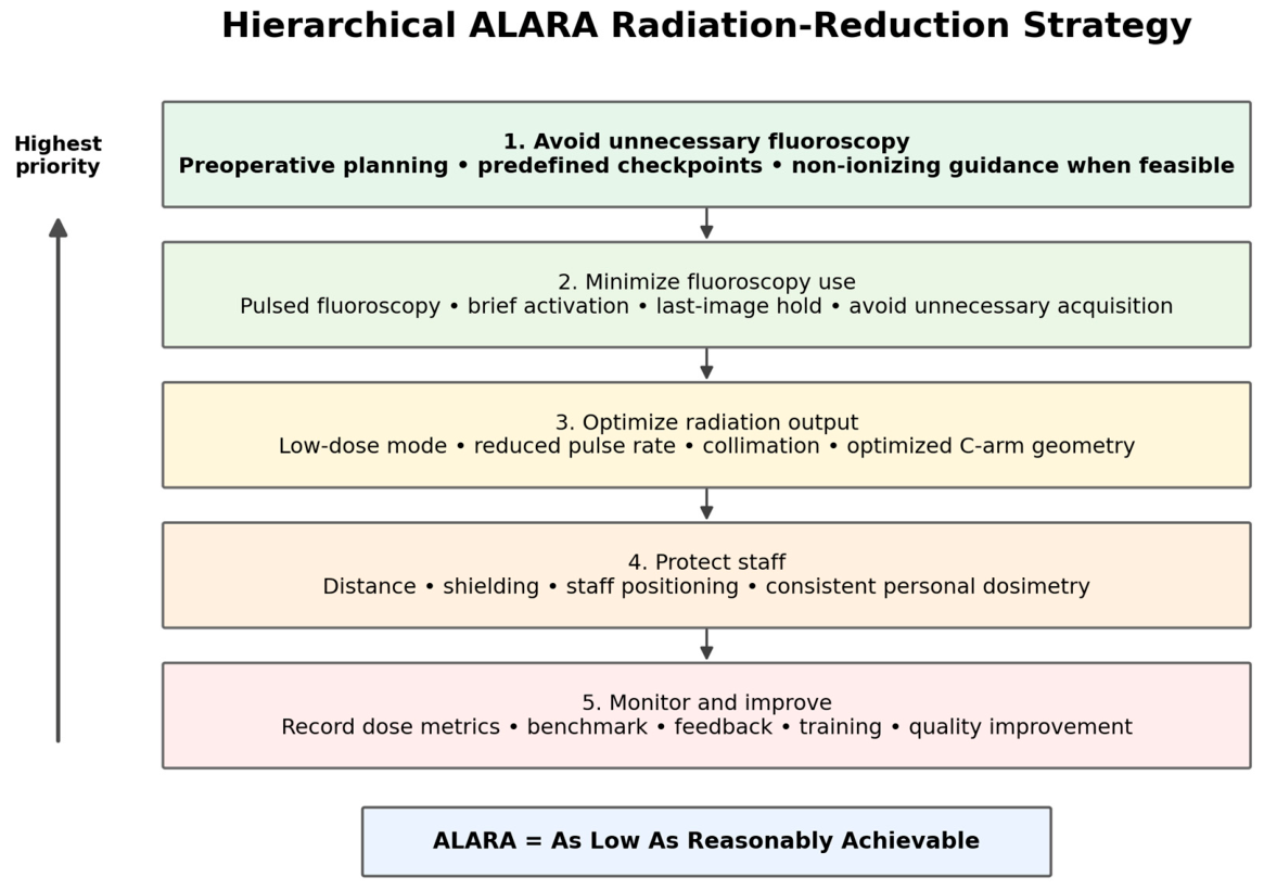
7. Practical Dose-Reduction Strategies Based on ALARA
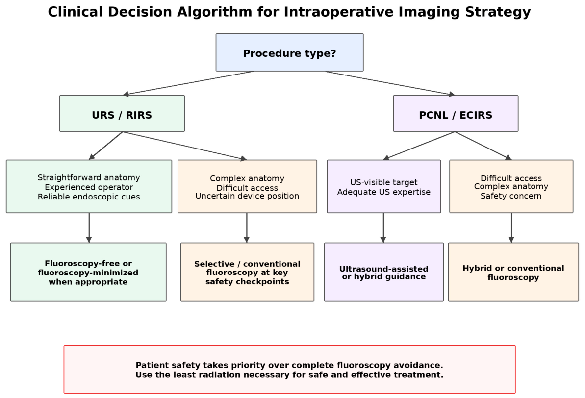
8. Fluoroscopy-Free and Fluoroscopy-Minimized Endourological Surgery
9. Alternative and Multimodal Guidance for PCNL and ECIRS
10. Emerging Image-Guided Technologies
11. Reporting Standards and Procedure-Specific Benchmarking
12. Limitations of This Review
13. Clinical Take-Home Recommendations
- Use fluoroscopy selectively at predefined safety-critical checkpoints rather than as continuous routine imaging.
- Do not use fluoroscopy time as the sole radiation metric; record KAP/DAP and cumulative air kerma when available, together with patient and procedural context.
- Consider fluoroscopy-free or fluoroscopy-minimized URS/RIRS in appropriately selected cases and experienced hands but prioritize procedural safety over an arbitrary zero-fluoroscopy target.
- Use ultrasound-assisted or hybrid ultrasound-fluoroscopy guidance during PCNL/ECIRS when local expertise and equipment are available.
- Treat navigation, augmented reality, robot-assisted guidance, computer vision, and AI-assisted dose optimization according to their evidence level; several remain emerging or experimental and require prospective clinical and dosimetric validation.
14. Conclusions
Supplementary Materials
Author Contributions
Funding
Institutional Review Board Statement
Informed Consent Statement
Data Availability Statement
Acknowledgments
Conflicts of Interest
Abbreviations
| 3D | three-dimensional |
| AI | artificial intelligence |
| ALARA | as low as reasonably achievable |
| AR | augmented reality |
| BMI | body mass index |
| CT | computed tomography |
| DAP | dose-area product |
| EAU | European Association of Urology |
| ECIRS | endoscopic combined intrarenal surgery |
| KAP | kerma–area product |
| PCNL | percutaneous nephrolithotomy |
| RCT | randomized controlled trial |
| RIRS | retrograde intrarenal surgery |
| SANRA | Scale for the Assessment of Narrative Review Articles |
| URS | ureteroscopy |
References
- European Association of Urology. EAU Guidelines on Urolithiasis; EAU Guidelines Office: Arnhem, The Netherlands, 2026; Available online: https://uroweb.org/guidelines/urolithiasis (accessed on 30 July 2026).
- Pearle, M.S.; Matlaga, B.R.; Antonelli, J.A.; Chi, T.; Hsi, R.S.; Kim, S.K.; Kirkby, E.; Knudsen, B.; Koo, K.; Maalouf, N.M.; et al. Surgical Management of Kidney and Ureteral Stones: AUA Guideline (2026) Part I: Evaluation and Treatment of Patients with Kidney and/or Ureteral Stones. J. Urol. 2026, 215, 113–123. [Google Scholar] [CrossRef] [Scilit]
- Pearle, M.S.; Matlaga, B.R.; Antonelli, J.A.; Chi, T.; Hsi, R.S.; Kim, S.K.; Kirkby, E.; Knudsen, B.; Koo, K.; Maalouf, N.M.; et al. Surgical Management of Kidney and Ureteral Stones: AUA Guideline (2026) Part III: Treatment of Patients with Kidney and/or Ureteral Stones and Future Directions. J. Urol. 2026, 215, 132–141. [Google Scholar] [CrossRef] [Scilit]
- International Commission on Radiological Protection. ICRP Publication 103. The 2007 Recommendations of the International Commission on Radiological Protection. Ann. ICRP 2007, 37, 9–34. [Google Scholar] [CrossRef] [Scilit]
- International Commission on Radiological Protection. ICRP Publication 118: ICRP Statement on Tissue Reactions and Early and Late Effects of Radiation in Normal Tissues and Organs—Threshold Doses for Tissue Reactions in a Radiation Protection Context. Ann. ICRP 2012, 41, 1–322. [Google Scholar] [CrossRef] [Scilit]
- Fisher, R.F.; Applegate, K.E.; Berkowitz, L.K.; Christianson, O.; Dave, J.K.; DeWeese, L.; Harris, N.; Jafari, M.E.; Jones, A.K.; Kobistek, R.J.; et al. AAPM Medical Physics Practice Guideline 12.a: Fluoroscopy Dose Management. J. Appl. Clin. Med. Phys. 2022, 23, e13526. [Google Scholar] [CrossRef] [Scilit]
- Lin, P.J.P.; Schueler, B.A.; Balter, S.; Strauss, K.J.; Wunderle, K.A.; LaFrance, M.T.; Kim, D.; Behrman, R.H.; Shepard, S.J.; Bercha, I.H. Accuracy and Calibration of Integrated Radiation Output Indicators in Diagnostic Radiology: A Report of the AAPM Imaging Physics Committee Task Group 190. Med. Phys. 2015, 42, 6815–6829. [Google Scholar] [CrossRef] [Scilit]
- Miller, D.L. Review of Air Kerma-Area Product, Effective Dose and Dose Conversion Coefficients for Non-Cardiac Interventional Fluoroscopy Procedures. Med. Phys. 2020, 47, 975–982. [Google Scholar] [CrossRef] [Scilit]
- Jaco, J.W.; Miller, D.L. Measuring and Monitoring Radiation Dose during Fluoroscopically Guided Procedures. Tech. Vasc. Interv. Radiol. 2010, 13, 188–193. [Google Scholar] [CrossRef] [Scilit]
- Baethge, C.; Goldbeck-Wood, S.; Mertens, S. SANRA—A Scale for the Quality Assessment of Narrative Review Articles. Res. Integr. Peer Rev. 2019, 4, 5. [Google Scholar] [CrossRef] [Scilit]
- Greene, D.J.; Tenggadjaja, C.F.; Bowman, R.J.; Agarwal, G.; Ebrahimi, K.Y.; Baldwin, D.D. Comparison of a Reduced Radiation Fluoroscopy Protocol to Conventional Fluoroscopy during Uncomplicated Ureteroscopy. Urology 2011, 78, 286–290. [Google Scholar] [CrossRef] [Scilit]
- Hein, S.; Schoenthaler, M.; Wilhelm, K.; Schlager, D.; Vach, W.; Wetterauer, U.; Miernik, A. Ultralow Radiation Exposure during Flexible Ureteroscopy in Patients with Nephrolithiasis—How Far Can We Go? Urology 2017, 108, 34–39. [Google Scholar] [CrossRef] [Scilit]
- Chung, J.W.; Kang, J.K.; Jung, W.; Oh, K.J.; Kim, H.W.; Shin, D.G.; Kim, B.S. The Efficacy and Safety of Radiation-Free Retrograde Intrarenal Surgery: A Prospective Multicenter-Based, Randomized, Controlled Trial. J. Urol. 2024, 211, 735–742. [Google Scholar] [CrossRef] [Scilit]
- Davis, N.F.; Tzelves, L.; Geraghty, R.; Lombardo, R.; Yuan, C.; Petrik, A.; Neisius, A.; Gambaro, G.; Jung, H.; Shepherd, R.; et al. Comparison of Treatment Outcomes for Fluoroscopic and Fluoroscopy-Free Endourological Procedures: A Systematic Review on Behalf of the European Association of Urology Urolithiasis Guidelines Panel. Eur. Urol. Focus 2023, 9, 938–953. [Google Scholar] [CrossRef] [Scilit]
- Gimenez, L.G.S.; Santana, D.S.; Lopes, G.M.M.; Cardoso, R.B.; Porto, B.C.; Passerotti, C.C.; Sardenberg, R.A.d.S.; Otoch, J.P.; DA Cruz, J.A.S. Comparison of Efficacy and Safety of Fluoroscopy-Free and Conventional Retrograde Ureteroscopy for Urolithiasis: A Systematic Review and Metanalysis of Randomized Controlled Trials. Minerva Urol. Nephrol. 2025, 77, 162–170. [Google Scholar] [CrossRef] [Scilit]
- Violette, P.D.; Szymanski, K.M.; Anidjar, M.; Andonian, S. Factors Determining Fluoroscopy Time during Ureteroscopy. J. Endourol. 2011, 25, 1837–1840. [Google Scholar] [CrossRef] [Scilit]
- Smith, D.L.; Heldt, J.P.; Richards, G.D.; Agarwal, G.; Brisbane, W.G.; Chen, C.J.; Chamberlin, J.D.; Baldwin, D.D. Radiation Exposure during Continuous and Pulsed Fluoroscopy. J. Endourol. 2013, 27, 384–388. [Google Scholar] [CrossRef] [Scilit]
- Weld, L.R.; Nwoye, U.O.; Knight, R.B.; Baumgartner, T.S.; Ebertowski, J.S.; Stringer, M.T.; Kasprenski, M.C.; Weld, K.J. Safety, Minimization, and Awareness Radiation Training Reduces Fluoroscopy Time during Unilateral Ureteroscopy. Urology 2014, 84, 520–525. [Google Scholar] [CrossRef] [Scilit]
- Usawachintachit, M.; Masic, S.; Chang, H.C.; Allen, I.E.; Chi, T. Ultrasound Guidance to Assist Percutaneous Nephrolithotomy Reduces Radiation Exposure in Obese Patients. Urology 2016, 98, 32–38. [Google Scholar] [CrossRef] [Scilit]
- Wenzler, D.L.; Abbott, J.E.; Su, J.J.; Shi, W.; Slater, R.; Miller, D.; Siemens, M.J.; Sur, R.L. Predictors of Radiation Exposure to Providers during Percutaneous Nephrolithotomy. Urol. Ann. 2017, 9, 55–60. [Google Scholar] [CrossRef] [Scilit]
- Danilovic, A.; Nunes, E.; Lipkin, M.E.; Ferreira, T.; Torricelli, F.C.M.; Marchini, G.S.; Srougi, M.; Nahas, W.C.; Mazzucchi, E. Low Dose Fluoroscopy During Ureteroscopy Does Not Compromise Surgical Outcomes. J. Endourol. 2019, 33, 527–532. [Google Scholar] [CrossRef] [Scilit]
- Hein, S.; Wilhelm, K.; Miernik, A.; Schoenthaler, M.; Suarez-Ibarrola, R.; Gratzke, C.; Salem, J.; Karapanos, L.; Netsch, C.; Becker, B.; et al. Radiation Exposure during Retrograde Intrarenal Surgery (RIRS): A Prospective Multicenter Evaluation. World J. Urol. 2021, 39, 217–224. [Google Scholar] [CrossRef] [Scilit]
- Vassileva, J.; Zagorska, A.; Basic, D.; Karagiannis, A.; Petkova, K.; Sabuncu, K.; Saltirov, I.; Sarica, K.; Skolarikos, A.; Stavridis, S.; et al. Radiation Exposure of Patients during Endourological Procedures: IAEA-SEGUR Study. J. Radiol. Prot. 2020, 40, 1390–1405. [Google Scholar] [CrossRef] [Scilit]
- Zampini, A.M.; Bamberger, J.N.; Gupta, K.; Gallante, B.; Atallah, W.M.; Gupta, M. Factors Affecting Patient Radiation Exposure During Prone and Supine Percutaneous Nephrolithotomy. J. Endourol. 2021, 35, 1448–1453. [Google Scholar] [CrossRef] [Scilit]
- Cheng, G.; Campbell, T.; Feng, C.; Quarrier, S.; Jain, R. Low-Dose Fluoroscopy Technique Drastically Decreases Patient Radiation Exposure during Percutaneous Nephrolithotomy. Urolithiasis 2023, 51, 11. [Google Scholar] [CrossRef] [Scilit]
- De Coninck, V.; Mortiers, X.; Hendrickx, L.; De Wachter, S.; Traxer, O.; Keller, E.X. Radiation Exposure of Patients during Endourological Procedures. World J. Urol. 2024, 42, 266. [Google Scholar] [CrossRef] [Scilit]
- De Coninck, V.; Ferretti, S.; Rico, L.; Chai, C.A.; Shrestha, A.; Gauhar, V.; Monga, M.; Gadzhiev, N.; Panthier, F.; Mortiers, X.; et al. Maximizing Safety in Stone Disease: The Impact of Strict As Low As Reasonably Achievable (ALARA) Principle Adherence on Radiation Exposure and Clinical Outcomes. World J. Urol. 2026, 44, 31. [Google Scholar] [CrossRef] [Scilit]
- Bagley, D.H.; Cubler-Goodman, A. Radiation Exposure during Ureteroscopy. J. Urol. 1990, 144, 1356–1358. [Google Scholar] [CrossRef] [Scilit]
- Lipkin, M.E.; Wang, A.J.; Toncheva, G.; Ferrandino, M.N.; Yoshizumi, T.T.; Preminger, G.M. Determination of Patient Radiation Dose during Ureteroscopic Treatment of Urolithiasis Using a Validated Model. J. Urol. 2012, 187, 920–924. [Google Scholar] [CrossRef] [Scilit]
- Ristau, B.T.; Dudley, A.G.; Casella, D.P.; Dwyer, M.E.; Fox, J.A.; Cannon, G.M.; Schneck, F.X.; Ost, M.C. Tracking of Radiation Exposure in Pediatric Stone Patients: The Time Is Now. J. Pediatr. Urol. 2015, 11, 339.e1–339.e5. [Google Scholar] [CrossRef] [Scilit]
- Graf, S.; Somani, B.K. Ureterorenoscopy for Stone Disease in Pregnancy: A Literature Review and Update. Curr. Opin. Urol. 2024, 34, 128–134. [Google Scholar] [CrossRef] [Scilit]
- Kumari, G.; Kumar, P.; Wadhwa, P.; Aron, M.; Gupta, N.P.; Dogra, P.N. Radiation Exposure to the Patient and Operating Room Personnel during Percutaneous Nephrolithotomy. Int. Urol. Nephrol. 2006, 38, 207–210. [Google Scholar] [CrossRef] [Scilit]
- Lowe, F.C.; Auster, M.; Beck, T.J.; Chang, R.; Marshall, F.F. Monitoring Radiation Exposure to Medical Personnel during Percutaneous Nephrolithotomy. Urology 1986, 28, 221–226. [Google Scholar] [CrossRef] [Scilit]
- Soufi Majidpour, H. Risk of Radiation Exposure during PCNL. Urol. J. 2010, 7, 87–89. [Google Scholar]
- Kim, K.P.; Miller, D.L.; Berrington de González, A.; Balter, S.; Kleinerman, R.A.; Ostroumova, E.; Simon, S.L.; Linet, M.S. Occupational Radiation Doses to Operators Performing Fluoroscopically Guided Procedures. Health Phys. 2012, 103, 80–99. [Google Scholar] [CrossRef] [Scilit]
- De Coninck, V.; Hendrickx, L.; Mortiers, X.; Somani, B.; Emiliani, E.; Sener, E.T.; Pietropaolo, A.; Jones, P.; Skolarikos, A.; Tailly, T.; et al. Radiation Exposure of Urologists during Endourological Procedures: A Systematic Review. World J. Urol. 2024, 42, 310. [Google Scholar] [CrossRef] [Scilit]
- Koolik, I.M.; Bock, M.E.; Antonelli, J.A. Managing Radiation Exposure to Endourologists during Pregnancy. Curr. Urol. Rep. 2025, 26, 59. [Google Scholar] [CrossRef] [Scilit]
- Acar, A.; Atoum, J.; Connor, P.S.; Pierre, C.; Lynch, C.N.; Kavoussi, N.L.; Wu, J.Y. NAVIUS: Navigated Augmented Reality Visualization for Ureteroscopic Surgery. In Medical Image Computing and Computer Assisted Intervention—MICCAI 2025; Springer Nature Switzerland: Cham, Switzerland, 2025; Volume 15970, pp. 433–443. [Google Scholar] [CrossRef] [Scilit]
- Yang, Y.H.; Wen, Y.C.; Chen, K.C.; Chen, C. Ultrasound-Guided versus Fluoroscopy-Guided Percutaneous Nephrolithotomy: A Systematic Review and Meta-Analysis. World J. Urol. 2019, 37, 777–788. [Google Scholar] [CrossRef] [Scilit]
- Arabzadeh Bahri, R.; Maleki, S.; Shafiee, A.; Shobeiri, P. Ultrasound versus Fluoroscopy as Imaging Guidance for Percutaneous Nephrolithotomy: A Systematic Review and Meta-Analysis. PLoS ONE 2023, 18, e0276708. [Google Scholar] [CrossRef] [Scilit]
- Chi, T.; Masic, S.; Li, J.; Usawachintachit, M. Ultrasound Guidance for Renal Tract Access and Dilation Reduces Radiation Exposure during Percutaneous Nephrolithotomy. Adv. Urol. 2016, 2016, 3840697. [Google Scholar] [CrossRef] [Scilit]
- Grasso, M.; Lang, G.; Taylor, F.C. Flexible Ureteroscopically Assisted Percutaneous Renal Access. Tech. Urol. 1995, 1, 39–43. Available online: https://pubmed.ncbi.nlm.nih.gov/9118366/ (accessed on 3 September 2026).
- Taguchi, K.; Yamashita, S.; Hamamoto, S.; Deguchi, R.; Kawase, K.; Okada, T.; Kato, T.; Ando, R.; Okada, A.; Kohjimoto, Y.; et al. Ureteroscopy-Assisted Puncture for Ultrasonography-Guided Renal Access Significantly Improves Overall Treatment Outcomes in Endoscopic Combined Intrarenal Surgery. Int. J. Urol. 2021, 28, 913–919. [Google Scholar] [CrossRef] [Scilit]
- Mozer, P.; Conort, P.; Leroy, A.; Baumann, M.; Payan, Y.; Troccaz, J.; Chartier-Kastler, E.; Richard, F. Aid to Percutaneous Renal Access by Virtual Projection of the Ultrasound Puncture Tract onto Fluoroscopic Images. J. Endourol. 2007, 21, 460–465. [Google Scholar] [CrossRef] [Scilit]
- Castellani, D.; Falsetti, F.; Spinozzi, L.; Gauhar, V.; Yuen, S.K.K.; Orecchia, L.; Pirola, G.M.; Maheshwari, P.N.; Sanguinetti, H.; Quarrier, S.; et al. Is Fluoroscopy Needed for Endourologic Treatment of Ureteral and Renal Stones? Results from a Systematic Review and Meta-Analysis of Randomized Studies by the FUTURE Collaborative of the Endourological Society. J. Endourol. 2026, 40, 667–683. [Google Scholar] [CrossRef] [Scilit]
- Fu, J.; Pecorella, M.; Iovene, E.; Palumbo, M.C.; Rota, A.; Redaelli, A.; Ferrigno, G.; De Momi, E. Augmented Reality and Human–Robot Collaboration Framework for Percutaneous Nephrolithotomy: System Design, Implementation, and Performance Metrics. IEEE Robot. Autom. Mag. 2024, 31, 25–37. [Google Scholar] [CrossRef] [Scilit]
- Zhong, Y.; Li, Y.; He, L.; Tang, Y.; Yang, Z.; Yu, X.; Zhang, J.; Li, W. Heavy-Metal-Free ZnSeTe Quantum Dots-Based Liquid Scintillators for Scalable X-Ray Imaging. Small 2026, 22, e13925. [Google Scholar] [CrossRef] [Scilit]
- Gupta, S.; Ali, S.; Goldsmith, L.; Turney, B.; Rittscher, J. Multi-Class Motion-Based Semantic Segmentation for Ureteroscopy and Laser Lithotripsy. Comput. Med. Imaging Graph. 2022, 101, 102112. [Google Scholar] [CrossRef] [Scilit]
- Simson, N.; Stonier, T.; Suleyman, N.; Hendry, J.; Salib, M.; Peacock, J.; Connor, M.; Jones, O.; Schuster-Bruce, J.; Bottrell, O.; et al. Defining a National Reference Level for Intraoperative Radiation Exposure in Urological Procedures: FLASH, a Retrospective Multicentre UK Study. BJU Int. 2020, 125, 292–298. [Google Scholar] [CrossRef] [Scilit]
- Gress, D.A.; Mahesh, M.; Dickey, K.W.; Angle, J.F.; Baldwin, D.D.; Balter, S.; Batchelor, W.; Bruedigan, L.; Davis, C.; Elder, D.; et al. Recommendations from the Blue Ribbon Panel on Fluoroscopy Safety. J. Am. Coll. Radiol. 2026, 23, 779–789. [Google Scholar] [CrossRef] [Scilit]



| Dose Metric | Definition | Interpretation and Limitations | Recommended Reporting or Use |
|---|---|---|---|
| Fluoroscopy time (s or min) | Total duration for which fluoroscopy is active | Simple and auditable; however, it does not account for dose rate, field size, pulse rate, patient size, geometry, magnification, or digital acquisition [6,7,8,9] | Report routinely, but not as the sole dose metric |
| Kerma-area product (KAP; historically DAP) (Gy·cm2) | Air kerma integrated over the irradiated field | Reflects X-ray output and field size; useful for population-level comparisons of stochastic risk, but not organ specific [6,7,8,9] | Patient-dose comparison and benchmarking |
| Cumulative air kerma (mGy or Gy) | Air kerma at the interventional reference point | Useful surrogate for potential skin dose; not equivalent to absorbed skin dose [6,7,9] | Report when available, especially for prolonged PCNL or ECIRS |
| Entrance skin dose (Gy) | Absorbed dose at the skin-entry site | Clinically intuitive, but requires direct measurement or model-based estimation | Consider in prolonged or high-dose procedures |
| Peak skin dose (Gy) | Highest absorbed dose to any skin region | Most relevant to deterministic skin injury; generally unavailable without dose mapping [6,9] | Dedicated dosimetry or unusually prolonged procedures |
| Effective dose (mSv) | Tissue-weighted estimate of whole-body stochastic risk | Allows approximate cross-procedure comparisons; calculated, population-based, and unsuitable for assessing deterministic effects [8] | Use only when a stated, validated conversion method is applied |
| Eye-lens equivalent dose (mSv) | Equivalent dose to the lens of the eye | Relevant to occupational cataract risk; strongly dependent on dosimeter placement [5,6] | Occupational studies using eye-level dosimetry |
| Extremity dose (mSv) | Equivalent dose to the hands or fingers | Relevant near the primary beam; ring dosimetry may not capture the maximally exposed finger | Consider in access-heavy PCNL or dedicated occupational studies |
| Staff whole-body dosimetry (mSv) | Occupational dose measured beneath or over protective garments | Supports longitudinal surveillance; inconsistent positioning reduces validity [1,4,5] | Specify dosimeter type, location, shielding, and monitoring interval |
| Study | Procedure/Population | Fluoroscopy Time | KAP/DAP | Air Kerma/Cumulative Radiation Dose | Effective Dose |
|---|---|---|---|---|---|
| Danilovic et al., 2019 [21] | Adult unilateral URS; ¼-dose vs. standard-dose fluoroscopy | 74.5 ± 84.8 vs. 88.3 ± 90 s | 0.23 ± 0.52 vs. 1.15 ± 2.74 mGy·m2 | 3.6 ± 4.5 vs. 16.2 ± 19.3 mGy | NR |
| Hein et al., 2021 [22] | RIRS; multicenter, before vs. after radiation-awareness training | 130.8 vs. 77.4 s | 565.8 vs. 357.8 * | NR | NR |
| Vassileva et al., 2020 [23] | Semirigid/flexible URS, RIRS, mini-PCNL, PCNL; 7 centers | 0.1–14 min overall | Semirigid URS 0.13–2.51; flexible URS 0.10–2.90; RIRS 0.80–1.79; mini-PCNL 1.39–9.90; PCNL 2.40–17.50 Gy·cm2 † | Maximum Ka,r 377 mGy | NR |
| Zampini et al., 2021 [24] | US-guided PCNL; supine vs. prone | 86.32 ± 7.7 vs. 51.00 ± 5.1 s | NR | NR | 2.92 ± 0.32 vs. 5.30 ± 0.70 mSv |
| Cheng et al., 2023 [25] | PCNL; standard-dose vs. low-dose protocol | 147.62 ± 73.94 vs. 123.59 ± 67.02 s | NR | 48.88 ± 36.84 vs. 11.68 ± 7.01 mGy | NR |
| De Coninck et al., 2024 [26] | Systematic review, 65 studies | Heterogeneous | Up to 8920 mGy·cm2 (URS); 28,700 mGy·cm2 (PCNL) | Up to 46.99 mGy (URS); 430.8 mGy (PCNL) | Up to 6.07 mSv (URS); 33 mSv (PCNL) |
| De Coninck et al., 2026 [27] | Strict-ALARA endourological cohort | Median 15 s (URS); 3 min 17 s (PCNL) | Median 158.5 mGy·cm2 (URS); 3440 mGy·cm2 (PCNL) | NR | NR |
| Procedure | Main Fluoroscopic Steps | Key Determinants | Radiation-Sparing Options |
|---|---|---|---|
| URS | Guidewire confirmation; retrograde pyelography; stone localization; dilation; stent positioning | Operative duration and surgeon-specific practice [16] | Reduced-radiation protocols [11]; pulsed fluoroscopy [17]; last-image hold [28]; fluoroscopy-free or minimized URS in selected patients [14,15] |
| RIRS | Retrograde pyelography; access sheath placement; collecting-system orientation; stent positioning | Independent dose determinants remain incompletely defined | Ultralow-dose or selective fluoroscopy [12]; fluoroscopy-free workflow for selected renal stones [13]; investigational navigation or AR [38] |
| PCNL | Collecting-system opacification; puncture; guidewire confirmation; tract dilation; sheath placement; residual-stone assessment | Body habitus; stone burden; staghorn configuration; operative and fluoroscopy duration [19,20] | Ultrasound-guided or hybrid access [19,39,40,41]; low-dose pulsed fluoroscopy and collimation [1]; endoscopically assisted puncture [42,43]; 3D planning [1] |
| ECIRS | Retrograde catheter placement; collecting-system mapping; puncture coordination; tract dilation; antegrade–retrograde device confirmation | Complex stone distribution and prolonged two-team procedures may increase imaging requirements | Retrograde target visualization and ultrasound-assisted puncture [42,43]; selective fluoroscopy; investigational fusion or navigation [44] |
| Approach | Radiation-Sparing Mechanism or Potential Role | Main Limitations | Evidence and Clinical Maturity |
|---|---|---|---|
| Fluoroscopy-free URS | Endoscopic and tactile guidance without planned fluoroscopy | Requires careful patient selection, operator experience, and a rescue strategy | Clinically supported. Systematic reviews and meta-analyses in selected patients [14,15] |
| Fluoroscopy-free RIRS | Standardized RIRS without planned fluoroscopy | Access sheath placement and orientation may be difficult; generalizability to complex stones is limited | Clinically supported. Prospective multicenter RCT and systematic-review evidence for selected renal stones ≤20 mm [13,14] |
| Ultrasound-guided PCNL | Real-time renal access without ionizing radiation during ultrasound-guided steps | Operator-dependent; access may be more difficult in patients with obesity or nondilated collecting systems | Clinically supported. Systematic reviews, meta-analyses, and prospective cohorts [19,39,40,41] |
| Combined ultrasound–fluoroscopy PCNL | Ultrasound for localization and puncture; selective fluoroscopy for confirmation | Reduces but does not eliminate radiation exposure | Clinically supported. Prospective and observational cohort evidence [19,41] |
| Endoscopically assisted puncture | Retrograde visualization of the target calyx and needle entry | Requires simultaneous retrograde access, team coordination, and often complementary imaging | Emerging. Small clinical series and a multicenter retrospective cohort [42,43] |
| Three-dimensional CT reconstruction | Patient-specific visualization of stones, the collecting system, and adjacent organs | Provides static preoperative information; requires segmentation and registration | Emerging. Observational planning studies summarized in the EAU guideline [1] |
| Image fusion and tracked navigation | Registers imaging with tracked instruments to improve spatial orientation | Registration error, motion, deformation, cost, and workflow burden | Emerging. Feasibility studies, phantom evaluation, and a single early clinical case [44] |
| AR- or robot-assisted PCNL access | Trajectory visualization and/or robotic puncture guidance | Experimental systems with limited clinical validation | Experimental. Experimental system and user evaluation [46] |
| NAVIUS for ureteroscopy | Electromagnetic scope tracking with 3D maps and AR overlays | Phantom-only evaluation; no clinical or radiation-outcome data | Experimental. Kidney-phantom user study [38] |
| Endoscopic computer vision | Automated segmentation of stones and laser fibers | Affected by turbid fluid, reflections, motion blur, bleeding, and debris | Emerging. Development and external-test dataset study [48] |
| AI-assisted dose optimization | Proposed support for dose settings, event detection, logging, and feedback | No endourology-specific prospective clinical validation | Experimental. Conceptual or proposed application |
| Domain | Minimum Variables |
|---|---|
| Study context | Study period; single- vs. multicenter setting; fluoroscopy system/model when available |
| Patient/stone | BMI/body habitus; stone location; size/volume; relevant anatomy; hydronephrosis; complex/staghorn configuration where applicable |
| Procedure | URS/RIRS/PCNL/mini-PCNL/ECIRS; operative time; access method; number of PCNL tracts; operator experience; trainee participation |
| Radiation | Fluoroscopy time; KAP/DAP; cumulative air kerma when available; pulse rate; dose mode; collimation; digital acquisitions |
| Guidance strategy | Conventional/minimized/free fluoroscopy; ultrasound; hybrid; endoscopic assistance; navigation/fusion; conversion/rescue fluoroscopy |
| Outcomes | Stone-free definition; postoperative imaging modality/timing; complications; reintervention; conversion/technical failure |
| Occupational studies | Staff role; dosimeter type/location; shielding; operator position; C-arm orientation; monitoring period |
Disclaimer/Publisher’s Note: The statements, opinions and data contained in all publications are solely those of the individual author(s) and contributor(s) and not of MDPI and/or the editor(s). MDPI and/or the editor(s) disclaim responsibility for any injury to people or property resulting from any ideas, methods, instructions or products referred to in the content. |
© 2026 by the authors. Licensee MDPI, Basel, Switzerland. This article is an open access article distributed under the terms and conditions of the Creative Commons Attribution (CC BY) license.
Share and Cite
Ishitsuka, N.; Utsumi, T.; Ikeda, R.; Sugimoto, T.; Kadono, Y.; Noro, T.; Suzuki, Y.; Iijima, S.; Sugizaki, Y.; Somoto, T.; et al. Intraoperative X-Ray Guidance for Endourological Stone Surgery: Fluoroscopic Workflow, Radiation Dose Optimization, and Emerging Image-Guided Technologies. Appl. Sci. 2026, 16, 9055. https://doi.org/10.3390/app16189055
Ishitsuka N, Utsumi T, Ikeda R, Sugimoto T, Kadono Y, Noro T, Suzuki Y, Iijima S, Sugizaki Y, Somoto T, et al. Intraoperative X-Ray Guidance for Endourological Stone Surgery: Fluoroscopic Workflow, Radiation Dose Optimization, and Emerging Image-Guided Technologies. Applied Sciences. 2026; 16(18):9055. https://doi.org/10.3390/app16189055
Chicago/Turabian StyleIshitsuka, Naoki, Takanobu Utsumi, Rino Ikeda, Tatsuharu Sugimoto, Yodai Kadono, Takahide Noro, Yuta Suzuki, Shota Iijima, Yuka Sugizaki, Takatoshi Somoto, and et al. 2026. "Intraoperative X-Ray Guidance for Endourological Stone Surgery: Fluoroscopic Workflow, Radiation Dose Optimization, and Emerging Image-Guided Technologies" Applied Sciences 16, no. 18: 9055. https://doi.org/10.3390/app16189055
APA StyleIshitsuka, N., Utsumi, T., Ikeda, R., Sugimoto, T., Kadono, Y., Noro, T., Suzuki, Y., Iijima, S., Sugizaki, Y., Somoto, T., Oka, R., Endo, T., Kamiya, N., & Suzuki, H. (2026). Intraoperative X-Ray Guidance for Endourological Stone Surgery: Fluoroscopic Workflow, Radiation Dose Optimization, and Emerging Image-Guided Technologies. Applied Sciences, 16(18), 9055. https://doi.org/10.3390/app16189055