1. Introduction
The long-term success of root canal treatment depends on effective cleaning, disinfection, and three-dimensional obturation of the root canal system. The complex root canal anatomy, particularly involving the apical delta, lateral canals, accessory canals, apical ramifications, and dentinal tubules, makes achieving an ideal obturation challenging [
1,
2]. Therefore, the use of materials capable of filling all irregularities in the canal system and creating an effective interface between dentin and the obturation material is of critical importance [
3].
Although the cold lateral condensation method, one of the traditional obturation techniques, has been used for many years, it may prove insufficient, particularly in adapting to intra-canal irregularities and filling lateral canals. Thermoplastic obturation techniques, developed to overcome these limitations, allow gutta-percha to be rendered fluid by heat, thereby providing better adaptation to the canal walls and enabling a more homogeneous filling [
4,
5]. Thermoplasticized injectable gutta-percha techniques are increasingly preferred today due to their clinical ease of use and potential for effective adaptation [
6].
Root canal filling materials used in modern endodontics possess distinct biomaterial properties; the chemical structures and physicochemical properties of these materials directly influence dentin–sealer interfacial adaptation and gap formation [
7,
8]. Epoxy resin-based sealers offer advantages such as high bonding strength, low solubility, and dimensional stability due to their polymeric structure, whereas calcium silicate-based bioceramic materials stand out for their biocompatibility, ability to induce hydroxyapatite formation, and potential to form chemical bonds with dentin [
9,
10]. On the other hand, calcium hydroxide-containing sealers have long been used in clinical practice due to their antibacterial and biological effects. The performance of these materials in clinical applications depends not only on their chemical structure but also on their interaction with the obturation techniques used [
11].
Heat generated during thermoplastic obturation may alter the physicochemical behavior of root canal sealers by affecting their viscosity, flow characteristics, setting reaction, and interaction with dentin [
12,
13]. Consequently, materials with different chemical compositions may not respond similarly under thermoplastic obturation conditions, potentially influencing their interfacial adaptation and gap formation. Specifically, while epoxy resin-based sealers demonstrate relative thermal stability, calcium silicate-based bioceramics may undergo premature setting or desiccation due to water loss when exposed to high temperatures, potentially compromising their adaptation [
12]. Therefore, evaluating sealer performance solely according to material classification may not adequately reflect their clinical behavior.
Although numerous studies have evaluated the sealing ability and adaptation of endodontic sealers, several knowledge gaps remain. Most previous investigations have examined cold lateral condensation or warm vertical compaction, whereas comparatively few have evaluated sealers under standardized thermoplasticized injectable gutta-percha conditions. The influence of heat generated during thermoplastic obturation on the interfacial behavior of sealers with different chemical bases is still incompletely characterized [
12,
13,
14,
15], and existing studies have predominantly investigated single sealer categories rather than providing direct comparisons among epoxy resin-, calcium silicate- and calcium hydroxide-based sealers under identical experimental conditions. Finally, segment-level interfacial data obtained with a single standardized SEM protocol remain limited.
The way in which interfacial discontinuities are quantified determines what an SEM-based outcome can and cannot show, and this methodological choice therefore requires explicit justification. Averaging gap width along the entire visible interface summarizes overall adaptation but may mask localized major discontinuities. Previous SEM studies have in fact used heterogeneous gap-width sampling strategies, ranging from recording the maximum gap in each section to reporting mean gap widths at predefined root levels [
16,
17]. Recording only a single maximum value is highly sensitive to one extreme measurement and may misrepresent the specimen, and no single SEM-derived metric is universally established for characterizing localized major interfacial discontinuities. As a compromise between these two approaches, the present study used the mean of the three widest visible discontinuities identified in each root segment (the three-largest-gap value). This study-specific outcome was chosen to retain sensitivity to localized major interfacial discontinuities while reducing the leverage of any single extreme value. It is not a validated measure of overall interfacial adaptation and is not interchangeable with the mean gap width of the entire interface or with the percentage of the defective interfacial area.
Recent evidence has clarified parts of this problem while leaving a specific gap. Heat applied during warm obturation modifies the setting time, flow, viscosity and film thickness of calcium silicate-based sealers in a formulation-dependent manner [
12,
13,
14,
15]. This applies even to hydraulic sealers specifically formulated for warm vertical obturation [
18]. SEM studies of marginal adaptation under warm vertical compaction have further reported that root level and the sealer × root level interaction, rather than obturation technique alone, are associated with gap width [
16]. SEM has also been applied to the direct detection of sealer at the dentin interface after warm vertical compaction, avoiding the limitations of dye-based approaches [
19]. Published data on BioRoot Flow, however, remain focused primarily on its biological properties [
20], and comparative interfacial data for this sealer under a thermoplastic protocol are scarce. To the best of our knowledge, ADSEAL, BioRoot Flow and Sealapex have not previously been compared directly at the segment level under a single standardized warm vertical obturation protocol using an identical SEM measurement protocol. The novelty of the present work therefore lies not in describing material classes but in providing product-specific and segment-specific interfacial data for these three commercially available sealers.
Accordingly, the objectives of this in vitro study were: (i) to measure the three-largest-gap value at the dentin–sealer interface by SEM after a standardized warm vertical obturation protocol; (ii) to compare this outcome among three commercially available sealers with different formulations (ADSEAL, BioRoot Flow and Sealapex); and (iii) to evaluate the effect of root segment (apical, middle, coronal) and of the sealer type × segment interaction. The null hypotheses were that the three-largest-gap value would not differ according to sealer type or root segment, and that no sealer-by-segment interaction would be observed.
2. Materials and Methods
2.1. Sample Size and Study Design
The reporting of this laboratory study was informed by the Preferred Reporting Items for Laboratory Studies in Endodontology (PRILE) 2021 guidelines [
21]. This in vitro study was conducted in the Faculty of Dentistry, Harran University, Sanliurfa, Turkey; scanning electron microscopy was performed at the Uluğ Bey Advanced Technology Application and Research Center (ULUTEM), Gaziantep University, Gaziantep, Turkey. The sample size was calculated using the G*Power (v3.1.9.7) software based on a one-way ANOVA (fixed effects, omnibus) model. Because no directly comparable preliminary dataset was available for a GEE-based calculation, the required sample size was approximated using a one-way ANOVA model based on the primary between-sealer comparison. A large standardized effect size of f = 0.40 was assumed. When the effect size was set to f = 0.40, the significance level to α = 0.05, and the power (1-β) to 0.80, the minimum sample size per group was determined to be 22. Taking into account potential sample losses and technical errors that may occur during the experimental process, a total of 90 teeth were included in the study, with 30 samples per group. For the final analysis, GEE was used instead of the one-way ANOVA model applied for the sample-size estimation, because measurements from the apical, middle, and coronal segments were obtained repeatedly from the same specimens and were therefore not statistically independent. This represents a change in the analytical model applied to the same prespecified primary comparison, not a change in the primary outcome.
This in vitro study was designed as a controlled, active-comparator investigation in which three commercially available sealers were compared for the three-largest-gap value under the same standardized warm vertical obturation protocol. A separate non-heated, positive, or negative control group was not included because the primary objective was to compare product-specific interfacial gap behavior under identical warm obturation conditions rather than to determine the independent effect of heat or to validate the measurement method against a reference material.
2.2. Sample Selection and Preparation
Inclusion criteria:
Single-rooted, single-canal incisors;
Teeth with fully developed roots (mature apices);
Teeth with preserved anatomical integrity of the root structure;
Teeth that have not previously undergone endodontic treatment.
Exclusion criteria:
Teeth with severe root curvature;
Teeth with root fractures or cracks;
Teeth with an immature apical structure;
Teeth with multiple canals;
Teeth with internal or external root resorption.
The extracted teeth were collected from the clinics of the Faculty of Dentistry, Harran University, Sanliurfa, Turkey. All teeth had been extracted for clinical indications unrelated to the present study, and no tooth was extracted solely for research purposes. Following collection, the specimens were assigned numerical identification codes, and no personally identifiable donor information was included in the research dataset. The suitability of the teeth was confirmed by examination under a stereomicroscope. Buccolingual and mesiodistal digital periapical radiographs were obtained to confirm that each tooth had a single root and a single canal; teeth with multiple canals or canal morphology that could not be clearly assessed were excluded. After soft and hard tissue residues were removed, the teeth were disinfected by soaking them in a 5.25% sodium hypochlorite solution for 1 h. The specimens were then rinsed with distilled water and stored in 0.9% saline solution at 4 °C; specimens were used within three months of extraction to minimize potential changes in dentin properties.
2.3. Root Canal Preparation
The crowns were removed at the cementoenamel junction using a water-cooled high-speed diamond bur, and the roots were standardized to a length of 10 mm. The working length was determined based on the visibility of a #10 K-type hand file through the apical foramen and adjusted by subtracting 1 mm from this length.
Root canals were prepared using HyFlex CM rotary instruments (Coltene/Whaledent, Altstätten, Switzerland) at a speed of 300 rpm and a torque of 2 Ncm to a final apical size of #25 with a 0.06 taper. After each instrument, canals were irrigated with 2 mL of 5.25% sodium hypochlorite delivered through a 30-gauge side-vented needle positioned 1 mm short of the working length. After preparation was completed, 5 mL of 17% EDTA was applied for 1 min to remove the smear layer, followed by a final rinse with 5 mL of 5.25% sodium hypochlorite for 1 min. The canals were then rinsed with 5 mL of distilled water and dried with sterile paper points.
To ensure standardization, all canal preparation and obturation procedures were performed by the same operator (T.D.) to minimize operator-related variability.
2.4. Root Canal Sealers and Grouping
Three different root canal sealers with distinct biomaterial properties were used in the study: epoxy resin-based (ADSEAL), calcium silicate-based (BioRoot Flow), and calcium hydroxide-based (Sealapex) (
Table 1).
Following completion of root canal preparation, the specimens were allocated to three groups (n = 30 per group) using a computer-generated random sequence. The random allocation sequence was generated by E.B. Specimen eligibility was assessed jointly by both authors (E.B. and T.D.); only specimens that both examiners independently agreed were eligible were included in the study, and group assignment according to the generated sequence was implemented by T.D. Before SEM evaluation, all specimens were relabeled using coded identification numbers that did not disclose the sealer group, and the examiner did not have access to the allocation list until image evaluation and gap measurements had been completed:
Group A: Epoxy resin-based sealer (ADSEAL);
Group B: Calcium silicate-based sealer (BioRoot Flow);
Group C: Calcium hydroxide-based sealer (Sealapex).
2.5. Obturation Protocol
In all groups, obturation was performed using a standardized warm vertical obturation protocol. All root canal sealers were prepared immediately before obturation according to the manufacturers’ instructions. ADSEAL (Meta Biomed, Cheongju, Republic of Korea) and Sealapex (Kerr, Orange, CA, USA), supplied as two-paste systems, were mixed in equal volumes (1:1) of base and catalyst on a sterile mixing pad until a homogeneous consistency was obtained. BioRoot Flow (Septodont, Saint-Maur-des-Fossés, France), supplied in a premixed syringe format, was used directly without manual mixing, according to the manufacturer’s instructions. The sealer was then introduced into the canal using a size #25 Lentulo spiral rotating at 300 rpm; the spiral was advanced to 1 mm short of the working length and slowly withdrawn while continuing to rotate in the same direction to distribute a thin layer of sealer along the canal walls. The same application protocol was used for all specimens, and excessive sealer application was avoided. A size-matched gutta-percha master cone (#25/0.06) was inserted to the working length. Down-pack was performed using a Fi-P Down-Pack heat plugger (Guilin Woodpecker Medical Instrument Co., Ltd., Guilin, China) set at 200 °C. The heat plugger was advanced to 4 mm short of the working length, creating a standardized 4-mm apical gutta-percha plug. Heat was applied according to the manufacturer’s instructions. The remaining canal space was incrementally backfilled with thermoplasticized gutta-percha using a Fi-G Gutta-Percha Obturation Device (Guilin Woodpecker Medical Instrument Co., Ltd., Guilin, China). During backfilling, the injection needle was positioned immediately above the apical gutta-percha plug and gradually withdrawn coronally as the thermoplasticized gutta-percha was delivered. Each increment was vertically compacted using a nickel–titanium hand plugger without excessive apical pressure. Both the Fi-P and Fi-G devices were operated at a temperature setting of 200 °C throughout the procedure. Because intracanal and sealer temperatures were not directly measured, the reported temperature refers exclusively to the device setting.
The same standardized obturation protocol was applied to all groups to enable comparison of the three sealers under identical thermoplastic obturation conditions.
2.6. Incubation and Sample Preparation
After obturation, all samples were incubated at 37 °C and 95% relative humidity for 7 days to allow the sealers to set before specimen preparation.
For the interface assessment, the roots were divided in half in the mesiodistal direction using an IsoMet precision saw (Buehler, Lake Bluff, IL, USA) equipped with a diamond disc under continuous water cooling in order to minimize heat- and cutting-related artifacts at the dentin–sealer interface. Continuous water irrigation was applied throughout the sectioning procedure.
2.7. SEM Analysis and Measurements
The section surfaces were smoothed with 400–1200 grit silicon carbide sandpaper. To remove debris generated during sectioning, the cut surfaces were treated with 17% EDTA for 5 s and immediately rinsed thoroughly with distilled water. Specimens were then dehydrated in ascending ethanol concentrations of 50%, 70%, 80%, 90%, 96%, and 100%, with 10 min at each step, and subsequently air-dried at room temperature. Specimens were mounted on aluminum SEM stubs using conductive carbon tape and sputter-coated with gold (approximately 10–15 nm thickness) using an Emitech SC7620 sputter coater (Quorum Technologies Ltd., Ashford, UK) under an argon atmosphere at 20 mA for 60 s prior to SEM examination.
SEM analysis was performed using a scanning electron microscope (JSM-6390LV, JEOL Ltd., Tokyo, Japan) at the Uluğ Bey Advanced Technology Application and Research Center (ULUTEM), Gaziantep University, Gaziantep, Turkey, operated at an accelerating voltage of 10 kV. Micrographs were recorded at ×500 magnification with a calibrated scale bar, and no post-acquisition digital image enhancement (for example, adjustment of brightness or contrast, or smoothing) was applied to any micrograph before measurement. Following longitudinal sectioning, before gap measurement, the better-preserved root half was selected by an examiner blinded to group allocation, based exclusively on predefined technical criteria, including continuity of the sectioned interface, absence of major cutting damage, and absence of polishing-related surface loss; gap width was not considered during half-selection, and the corresponding half was excluded from the analysis. The dentin–sealer interface was systematically examined throughout three standardized 3-mm root segments—apical (0–3 mm), middle (3–6 mm), and coronal (6–9 mm)—using SEM micrographs acquired at magnifications ranging from ×200 to ×1000. Initial screening was performed at lower magnifications to evaluate the entire interface, whereas higher magnifications were used for detailed gap measurements. An interfacial gap was defined as any visible linear separation between the dentin surface and the root canal sealer with no direct physical contact between the two surfaces. To distinguish true interfacial gaps from sectioning-, polishing-, or dehydration-related artifacts, the following pre-determined exclusion criteria were applied: voids confined entirely within the filling material, dentinal microcracks, and features morphologically consistent with polishing scratches or preparation artifacts not directly involving the dentin–sealer interface were excluded from the analysis. Within each root segment, the entire visible dentin–sealer interface was systematically scanned, and the three widest interfacial gaps identified in that segment were measured perpendicular to the interface using the integrated JEOL SEM Control User Interface, version 8.01, calibrated according to the SEM scale bar. The number of micrographs acquired per root segment was not fixed, because the entire visible dentin–sealer interface was systematically screened at low magnification, and higher-magnification micrographs were obtained as required to document and measure the widest interfacial discontinuities; screening and field identification were completed before the final three-largest-gap value was calculated. The examiner had no access to the specimen allocation list during image evaluation and measurement; nevertheless, complete perceptual blinding could not be guaranteed, as material-specific morphological characteristics may have been distinguishable in some SEM images. The arithmetic mean of these three measurements was recorded as the representative three-largest-gap value for the corresponding root segment. If no visible interfacial discontinuity was identified within a root segment, the three-largest-gap value was recorded as 0 µm; if fewer than three discontinuities were visible, the mean was calculated using only the discontinuities present. Because no universally established SEM-derived metric exists for characterizing localized major dentin–sealer interfacial discontinuities, this study-specific, three-largest-gap-oriented outcome was selected to characterize localized areas of greatest interfacial discontinuity rather than the average adaptation of the entire interface; this approach was chosen to reduce the influence of a single extreme measurement while retaining sensitivity to localized major interfacial discontinuities. This outcome characterizes localized areas of greatest discontinuity and should not be interpreted as a validated measure of overall interfacial adaptation. All measurements were performed by a single examiner blinded to group allocation. To assess intra-examiner reliability, 20% of the specimens (18 of 90 teeth) were randomly re-evaluated after a two-week interval. Agreement between the two measurement sessions was quantified with a two-way random-effects, absolute-agreement, single-measures intraclass correlation coefficient (ICC = 0.91, based on 54 paired segment-level measurements). The mean difference between the first and the second session (first minus second) was −0.20 µm, corresponding to marginally higher values at re-measurement, and did not differ from zero (paired t-test, p = 0.754), indicating no systematic drift between sessions.
This value corresponds to good-to-excellent agreement [
22]. Reproducibility was assessed by repeated measurements rather than by a separate formal calibration exercise, and inter-examiner reliability was not applicable because all measurements were performed by a single examiner. The device setting of 200 °C refers to the obturation unit only; neither intracanal nor sealer temperature was measured at any stage of this study.
2.8. Statistical Analysis
Statistical analyses were performed using IBM SPSS Statistics v23 (IBM Corp., Armonk, NY, USA). Because measurements from the apical, middle, and coronal segments were obtained from the same specimen, generalized estimating equations (GEEs) were used to account for within-specimen correlation. The GEE model specified a Gaussian distribution with an identity link function, specimen ID as the subject variable, and root segment as the within-subject variable. An exchangeable working correlation matrix was specified, and robust (sandwich) standard errors were used to reduce sensitivity to misspecification of the working covariance structure. Bonferroni-adjusted pairwise comparisons were performed, and statistical significance was set at p < 0.05.
GEE was selected because each specimen contributed three correlated measurements (apical, middle and coronal segments), which violates the independence assumption of conventional ANOVA and of independent-sample tests; GEE accounts for within-specimen correlation and estimates population-averaged effects. Bonferroni adjustment was applied to control the family-wise Type I error rate arising from multiple pairwise comparisons. A linear mixed-effects model would have been an acceptable alternative; GEE was preferred because the research question concerned population-averaged sealer and segment effects rather than subject-specific random effects. A conventional post hoc (observed) power analysis was not performed, because observed power is a deterministic function of the obtained p-value and provides no information beyond the reported effects and their uncertainty.
Bonferroni adjustment for the segment-specific comparisons was applied across the full family of nine pairwise contrasts (three sealer pairs × three root segments) rather than within each segment separately. Effect estimates are reported with unadjusted 95% confidence intervals, whereas multiplicity was controlled using Bonferroni-adjusted p-values; an interval that excludes zero may therefore accompany an adjusted p-value above 0.05. Three sensitivity analyses were performed to assess the robustness of the primary model: exclusion of the single most extreme observation, exclusion of all observations exceeding the 3 × IQR criterion, and refitting on a log-transformed outcome, log(gap + 1), the constant being required because 12 segment-level observations were 0 µm. Distributional behavior was inspected using histograms, quantile–quantile plots, the Fisher–Pearson skewness coefficient and the Shapiro–Wilk test.
4. Discussion
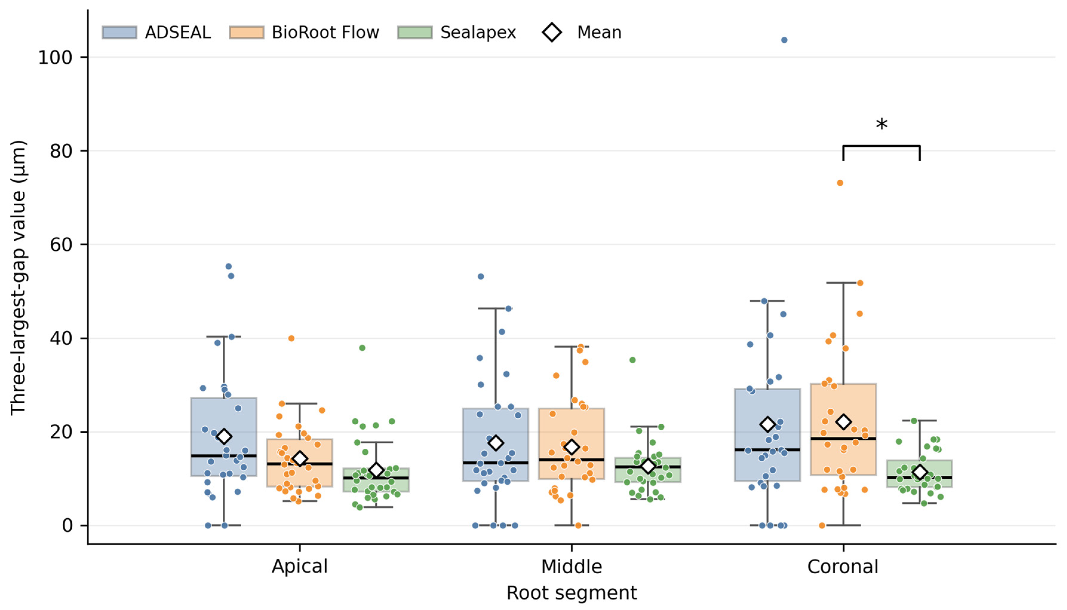
This in vitro study compared the three-largest-gap values of three commercially available sealers under a standardized warm vertical obturation protocol. In the primary raw-scale GEE, sealer type significantly affected the three-largest-gap value (p < 0.001) and the sealer type × segment interaction was significant (p = 0.021), whereas root segment alone was not (p = 0.083). The only statistically significant segment-specific pairwise difference was found in the coronal segment, where BioRoot Flow (22.13 ± 16.06 µm) showed a wider three-largest-gap value than Sealapex (11.40 ± 4.34 µm); ADSEAL (21.61 ± 20.30 µm) differed significantly from neither product. Although this coronal contrast was statistically significant after Bonferroni adjustment (estimated difference 10.73 µm; adjusted p = 0.003), the relatively wide unadjusted 95% confidence interval (4.87 to 16.58 µm) indicates appreciable uncertainty regarding the exact magnitude of the difference. In the secondary raw-scale main-effect summary, Sealapex showed the lowest overall mean value (11.95 ± 5.87 µm) compared with ADSEAL (19.38 ± 16.17 µm) and BioRoot Flow (17.75 ± 12.11 µm). The observed behavior of the tested products could not be inferred from their nominal material categories alone.
The combination of a non-significant segment main effect with a significant sealer × segment interaction is not a statistical contradiction. The main effect of segment tests whether the three-largest-gap value differs between the apical, middle and coronal segments when the three sealers are averaged together, whereas the interaction tests whether the pattern across segments is the same for every sealer. A segment-related difference that is present for some products and absent or attenuated for others can therefore cancel out in the pooled main effect while still producing a significant interaction. This is what was observed here. ADSEAL and BioRoot Flow showed their descriptively highest values in the coronal segment, whereas Sealapex remained comparatively stable across the three segments (11.77 ± 7.25, 12.68 ± 5.78 and 11.40 ± 4.34 µm in the apical, middle and coronal segments, respectively). It should be emphasized, however, that none of the within-sealer comparisons between segments reached statistical significance. This divergence in pattern is therefore descriptive rather than an established segment effect for any individual product. For this reason, the segment-specific comparisons, rather than the pooled main effects, were treated as the primary interpretation.
The lower three-largest-gap values observed for the Sealapex group compared to the other groups in this study indicate that sealer performance cannot be explained solely by general classifications such as “bioceramic” or “epoxy resin”. These findings indicate that physicochemical properties such as viscosity, flowability, curing mechanism, and interaction with dentin may play a role in three-largest-gap behavior [
12,
13]. However, these parameters were not directly measured in the present study, and this explanation remains hypothetical. Although interfacial discontinuities may theoretically be relevant to root filling integrity, the present SEM findings cannot directly predict leakage, bond strength, restorative performance, or clinical outcomes. Similar variability in interfacial adaptation among different root canal sealers has also been reported in previous experimental studies, supporting the concept that sealer performance is product- and technique-dependent [
23,
24].
The present findings suggest that sealer behavior under thermoplastic obturation conditions may be product-specific. Although the same obturation protocol was applied across all groups, the emergence of significant differences between materials, particularly in the coronal segment, suggests that factors related to heat and flowability may create material-specific variations [
25]. Previous experimental studies have demonstrated that heat exposure may alter the physicochemical properties of endodontic sealers depending on their composition, indicating that the compatibility between the sealer and the obturation technique should be considered [
26]. However, because no non-heated obturation control group was included, the independent effect of heat or obturation technique could not be determined. This indicates that thermoplastic techniques may not provide uniform advantages for all sealer types. Thermoplasticized injectable obturation exposes the sealer to elevated temperatures during gutta-percha delivery. Although the heat is primarily applied to gutta-percha, thermal energy may transiently influence the physicochemical behavior of the sealer, including its viscosity, flowability, film thickness, and setting kinetics. These alterations may contribute to differences in three-largest-gap behavior among sealers with different chemical compositions; however, actual intracanal or sealer temperature was not measured in the present study, and the observed differences cannot be attributed directly to a measured temperature [
13].
Although calcium silicate-based sealers are generally considered bioactive, their hydration reaction and setting behavior may be influenced by the clinical environment and obturation technique. Under thermoplastic conditions, heat exposure and rapid manipulation may affect the early setting characteristics of hydraulic calcium silicate sealers, potentially influencing their adaptation to dentin [
12].
Prospective clinical data are available for bioceramic sealers used in combination with warm gutta-percha obturation techniques [
27]; however, clinical outcomes do not establish superior interfacial adaptation. The present findings further indicate that their performance may vary depending on the obturation technique and the evaluation method [
12]. This suggests that the assumption that bioceramic materials consistently outperform other sealers under all conditions should be interpreted with caution.
A systematic review and meta-analysis have compared bioceramic sealers with AH Plus in terms of dentinal tubule penetration and antimicrobial effectiveness [
28]; interfacial adaptation, however, was not among its outcomes, and superiority based on material class alone should therefore not be assumed. Dasari et al. [
24] evaluated BioRoot RCS, a related but distinct calcium silicate-based sealer, under different obturation techniques and reported better interfacial adaptation with warm vertical condensation. Donnermeyer et al. [
29], using a different experimental approach, investigated the physico-chemical response of hydraulic calcium silicate-based sealers, including BioRoot RCS, to simulate intracanal heat application rather than their interfacial adaptation. Taken together, these findings suggest that both obturation technique and thermal exposure may influence the behavior of hydraulic calcium silicate-based sealers; however, direct comparison with BioRoot Flow should be made cautiously, as the two formulations are not identical. In this study, the technique was kept constant and only the sealer type was changed. Therefore, the interfacial performance of BioRoot Flow under the present experimental conditions may have been influenced by the obturation technique.
Vazquez-Alcaraz et al. [
30] reported that root canal anatomy influences obturation success and that material behavior may vary by segment. In the present study, although root segment alone did not significantly influence the three-largest-gap value, a significant interaction between sealer type and root segment was observed. This finding suggests that the three-largest-gap value is influenced by both material characteristics and anatomical factors rather than by root segment alone. The coronal segment, which generally has a larger canal diameter and receives a greater volume of thermoplasticized gutta-percha, may exhibit different flow dynamics and sealer distribution compared with the apical segment. These factors may explain the product-specific differences observed in the three-largest-gap value, particularly in the coronal segment. These explanations remain hypothetical because canal dimensions, sealer volume, gutta-percha volume, and local temperature were not quantified in the present study.
Majumdar et al. [
23] reported that different sealers showed significant differences in terms of dentin penetration and interface adaptation. However, improved penetration does not necessarily correspond to a reduced three-largest-gap value, suggesting that these parameters may not be directly correlated.
Shandilya et al. [
31] reported that bioceramic sealers demonstrated better adaptation. However, our findings are not fully consistent with this result; BioRoot Flow did not show lower three-largest-gap values than Sealapex under the tested protocol. This discrepancy highlights the influence of experimental conditions, material formulation, and evaluation methods.
Direct comparison with published data on other commercial products requires caution, because obturation technique, sealer formulation, evaluation method and root level differ across studies. Shandilya et al. [
31] compared BioRoot RCS, MTA Fillapex and Sealapex by SEM and reported the smallest gaps for BioRoot RCS, with Sealapex better adapted than MTA Fillapex in the middle third but not in the cervical or apical thirds; under the thermoplastic protocol and outcome definition used here, a calcium silicate advantage was not reproduced. Kirthiga et al. [
3] likewise evaluated BioRoot RCS in terms of marginal adaptation. It must be emphasized that BioRoot RCS and BioRoot Flow are not the same material: BioRoot RCS is a powder–liquid formulation, whereas BioRoot Flow is a ready-to-use pre-mixed sealer with a different vehicle and radiopacifier composition [
20], and their interfacial behavior cannot be assumed to be equivalent. With respect to epoxy resin-based sealers, AH Plus has served as the reference comparator in SEM-based adaptation studies, including that of Majumdar et al. [
23]; ADSEAL is a different epoxy resin-based product and the same caution applies. Absolute gap widths also vary by an order of magnitude between studies: mean gap widths of approximately 0.8–5.8 µm have been reported when the average gap across multiple interface zones was measured [
16], compared with the 11–22 µm range obtained here. This difference is expected, because the three-largest-gap value deliberately targets the widest local discontinuities rather than the average interface, and it illustrates why values derived from different outcome definitions should not be compared numerically.
The finding that Sealapex exhibited lower three-largest-gap values suggests that this product may exhibit favorable three-largest-gap behavior under the tested conditions. The lower values observed for Sealapex may be related to product-specific rheological and setting characteristics. However, flow, film thickness, dimensional change, and setting kinetics were not measured in the present study; therefore, the mechanisms underlying this finding remain speculative. Accordingly, the results should be interpreted as product-specific outcomes under the tested protocol rather than evidence of the general superiority of calcium hydroxide-based sealers.
Several mechanisms may plausibly contribute to the coronal pattern. The coronal segment generally has a larger canal diameter, receives a greater volume of thermoplasticized gutta-percha and accommodates a thicker sealer layer, and shrinkage of a larger bulk of backfill gutta-percha upon cooling has been proposed as a cause of wider coronal gaps [
16,
32]. Heat transfer during warm vertical compaction is also greatest in the coronal and middle thirds and limited apically [
33], so any formulation-dependent thermal effect on flow, viscosity or setting kinetics [
14,
15] would be expected to manifest preferentially in these segments. Consistent with this, an SEM study of two calcium silicate-based sealers under warm obturation reported that coronal gaps were consistently wider than apical gaps and that both root level and the sealer × root level interaction were significantly associated with gap width [
16]. These explanations remain hypothetical in the present study, because canal dimensions, sealer volume, gutta-percha volume and local temperature were not quantified.
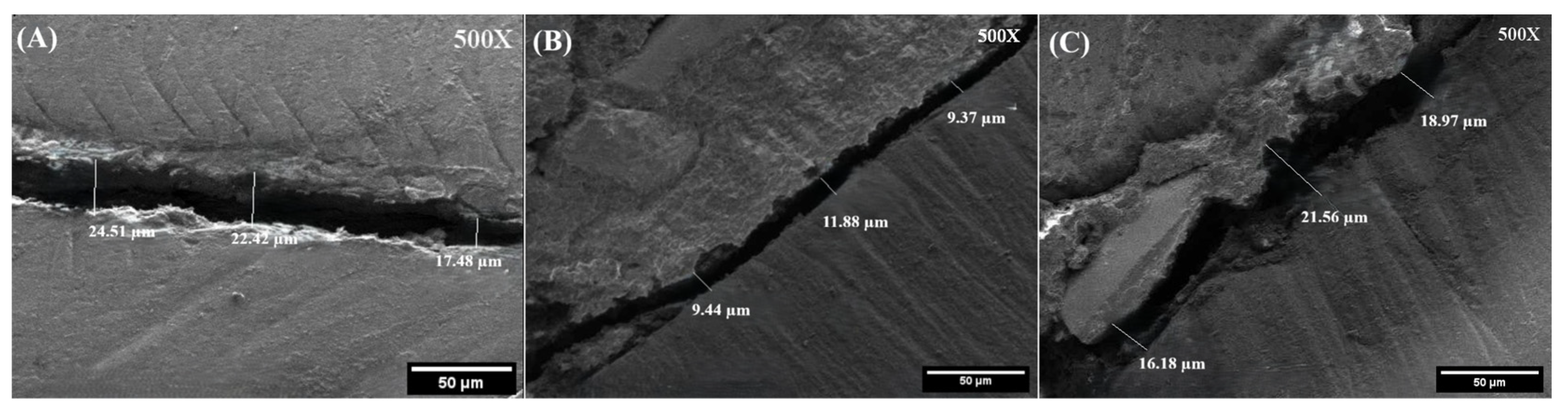
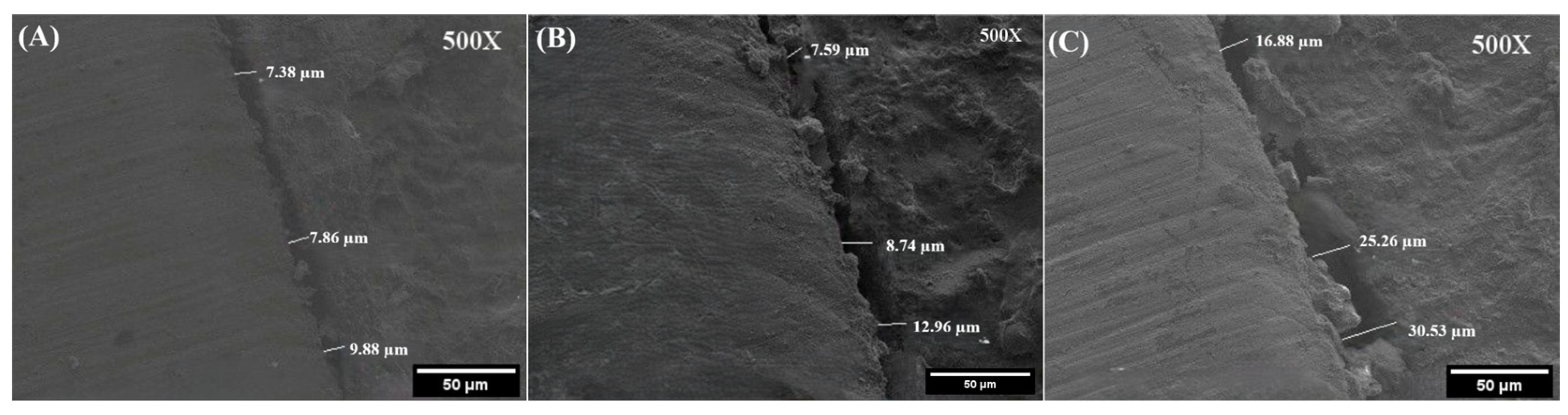
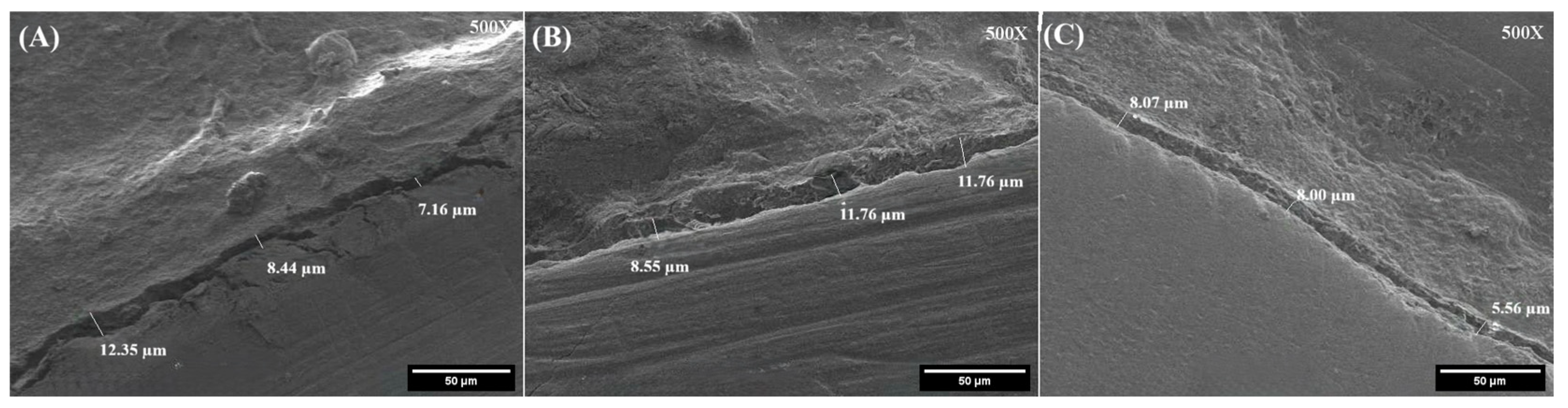
In the selected representative SEM sections, interfacial discontinuities appeared to be nonuniformly distributed along the dentin–sealer interface. The irregular morphology observed in some ADSEAL and BioRoot Flow sections may represent localized interfacial discontinuities; however, the origin of these features could not be determined from the present analysis. In the selected representative SEM sections, the Sealapex group appeared to exhibit narrower localized discontinuities, in agreement with the measured three-largest-gap values.
The present analysis was morphological. Energy-dispersive X-ray spectroscopy (EDX) was not performed because elemental mapping of the interface was not among the prespecified outcomes of this study. EDX can characterize the elemental composition of a specimen surface and has been used together with SEM to compare surface ultrastructure and elemental composition before and after heat application [
14]. However, elemental co-localization alone does not establish that a chemical bond has formed, and it does not by itself distinguish a genuine interfacial discontinuity from a preparation artifact. Accordingly, the present findings cannot be interpreted as evidence either for or against chemical bonding between any of the tested sealers and dentin, and studies combining interfacial morphology with elemental and chemical characterization are warranted.
The clinical meaning of the gap widths reported here should not be overstated. No validated threshold exists at which an SEM-measured localized interfacial gap of 11, 15 or 22 µm can be said to predict microleakage, bacterial penetration, retreatment or clinical failure. What can reasonably be stated is that interfacial gaps even in the low-micrometre range have been noted to exceed the dimensions of many oral microorganisms [
16], so the discontinuities measured here are not, on dimensional grounds alone, too small to be biologically relevant. However, the presence of a two-dimensional SEM discontinuity does not demonstrate the existence of a continuous leakage pathway along the root canal, and this remains an argument about plausibility rather than about demonstrated leakage. Clinical outcome data, moreover, do not map simply onto interfacial morphology: a randomized controlled trial comparing sealer-based obturation with warm vertical compaction reported comparable healing outcomes between techniques [
34]. The present findings should therefore be regarded as morphological and hypothesis-generating rather than as a ranking of clinical performance.
One of the strengths of this study is the standardization of all experimental procedures. Furthermore, SEM analysis enabled direct morphological evaluation of the dentin–sealer interface. Although SEM provides high-resolution evaluation of the dentin–sealer interface, it represents only a two-dimensional assessment of the sectioned surface and cannot quantify the three-dimensional distribution of voids throughout the root canal filling [
35,
36]. Moreover, this study evaluated interfacial morphology rather than true leakage or bond strength; therefore, the findings should be interpreted within the limitations of SEM-based analysis. A further limitation concerns the measurement approach itself: rather than characterizing the entire dentin–sealer interface, this study quantified the three widest visible discontinuities in each root segment, and the reported values therefore represent a three-largest-gap-oriented outcome rather than an overall mean adaptation of the interface. Although the same approach was applied identically to all groups by a single examiner blinded to group allocation, selecting the widest gaps may have emphasized localized interfacial defects rather than the general quality of the interface.
This study has several limitations that constrain the interpretation of its findings. Only one commercial product was tested per material class, so the results should not be generalized to epoxy resin-, calcium silicate- or calcium hydroxide-based sealers as categories but interpreted as applying specifically to the tested products under the tested protocol. No non-heated (cold obturation) control group was included; the study was designed as an active-comparator comparison of three products under one clinically relevant protocol, and consequently the independent effect of heat on gap formation cannot be isolated from these data. Neither intracanal nor sealer temperature was measured, so the observed differences cannot be attributed to a measured thermal exposure. The outcome is a study-specific morphological metric and not a validated measure of overall adaptation, leakage or bond strength, and EDX or other elemental characterization was not performed. All measurements were made by a single examiner, so although intra-examiner reproducibility was good to excellent, inter-examiner reliability could not be assessed. Sectioning, polishing, ethanol dehydration and SEM vacuum conditions may have introduced or enlarged interfacial discontinuities, and only the better-preserved root half was evaluated. Although specimens were incubated for seven days at 37 °C and 95% relative humidity before preparation, complete setting was not independently verified for any of the materials; material-specific setting kinetics therefore cannot be excluded as a source of variability in interfacial behavior under the specimen-preparation procedures used. The outcome distribution was right-skewed, and the statistical significance of the principal findings was not invariant to the analysis scale, as detailed below. Finally, the in vitro design and the absence of long-term aging limit direct extrapolation to clinical conditions. Future studies incorporating a non-heated control group, direct intracanal thermometry, elemental characterization of the interface and complementary three-dimensional or mechanical methods, such as micro-computed tomography and bond strength testing, are recommended.
Two features of the data deserve comment. The first is the magnitude of the dispersion. Standard deviations approaching the mean are an expected property of this outcome rather than an anomaly. Because the metric selects the three widest local discontinuities in each segment instead of averaging the interface, it is, by construction, sensitive to extreme localized defects. Tooth-to-tooth anatomical variation, local sealer distribution, interface morphology and the residual error of manual SEM measurement all add further variance. Verification against the specimen-level data confirmed that the plotted error bars represent standard deviations and that all cell statistics were correctly computed, and individual observations are now displayed in
Figure 1 so that the distribution, rather than a summary of it, is visible. The second is that the statistical findings warrant cautious interpretation. Although the primary raw-scale GEE identified a significant sealer type × segment interaction (
p = 0.021), the strength of this evidence decreased as the influence of extreme observations was reduced. The interaction remained significant after exclusion of the single most extreme observation (
p = 0.036), but it did not reach the conventional significance threshold after exclusion of all three observations exceeding the 3 × IQR criterion (
p = 0.058) or after log transformation of the outcome (
p = 0.082). The sealer main effect followed the same pattern: it remained significant in both exclusion analyses (
p < 0.001) but reached only
p = 0.080 on the log scale. These sensitivity analyses should be regarded as tests of robustness rather than as competing primary models. They indicate that the statistical significance of the interaction is sensitive to the analytical scale and to the weighting of high observations. Importantly, the ordering and direction of the group means remained unchanged across all specifications. The data therefore support a consistent descriptive pattern of product-specific segment behavior, but not definitive evidence of a fully robust interaction effect. The interaction should accordingly be considered suggestive and hypothesis-generating and should be confirmed in an independent, adequately powered study.
The present findings indicate that the nominal biomaterial category did not fully explain the product-specific three-largest-gap behavior observed under the tested protocol. Differences in formulation and interaction with the obturation procedure may have contributed to the findings, although the relevant physicochemical properties were not directly measured. These laboratory findings support the need for product-specific evaluation of root canal sealers under different obturation conditions and should be confirmed using complementary three-dimensional, mechanical, leakage, and clinical outcome assessments.