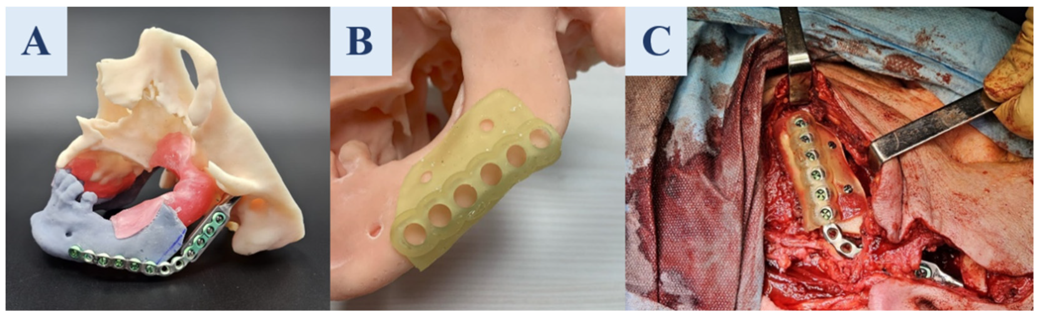
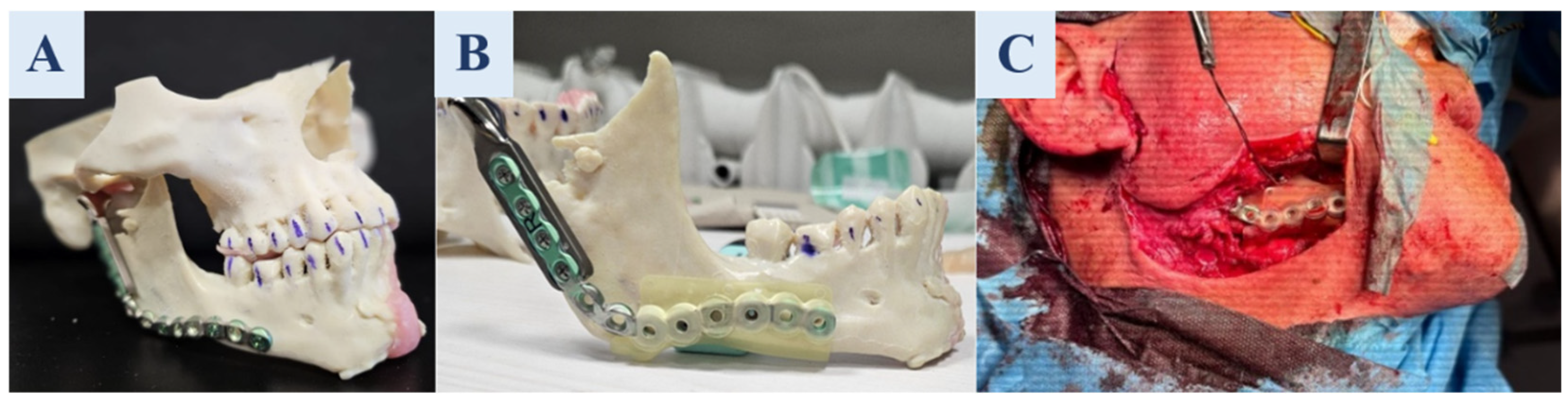
A Closed-Loop Digital–Physical Workflow for Patient-Specific Surgical Guides in Maxillofacial Reconstruction: A Clinical Feasibility Study

 ,
,  ,
,  and
and 
Abstract
1. Introduction
2. Materials and Methods
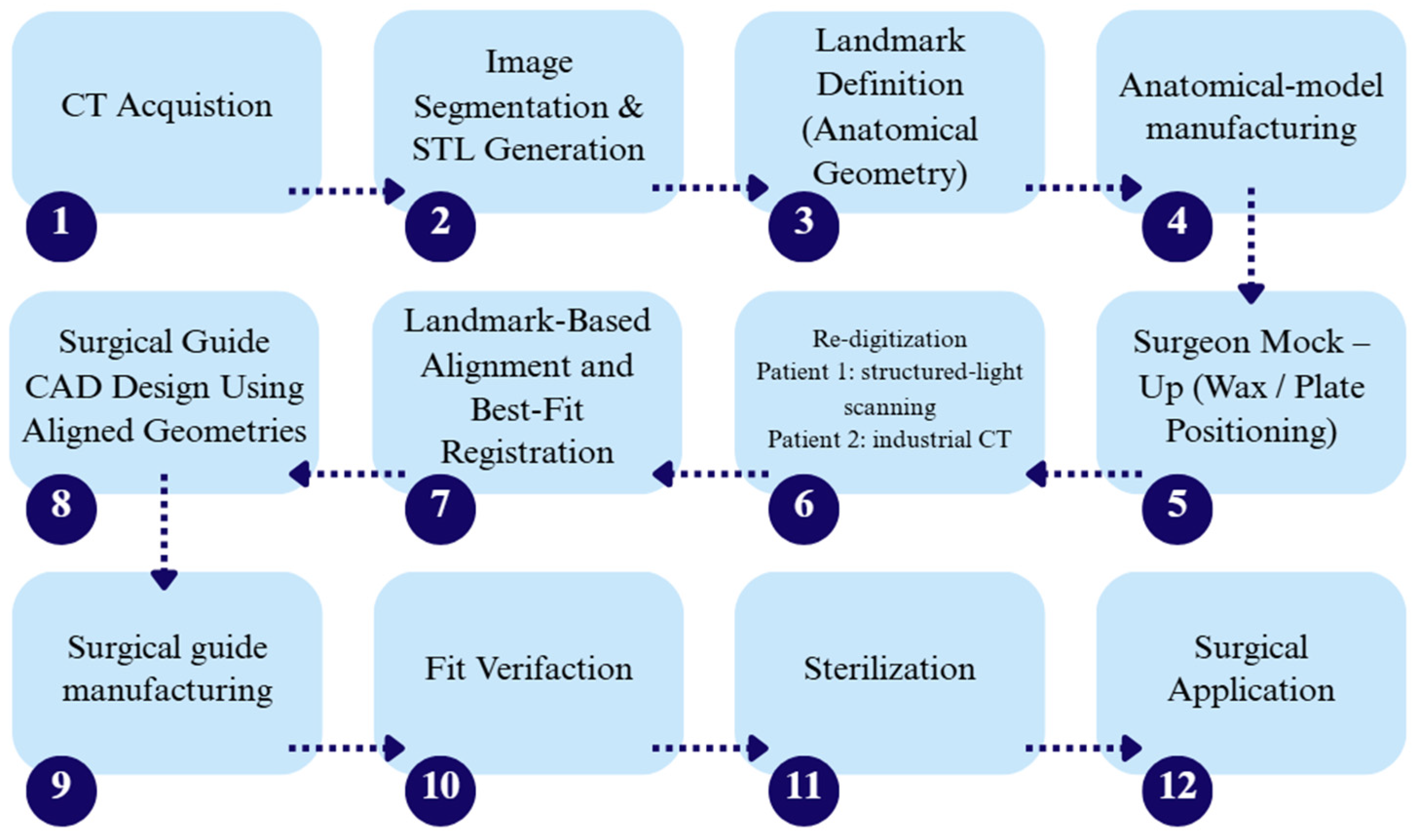
2.1. Concept and Plan of the Work
2.2. Overview Concerning the Cases of Patients Considered for the Research
Supplementary Clinical Implementations
2.3. Manufacturing Methodology
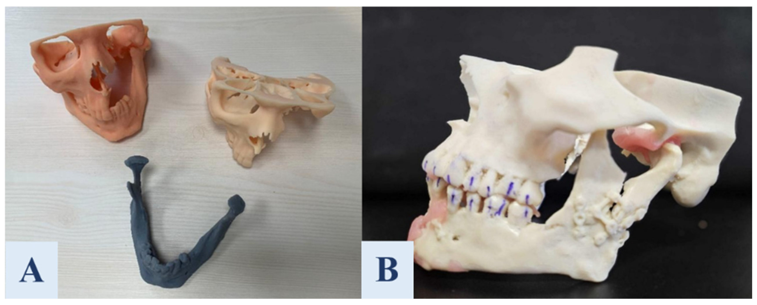
2.3.1. Generation and Additive Manufacturing of Preoperative Anatomical Models
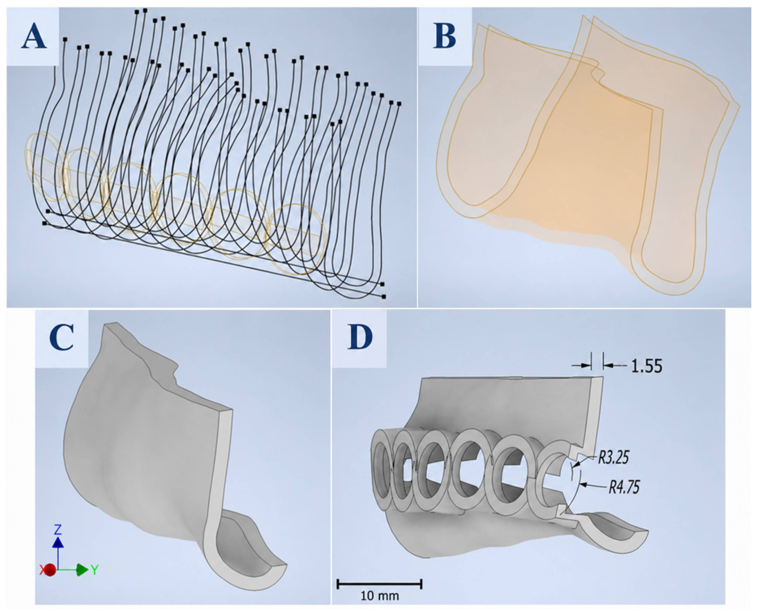
2.3.2. Designing of the Customized Surgical Guides
2.4. Physician Evaluation Methodology
- The craniofacial models accurately represented the patients’ anatomical structures.
- The surgical guides were precisely matched to the patient’s anatomical structures and the reconstruction plate.
- The surface quality of the craniofacial models was satisfactory.
- The surface quality of the surgical guides was satisfactory.
- The dimensional and shape accuracy of the surgical guides was satisfactory.
- The durability of the craniofacial models was satisfactory.
- The durability of the surgical guides was satisfactory.
- The craniofacial models significantly facilitated the surgical planning process.
- The surgical guides were easy and intuitive to use during the procedure.
- The surgical guides improved the precision of the surgical procedure.
- The use of surgical guides shortened the operation time.
- The customization of surgical guides for the specific patient case positively influenced the reconstruction outcomes.
3. Results
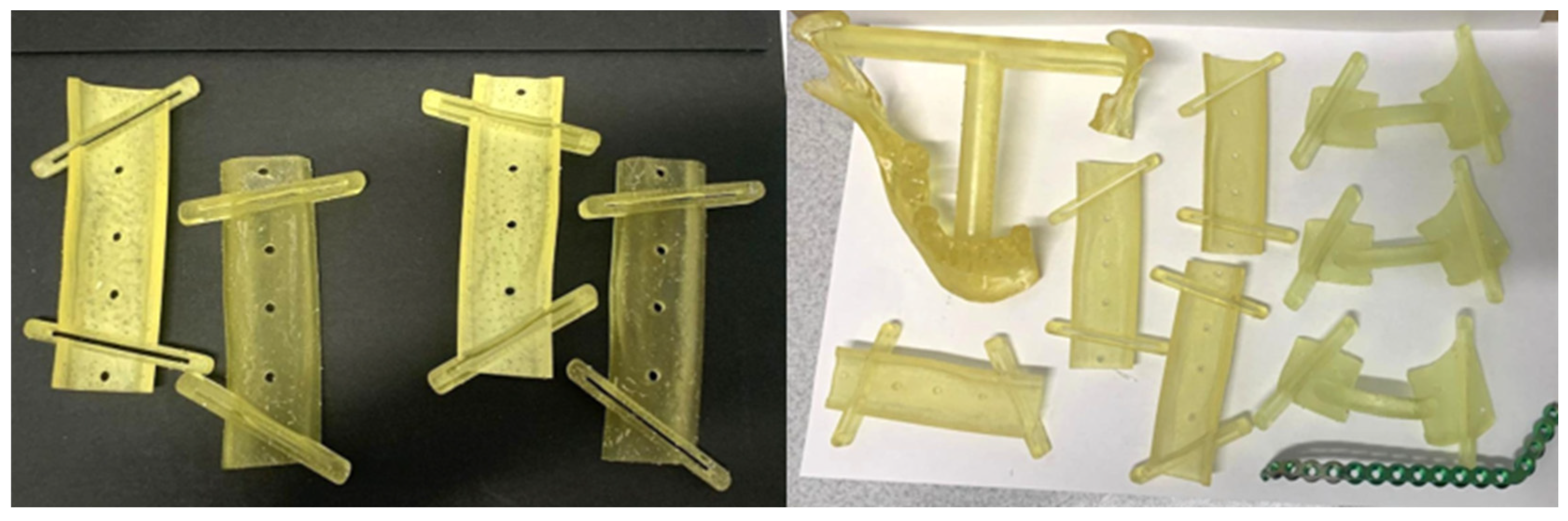
3.1. Using of the Surgical Guides for the Maxillofacial Reconstruction
3.2. Physician Assessment Results
3.3. Economic Impact of Manufacturing Surgical Guides
4. Discussion
5. Conclusions
Author Contributions
Funding
Institutional Review Board Statement
Informed Consent Statement
Data Availability Statement
Conflicts of Interest
References
- Tan, L.; Wang, Z.; Jiang, H.; Han, B.; Tang, J.; Kang, C.; Zhang, N.; Xu, Y. Full color 3D printing of anatomical models. Clin. Anat. 2022, 35, 598–608. [Google Scholar] [CrossRef] [Scilit] [PubMed]
- Lai, J.; Wang, C.; Wang, M. 3D printing in biomedical engineering: Processes, materials, and applications. Appl. Phys. Rev. 2021, 8, 021322. [Google Scholar] [CrossRef] [Scilit]
- Sheoran, A.J.; Kumar, H.; Arora, P.K.; Moona, G. Bio-medical applications of additive manufacturing: A review. Procedia Manuf. 2020, 51, 663–670. [Google Scholar] [CrossRef] [Scilit]
- Kumar, R.; Kumar, M.; Chohan, J.S. The role of additive manufacturing for biomedical applications: A critical review. J. Manuf. Process 2021, 64, 828–850. [Google Scholar] [CrossRef] [Scilit]
- King, C.; Shafi, A.; Burke, E. Optimising the management of concurrent symphyseal/parasymphyseal and bilateral extracapsular condylar fractures using three-dimensional printing. Oral Maxillofac. Surg. 2020, 24, 217–219. [Google Scholar] [CrossRef] [Scilit] [PubMed]
- Aragón-Niño, Í.; Moreiras-Sanchez, Á.D.; de Vera, J.-L.d.C.-P.; Pampín-Martinez, M.M.; Barajas-Blanco, M.; Cebrián-Carretero, J.L. 3D designed in-house positioning and cutting guide system for genioplasty surgery. Technical note and case report. J. Clin. Exp. Dent. 2023, 15, e870–e873. [Google Scholar] [PubMed]
- Li, S.; Mi, L.; Bai, L.; Liu, Z.; Li, L.; Wu, Y.; Chen, L.; Bai, N.; Sun, J.; Liu, Y. Application of 3D printed titanium mesh and digital guide plate in the repair of mandibular defects using double-layer folded fibula combined with simultaneous implantation. Front. Bioeng. Biotechnol. 2024, 12, 1350227. [Google Scholar] [CrossRef] [Scilit] [PubMed]
- Zoabi, A.; Redenski, I.; Oren, D.; Kasem, A.; Zigron, A.; Daoud, S.; Moskovich, L.; Kablan, F.; Srouji, S. 3D Printing and Virtual Surgical Planning in Oral and Maxillofacial Surgery. J. Clin. Med. 2022, 11, 2385. [Google Scholar] [CrossRef] [Scilit] [PubMed]
- Palazzo, L.; Guerra, D.; Frezzato, I.; Frezzato, A.; Russo, N.; Rossi, C. 3D printing in surgical preparation of complex cases: Gran-ulomatous and cystic periapical lesions resolution. Dent. Cadmos 2022, 90, 616–632. Available online: https://www.dentalcadmos.com/en/la-stampa-3d-nella-preparazione-chirurgica-dei-casi-complessi-risoluzione-di-lesioni-periapicali-granulomatose-e-cistiche-2/ (accessed on 26 August 2026).
- Roy, I.D.; Desai, A.P.; Ramyasri, M.; Mukesh, K. The role of patient specific implants in the oral and maxillofacial region. Med. J. Armed Forces India 2024, 80, 387–391. [Google Scholar] [CrossRef] [Scilit] [PubMed]
- Araujo, C.H.; Aguirre, J.A.; Puig, L. 3D Printing Applications and Extended Realities in Medicine: Systematic Review. 3D Print. Addit. Manuf. 2024, 13, 142–156. [Google Scholar] [CrossRef] [Scilit] [PubMed]
- Zeller, A.N.; Neuhaus, M.T.; Fresenborg, S.; Zimmerer, R.M.; Jehn, P.; Spalthoff, S.; Gellrich, N.-C.; Dittmann, J.A. Accurate and cost-effective mandibular biomodels: A standardized evaluation of 3D-Printing via fused layer deposition modeling on soluble support structures. J. Stomatol. Oral Maxillofac. Surg. 2021, 122, 355–360. [Google Scholar] [CrossRef] [Scilit] [PubMed]
- Nizam, M.; Purohit, R.; Taufik, M. Materials for 3D printing in healthcare sector: A review. Proc. Inst. Mech. Eng. H 2024, 238, 939–963. [Google Scholar] [CrossRef] [Scilit] [PubMed]
- Velasco, I.; Vahdani, S.; Ramos, H. Low-cost method for obtaining medical rapid prototyping using desktop 3D printing: A novel technique for mandibular reconstruction planning. J. Clin. Exp. Dent. 2017, 9, e1103–e1108. [Google Scholar] [CrossRef] [Scilit] [PubMed]
- Bozkurt, Y.; Karayel, E. 3D printing technology; methods, biomedical applications, future opportu-nities and trends. J. Mater. Res. Technol. 2021, 14, 1430–1450. [Google Scholar] [CrossRef] [Scilit]
- Abdulhameed, O.; Al-Ahmari, A.; Ameen, W.; Mian, S.H. Additive manufacturing: Challenges, trends, and applications. Adv. Mech. Eng. 2019, 11, 168781401882288. [Google Scholar] [CrossRef] [Scilit]
- Kumar, R.; Mehdi, H.; Bhati, S.S.; Arunkumar, M.; Mishra, S.; Lohumi, M.K. A comprehensive review of advancements in additive manufacturing for 3D printed medical components using diverse materials. Discov. Mater. 2025, 5, 152. [Google Scholar] [CrossRef] [Scilit]
- Amaya-Rivas, J.L.; Perero, B.S.; Helguero, C.G.; Hurel, J.L.; Peralta, J.M.; Flores, F.A.; Alvarado, J.D. Future trends of additive manufacturing in medical applications: An overview. Heliyon 2024, 10, e26641. [Google Scholar] [CrossRef] [Scilit] [PubMed]
- Unkovskiy, A.; Schmidt, F.; Beuer, F.; Li, P.; Spintzyk, S.; Fernandez, P.K. Stereolithography vs. Direct Light Processing for Rapid Manufacturing of Complete Denture Bases: An In Vitro Accuracy Analysis. J. Clin. Med. 2021, 10, 1070. [Google Scholar] [CrossRef] [Scilit] [PubMed]
- Murtezani, I.; Sharma, N.; Thieringer, F.M. Medical 3D printing with a focus on Point-of-Care in Cranio- and Maxillofacial Surgery. A systematic review of literature. Ann. 3D Print. Med. 2022, 6, 100059. [Google Scholar] [CrossRef] [Scilit]
- Dean, D.; Olivas-Alanis, L.H.; Thurston, B.; Vazquez, J.; Hoelzle, D.; Groeber, M.; Niezgoda, S.; Morris, J.; Emam, H.; Skoracki, R.; et al. Virtual Surgical Planning for Point-of-Care Manufacturing. Procedia CIRP 2024, 125, 90–95. [Google Scholar] [CrossRef] [Scilit]
- Pöppe, J.P.; Spendel, M.; Griessenauer, C.J.; Gaggl, A.; Wurm, W.; Enzinger, S. Point-of-Care 3-Dimensional-Printed Polyetherether-ketone Customized Implants for Cranioplastic Surgery of Large Skull Defects. Oper. Neurosurg. 2024, 27, 449–454. [Google Scholar] [CrossRef] [Scilit] [PubMed]
- Ostaș, D.; Almășan, O.; Ileșan, R.R.; Andrei, V.; Thieringer, F.M.; Hedeșiu, M.; Rotar, H. Point-of-Care Virtual Surgical Planning and 3D Printing in Oral and Cranio-Maxillofacial Surgery: A Narrative Review. J. Clin. Med. 2022, 11, 6625. [Google Scholar] [CrossRef] [Scilit] [PubMed]
- Dadhich, A.; Nilesh, K.; Shah, S.; Saluja, H. Three-dimensional printing in maxillofacial surgery: A quantum leap in future. Natl. J. Maxillofac. Surg. 2022, 13, S203–S211. [Google Scholar] [PubMed]
- Rodríguez-Arias, J.P.; Moreiras-Sánchez, Á.D.; Gutiérrez-Venturini, A.; Pampín, M.; González, J.; Morán, M.; Del Castillo, J.; Navarro-Cuéllar, C.; Cebrián, J. In-House 3D Printed Positioning and Cutting Guide System for Mandibular Reconstruction. Protocol and case report. J. Clin. Exp. Dent. 2024, 16, e229–e235. [Google Scholar] [CrossRef] [Scilit] [PubMed]
- Kämmerer, P.W.; Müller, D.; Linz, F.; Peron, P.F.; Pabst, A. Patient-specific 3D-printed cutting guides for high oblique sagittal osteotomy—An innovative surgical technique for nerve preservation in orthognathic surgery. J. Surg. Case Rep. 2021, 2021, rjab345. [Google Scholar] [CrossRef] [Scilit] [PubMed]
- D’Alpaos, D.; Badiali, G.; Ceccariglia, F.; Nosrati, A.; Tarsitano, A. Use of CAD/CAM Workflow and Patient-Specific Implants for Maxillary Reconstruction: A Systematic Review. J. Clin. Med. 2026, 15, 647. [Google Scholar] [CrossRef] [Scilit] [PubMed]
- Bou Zeid, N.; Arias, E.; Alkureishi, L.W.T. 3D Printing in Craniofacial Surgery. Plast. Aesthet. Res. 2024, 11, 34. [Google Scholar] [CrossRef] [Scilit]
- Mann, S.E.; Moore, E.T.; Hill, M.J.; Campa, K.A.; Anderson, S.H.; Glasgow, M.J. Outcomes of Orbit Reconstructive Surgery Utilizing 3D-Printed Surgical Guides. Laryngoscope 2025, 135, 4178–4184. [Google Scholar] [CrossRef] [Scilit] [PubMed]
- Ruggiero, F.; Bianchi, A.; Marcelli, E.; Jeelani, O.; Zucchelli, M.; Cercenelli, L.; Borghi, A. Virtual Surgical Planning in Craniofacial Surgery. In Atlas of Virtual Surgical Planning and 3D Printing for Cranio-Maxillo-Facial Surgery; Tel, A., Robiony, M., Eds.; Springer: Cham, Switzerland, 2025. [Google Scholar] [CrossRef] [Scilit]
- Rana, M.; Buchbinder, D.; Sánchez Aniceto, G.; Mast, G. Patient-Specific Solutions for Cranial, Midface, and Mandible Reconstruction Following Ablative Surgery: Expert Opinion and a Consensus on the Guidelines and Workflow. Craniomaxillofac. Trauma Reconstr. 2025, 18, 15. [Google Scholar] [CrossRef] [Scilit] [PubMed]
- Philippe, B. Accuracy of position of cutting and drilling guide for sagittal split guided surgery: A proof of concept study. Br. J. Oral Maxillofac. Surg. 2020, 58, 940–946. [Google Scholar] [CrossRef] [Scilit] [PubMed]
- Vranckx, J.J.; Desmet, O.; Bila, M.; Wittesaele, W.; Wilssens, N.; Poorten, V.V. Maxillomandibular Reconstruction Using Insourced Virtual Surgical Planning and Homemade CAD/CAM: A Single-Center Evolution in 75 Patients. Plast. Reconstr. Surg. 2023, 152, 143e–154e. [Google Scholar] [CrossRef] [Scilit] [PubMed]

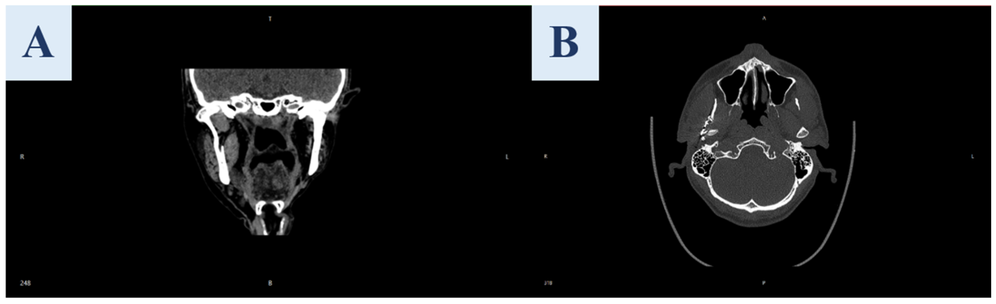


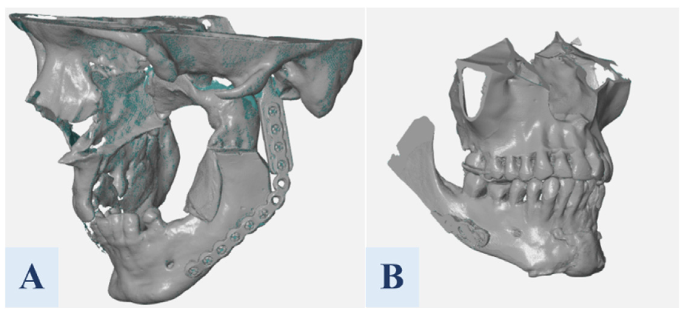



| Model | Material | Printer | Estimated Print Time | Layer Thickness |
|---|---|---|---|---|
| Patient 1 | ||||
| Face–skull | Anycubic basic resin skin | Phrozen sonic mighty 8K | 13 h 37 min | 0.05 mm |
| Mandible | Phrozen aqua resin grey 8K | Phrozen sonic mighty 4K | 7 h 31 min | 0.05 mm |
| Maxilla | Anycubic basic resin light beige | Phrozen sonic mighty 8K | 13 h 46 min | 0.05 mm |
| Patient 2 | ||||
| Mandible | Stratasys Vero | 3D Stratasys J5 MediJet | 22 h 27 min | 18 μm |
| Maxilla | ||||
| Surgical Guide | Material | Printer | Estimated Print Time | Layer Thickness |
|---|---|---|---|---|
| Patient 1 | NextDent surgical guide | Phrozen sonic mighty 8K | 3 h 31 min | 0.05 mm |
| Patient 2 | NextDent surgical guide | Phrozen sonic mighty 8K | 2 h 50 min | 0.05 mm |
| Questionnaire Item | Physician 1 | Physician 2 | Physician 3 | Physician 4 | Physician 5 | Physician 6 | Median | Range |
|---|---|---|---|---|---|---|---|---|
| Anatomical representation by models | 5 | 5 | 5 | 5 | 5 | 4 | 5 | 4–5 |
| Guide fit to anatomy and plate | 4 | 4 | 5 | 5 | 4 | 4 | 4 | 4–5 |
| Model surface quality | 5 | 4 | 4 | 4 | 4 | 5 | 4 | 4–5 |
| Guide surface quality | 4 | 5 | 4 | 5 | 4 | 4 | 4 | 4–5 |
| Guide dimensional and shape accuracy | 3 | 4 | 4 | 4 | 5 | 4 | 4 | 3–5 |
| Model durability | 4 | 4 | 5 | 5 | 5 | 5 | 5 | 4–5 |
| Guide durability | 4 | 5 | 5 | 5 | 5 | 2 | 5 | 2–5 |
| Support for surgical planning | 2 | 4 | 5 | 5 | 5 | 5 | 5 | 2–5 |
| Ease of intraoperative use | 5 | 4 | 5 | 4 | 5 | 4 | 4.5 | 4–5 |
| Perceived improvement in surgical precision | 5 | 5 | 5 | 5 | 5 | 5 | 5 | 5 |
| Perceived reduction in operating time | 5 | 5 | 5 | 5 | 5 | 5 | 5 | 5 |
| Perceived effect on reconstruction outcome | 5 | 5 | 5 | 5 | 5 | 5 | 5 | 5 |
| Process | Time | Cost [USD] |
|---|---|---|
| Patient 1 | ||
| Design | 12 h | 238.94 |
| 3D Scanning | 1 h | 27.69 |
| Printing (Total time) | 42 h 30 min | 23.07 |
| Total | 55 h 30 min | 289.70 |
| Patient 2 | ||
| Design | 7 h | 139.38 |
| Computed Tomography | 15 min | 476.40 |
| Printing | 25 h | 227.77 |
| Total | 32 h 15 min | 843.55 |
| Feature | Direct CT-Based Workflow | Physical-Model Planning Without Digital Update | Proposed Closed-Loop Workflow |
|---|---|---|---|
| Starting geometry | CT-derived digital model | CT-derived digital model and printed physical model | CT-derived digital model and printed physical model |
| Physical surgeon-led simulation | Optional or absent | Yes | Yes |
| Adaptation of the reconstruction plate on the physical model | Optional | Yes | Yes |
| Return of surgeon-introduced physical modifications to the digital environment | No | Usually no | Yes, through re-digitization, landmark-based alignment, and surface-based best-fit registration |
| Role of FFD | Not applicable | Not applicable | Considered as a limited supporting approach for localized geometry updating |
| Final guide-design reference | Initial virtual plan | Usually the initial virtual plan or manually transferred information | Aligned CT-derived and re-digitized geometries representing the surgeon-approved physical configuration |
| Main potential advantage | Shorter and simpler digital workflow | Direct tactile planning and manual plate adaptation | Traceable transfer of surgeon-introduced physical modifications into the subsequent CAD process |
| Main additional requirement | Reliable virtual planning and direct CAD design | Manufacturing of an anatomical model | Anatomical-model manufacturing, physical simulation, re-digitization, registration, and additional CAD processing |
| Evidence provided in the present study | Not evaluated as a control workflow | Not evaluated as a control workflow | Technical feasibility in two detailed cases and adapted use in eight supplementary cases; no evidence of quantitative superiority |
Disclaimer/Publisher’s Note: The statements, opinions and data contained in all publications are solely those of the individual author(s) and contributor(s) and not of MDPI and/or the editor(s). MDPI and/or the editor(s) disclaim responsibility for any injury to people or property resulting from any ideas, methods, instructions or products referred to in the content. |
© 2026 by the authors. Licensee MDPI, Basel, Switzerland. This article is an open access article distributed under the terms and conditions of the Creative Commons Attribution (CC BY) license.
Share and Cite
Smolarek, E.; Górski, F.; Kłak, D.; Żukowska, M.; Słowik, Ł.; Vitkovic, N.; Pǎcurar, R. A Closed-Loop Digital–Physical Workflow for Patient-Specific Surgical Guides in Maxillofacial Reconstruction: A Clinical Feasibility Study. Appl. Sci. 2026, 16, 8604. https://doi.org/10.3390/app16178604
Smolarek E, Górski F, Kłak D, Żukowska M, Słowik Ł, Vitkovic N, Pǎcurar R. A Closed-Loop Digital–Physical Workflow for Patient-Specific Surgical Guides in Maxillofacial Reconstruction: A Clinical Feasibility Study. Applied Sciences. 2026; 16(17):8604. https://doi.org/10.3390/app16178604
Chicago/Turabian StyleSmolarek, Emilia, Filip Górski, Dominik Kłak, Magdalena Żukowska, Łukasz Słowik, Nikola Vitkovic, and Rǎzvan Pǎcurar. 2026. "A Closed-Loop Digital–Physical Workflow for Patient-Specific Surgical Guides in Maxillofacial Reconstruction: A Clinical Feasibility Study" Applied Sciences 16, no. 17: 8604. https://doi.org/10.3390/app16178604
APA StyleSmolarek, E., Górski, F., Kłak, D., Żukowska, M., Słowik, Ł., Vitkovic, N., & Pǎcurar, R. (2026). A Closed-Loop Digital–Physical Workflow for Patient-Specific Surgical Guides in Maxillofacial Reconstruction: A Clinical Feasibility Study. Applied Sciences, 16(17), 8604. https://doi.org/10.3390/app16178604