Article

Preserving Heritage Spaces by a Tea Tree Oil-Loaded Silica Microparticles System for Air Purification


by
Anna Laura Tassi
1,
Pilar Bosch-Roig
2,*,
Andrea Bernardos
3,4,
María Cebriá-Mendoza
3,
María Dolores Marcos
3,4,
Ramón Martínez-Máñez
3,4,
Daniela Pinna
5,
Alessia Santiglia
1,
Laura Santagostini
1 and
Vittoria Guglielmi
1,*
1
Department of Chemistry, University of Milan, Via Golgi 19, 20133 Milan, Italy
2
Instituto Universitario de Restauración del Patrimonio, Universitat Politècnica de València, Camino de Vera, s/n, 46022 Valencia, Spain
3
Instituto Interuniversitario de Investigación de Reconocimiento Molecular y Desarrollo Tecnológico (IDM), Universitat Politècnica de Valencia and Universitat de València, Camino de Vera, s/n, 46022 Valencia, Spain
4
CIBER de Bioingeniería, Biomateriales y Nanomedicina, Instituto de Salud Carlos III, 28029 Madrid, Spain
5
Department of Chemistry ‘Giacomo Ciamician’, University of Bologna, Via Francesco Selmi 3, 40126 Bologna, Italy
*
Authors to whom correspondence should be addressed.
Appl. Sci. 2026, 16(15), 7674; https://doi.org/10.3390/app16157674
Submission received: 15 May 2026 / Revised: 27 July 2026 / Accepted: 29 July 2026 / Published: 2 August 2026
(This article belongs to the Special Issue Non-Destructive Techniques for Heritage Conservation)

Featured Application

This research aims to develop a cost-effective and easy-to-use technology for improving indoor air quality through the reduction of fungal and bacterial contamination. While the primary objective of the project is the conservation of cultural heritage, the proposed tool also has broader applications as a passive air purification system for residential and occupational settings, contributing to healthier indoor environments by lowering harmful microbial loads.

Abstract

Airborne fungi and bacteria pose a severe challenge for conservators, as they can settle on artistic surfaces, leading to chemical, physical and aesthetic degradation of artworks. A new air-cleaning system has been developed to reduce indoor airborne microorganisms by leveraging the antimicrobial potential of Tea Tree (Melaleuca alternifolia) Essential Oil (TTEO) encapsulated within the pores of MCM-41 mesoporous silica microparticles (MCM-TTEO), thereby extending its antimicrobial activity and enhancing its efficacy. Experimental data showed a progressive release of TTEO from the MCM-TTEO system during the 5 weeks monitoring. The release rate was quite linear in the first week, reaching about 16.2%, before plateauing at approximately 30% in the next weeks. A comparison of the performances of the MCM-TTEO system with those of the non-encapsulated (‘free’) TTEO was performed using a comprehensive air quality monitoring approach, which tracked temperature, relative humidity, microbial loads and particulate matter. Results demonstrate that the MCM-TTEO system maintained higher biocidal activity over the 5-week study period than the free EO. Furthermore, the biocidal efficacy increased when the MCM-TTEO system was used in direct contact with microorganisms. These promising findings highlight the potential for exploring different combinations of the two components in both passive and active configurations, including the development of air filtration systems.

1. Introduction

Cultural heritage assets are exposed to a range of environmental deterioration factors. In the field of conservation, it is well established that elevated concentrations of airborne microorganisms in exhibition spaces and storage facilities not only pose health risks to staff and visitors, reducing life expectancy and exacerbating various pathologies, but may also cause irreversible damage to books, archives, cultural artifacts, and monuments [1,2,3,4]. However, unlike the standards established for the protection of human health, threshold values for air pollutants affecting cultural heritage remain poorly defined (Spanish standards UNE developed by AENOR 2024; European Commission 1993) [5]. Consequently, several mitigation strategies have been adopted, including air quality management policies, the application of protective coatings, and the implementation of climate-controlled environments.
Assessing environmental quality involves monitoring of temperature, relative humidity, light exposure, and atmospheric aerosols—metastable suspensions of solid particles and liquid droplets—as these parameters provides insight into pollutant sources, formation mechanisms, and their roles in degradation processes [6]. Among natural contributors, bioaerosols and other forms of biogenic matter play a major role in the degradation of artistic materials [7]. Microorganisms associated with these aerosols, primarily fungi and bacteria, can generate acids, enzymes and pigmented metabolites, as well as mechanical penetration, contributing to biodeterioration. Under favorable environmental conditions such as dust accumulation, elevated temperatures, and high relative humidity, biofilms may develop, affecting the mechanical and chemical properties of both organic and inorganic materials, as well as their aesthetic appearance [8,9].
Specifically, fungi proliferate in humid, polluted environments, frequently colonizing the surfaces of cultural artifacts and monuments. Bacteria contribute to stone weathering and cellulose degradation by producing different organic and inorganic acids, including both sulfuric and nitric acids. The primary sources of airborne bacteria and fungi in indoor environments include the ambient atmosphere, resuspended dust, mold, human and pet occupants, plants, ductwork, and air conditioning (HVAC) systems [8].
Different products, protocols and techniques were developed and investigated to reduce Cultural Heritage (CH) biodeterioration risk in indoor spaces. However, many of these approaches present significant limitations, including toxicity and limited long-term effectiveness [10]. For example, physical-mechanical air-cleaning methods, such as ozone and UV light, may adversely affect artistic and historical materials, and chemical air-cleaning systems must be avoided due to their high human toxicity [11,12].
Aligned with the principles of preventive conservation, this work offers a proactive approach to address biodeterioration before it occurs. The International Center for the Study of the Preservation and Restoration of Cultural Property (ICCROM) promotes this methodology, defining it as a comprehensive set of measures and actions designed to prevent or minimize future deterioration or loss. These actions are typically applied to the surroundings or context of an artwork—or a collection of items—regardless of their age or condition. Consequently, preventive conservation is characterized by its indirect nature, avoiding direct interference with the materials, structures, or original appearance of the cultural artworks themselves [13,14].
Our research focuses on developing an eco-sustainable, preventive conservation strategy to mitigate microbial loads within indoor Cultural Heritage (CH) environments. Previous studies have shown the potential of the cold diffusion of Tea Tree pure essential oil to reduce microorganisms present on the unventilated indoor space of Santos Juanes Church [15]. The tool consists of a waterless, heatless diffusion device that disperses pure essential oils as microparticles without affecting the artistic surfaces [15].
The antimicrobial activity of TTEO is primarily attributed to its volatile components, particularly terpinen-4-ol (35–45%) and 1,8-cineole (1–6%). Nonetheless, additional components such as α-terpineol, terpinolene, and α- and γ-terpinene are also present and may contribute to its overall biocidal efficacy [15,16,17]. The antimicrobial properties of essential oils are generally associated with their volatile fractions. Nevertheless, their intense sensory impact, high chemical reactivity, rapid evaporation, and limited water solubility pose significant challenges that must be overcome prior to practical application [18,19].
To address this, previous studies by part of our research group enhanced the antimicrobial efficacy of essential oils (EOs) by their encapsulation into MCM-41 mesoporous silica nanoparticles to reduce evaporation, subsequently applying EO-loaded MCM-41 particles as a protective coating on Carrara marble [20].
MCM-41-type microparticles were first developed in the 1990s as part of the M41S molecular sieve family. The main characteristic of these materials is their ordered pore distribution, with pore sizes ranging from 2 to 10 nm. In addition, these materials have a high pore volume, on the order of 1 cm3/g, and a high specific surface area ranging from 500 m2/g to 1000 m2/g. These characteristics make them potentially very useful materials in processes that require the adsorption of significant amounts of bulky molecules [21]. Using Transmission Electron Microscopy (TEM), the distinctive hexagonal pattern of MCM-41’s channels can be observed, as well as the periodicity of the structure, also highlighted by X-ray diffraction [22]. The research we report herein connects to work by Bernardos et al., which used mesoporous silica nanoparticles to mitigate the volatility of essential oil components, such as carvacrol, thymol, eugenol, and cinnamaldehyde, in food packaging [23]. In the field of Cultural Heritage, these materials are widely used in various applications, including coatings for cultural heritage materials as mentioned above [24,25,26].
To design a passive-release air purification system, we selected microsized mesoporous silica rather than mesoporous silica nanoparticles, thereby eliminating the risk of nanoparticle dispersion and subsequent inhalation hazards for operators [27,28]. While we reviewed the literature regarding the inhalation cytotoxicity of tea tree essential oil (TTEO), findings remain inconclusive [29]. Nevertheless, high concentrations of TTEO can be toxic if ingested and may induce skin irritation or allergic reactions in susceptible individuals [29]. Existing data indicate that TTEO toxicity and its compatibility with air cleaning systems are strictly dose-dependent; thus, utilizing low concentrations can effectively minimize the risk of adverse effects [30]. To mitigate TTEO’s high volatility and reduce the volume required, our approach encapsulates the oil within mesoporous silica microparticles, ensuring a controlled release that enhances and prolongs its antimicrobial efficacy [21,31], thereby ensuring the long-term efficacy of the air-cleaning system.
Accordingly, MCM-41 microparticles were synthesized and loaded with TTEO. The resulting MCM-TTEO system was characterized by evaluating both essential oil encapsulation and evaporation rates. Subsequently, laboratory indoor tests were conducted to evaluate the effectiveness and durability of the developed MCM-TTEO system against free TTEO. Oil encapsulation efficiency was assessed via evaporation release curves, comparing the release profiles of the free and encapsulated essential oils. Additionally, changes in particulate matter, temperature, relative humidity, and microbial reduction were analyzed and compared between the two configurations. Because the MCM-TTEO system operates passively, the potential necessity for external activation, specifically via temperature or moisture fluctuations, was also investigated to determine if the microencapsulated formulation yields superior operational outcomes compared to free TTEO.

2. Materials and Methods

2.1. Hydrothermal Synthesis of Mesoporous MCM-41 Microparticles

The synthesis of MCM-41 type mesoporous silica microparticles was based on the use of triethanolamine ligands as hydrolytic inorganic precursors, and surfactants as porogen species [23]. To 0.98 gr NaOH dissolved in 2 mL of H2O, 52 gr of triethanolamine were added under stirring. The mixture was heated to 120 °C to homogenize the components, and then cooled to 70 °C, keeping the temperature controlled through a VWR VT-5 penetration thermometer (VWR International Eurolab, Llinars del Vallès, Barcelona, Spain). Then, 22 mL of tetraethyl orthosilicate (TEOS) were added dropwise, under stirring. The system was then heated up to 118 °C, and 9.36 g of n-cetyltrimethylammonium bromide (CTAB) were added. Finally, the reaction mixture was cooled to 70 °C, and 180 mL of H2O were added. The resulting mixture was stirred at room temperature for 1 h, transferred to a PTFE vessel and maintained at 100 °C overnight (or 24 h). As the pH of the system was extremely alkaline (pH 10), the obtained suspension was centrifugated with a 22-5810R centrifuge (Eppendorf Ibérica S.L.U., San Sebastián de los Reyes, Madrid, Spain) for 18 min at room temperature (20 °C), speed 10,000 rpm. The solid particles were separated from the liquid and washed three times with EtOH and water, to reach an almost neutral pH (pH 7–7.5); then the microparticles were dried at 70 °C for 24 h; 3.54 g of microparticles were obtained. To prepare the final porous material, the obtained microparticles were calcinated at 550 °C for 5 h to remove the template phase. Tetraethyl orthosilicate (TEOS), n-cetyltrimethylammonium bromide (CTAB), sodium hydroxide (NaOH) and triethanolamine (TEA) were purchased from Sigma–Aldrich (Merck Group, Madrid, Spain) and used as received.

2.2. Encapsulation of TTEO on MCM-41 Microparticles

Seven vials were prepared with 150 mg of calcinated MCM-41 mesoporous silica microparticles each. In each vial, 150 µL of TTEO (Melaleuca alternifolia essential oil from Pranarôm International, Barcelona, Spain) was added, and the vial was closed and sealed with parafilm. Then, the vials were vortexed and held in the oven at 42 °C for 24 h. After that, the process of encapsulation of the oil on the MCM-41 material was quantified.

2.3. Microparticles Characterization

Powder X-ray diffraction (PXRD) and thermogravimetric analysis (TGA) techniques were employed for materials characterization. The MCM-41 microparticles obtained were characterized by PXRD analysis before and after calcination, and diffractograms were compared to data available in the literature. The diffractograms were acquired in 11 min and an angular range (2θ) from 0.04 °C to 8.3 °C, with a 0.2 mm filter for the X-Ray source, and a 3 mm filter for the detector. Spectra were obtained from the ‘Diffrac Plus XRD Commander V.2.5’ software, while ‘EVA V14.0.0.0’ software (Bruker Corporation, Madrid, Spain) was used to convert the data. Moreover, in order to obtain the TTEO content inside the pores after the synthesis of MCM-TTEO, TGA allowed us to calculate the organic matter content. The TGAs were carried out on a TGA/SDTA 851e balance (Mettler Toledo, Greifensee, Switzerland) using an oxidant atmosphere (air, 80 mL min−1) with a heating program consisting of a heating ramp of 10 °C min−1 from 393 to 1273 K and then a final isothermal heating step at 1273 K for 30 min.

2.4. In-Lab Indoor Passive Air Cleaning Systems Experimentation

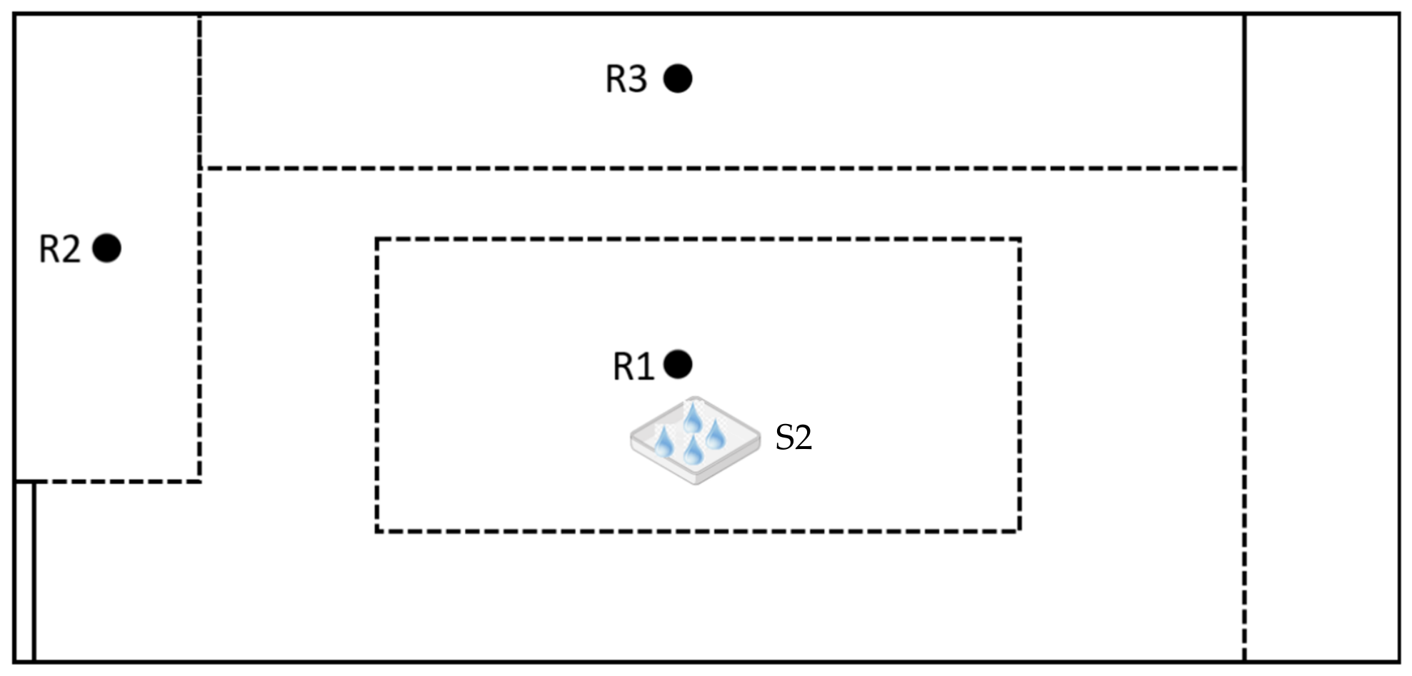
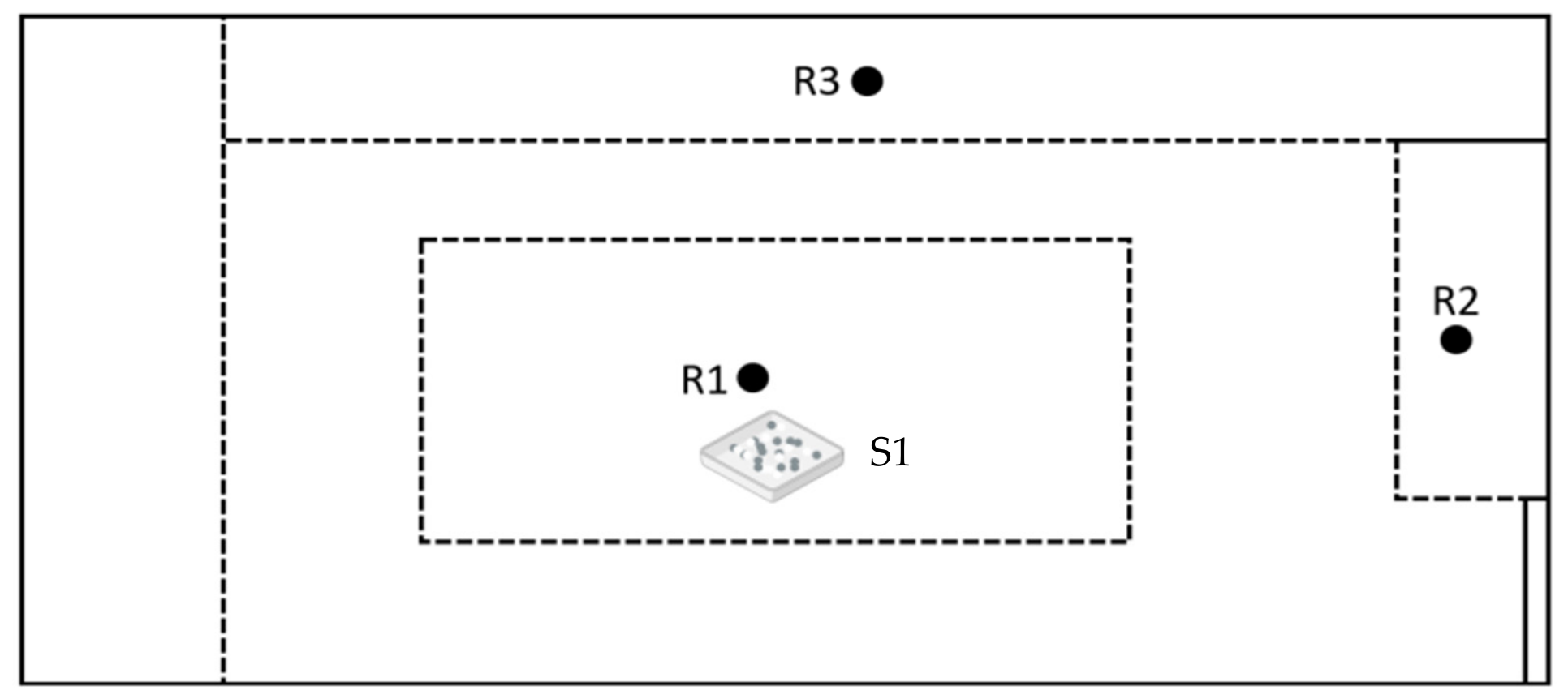
The evaporation rate and the antimicrobial activity of the two systems studied (TTEO free and MCM-TTEO) were evaluated by developing an in-lab passive air cleaning indoor experimentation. The indoor experiments were conducted in two adjacent, windowless university rooms of identical dimensions (3.25 m × 4.42 m × 4.42 m) and structural layout, served only by a mechanical HVAC system. Prior to testing, baseline air quality and microbial loads were assessed in both rooms to confirm uniform initial conditions. To minimize environmental variability, both spaces remained unoccupied with restricted accesses to prevent door openings, and the HVAC systems were operated only as ventilating systems, under identical settings throughout the study. Room 1 was used to test the system obtained encapsulating TT EO in microparticles (MCM-TTEO), while Room 2 was used to evaluate the behaviour of free TTEO. The two systems were positioned in the middle of the room, in a squared capless Petri dish to allow passive evaporation. Air quality was monitored before and during treatment in three positions in each room (see Appendix AFigure A1 and Figure A2) in order to account for spatial variability. To minimize possible differences between the two rooms, data obtained during treatment were compared to the baseline microbial load (T0) of each room, measured prior to treatment, to normalize room-to-room differences in absolute bacterial counts.

2.4.1. Evaporation Test of EOs

The amount of TTEO released from the microparticles was evaluated and compared with the amount of free EO evaporated to determine TTEO evaporation over time. The release test was performed in the same rooms described above. Petri dishes containing 10 μL free TTEO or 5 mg of MCM-TTEO were placed in the middle of the room to monitor spontaneous release into the air. The Petri dishes were sealed at specific time intervals; after sealing, they were weighed to evaluate the residual oil mass with an analytical balance. The mass was measured before starting the evaporation test (m0) and then repeated at different time points (mt) corresponding to 1 h, 8 h, 24 h, 48 h, 72 h, and 1, 2, 3, 4, 5 weeks. The percentage of evaporation release was determined using the formula:
%   e v a p o r a t i o n   r e l e a s e = m 0 m t m 0 × 100
Furthermore, the accuracy in determining the amount of TTEO released from the microparticles was improved through TGA of the particles after passive release by exposure to air. The analyses were conducted by sampling 2 mg of MCM-TTEO from each Petri dish placed in Room 1, after determining the residual mass at the time points indicated above (1 h, 8 h, 24 h, 48 h, 72 h, and 1, 2, 3, 4, 5 weeks). TGA thus enabled the determination of the residual EO in the microparticles, allowing for the calculation of the TTEO evaporation rate from MCM-41.

2.4.2. Air Quality Experimentation

The two rooms described above were also used for in-lab air quality experimentation, to assess the effectiveness of the air cleaning systems [15]. Air quality monitoring included hourly recording of temperature and relative humidity data by a Tzone TempU 03 multipurpose Datalogger (JUANJUAN, Guangzhou, China). Airborne particulate matter (PM) was evaluated using an Airy technology P311 Laser Particle counter (Airy Technology, Inc., Stoughton, MA, USA) which detects particles of 0.3 μm and 5 μm in the air over a 1 min sampling period at a flow rate of 2.83 L/min. Microbiological analysis of the air for bacteria and fungi was performed by the SAS SUPER 100/180 (VWR 21; International PBI S.p.A., Milan, Italy), directing an airflow of 100 L onto Petri dishes containing Plate Count Agar (Scharlau, Barcelona, Spain) for bacterial growth and Sabouraud Chloramphenicol Agar (Scharlau, Barcelona, Spain) for fungal growth. Measurements were taken before any treatment (t0) and at the same time points used to evaluate EO evaporation (1 h, 8 h, 24 h, 48 h, 72 h, and 1, 2, 3, 4, 5 weeks), during the operation of TTEO or MCM-TTEO cleaning systems in rooms 1 and 2.

2.5. In-Lab Direct Contact Antimicrobial Efficacy Evaluation

To better evaluate antimicrobial activity of the two air cleaning systems prepared, free TTEO and TTEO-loaded MCM-41 microparticles were directly applied on the surface of the microbial culture media contained in the Petri dishes, before collecting the air volume using the SAS instrumentation. Free TTEO and MCM-TTEO were diluted in ultrapure sterile water at concentration of 1%w (free TTEO) and 22.3 mg/mL (MCM-TTEO), respectively. Each solution was applied to a Petri dish using a sterile Digralsky spreader (1.3 mL of free EO, or the equivalent of the MCM-TTEO systems) (VWR International Eurolab, Barcelona, Spain). Subsequently, the Petri dishes were placed into the SAS, and air samples were collected in triplicate in each of the test rooms, as described above. After using the SAS equipment to sample the air from the two tested rooms, the Petri dishes were incubated for 48 h (for bacteria) and 5 days (for fungi) at 30 °C and 100% relative humidity (RH).

2.6. Statistical Evaluation

Measurements were performed in triplicate (n = 3) in each room via SAS air sampling across three distinct positions (Appendix A, Figure A1 and Figure A2) to account for intra-chamber spatial and technical variability. Results are presented as mean values ± standard deviation (SD). Statistical analysis was carried out using Student’s t-test to evaluate differences between treatments (free TTEO vs. MCM-TTEO). Differences between the analyzed EOs treatments were considered statistically significant at p < 0.05, and reported where applicable. For statistical comparison, data were aggregated to obtain representative mean values for each treatment, reducing the impact of temporal autocorrelation associated with repeated measurements over time.

3. Results and Discussion

3.1. Synthesis and Physico-Chemical Characterization

The designed MCM-TTEO microparticles consist of two components: MCM-41 as the inorganic support and TTEO as a naturally occurring antimicrobial essential oil. MCM-41 was selected due to its high surface area and excellent loading capacity. Material preparation was carried out in two consecutive stages: first, the synthesis of the mesoporous silica microparticles, followed by the loading of TTEO into the mesopores of the silica matrix to yield MCM-TTEO.
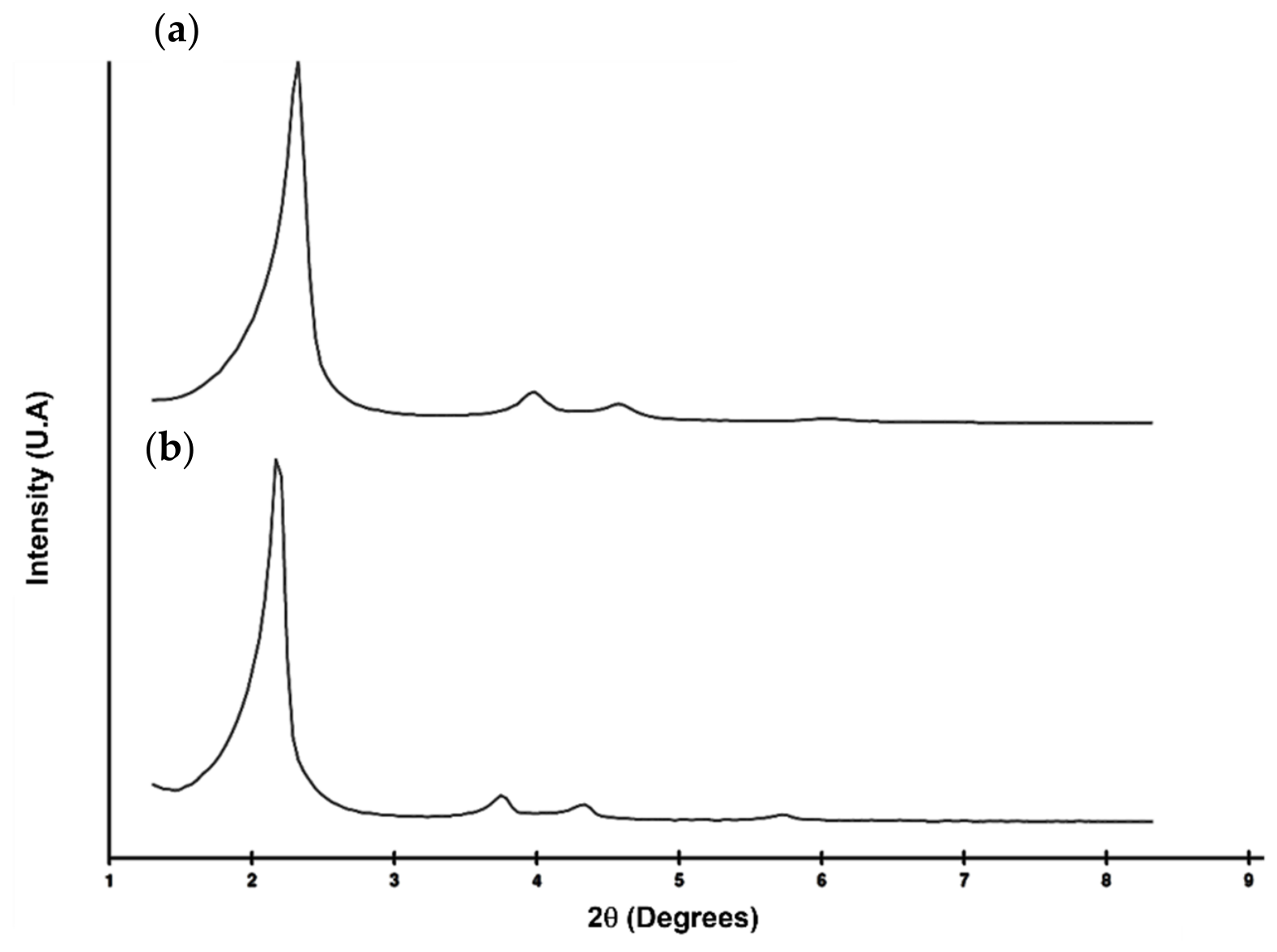
The microparticles obtained were characterized by X-ray diffraction. The low-angle powder X-ray diffraction pattern of the as-synthesized MCM-41 exhibits the characteristic reflections of a 2D hexagonally ordered mesostructure (space group p6mm). A strong, well-defined diffraction peak was observed at approximately 2θ ≈ 2°, corresponding to the (100) plane and confirming the formation of an ordered array of mesoporous channels. Two additional, weaker reflections, located in the 2θ ≈ 3–5° region and indexed as the (110) and (200) planes, were also present. Upon calcination, the X-ray diffraction pattern exhibits the expected structural modifications associated with the removal of the organic surfactant template; a small shift of all reflections toward higher 2θ values was observed, consistent with a contraction of the unit cell due to framework shrinkage during thermal treatment and template removal. The persistence of these three low-angle reflections after calcination confirms that MCM-41 mesostructure is retained despite the surfactant removal (see Figure 1).
Thermogravimetric analysis (TGA) was employed to quantify the amount of essential oil loaded onto the silica microparticles. The TTEO content in the particles was determined to be 43.47%, corresponding to 434.7 mg of TTEO per gram of the MCM-TTEO system.

3.2. In-Lab Indoor Evaluation of the Passive Air Cleaning Systems

3.2.1. Evaporation Assay

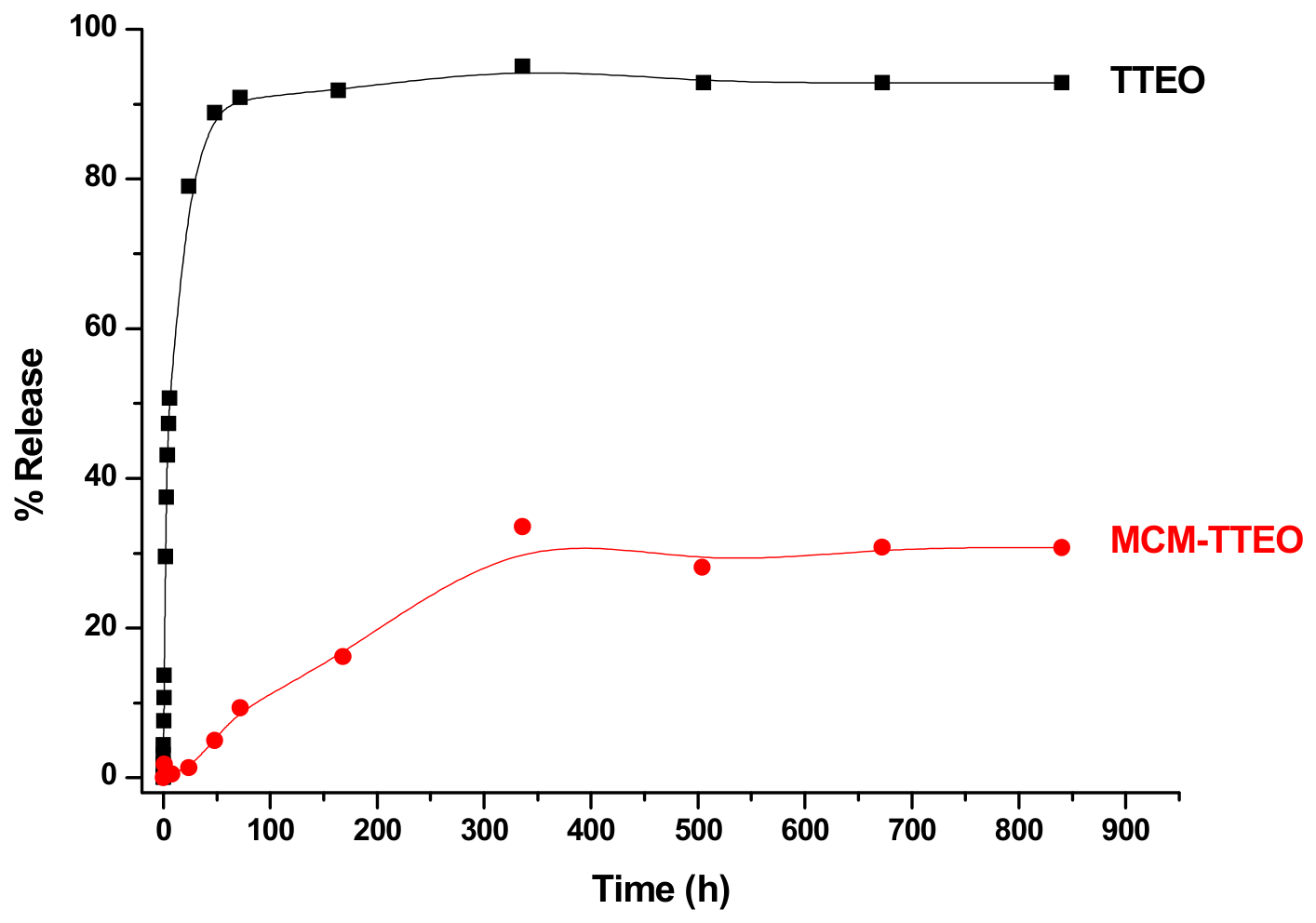
The passive release of TTEO from MCM-41 microparticles was studied in an indoor environment and compared with the spontaneous evaporation of free TTEO (Appendix AFigure A1 and Figure A2). Data obtained confirmed the high retention capacity of MCM-41: after nearly 5 weeks (840 h), less than 30% of the encapsulated essential oil had been released, whereas nearly 95% of the free oil had evaporated after 5 weeks (Figure 2). Notably, approximately 80% of the non-encapsulated TTEO volatilized within the first week (168 h). By contrast, the TTEO confined within the MCM-41 matrix exhibits a linear and sustained release profile, with the cumulative release reaching approximately 30% after two weeks (350 h). These results highlight that the mesoporous support significantly enhances stability and ensures a controlled-release behavior.
To confirm the behaviour observed, TGA analyses were performed on MCM-TTEO system to accurately quantify the amount of essential oil still loaded onto the silica microparticles (Table 1) and to evaluate whether the support is suitable for the effective delivery of the EO. During the first hours (1 h, 8 h, 24 h) of the exposure to air, the TTEO was released from the MCM-41 slowly, resulting in 1.4, 5.0 and 9.3% of the organic matter being released. After the first week, TGA revealed a further decrease in the amount of TTEO retained within the microparticles, compared to the initial content. Data obtained indicate that, at this step, the oil accounted for 37.72% of the total mass sample (MCM-41 microparticles + tea tree essential oil); this suggests that 16.2% of the organic matter was released. This value reflects the total amount of TTEO remaining on the surface of the material and that confined within the mesoporous silica structure. At the end of the experiment (2 to 5 weeks), the percentage of the organic matter released was approximately 30%, confirming the results obtained by the gravimetric determination of EO mass lost during the release assay.

3.2.2. Environmental Conditions Assay

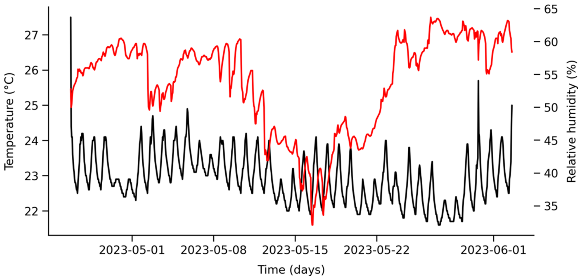
The monitoring of environmental conditions (temperature and relative humidity RH) in the studied test rooms (see Section 2.4) throughout the experiment shows average temperatures of 22.9 °C and relative humidity of 53.7%, respectively. Clear nighttime variations are evident due to the automatic climate control, which switches off the rooms at night (Figure 3). Furthermore, an important RH drop occurs during the second and third week of the experiment, with a significant reduction to 32% on 16 May.

3.2.3. In-Lab Indoor Airborne Particulate Matter Assay

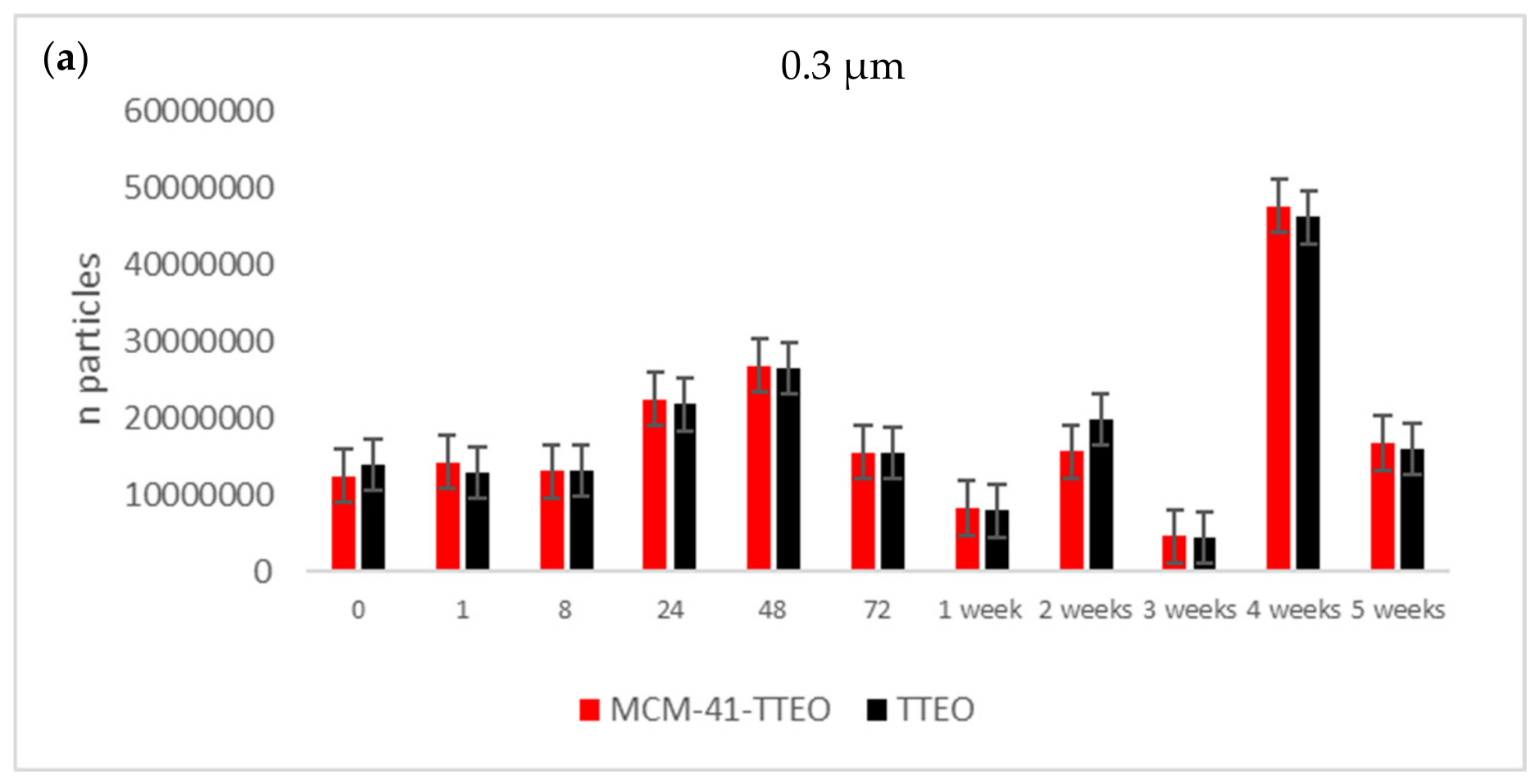
Airborne particulate matter (PM) was also assessed, measuring 0.3 μm and 5 μm fractions in the air over a 1 min-sampling period (see Section 2.4.2). The highest concentrations of 0.3 µm particles were registered at 48 h and 4 weeks into the experiment (Figure 4). These peaks coincide with the lowest microorganism levels detected in the test rooms, suggesting a relation to the higher evaporation of the EOs’ volatile components. Other authors have shown similar increases in PM when EOs are diffused in indoor environments [16,32].
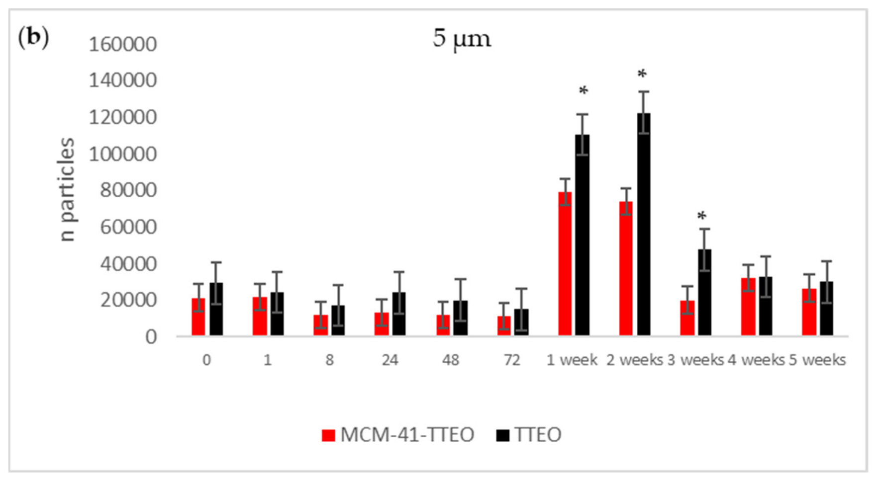
The particle size data support the observed microbiological trends. A reduction in microorganisms during the first 72 h of treatment is temporally associated with a reduction in PM 5.0 µm fraction (Figure 4). Subsequently, an increase in PM 5.0 µm levels during weeks 1 and 2 coincides with the observed microbial recolonization of the tested rooms. Airborne biological particles within the PM 5.0 µm size range are known to include fungal spores and bacterial aggregates, which may explain the parallel trends observed [33]. These parallel trends suggest a potential link between PM dynamics and microbial behavior, although further statistical analysis would be required to confirm this relationship.

3.2.4. In-Lab Indoor Evaluation of the Antimicrobial Activity of the Passive Air Cleaning Systems

The two indoor rooms (Section 2.4) were used to evaluate the antimicrobial efficacy of the developed air cleaning system. Free TTEO and MCM-TTEO were deposited onto 90 mm Petri dishes inside the rooms to facilitate essential oil (EO) evaporation. As described above, air samples were collected from three locations per room at eleven time points (baseline, 1 h, 8 h, 24 h, 48 h, 72 h, and weeks 1, 2, 3, 4, and 5), corresponding to various oil release stages for both formulations.
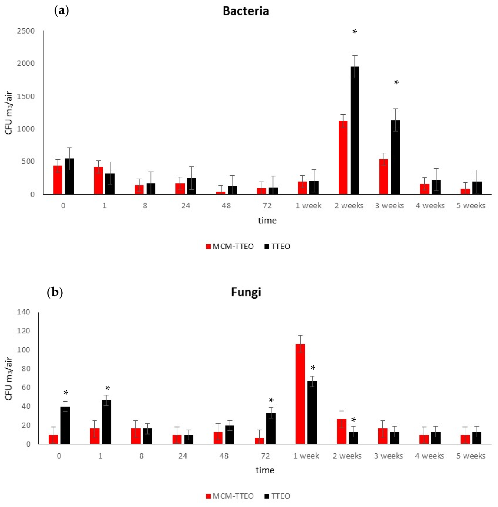
As shown in Figure 5, both treatments (free TTEO and MCM-TTEO) reduced microbial counts for up to 1 week for bacteria and 72 h for fungi. Between weeks 1 and 2, a temporary increase in both fungi and bacteria was observed, followed by a gradual decline to below-baseline levels by week 5. This transient surge may stem from the sampling procedure: opening the room door during collection likely introduced ambient contaminants. Additionally, fluctuations in relative humidity (RH%) and temperature (Section 3.2.2) may have influenced microbial dynamics.
When comparing the two formulations, MCM-TTEO induced a slightly greater initial reduction in bacterial counts at 48 h (up to 89%) compared to free TTEO (78%). Following the recolonization peak at week 2, where higher bacterial loads occurred in the free TTEO room (1953 CFU m3air−1) than in the MCM-TTEO room (about 1126 CFU m3air−1), both treatments ultimately reduced air-borne bacteria below baseline by week 5. However, MCM-TTEO exhibited higher efficacy, achieving an 81% reduction compared to 64% for free TTEO (Figure 5).
The antimicrobial activity observed in this study can be mainly attributed to the release of TTEO, whose bioactive compounds are known to exert antimicrobial effects through membrane disruption and alteration of cellular functions [34,35]. In this context, the mesoporous silica support is not expected to exhibit direct antimicrobial activity; however, it plays a key role in modulating the release of the essential oil. These results indicate that encapsulation within the porous structure of the MCM likely contributes to a more sustained release profile, enhancing the persistence and effectiveness of the antimicrobial action compared to the free essential oil.
Similar results were obtained for the short-term activity against fungi, with MCM-TTEO system inducing a higher initial reduction than free TTEO. However, week 2 recolonization was higher in the MCM-TTEO room (106 CFU m3air−1) than in the free TTEO room (66 CFU m3air−1). By week 5, both treatments had gradually reduced airborne fungal loads to below-baseline levels (Figure 5).
Correlating the temperature and relative humidity (RH) time series (Section 3.2.2) reveals that temperature remained relatively stable throughout the study (approximately 23 °C). Conversely, a marked drop in relative humidity occurred during the intermediate phase (reaching 37% on May 16), followed by a recovery in week 4 (51.6%) and a further increase in week 5 (60.4%). This transient reduction in RH aligns with the observed recolonization phases (week 1 for fungi and week 2 for bacteria). A lower RH likely impairs the release of essential oil molecules, thereby diminishing their antimicrobial efficacy and enabling microbial recolonization. Then, as relative humidity levels recovered, the performance of the air purification system was restored.

3.3. In-Lab Indoor Evaluation of the Direct Contact Antimicrobial Activity of the EO-Based Systems

In vitro tests were conducted in the two designated rooms to evaluate whether the antimicrobial efficacy of the developed air cleaning systems could be enhanced through active contact. These tests assessed the combined influence of temperature, humidity, and direct contact between airborne microorganisms and the biocide, determining whether these parameters stimulate tea tree essential oil release from the microparticles and augment the overall biocidal effect. Specifically, the surfaces of microbial culture media in Petri dishes were treated with either free EO or the MCM-TTEO system and subjected to SAS air sampling within the test rooms. Following exposure, the Petri dishes were incubated at 30 °C and 100% RH for 48 h (bacteria) or 5 days (fungi). Results obtained were then compared with the passive-release system data described above (Section 3.2.4).
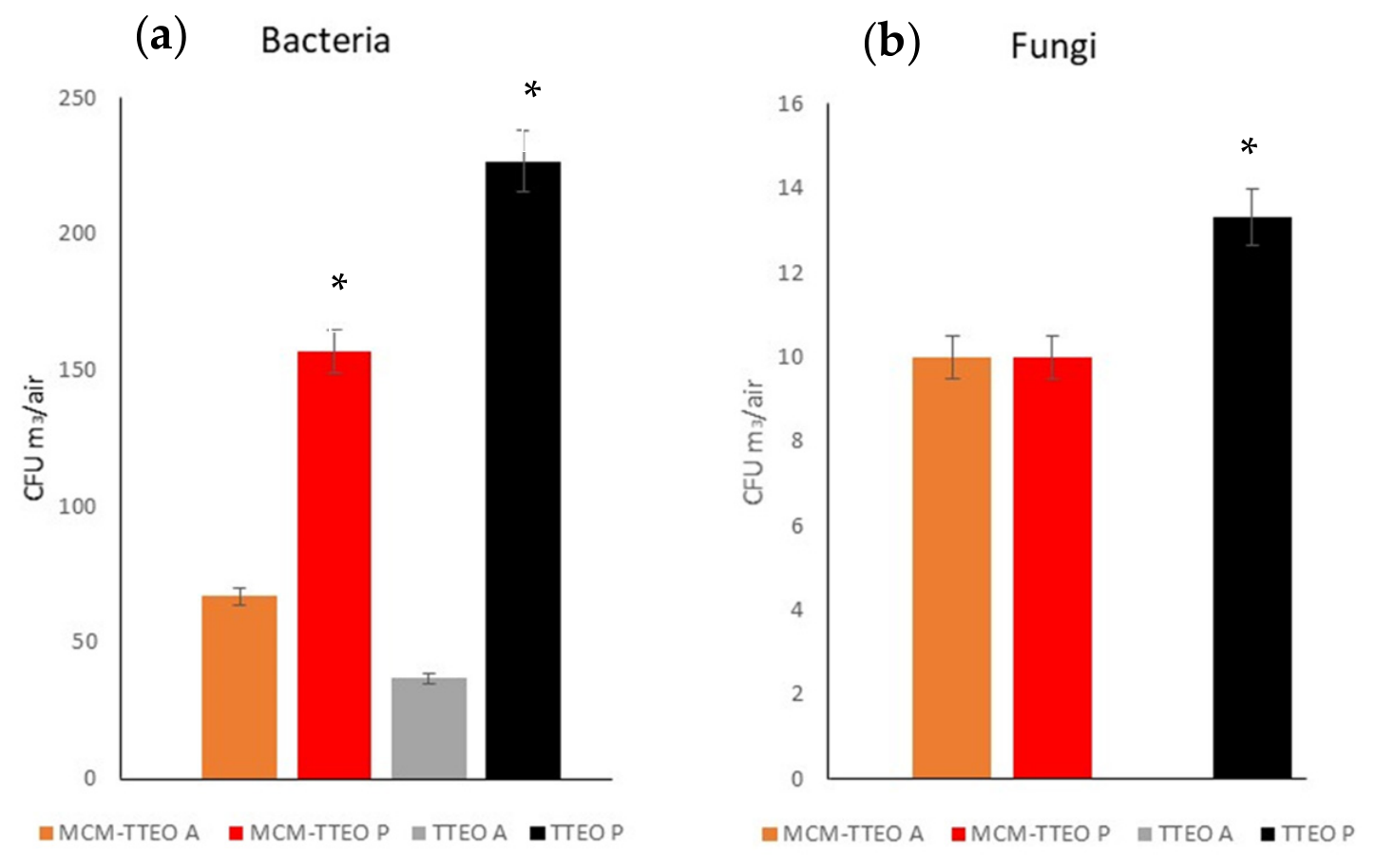
As shown in Figure 6, direct interaction between the biocide and microorganisms significantly reduced bacterial counts compared to the passive-release system for both free and encapsulated TTEO, with free TTEO exhibiting slightly higher bactericidal activity (Figure 6a). Similar trends were observed for fungal reduction with free TTEO (Figure 6b); however, no significant difference was detected between contact or passive MCM-TTEO systems. Consequently, a direct-contact cleaning system may offer enhanced efficacy in reducing airborne bacteria in indoor environments.
The sustained antimicrobial activity highlighted by the micro-MCM-41 system underscores the vital role of host–guest structural architectures in optimizing plant-derived biocides for indoor air treatment. Unencapsulated essential oil terpenes, such as thymol, carvacrol, and terpinen-4-ol, typically suffer from rapid atmospheric evaporation, leading to short-lived efficacy and excessive burst-release kinetics [20]. Consistent with literature data, our findings confirm the capacity of micro-MCM-41 matrices to serve as exceptional structural reservoirs. Their ordered microscale pore channels prioritize long-term payload retention, significantly suppressing premature volatilization and extending the release window over multiple weeks. As the microscale carrier remains localized and could not readily penetrate cellular membranes, its antimicrobial action relies on the controlled, continuous diffusion of active volatile vapors directly into the surrounding microenvironment [36]. Consequently, micro-MCM-41 systems drastically optimize the functional and economic efficiency of natural extracts, maintaining prolonged microbial suppression while utilizing lower total concentrations of essential oil.
Crucially, the volatilization and antimicrobial efficacy of encapsulated essential oils are intrinsically tied to environmental conditions—particularly relative humidity (RH) and ambient temperature, which govern vapor pressure dynamics and microbial recolonization rates. Air purification platforms must operate in tandem with robust environmental monitoring, implemented through the use of nanomaterials capable of accurately detecting specific gaseous pollutants. For instance, advanced chemical sensors, such as WO3/CNT hybrids and flexible WO3/MoS2 heterojunctions enable continuous, low-power detection of gaseous pollutants as NO2 [37]. Furthermore, recent advances in traceable Indoor Environmental Quality (IEQ) monitoring, such as low-cost multi-sensor platforms (e.g., the PROMET&O framework), demonstrate the feasibility of real-time, multi-parameter tracking [38]. Integrating metrologically calibrated multi-sensor modules capable of simultaneously measuring microclimatic drivers (temperature, RH) and air quality indicators (Volatile Organic Compounds, Particulate Matter, CO2) provides essential contextual data to predict and counteract fluctuations in biocide release efficacy. This establishes a clear synergy with micro-MCM-41 sustained-release filtration, enabling the creation of an adaptive HVAC filtration ecosystem. In delicate cultural heritage environments, archives, and healthcare facilities, such a closed-loop system can dynamically modulate ventilation, humidity, and active biocide dispersion based on real-time sensor feedback, ensuring continuous airborne pathogen suppression while safeguarding sensitive substrates against biofouling and environmental degradation.

4. Conclusions

This study presents a successful proof-of-concept for a sustainable, non-invasive air cleaning system based on Tea Tree Essential Oil (TTEO) encapsulated within MCM-41 mesoporous silica microparticles (MCM-TTEO). Designed specifically to overcome the material incompatibility of conventional industrial biocides with delicate cultural heritage substrates, the microencapsulation approach resolves the high volatility of natural essential oils. In vitro studies confirmed that while free TTEO underwent nearly complete evaporation (approximately 95%) within one week, MCM-TTEO restricted early release to approximately 15%, maintaining a sustained volatilization rate of about 30% from weeks 2 through 5.
When deployed in real indoor testing environments, MCM-TTEO demonstrated superior, rapid bactericidal and short-term fungicidal activity compared to free oil, effectively suppressing microbial recolonization despite a significantly lower volatilization footprint (5.76% of total loaded oil).
Environmental monitoring revealed that fluctuations in relative humidity (RH) strongly influence efficacy: temporary drops in RH impaired essential oil volatilization and facilitated transient microbial recolonization, whereas RH recovery restored system performance. Furthermore, direct-contact assays on culture media demonstrated that moisture and temperature actively trigger the microparticle release mechanism, enhancing immediate biocidal action.
Overall, MCM-TTEO offers a safer, indirect strategy to protect heritage surfaces by preventing airborne biocolonization without direct chemical application onto fragile artifacts. Future work should explore longer observation periods in semi-confined spaces to optimize oil dosage. Additionally, covalent functionalization of the MCM-41 matrix could enable the development of active air filtration systems, allowing seamless integration into climate-control protocols alongside temperature and relative humidity management in museums and archives.

Author Contributions

Conceptualization, P.B.-R.; methodology, P.B.-R. and A.B.; formal analysis, A.L.T., A.B. and M.C.-M.; investigation, A.L.T., A.B., P.B.-R. and M.C.-M.; resources, P.B.-R., M.D.M., R.M.-M. and A.B.; data curation, A.L.T., A.B. and P.B.-R.; writing—original draft preparation, A.L.T., A.S., L.S., P.B.-R. and A.B.; writing—review and editing, A.L.T., P.B.-R., M.D.M., R.M.-M., A.B., L.S., A.S. and V.G.; supervision, D.P. All authors have read and agreed to the published version of the manuscript.

Funding

Erasmus + Placement 2023/2024 Scholarship funded A.L.T. staying in Valencia and the Santos Juanes Project funds the lab fungible material. This research was funded by the Spanish Government (projects PID2021-126304OB-C41, PID2021-128141OB-C22, PID2024-155683OB-C41 and PID2024-162542OB-I00), MCUI/FEDER, EU and CIBER-Consorcio Centro de Investigación Biomédica en Red (CB06/01/2012), Instituto de Salud Carlos III, Ministerio de Ciencia e Innovación and the Generalitat Valenciana (CIPROM/2021/007).

Institutional Review Board Statement

Not applicable.

Informed Consent Statement

Not applicable.

Data Availability Statement

The original contributions presented in this study are included in the article. Further inquiries can be directed to the corresponding author.

Conflicts of Interest

The authors declare no conflicts of interest.

Appendix A

Figure A1. Representation of Room 1, where the labels ‘R3’, ‘R2’and ‘R1’ indicate the positions of the replicates inside the rooms. Label ‘S1’ indicates the square Petri Dish with the MCM-TTEO microparticles.
Applsci 16 07674 g0a1
Figure A2. Representation of Room 2, where the labels ‘R3’, ‘R2’and ‘R1’ indicate the positions of the replicates inside the room. Label ‘S2’ indicates the square Petri Dish with the free essential oil.
Applsci 16 07674 g0a2

References

  1. Gorbushina, A. Bacterial and fungal diversity and biodeterioration problems in mural painting environments of St. Martins church (Greene–Kreiensen, Germany). Int. Biodet. Biodegrad. 2004, 53, 13–24. [Google Scholar] [CrossRef] [Scilit]
  2. Dias, L.; Rosado, T.; Candeias, A.; Mirão, J.; Caldeira, A.T. Linking ornamental stone discolouration to its biocolonisation state. Build. Environ. 2020, 180, 106934. [Google Scholar] [CrossRef] [Scilit]
  3. Sokhi, R.S.; Moussiopoulos, N.; Baklanov, A.; Bartzis, J.; Coll, I.; Finardi, S.; Friedrich, R.; Geels, C.; Grönholm, T.; Halenka, T.; et al. Advances in Air Quality Research—Current and Emerging Challenges. Atmos. Chem. Phys. 2022, 22, 4615–4703. [Google Scholar] [CrossRef] [Scilit]
  4. Pye, H.O.T.; Nenes, A.; Alexander, B.; Ault, A.P.; Barth, M.C.; Clegg, S.L.; Collett, J.L., Jr.; Fahey, K.M.; Hennigan, C.J.; Herrmann, H.; et al. The Acidity of Atmospheric Particles and Clouds. Atmos. Chem. Phys. 2020, 20, 4809–4888. [Google Scholar] [CrossRef] [Scilit] [PubMed]
  5. European Commission. Indoor Air Quality & Its Impact on Man; Report No. 12: Biological Particles in Indoor Environments; Office for Official Publications of the European Communities: Luxembourg, 1993. [Google Scholar]
  6. Degrendele, C.; Kanduč, T.; Kocman, D.; Lammel, G.; Cambelová, A.; Dos Santos, S.G.; Horvat, M.; Kukučka, P.; Holubová Šmejkalová, A.; Mikeš, O.; et al. NPAHs and OPAHs in the Atmosphere of Two Central European Cities: Seasonality, Urban-to-Background Gradients, Cancer Risks and Gas-to-Particle Partitioning. Sci. Total Environ. 2021, 793, 148528. [Google Scholar] [CrossRef] [Scilit] [PubMed]
  7. Tiano, P. Biodegradation of Cultural Heritage: Decay Mechanisms and Control Methods. In Proceedings of the 9th ARIADNE Workshop “Historic Material and their Diagnostic” ARCCHIP, Prague, Czech Republic, 22–28 April 2002; Available online: http://www.arcchip.cz/w09/w09_tiano.pdf (accessed on 5 May 2026).
  8. Angeles Flores, G.; Venanzoni, R.; Martino, S.; Angelini, P. Filamentous Fungi and the Biodeterioration of Organic Cultural Heritage Materials: A Systematic Review of Mechanisms, Risks, and Preventive Conservation Strategies. Microorganisms 2026, 14, 526. [Google Scholar] [CrossRef] [Scilit] [PubMed]
  9. Ramos, C.C.R.; Roque, J.L.A.; Sarmiento, D.B.; Suarez, L.E.G.; Sunio, J.T.P.; Tabungar, K.I.B.; Tengco, G.S.C.; Rio, P.C.; Hilario, A.L. Use of Ultraviolet-C in Environmental Sterilization in Hospitals: A Systematic Review on Efficacy and Safety. Int. J. Health Sci. 2020, 14, 52–65. [Google Scholar]
  10. Pavlogeorgatos, G. Environmental Parameters in Museums. Build. Environ. 2003, 38, 1457–1462. [Google Scholar] [CrossRef] [Scilit]
  11. Camuffo, D. Microclimate for Cultural Heritage: Conservation, Restoration, and Maintenance of Indoor and Outdoor Monuments, 3rd ed.; Elsevier: Amsterdam, The Netherlands, 2019; pp. 350–375. [Google Scholar]
  12. Valentín, N.; Fazio, A.T. Análisis de la Incidencia del SARS-CoV-2 en Bienes Culturales. Sistemas de Desinfección. Fundamentos y Estrategias de Control; Grupo Español de Conservación (GE-IIC): Madrid, Spain, 2020; pp. 1–26. [Google Scholar]
  13. Lucchi, E. Review of Preventive Conservation in Museum Buildings. J. Cult. Herit. 2018, 29, 180–193. [Google Scholar] [CrossRef] [Scilit]
  14. Colalucci, G. Michelangelo Buonarroti: Restoration of the Frescoes on the Vaulted Ceiling and the Last Judgment in the Sistine Chapel. Conserv. Sci. Cult. Herit. 2016, 16, 89–108. [Google Scholar]
  15. Bosch Roig, P.; Díaz Alonso, J.; Vivo Soria, E. Propuesta de Monitorización Integral de La Calidad Ambiental Del Interior de La Iglesia de Los Santos Juanes de Valencia. Arch. Arte Valencia. 2020, 101, 179–192. [Google Scholar]
  16. Díaz-Alonso, J.; Bernardos, A.; Regidor-Ros, J.L.; Martínez-Máñez, R.; Bosch-Roig, P. Innovative Use of Essential Oil Cold Diffusion System for Improving Air Quality on Indoor Cultural Heritage Spaces. Int. Biodeterior. Biodegrad. 2021, 162, 105251. [Google Scholar] [CrossRef] [Scilit]
  17. Nazzaro, F.; Fratianni, F.; De Martino, L.; Coppola, R.; De Feo, V. Effect of Essential Oils on Pathogenic Bacteria. Pharmaceuticals 2013, 6, 1451–1474. [Google Scholar] [CrossRef] [Scilit] [PubMed]
  18. Krist, S.; Sato, K.; Glasl, S.; Hoeferl, M.; Saukel, J. Antimicrobial Effect of Vapours of Terpineol, (R)-(–)-Linalool, Carvacrol, (S)-(–)-Perillaldehyde and 1,8-Cineole on Airborne Microbes Using a Room Diffuser. Flavour Fragr. J. 2008, 23, 353–356. [Google Scholar] [CrossRef] [Scilit]
  19. Laird, K.; Phillips, C. Vapour phase: A potential future use for essential oils as antimicrobials? Lett. Appl. Microbiol. 2012, 54, 169–174. [Google Scholar] [CrossRef] [Scilit] [PubMed]
  20. Campostrini, A.; Sala-Luis, A.; Bosch-Roig, P.; Ghedini, E.; Signoretto, M.; Menegazzo, F. Mesoporous Silica and Vegetal Extracts Combined as Sustainable Stone Heritage Protection against Biodeterioration. Appl. Microbiol. Biotechnol. 2025, 109, 99. [Google Scholar] [CrossRef] [Scilit] [PubMed]
  21. Vallet-Regí, M.; Colilla, M.; Izquierdo-Barba, I.; Manzano, M. Mesoporous Silica Nanoparticles for Drug Delivery: Current Insights. Mol. J. Synth. Chem. Nat. Prod. Chem. 2017, 23, 47. [Google Scholar] [CrossRef] [Scilit] [PubMed]
  22. Popova, T.; Tzankov, B.; Voycheva, C.; Spassova, I.; Kovacheva, D.; Tzankov, S.; Aluani, D.; Tzankova, V.; Lambov, N. Mesoporous Silica MCM-41 and HMS as Advanced Drug Delivery Carriers for Bicalutamide. J. Drug Deliv. Sci. Technol. 2021, 62, 102340. [Google Scholar] [CrossRef] [Scilit]
  23. Bernardos, A.; Marina, T.; Žáček, P.; Pérez-Esteve, É.; Martínez-Mañez, R.; Lhotka, M.; Kouřimská, L.; Pulkrábek, J.; Klouček, P. Antifungal Effect of Essential Oil Components against Aspergillus niger When Loaded into Silica Mesoporous Supports. J. Sci. Food Agric. 2015, 95, 2824–2831. [Google Scholar] [CrossRef] [Scilit] [PubMed]
  24. Alfieri, I.; Lorenzi, A.; Ranzenigo, L.; Lazzarini, L.; Predieri, G.; Lottici, P.P. Synthesis and Characterization of Photocatalytic Hydrophobic Hybrid TiO2-SiO2 Coatings for Building Applications. Build. Environ. 2017, 111, 72–79. [Google Scholar] [CrossRef] [Scilit]
  25. Ruggiero, L.; Bartoli, F.; Fidanza, M.R.; Zurlo, F.; Marconi, E.; Gasperi, T.; Tuti, S.; Crociani, L.; Di Bartolomeo, E.; Caneva, G.; et al. Encapsulation of environmentally-friendly biocides in silica nanosystems for multifunctional coatings. Appl. Surf. Sci. 2020, 514, 145908. [Google Scholar] [CrossRef] [Scilit]
  26. Bartoli, F.; Corradi, L.; Hosseini, Z.; Privitera, A.; Zuena, M.; Kumbaric, A.; Graziani, V.; Tortora, L.; Sodo, A.; Caneva, G. In vitro viability tests of new ecofriendly nanosystems incorporating essential oils for long-lasting conservation of stone artworks. Gels 2024, 10, 132. [Google Scholar] [CrossRef] [Scilit] [PubMed]
  27. Liu, J.Y.; Sayes, C.M. A toxicological profile of silica nanoparticles. Toxicol. Res. 2022, 11, 565–582. [Google Scholar] [CrossRef] [Scilit] [PubMed]
  28. Kamikubo, Y.; Sakairi, H. Silica nanoparticle toxicity: Cellular mechanisms, neurotoxicological concerns, and environmental perspectives. Front. Nanotechnol. 2025, 7, 1629722. [Google Scholar] [CrossRef] [Scilit]
  29. Hammer, K.A.; Carson, C.F.; Riley, T.V.; Nielsen, J.B. A Review of the Toxicity of Melaleuca alternifolia (Tea Tree) Oil. Food Chem. Toxicol. 2006, 44, 616–625. [Google Scholar] [CrossRef] [Scilit] [PubMed]
  30. Kozics, K.; Bučková, M.; Puškárová, A.; Kalászová, V.; Cabicarová, T.; Pangallo, D. The Effect of Ten Essential Oils on Several Cutaneous Drug-Resistant Microorganisms and Their Cyto/Genotoxic and Antioxidant Properties. Molecules 2019, 24, 4570. [Google Scholar] [CrossRef] [Scilit] [PubMed]
  31. Vijeth, S.; Heggannavar, G.B.; Kariduraganavar, Y.M. Encapsulating Wall Materials for Micro-/Nanocapsules. In Microencapsulation—Processes, Technologies and Industrial Applications; Salaün, F., Ed.; IntechOpen: London, UK, 2019. [Google Scholar]
  32. Angulo-Milhem, S.; Verriele, M.; Nicolas, M.; Thevenet, F. Indoor use of essential oils: Emission rates, exposure time and impact on air quality. Atmos. Environ. 2021, 244, 117863. [Google Scholar] [CrossRef] [Scilit]
  33. Reponen, T.; Grinshpun, S.A.; Conwell, K.L.; Wiest, J.; Anderson, M. Aerodynamic versus physical size of spores: Measurement and implication for respiratory deposition. Grana 2001, 40, 119–125. [Google Scholar] [CrossRef] [Scilit]
  34. Nazzaro, F.; Fratianni, F.; Coppola, R.; De Feo, V. Essential Oils and Antifungal Activity. Pharmaceuticals 2017, 10, 86. [Google Scholar] [CrossRef] [Scilit] [PubMed]
  35. Cai, J.; Yan, R.; Shi, J.; Chen, J.; Long, M.; Wu, W.; Kuca, K. Antifungal and Mycotoxin Detoxification Ability of Essential Oils: A Review. Phytother. Res. 2022, 36, 62–72. [Google Scholar] [CrossRef] [Scilit] [PubMed]
  36. Gulin-Sarfraz, T.; Kalantzopoulos, G.N.; Haugen, J.-E.; Axelsson, L.; Raanaas Kolstad, H.; Sarfraz, J. Controlled release of volatile antimicrobial compounds from mesoporous silica nanocarriers for active food packaging applications. Int. J. Mol. Sci. 2022, 23, 7032. [Google Scholar] [CrossRef] [Scilit] [PubMed]
  37. Gao, M.; Guo, J.; Yang, X.; Zhou, Y.; dos Santos Laranjeira, J.A.; Sambrano, J.R.; Zhang, W. From Sensors to System-Level Integration: Flexible WO3/MoS2 Heterojunction NO2 Gas Sensor. Sens. Actuators B Chem. 2026, 451, 139328. [Google Scholar] [CrossRef] [Scilit]
  38. Fissore, V.I.; Arcamone, G.; Astolfi, A.; Barbaro, A.; Carullo, A.; Chiavassa, P.; Clerico, M.; Fantucci, S.; Fiori, F.; Gallione, D.; et al. Multi-Sensor Device for Traceable Monitoring of Indoor Environmental Quality. Sensors 2024, 24, 2893. [Google Scholar] [CrossRef] [Scilit] [PubMed]
Figure 1. Characterization of MCM-41 microparticles by normalized powder X-ray diffraction: (a) calcinated MCM-41 microparticles, (b) MCM-41 microparticles as obtained from synthesis reaction.
Applsci 16 07674 g001
Figure 2. Release curve of EO from the MCM-TTEO system (red dots) compared with evaporation of the free TTEO (black squares).
Applsci 16 07674 g002
Figure 3. Temporal evolution of indoor temperature (°C, black line) and relative humidity (%, red line) during the experimental period in Room 1, where the experiment of spontaneous release from the MCM-TTEO air cleaning system was performed.
Applsci 16 07674 g003
Figure 4. Mean values and standard deviations of (a) PM 0.3 μm, and (b) PM 5.0 μm obtained during testing of both air cleaning systems: results for free TTEO are reported with black bars, while those for MCM-TTEO are reported with red bars. Asterisks indicate significant differences between treatments (* p < 0.05).
Applsci 16 07674 g004aApplsci 16 07674 g004b
Figure 5. Mean values and standard deviations of microbial counts obtained testing both air cleaning systems; graphs report counts relative to (a) bacteria, and (b) fungi. Results obtained with free TTEO are reported with black bars, while those for MCM-TTEO are reported with red bars. Asterisks indicate significant differences between treatments.
Applsci 16 07674 g005
Figure 6. Mean microbial counts (CFU) recovered for free TTEO and MCM-TTEO applied under contact (A) or passive (P) air cleaning system for (a) bacteria and (b) fungi. Data are presented as mean ± standard deviation (SD) (n = 3). Asterisks indicate significant differences between treatments (* p < 0.05).
Applsci 16 07674 g006
Table 1. The table reports the amount of organic matter (the TTEO encapsulated on the MCM-41) detected during the thermogravimetric analysis performed at different time points of experimentation. The fraction detected is correlated with the respective temperature of analysis.
Time of Release(W/W) % of Organic Matter Released
0 s0.0
24 h1.4
48 h5.0
72 h9.3
1 week16.2
2 weeks33.5
3 weeks28.1
4 weeks30.8
5 weeks30.7
Disclaimer/Publisher’s Note: The statements, opinions and data contained in all publications are solely those of the individual author(s) and contributor(s) and not of MDPI and/or the editor(s). MDPI and/or the editor(s) disclaim responsibility for any injury to people or property resulting from any ideas, methods, instructions or products referred to in the content.