1. Introduction
The increasing demand for esthetic dental treatment has substantially changed the planning of prosthodontic and interdisciplinary rehabilitation. Contemporary prosthodontics is no longer focused solely on replacing or masking dental deficiencies, but increasingly follows a biologically oriented and minimally invasive philosophy aimed at preserving enamel, dentin, periodontal tissues, and pulp vitality whenever possible [
1,
2,
3,
4]. This shift has been facilitated by adhesive dentistry, improved ceramic and polymer-based restorative materials, digital workflows, and additive treatment concepts, which together allow clinicians to achieve esthetic and functional outcomes with reduced biological cost [
1,
2,
3,
4].
Within this biologically driven approach, orthodontic pretreatment has become an important adjunct to esthetic and prosthodontic treatment planning. Tooth repositioning before restorative rehabilitation may improve spatial relationships, correct malalignment, redistribute restorative volume, optimize incisal edge position and gingival architecture, and reduce the need for aggressive tooth preparation [
5,
6,
7,
8]. In selected cases, orthodontic alignment may therefore convert a subtractive restorative plan into a more conservative additive or minimally invasive solution [
5,
8]. This concept is particularly relevant in adult patients seeking esthetic improvement, in whom the treatment sequence often includes orthodontic alignment, tooth whitening, and, when needed, definitive adhesive restorations.
Tooth whitening has become an established component of contemporary esthetic dentistry because it can improve tooth color without removing hard dental tissue [
9,
10,
11,
12,
13]. Professionally supervised at-home bleaching with low-concentration carbamide peroxide, especially 10% carbamide peroxide used in custom trays, has been extensively documented since the introduction of nightguard vital bleaching and remains a conservative treatment option for vital tooth discoloration [
9,
10,
11,
12]. However, when tooth alignment and whitening are performed as separate procedures, the total treatment time may be prolonged, additional appliances may be required, and patient compliance may be compromised.
The widespread use of clear aligner therapy has created a potential opportunity to combine orthodontic tooth movement with dental bleaching. Because clear aligners are removable, custom-made, and closely adapted to the dentition, they may potentially serve as carriers for bleaching agents during orthodontic treatment [
14,
15,
16,
17]. This approach could eliminate the need for additional bleaching trays, reduce the number of clinical and laboratory procedures, shorten the overall esthetic treatment sequence, and improve patient convenience. Existing clinical evidence suggests that bleaching during clear aligner treatment can improve tooth shade, including protocols using carbamide peroxide or hydrogen peroxide gels within the aligner [
14,
15,
16,
17]. Nevertheless, the available evidence remains limited, and most studies have focused primarily on clinical whitening outcomes, plaque indices, gingival parameters, or patient-related outcomes rather than on the possible effects of bleaching agents on aligner materials [
16,
17,
18].
This limitation is clinically relevant because clear aligners are not passive trays but active orthodontic appliances. Their performance depends on polymer composition, thermoforming behavior, elastic modulus, stress relaxation, force decay, dimensional stability, optical properties, surface integrity, and resistance to intraoral aging [
19,
20,
21,
22,
23]. Aligners are exposed to saliva, temperature fluctuations, masticatory and parafunctional loading, cleaning agents, dietary components, and, in combined treatment protocols, bleaching products. Even subtle changes in polymer structure or thermomechanical behavior may influence aligner stiffness, fit, force delivery, or transparency. Therefore, the simultaneous use of bleaching agents and clear aligners should not be evaluated only from the perspective of tooth color change, but also from the standpoint of material stability.
Polyethylene terephthalate glycol-modified (PETG) materials and related thermoplastics are commonly used in clear aligner fabrication because of their transparency, formability, and clinically acceptable mechanical behavior [
19,
20,
21,
22,
23]. However, clear aligner polymers are sensitive to clinically relevant environmental factors, including thermoforming, intraoral moisture, thermocycling, brushing, cleaning protocols, and chemical exposure [
20,
21,
22,
23,
24,
25,
26,
27]. These factors may affect surface roughness, mass stability, optical properties, water sorption/desorption behavior, and thermomechanical response in orthodontic thermoplastic and 3D-printed aligner materials [
22,
23,
24,
25,
26,
27,
28,
29]. Accordingly, the introduction of bleaching agents into aligners should be considered not only a clinical bleaching protocol but also a material-exposure condition that may influence polymer stability.
Carbamide peroxide decomposes into hydrogen peroxide and urea in an aqueous gel environment. Because PETG contains ester and ether-related linkages, exposure to peroxide and moisture may theoretically promote surface oxidation, hydrolysis, or chain scission. Such reactions may alter the carbonyl, hydroxyl, or C–O–C regions of the infrared spectrum and may also affect molecular mobility. Conversely, water loss, physical aging, residual-stress relaxation, or densification of the amorphous phase may restrict segmental mobility and produce a small increase in Tg without a major change in the infrared chemical fingerprint. Therefore, both low-level chemical modification and non-chemical thermophysical rearrangement were considered plausible responses to intra-aligner bleaching [
29,
30].
Dynamic mechanical analysis (DMA) is particularly suitable for evaluating thermomechanical transitions in polymeric aligner materials because it detects temperature-dependent changes in storage modulus, loss modulus, and damping behavior [
20,
25,
28]. Fourier-transform infrared spectroscopy (FTIR), in contrast, enables assessment of the material’s chemical fingerprint and can identify changes in characteristic functional groups following exposure to external agents [
24,
29,
31]. The combined use of DMA and FTIR therefore provides complementary information: DMA evaluates thermomechanical response and polymer chain mobility, whereas FTIR assesses chemical stability.
The present study used aligners retrieved after clinical wear rather than specimens exposed only under simplified laboratory immersion conditions. The aim was to evaluate whether intra-aligner application of 10% carbamide peroxide during clinical use affects the onset glass transition temperature or the qualitative FTIR spectral characteristics of Nuvola® clear aligners. The null hypothesis was that clinical wear combined with 10% carbamide peroxide bleaching would not produce detectable differences in these outcomes compared with unused aligners and clinically worn aligners not exposed to the bleaching agent.
2. Materials and Methods
2.1. Study Design and Experimental Groups
This experimental material study evaluated the thermomechanical and chemical behavior of clinically used Nuvola® clear aligners (Nuvola World Srl, Vicenza, Italy) after intra-aligner application of 10% carbamide peroxide. The study was designed to reproduce a clinically relevant exposure model, in which aligners were retrieved after patient use, rather than being exposed only under simplified laboratory immersion conditions.
Five experimental groups were investigated: unused aligners not exposed to intraoral conditions or bleaching treatment (control group); aligners worn intraorally for 14 days without bleaching treatment (wear-only group); aligners worn intraorally for 14 days with intra-aligner application of 10% carbamide peroxide for 3 days; aligners worn intraorally for 14 days with intra-aligner application of 10% carbamide peroxide for 7 days; and aligners worn intraorally for 14 days with intra-aligner application of 10% carbamide peroxide for 14 days. The final sample consisted of 30 independent aligners, with six aligners allocated to each experimental group. One DMA specimen was prepared from each aligner; therefore, 30 independent DMA specimens were analyzed in total. Each aligner contributed to only one experimental group.
All clinically worn aligners were used according to the prescribed clinical wear protocol. In the bleaching groups, 10% carbamide peroxide gel (Opalescence™ PF 10%, Ultradent Products, Inc., South Jordan, UT, USA) was applied to the inner surface of the aligner in the region corresponding to the maxillary right second premolar to the maxillary left second premolar (FDI teeth 15–25). After completion of the assigned wear and bleaching period, the aligners were retrieved, rinsed to remove visible debris and residual bleaching gel, dried, and stored under standardized laboratory conditions until specimen preparation.
2.2. Specimen Preparation
Specimens for dynamic mechanical analysis were harvested from the central incisor region of each aligner. This region was selected because it presents relatively low anatomical curvature compared with posterior or canine regions, thereby allowing more reproducible specimen preparation and reducing variability related to complex thermoformed geometry. After sectioning, the specimens were subjected to a standardized constant load of 10 kg for 10 days at ambient laboratory temperature to maximize flatness and stabilize the polymer structure before testing.
Rectangular specimens with nominal dimensions of 15 mm × 3 mm × 0.7 mm were prepared. Specimen thickness and width were measured at three locations using a caliper. The predefined dimensional acceptance criterion was ±5% of the target dimensions. All prepared specimens met this criterion; therefore, no specimens were excluded or replaced. A total of 30 specimens were included in the DMA analysis, with six independent specimens in each experimental group. The flattening procedure was applied uniformly to all groups.
2.3. Dynamic Mechanical Analysis (DMA)
Thermomechanical measurements were performed using a DMA 1 dynamic mechanical analyzer (Mettler-Toledo GmbH, Schwerzenbach, Switzerland) equipped with a tensile specimen holder. The initial gauge length was 7.5 mm. Each specimen underwent a single temperature scan to avoid additional effects associated with repeated thermal cycling and thermal history. Data acquisition and analysis were performed using STARe Excellence Thermal Analysis Software, version 16.3. Temperature, force, and displacement calibrations were performed according to the manufacturer’s recommendations.
DMA was performed under ambient air conditions. Temperature scans were conducted from 25 °C to 100 °C using a heating rate of 2 °C/min, an oscillation frequency of 1 Hz, and a displacement amplitude of 10 μm. The selected heating rate represented a compromise between accurately characterizing the α-relaxation region and minimizing additional temperature-induced changes during the measurement.
The damping factor (tan δ = E″/E′) was continuously recorded as a function of temperature and selected as the primary outcome because this ratio reduces the influence of small differences in specimen cross-section. The glass transition temperature (Tg) was determined from the onset of the α-relaxation rather than from the tan δ peak maximum. This approach was selected because the onset temperature corresponds to the initiation of cooperative segmental chain mobility within the polymer matrix and therefore represents the beginning of thermomechanical softening. For orthodontic aligner applications, identifying the temperature at which the material begins to lose mechanical stability was considered more clinically relevant than determining the peak relaxation temperature. After the operator selected the α-relaxation region, STARe Excellence Thermal Analysis Software, version 16.3, automatically constructed two tangents: one corresponding to the pre-transition baseline and the other to the rising portion of the α-relaxation curve. Tg onset was defined as the temperature at the intersection of these two tangents. Formal measurement uncertainty for the complete DMA procedure, including specimen preparation and Tg-onset determination, was not independently quantified; therefore, statistically significant differences smaller than 1 °C were interpreted cautiously.
2.4. Fourier-Transform Infrared Spectroscopy
Fourier-transform infrared spectroscopy was performed to confirm the chemical composition of the investigated aligner material and to evaluate its chemical stability following exposure to the bleaching agent. Spectra were recorded using a Bruker Alpha FTIR spectrometer (Bruker Optics, Ettlingen, Germany). Spectral acquisition was performed in the wavenumber range of 4000–400 cm−1 with a spectral resolution of 4 cm−1. For each sample, 10 consecutive scans were collected and averaged. Spectral acquisition and data processing were carried out using OPUS software version 7.2.
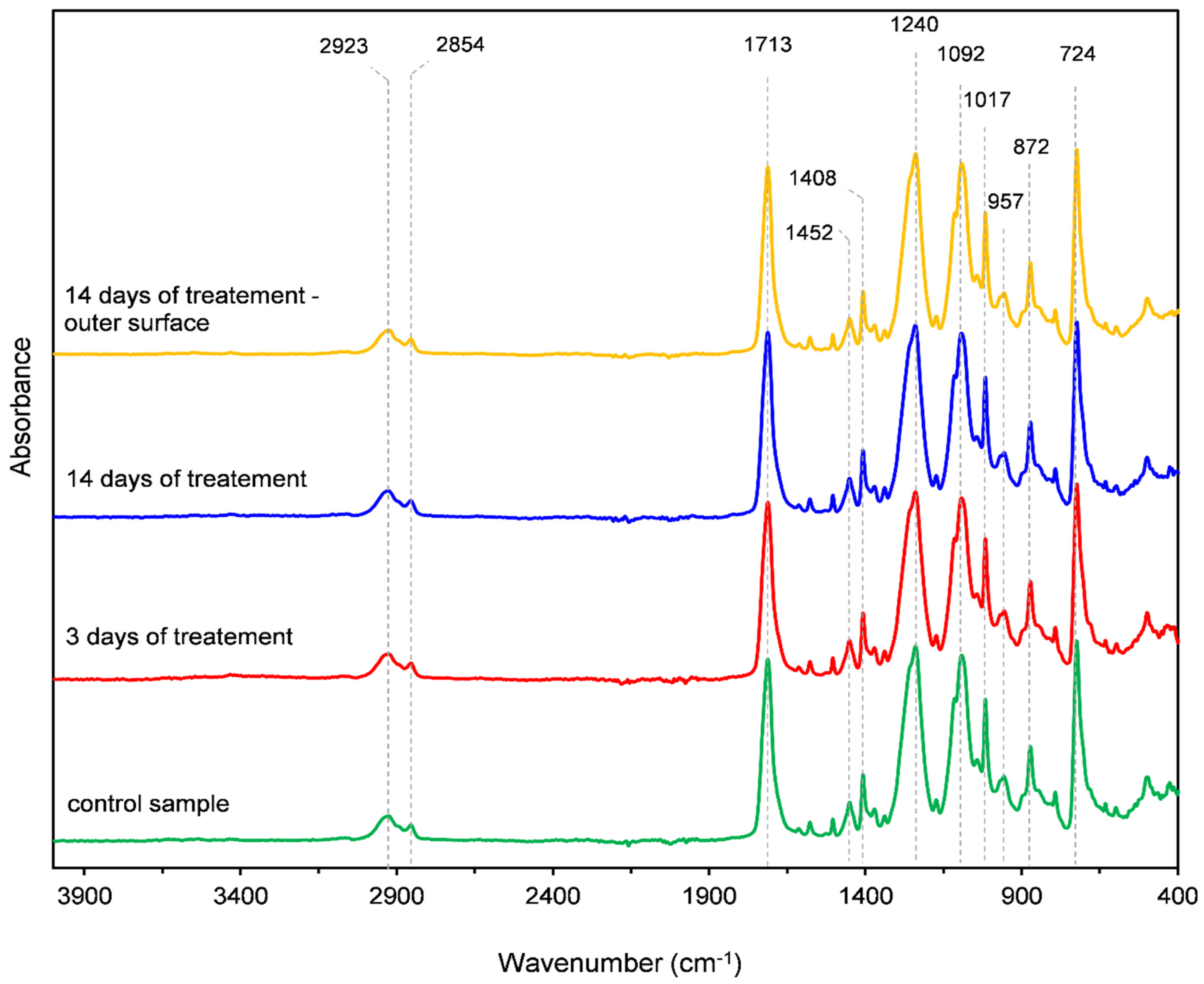
Representative spectra were obtained from samples collected from both the inner and outer surfaces of the aligner in the region corresponding to the central incisor, canine and second premolar. Inner-surface samples included the untreated control specimen and specimens exposed to 10% carbamide peroxide for 3 and 14 days. In addition, the outer surface of a specimen exposed to 14 days of bleaching treatment was analyzed to determine whether the surface not directly exposed to the bleaching gel showed a comparable spectral profile.
The spectra were evaluated by identifying characteristic absorption bands of polyethylene terephthalate glycol-modified material, more specifically, poly(ethylene glycol-co-1,4-cyclohexanedimethanol terephthalate). The main evaluated bands included C–H stretching vibrations of methylene groups at 2923 cm−1 and 2854 cm−1, ester carbonyl C=O stretching at 1713 cm−1, CH2 bending of glycol units at 1452 cm−1, in-plane C–H bending and C–C stretching at 1408 cm−1, ester C–(=O)–O vibrations at approximately 1262–1240 cm−1, symmetric C–O–C stretching at 1092 cm−1, cyclohexane ring-associated vibrations at 957 cm−1, out-of-plane C–H/C–C–C bending at 872 cm−1, and aromatic C–H out-of-plane bending at 724 cm−1. Spectra from untreated and bleaching-treated specimens were compared qualitatively with respect to the appearance of new absorption bands, disappearance of characteristic bands, or shifts in peak position. All prepared FTIR specimens were analyzed; because spectra within each experimental condition showed the same PETG-related band pattern, one representative spectrum per condition is shown for clarity. FTIR spectra were not subjected to inferential statistical testing.
2.5. Statistical Analysis
Statistical analysis was performed using IBM SPSS Statistics 29.0.2.0. [
32]. Descriptive statistics were calculated for Tg values and expressed as mean, standard deviation, 95% confidence interval, and minimum to maximum range. The normality of Tg data within each group was assessed using the Shapiro–Wilk test. Homogeneity of variance was evaluated using Levene’s test. Because Levene’s test indicated unequal variances, differences among experimental groups were analyzed using Welch’s analysis of variance. Post hoc pairwise comparisons were performed using the Games–Howell test. The level of statistical significance was set at α = 0.05.
4. Discussion
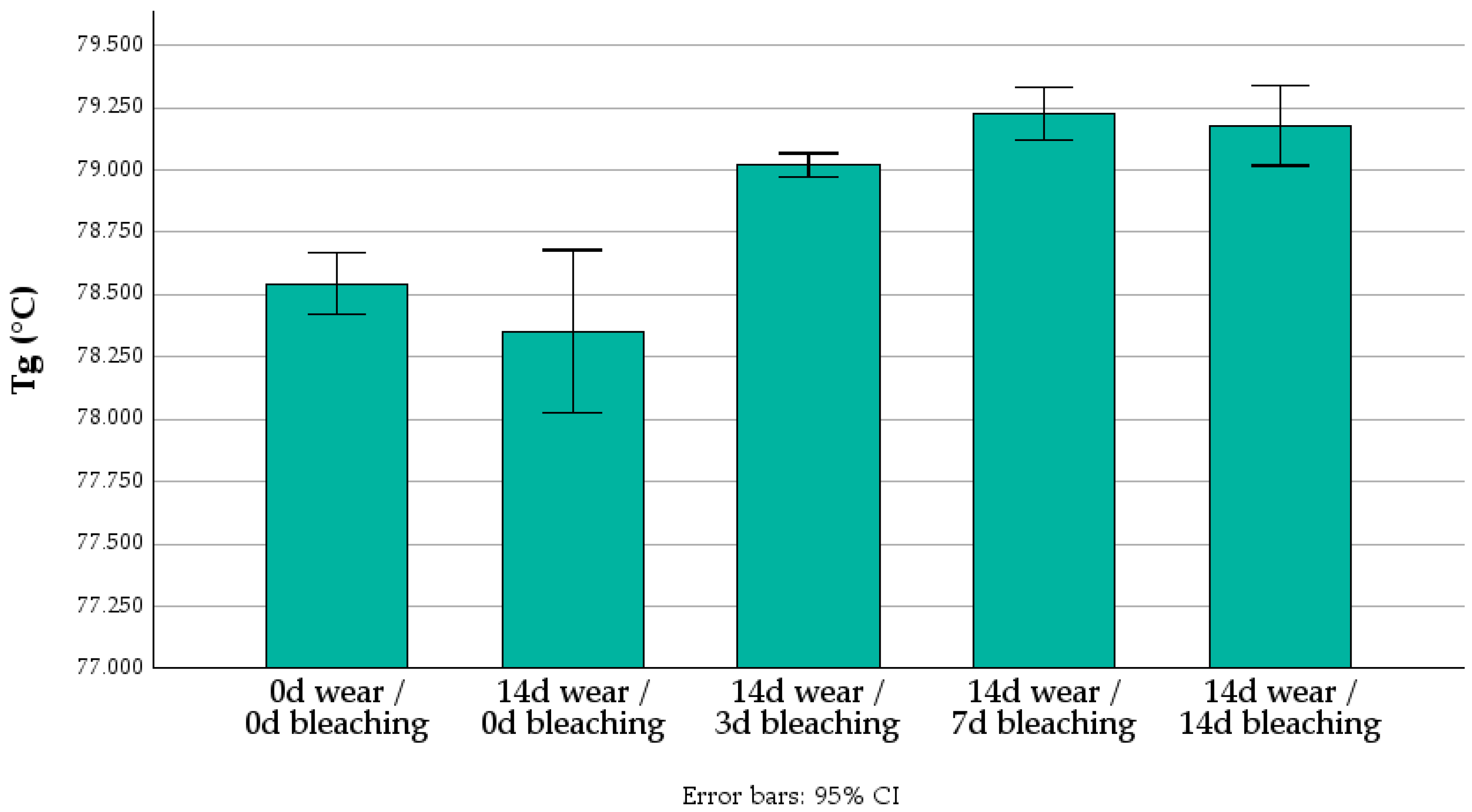
This study evaluated whether intra-aligner application of 10% carbamide peroxide during clinical use of Nuvola® aligners affects onset Tg or the qualitative PETG-related infrared fingerprint. Because the aligners were retrieved after clinical wear, the exposure model incorporated intraoral conditions rather than only laboratory immersion. Bleaching exposure was associated with a statistically significant but small increase in onset Tg, with maximum mean differences below 1 °C and no monotonic exposure-duration trend. Qualitative FTIR did not reveal major changes in PETG-related band positions or the presence of characteristic absorption bands.
The clinical relevance of this question is linked to the increasing combination of clear aligner therapy with tooth whitening procedures. Clear aligners may be used as carriers for bleaching agents, offering a convenient approach for patients seeking simultaneous orthodontic and esthetic treatment [
14,
16,
33]. Previous clinical reports and studies have shown that tooth bleaching during clear aligner treatment can be effective, including protocols based on 10% carbamide peroxide [
14,
16,
33]. However, most available evidence has focused on whitening efficacy, tooth shade change, plaque accumulation, or gingival indices, rather than on the possible effects of bleaching agents on the aligner material itself [
16,
17,
18,
33]. This distinction is important because clear aligner performance depends not only on tooth whitening efficacy, but also on preservation of the material’s mechanical, optical, surface, dimensional, and chemical properties during clinical wear.
The bleaching-treated groups showed statistically higher Tg values than both the control and wear-only groups. From a polymer science perspective, Tg is related to the segmental mobility of polymer chains. A slight increase in Tg may indicate reduced chain mobility, physical aging, changes in free volume, residual stress relaxation, or subtle rearrangement of the amorphous phase [
20,
25,
28,
29,
34,
35,
36,
37,
38]. However, the magnitude of the increase observed in this study was very small. Mean Tg values ranged from 78.35 °C to 79.23 °C, and the maximum mean difference between groups was below 1 °C. This distinction between statistical significance and practical relevance is essential. With low within-group variability, small differences may reach statistical significance, but they do not necessarily indicate clinically meaningful deterioration or functional impairment of the material.
The absence of a consistent exposure-duration trend further supports a cautious interpretation. The 7-day group had the highest mean Tg, but the 14-day group did not differ from either the 3-day or 7-day group. Thus, the data indicate an effect associated with bleaching exposure but do not demonstrate progressive change from 3 to 14 days. This pattern is more compatible with minor thermophysical rearrangement than with a simple cumulative degradation process [
20,
29,
36,
38,
39,
40,
41].
The present findings should be interpreted in relation to the study by Khashashneh et al., who evaluated the effect of 10% carbamide peroxide on Invisalign aligners under controlled in vitro conditions [
18]. Their investigation involved a different commercial aligner system and therefore a different proprietary polymer formulation from the PETG-related Nuvola
® material examined in the present study. In addition, Khashashneh et al. exposed the aligners to carbamide peroxide in a laboratory model, whereas the present study evaluated Nuvola
® aligners retrieved after actual patient wear. Consequently, the specimens investigated here were exposed not only to the bleaching agent but also to saliva, temperature fluctuations, functional loading, cleaning procedures, and patient-specific intraoral conditions.
The outcomes assessed in the two studies also differed. Khashashneh et al. primarily evaluated tensile strength, translucency, hardness, and surface roughness and reported that 10% carbamide peroxide did not significantly affect tensile strength or translucency but reduced hardness and increased surface roughness [
18]. In contrast, the present study focused on the onset glass transition temperature and the infrared chemical fingerprint of the material. DMA reflects temperature-dependent segmental mobility and the thermomechanical response of the specimen, whereas FTIR evaluates changes in characteristic chemical bonds at the analyzed surface. Therefore, the absence of major FTIR-detectable changes and the presence of only a small Tg increase in the present study do not contradict the surface and hardness changes reported for Invisalign aligners. Rather, the differences may be explained by the distinct material formulations, exposure models, analytical methods, and measured properties.
Similar property-specific responses have been reported after intraoral exposure and aging of clear aligner materials, with surface morphology, chemical characteristics, mechanical behavior, and optical properties changing to different extents depending on polymer composition and exposure conditions [
34,
35,
42,
43]. Taken together, the available evidence suggests that bleaching-related effects are both material-specific and property-specific and should not be interpreted as generalized deterioration of all aligner characteristics.
The FTIR results are particularly relevant for interpreting the DMA findings. All spectra showed characteristic bands consistent with PETG, including C–H stretching, ester carbonyl C=O stretching, C–O–C stretching, aromatic ring-associated vibrations, and cyclohexane-related bands. Following exposure to 10% carbamide peroxide, the spectra remained highly overlapping, with no detectable shifts in the positions of the principal absorption bands, no appearance of new bands, and no disappearance of characteristic PETG-related peaks. ATR-FTIR has been widely used for the chemical characterization of orthodontic aligner materials and for distinguishing PETG-based, polyurethane-based, and other thermoplastic formulations [
24,
31,
36,
37]. Within the sensitivity of the applied qualitative FTIR analysis, these results indicate that no major alteration of the PETG-related chemical fingerprint was detected after clinical use with 10% carbamide peroxide. However, the absence of new bands or evident peak shifts should not be interpreted as definitive evidence that no chemical modification occurred. Weak oxidation- or hydrolysis-related changes may have been masked by the strong intrinsic ester carbonyl band of PETG or may have remained below the detection sensitivity of the applied qualitative analysis.
Fuentes et al. reported changes in the carbonyl, hydroxyl, and C–O–C regions of hydrogen-peroxide-treated FDM-printed PETG, which they attributed to oxidative modification and ester-bond cleavage [
30]. Although the sterilization conditions, manufacturing process, and exposure intensity used in their study differed substantially from the present clinical bleaching protocol, their findings demonstrate that peroxide-related modification of PETG is chemically plausible. Therefore, the absence of evident changes in the present qualitative FTIR spectra cannot exclude low-level oxidation or hydrolysis below the sensitivity of the applied analytical method.
The combined DMA and FTIR findings should not be considered contradictory. DMA is sensitive to thermal transitions and changes in polymer-chain mobility, whereas FTIR primarily detects alterations in chemical functional groups and molecular bonding. Consequently, a small Tg shift may occur without clearly detectable FTIR changes when the underlying response is predominantly physical or thermophysical rather than associated with extensive chemical degradation. Possible mechanisms include physical aging, changes in free volume, residual-stress relaxation, water-related effects, or subtle rearrangement of the amorphous phase. Comparable distinctions have been reported in the clear aligner literature, where thermoforming, water exposure, thermocycling, intraoral aging, and manufacturing-related factors may influence mechanical, optical, surface, or thermal properties without producing major FTIR-detectable changes [
20,
24,
25,
29,
36,
37,
38]. The present findings therefore suggest that 10% carbamide peroxide may slightly influence the thermal response of clinically worn Nuvola
® aligners, while no major chemical modification was detectable using the applied qualitative FTIR protocol. Nevertheless, low-level oxidation, hydrolysis, or chain scission cannot be excluded.
The clinical interpretation must also account for the fact that orthodontic aligners are viscoelastic appliances. Their performance depends on stiffness, elastic recovery, stress relaxation, force decay, material thickness, fit, attachment geometry, programmed tooth movement, and intraoral aging. Previous studies have demonstrated differences in stress-relaxation behavior among clear aligner materials and have shown that aging, thermoforming, water immersion, and thermocycling may affect their mechanical and thermal properties [
20,
36,
38]. Furthermore, micro-computed tomography studies have demonstrated differences among aligner systems in thickness distribution and tooth–aligner gap and have shown that thermoforming may influence these parameters [
21,
39,
40]. Tg is a thermal-transition parameter and should not be regarded as a direct surrogate for orthodontic force delivery, stiffness at oral temperature, elastic recovery, stress relaxation, or dimensional stability. Therefore, clinically relevant aligner performance cannot be inferred from Tg and FTIR measurements alone.
This consideration is particularly important for Nuvola
® aligners because their clinical predictability and effectiveness depend on the interaction between material behavior, aligner fit, attachment configuration, and planned biomechanics. Nuvola
® aligners have been evaluated clinically for dentoalveolar transverse changes, while micro-CT studies have included Nuvola
® among aligner systems assessed for fit and thickness [
39,
40,
41]. The present study contributes material-level evidence obtained after actual patient wear and bleaching exposure. However, it does not establish whether the observed small Tg shift affects orthodontic force delivery, aligner adaptation, transparency, dimensional stability, or patient-perceived performance.
A strength of the present study is that the investigated aligners were retrieved after clinical use rather than being exposed exclusively under simplified laboratory conditions. This design increases clinical relevance because the specimens were subjected to real intraoral factors, including saliva, temperature variations, oral-hygiene habits, functional loading, and patient-specific wear patterns. Another strength is the combined use of DMA and FTIR. DMA enabled the detection of subtle differences in thermal-transition behavior, whereas FTIR was used to determine whether these differences were accompanied by detectable changes in the polymer chemical fingerprint. This combined approach provides a more balanced interpretation than either method alone. If only DMA had been used, the statistically significant Tg increase might have been overinterpreted as evidence of material degradation. Conversely, if only FTIR had been used, subtle thermophysical changes in polymer-chain mobility might not have been detected.
Several limitations must nevertheless be acknowledged. First, although the aligners were retrieved after clinical use, the study design does not allow complete separation of the individual contributions of intraoral aging, salivary exposure, temperature fluctuations, mechanical loading, oral-hygiene habits, diet, and exposure to 10% carbamide peroxide. Second, the study evaluated Tg and FTIR spectra but did not assess surface roughness, hardness, tensile strength, flexural properties, elastic recovery, force decay, stress relaxation, color stability, translucency, water sorption, dimensional stability, or tooth–aligner fit. These outcomes are clinically relevant because previous studies have demonstrated that oral exposure, cleaning protocols, thermocycling, water immersion, and material aging may affect the surface, optical, mechanical, dimensional, and thermomechanical properties of aligner polymers [
18,
26,
27,
28,
29,
34,
35,
36,
37,
38,
42,
43]. Consequently, the present results should not be interpreted as evidence that the investigated bleaching protocol has no effect on the overall clinical performance of the aligner.
Third, FTIR spectra were interpreted qualitatively. Quantitative analysis based on normalized peak heights, integrated peak areas, hydroxyl- or carbonyl-related indices, or difference spectra could provide greater sensitivity for detecting subtle chemical changes. The strong intrinsic ester carbonyl absorption of PETG may also obscure weak oxidation-related changes. Fourth, formal measurement uncertainty for the complete DMA procedure, including specimen preparation, clamping, temperature calibration, and Tg-onset determination, was not independently quantified. Accordingly, statistically significant differences smaller than 1 °C should be interpreted as detectable within the present dataset but not necessarily as exceeding the uncertainty of the complete analytical procedure. Fifth, the prolonged flattening procedure was applied consistently to all experimental groups; however, its potential influence on residual stresses, physical aging, or molecular rearrangement cannot be completely excluded. Finally, the findings apply specifically to the investigated Nuvola® material and the 10% carbamide peroxide protocol used in this study and should not be generalized to other aligner brands, polymer formulations, bleaching agents, concentrations, or exposure regimens.
Future studies should determine whether the small Tg differences observed after intra-aligner use of 10% carbamide peroxide are accompanied by clinically relevant changes in surface, optical, mechanical, or fit-related properties. In particular, further investigations should evaluate hardness, surface roughness, translucency, color stability, tensile behavior, elastic recovery, stress relaxation, orthodontic force delivery, water sorption and desorption, dimensional stability, and micro-CT-based aligner fit after clinical use [
18,
28,
29,
36,
37,
38,
39,
40]. Quantitative spectroscopic analyses and reference specimens subjected to controlled oxidative aging would also improve the ability to detect low-level chemical modification. Such studies are required to determine whether the use of bleaching gel inside clear aligners preserves not only the principal polymer chemical fingerprint but also the biomechanical, dimensional, surface, and esthetic performance required during the intended clinical wear interval.




 ,
,