


Abstract
Natural bioactive compounds play an increasingly important role in modern cosmetic formulations, particularly in the context of efficacy, safety, and regulatory compliance. Among them, vitamin A-related compounds have attracted significant attention due to their well-documented effects on skin renewal, collagen synthesis, and photoaging prevention. This review provides a comprehensive overview of natural vitamin A derivatives and structurally related carotenoids used in cosmetic applications, including retinol, retinal, retinyl esters, β-carotene, lycopene, zeaxanthin, astaxanthin, lutein, and fucoxanthin. The article critically examines their intracellular mechanisms of action, distinguishing between canonical retinoid signaling pathways mediated by nuclear receptors (RAR/RXR) and the predominantly antioxidant activities of carotenoids. Particular attention is given to the metabolic conversion of provitamin A compounds and their relevance to biological activity in the skin. Furthermore, recent regulatory restrictions on the use of retinol and retinyl esters, especially within the European Union, are discussed in the context of formulation challenges and the growing interest in naturally derived alternatives. By integrating mechanistic insights with regulatory and application-oriented perspectives, this review highlights the potential and limitations of natural vitamin A-related compounds and outlines future directions for their use in safe and effective cosmetic products.
Keywords:
vitamin A; retinoids; carotenoids; cosmetic products; natural bioactives; cosmetics safety 1. Introduction
Vitamin A constitutes a fat -oluble group of compounds essential for the functioning of the human organism. It is required for the control of stem cell functions, as well as cell differentiation and metabolism. It takes place in the development of the human embryo, tissues and organs, including the nervous system. In mature organisms, it supports overall functioning by maintaining central nervous system homeostasis, vision, immunity, hemopoiesis, bone metabolism, and skin condition [1,2]. Some classifications reserve the term vitamin A solely for all-trans-retinol. However, other natural derivatives, such as retinyl esters of animal origin, are metabolized in the human organism into retinol. Similarly, some carotenoids are converted into retinol and are called provitamin A. The main biologically active molecules derived from retinol during oxidation are 11-cis-retinal and all-trans-retinoic acid (ATRA, tretinoin). 11-Cis-retinal is involved in visual function. All-trans-retinoic acid is a transcription factor; it is a ligand in the RAR/RXR nuclear receptor. As soon as it binds to this receptor complex, the cascade of intranuclear reactions causing transcription of designated genes begins [1,3,4].
From the chemical point of view, vitamin A forms are characterized by an unsaturated isoprenoid chain structure. In addition to chemical structure, a classification of a certain compound as a “vitamin A form” is also based on biological function in an organism. As a result, the term retinoids was established to cover the group of compounds which do not necessarily share the isoprenoid pattern in their structure but exert their molecular activity via retinoid nuclear receptors. They could also be classified as vitamin A-related compounds [4].
Taking into consideration the biological functions in the skin, retinoids gain attention in several dermatological conditions, including therapy of acne vulgaris, psoriasis, photoaged skin, hyperpigmentation, and keratosis pilaris. Especially, all-trans-retinoic acid is classified as a “gold standard” in anti-aging regimens. Straight from dermatology, its usage shifted to cosmetology, although some retinoids such as tretinoin, isotretinoin, and adapalene remain prescription-only medicines [5,6,7]. Other retinoids, including retinol, retinal, and retinyl esters, are allowed to be used in cosmetic products. They became extremely popular in beauty routines due to their biological efficacy. Retinol has been shown in many clinical evaluations to improve fine lines and wrinkles, hyperpigmentation, skin roughness, and the appearance of photoaged skin. While the efficacy of retinoids is well established, attention should be drawn to the adverse effects that may accompany their topical application. Notably, a significant proportion of patients experience troublesome side effects following the use of prescription retinoids. For this reason, cosmetic-grade concentrations are often considered more appropriate, and consequently, cosmetic products containing permitted retinoid compounds have gained widespread use [8,9].
Carotenoids may serve as provitamin A, though not all are efficiently converted into retinol in the human body. As natural pigments, they absorb light within a specific spectral range, typically with an absorption maximum around 450 nm; however, their UV protection properties remain a subject of debate. They are used in cosmetic products primarily for their antioxidant properties [10,11,12].
The aim of this review is to critically assess the role of natural vitamin A-related compounds encompassing both retinoids and carotenoids, in cosmetic applications, with particular emphasis on their distinct and overlapping mechanisms of action at the molecular and cellular level. By systematically comparing efficacy, stability, and bioavailability of these structurally related compound groups, this review seeks to identify existing limitations in current formulation strategies, highlight inconsistencies and methodological shortcomings in the available evidence, and delineate unresolved knowledge gaps that impede the translation of mechanistic insights into evidence-based cosmetic practice.
2. Materials and Methods
The literature search was conducted using electronic databases including PubMed, Scopus, Web of Science, and Google Scholar. The search was performed between April and May 2026. The following search terms were applied: “retinoid cosmetics”, “retinoid skin”, “retinol cosmetics”, “retinoid adverse effects”, “carotenoid cosmetics”, and “carotenoid skin”. A complementary search was performed to identify literature on specific cosmetic ingredients, including retinal, retinyl esters, β-carotene, astaxanthin, zeaxanthin, fucoxanthin, lutein, and lycopene, combined with the terms “skin”, “cosmetics”, or “topical application”. No restriction on publication year was applied to capture both foundational and recent studies in the field. Publications were included if they were written in English and addressed cosmetic, dermatological, or mechanistic aspects of retinoids or carotenoids relevant to skin, encompassing original research articles, clinical studies, and peer-reviewed reviews. Publications were excluded if they focused exclusively on systemic or pharmacological applications with no relevance to cosmetic use. The usage of compounds in cosmetic products was confirmed via the CosIng database, and regulatory restrictions were verified against applicable cosmetic regulations. ChatGPT v5.5 was used to assist in preparing Figures 4 and 6, which were subsequently reviewed and revised by the authors.
3. Classification of Natural Vitamin A-Related Compounds
3.1. Retinoids
Retinoids are natural or synthetic compounds related to vitamin A. The family includes retinol, retinal, retinoic acid, and retinyl esters as the main functional forms [13]. Naturally occurring retinoids include all-trans retinoic acid, retinol, retinaldehyde (retinal), 9-cis-retinoic acid, 13-cis-retinoic acid, and retinyl esters [14]. Retinoids are classified into four generations according to molecular structure and receptor selectivity. For first-generation representatives, retinoid activity increases in the following order: retinyl esters, retinol, retinaldehyde, retinoic acid, while tolerance shows the reverse order [15]. Figure 1 presents chemical structures of first-generation natural retinoids.
Figure 1.
Chemical structures of selected first-generation retinoids in all-trans isomeric form.
First-generation natural retinoids are characterized by a β-ionone ring and a polyene side chain. Retinol is an alcohol, retinal—aldehyde, and retinoic acid, which possesses a carboxylic group, while retinyl acetate and palmitate are organic esters. These compounds contain multiple unsaturated double bonds in their structures, which determine the possible absorption of certain wavelengths of light but also cause instability. Analysis of their chemical structures also clearly indicates their lipophilic character.
3.1.1. Retinol
Retinol is the alcohol form of vitamin A. It contains a hydroxyl group, which distinguishes it chemically from retinal and retinoic acid [16]. In enterocytes and target tissues, retinol binds cellular retinol-binding protein, and in the blood, it is transported mainly with retinol-binding protein, often together with transthyretin [4]. Retinol is converted to retinaldehyde, which is then oxidized to retinoic acid in a two-step reaction [13]. When applied topically, retinol must be converted to retinoic acid via retinaldehyde to exert its biological activity [15]. Retinol is also described as an important regulator of epidermal cell growth, normal cell differentiation, and cell maintenance [17]. In blood and tissues, retinol and its esters together account for more than 99% of all retinoids present in the skin [18].
3.1.2. Retinal
Retinal is the aldehyde form of vitamin A. It contains an aldehyde group, distinguishing it chemically from retinol and retinoic acid. Retinol can be oxidized to retinal by retinol dehydrogenases, and retinal can then be further oxidized to retinoic acid by retinaldehyde dehydrogenases, including RALDH1 and RALDH3 [19]. Retinal is especially important for vision [16]. The most important retinal species in the eye is 11-cis-retinal, because it forms the chromophore of rhodopsin when bound to opsin [20]. When light hits rhodopsin, 11-cis-retinal isomerizes to all-trans-retinal, and that change triggers opsin activation, which is the molecular start of visual signaling [21]. All-trans-retinal is then reduced back to all-trans-retinol in photoreceptors and carried to the retinal pigment epithelium, where it is re-esterified by LRAT, helping maintain the visual cycle [20]. Outside the eye, retinaldehyde can inhibit 9-cis retinoic acid-driven RXR/PPARγ signaling in adipose tissue, which suppresses adipogenesis and lipid accumulation, and can also promote thermogenesis and the browning of white fat by activating RAR-related pathways. Raising retinaldehyde levels by blocking RALDH1 protects mice from diet-induced obesity and diabetes [19]. Retinoids, including retinal, can also isomerize into mixtures of trans- and cis-isomers under photoirradiation [17].
3.1.3. Retinyl Esters
Retinyl esters are chemically stable, hydrophobic, and largely inert compounds [20]. Retinyl palmitate is described as the principal storage form of retinol in humans and animals. Animal-derived foods such as liver, milk, and butter contain retinyl esters directly. Vitamin A from animal sources is mainly acquired as retinyl esters such as palmitate, propionate, and acetate [17,18,21]. In the intestine, dietary retinyl esters must first be hydrolyzed to retinol before absorption, driven mainly by pancreatic triglyceride lipase, lipase-related protein 2, and intestinal phospholipase B. Once inside the enterocyte, retinol binds CRBP2 and is transported to the endoplasmic reticulum, where LRAT re-esterifies it into retinyl esters, with LRAT accounting for approximately 90% of retinyl ester formation in enterocytes. These retinyl esters are then packaged into chylomicrons and transported through the lymph and blood to the liver. The liver is the major retinoid reservoir, accounting for around 70% of chylomicron uptake [20,21]. Under adequate dietary conditions, the liver holds more than 95% of its neutral retinoid pool as retinyl esters, mainly retinyl palmitate and retinyl stearate, stored in lipid droplets inside hepatocytes and stellate cells [22]. Hepatic stellate cells hold about 90% to 95% of hepatic retinoids in lipid droplets, with LRAT being solely responsible for retinyl ester synthesis in these cells. This reserve can maintain physiological levels of vitamin A for months during periods of low intake [4]. When the body needs vitamin A, stored retinyl esters are hydrolyzed back to retinol, which is exported bound to retinol-binding protein and circulates with transthyretin to reduce renal loss [20]. Retinyl palmitate can be enzymatically hydrolyzed back to retinol in vivo [17]. When applied topically, retinyl esters are hydrolyzed to retinol, which is then converted to biologically active retinoic acid via retinaldehyde [15]. Retinyl esters, including retinyl acetate and retinyl palmitate, can also isomerize into mixtures of trans- and cis-isomers under photoirradiation [17].
3.2. Carotenoids
Carotenoids are lipophilic pigments found in plants, animals, and microorganisms that must be obtained through diet because the human body does not produce them. Some carotenoids, such as β-carotene, can be converted to vitamin A in the human body and are therefore called provitamin A carotenoids. Their conjugated double-bond system is responsible for photoprotective effects, including absorption of light, quenching of singlet oxygen, and scavenging of free radicals [22]. Carotenoids are also described as tetraterpenes responsible for red, yellow, and orange coloration in fruits, vegetables, roots, flowers, fish, invertebrates, and birds, and as compounds with antioxidant activity that can prevent free radicals from attacking skin tissue [23]. Carotenoids are divided into two groups: carotenes, which consist exclusively of carbon and hydrogen atoms, and xanthophylls, which additionally contain oxygen atoms [23].
3.2.1. Provitamin A Carotenoids
β-carotene (Figure 2) is described as the most widely distributed carotenoid in foods, found mainly in yellow-orange and dark green fruits and vegetables, specifically in carrots, palm oil, mango, sweet potato, and apricot [24,25]. Metabolically, β-carotene is converted to all-trans-retinal, then to all-trans-retinol and all-trans-retinoic acid [18]. Provitamin A carotenoids are important in the fight against vitamin A deficiency and are beneficial for the skin through the production of retinoic acid, which plays important roles in keratinocyte proliferation, epidermal differentiation, keratinization, reduction of inflammation and oxidation, and enhancement of penetration of topical agents. Dietary β-carotene can protect against UV-induced erythema, with daily doses of approximately 10 mg for about 10 weeks demonstrating this benefit. A β -carotene-rich algal powder has also been shown to reduce the severity of mild chronic plaque-type psoriasis in adults [24]. Other provitamin A carotenoids are α-carotene and β-cryptoxanthin; they also possess antioxidant activity, however, their usage in cosmetic products is less pronounced.
Figure 2.
Chemical structure of β-carotene.
3.2.2. Non-Provitamin A Carotenoids
The chemical structures of selected non-provitamin A carotenoids are presented in Figure 3. Although these compounds are structurally related to β-carotene, subtle structural modifications prevent their conversion into the biologically active form of vitamin A. Their biological activity is primarily attributed to the presence of conjugated unsaturated bonds within their molecular structure. Particular attention should also be given to the hydroxyl and keto functional groups present in lutein, zeaxanthin, and astaxanthin, which further enhance their antioxidant properties.
Figure 3.
Chemical structures of selected non-provitamin A carotenoids: lycopene, lutein, zeaxanthin, and astaxanthin.
Lycopene
Lycopene constitutes the most common carotenoid found in tomatoes, both fresh and pomace [26]. It possesses strong antioxidant activity, and for many years, it was considered a strong photoprotectant. Animal as well as human studies confirmed that dietary lycopene is able to change the color of the skin, lowering the UV-dose causing erythema. It was also shown to effectively affect transcription of genes related to inflammation (IL-6, TNF-α), oxidative stress, photodermatoses, and photoaging [27]. Dietary supplementation with lycopene was shown to improve parameters of skin structure in in vivo human studies [28]. Moreover, cutaneous concentration of lycopene was shown to significantly correlate with lower levels of skin roughness in healthy volunteers [29]. Beneficial results of topically applied lycopene were also observed, especially in terms of the prevention of cutaneous damage caused by free radicals [30]. Many studies proved that this molecule is an effective antioxidant, which is its main beneficial effect on human organisms.
Zeaxanthin
Zeaxanthin belongs to the xanthophylls, which are the oxygenated derivatives of carotenoids [25]. Zeaxanthin is found in foods such as corn, egg yolk, leafy greens, and fortified foods [31]. It is found in the epidermis, dermis, and subcutaneous fat, and in human skin, β-carotene and lycopene are present in greater amounts than zeaxanthin and lutein [25].
Zeaxanthin is one of the main carotenoids concentrated in the human retina and macula, and together with lutein, it accumulates selectively in the macula lutea [24,31]. Zeaxanthin is also described as one of the predominant carotenoids in the macular pigment of the human retina, and lower retinal levels are associated with age-related macular degeneration, cataracts, and other ophthalmologic diseases [32]. Lutein and zeaxanthin help protect the macula from blue light damage, improve visual acuity, and act as antioxidants that scavenge reactive oxygen species. Higher intake of lutein and zeaxanthin is also associated with lower risk of age-related macular degeneration and cataracts [31]. Zeaxanthin, together with lutein and astaxanthin, has also been shown to protect DNA in neuroblastoma cells exposed to reactive nitrogen species [32]. For skin health, a zeaxanthin-based dietary supplement and topical serum have been shown to improve hydration and reduce wrinkle count in female subjects. Supplementation with lutein and zeaxanthin isomers has been reported to improve skin tone [25].
Astaxanthin
Astaxanthin is classified as a xanthophyll carotenoid [31]. It possesses hydroxyl and keto groups at both ends of its conjugated polyene chain, and this structure is central to its ability to quench singlet oxygen, scavenge free radicals, and chelate metal ions [33]. Astaxanthin is synthesized from β-carotene by the sequential action of a hydroxylase and a ketolase, and it is described as a xanthophyll of great interest in animal nutrition and human health [34]. It occurs naturally in microalgae, yeast, salmon, shrimp, lobster, and crustacean by-products [32]. It is also abundant in green algae and in fish that feed on algae [31]. Astaxanthin can neutralize reactive oxygen species approximately ten times more effectively than zeaxanthin, lutein, tunaxanthin, canthaxanthin, and β-carotene. Because of its stronger antioxidant profile, astaxanthin is considered a promising dietary supplement with potential therapeutic uses in inflammation-related, cardiovascular, neurodegenerative, and aging-related conditions [33]. Its antioxidant activity is also linked to potential benefits in conditions associated with oxidative stress, such as cardiovascular disease, macular degeneration, cancer, obesity, and hypertension [32]. Astaxanthin combined with collagen hydrolysate improves facial elasticity and decreases matrix metalloproteinase-1 and -12 expression [25]. It may also protect against erythema and reduce wrinkling [24]. Combined use of oral and topical astaxanthin has been shown to improve skin wrinkles, age spot size, skin texture, moisture content, and corneocyte condition, as demonstrated in in vivo human clinical studies [35].
Lutein
Lutein belongs to the xanthophylls; it is found in green vegetables, fruits, eggs, and marigold flower petals. It is also a primary metabolite in microalgae, in which it takes part in light harvesting and photosynthesis as well as acts as a protectant against photo-oxidative damage [36]. In humans, it is primarily used in ophthalmology as it has been shown to significantly lower the incidence of age-related macular degeneration. Its utilization in cosmetology is related to antioxidant activity as well as the potential to filter out blue light from the visible light spectrum. Lutein also showed other beneficial skin-related properties, such as reduced UV-B-induced immunosuppression, inflammation, and skin thickening, and the development of apoptotic cells in the skin, as tested in nude mice. Additionally, lutein was shown to improve skin hydration, lipid content, and flexibility, and reduce lipid peroxidation. Cosmetic products containing lutein could be recommended as anti-photoaging formulations to prevent damage caused by radiation, as well as skin lightening and whitening products [37]. In vitro studies in human keratinocytes showed that lutein upregulates HAS3 gene expression, causing increased hyaluronan synthesis. Moreover, it significantly increased retinoic acid-responsive element-dependent transcript activity [38]. The anti-inflammatory effect of lutein was proven in human keratinocytes and macrophages. The molecule increased the expression of interleukin-6, affected cyclooxygenase-2, and enhanced the matrix-metallopeptidase-9 level. It was also shown to inhibit the activation of redox-sensitive AP-1 pathway by suppressing the activation of p38 and c-Jun-N-terminal [39].
Fucoxanthin
The main natural source of fucoxanthin is marine microalgae. Similar to other xanthophylls, it possesses strong antioxidant and anti-inflammatory properties as well as the ability to absorb UV-light [40,41,42]. In vitro tests showed its ability to enhance procollagen synthesis, inhibit matrix metalloproteinases, and suppress melanin synthesis, indicating its great potential in cosmetic applications, which was subsequently proved in vivo human studies, which showed anti-wrinkle activity of fucoxanthin concentrate derived from Phaeodactylum tricornutum [43,44]. Studies on its molecular mechanism of action showed that the molecule inhibits pro-inflammatory cytokines such as IL-1β, IL-6, and TNF-α by regulating both NF-κB and NLRP3 inflammasome activation [45].
4. Intracellular Mechanisms of Action
4.1. Retinoid Signaling Pathways
4.1.1. Conversion: Retinol—Retinal—Retinoic Acid
The synthesis of retinoic acid (RA) from vitamin A proceeds through two main oxidative steps. In the first step, retinol is taken up from the bloodstream, where it is transported bound to RBP4, and enters the cell via the membrane receptor STRA6. Retinol is then converted to retinaldehyde by two classes of enzymes: cytoplasmic alcohol dehydrogenases (ADH1, ADH7) and microsomal retinol dehydrogenases, particularly RDH10. To prevent excess RA production, retinaldehyde can be converted back to retinol by DHRS3, which physically interacts with RDH10 to form a complex regulating the rate of RA synthesis. In the second, irreversible step, retinaldehyde is oxidized to RA by three retinaldehyde dehydrogenases, ALDH1A1 and ALDH1A2, each with distinct patterns of tissue and temporal expression. ALDH1A2 is responsible for nearly all RA production during early mouse embryogenesis. RA is subsequently degraded by three enzymes of the CYP26 family (CYP26A1, CYP26B1, CYP26C1), ensuring a short half-life of approximately 1 h and restricting its activity to specific tissues [46,47].
4.1.2. Receptors: RAR/RXR
Retinoic acid acts as a ligand for nuclear retinoic acid receptors, RAR and retinoid X receptors, RXR. RAR is activated primarily by all-trans-retinoic acid, while RXR is activated by 9-cis-retinoic acid [48]. Each RAR requires heterodimerization with an RXR to bind DNA and regulate target genes. RXR additionally serves as a universal heterodimerization partner for many other nuclear receptors, including PPAR, LXR, FXR and Nurr1. All three RAR isoforms have been studied in heterodimers with RXRα, and RARA, RXRA, and RXRB are broadly expressed across tissues, suggesting that most tissues are potential targets of RA signaling. Each receptor shares a common architecture: an N-terminal A/B segment, a DNA-binding domain (DBD) with two α-helices and eight conserved zinc-coordinating cysteines, and a C-terminal ligand-binding domain (LBD) composed of 12 α-helices arranged in a three-layer sandwich [46,48,49,50].
4.1.3. Influence on Gene Expression
The RAR–RXR heterodimer functions as a ligand-dependent transcription factor. In the absence of a ligand, the AF-2 domain of RXR promotes interaction with a co-repressor complex, which blocks transcription of the target gene. Upon ligand binding, a conformational change in the LBD promotes recruitment of a co-activator complex containing histone acetyltransferases and p160/SRC proteins, leading to chromatin opening and transcriptional activation. Gene specificity arises from recognition of direct repeat (DR) response elements with varying spacings (DR0–DR5). RAR-RXR preferentially binds DR5 and DR2 elements [50]. Binding at DR5 leads to transcriptional activation, while binding at DR1 results in repression; all-trans RA enhances both responses. Examples of genes regulated through this pathway include Hoxa1, Hoxb1, Hoxb4, Hoxd4, Rarb, Cyp26a1, Pitx2, and Fgf8 [1,47,48,49,50].
4.1.4. Influence on Keratinocytes
Retinoids are potent regulators of keratinocyte proliferation and differentiation, with their effects mediated through binding to nuclear RAR and RXR receptors. By regulating epidermal cellular division and differentiation, they exert numerous major effects on the formation of the layered epidermal structure. In normal human skin, RAR-γ and RXR-α represent the majority of cutaneous retinoid receptors [51]. Retinoids promote keratinization in keratinocytes of photodamaged skin, and retinoic acid also stimulates keratinocyte proliferation [52]. ATRA regulates skin cell proliferation and differentiation, and in mouse models, ATRA treatment led to parakeratosis and abnormal keratinocyte differentiation, with the number of epidermal cell layers increasing from approximately 3 to 8 and epidermal thickness rising 2.5-fold [53]. Retinoids also selectively and dramatically upregulate the mRNA expression of P2Y2 receptors in normal human epidermal keratinocytes, facilitating ATP/P2Y2 autocrine signaling that drives basal keratinocyte proliferation [54]. RORα4, the predominant isoform of the retinoid-related orphan receptor RORα in human keratinocytes, positively regulates both early differentiation markers (keratin 1/10, involucrin) and late differentiation markers (loricrin, filaggrin). Silencing RORα significantly reduces expression of these differentiation markers without affecting basal layer markers [55]. ATRA causes the dysregulation of numerous keratins and keratin-associated proteins, indicating abnormal terminal differentiation. It also reduces Claudin-1 expression while upregulating Claudin-4, suggesting a dual effect on epidermal barrier function [53]. In studies using the HaCaT human keratinocyte cell line, trifarotene, a selective RARγ agonist and functional analogue of retinoic acid, significantly reduced the expression of phosphorylated JNK and phosphorylated c-Jun [52].
4.1.5. Influence on Collagen and MMPs
UV irradiation damages skin connective tissue through two simultaneous mechanisms: inhibiting procollagen synthesis and stimulating collagen breakdown via MMPs. A single UV exposure reduces type I and type I procollagen mRNA by approximately 40% and type III procollagen mRNA by approximately 60% within 24 h [56]. At the same time, UV irradiation significantly upregulates MMP-3 and MMP-13 protein expression [53] as well as collagenases MMP-1, MMP-8, and MMP-13 [56]. Via the inhibition of the JNK/c-Jun pathway, retinoic acid blocks activation of the transcription factor AP-1, which regulates the expression of matrix metalloproteinases MMP1, MMP3, and MMP9. Phosphorylation of JNK leads to activation of c-Jun, which, together with c-Fos, forms AP-1, which, in turn, upregulates MMP expression at both the gene and protein level; retinoids interrupt this cascade by inhibiting JNK [52]. This mechanism is further supported by evidence that ATRA and RAR agonists significantly suppress c-Jun protein levels, and that this suppression is reversed by RAR antagonist co-treatment, confirming the RAR-mediated nature of the pathway.
Retinoids also directly suppress specific MMPs. ATRA significantly reduced MMP-3 and MMP-13 levels in photoaged mouse skin, and this effect was reproduced by a selective RAR agonist but not by an RXR agonist, confirming RAR dependence [53]. Similarly, in HaCaT keratinocytes, acitretin reduced MMP13 at both the mRNA and protein levels, and in a psoriasis mouse model, tazarotene decreased MMP13 expression in skin lesions. The combination of retinoids with NB-UVB irradiation produced synergistic inhibitory effects on MMP13 expression [57]. As a consequence, retinoic acid and its derivatives reduce MMP secretion, limit collagen degradation, and increase total collagen content in the skin [52]. ATRA increased collagen content by 29.83% and a selective RAR agonist by 25.00% relative to the UV control group, with these effects blocked by an RAR antagonist but unaffected by an RXR antagonist [53]. Furthermore, pretreatment of human skin with all-trans retinoic acid (0.1%) for 24 h before UV irradiation substantially protects against UV-induced loss of both type I and type III procollagen mRNA and protein, and in chronically photodamaged skin, treatment with all-trans retinoic acid increases type I procollagen expression [56].
4.2. Carotenoids: Antioxidant and Photoprotective Mechanisms
4.2.1. Scavenging ROS
Carotenoids are among the most effective physical and chemical quenchers of ROS [58]. Their antioxidant properties stem from conjugated C=C double bonds: the greater their number, the higher the antioxidant efficacy [59]. Three main mechanisms are described: physical quenching of singlet oxygen with a rate constant near the diffusion-controlled limit, chemical quenching leading to irreversible molecular modifications, and free radical scavenging via electron transfer, radical addition, or hydrogen atom transfer [58,60].
Among carotenoids, lycopene shows the highest antioxidant efficiency, followed by α-carotene, β-cryptoxanthin, β-carotene, zeaxanthin, and lutein [59]. In vitro studies on human skin fibroblasts confirmed that lycopene required only 0.05 nmol/mg protein for UVB photoprotection, compared to 0.40 and 0.30 nmol/mg for β-carotene and lutein, respectively [27]. The antioxidant potential of astaxanthin exceeds vitamin E (100×), vitamin C (6000×), and β-carotene (5×) in lipid peroxidation assays. Additionally it activates the Nrf2/HO-1 pathway, upregulating SOD2, catalase, and GPX1 [27,61].
4.2.2. Protection Against UV Radiation
UV radiation generates approximately 50% of the free radicals found in skin. Carotenoids accumulate in the epidermis and dermis, acting as natural filters for blue light and near-UV. Following UV irradiation, carotenoid levels in the stratum corneum drop significantly. Lycopene decreases immediately, while β-carotene remains stable for 30 to 60 min before declining sharply. Recovery to baseline takes up to three days [27,62].
Clinical evidence confirms photoprotective effects, though only after a minimum of 10 weeks of supplementation at doses above 12 mg/day [63]. Consumption of tomato paste (40 g/day, 16 mg lycopene) for 10 weeks reduced UV-induced erythema by 40% compared to controls [27]. In a randomized, double-blind, placebo-controlled trial, oral supplementation with a tomato nutrient complex and lutein suppressed UV-induced upregulation of HO-1, ICAM-1, and MMP-1. Notably, a mixture of tomato carotenoids provided greater photoprotection than lycopene alone, suggesting synergistic interactions [64].
4.2.3. Effect on Oxidative Stress
UV radiation is considered the primary source of oxidative stress in human skin [63]. Carotenoid concentration in the stratum corneum reflects overall antioxidant status—the higher the concentration, the faster the neutralization of free radicals. High carotenoid levels correlate with low stress and a carotenoid-rich diet, whereas low levels are associated with smoking and disease [59,62].
Carotenoids reduce oxidative stress markers, including ICAM-1, HO-1, MMP, malondialdehyde (MDA), and 4-hydroxyalkenals. In a clinical trial, 4 mg/day of ASX for 4 weeks reduced MDA levels by 11.2% after 15 days and 21.7% after 29 days. Carotenoids act through both direct (energy transfer) and indirect pathways, the latter involving transcription factors Nrf2 and NF-κB. It should be noted that, since carotenoids are not synthesized by the human body, their accumulation in the skin depends entirely on diet [27,59,61].
4.2.4. Effect on Inflammation
Chronic oxidative stress drives persistent inflammation underlying most skin damage. Carotenoids suppress inflammatory responses by modulating NF-κB/MAPK signaling, thereby inhibiting pro-inflammatory gene expression. ASX blocks nuclear translocation of NF-κB p65 subunit, inhibits IKK kinase, suppresses IL-1β, IL-6, and TNF-α expression, and reduces COX-2, iNOS, and prostaglandin E2 release in UV-irradiated keratinocytes. Oral β-carotene supplementation reduced pro-inflammatory cytokines (IL-1β, IL-6, IL-4, TNF-α) and MMP activity in a mouse model. Lutein and zeaxanthin decreased UV-induced skin inflammation, apoptotic cell count, and mast cell infiltration in hairless mice, while also increasing tumor-free survival in a photocarcinogenesis model. Topical application of creams containing 0.2% carotenoids showed efficacy against pruritus and inflammation in atopic dermatitis patients [27,59,61,63].
4.3. Comparative Mechanistic Insights
While both retinoids and carotenoids play important roles in skin biology, they operate through fundamentally distinct mechanisms [1,65]. Retinoids exert their effects primarily through receptor-mediated genomic signaling, binding to nuclear retinoic acid receptors (RARs) and retinoid X receptors (RXRs) to regulate gene expression involved in cell differentiation, proliferation, and extracellular matrix synthesis [1,9]. This direct mode of action accounts for their well-established potency in enhancing skin texture, diminishing fine lines, and augmenting epidermal and dermal thickness. However, the same receptor-mediated potency underlies their characteristic side effects, including irritation, dryness, peeling, and photosensitivity, which necessitate gradual introduction into skincare routines [9,66].
In contrast, carotenoids act predominantly through indirect, redox-based mechanisms, scavenging reactive oxygen species and modulating signaling pathways such as NF-kB, AP-1, and Nrf2/ARE in keratinocytes and fibroblasts [63,65]. Although this mechanism generally results in a slower, more gradual onset of cosmetic benefits, it offers a markedly improved safety profile with minimal irritation potential, even with prolonged topical use [59,63]. Notably, astaxanthin demonstrates exceptional antioxidant capacity, reported to be a greater antioxidant agent than vitamin C, vitamin E, and beta-carotene [67,68], while clinical trials have confirmed its protective role against UV-induced skin deterioration [69].
Critically, these two compound classes are not mutually exclusive: rather, their complementary mechanisms suggest strong synergy when combined, with antioxidants potentially buffering the oxidative burden and irritation associated with retinoid regimens [24,66,70]. Table 1 provides a side-by-side comparison of these classes across mechanistic, efficacy, safety, and formulation dimensions to contextualize the discussion of natural bioactives that follows. Additionally, Figure 4 illustrates simplified comparison of biological effects of retinoids and carotenoids in cosmetic applications.
Table 1.
Comparison of retinoids and carotenoids in cosmetic applications.
Figure 4.
Comparison of biological effects of retinoids and carotenoids in cosmetic applications (RDH—Retinol Dehydrogenase, RALDH—Retinaldehyde Dehydrogenase, RAR—Retinoic Acid Receptor, RXR—Retinoid X Receptor, RARE—Retinoic Acid Response Element, ROS—Reactive Oxygen Species, PPAR—Peroxisome Proliferator-Activated Receptor, RORα—Retinoic Acid Receptor-Related Orphan Receptor Alpha, COL1A1/COL3A1—Collagen Type I/III Alpha Chain Genes, TIMP—Tissue Inhibitor of Metalloproteinases, MMP—Matrix Metalloproteinase, AP-1—Activator Protein 1, HAT—Hydrogen Atom Transfer, HO-1—Heme Oxygenase 1, SOD—Superoxide Dismutase, GPX—Glutathione Peroxidase, ICAM-1—Intercellular Adhesion Molecule 1, NF-κB—Nuclear Factor Kappa B, Nrf2—Nuclear Factor Erythroid 2–Related Factor 2, TNF-α—Tumor Necrosis Factor Alpha, IL-1β—Interleukin 1 Beta, and 1O2—Singlet Oxygen).
4.3.1. Direct vs. Indirect Action
In the context of topical application, retinoids act directly, as active forms such as all-trans-retinoic acid bind to nuclear receptors (RAR, RXR, PPAR, and ROR) in skin cells without requiring metabolic activation. Topically applied retinol undergoes a two-step oxidation to retinoic acid within target cells, the biologically active ligand. Retinyl esters require three conversion steps and show reduced anti-wrinkle efficacy in topical formulations compared to tretinoin [4,71].
Carotenoids applied topically operate primarily through redox-based mechanisms, neutralizing free radicals and reducing oxidative stress in skin cells without directly binding nuclear receptors [72]. Lutein in nanocrystal formulations demonstrates improved skin penetration compared to conventional forms, enhancing local antioxidant delivery [73]. Any receptor-mediated activity of provitamin A carotenoids remains indirect and requires enzymatic conversion, which is limited in skin tissue [74].
4.3.2. Receptor-Mediated vs. Redox Mechanisms
Topically applied retinoids act through a well-defined genomic pathway in skin cells: binding to RAR/RXR heterodimers leads to formation of the RARE complex and subsequent modulation of genes regulating keratinocyte proliferation, collagen synthesis, and MMP inhibition [72,75]. Nanoparticle formulations, such as solid lipid nanoparticles (SLNs) and liposomes, are used to improve skin penetration and reduce local irritation while maintaining receptor-mediated efficacy [73]. Their redox activity in topical use is marginal and considered secondary [4]. Topically applied carotenoids act primarily through redox mechanisms in the epidermis and dermis, quenching singlet oxygen and scavenging free radicals via electron transfer, hydrogen abstraction, and radical addition [76]. Astaxanthin additionally activates the Nrf2/ARE pathway in skin cells, upregulating GPX1, catalase, HO-1, and SOD2 [75]. Carotenoids also modulate NF-κB and AP-1 signaling pathways in keratinocytes and fibroblasts, but this remains secondary to their antioxidant activity [74].
5. Cosmetic Applications and Efficacy
5.1. Cutaneous Bioavailability of Retinoids and Carotenoids
The bioavailability of retinoids and carotenoids following topical application remains a significant challenge. Since cosmetic products are primarily intended for external use, this aspect is of particular importance in cosmetology. The limited ability of these compounds to penetrate the epidermal barrier is largely determined by their physicochemical properties, particularly their lipophilic nature (high logP values) and relatively high molecular weights.
The delivery of retinoids represents a considerable challenge because, on the one hand, these compounds should exhibit efficient skin penetration due to their affinity for the lipid-rich stratum corneum, while on the other hand, they are chemically unstable and susceptible to degradation by external environmental factors. Retinol, retinal, and retinoic acid are lipophilic molecules but also contain hydrophilic structural elements, which provide a more balanced penetration profile. In contrast, retinyl esters, particularly retinyl palmitate, possess substantially higher lipophilicity.
Recent studies evaluating transcutaneous permeation of retinol from semisolid cosmetic preparations showed that after topical administration the molecule was mainly concentrated in the stratum corneum and did not enter the blood in in vivo tests in mice and rats. Similar results were obtained in vitro; retinol was not able to cross the skin barrier after a 24 h test in Franz diffusion cells involving pig, mouse, and rat skin [77]. These results suggest that the efficacy of retinoids may be limited due to poor bioavailability.
Retinyl palmitate is more chemically stable than other first-generation retinoids, and it also exhibits a tendency to accumulate within the stratum corneum. However, permeation studies involving retinyl palmitate have demonstrated its ability to reach the dermis. Moreover, its penetration efficiency was significantly enhanced through the use of appropriately designed delivery systems [78].
Topically applied carotenoids are characterized by a limited ability to penetrate into the viable layers of the epidermis and the dermis. Instead, they tend to accumulate predominantly within the outer layers of the epidermis, thereby contributing to the skin’s antioxidant reservoir [62,79]. For example, in in vivo human studies, β-carotene was shown to be retained primarily within the outermost layers of the stratum corneum [80,81].
Several strategies were implemented to enhance epidermis penetration of retinoids and carotenoids, most of which include various carrier systems such as nanostructured lipid carriers, solid lipid nanoparticles, nanoemulsion [79,80,82,83], nanocrystals [84], self-nano-emulsifying drug delivery systems [85], β-cyclodextrin, microspheres, liposomes, niosomes [78,86], and nanocapsules [81]. In most cases, those carriers improved permeation and were likely to enhance biological activity. They also protected the vulnerable structures of carotenoids against external factors and enhanced their stability. In the case of retinoids, the utilization of carriers may also prevent the occurrence of adverse effects [86].
5.2. Anti-Aging and Skin Renewal
5.2.1. Retinoids
In cosmetic formulations, ATRA is replaced by its precursors, retinol (ROH), retinaldehyde, and retinyl esters, which show comparable efficacy with a better tolerability profile. Once applied to the skin, these compounds are converted to retinoic acid, which regulates the expression of genes responsible for barrier function, anti-inflammatory activity, and extracellular matrix remodeling [14]. At the tissue level, topically applied retinoids stimulate fibroblasts to produce type I collagen, fibronectin, and elastin via the TGF-β/CTGF pathway, while protecting collagen from degradation by matrix metalloproteinases (MMPs) and upregulating their tissue inhibitors (TIMPs) [18]. Topical 0.3% ROH increases epidermal thickness from approximately 45 µm to 95 µm, whereas 1% ROH elevates it to 120 µm, with the lower concentration achieving this at a reduced risk of irritation. Both concentrations significantly increase fibrillin-rich microfibril (FRM) deposition in the dermis, and ROH may support collagen deposition even more effectively than ATRA [14]. Retinoids also inhibit tyrosinase activity and melanosome transfer to keratinocytes, reducing melanogenesis and hyperpigmentation [87]. A noteworthy cosmeceutical innovation is the RTF complex, a topical formulation combining low-dose retinol, HPR (a synthetic retinoid ester), pea peptide, and an antioxidant blend. In a clinical study, RTF increased skin hydration by 20% above baseline and elasticity by 64% after 8 weeks of topical application, while reducing transepidermal water loss (TEWL) by 38%, with no participants reporting irritation [14,88].
5.2.2. Topical Carotenoids
Topically applied carotenoids exert anti-aging effects mainly through antioxidant mechanisms. Higher carotenoid concentrations in the stratum corneum slow the degradation of type I collagen and may encourage its new synthesis by fibroblasts, by reducing the oxidative burden on both fibroblasts and the extracellular matrix [59]. Among carotenoids with documented topical applications, lycopene-based nanoemulsions stand out: a nanoemulsion containing 35% lycopene and 35% propolis inhibited collagenase activity by up to 37%, offering direct protection of dermal collagen [89]. A kale extract rich in carotenoids applied topically prevented type I collagen degradation in the dermis and improved the extracellular matrix in proportion to the duration of application. Topical application of lutein and zeaxanthin improves skin hydration, elasticity, and surface lipid levels [63].
5.2.3. Mechanistic Comparison
Both ingredient classes influence collagen turnover and MMP activity, but through different pathways. Retinoids act via nuclear receptor activation (RAR/RXR) and direct gene regulation, producing a fast and potent tissue response. Carotenoids act indirectly by reducing the oxidative burden on fibroblasts and the extracellular matrix, thereby limiting MMP activity [18,59]. One relevant safety difference is the pro-oxidant risk associated with retinol; it can undergo oxidation to toxic products, which is why vitamin E is often included as a protective co-factor in retinol formulations. Carotenoids at cosmetic concentrations do not carry this risk. It is also worth noting that α, β and γ carotene are provitamin A-active, meaning their oxidation within the skin yields retinol, with one molecule of β-carotene capable of generating two molecules of retinol [59,62]. This biochemical link makes topically applied carotenoids indirect contributors to the retinoid activity pool in the skin.
5.3. Photoprotection
5.3.1. Mechanism of Action
The photoprotective activity of topically applied carotenoids comes mainly from their antioxidant properties, specifically the quenching of singlet oxygen (1O2) and scavenging of ROS generated by radiation, rather than from direct UV absorption [58,63]. An important point often overlooked in formulation design is that UV radiation generates only around 50% of the free radicals found in the skin; the remaining 50% come from visible light and infrared radiation. This makes carotenoid-based antioxidants relevant not just for UV protection but across the full spectrum of solar radiation. Upon UV exposure, skin carotenoids are rapidly consumed, lycopene drops immediately, while β-carotene declines within 30 to 60 min, and recovery to baseline takes 2 to 4 days [62]. This highlights the importance of maintaining adequate carotenoid levels in formulations applied before sun exposure.
5.3.2. Topical Formulations
The protective capacity of carotenoid-based topical formulations is well illustrated by lycopene nanoemulsions. In vitro testing showed that a nanoemulsion with 35% lycopene and 35% propolis achieved an SPF of 10.9 and the highest UVA parameter values among all tested formulations. The combination of lycopene with propolis also modulates UVA-induced oxidative stress by dampening pro-oxidant processes and reinforcing antioxidant defenses in the skin [89].
5.3.3. Full-Spectrum Protection and Formulation Strategies
A finding with direct practical relevance is that standard UV filters do not protect against the full solar spectrum. An SPF 50 sunscreen without antioxidants offers no protection against violet-blue visible light, and shielding the skin from IR-A and IR-B requires the inclusion of topical antioxidants, since conventional absorbers and reflectors are largely ineffective in this range. Sunscreens formulated with antioxidants, including carotenoids, have shown the ability to protect across the UV-B to IR-B range: in a study using an SPF 30 sunscreen with antioxidants, no significant decline in either carotenoid levels or type I collagen was observed after sun exposure, whereas unprotected skin showed a ~14% drop in carotenoids and ~18% in collagen [59]. Among individual carotenoids, topical lutein reduces UVA/UVB-induced gene expression and reduces MMP-1, HO-1, and ICAM-1 as markers of oxidative stress and photoaging. Topical astaxanthin shows stronger photoprotective activity than β-carotene in skin fibroblasts, achieves higher cellular uptake, and inhibits UVA-induced MMP-1 and fibroblast elastase [63]. Phytoene and phytofluene, colorless carotenoid precursors that absorb UV-B and UV-A, are gaining interest in sunscreen formulation for their complementary photoprotective contribution [58].
5.4. Acne and Skin Barrier Function
5.4.1. Retinoids in Acne Dermocosmetics
Topically applied retinoids address acne through several mechanisms at once. Their main comedolytic effect comes from normalizing keratinocyte desquamation by reducing proliferation and promoting differentiation, which prevents and resolves microcomedones, the starting point of all acne lesion types [87,90]. This results in the unblocking of follicular ostia from accumulated sebum. Their anti-inflammatory action works through inhibition of Toll-like receptor (TLR) signaling, leukocyte migration, and the AP-1 pathway, all of which suppress cytokine release. Topical retinoids reduce both non-inflammatory lesions, such as comedones, and inflammatory ones, such as papules and pustules, with comparable efficacy across both types. They also inhibit melanosome transfer to keratinocytes, limiting post-inflammatory hyperpigmentation (PIH), a common cosmetic concern after acne [87,90]. In dermocosmetology, retinoid derivatives such as hydroxypinacolone retinoate combined with retinol in glycospheres are found in over-the-counter formulations designed to target several acne mechanisms at the same time [91].
5.4.2. Retinoids and the Skin Barrier
Topically applied retinoids strengthen the skin barrier and protect it against environmental stressors [87]. The RTF complex confirmed this with a 38% reduction in TEWL after 8 weeks of use [14,88]. However, in the first weeks of application, a transient barrier disruption often occurs: corneocyte organization becomes disturbed, cohesion is lost, and signs of retinoid dermatitis such as scaling, dryness, and irritation appear. This usually resolves within 2 to 4 weeks as the corneocyte arrangement normalizes. The severity of this reaction is more closely tied to individual skin sensitivity than to the specific retinoid used, its concentration, or the formulation vehicle [90].
5.4.3. Barrier Support and Formulation Strategies
Several practical approaches help manage retinoid-induced barrier disruption. Using gentle, lipid-free cleansers or syndet bars avoids further compromising the barrier during retinoid use. Moisturization may actually have a greater impact on retinoid tolerability than intrinsic formulation factors, by conditioning the SC, reducing irritation, improving hydration, and keeping skin pH at an appropriate level [90]. Dermocosmetology formulations for acne-prone skin typically include barrier-supporting ingredients such as ceramides, glycerin, panthenol, niacinamide, thermal water, and mannose, which strengthen the epidermal barrier and reduce the side effects of retinoid use [87,91]. The recommended pH for acne skin care formulations falls between 4.7 and 5.75 [91].
5.4.4. Role of Topical Carotenoids in Barrier Function
Although carotenoids are not a core ingredient in acne dermcosmetics, some of them offer complementary benefits worth noting. Topically applied fucoxanthin restores filaggrin levels and supports skin barrier formation, making it a candidate for formulations targeting barrier dysfunction in inflammatory skin conditions. More broadly, carotenoids have anti-inflammatory effects through inhibition of iNOS, COX-2, NF-κB, TNF-α, IL-1β, and IL-6, which can support the anti-inflammatory goals of acne cosmetic routines [63].
6. Safety and Regulatory Aspects
6.1. Chemical Instability and Adverse Effects of Vitamin A-Related Compounds
The chemical structures of natural retinoids are characterized by the presence of multiple conjugated double bonds, which, on the one hand, contribute to their biological activity, including antioxidant properties and the promotion of differentiation of keratinized epithelial cells. On the other hand, these structural features also render retinoids highly susceptible to photodegradation and may increase the risk of adverse effects. Of particular concern is the potential misinterpretation of retinoids as agents that protect against UV-induced skin damage, leading to their inappropriate use during sun exposure or tanning. Such misuse may result in intensified adverse reactions.
Phototoxic and photocarcinogenic effects have been demonstrated for several retinoids, including retinol, retinoic acid, retinyl acetate, and retinyl palmitate. These effects are largely associated with the formation of numerous photoproducts generated through chemical transformations induced by solar radiation, particularly ultraviolet (UV) exposure [92]. In particular, photochemical reactions of retinoids include photoisomerization, photopolymerization, photooxidation, and photodegradation. Excited retinoid species formed after photoirradiation exert toxicity to skin tissues both directly, by toxic photoproducts and indirectly, by production of ROS, which further induce lipid peroxidation and cause DNA damage [17]. Moreover, it has been demonstrated that UV radiation may alter retinoid-induced gene expression, among others, through modifications in retinoid metabolic pathways and changes in their ability to interact with specific nuclear receptors [17,92].
Topical retinoid therapy can lead to local skin irritation, including dryness, peeling, erythema and pruritus. Many patients report a decreased tolerance to sunlight exposure while on therapy. Furthermore, ultraviolet light decreases the expression of retinoid receptors in the skin, thereby decreasing their effectiveness [93]. Recently published analysis of available openFDA cosmetovigilance data revealed that retinoid anti-aging products showed strong swelling signals in susceptible individuals [94]. However, some reports on cosmetic formulations containing selected retinoids such as retinal and double-conjugated retinoid did not demonstrate skin irritation, which may support the findings that this adverse effect appears only in susceptible individuals [95,96].
Carotenoids, due to the presence of multiple conjugated double bonds, are also chemically unstable; they can be easily oxidized in the presence of light, heat, oxygen, acids, and metal ions. Many studies have proved that structural changes of carotenoids, including cyclization, migration of double bonds, and the addition of oxygen molecules, result in the formation of epoxy-carotenoids and apocarotenoids, which are deprived of beneficial biological activity [97,98]. Apocarotenoids, such as short-chain carbonyl compounds, are supposed to possess pro-oxidative properties, which may be harmful to organisms. However, these are multifactorial processes which need to be further clarified [98]. Carotenoids are considered safe and non-irritating cosmetic raw materials. No specific adverse reactions were classified after their external application.
6.2. Regulatory Restrictions
The application of cosmetic raw materials is subject to strict regulation by relevant authorities, including the Scientific Committee on Consumer Safety (SCCS). In its recent assessment, the Committee affirmed the safety of vitamin A for cosmetic use while acknowledging that cumulative population exposure to this compound may exceed established safe intake thresholds. In view of this position, cosmetic products containing vitamin A at elevated concentrations pose a potential risk to public health. Consequently, the permitted levels of retinol, retinyl acetate, and retinyl palmitate were capped at 0.05% in body lotions and 0.3% in other leave-on and rinse-off formulations. Furthermore, appropriate warning labeling is required to alert consumers who may already be obtaining vitamin A through dietary sources or supplements, thereby reducing the risk of inadvertent overexposure [99]. The entry number 376 in Annex III for European Commission Regulation (EC) No 1223/2009 on cosmetic products contains the above-mentioned restrictions of these certain retinoids [100]. However, such restrictions were not introduced outside the European Union. It is also worth mentioning that retinal was not a subject of the restrictions, which can lead to its increased use in cosmetic products. Moreover, there is growing interest in plant-based alternatives like bakuchiol and some diterpens, which mimic the molecular mechanism of action of retinoids, may offer similar anti-aging benefits, and are sometimes called “plant-derived retinol”.
Carotenoids, including β-carotene, zeaxanthin, astaxanthin, fucoxanthin, and lycopene, are indexed in Cosmetic Ingredients Database (CosIng [101]) as skin conditioning and/or antioxidant raw materials. β-carotene and lycopene are also indexed in Annex IV for European Commission Regulation (EC) No 1223/2009 on cosmetic products as colorants with CI 40800 and CI 75125, respectively [100]. A collective entry “carotenoids” indicates them as “a group of related compounds including alpha, beta and gamma carotene and lycopene” with antioxidant, skin-protecting, and UV-absorbing functions in cosmetic products. Additional entry “xanthophylls” refers to “a class of oxygen-containing carotenoids found throughout nature, particularly in plant leaves, egg yolks, nettles, algae and the petals of yellow flowers” with skin conditioning function in cosmetic products. No restrictions on concentration or usage are provided for any indexed carotens and xanthophylls [101].
6.3. Impact on Formulation Strategies
Studies conducted for cosmetic products containing retinoids such as retinol and retinyl palmitate, and also β-carotene, showed instability of these active compounds reaching 80% degradation of the initial content after 6 months at 25 °C. Moreover, light degradation was shown to be more pronounced than temperature-induced degradation. Additionally, in some cases, analysis of the cosmetic products showed that the detected amount of retinoid was significantly lower than declared [102]. To overcome these problems, several encapsulation strategies are used, including microencapsulation, nanoencapsulation and supercritical encapsulation [97,98]. Lipid-based carriers seem well-suited for cosmetics applications due to enhanced solubility of lipophilic compounds like retinoids and advantages in terms of biocompatibility. They include liposomes, penetration-enhancer vesicles, ethosomes, niosomes, nanoemulsions, solid lipid nanoparticles, and nanostructured lipid carriers [13].
Considering the chemical structure and physicochemical properties of retinoids, their activity while incorporated into cosmetic products depends not only on the formulation itself but also on the practices adopted by manufacturers. The multifaceted nature of this issue should therefore be taken into account, encompassing the well-established biological properties of retinoids, which have been confirmed in numerous studies, as well as safety considerations and, ultimately, the role and responsibilities of cosmetic manufacturers.
7. Alternatives and Future Perspectives
As mentioned above, retinol may cause adverse effects and is not well tolerated by all consumers. Furthermore, the European Union has recently imposed restrictions on its maximum permitted concentration in cosmetic formulations. For these reasons, there is a growing interest in alternative approaches involving innovative active ingredients capable of delivering comparable benefits upon application, most notably anti-aging and anti-wrinkle effects, while simultaneously offering a more favorable safety profile. One such ingredient is bakuchiol, whose chemical structure is shown in Figure 5.
Figure 5.
Chemical structure of bakuchiol.
Bakuchiol is often referred to as a “plant-based retinol”, although it cannot by any means be classified as a retinoid. It constitutes a phenol derivative and possesses no conjugated double bonds. However, the versatile and well-documented beneficial effects of this molecule on the skin are difficult to overlook. Its activity was proven in several pre-clinical as well as clinical studies. It showed positive effects, comparable to those achieved by topical retinoids, in photoaging, acne, and post-inflammatory hyperpigmentation. Some studies have also shown a beneficial effect in psoriasis by normalizing keratinocyte activity. Molecular mechanism studies suggested that bakuchiol can exhibit retinol-like activity in regulating gene expression [103,104,105]. Furthermore, in vitro studies have also demonstrated the ability of pimaradienoic, pimaric and abietic acids, naturally occurring diterpenes, to bind to retinoic acid receptors [106]. Other plant-derived compounds were also tested in this regard, mostly derivatives that affect extracellular matrix formation and keratinocyte proliferation [107].
Another promising strategy to optimize efficacy and safety of retinoids is an approach in which retinoid molecules are coupled with other active compounds, forming so-called double-conjugated retinoid. There were reported double-conjugated retinoid/alpha hydroxy acid compounds (e.g., retinol with glycolic or lactic acid), which proved beneficial effects in photodamaged skin in in vivo human studies [108,109]. This approach leads to both minimizing irritation and optimizing delivery, because the two active molecules are gradually released in the hydrolysis process of ester bonds, taking part in the skin. Double-conjugated retinoid/alpha hydroxy acid compounds were also compared with retinol and tretinoin. Topical formulations containing these three actives were tested in subjects with mild to severe photoaging. Double-conjugated retinoid/alpha hydroxy acid product not only significantly improved photodamage and skin hydration but also caused less erythema and was more tolerable [110].
Carotenoids are valuable active ingredients of cosmetics due to their antioxidant activity, yet novel sources of naturally-derived compounds from those groups are still investigated as an alternative to chemical synthesis. Several carotenoids, such as β-carotene, astaxanthin, canthaxanthin, lutein, echinenone, violaxanthin, and fucoxanthin, were identified in various microalgae. Especially fucoxanthin and lutein were found in numerous microalgae species [11,111,112]. Future perspectives are mostly related to novel advanced effective extraction methods such as microwave-assisted extraction, ultrasound-assisted extraction, supercritical CO2 extraction, supercritical fluid extraction, freeze and thaw extraction, and deep eutectic solvent [113,114]. Additionally, future perspectives include searching for novel organisms that could be used as natural sources of carotenoids. Microalgae are extensively investigated as green fabrics able to synthesize many bioactive metabolites, including carotenoids. Many attempts were made to enable industrial-scale synthesis, which still faces challenges such as high costs related to upstream and downstream processes. Reported studies showed the potential of microalgae for effective biosynthesis of, among others, lutein, astaxanthin, and β-carotene [36,115]. Some microorganisms, such as Saccharomyces cerevisiae, Escherichia coli, and Yarrowia lipolytica, also proved effective in the synthesis of carotenoids [116,117,118].
Despite the well-established role of retinoids and carotenoids as bioactive compounds in cosmetic formulations, their future development remains strongly dependent on overcoming several formulation, mechanistic, and regulatory challenges. Both groups of compounds exhibit significant physicochemical limitations, including susceptibility to oxidation and photodegradation, as well as limited stability in conventional formulations, which directly affect their efficacy and shelf-life. In the case of retinoids, irritation potential and dose-dependent toxicity remain major concerns, whereas carotenoids are additionally limited by their pronounced lipophilicity and poor aqueous solubility, complicating their incorporation into cosmetically elegant and stable formulations.
One of the most promising future directions involves the development of advanced delivery systems designed to improve stability, control release kinetics, and optimize skin deposition. However, one of the major unresolved scientific challenges concerns the actual bioavailability and skin penetration behavior of these compounds. Current literature remains inconsistent, especially regarding their ability to effectively penetrate the stratum corneum and reach viable epidermal layers in concentrations sufficient to exert biological activity. While some studies suggest that topical retinoids readily penetrate through transepidermal and follicular pathways and achieve therapeutically relevant cutaneous concentrations, other reports indicate limited penetration of certain retinoid derivatives commonly used in cosmetic products. Similar discrepancies exist for carotenoids, where some evidence supports their accumulation within skin lipids and protective deposition in superficial epidermal layers, whereas other studies question whether topical delivery alone can achieve meaningful bioavailability comparable to systemic administration. These contradictory findings highlight the urgent need for standardized methodologies for evaluating skin penetration, biodistribution, and local pharmacokinetics in cosmetic research.
Although the well-established market position of retinoids and carotenoids appears to be secure, future research could focus not only on formulation optimization but also on establishing robust in vivo efficacy models, long-term comparative clinical studies, and validated biomarkers capable of objectively assessing biological activity in the skin. Personalized dermocosmetics may also represent an emerging direction, where formulation design considers individual variability in skin type, barrier integrity, age, and oxidative status.
In Figure 6, a schematic representation of translational challenges and development considerations in cosmetic formulations with retinoid and carotenoids.
Figure 6.
Translational challenges and future development considerations for cosmetic formulations containing retinoids and carotenoids.
8. Conclusions
Natural vitamin A-related compounds represent an important group of cosmetic active ingredients with well-documented effects on skin physiology, photoaging prevention, and maintenance of skin homeostasis. Although retinoids and carotenoids share certain structural similarities and, in selected cases, metabolic relationships, they differ substantially in their mechanisms of action, potency, safety profile, and regulatory status.
Retinoids remain among the most effective cosmetic ingredients for stimulation of epidermal renewal, regulation of keratinocyte differentiation, enhancement of collagen synthesis, and inhibition of matrix metalloproteinases. Their activity is primarily receptor-mediated and depends on retinoic acid signaling through RAR/RXR nuclear receptors. In contrast, carotenoids act mainly through indirect antioxidant and photoprotective mechanisms involving scavenging of reactive oxygen species and modulation of oxidative stress and inflammatory pathways. Provitamin A carotenoids, particularly β-carotene, form a mechanistic link between these two groups, although their conversion to retinoic acid in skin tissue appears limited.
Despite their high efficacy, retinoids are associated with formulation and safety challenges, including poor bioavailability, photoinstability, irritation potential, barrier disruption, and photosensitivity. Recent European Union regulations limiting the concentration of retinol, retinyl acetate, and retinyl palmitate to 0.05% in body lotions and 0.3% in other cosmetic formulations are expected to significantly influence future formulation strategies and increase interest in alternative compounds. In contrast, carotenoids currently remain unrestricted in cosmetic regulations and demonstrate a more favorable tolerability profile, although their biological activity is generally less potent compared to retinoids. Moreover, there are characterized challenges regarding their (photo)stability, as well as differences between dietary and topical effects.
Future perspectives are associated with the development of advanced delivery systems improving stability, skin penetration, and bioavailability of vitamin A-related compounds, as well as with the growing interest in safer retinoid alternatives such as bakuchiol and naturally occurring diterpenes. Additionally, increasing attention is being paid to sustainable and naturally derived sources of carotenoids, particularly microalgae and microbial biosynthesis, supported by modern extraction and biotechnological approaches.
Overall, natural vitamin A-related compounds continue to play a major role in modern cosmetic science, and further progress in formulation technology, safety optimization, and sustainable sourcing will likely determine their future applications in dermocosmetic products.
Author Contributions
Writing—original draft preparation, K.Ł. and A.G.-K.; writing—review and editing, K.Ł. and A.G.-K.; conceptualization, A.G.-K. All authors have read and agreed to the published version of the manuscript.
Funding
This research received no external funding.
Institutional Review Board Statement
Not applicable.
Data Availability Statement
Data sharing is not applicable.
Conflicts of Interest
The authors declare no conflicts of interest.
Abbreviations
The following abbreviations are used in this manuscript:
| ADH1 | Alcohol Dehydrogenase |
| ADH7 | Alcohol Dehydrogenase 7 |
| AF-2 | Activation Function 2 |
| ALDH1A1 | Aldehyde Dehydrogenase 1 Family Member A1 |
| ALDH1A2 | Aldehyde Dehydrogenase 1 Family Member A2 |
| AP-1 | Activator Protein 1 |
| ARE | Antioxidant Response Element |
| ASX | Astaxanthin |
| ATP/P2Y2 | Adenosine Triphosphate/P2Y2 receptor pathway |
| ATRA | All-Trans Retinoic Acid |
| CI | Color Index |
| CosIng | Cosmetic Ingredients Database |
| COX-2 | Cyclooxygenase-2 |
| CRBP2 | Cellular Retinol-Binding Protein 2 |
| CTGF | Connective Tissue Growth Factor |
| Cyp26a1 | Cytochrome P450 26A1 gene |
| CYP26 | Cytochrome P450 Family 26 |
| CYP26A1 | Cytochrome P450 Family 26 Subfamily A Member 1 |
| CYP26B1 | Cytochrome P450 Family 26 Subfamily B Member 1 |
| CYP26C1 | Cytochrome P450 Family 26 Subfamily C Member 1 |
| DBD | DNA-Binding Domain |
| DHRS3 | Dehydrogenase/Reductase SDR family member 3 |
| DNA | Deoxyribonucleic Acid |
| DR | Direct Repeat |
| DR0–DR5 | Direct Repeat spacer lengths 0 to 5 |
| E2 | Estradiol |
| EC | European Commission |
| c-Fos | Fos proto-oncogene, AP-1 subunit |
| Fgf8 | Fibroblast Growth Factor 8 |
| FXR | Farnesoid X Receptor |
| GPX1 | Glutathione Peroxidase 1 |
| HaCaT | Human adult low Calcium high Temperature keratinocyte cell line |
| HAS3 | Hyaluronan Synthase 3 |
| HO-1 | Heme Oxygenase 1 |
| Hoxa1 | Homeobox A1 |
| Hoxb1 | Homeobox B1 |
| Hoxb4 | Homeobox B4 |
| Hoxd4 | Homeobox D4 |
| HPR | Hydroxypinacolone Retinoate |
| ICAM-1 | Intercellular Adhesion Molecule 1 |
| IKK | IκB Kinase |
| IL-1β | Interleukin 1 beta |
| IL-4 | Interleukin 4 |
| IL-6 | Interleukin 6 |
| iNOS | Inducible Nitric Oxide Synthase |
| JNK | c-Jun N-terminal Kinase |
| LBD | Ligand-Binding Domain |
| LRAT | Lecithin:Retinol Acyltransferase |
| LXR | Liver X Receptor |
| MAPK | Mitogen-Activated Protein Kinase |
| MDA | Malondialdehyde |
| MMP | Matrix Metalloproteinase |
| MMP-1 | Matrix Metalloproteinase 1 (interstitial collagenase) |
| MMP-3 | Matrix Metalloproteinase 3 (stromelysin-1) |
| MMP-8 | Matrix Metalloproteinase 8 (neutrophil collagenase) |
| MMP-9 | Matrix Metalloproteinase 9 (gelatinase B) |
| MMP-13 | Matrix Metalloproteinase 13 (collagenase-3) |
| MMPs | Matrix Metalloproteinases |
| mRNA | Messenger RNA |
| NB-UVB | Narrow-Band Ultraviolet B |
| NF-κB | Nuclear Factor kappa-light-chain-enhancer of activated B cells |
| NLRP3 | NOD-like receptor family pyrin domain containing 3 |
| Nrf2 | Nuclear factor erythroid 2-related factor 2 |
| Nurr1 | Nuclear Receptor Related 1 (NR4A2) |
| P2Y2 | Purinergic Receptor P2Y2 |
| p38 | p38 Mitogen-Activated Protein Kinase |
| p65 | RelA (NF-κB subunit) |
| p160/SRC | p160 Steroid Receptor Coactivator family |
| pH | Power of Hydrogen |
| PIH | Post-Inflammatory Hyperpigmentation |
| Pitx2 | Paired-like homeodomain transcription factor 2 |
| PKC | Protein Kinase C |
| PPAR PPARγ | Peroxisome Proliferator-Activated Receptor Peroxisome Proliferator-Activated Receptor gamma |
| RA | Retinoic Acid |
| Raf | Rapidly Accelerated Fibrosarcoma kinase |
| RALDH1 | Retinaldehyde Dehydrogenase 1 |
| RALDH3 | Retinaldehyde Dehydrogenase 3 |
| RAR | Retinoic Acid Receptor |
| RARA | Retinoic Acid Receptor alpha |
| Rarb | Retinoic Acid Receptor beta gene |
| RARE | Retinoic Acid Response Element |
| RARγ | Retinoic Acid Receptor gamma |
| RBP4 | Retinol-Binding Protein 4 |
| RDH10 | Retinol Dehydrogenase 10 |
| ROH | Retinol |
| RORα | Retinoic Acid Receptor-related Orphan Receptor alpha |
| RTF | Retinol Topical Formulation |
| RXR | Retinoid X Receptor |
| RXRα | Retinoid X Receptor alpha |
| RXRA | Retinoid X Receptor alpha (gene) |
| RXRB | Retinoid X Receptor beta |
| SC | Stratum Corneum |
| SCCS | Scientific Committee on Consumer Safety |
| SLNs | Solid Lipid Nanoparticles |
| SOD2 | Superoxide Dismutase 2 |
| STRA6 | Stimulated by Retinoic Acid 6 |
| TEWL | Transepidermal Water Loss |
| TGF-β | Transforming Growth Factor beta |
| TIMPs | Tissue Inhibitors of Metalloproteinases |
| TLR | Toll-Like Receptor |
| TNF-α | Tumor Necrosis Factor alpha |
| UV | Ultraviolet |
| UV-A | Ultraviolet A |
| UV-B | Ultraviolet B |
References
- Gudas, L.J. Retinoid Metabolism: New Insights. J. Mol. Endocrinol. 2022, 69, T37–T49. [Google Scholar] [CrossRef] [Scilit]
- Brown, G. Retinoic Acid Receptor Regulation of Decision-Making for Cell Differentiation. Front. Cell Dev. Biol. 2023, 11, 1182204. [Google Scholar] [CrossRef] [Scilit]
- Ask, N.M.; Leung, M.; Radhakrishnan, R.; Lobo, G.P. Vitamin A Transporters in Visual Function: A Mini Review on Membrane Receptors for Dietary Vitamin a Uptake, Storage, and Transport to the Eye. Nutrients 2021, 13, 3987. [Google Scholar] [CrossRef] [Scilit] [PubMed]
- Carazo, A.; Macáková, K.; Matoušová, K.; Krčmová, L.K.; Protti, M.; Mladěnka, P. Vitamin A Update: Forms, Sources, Kinetics, Detection, Function, Deficiency, Therapeutic Use and Toxicity. Nutrients 2021, 13, 1703. [Google Scholar] [CrossRef] [Scilit] [PubMed]
- Griffiths, T.W.; Watson, R.E.B.; Langton, A.K. Skin Ageing and Topical Rejuvenation Strategies. Br. J. Dermatol. 2023, 189, I17–I23. [Google Scholar] [CrossRef] [Scilit] [PubMed]
- Grewal, P.; Motamedi, M.; Chehade, A.; Sanghera, R. A Clinician’s Guide to Topical Retinoids. J. Cutan. Med. Surg. 2022, 26, 71–78. [Google Scholar]
- Lupo, M.P. Antioxidants and Vitamins in Cosmetics. Clin. Dermatol. 2001, 19, 467–473. [Google Scholar] [CrossRef] [Scilit] [PubMed]
- Farris, P. SUPPLEMENT ARTICLE: Retinol: The Ideal Retinoid for Cosmetic Solutions. J. Drugs Dermatol. 2022, 21, s4–s10. [Google Scholar] [CrossRef] [PubMed]
- Quan, T. Human Skin Aging and the Anti-Aging Properties of Retinol. Biomolecules 2023, 13, 1614. [Google Scholar] [CrossRef] [Scilit] [PubMed]
- Stahl, W.; Sies, H. Antioxidant Activity of Carotenoids. Mol. Asp. Med. 2003, 24, 345–351. [Google Scholar] [CrossRef] [Scilit] [PubMed]
- Sathasivam, R.; Ki, J.S. A Review of the Biological Activities of Microalgal Carotenoids and Their Potential Use in Healthcare and Cosmetic Industries. Mar. Drugs 2018, 16, 26. [Google Scholar] [CrossRef] [Scilit] [PubMed]
- Anunciato, T.P.; da Rocha Filho, P.A. Carotenoids and Polyphenols in Nutricosmetics, Nutraceuticals, and Cosmeceuticals. J. Cosmet. Dermatol. 2012, 11, 51–54. [Google Scholar] [CrossRef] [Scilit] [PubMed]
- Zhong, J.; Zhao, N.; Song, Q.; Du, Z.; Shu, P. Topical Retinoids: Novel Derivatives, Nano Lipid-Based Carriers, and Combinations to Improve Chemical Instability and Skin Irritation. J. Cosmet. Dermatol. 2024, 23, 3102–3115. [Google Scholar] [CrossRef] [Scilit] [PubMed]
- Mambwe, B.; Mellody, K.T.; Kiss, O.; O’Connor, C.; Bell, M.; Watson, R.E.B.; Langton, A.K. Cosmetic Retinoid Use in Photoaged Skin: A Review of the Compounds, Their Use and Mechanisms of Action. Int. J. Cosmet. Sci. 2025, 47, 45–57. [Google Scholar] [CrossRef] [Scilit] [PubMed]
- Milosheska, D.; Roškar, R. Use of Retinoids in Topical Antiaging Treatments: A Focused Review of Clinical Evidence for Conventional and Nanoformulations. Adv. Ther. 2022, 39, 5351–5375. [Google Scholar] [CrossRef] [Scilit] [PubMed]
- Takahashi, N. Inhibitory Effects of Vitamin A and Its Derivatives on Cancer Cell Growth Not Mediated ByRetinoic Acid Receptors. Pharm. Soc. Jpn. 2022, 45, 1213–1224. [Google Scholar]
- Tolleson, W.H.; Cherng, S.-H.; Xia, Q.; Boudreau, M.; Yin, J.J.; Wamer, W.G.; Howard, P.C.; Yu, H.; Fu, P.P. Photodecomposition and Phototoxicity of Natural Retinoids. Int. J. Environ. Res. Public Health 2005, 2, 147–155. [Google Scholar] [CrossRef] [Scilit] [PubMed]
- Michalak, M.; Pierzak, M.; Kręcisz, B.; Suliga, E. Bioactive Compounds for Skin Health: A Review. Nutrients 2021, 13, 203. [Google Scholar] [CrossRef] [Scilit] [PubMed]
- Saeed, A.; Dullaart, R.P.F.; Schreuder, T.C.M.A.; Blokzijl, H.; Faber, K.N. Disturbed Vitamin A Metabolism in Non-Alcoholic Fatty Liver Disease (NAFLD). Nutrients 2018, 10, 29. [Google Scholar] [CrossRef] [Scilit] [PubMed]
- Chelstowska, S.; Widjaja-Adhi, M.A.K.; Silvaroli, J.A.; Golczak, M. Molecular Basis for Vitamin A Uptake and Storage in Vertebrates. Nutrients 2016, 8, 676. [Google Scholar] [CrossRef] [Scilit] [PubMed]
- Haaker, M.W.; Vaandrager, A.B.; Helms, J.B. Retinoids in Health and Disease: A Role for Hepatic Stellate Cells in Affecting Retinoid Levels. Biochim. Biophys. Acta Mol. Cell Biol. Lipids 2020, 1865, 158674. [Google Scholar] [CrossRef] [Scilit] [PubMed]
- Harrison, E.H. Mechanisms of Transport and Delivery of Vitamin A and Carotenoids to the Retinal Pigment Epithelium. Mol. Nutr. Food Res. 2019, 63, e1801046. [Google Scholar] [CrossRef] [Scilit] [PubMed]
- Shah, F.I.; Imran, H.; Akram, F.; Khalid, T.; Shehzadi, S. Marine Carotenoids: Unlocking Advanced Antioxidant Mechanisms and Therapeutic Applications for Oxidative Stress. Mol. Biotechnol. 2026, 68, 969–986. [Google Scholar] [CrossRef] [Scilit] [PubMed]
- Meléndez-Martínez, A.J.; Stinco, C.M.; Mapelli-Brahm, P. Skin Carotenoids in Public Health and Nutricosmetics: The Emerging Roles and Applications of the UV Radiation-Absorbing Colourless Carotenoids Phytoene and Phytofluene. Nutrients 2019, 11, 1093. [Google Scholar] [CrossRef] [Scilit] [PubMed]
- Michalak, M. Plant-Derived Antioxidants: Significance in Skin Health and the Ageing Process. Int. J. Mol. Sci. 2022, 23, 585. [Google Scholar] [CrossRef] [Scilit] [PubMed]
- Maciejczyk, E.; Wajs-Bonikowska, A.; Batory, M.; Budzisz, E. Tomato Pomace: Underestimated Sustainable Cosmetic/Pharmaceutical Raw Source. Molecules 2025, 31, 53. [Google Scholar] [CrossRef] [Scilit] [PubMed]
- Flieger, J.; Raszewska-Famielec, M.; Radzikowska-Büchner, E.; Flieger, W. Skin Protection by Carotenoid Pigments. Int. J. Mol. Sci. 2024, 25, 1431. [Google Scholar] [CrossRef] [Scilit] [PubMed]
- Heinrich, U.; Tronnier, H.; Stahl, W.; Béjot, M.; Maurette, J.M. Antioxidant Supplements Improve Parameters Related to Skin Structure in Humans. Skin Pharmacol. Physiol. 2006, 19, 224–231. [Google Scholar] [CrossRef] [Scilit] [PubMed]
- Darvin, M.; Patzelt, A.; Gehse, S.; Schanzer, S.; Benderoth, C.; Sterry, W.; Lademann, J. Cutaneous Concentration of Lycopene Correlates Significantly with the Roughness of the Skin. Eur. J. Pharm. Biopharm. 2008, 69, 943–947. [Google Scholar] [CrossRef] [Scilit] [PubMed]
- Andreassi, M.; Stanghellini, E.; Ettorre, A.; Di Stefano, A.; Andreassi, L. Antioxidant Activity of Topically Applied Lycopene. J. Eur. Acad. Dermatol. Venereol. 2004, 18, 52–55. [Google Scholar] [CrossRef] [Scilit] [PubMed]
- Abdel-Aal, E.S.M.; Akhtar, H.; Zaheer, K.; Ali, R. Dietary Sources of Lutein and Zeaxanthin Carotenoids and Their Role in Eye Health. Nutrients 2013, 5, 1169–1185. [Google Scholar] [CrossRef] [Scilit] [PubMed]
- Seabra, L.M.J.; Pedrosa, L.F.C. Astaxanthin: Structural and Functional Aspects. Rev. Nutr. 2010, 23, 1041–1050. [Google Scholar] [CrossRef] [Scilit]
- Brotosudarmo, T.H.P.; Limantara, L.; Setiyono, E.; Heriyanto. Structures of Astaxanthin and Their Consequences for Therapeutic Application. Int. J. Food Sci. 2020, 2020, 2156582. [Google Scholar] [CrossRef] [Scilit] [PubMed]
- Martín, J.F.; Gudiña, E.; Barredo, J.L. Conversion of β-Carotene into Astaxanthin: Two Separate Enzymes or a Bifunctional Hydroxylase-Ketolase Protein? Microb. Cell Fact. 2008, 7, 3. [Google Scholar] [CrossRef] [Scilit] [PubMed]
- Tominaga, K.; Hongo, N.; Karato, M.; Yamashita, E. Cosmetic Benefits of Astaxanthin on Humans Subjects. Acta Biochim. Pol. 2012, 59, 43–47. [Google Scholar] [CrossRef] [Scilit] [PubMed]
- Kadri, M.S.; Singhania, R.R.; Anisha, G.S.; Gohil, N.; Singh, V.; Patel, A.K.; Patel, A.K. Microalgal Lutein: Advancements in Production, Extraction, Market Potential, and Applications. Bioresour. Technol. 2023, 389, 129808. [Google Scholar] [CrossRef] [Scilit] [PubMed]
- Akkewar, A.S.; Mishra, K.A.; Kamble, M.G.; Kumar, S.; Dey, J.; Sethi, K.K. A Mechanistic Review on Growing Multiple Therapeutic Applications of Lutein and Its Global Market Research. Phytother. Res. 2024, 38, 3190–3217. [Google Scholar] [CrossRef] [Scilit] [PubMed]
- Sayo, T.; Sugiyama, Y.; Inoue, S. Lutein, a Nonprovitamin A, Activates the Retinoic Acid Receptor to Induce HAS3-Dependent Hyaluronan Synthesis in Keratinocytes. Biosci. Biotechnol. Biochem. 2013, 77, 1282–1286. [Google Scholar] [CrossRef] [Scilit] [PubMed]
- Oh, J.; Kim, J.H.; Park, J.G.; Yi, Y.S.; Park, K.W.; Rho, H.S.; Lee, M.S.; Yoo, J.W.; Kang, S.H.; Hong, Y.D.; et al. Radical Scavenging Activity-Based and AP-1-Targeted Anti-Inflammatory Effects of Lutein in Macrophage-like and Skin Keratinocytic Cells. Mediat. Inflamm. 2013, 2013, 787042. [Google Scholar] [CrossRef] [Scilit] [PubMed]
- Smeriglio, A.; Lionti, J.; Ingegneri, M.; Burlando, B.; Cornara, L.; Grillo, F.; Mastracci, L.; Trombetta, D. Xanthophyll-Rich Extract of Phaeodactylum Tricornutum Bohlin as New Photoprotective Cosmeceutical Agent: Safety and Efficacy Assessment on In Vitro Reconstructed Human Epidermis Model. Molecules 2023, 28, 4190. [Google Scholar] [CrossRef] [Scilit] [PubMed]
- Chini Zittelli, G.; Lauceri, R.; Faraloni, C.; Silva Benavides, A.M.; Torzillo, G. Valuable Pigments from Microalgae: Phycobiliproteins, Primary Carotenoids, and Fucoxanthin. Photochem. Photobiol. Sci. 2023, 22, 1733–1789. [Google Scholar] [CrossRef] [Scilit] [PubMed]
- Neumann, U.; Derwenskus, F.; Flister, V.F.; Schmid-Staiger, U.; Hirth, T.; Bischoff, S.C. Fucoxanthin, a Carotenoid Derived from Phaeodactylum Tricornutum Exerts Antiproliferative and Antioxidant Activities in Vitro. Antioxidants 2019, 8, 183. [Google Scholar] [CrossRef] [Scilit] [PubMed]
- Young Kang, S.; Kang, H.; Eun Lee, J.; Song Jo, C.; Bae Moon, C.; Ha, J.; Sung Hwang, J.; Choi, J. Antiaging Potential of Fucoxanthin Concentrate Derived from Phaeodactylum Tricornutum. J. Cosmet. Sci. 2020, 71, 53–64. [Google Scholar]
- Abu Bakar, M.F.; Mohd Setapar, S.H. Fucoxanthin: An Emerging Ingredient in Cosmeceutical Applications. J. Dermatol. Sci. Cosmet. Technol. 2025, 2, 100085. [Google Scholar] [CrossRef] [Scilit]
- Lee, A.H.; Shin, H.Y.; Park, J.H.; Koo, S.Y.; Kim, S.M.; Yang, S.H. Fucoxanthin from Microalgae Phaeodactylum Tricornutum Inhibits Pro-Inflammatory Cytokines by Regulating Both NF-ΚB and NLRP3 Inflammasome Activation. Sci. Rep. 2021, 11, 543. [Google Scholar] [CrossRef] [Scilit] [PubMed]
- Berenguer, M.; Duester, G. Retinoic Acid, RARs and Early Development. J. Mol. Endocrinol. 2022, 69, T59–T67. [Google Scholar] [CrossRef] [Scilit] [PubMed]
- Thompson, B.; Katsanis, N.; Apostolopoulos, N.; Thompson, D.C.; Nebert, D.W.; Vasiliou, V. Genetics and Functions of the Retinoic Acid Pathway, with Special Emphasis on the Eye. Hum. Genom. 2019, 13, 61. [Google Scholar] [CrossRef] [Scilit]
- Sharma, S.; Shen, T.; Chitranshi, N.; Gupta, V.; Basavarajappa, D.; Sarkar, S.; Mirzaei, M.; You, Y.; Krezel, W.; Graham, S.L.; et al. Retinoid X Receptor: Cellular and Biochemical Roles of Nuclear Receptor with a Focus on Neuropathological Involvement. Mol. Neurobiol. 2022, 59, 2027–2050. [Google Scholar] [CrossRef] [Scilit] [PubMed]
- Rastinejad, F. Retinoic Acid Receptor Structures: The Journey from Single Domains to Full-Length Complex. J. Mol. Endocrinol. 2022, 69, T25–T36. [Google Scholar] [CrossRef] [Scilit] [PubMed]
- Penvose, A.; Keenan, J.L.; Bray, D.; Ramlall, V.; Siggers, T. Comprehensive Study of Nuclear Receptor DNA Binding Provides a Revised Framework for Understanding Receptor Specificity. Nat. Commun. 2019, 10, 2514. [Google Scholar] [CrossRef] [Scilit] [PubMed]
- Beckenbach, L.; Baron, J.M.; Merk, H.F.; Löffler, H.; Amann, P.M. Retinoid Treatment of Skin Diseases. Eur. J. Dermatol. 2015, 25, 384–391. [Google Scholar] [CrossRef] [Scilit] [PubMed]
- Fei, X.; Yang, L.Z.; Zhang, J.; Li, X.; Pan, M.; Xu, G.; Zhang, C.; Liu, F.; Fang, W. Trifarotene Alleviates Skin Photoaging Injury by Inhibition of JNK/c-Jun/MMPs. Acta Pharm. 2024, 74, 461–478. [Google Scholar] [CrossRef] [Scilit] [PubMed]
- Li, J.; Li, Q.; Geng, S. All-Trans Retinoic Acid Alters the Expression of the Tight Junction Proteins Claudin-1 and -4 and Epidermal Barrier Function-Associated Genes in the Epidermis. Int. J. Mol. Med. 2019, 43, 1789–1805. [Google Scholar] [CrossRef] [Scilit] [PubMed]
- Fujishita, K.; Koizumi, S.; Inoue, K. Upregulation of P2Y2 Receptors by Retinoids in Normal Human Epidermal Keratinocytes. Purinergic Signal. 2006, 2, 491–498. [Google Scholar] [CrossRef] [Scilit] [PubMed]
- Dai, J.; Brooks, Y.; Lefort, K.; Getsios, S.; Dotto, G.P. The Retinoid-Related Orphan Receptor RORα Promotes Keratinocyte Differentiation via FOXN1. PLoS ONE 2013, 8, e70392. [Google Scholar] [CrossRef] [Scilit] [PubMed]
- Fisher, G.J.; Datta, S.; Wang, Z.; Li, X.-Y.; Quan, T.; Chung, J.H.; Kang, S.; Voorhees, J.J. C-Jun-Dependent Inhibition of Cutaneous Procollagen Transcription Following Ultraviolet Irradiation Is Reversed by All-Trans Retinoic Acid. J. Clin. Investig. 2000, 106, 663–670. [Google Scholar] [PubMed]
- Xi, C.; Xiong, C.; Wang, H.; Liu, Y.; Luo, S. Combination of Retinoids and Narrow-Band Ultraviolet B Inhibits Matrix Metalloproteinase 13 Expression in HaCaT Keratinocytes and a Mouse Model of Psoriasis. Sci. Rep. 2021, 11, 13328. [Google Scholar] [CrossRef] [Scilit] [PubMed]
- Fiedor, J.; Burda, K. Potential Role of Carotenoids as Antioxidants in Human Health and Disease. Nutrients 2014, 6, 466–488. [Google Scholar] [CrossRef] [Scilit] [PubMed]
- Darvin, M.E.; Lademann, J.; von Hagen, J.; Lohan, S.B.; Kolmar, H.; Meinke, M.C.; Jung, S. Carotenoids in Human Skin In Vivo: Antioxidant and Photo-Protectant Role against External and Internal Stressors. Antioxidants 2022, 11, 1451. [Google Scholar] [CrossRef] [Scilit] [PubMed]
- Terao, J.; Minami, Y.; Bando, N. Singlet Molecular Oxygennquenching Activity of Carotenoids: Relevance to Protection of the Skin from Photoaging. J. Clin. Biochem. Nutr. 2011, 48, 57–62. [Google Scholar] [CrossRef] [Scilit] [PubMed]
- Davinelli, S.; Nielsen, M.E.; Scapagnini, G. Astaxanthin in Skin Health, Repair, and Disease: A Comprehensive Review. Nutrients 2018, 10, 522. [Google Scholar] [CrossRef] [Scilit] [PubMed]
- Darvin, M.E.; Sterry, W.; Lademann, J.; Vergou, T. The Role of Carotenoids in Human Skin. Molecules 2011, 16, 10491–10506. [Google Scholar] [CrossRef] [Scilit]
- Balić, A.; Mokos, M. Do We Utilize Our Knowledge of the Skin Protective Effects of Carotenoids Enough? Antioxidants 2019, 8, 259. [Google Scholar] [CrossRef] [Scilit] [PubMed]
- Petruk, G.; Del Giudice, R.; Rigano, M.M.; Monti, D.M. Antioxidants from Plants Protect against Skin Photoaging. Oxid. Med. Cell. Longev. 2018, 2018, 1454936. [Google Scholar] [CrossRef] [Scilit] [PubMed]
- Edge, R.; Truscott, T.G. Singlet Oxygen and Free Radical Reactions of Retinoids and Carotenoids—A Review. Antioxidants 2018, 7, 5. [Google Scholar] [CrossRef] [Scilit] [PubMed]
- Ahmad, J. Lipid Nanoparticles Based Cosmetics with Potential Application in Alleviating Skin Disorders. Cosmetics 2021, 8, 84. [Google Scholar] [CrossRef] [Scilit]
- Bjørklund, G.; Gasmi, A.; Lenchyk, L.; Shanaida, M.; Zafar, S.; Mujawdiya, P.K.; Lysiuk, R.; Antonyak, H.; Noor, S.; Akram, M.; et al. The Role of Astaxanthin as a Nutraceutical in Health and Age-Related Conditions. Molecules 2022, 27, 7167. [Google Scholar] [CrossRef] [Scilit] [PubMed]
- Dose, J.; Matsugo, S.; Yokokawa, H.; Koshida, Y.; Okazaki, S.; Seidel, U.; Eggersdorfer, M.; Rimbach, G.; Esatbeyoglu, T. Free Radical Scavenging and Cellular Antioxidant Properties of Astaxanthin. Int. J. Mol. Sci. 2016, 17, 103. [Google Scholar] [CrossRef] [Scilit] [PubMed]
- Ito, N.; Seki, S.; Ueda, F. The Protective Role of Astaxanthin for UV-Induced Skin Deterioration in Healthy People—A Randomized, Double-Blind, Placebo-Controlled Trial. Nutrients 2018, 10, 817. [Google Scholar] [CrossRef] [Scilit] [PubMed]
- Haykal, D.; Flament, F.; Balooch, G.; Mora, P.; Kovylkina, N.; Calixto, L.S.; Mercurio, D.G.; Sachdev, M.; Sundaram, H. Integrative Dermatology for Longevity: The Synergy of Topical and Internal Approaches. Dermatol. Ther. 2026, 16, 2639–2660. [Google Scholar] [CrossRef] [Scilit] [PubMed]
- Zasada, M.; Budzisz, E. Retinoids: Active Molecules Influencing Skin Structure Formation in Cosmetic and Dermatological Treatments. Postep. Dermatol. Alergol. 2019, 36, 392–397. [Google Scholar] [CrossRef] [Scilit] [PubMed]
- Tomas, M.; Günal-Köroğlu, D.; Kamiloglu, S.; Ozdal, T.; Capanoglu, E. The State of the Art in Anti-Aging: Plant-Based Phytochemicals for Skin Care. Immun. Ageing 2025, 22, 5. [Google Scholar] [CrossRef] [Scilit] [PubMed]
- Raszewska-Famielec, M.; Flieger, J. Nanoparticles for Topical Application in the Treatment of Skin Dysfunctions—An Overview of Dermo-Cosmetic and Dermatological Products. Int. J. Mol. Sci. 2022, 23, 15980. [Google Scholar] [CrossRef] [Scilit] [PubMed]
- Milani, A.; Basirnejad, M.; Shahbazi, S.; Bolhassani, A. Carotenoids: Biochemistry, Pharmacology and Treatment. Br. J. Pharmacol. 2017, 174, 1290–1324. [Google Scholar] [CrossRef] [Scilit] [PubMed]
- Hyeraci, M.; Papanikolau, E.S.; Grimaldi, M.; Ricci, F.; Pallotta, S.; Monetta, R.; Minafò, Y.A.; Di Lella, G.; Galdo, G.; Abeni, D.; et al. Systemic Photoprotection in Melanoma and Non-Melanoma Skin Cancer. Biomolecules 2023, 13, 1067. [Google Scholar] [CrossRef] [Scilit] [PubMed]
- Black, H.S.; Boehm, F.; Edge, R.; Truscott, T.G. The Benefits and Risks of Certain Dietary Carotenoids That Exhibit Both Anti-and pro-Oxidative Mechanisms—A Comprehensive Review. Antioxidants 2020, 9, 264. [Google Scholar] [CrossRef] [Scilit] [PubMed]
- Wang, Y.; Zhang, Q.; Wei, Y.; Cai, X.; Li, Z.; Wu, Q.; Zhang, X.; Deng, C.; Shu, P.; Xiang, Q. Retinol Semisolid Preparations in Cosmetics: Transcutaneous Permeation Mechanism and Behaviour. Sci. Rep. 2024, 14, 22793. [Google Scholar] [CrossRef] [Scilit] [PubMed]
- Clares, B.; Calpena, A.C.; Parra, A.; Abrego, G.; Alvarado, H.; Fangueiro, J.F.; Souto, E.B. Nanoemulsions (NEs), Liposomes (LPs) and Solid Lipid Nanoparticles (SLNs) for Retinyl Palmitate: Effect on Skin Permeation. Int. J. Pharm. 2014, 473, 591–598. [Google Scholar] [CrossRef] [Scilit] [PubMed]
- Gu, L.; Wang, W.; Wu, B.; Ji, S.; Xia, Q. Preparation and in Vitro Characterization Studies of Astaxanthin-Loaded Nanostructured Lipid Carriers with Antioxidant Properties. J. Biomater. Appl. 2023, 38, 292–301. [Google Scholar] [CrossRef] [Scilit] [PubMed]
- Maretti, E.; Leo, E.; Rustichelli, C.; Truzzi, E.; Siligardi, C.; Iannuccelli, V. In Vivo β-Carotene Skin Permeation Modulated by Nanostructured Lipid Carriers. Int. J. Pharm. 2021, 597, 120322. [Google Scholar] [CrossRef] [Scilit] [PubMed]
- Teixeira, Z.; Zanchetta, B.; Melo, B.A.G.; Oliveira, L.L.; Santana, M.H.A.; Paredes-Gamero, E.J.; Justo, G.Z.; Nader, H.B.; Guterres, S.S.; Durán, N. Retinyl Palmitate Flexible Polymeric Nanocapsules: Characterization and Permeation Studies. Colloids Surf. B Biointerfaces 2010, 81, 374–380. [Google Scholar] [CrossRef] [Scilit] [PubMed]
- Mitri, K.; Shegokar, R.; Gohla, S.; Anselmi, C.; Müller, R.H. Lipid Nanocarriers for Dermal Delivery of Lutein: Preparation, Characterization, Stability and Performance. Int. J. Pharm. 2011, 414, 267–275. [Google Scholar] [CrossRef] [Scilit] [PubMed]
- Teeranachaideekul, V.; Boribalnukul, P.; Morakul, B.; Junyaprasert, V.B. Influence of Vegetable Oils on In Vitro Performance of Lutein-Loaded Lipid Carriers for Skin Delivery: Nanostructured Lipid Carriers vs. Nanoemulsions. Pharmaceutics 2022, 14, 2160. [Google Scholar] [CrossRef] [Scilit] [PubMed]
- Mitri, K.; Shegokar, R.; Gohla, S.; Anselmi, C.; Müller, R.H. Lutein Nanocrystals as Antioxidant Formulation for Oral and Dermal Delivery. Int. J. Pharm. 2011, 420, 141–146. [Google Scholar] [CrossRef] [Scilit] [PubMed]
- Ponto, T.; Latter, G.; Luna, G.; Leite-Silva, V.R.; Wright, A.; Benson, H.A.E. Novel Self-Nano-Emulsifying Drug Delivery Systems Containing Astaxanthin for Topical Skin Delivery. Pharmaceutics 2021, 13, 649. [Google Scholar] [CrossRef] [Scilit] [PubMed]
- Jeon, H.S.; Seo, J.E.; Kim, M.S.; Kang, M.H.; Oh, D.H.; Jeon, S.O.; Jeong, S.H.; Choi, Y.W.; Lee, S. A Retinyl Palmitate-Loaded Solid Lipid Nanoparticle System: Effect of Surface Modification with Dicetyl Phosphate on Skin Permeation in Vitro and Anti-Wrinkle Effect in Vivo. Int. J. Pharm. 2013, 452, 311–320. [Google Scholar] [CrossRef] [Scilit] [PubMed]
- Zegarska, B.; Rudnicka, L.; Narbutt, J.; Barańska-Rybak, W.; Bergler-Czop, B.; Chlebus, E.; Czarnecka-Operacz, M.; Czuwara, J.; Kaszuba, A.; Nowicki, R.; et al. Dermocosmetics in Dermatological Practice. Recommendations of the Polish Dermatological Society. Part 1. Przegl. Dermatol. 2023, 110, 121–132. [Google Scholar] [CrossRef] [Scilit]
- Cook, B.; Riggs, M.; Holley, K.C.; Knaggs, H.; Diwakar, G.; Lephart, E.D. Effects of Retinol, Natural Pea Peptide and Antioxidant Blend in a Topical Formulation: In Vitro and Clinical Evidence. Dermatol. Ther. 2025, 15, 189–200. [Google Scholar] [CrossRef] [Scilit] [PubMed]
- Butnariu, M.V.; Giuchici, C.V. The Use of Some Nanoemulsions Based on Aqueous Propolis and Lycopene Extract in the Skin’s Protective Mechanisms against UVA Radiation. J. Nanobiotechnol. 2011, 9, 3. [Google Scholar] [CrossRef] [Scilit] [PubMed]
- Leyden, J.; Stein-Gold, L.; Weiss, J. Why Topical Retinoids Are Mainstay of Therapy for Acne. Dermatol. Ther. 2017, 7, 293–304. [Google Scholar] [CrossRef] [Scilit]
- Thiboutot, D.; Layton, A.M.; Traore, I.; Gontijo, G.; Troielli, P.; Ju, Q.; Kurokawa, I.; Dreno, B. International Expert Consensus Recommendations for the Use of Dermocosmetics in Acne. J. Eur. Acad. Dermatol. Venereol. 2025, 39, 952–966. [Google Scholar] [CrossRef] [Scilit] [PubMed]
- Fu, P.P.; Cheng, S.H.; Coop, L.; Xia, Q.; Culp, S.J.; Tolleson, W.H.; Wamer, W.G.; Howard, P.C. Photoreaction, Phototoxicity, and Photocarcinogenicity of Retinoids. J. Environ. Sci. Health C Environ. Carcinog. Ecotoxicol. Rev. 2003, 21, 165–197. [Google Scholar] [CrossRef] [Scilit] [PubMed]
- Khalil, S.; Bardawil, T.; Stephan, C.; Darwiche, N.; Abbas, O.; Kibbi, A.G.; Nemer, G.; Kurban, M. Retinoids: A Journey from the Molecular Structures and Mechanisms of Action to Clinical Uses in Dermatology and Adverse Effects. J. Dermatol. Treat. 2017, 28, 684–696. [Google Scholar] [CrossRef] [Scilit] [PubMed]
- Sun, Z.; Xiang, H.; Fan, Z. Facial Skincare Adverse Event Atlas and Safety Signals From OpenFDA Cosmetic Reports: A Disproportionality Analysis. J. Cosmet. Dermatol. 2026, 25, e70712. [Google Scholar] [CrossRef] [Scilit] [PubMed]
- Konisky, H.; Bowe, W.P.; Yang, P.; Kobets, K. The Clinical Efficacy and Tolerability of a Novel Retinaldehyde Serum with Firming Peptides to Improve Skin Texture and Signs of Photoaging. J. Drugs Dermatol. 2024, 23, 992–997. [Google Scholar] [CrossRef] [Scilit] [PubMed]
- Kaufman, J.; Callender, V.D.; Young, C.; Jones, P.; Wortzman, M.; Nelson, D.B. Efficacy and Tolerability of a Retinoid Eye Cream for Fine to Moderate Wrinkles of the Periorbital Region. J. Drugs Dermatol. 2022, 21, 932–937. [Google Scholar] [CrossRef] [Scilit] [PubMed]
- González-Peña, M.A.; Ortega-Regules, A.E.; Anaya de Parrodi, C.; Lozada-Ramírez, J.D. Chemistry, Occurrence, Properties, Applications, and Encapsulation of Carotenoids—A Review. Plants 2023, 12, 313. [Google Scholar] [CrossRef] [Scilit] [PubMed]
- Ribeiro, D.; Freitas, M.; Silva, A.M.S.; Carvalho, F.; Fernandes, E. Antioxidant and Pro-Oxidant Activities of Carotenoids and Their Oxidation Products. Food Chem. Toxicol. 2018, 120, 681–699. [Google Scholar] [CrossRef] [Scilit] [PubMed]
- Commission Regulation (EU) 2024/996 of 3 April 2024 Amending Regulation (EC) No 1223/2009 of the European Parliament and of the Council as Regards the Use of Vitamin A, Alpha-Arbutin and Arbutin and Certain Substances with Potential Endocrine Disrupting Properties in Cosmetic Products. Available online: https://eur-lex.europa.eu/eli/reg/2024/996/oj/eng (accessed on 31 May 2026).
- European Commission. Regulation (EC) No 1223/2009 of the European Parliament and of the Council of 30 November 2009 on Cosmetic Products. Off. J. Eur. Union 2009, 50, 59–209. [Google Scholar]
- Available online: https://ec.europa.eu/growth/tools-databases/cosing/ (accessed on 31 May 2026).
- Temova Rakuša, Ž.; Škufca, P.; Kristl, A.; Roškar, R. Retinoid Stability and Degradation Kinetics in Commercial Cosmetic Products. J. Cosmet. Dermatol. 2021, 20, 2350–2358. [Google Scholar] [CrossRef] [Scilit] [PubMed]
- Puyana, C.; Chandan, N.; Tsoukas, M. Applications of Bakuchiol in Dermatology: Systematic Review of the Literature. J. Cosmet. Dermatol. 2022, 21, 6636–6643. [Google Scholar] [CrossRef] [Scilit] [PubMed]
- Chaudhuri, R.K.; Bojanowski, K. Bakuchiol: A Retinol-like Functional Compound Revealed by Gene Expression Profiling and Clinically Proven to Have Anti-Aging Effects. Int. J. Cosmet. Sci. 2014, 36, 221–230. [Google Scholar] [CrossRef] [Scilit] [PubMed]
- Dhaliwal, S.; Rybak, I.; Ellis, S.R.; Notay, M.; Trivedi, M.; Burney, W.; Vaughn, A.R.; Nguyen, M.; Reiter, P.; Bosanac, S.; et al. Prospective, Randomized, Double-Blind Assessment of Topical Bakuchiol and Retinol for Facial Photoageing. Br. J. Dermatol. 2019, 180, 289–296. [Google Scholar] [CrossRef] [Scilit] [PubMed]
- Tanabe, H.; Yasui, T.; Kotani, H.; Nagatsu, A.; Makishima, M.; Amagaya, S.; Inoue, M. Retinoic Acid Receptor Agonist Activity of Naturally Occurring Diterpenes. Bioorg. Med. Chem. 2014, 22, 3204–3212. [Google Scholar] [CrossRef] [Scilit] [PubMed]
- Sadgrove, N.J.; Oblong, J.E.; Simmonds, M.S.J. Inspired by Vitamin A for Anti-Ageing: Searching for Plant-Derived Functional Retinoid Analogues. Ski. Health Dis. 2021, 1, e36. [Google Scholar] [CrossRef] [Scilit] [PubMed]
- Grimes, P.E.; Nelson, D.B. Evaluation of an Advanced Antioxidant and Double-Conjugated Retinoid/AHA Cream in Participants with FST IV–V. J. Cosmet. Dermatol. 2024, 23, 1291–1297. [Google Scholar] [CrossRef] [Scilit] [PubMed]
- Hartman, C.L.; Dyck, R.M.; Nelson, D.B. A Pilot Study Examining a Double-Conjugated, Retinoid-Based Skincare Regimen for Darker, Blemish-Prone Skin. J. Cosmet. Dermatol. 2024, 23, 496–501. [Google Scholar] [CrossRef] [Scilit] [PubMed]
- McDaniel, D.H.; Mazur, C.; Wortzman, M.S.; Nelson, D.B. Efficacy and Tolerability of a Double-Conjugated Retinoid Cream vs. 1.0% Retinol Cream or 0.025% Tretinoin Cream in Subjects with Mild to Severe Photoaging. J. Cosmet. Dermatol. 2017, 16, 542–548. [Google Scholar] [CrossRef] [Scilit] [PubMed]
- Wijesinghe, W.A.J.P.; Jeon, Y.J. Biological Activities and Potential Cosmeceutical Applications of Bioactive Components from Brown Seaweeds: A Review. Phytochem. Rev. 2011, 10, 431–443. [Google Scholar] [CrossRef] [Scilit]
- Thomas, N.V.; Kim, S.K. Beneficial Effects of Marine Algal Compounds in Cosmeceuticals. Mar. Drugs 2013, 11, 146–164. [Google Scholar] [CrossRef] [Scilit] [PubMed]
- Ninčević Grassino, A.; Rimac Brnčić, S.; Badanjak Sabolović, M.; Šic Žlabur, J.; Marović, R.; Brnčić, M. Carotenoid Content and Profiles of Pumpkin Products and By-Products. Molecules 2023, 28, 858. [Google Scholar] [CrossRef] [Scilit] [PubMed]
- Fatima, I.; Munir, M.; Qureshi, R.; Hanif, U.; Gulzar, N.; Sheikh, A.A. Advanced Methods of Algal Pigments Extraction: A Review. Crit. Rev. Food Sci. Nutr. 2024, 64, 9771–9788. [Google Scholar] [CrossRef] [Scilit] [PubMed]
- Imbimbo, P.; Giustino, E.; Ferrara, A.; Alvarez-Rivera, G.; Annaz, H.; Ibanez, E.; Di Meo, M.C.; Zarrelli, A.; Monti, D.M. Unveiling the Potential of Pseudococcomyxa Simplex: A Stepwise Extraction for Cosmetic Applications. Appl. Microbiol. Biotechnol. 2024, 108, 390. [Google Scholar] [CrossRef] [Scilit] [PubMed]
- Qin, Z.; Liu, M.; Ren, X.; Zeng, W.; Luo, Z.; Zhou, J. De Novo Biosynthesis of Lutein in Yarrowia lipolytica. J. Agric. Food Chem. 2024, 72, 5348–5357. [Google Scholar] [CrossRef] [Scilit] [PubMed]
- Bian, Q.; Zhou, P.; Yao, Z.; Li, M.; Yu, H.; Ye, L. Heterologous Biosynthesis of Lutein in S. Cerevisiae Enabled by Temporospatial Pathway Control. Metab. Eng. 2021, 67, 19–28. [Google Scholar] [CrossRef] [Scilit] [PubMed]
- Seon, Y.P.; Humin, E.; Lee, M.H.; Lee, S.Y. Metabolic Engineering of Escherichia coli with Electron Channelling for the Production of Natural Products. Nat. Catal. 2022, 5, 726–737. [Google Scholar] [CrossRef] [Scilit]
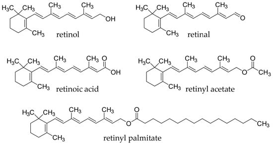
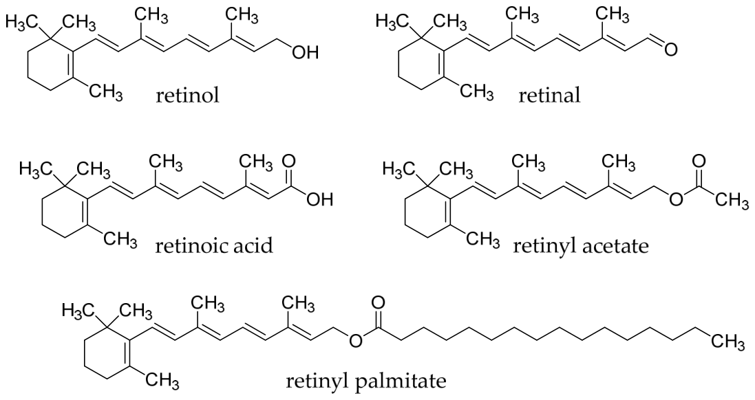
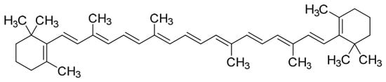
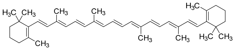
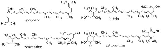
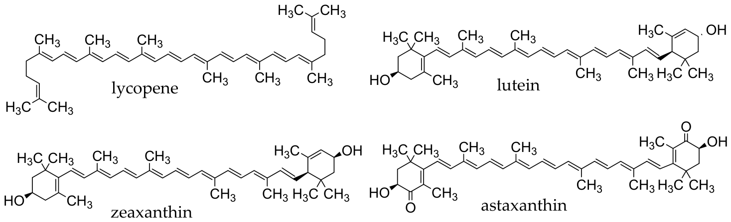
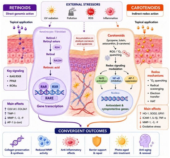
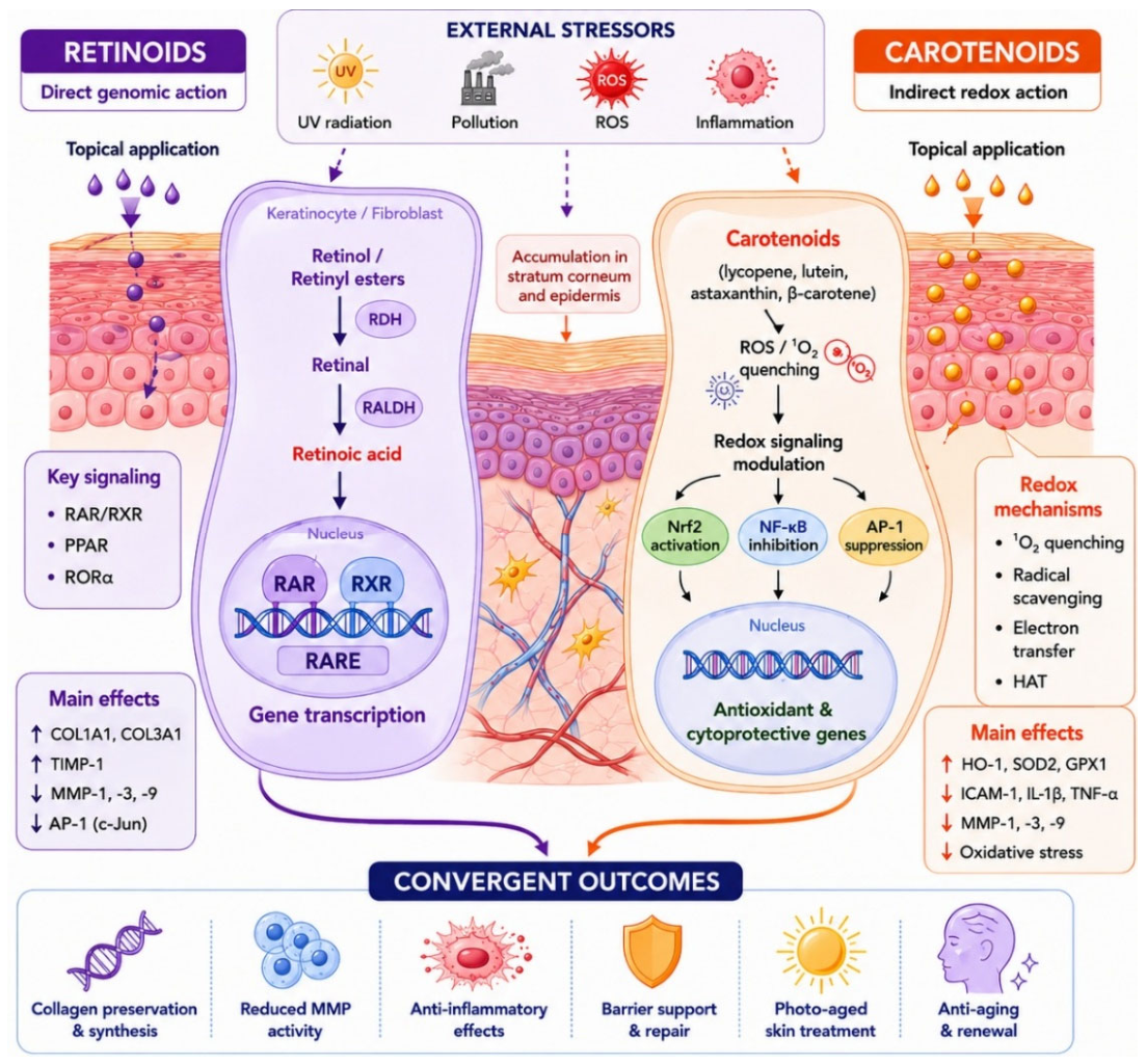
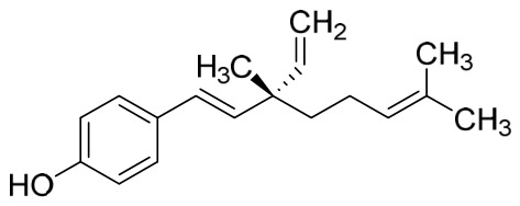
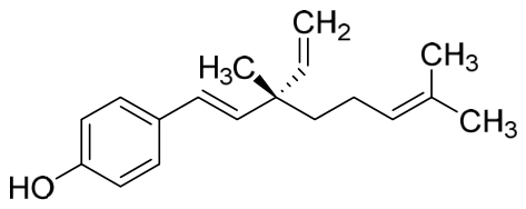
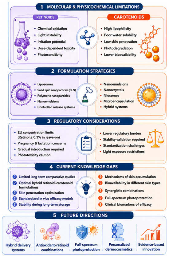
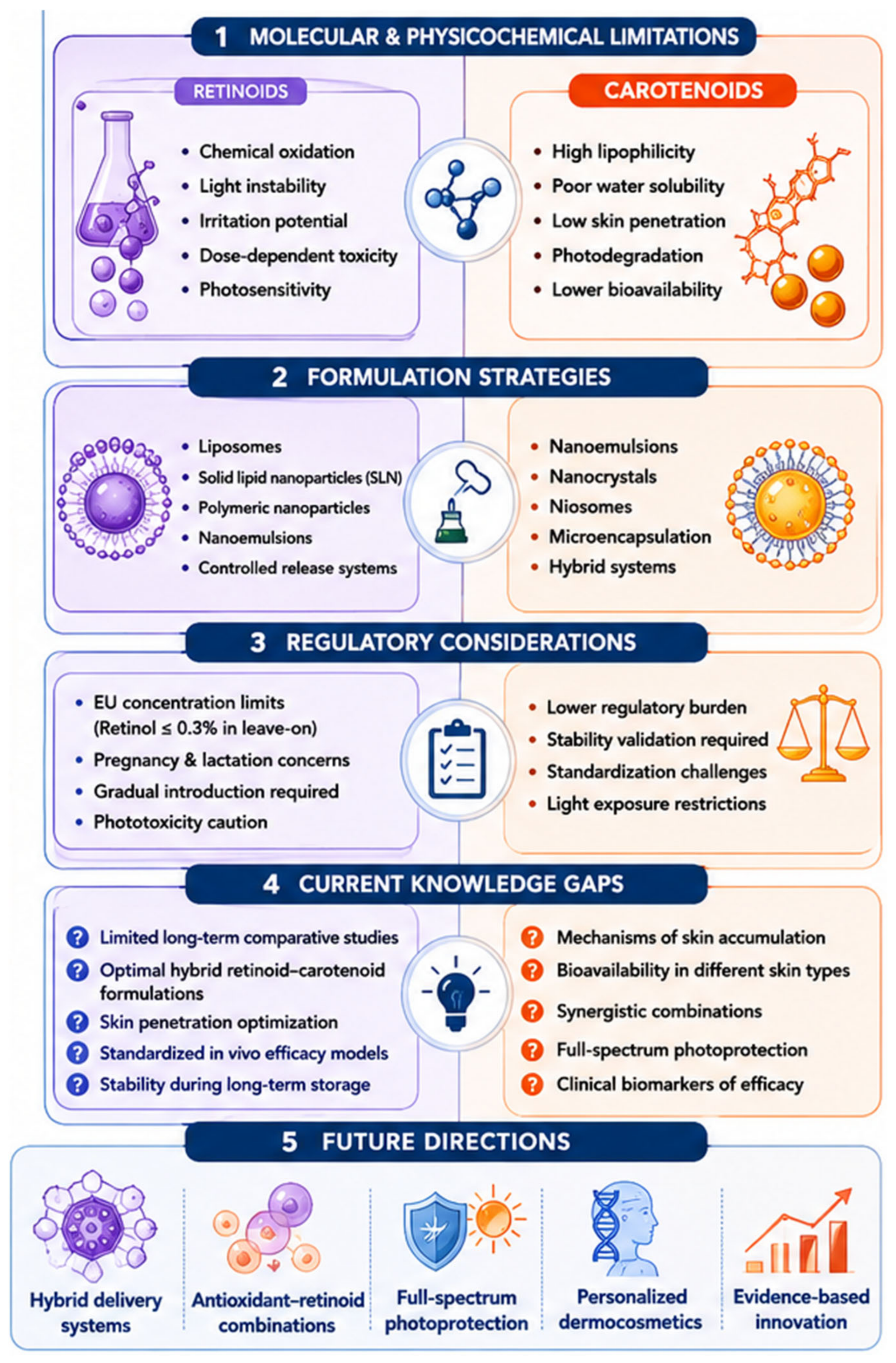

Disclaimer/Publisher’s Note: The statements, opinions and data contained in all publications are solely those of the individual author(s) and contributor(s) and not of MDPI and/or the editor(s). MDPI and/or the editor(s) disclaim responsibility for any injury to people or property resulting from any ideas, methods, instructions or products referred to in the content. |
© 2026 by the authors. Licensee MDPI, Basel, Switzerland. This article is an open access article distributed under the terms and conditions of the Creative Commons Attribution (CC BY) license.