1. Introduction
Implant therapy is currently considered the gold standard for the replacement of teeth lost primarily as a consequence of untreated periodontitis or extensive carious lesions. Although numerous clinical intervention studies and systematic reviews with meta-analyses have reported survival rates exceeding 90% after 10 years of follow-up, dental implants should not be regarded as a fail-safe therapeutic solution, as they remain susceptible to both biological and mechanical complications and failures [
1,
2].
Peri-implantitis represents the leading biological cause of implant failure and is defined as an infectious disease affecting the tissues surrounding an osseointegrated dental implant. Because peri-implantitis is characterized by a progressive corono-apical pattern of tissue destruction, any disruption of osseointegration at the crestal level may significantly increase the risk of implant failure. It is well established that, during the first year of function, peri-implant marginal bone undergoes a remodeling process of unpredictable magnitude and multifactorial etiology. In this context, peri-implant tissue stability is of paramount importance, as several studies have demonstrated a direct association between early marginal bone loss and the subsequent risk of peri-implantitis. In particular, Windael et al. (2021) reported that early marginal bone loss exceeding 0.5 mm was associated with an odds ratio of 5.43 for the development of peri-implantitis [
3]. Consequently, the assessment of marginal peri-implant bone-level changes over time is widely considered one of the key indicators of implant treatment success [
4].
Early marginal bone loss is described in the literature as a non-infectious, multifactorial process resulting in peri-implant marginal bone resorption, with its greatest impact occurring within the first 12 months following implant placement [
5]. This phenomenon may be influenced by several factors related to the surgical procedure, the prosthetic restoration, or the implant design itself. These factors include flap elevation [
6], excessive surgical trauma such as over-torqueing [
7] or overheating during osteotomy preparation [
8,
9], inadequate bone width or insufficient implant diameter [
10], implant malpositioning [
11], reduced vertical soft tissue thickness, bacterial leakage at the implant–abutment interface due to inadequate sealing, insufficient abutment height, the use of platform-matching rather than platform-switching configurations [
12], and repeated disruption of the peri-implant mucosal seal resulting from multiple abutment disconnections and reconnections [
13,
14].
One of the limitations of conventional prosthetic protocols for implant-supported restorations is the need for repeated removal and reconnection of abutments. Multiple disconnection and reconnection events have been associated with adverse effects on peri-implant marginal bone levels. Frequent manipulation of secondary implant components may mechanically traumatize the circumferential peri-implant mucosal barrier, potentially leading to inflammation and subsequent marginal bone loss. In a landmark animal study conducted in the mid-1990s, Abrahamsson et al. demonstrated that repeated abutment disconnections (five times) resulted in apical migration of the supracrestal connective tissue attachment, accompanied by crestal bone resorption [
15]. Conversely, a subsequent study showed that limiting the number of disconnection/reconnection episodes to two did not result in statistically significant differences in either hard- or soft-tissue outcomes [
16].
To minimize the effects of repeated abutment manipulation and preserve peri-implant hard and soft tissues, the one-abutment one-time (OTA) protocol was introduced [
17]. Although several studies have reported significantly greater marginal bone loss—approximately 0.2 mm—in cases involving multiple abutment disconnections, the actual clinical relevance of such differences remains unclear. This issue is particularly relevant in cases of subcrestal implant placement, a treatment approach frequently combined with the OTA protocol. At present, the literature contains only limited clinical evidence from studies with robust methodological designs evaluating this association [
18].
Subcrestal implant placement has been advocated because the position of the implant–abutment interface (IAI) may play a critical role in achieving optimal esthetic outcomes. In esthetically demanding situations, particularly in the anterior maxilla, the IAI is often positioned more apically to provide adequate space for the development of a gradual and natural emergence profile. However, early animal studies reported increased marginal bone loss associated with subcrestal implant placement. These findings have largely been attributed to the use of implants featuring flat-to-flat implant–abutment connections, which offer inferior sealing capabilities compared with contemporary connection designs capable of reducing bacterial leakage under functional loading.
Indeed, the implant–abutment interface should be considered a critical determinant of marginal bone stability, as it represents a potential source of chronic irritation resulting from both micromovement and bacterial contamination. Bacterial infiltration through the microgap at the implant–abutment interface, combined with mechanical loading, has long been regarded as a major factor limiting the widespread adoption of subcrestal placement protocols with earlier implant systems.
Nevertheless, the increasing use of implants incorporating conical internal connections and platform-switching concepts has contributed to the growing popularity of subcrestal implant placement over the past decade. Consequently, several systematic reviews, with and without meta-analyses, have been published on this topic. Despite this growing body of evidence, the literature remains inconclusive regarding the medium- and long-term reliability of subcrestal implant placement and its potential influence on peri-implant clinical parameters, including probing depth and bleeding on probing. Furthermore, there is a paucity of controlled clinical studies investigating two-piece implants placed specifically 2 mm below the crestal bone level.
The aim of the present study was to report and discuss the clinical and radiographic outcomes after 2 and 3 years of follow-up from a randomized controlled trial comparing implants placed 2 mm subcrestally with implants placed at the crestal level. The results from the first 12 months of follow-up have been published previously [
18]. The outcomes presented herein represent a continuation of the observational period of the same patient cohort.
2. Materials and Methods
Detailed materials and methods were previously published together with the 12-month follow-up results of the clinical investigation [
18]. Here, the 36-month follow-up data were evaluated including the radiographic and biometric parameters.
2.1. Patient Selection
This single-blind, parallel-arm, randomized controlled clinical trial received ethical approval from the University-Hospital ethics committee (approval number N4078) and was registered on ClinicalTrials.gov (NCT06182670; accessed 27 December 2023). The study adhered to the Declaration of Helsinki (2013 Fortaleza revision) and followed CONSORT reporting guidelines. Patient recruitment took place between April 2021 and May 2023 at the Section of Periodontics, School of Dentistry, Department of Surgical Specialties, Radiological Science, and Public Health, University of Brescia.
2.2. Surgical and Prosthetic Procedures
Detailed surgical and prosthetic procedures of this study were published elsewhere (Mensi et al. 2024 [
18]).
2.3. Radiographic Evaluation
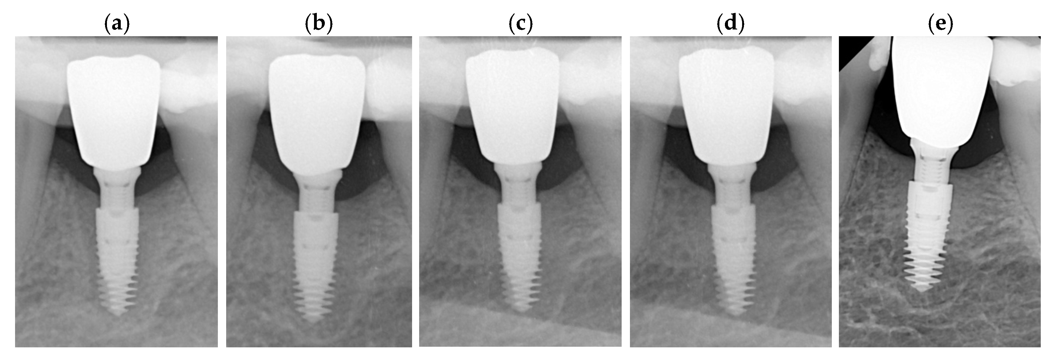
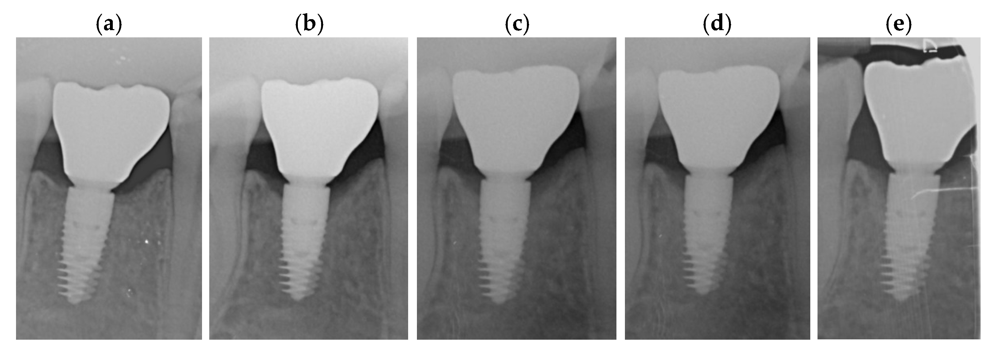
Each periapical radiograph which was evaluated to assess marginal bone modification (MBM) was retrieved by means of a standardized parallel technique (FONA™ Dental Image Plates, Assago, MI, Italy). Subsequently, the images underwent analysis by dedicated software (ImageJ v1.54r), National Institute of Health, Bethesda, MA, USA). Prior to performing the measurements, radiographs were calibrated using the implant diameter and length as references to reduce any possible distortion. All measurements were performed twice by a single, calibrated operator (ES), and each was repeated at three separate time points. The calibration process was conducted by assessing MBM (BL and BR) on 10 radiographs not included in the study. Intra-examiner reliability was investigated using the intra-class correlation coefficient (ICC = 0.91). MBM measurements were recorded at both the mesial and distal sites; hence, the mean value was calculated for each included implant. The radiographs were taken at surgical placement (T0), prosthetic delivery (T1), and after 6 (T2) and 12 months (T3) of function. The most coronal level of the bone-to-implant contact at surgery was defined as the marginal bone level (MBL).
During MBM evaluation, a distinction was made between bone remodeling (BR) and bone loss (BL). Bone loss was defined as the distance from the initial MBL to the first radiographically detectable bone-to-implant contact apical to the implant neck. Bone remodeling was defined as the distance from the initial MBL to the first point of contact coronal to the implant neck. Apical MBM values were recorded as negative numbers.
MBM was assessed at baseline (T0), at crown delivery (T1), and during follow-up visits at 6 months (T2), 12 months (T3), 24 months (T4) and 36 months (T5) (
Figure 1a–e and
Figure 2a–e).
2.4. Clinical Evaluation
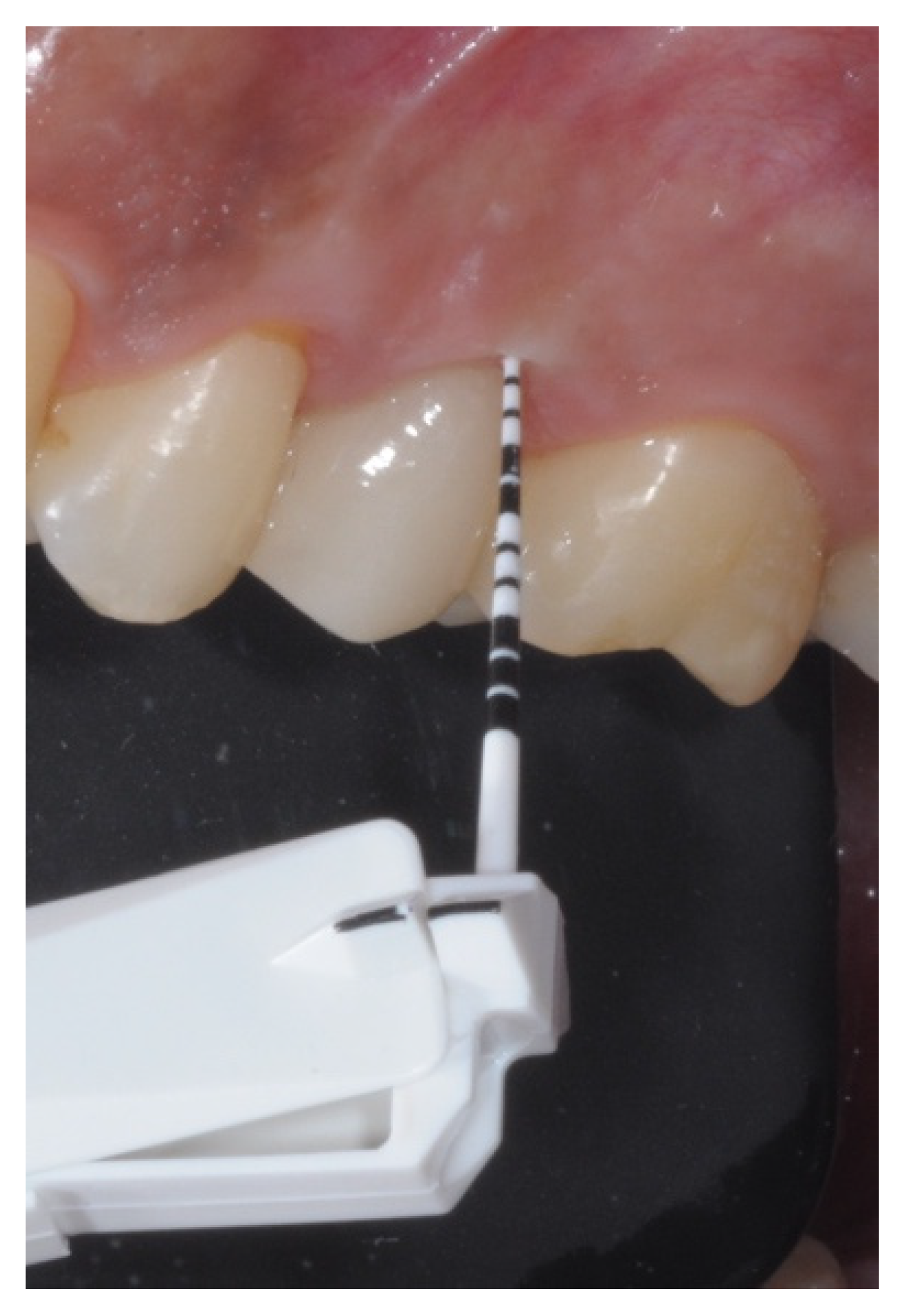
Peri-implant biometric parameters related to soft tissue were recorded using a calibrated periodontal probe (Vivacare TPS probe, Ivoclar Vivadent, Schaan, Liechtenstein) (
Figure 3). Probing depth (PD) was assessed at four sites—mesial, distal, buccal, and palatal—according to the classification described by Mombelli and Lang (1994) [
19]. Bleeding on probing (BOP) and plaque index (PI) were evaluated at the implant site following the criteria outlined by Trombelli et al. (2018) [
20] and O’Leary (1972) [
21], respectively. PD, BOP, and PI were measured at 6, 12, 24, and 36 months after delivery of the final prosthetic restoration.
2.5. Maintenance Protocol
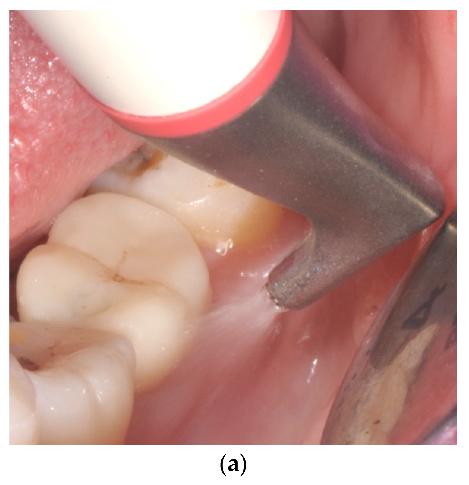
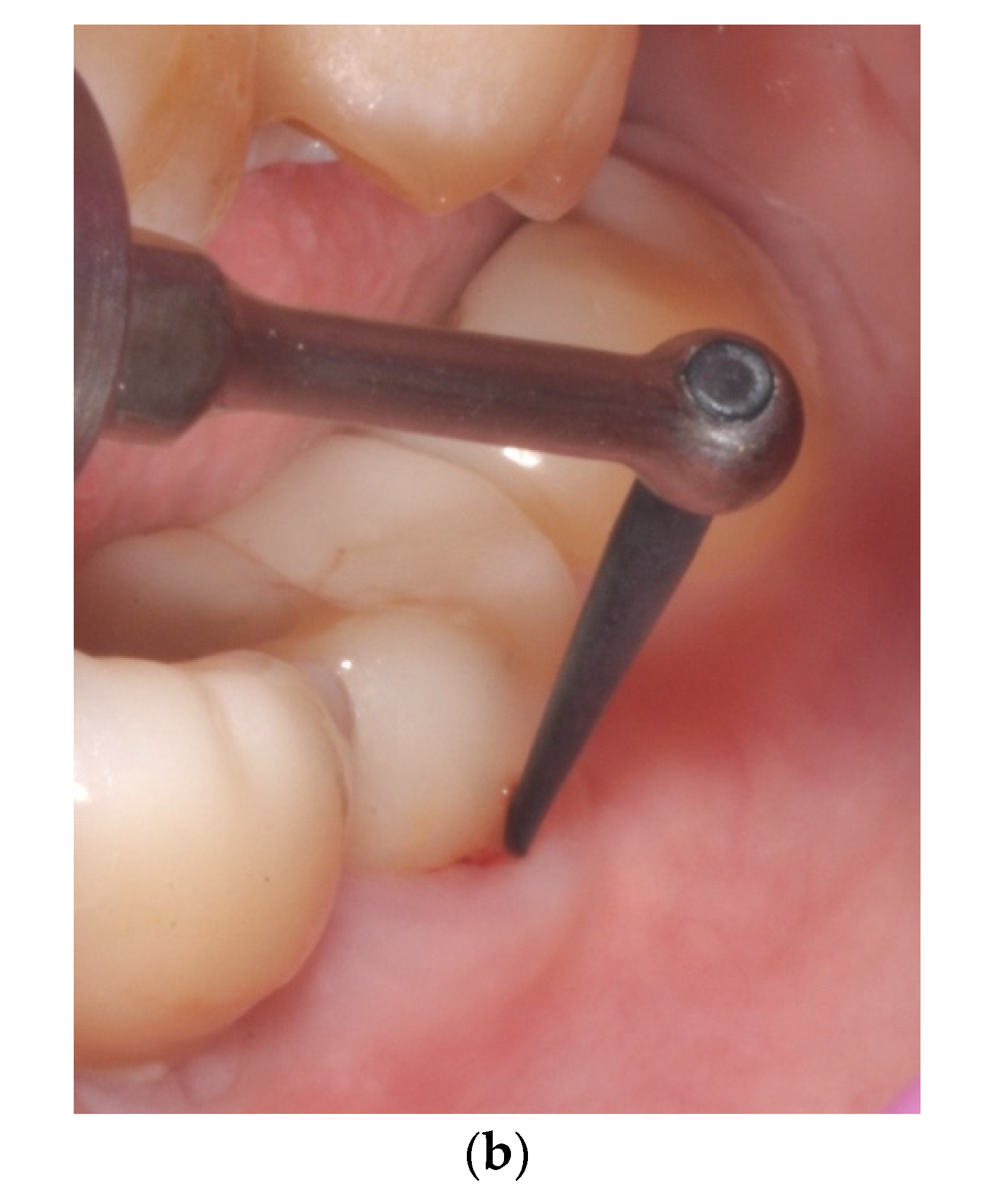
All subjects underwent professional oral hygiene every six months as part of supportive periodontal therapy delivered following the Guided Biofilm Therapy (GBT) approach. The implant-focused procedure involved subgingival airflowing with erythritol powder (PLUS powder
®, EMS, Nyon, Switzerland) within the peri-implant sulcus to ensure the removal of supra- and subgingival biofilm and plaque deposits in combination with the peek carbon slip tip (PI max
®, EMS, Nyon, Switzerland) (
Figure 4a,b). Reinforcement of oral hygiene measures for home care was provided every six months.
2.6. Sample Size and Randomization
The sample size was determined based on a two-parallel-group design with equal group allocation, using an independent samples t-test. The calculation assumed one implant and one measurement per patient, a standard deviation of 0.5 mm, and a minimum expected difference of 0.5 mm between groups. Under these assumptions, a total of 34 patients would be needed to achieve at least 80% statistical power at a 5% significance level. To account for an anticipated dropout rate of 15%, the final sample size was increased to 40 patients.
The randomization sequence was generated by a biostatistician using a block randomization method with variable block sizes (4, 6, and 8). Allocation concealment was guaranteed by opaque envelopes containing sequential numbered codes associated with each enrolled patient.
2.7. Statistical Analysis
All quantitative data, MBM, bone loss, and PPD, were modeled using Generalized Estimation Equation (GEE) models to account for the within-patient clustering effect, assuming an exchangeable correlation structure and a normal distribution. BOP and PI were modeled as counts (number of bleeding sites over the total number of patient sites) using a GEE assuming a Poisson distribution. All results are reported as effects estimates and corresponding 95% confidence intervals. All tests were two-sided and assumed a 5% significance level. All the analyses were performed using R (version 4.5.1).
3. Results
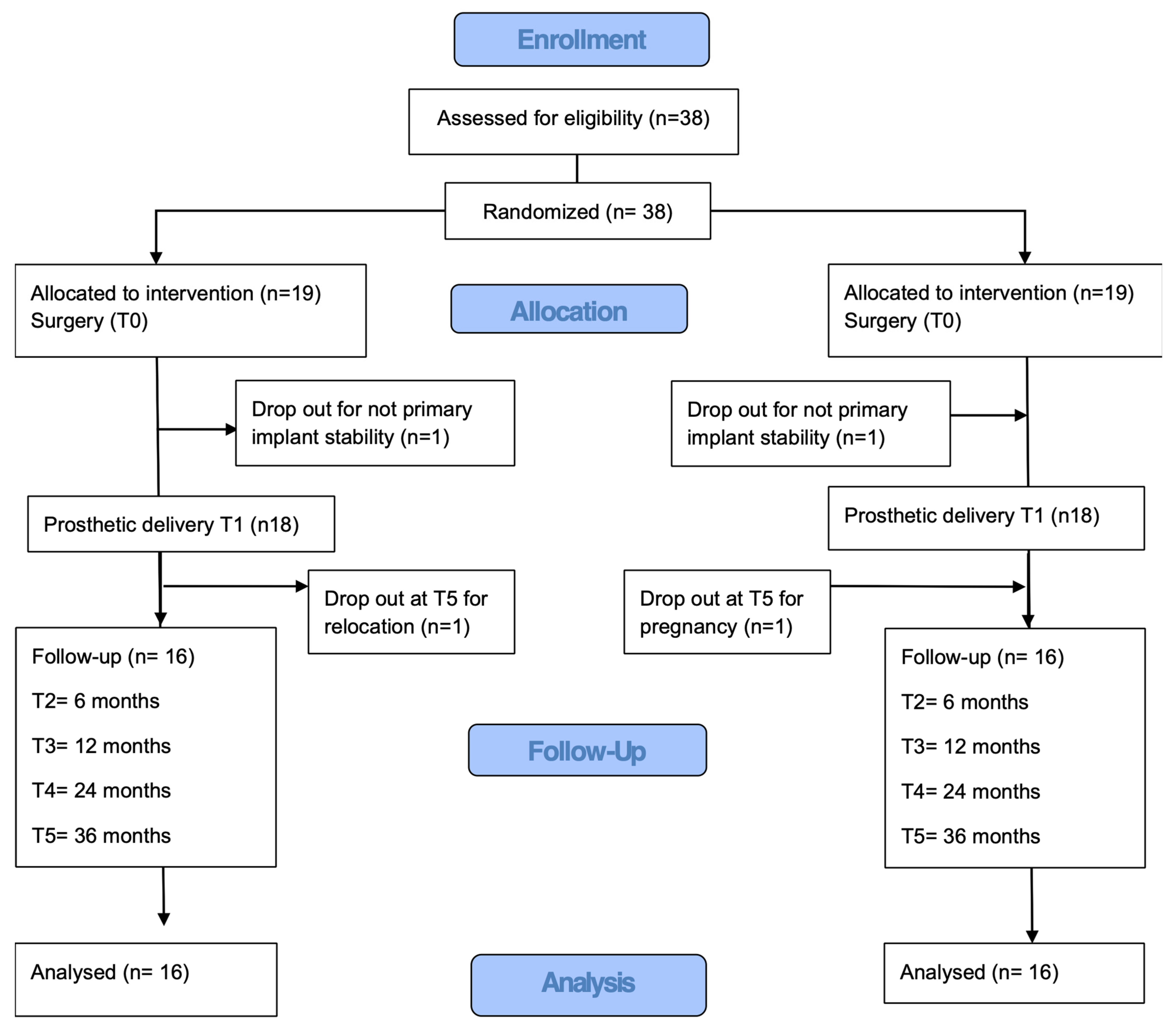
Thirty-eight patients were initially enrolled in the previous study. Two implants were considered dropouts because of insufficient primary implant stability (<35 Ncm) at implant placement, leaving 36 patients (14 males and 22 females) who completed the 12-month evaluation and who were included in the present study. The mean age was 48.29 (15.03) in the control group and 51.71 (12.75) in the test group. During the follow-up of the current investigation, two patients were lost to follow-up due to relocation and pregnancy. Therefore, data from 34 patients were available for the final analysis (
Figure 5). Patients’ demographic characteristics are reported in
Table 1.
MBMs for both groups from baseline (T0) to the prosthetic loading (T1), six-month follow-up visit (T2), 1-year follow-up, 2-year follow-up, and 3-year follow-up visit (T3) were statically significant (
p < 0.01), as shown in
Table 2.
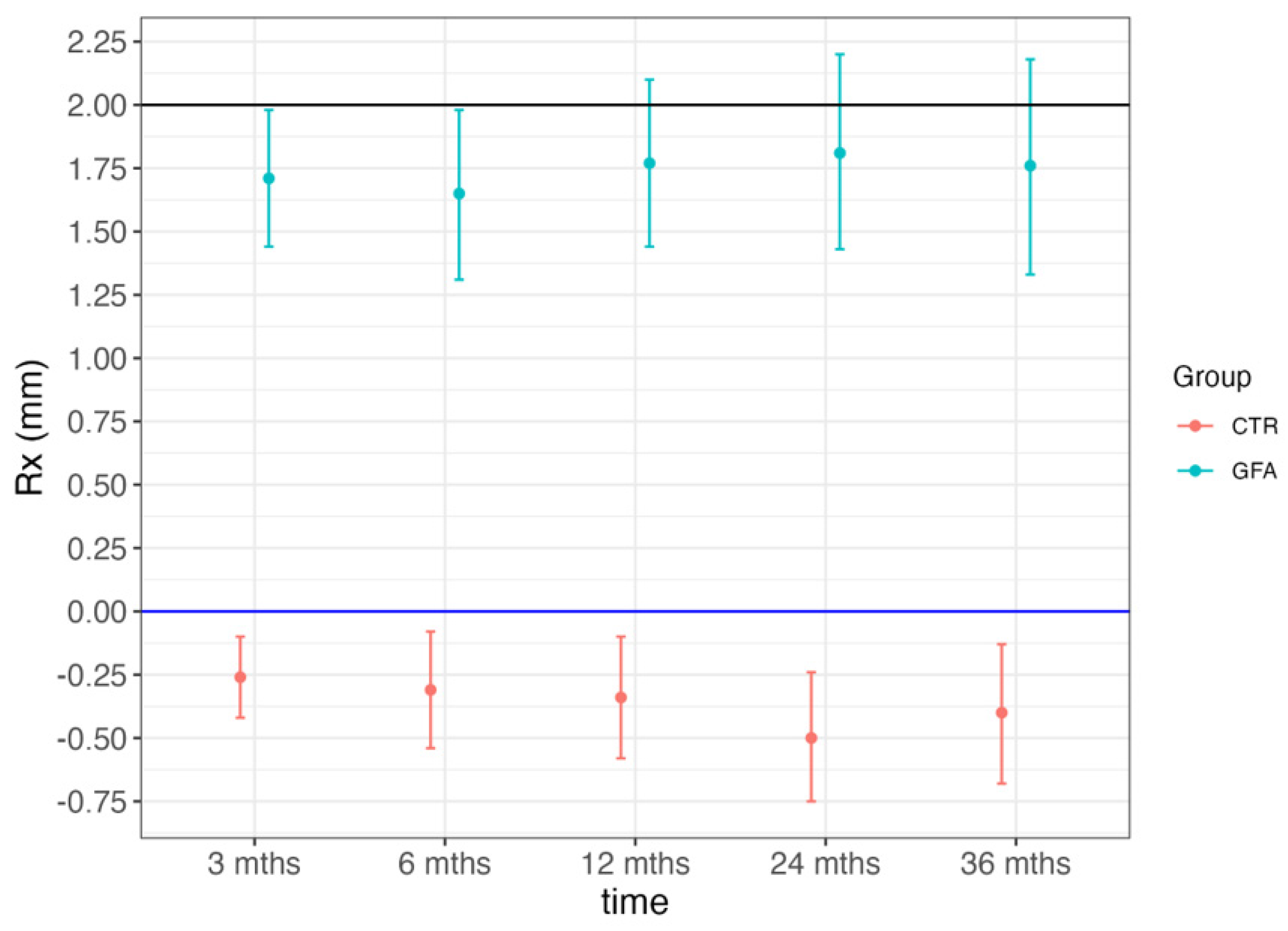
To be more precise, after 2 years of functional loading, the mean MBM in the control group was −0.68 mm as compared to the baseline (T0) and −0.55 mm in the test group. After 3 years of functional loading, the mean MBM in the control group was −0.58 mm as compared to the baseline (T0) and −0.61 mm in the test group. Nevertheless, no statistically significant difference was noticed when the MBM values in the two study groups were compared, neither at the 2-year follow-up nor 3-year follow-up (
p > 0.05) (
Table 2).
Similarly to what was observed in the analysis of the 12-month data, even after 2 and 3 years of follow-up, the control group exhibited a mean bone loss (BL) equivalent to the MBM values. Contrarily, in the test group, MBM variations over time remained within the initial subcrestal positioning value as no BL was observed. Therefore, the bone loss for the test group equals zero at all time points and up to 3 years (
Table 3).
Visual summary of MBM and BL changes over time is shown in
Graph 1.
Graph 1.
Marginal bone modification and bone loss at different time points in test (GFA) and control (CTR) group.
Graph 1.
Marginal bone modification and bone loss at different time points in test (GFA) and control (CTR) group.
Peri-implant-related biometric parameter variations over time are reported in
Table 4 and
Table 5.
PPD values amounted to 2.10 and 2.97 after 2 years and 2.03 and 2.78 after 3 years in the test and control group, respectively. No statistically significant difference was observed when comparing PPD values at T4 and T5 with baseline values (3 months) (p > 0.05).
BoP percentages amounted to 31% and 15% after 2 years and 13% and 11% after 3 years in the test and control group, respectively. A statistically significant difference was observed when comparing BoP values at 2 years (p< 0.01), although no significance was noticed after 3 years (p > 0.05).
PI percentages amounted to 22% and 12% after 2 years and 11% and 5% after 3 years in the test and control group, respectively. No statistically significant difference was observed when comparing PI values at T4 and T5 with baseline values (6 months) (p > 0.05).
4. Discussion
The primary aim of this randomized controlled clinical trial was to evaluate marginal bone modifications (MBMs) around one-stage, platform-switched implants with an internal conical connection placed either at the crestal level or 2 mm below the bone crest. As reported in the previous publication presenting the 12-month outcomes, supracrestal tissue height was not considered when determining implant insertion depth. Consequently, a baseline discrepancy in soft tissue thickness between the test and control groups cannot be excluded.
According to the results of the present investigation, no statistically significant differences in radiographic marginal bone changes were observed between the test and control groups after either 2 or 3 years of follow-up. These findings are consistent with those reported in previous clinical studies evaluating marginal bone levels around subcrestally placed implants characterized by platform switching and internal conical connections. In this regard, a multicenter crossover randomized controlled trial conducted by Stacchi et al. demonstrated no significant differences between implants placed 1 mm and 2 mm below the bone crest after 12 months of observation. Nevertheless, the authors suggested that deeper implant placement (2 mm) might reduce the likelihood of implant exposure within the oral cavity, thereby potentially acting as a protective factor in the primary prevention of peri-implantitis [
22].
At present, however, this hypothesis remains speculative, as clinical studies specifically designed to investigate peri-implantitis incidence as a primary outcome are required before definitive conclusions can be drawn. Although these observations should be interpreted cautiously due to the relatively short follow-up period, they open interesting clinical perspectives that may already be considered during surgical treatment planning. Supporting this concept, Lops et al., in a retrospective analysis of 410 implants placed at least 1 mm subcrestally, reported no cases of peri-implantitis and a mean marginal bone loss of only 0.09 ± 0.68 mm after an average follow-up period of 2.72 years [
23]. Furthermore, stable marginal bone levels up to three years were also reported in a recent randomized controlled trial by Lops et al., which found no significant differences between implants placed 1 mm subcrestally using either static guided surgery or a freehand surgical approach [
24].
The results of the present clinical trial corroborate these observations over a three-year follow-up period, suggesting that a stable implant–abutment connection can be maintained over time, thereby preserving peri-implant marginal bone levels in the short to medium term. To the best of the authors’ knowledge, this is the first randomized controlled trial directly comparing the same implant system, characterized by platform switching and an internal conical connection, placed either equicrestally or 2 mm subcrestally.
The importance of a precise and stable implant–abutment interface, as well as a reliable prosthetic connection, has also been highlighted in a recent case series. Although lacking a control group, the authors demonstrated that the prosthetic interface could remain stable in a subcrestal position for up to two years [
25].
As emphasized in a recent systematic review and meta-analysis by Cruz et al. (2022) [
26], the influence of subcrestal implant placement on peri-implant clinical and biometric parameters remains poorly understood. Existing clinical studies only occasionally report these outcomes, and when they do, the data are often collected and presented inconsistently [
26]. This aspect is particularly relevant because the maintenance of the peri-implant mucosal tunnel plays a crucial role in both the development and management of peri-implant mucositis.
In a cohort study involving 19 patients, Chan et al. demonstrated that deeper mucosal tunnel depths (≥3 mm) were associated with a higher prevalence of peri-implant mucositis and frequently required prosthesis removal combined with professional mechanical therapy to achieve disease resolution [
27]. Conversely, in cases characterized by shallow tunnel depths (≤1 mm), crown removal was unnecessary, and mucositis could be successfully managed through oral hygiene measures alone. It is well established that adequate access for daily plaque control and professional maintenance around implant-supported restorations is essential for the prevention and management of peri-implant inflammatory diseases [
28,
29].
In the present study, probing pocket depth (PPD) values differed significantly between the test and control groups after 12 months; however, this difference was no longer observed at the 2- and 3-year follow-up examinations. Similarly, bleeding on probing (BoP) values showed statistically significant differences between groups at 1 and 2 years but not after 3 years.
The transient differences observed in BoP are likely attributable primarily to differences in prosthetic design rather than to implant positioning itself. The test group was restored with mucosal-supported prostheses incorporating cantilever extensions, which may have complicated plaque control procedures for patients. This may explain the higher bleeding scores observed at the 12-month evaluation when compared with the control group. However, analysis of the temporal trends within each group revealed a progressive reduction in both plaque and bleeding indices at 24 and 36 months. Although intra-group statistical analyses were not available, this reduction appears clinically relevant. A similar trend was observed in the control group, albeit to a lesser extent.
A slight but consistent difference remained in favor of the control group, likely reflecting the increased complexity of hygiene procedures associated with the prosthetic design used in the test group, particularly because of the presence of buccal and palatal cantilever extensions. Importantly, the observed increases in plaque accumulation and bleeding appear to be primarily related to mucosal contact with the prosthesis rather than to biofilm penetration along the transmucosal pathway.
Notably, plaque index (PI) values remained relatively low, reaching 5% and 11% in the control and test groups, respectively, and no direct correlation between plaque accumulation and bleeding was observed. This finding suggests that mucosal compression may have contributed more substantially to BoP values than plaque accumulation itself. From both a clinical and biological standpoint, this phenomenon is likely of limited pathological significance and may be more comparable to the tissue response observed beneath pontic areas in tooth-supported fixed prostheses than to true peri-implant inflammatory bleeding.
A key factor contributing to the progressive improvement in clinical parameters over time was the implementation of a structured supportive care program consisting of six-monthly Guided Biofilm Therapy (GBT) sessions. This protocol not only ensured effective professional biofilm removal but also provided continuous patient education, motivation, and individualized recommendations regarding home-care products tailored to the specific morphology of each prosthetic restoration.
Recent evidence has demonstrated that GBT is effective in the management of peri-implant mucositis and may reduce the need for adjunctive chemical antiseptics [
30]. Consequently, both study groups exhibited progressive improvements in clinical outcomes over time, with slightly more favorable results observed in restorations characterized by a more physiological S-shaped emergence profile, as typically found in bone-level restorations.
Furthermore, the bleeding occasionally observed in the test group appeared more frequently associated with irritation of the supporting mucosa caused by contact with the zirconia prosthesis and coronal soft tissue creep, which may result in excessive tissue compression. This condition should be distinguished from true bleeding elicited during probing of the peri-implant sulcus, as observed in the control group, which may be considered biologically more relevant.
The clinical data collected at 12, 24, and 36 months revealed distinct temporal trends but, most importantly, demonstrated progressive improvements in both groups. This favorable evolution can be directly attributed to the structured maintenance program, which included six-monthly follow-up visits managed by a dental hygienist. During each maintenance session, the GBT protocol was performed, incorporating not only professional biofilm removal but also systematic biofilm disclosure and individualized patient education, motivation, and reinforcement of oral hygiene practices.
This aspect represents one of the key findings of the present study and warrants particular emphasis, as it provides a plausible explanation for the progressive reductions in both bleeding on probing and plaque accumulation observed over time. The results suggest that regular maintenance appointments at six-month intervals, combined with continuous patient education and motivation, can not only maintain peri-implant health but may also lead to progressive improvements in clinical parameters. From this perspective, GBT represents a valuable tool not only for biofilm control but also for minimizing the risk of biological complications, particularly in complex prosthetic rehabilitations. Nevertheless, the aforementioned observations regarding prosthetic design, mucosal compression, plaque accumulation and bleeding are merely descriptive and speculative in nature. In fact, many of the prosthetic variables that may play a critical role were not directly measured or reported in this randomized controlled trial. It should be borne in mind that, even if the proposed mechanisms represent plausible biological interpretations of the results, they were not experimentally verified as primary outcomes. For these reasons, further clinical trials characterized by adequate methodology and follow-up are needed.
Overall, the findings of this study underscore the critical role of long-term supportive care and patient compliance in achieving and maintaining biological success around dental implants.
6. Conclusions
Within the limitations of the present study, implants featuring an internal conical connection and platform-switching design, placed 2 mm below the bone crest and restored according to a one-abutment one-time protocol with screw-retained crowns, demonstrated marginal bone changes comparable to those observed around equicrestally placed bone-level implants.
Notably, no clinically relevant crestal bone loss leading to exposure of the implant body was observed around subcrestally placed implants throughout the three-year follow-up period. Although some degree of radiographic bone remodeling occurred, it never resulted in exposure of the implant surface within the oral cavity.
Differences in peri-implant clinical parameters, such as probing depth and bleeding on probing, appear to be more closely related to prosthetic design characteristics than to implant placement depth itself. However, this conclusion should be limited to implant systems featuring internal conical connections and platform-switching configurations and cannot be generalized to all implant designs.
Furthermore, these differences appear to become clinically negligible when patients are enrolled in a structured maintenance program supported by contemporary biofilm management protocols and reinforced through continuous patient education and motivation regarding home oral hygiene procedures.
Although the results of this study are encouraging, the three-year follow-up period remains relatively short. Additional well-designed clinical trials with longer observation periods are needed to clarify the influence of implant positioning relative to the bone crest, with or without the one-abutment one-time protocol, on peri-implant marginal bone stability over the medium and long term.




 ,
,