Article

Genomic and Metabolomic Profiling of Streptomyces anulatus 89: Molecular Phylogeny and Biosynthesis of Antitumor Antibiotics

1
Department of General and Soil Microbiology, D.K. Zabolotny Institute of Microbiology and Virology, National Academy of Sciences of Ukraine, Akademika Zabolotnoho Str., 154, 03143 Kyiv, Ukraine
2
Microbial Culture Collection of Antibiotic Producers, Faculty of Biology, Ivan Franko National University of Lviv, Hrushevskoho Street 4, 79005 Lviv, Ukraine
3
Department of Technologies of Medical Diagnostics and Treatment, ESC “Institute of Biology and Medicine”, Taras Shevchenko National University of Kyiv, Volodymyrska Street 60, 01033 Kyiv, Ukraine
4
Department of Molecular Immunology, Palladin Institute of Biochemistry, National Academy of Sciences of Ukraine, Leontovycha Street, 9, 01054 Kyiv, Ukraine
5
Department of Reproduction of Viruses, D.K. Zabolotny Institute of Microbiology and Virology, National Academy of Sciences of Ukraine, Akademika Zabolotnoho Str., 154, 03143 Kyiv, Ukraine
6
Department of Ecology, Faculty of Humanities and Natural Science, University of Presov, 08001 Presov, Slovakia
*
Authors to whom correspondence should be addressed.
Appl. Sci. 2026, 16(13), 6743; https://doi.org/10.3390/app16136743 (registering DOI)
Submission received: 8 June 2026 / Revised: 30 June 2026 / Accepted: 2 July 2026 / Published: 6 July 2026

Abstract

Background: Soil streptomycetes, particularly those isolated from extreme environments, are valuable sources of bioactive compounds. Their genomes encode a large number of biosynthetic gene clusters (BGCs), many of which can be simultaneously expressed. Methods: Molecular genetic methods were employed to identify Streptomyces anulatus 89 (Illumina NovaSeq 2 × 150 bp). Whole-genome phylogeny based on orthologous genes was employed using the Bacterial and Viral Bioinformatics Resource Centre services. Liquid chromatography–mass spectrometry analysis of biomass extract was carried out to identify antibiotics. Bioassays on cell lines were employed to evaluate the cytotoxicity and antitumor activity of the crude extract of the S. anulatus 89 strain. Results: Genome analysis identified 36 BGCs associated with secondary metabolites. The strain synthesized nactins, pladienolide, phenazinomycin, and 21-hydroxyoligomycin. The biomass extract demonstrated cytotoxicity against cancer cells and induced apoptosis. The A549 and A431 cell lines were the most sensitive. Changes in tumor cell morphology included rounding, shrinkage, increased granularity, and vacuolization. Conclusions: The ability of S. anulatus 89 to simultaneously synthesize different classes of anticancer antibiotics was reported. The investigated crude extract exhibited pronounced antitumor activity, making it a promising candidate for further studies. The underlying hypothesis suggested that strains with broad adaptive potential may serve as promising producers of natural products with antitumor properties.

1. Introduction

Actinobacteria of the genus Streptomyces are Gram-positive, spore-forming bacteria that are widely distributed across diverse ecosystems, ranging from soils to aquatic environments. Their genomes encode a large number of biosynthetic gene clusters (BGCs), whose expression provides them with high adaptive potential and bioactivity [1]. They are able to survive under adverse environmental conditions, including osmotic, acidic, and thermal stress, as well as in ecosystems contaminated with heavy metals, which makes their genomes valuable resources for the discovery of encoded BGCs and novel natural products [2]. During their life cycles, streptomycetes excrete a wide spectrum of extracellular molecules, e.g., hydrolytic enzymes such as proteases, cellulases, lipases, pectinases, and chitinases, as well as a large number of secondary metabolites. These compounds are widely utilized across diverse fields, ranging from industry to medicine, where they are used for their therapeutic properties [3]. Secondary metabolites are essential for their survival, enhancing competitiveness within the environment and increasing tolerance to stress from biotic and abiotic factors while simultaneously holding great promise for applications in biotechnology. These include osmoprotectants, pigments, antioxidants, and metabolites with signaling functions, among others. Consequently, extreme environments represent valuable reservoirs for isolating Streptomyces strains with unique metabolic profiles [4,5]. For example, saline biotopes are frequently marked by additional extreme environmental conditions, including high alkalinity, limited oxygen availability, nutrient scarcity, and intense solar irradiation, which vary depending on the geographical region. Several antitumor and antimicrobial compounds have been isolated from actinomycetes inhabiting saline environments, including salternamides, salinazinones, and xiamycins from Streptomyces; erythronolides and actinopolysporins from Actinopolyspora; and borrelidins together with nocarbenzoxazoles from Nocardiopsis. These metabolites encompass diverse chemical classes such as anthraquinones, benzofurans, sesquiterpenoids, oxazinones, and macrolides. Nevertheless, reports on bioactive metabolites produced by Streptomyces from saline biotopes remain scarce compared with those from other extreme ecosystems.
Approximately 70–80% of all natural bioactive compounds with pharmacological applications are products of the metabolic activity of these bacteria [1], including doxorubicin, daunorubicin, actinomycin D, mitomycin C, and bleomycin, which are among the most widely used anticancer agents [6]. They can affect tumor cells through several key mechanisms: they may induce apoptosis; suppress cell cycle progression at the G1, S, or G2/M phases; inhibit angiogenesis; modulate signaling pathways; and alter tumor cell metabolism. Many natural products also possess the ability to sensitize cancer cells to oxidative stress induced by chemo- and radiotherapy by limiting their antioxidant capacity. Inhibition of antioxidant defenses within tumors reduces their ability to counterbalance oxidative stress, ultimately leading to cell death. Notably, the same natural compound may exhibit both antioxidant and prooxidant properties, depending on factors such as concentration, cell type, duration of exposure, and environmental conditions [7,8].
Under laboratory conditions, several BGCs can be expressed, many of which are responsible for the production of metabolites with antitumor properties [9]. The discovery of new, non-toxic natural antitumor metabolites is essential for overcoming tumor resistance and mitigating the adverse effects of chemotherapy. Insights into the biosynthetic potential of Streptomyces spp. provide a foundation for exploring antitumor compounds, optimizing their structural modifications to enhance selectivity and reduce cytotoxicity toward normal cells, and bridging knowledge gaps regarding the genetically determined traits of individual strains. Consequently, the pharmaceutical potential of local Streptomyces strains residing in coastal saline zones of Ukraine warrants deeper investigation, particularly in light of the continuously growing demand for novel antitumor agents. This study aimed to perform whole-genome sequencing of the S. anulatus 89 strain, isolated from saline soil, to assess its genome-determined adaptive potential and capacity to synthesize antibiotics, and to evaluate its effects on tumor cell lines under in vitro conditions. It is hypothesized that strains with broad adaptive potential may serve as promising producers of natural products with antitumor properties.

2. Materials and Methods

2.1. Whole-Genome Sequencing, Рhylogenetic Analysis, and Genome Annotation of S. anulatus 89

The strain S. anulatus 89 was isolated from soil in the Mykolaiv region of Ukraine (46.8° N, 31.9° E) in 2016. The sampling site is located within the coastal zone of Southern Ukraine, specifically in southern chernozem soil near the Black Sea shore. The site is characterized by a temperate continental climate with maritime influence, average annual temperatures of approximately 11–13 °C, and summer peaks reaching 34 °C. Annual precipitation was moderate (around 450–500 mm), with frequent summer droughts and high evapotranspiration. Coastal soils in this zone are subject to periodic salinity fluctuations due to marine aerosols and groundwater intrusion, creating conditions of osmotic stress. These environmental parameters define the sampling location as an ecologically variable habitat with seasonal extremes of heat, drought, and moderate salinity [10]. No chemical treatment was applied prior to sampling, and the strain was cultivated on GYM medium [11]. Isolation of DNA was carried out using the DNeasy® UltraClean® Microbial Kit (QIAGEN, Hilden, Germany) according to the manufacturer’s instructions. DNA quality was examined using a NanoDrop spectrophotometer (Thermo Fisher Scientific, Waltham, MA, USA). 16S rRNA (using the Sanger method) and whole-genome sequencing (Illumina NovaSeq) were performed at Explogen LLC (Lviv, Ukraine). De novo genome assembly was performed using Velvet Assembler (v1.2.10) [12], and plasmids were searched using PlasmidFinder (v2.1.6) [13]. The quality of the genome assembly was verified using CheckM [14]. The whole genome sequence was deposited in the NCBI database under GenBank accession number GCA_053710995.1. Phylogenetic analysis was performed using the Bacterial and Viral Bioinformatics Resource Center (BV-BRC) Phylogenetic Tree Building pipeline [15]. A set of 500 global single-copy orthologous genes (PGFams) was randomly selected from the analyzed genomes. Both protein and nucleotide sequences were aligned using MAFFT [16]. The concatenated alignments were then used to infer a maximum likelihood phylogenetic tree using RAxML version 8.2.12 [17]. The best protein substitution model was identified as LG + F. Branch support was calculated using the RAxML Fast Bootstrapping method with 100 replicates. The phylogenetic tree was visualized using the iTOL (v6) resource [18]. Average Nucleotide Identity (ANI) was calculated using the FastANI algorithm implemented in DFAST 1.3.9 (DDBJ Fast Annotation and Submission Tool) [19]. Rapid genome annotation was carried out using the RAST prokaryotic genome annotation service (Rapid Annotations using Subsystems Technology) [20]. Comparative genomic analysis, including the identification of orthologous gene clusters and strain-specific proteins, was performed using the OrthoVenn3 web platform (2023) [21]. Searching and identification of putative clusters of secondary metabolites were performed using antiSMASH version 8 [22]. Alignment and visualization of homology clusters were performed using Clinker v0.0.32 [23].

2.2. LC-МS Analysis of Crude Biomass Extracts of S. anulatus 89

The strain was cultivated by submerged fermentation for 7 days at 28 °C in a shaker incubator at 240 rpm, as previously described [24]. Metabolites were extracted with ethanol (3:1) from the biomass, evaporated, and dissolved in 250 μL of methanol [19]. LC-HRMS data were collected on a Dionex Ultimate 3000 RSLC system using a BEH C18 column (100 × 2.1 mm, 1.7 µm particle size; Waters, Eschborn, Germany). Separation of 1 µL of the sample was achieved using a linear gradient of solvent B (acetonitrile with 0.1% of formic acid) against solvent A (water with 0.1% of formic acid) at a flow rate of 600 µL/min and 45 °C. The gradient started with a 0.5 min isocratic step at 5% B and then increased to 95% B over 18 min, ending with a 2 min step at 95% before re-equilibration under the initial conditions. UV spectra were acquired using DAD in the range of 200 to 600 nm. The mass-spectrometric data was collected on a maXis 4G hr-ToF ultrahigh-resolution mass spectrometer (Bruker Daltonics, Bremen, Germany) using the Apollo II ESI source. Mass spectra were acquired in centroid mode over a mass range from 150 to 2500 m/z at a 2 Hz scan rate [25]. LC-MS data were processed using Compass Data Analysis 6.1 software.

2.3. Analysis of the Cytotoxicity and Antitumor Properties of the Biomass Extract of S. anulatus 89

Cancer cells from the Нер-2 (human laryngeal carcinoma, ECACC 86030501), A431 (epidermoid carcinoma with EGFR/ErbB-1 overexpression, ECACC 85090402), and A549 (human lung adenocarcinoma, ECACC 86012804) cell lines and the normal cell line Vero (kidney of the African Green Monkey, ECACC 84113001) were obtained from the Cell Bank of the Kavetsky Institute of Experimental Pathology, Oncology, and Radiobiology of the National Academy of Sciences of Ukraine (Kyiv, Ukraine). These cell lines were selected to provide an initial evaluation of the antitumor potential of the S. anulatus 89 crude extract across a panel of human epithelial cancer models. The Vero cell line was included in the experimental design as a continuously growing, non-tumorigenic mammalian cell line that has been widely used in toxicological and pharmacological studies, including the screening of microbial metabolites and natural products. The cells were cultured in a medium consisting of 46% Dulbecco’s Modified Eagle’s Medium (DMEM, Sigma-Aldrich, St. Louis, MO, USA) and 46% RPMI-1640 (Sigma-Aldrich, St. Louis, MO, USA), supplemented with 8% (v/v) fetal bovine serum (FBS, Sigma, St. Louis, MO, USA) and 100 U/mL of gentamicin (Biowest, Nuaillé, France). The S. anulatus 89 biomass extract stock solution contained 25.8 mg/mL of dry weight and was sterilized by filtration through a 0.22 μm filter before use.

2.3.1. Cell Morphology Characterization

Photographs documenting the interaction of cells with the ethanolic biomass extract of S. anulatus 89 at different dilutions were taken over a 48 h period. Morphological changes were characterized using an inverted light microscope Axiovert 40 CFL (Carl Zeiss Jena, Germany) at 70× magnification. Alterations in epithelial cell morphology resulting from treatment with the extract were systematically observed and recorded.

2.3.2. Cytotoxic Activity of Extract Against Cancer and Normal Cells

Cell viability was assessed using the MTT assay (3-(4,5-dimethylthiazol-2-yl)-2,5-diphenyltetrazolium bromide) (Sigma-Aldrich, St. Louis, MO, USA) following exposure to the ethanolic biomass extract. Hep-2, A431, A549, and Vero cells were seeded at densities of 1.7 × 10⁵, 2.0 × 10⁵, 2.0 × 10⁵, and 2.2 × 10⁵ cells/mL, respectively, in 96-well plates and incubated for 24 h at 37 °C in a humidified atmosphere containing 5% CO2 before treatment. Cells were then exposed to serial two-fold dilutions of the extract (ranging from 1:20 to 1:1280). Non-exposed cells served as negative controls. After 48 h of incubation, the culture medium was removed, cells were washed with phosphate-buffered saline (PBS; Biowest, Nuaillé, France), and 20 µL of MTT solution (NeoFrexx, Einhausen, Germany, 5 mg/mL in PBS) was added to each well. Plates were incubated for an additional 3 h at 37 °C with 5% CO2. The resulting purple formazan crystals were solubilized with 150 µL of 96% ethanol per well. Absorbance was measured at 570 nm using a Multiskan FC microplate reader (Thermo Scientific, Waltham, MA, USA). Cell viability (%) was expressed as the absorbance of treated cells relative to untreated controls (considered to present 100% viability). A reduction in viability was interpreted as an indication of increased cytotoxicity. The percentage of cell viability was calculated according to Equation (1):
C e l l   v i a b i l i t y % = A b s o r b a n c e   o f   t r e a t e d   c e l l s A b s o r b a n c e   o f   c o n t r o l   c e l l s × 100 % .
The 50% half-maximal inhibitory concentration (IC50) was determined from the dose–response curve, and the mean IC50 (±SD) value of the ethanol extract solution was calculated from three independent experiments [26].

2.3.3. Determination of Apoptosis Stages

Cells were plated in flat-bottom 6-well plates at a density of 1 × 10⁶ cells per well and left overnight to ensure proper adherence before exposure to S. anulatus 89 extract and pure ethanol (Sigma-Aldrich, Taufkirchen, Germany). After 24 h of treatment, both detached cells and adherent cells (harvested with 30 mM EDTA in PBS, Sigma-Aldrich, Taufkirchen, Germany) were collected, combined, and centrifuged at 2000 rpm for 5 min. The resulting pellets were washed twice with cold PBS. Cell populations (viable, early apoptotic, late-apoptotic, and dead) at varying extract dilutions (1:320, 1:640, and 1:1280) were quantified by flow cytometry with the Annexin V-EGFP/PI dual-staining method using the Annexin V-FITC Apoptosis Detection Kit (Sigma-Aldrich, Taufkirchen, Germany). Samples were simultaneously exposed to recombinant Annexin V-EGFP (2.5 μg/sample) for 30 min and PI (50 μg/mL) for 5 min at 4 °C in the dark and then washed to remove unbound dyes. Flow cytometric analysis was performed, with each determination based on the acquisition of 10,000 events. Fluorescence was detected using a DxFLEX Flow Cytometer (Beckman Coulter, Indianapolis, IN, USA) with FITC-A (Annexin V-EGFP) and ECD-A (PI) channels, and data were processed using Kaluza Analysis Software 2.1 [27,28].

2.4. Statistical Analysis

The experiments were carried out in triplicate (n = 3), and each value is presented as the mean ± standard deviation (SD). For LC-MS analysis, parts-per-million (ppm) values were calculated using Equation (2):
p p m = m m × 10 6 ,
where Δm represents the difference between the measured and theoretical mass, and m denotes the theoretical mass.

3. Results

3.1. Identification and Genome Annotation of S. anulatus 89

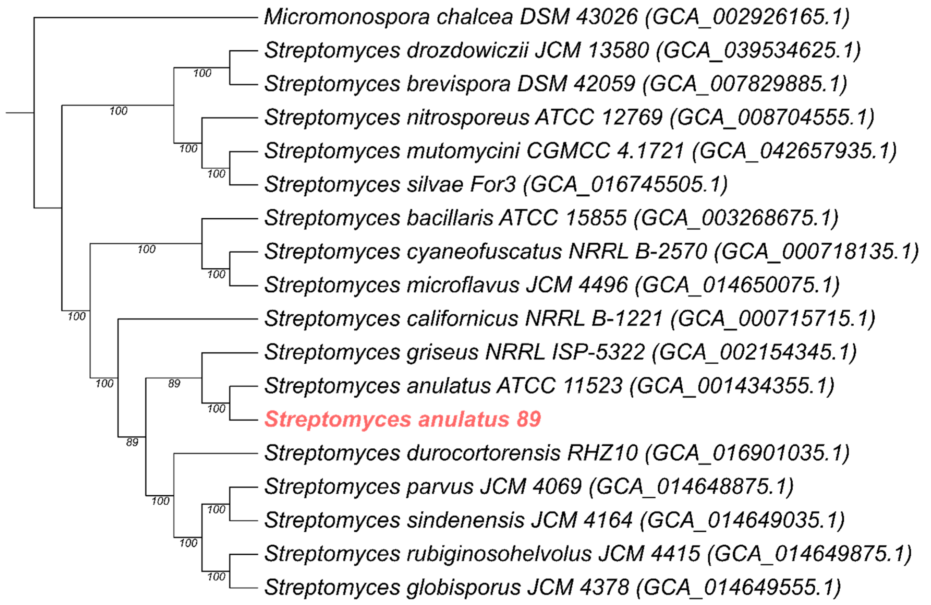
Primary identification of the strain was performed based on 16S rRNA gene sequence analysis, which allowed us to classify it as Streptomyces. For further research, the genome of strain 89 was sequenced. The final draft assembly of the genome consists of 57 contigs with a total length of 8,478,849 nucleotide pairs (np) and a high G + C content of 71.5%. In addition, a plasmid 67,039 bp in length with a G + C content of 67.7% was identified. The assembly demonstrated high completeness, as indicated by an N50 value of 346,545 bp and an L50 value of 10. The quality of the assembly was further verified using CheckM v1.2.5, which revealed a genome completeness of 99.91% and a minimal contamination level of 0.19%, confirming the high purity and reliability of the genomic data. Genomic annotation enabled the identification of 7555 coding sequences (CDS), 83 tRNAs, and 2 rRNAs, resulting in a high coding ratio of 87.7%. A whole-genome phylogenetic analysis based on 500 orthologous genes showed that strain 89 was grouped with S. anulatus ATCC 11523, and together they form a common group with S. griseus (Figure 1). Average Nucleotide Identity (ANI) showed that the strain is 97.63% similar to S. anulatus ATCC 11523 and 97.48% similar to S. anulatus JCM 4721, supporting its classification as strain 89 of the species S. anulatus. To explore the specific genetic features that distinguish this newly identified strain from its close relatives, a comparative genomic analysis using OrthoVenn3 between the studied strain 89 and the reference strains ATCC 11523 and JCM 4721 was performed. This analysis identified 52 proteins uniquely present in strain 89. This strain-specific set of proteins includes numerous phage fragments, transposases, and components of antiviral defense mechanisms. Furthermore, the genome of strain 89 encodes a protein containing GAF and ANTAR domains, as well as an oxidoreductase from the short-chain dehydrogenase/reductase (SDR) family; both are potentially involved in reducing the impact of environmental stressors, particularly heavy metal toxicity and oxidative stress. Additionally, the identification of a copper-transporting P-type ATPase, the enzyme responsible for the active cellular efflux of copper, suggests the presence of a potent heavy metal detoxification system. Taken together, the presence of these genetic markers associated with environmental adaptation and resistance provides strong support for the hypothesis that strain 89 evolved under significant selective pressure in its native soil environment.
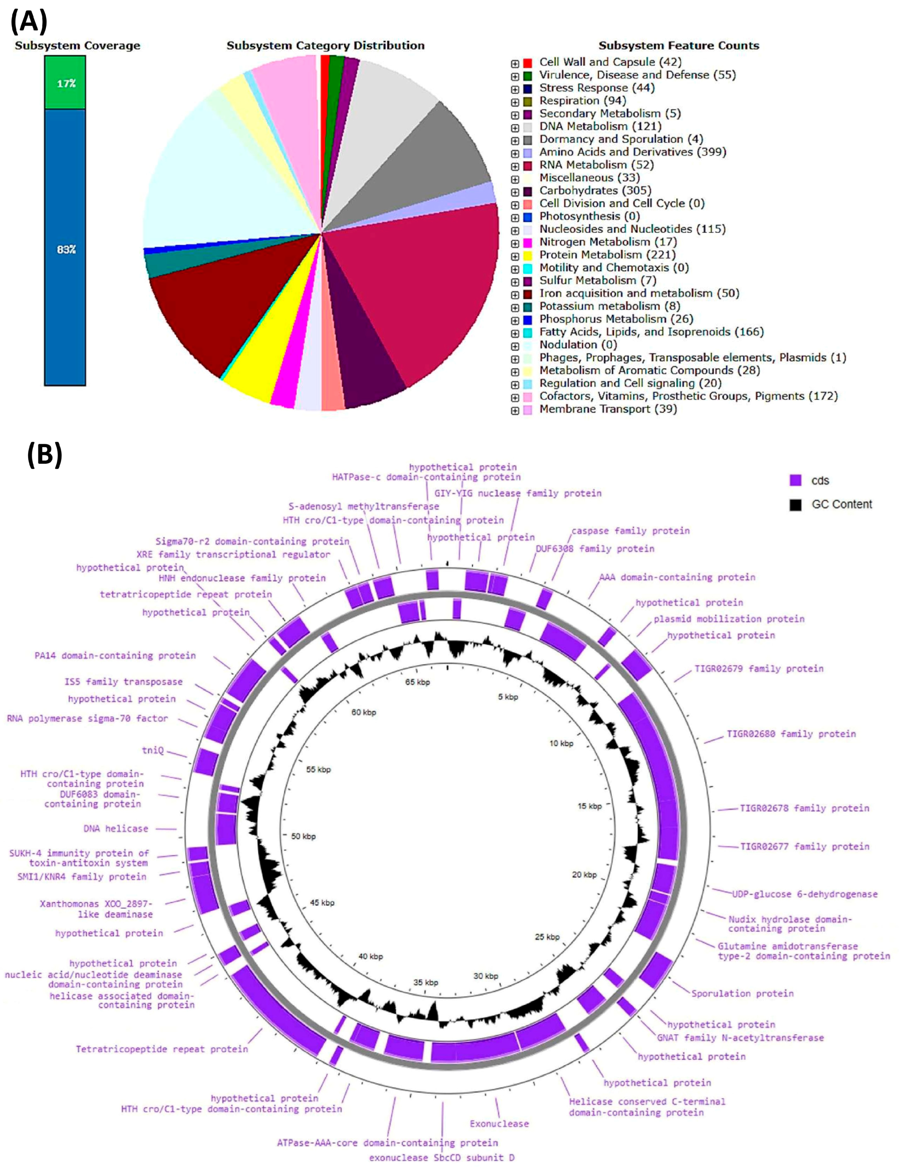
The graphical annotation of the genome of S. anulatus 89 reveals that 17% of its content was identified as encoding functional subsystems (Figure 2A).
Notably, no genes encoding toxins, superantigens, adhesion factors, or determinants of virulence or pathogenicity were detected, allowing the strain to be classified within Risk Group 1. A broad genomic adaptative potential was identified, associated with osmotic, periplasmic, oxidative, and nitrosative stresses as well as resistance to carbon starvation. The genome also encoded siderophore BGCs, enabling the strain to effectively compete with other soil microorganisms for limited iron resources. Among cofactors, vitamins, prosthetic groups, and pigments, genes for the biosynthesis of thiamin (10); pyridoxine (13); biotin (29); folate and pteridines (61); riboflavin, FMN, and FAD (14); and tetrapyrroles (10) were identified. This repertoire indicates the strain’s ability to sustain essential enzymatic reactions required for central metabolism, respiration, and secondary-metabolite production. Plasmid annotation identified 53 protein-coding genes; their positions and predicted functions are summarized in Figure 2B. Such genetic capacity not only likely contributes to resilience under nutrient-limited soil conditions but also provides a competitive advantage by supporting robust growth, sporulation, and antibiotic biosynthesis, highlighting the versatile metabolic potential of S. anulatus 89 and its ecological role as a self-sufficient and adaptive soil microorganism.
To better understand the biosynthetic potential of S. anulatus 89, BGCs responsible for secondary-metabolite production were analyzed in the assembled genome (Table 1). The analysis identified 36 putative clusters associated with the biosynthesis of secondary metabolites. BGCs with high homology included those responsible for the biosynthesis of ectoine, AmfS, melanin, naringenin, coelichein, geosmin, alkylresorcinol, 2-methylisoborneol, desferrioxamin B, and isorenieratene. Among the encoded BGCs with a high level of homology, particularly notable were the SGR PTMs (polycyclic tetramate macrolactams, T1PKS–NRPS products combining polyketide and peptide modules that form a macrolactam ring with a tetramate moiety) and the macrolide antibiotics Warkmycin CS1 and CS2.
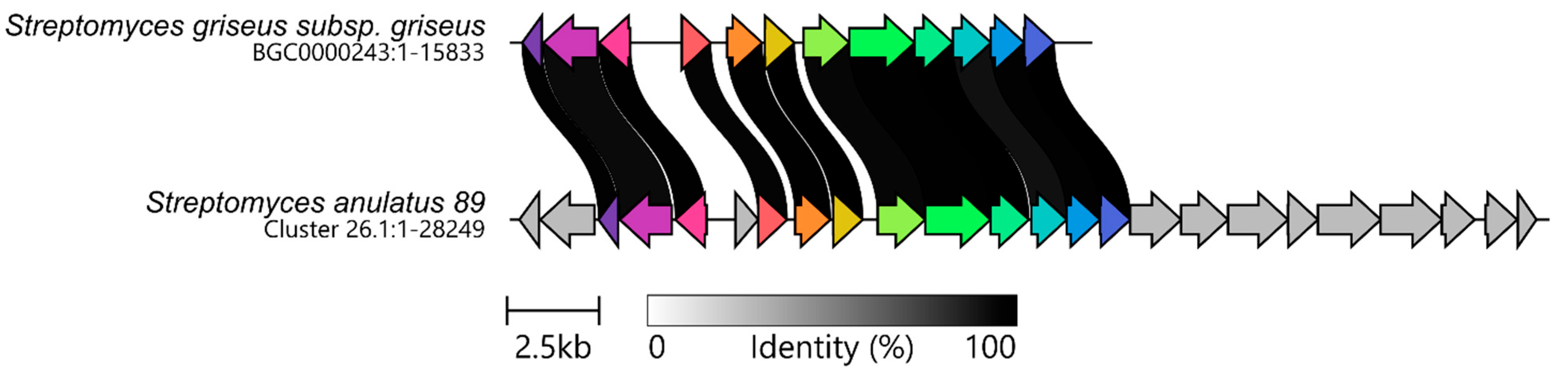
To identify regions lacking classical core biosynthetic genes, the antiSMASH “loose” cluster search strategy was deliberately employed. Manual inspection of the output confirmed that the additional 22 identified regions were not fragmented assembly artifacts at contig boundaries but rather structurally non-canonical BGCs, including several associated with primary metabolism. A notable example of such an unconventional BGC is cluster 26.1. Despite lacking a canonical core gene profile, it exhibited high sequence similarity with the nonactin BGC of S. griseus subsp. griseus (Figure 3), which is responsible for the production of a macrotetrolide compound with well-documented antibacterial and antitumor activities [29]. Comparative analysis revealed that the two clusters are largely conserved. However, in S. anulatus 89, an additional response-regulator-type transcription factor was identified within the cluster, suggesting a potential strain-specific role in the regulation of nonactin biosynthesis.

3.2. Biosynthesis of Antibiotics by S. anulatus 89

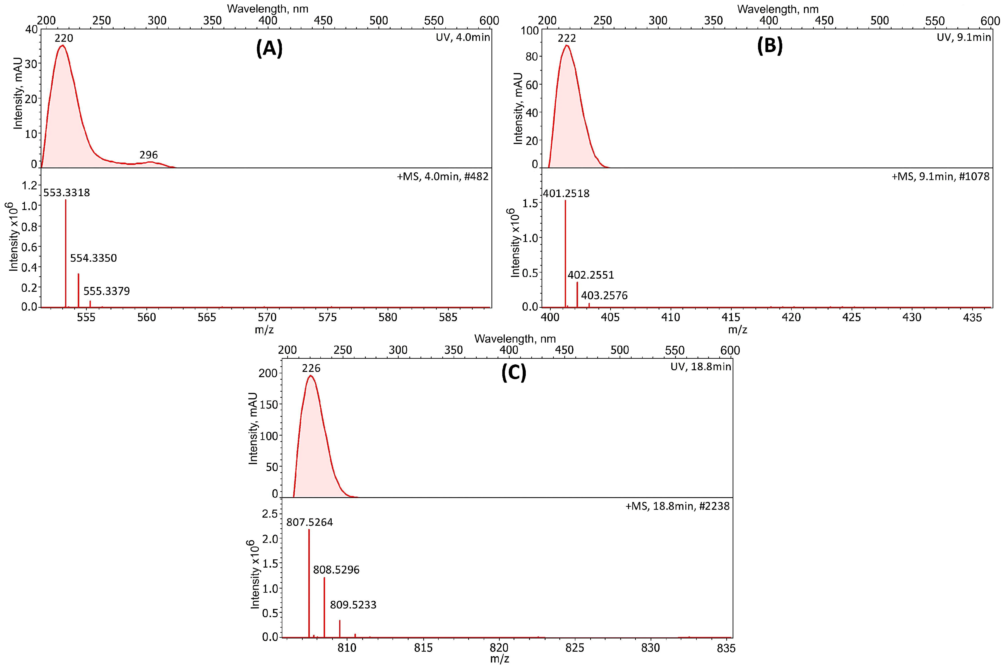
Based on LC-MS analysis of the crude ethanolic biomass extract of S. anulatus 89 and the comparison of m/z values, UV spectra, and retention times of compounds with reference compound databases, together with calculation of corresponding ppm values, the antibiotics synthesized by the strain under cultivation conditions were initially identified (Table 2).
S. anulatus 89 synthesized pladienolide, phenazinomycin, and 21-hydroxyoligomycin (Figure 4). All three metabolites are rare natural products of Streptomyces spp. with potent anticancer and cytotoxic activity that interfere with essential cellular processes, making them part of the broader class of antitumor antibiotics.
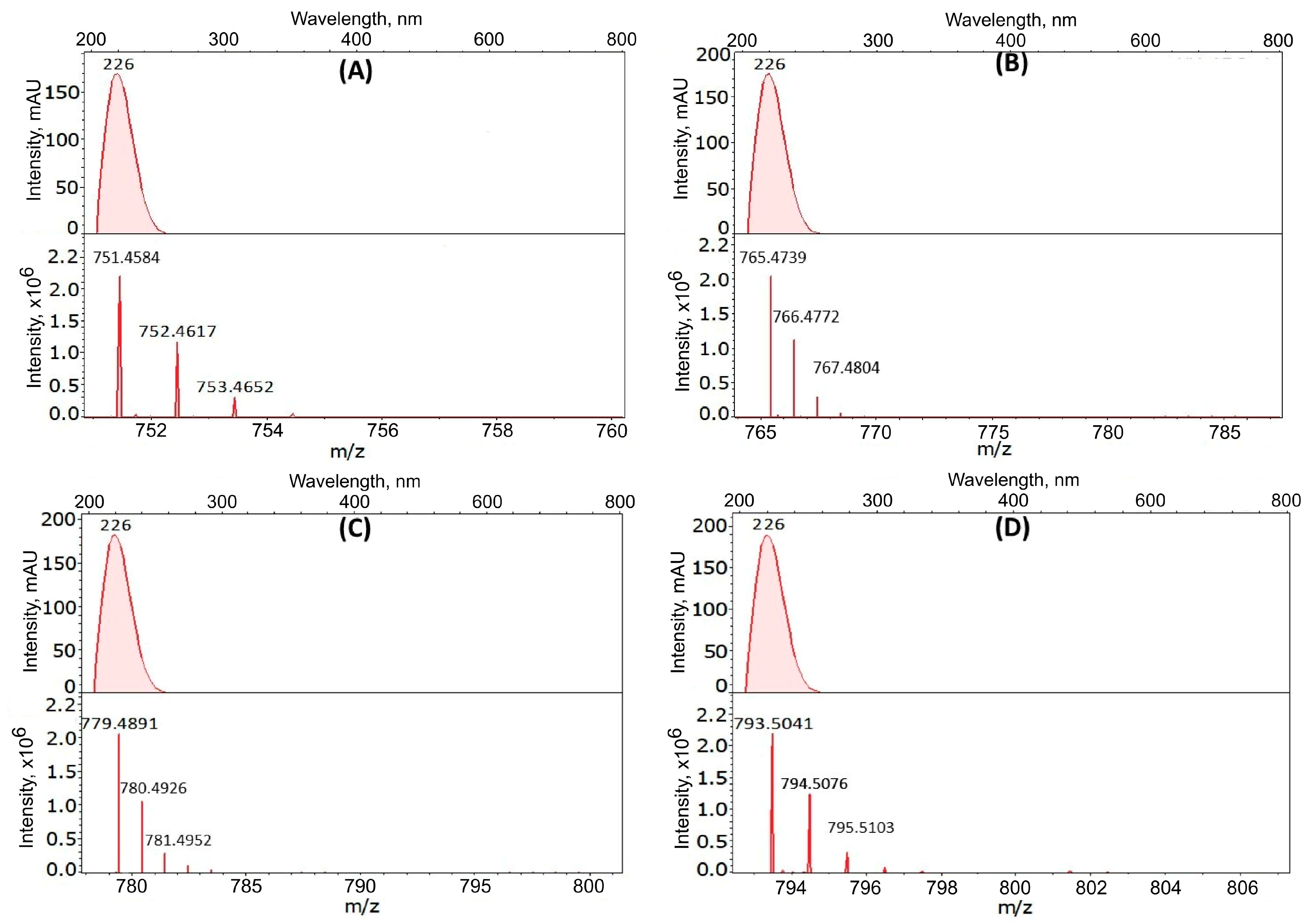
Pladienolides are well-characterized macrolide antibiotics, originally discovered in S. platensis, exhibiting antitumor properties but differing in the extent of their bioactivity [24]. 21-Hydroxyoligomycin is a derivative of oligomycin A and was first described in 2007 as being produced by S. cyaneogriseus ssp. noncyanogenus, strain LL-F28249 [30]. Members of the oligomycin family exhibit diverse biological activities, including pronounced anticancer and immunosuppressive effects [29]. Based on the LC-MS analysis of the crude ethanolic biomass extract of S. anulatus 89, four macrotetrolide ionophore antibiotics (nactins), namely, monactin, dinactin, trinactin, and tetranactin, were identified (Figure 5). These compounds are well recognized for their ability to disrupt mitochondrial function through ion transport and are associated with notable antitumor properties [31].

3.3. Antitumor Properties of the Ethanolic Biomass Extract of S. anulatus 89

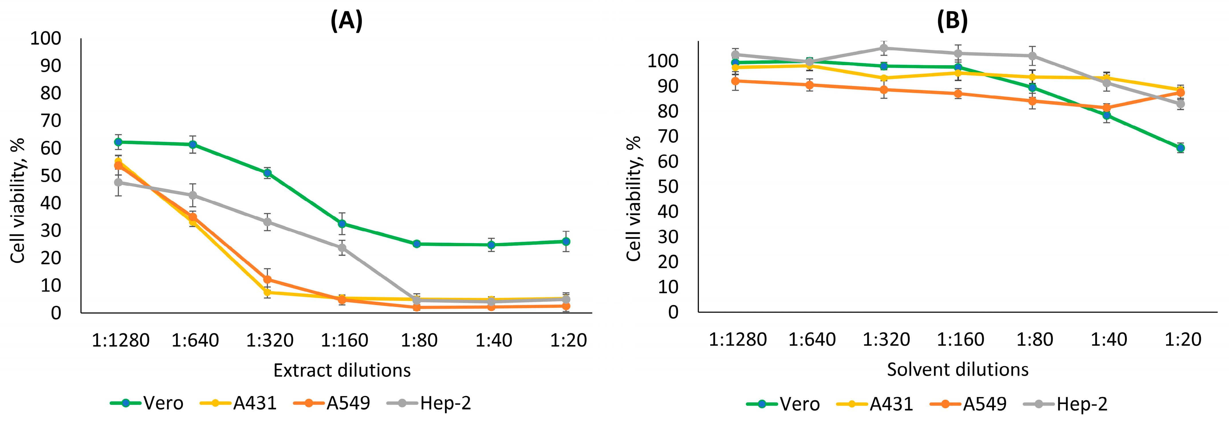
The cytotoxic effect of the crude ethanolic biomass extract of S. anulatus 89 was examined on three cancer cell lines (A431, A549, and Hep-2) and one normal cell line (Vero) using the MTT assay. The extract of S. anulatus 89 inhibited the viability and proliferation of all tested cells in a dilution-dependent manner. The extract exhibited a significantly lower effect on noncancerous Vero cells, as at dilutions of 1:320–1:1280, cell viability remained 51–63% (Figure 6). At the same dilutions, the viability of cancer cells was reduced to 12–54%. The extract proved to be most toxic against the cancer cell lines A431 and A549. It is important to note that 96% ethanol (used as the extraction solvent) was not toxic to any of the cell lines tested, since at dilutions of 1:20–1:1280, cell viability remained within 66–100%.
Despite the fact that the extract of S. anulatus 89 inhibited the growth of all tested cell cultures, it exhibited a selective cytotoxic effect (3–4-fold higher) on the A431, A549, and Hep-2 cells relative to normal epithelial cells (Table 3). The half-maximal inhibitory concentration (IC50) of the extract for the A431, A549, and Hep-2 cancerous cell lines was 1:1090, 1:1119, and 1:914, respectively, whereas for the noncancerous Vero cell line, it was 1:313. Although the selectivity indices (SIs) were moderately low, dose-dependent cytotoxicity assays demonstrated that all three malignant cell lines (Hep-2, A549, and A431) exhibited a dramatic reduction in cell viability at dilutions ranging from 1:160 to 1:20, whereas Vero cells maintained substantially higher viability despite an eight-fold increase in exposure to metabolites present in the crude ethanolic extract of S. anulatus 89. These findings indicate that malignant cells were substantially more sensitive to the antibiotics present in the crude extract than Vero cells. Although the Vero cells were exposed to concentrations exceeding their IC50 by approximately 2- to 15-fold, they retained higher viability than the cancer cell lines, indicating moderate in vitro selectivity of the extract towards malignant cells.
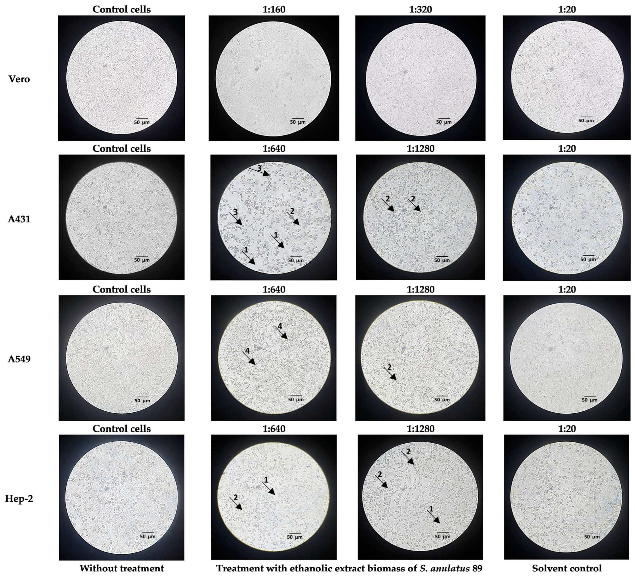
Cell morphology was analyzed using an inverted microscope. In the absence of the ethanolic biomass extract of S. anulatus 89, after 48 h of growth, both cancer and normal cells exhibited typical polygonal or spindle shapes and a clear and slightly flattened morphology and formed tight monolayers, often with visible nuclei and distinct cell membranes (Figure 7). In contrast, significant changes in the morphology of all tumor cell lines (Hep-2, A549, and A431) were observed even when the extract was applied at the highest dilutions (1:640 and 1:1280). Cancer cells lost their typical morphology and displayed altered shapes, including rounding, shrinkage, increased granularity, cell elongation, loss of intercellular contacts, enlargement, vacuolization of the cytoplasm, and disruption of the cell monolayer (Figure 7). Smaller changes were detected in normal Vero cells at much lower extract dilutions (1:160 and 1:320).
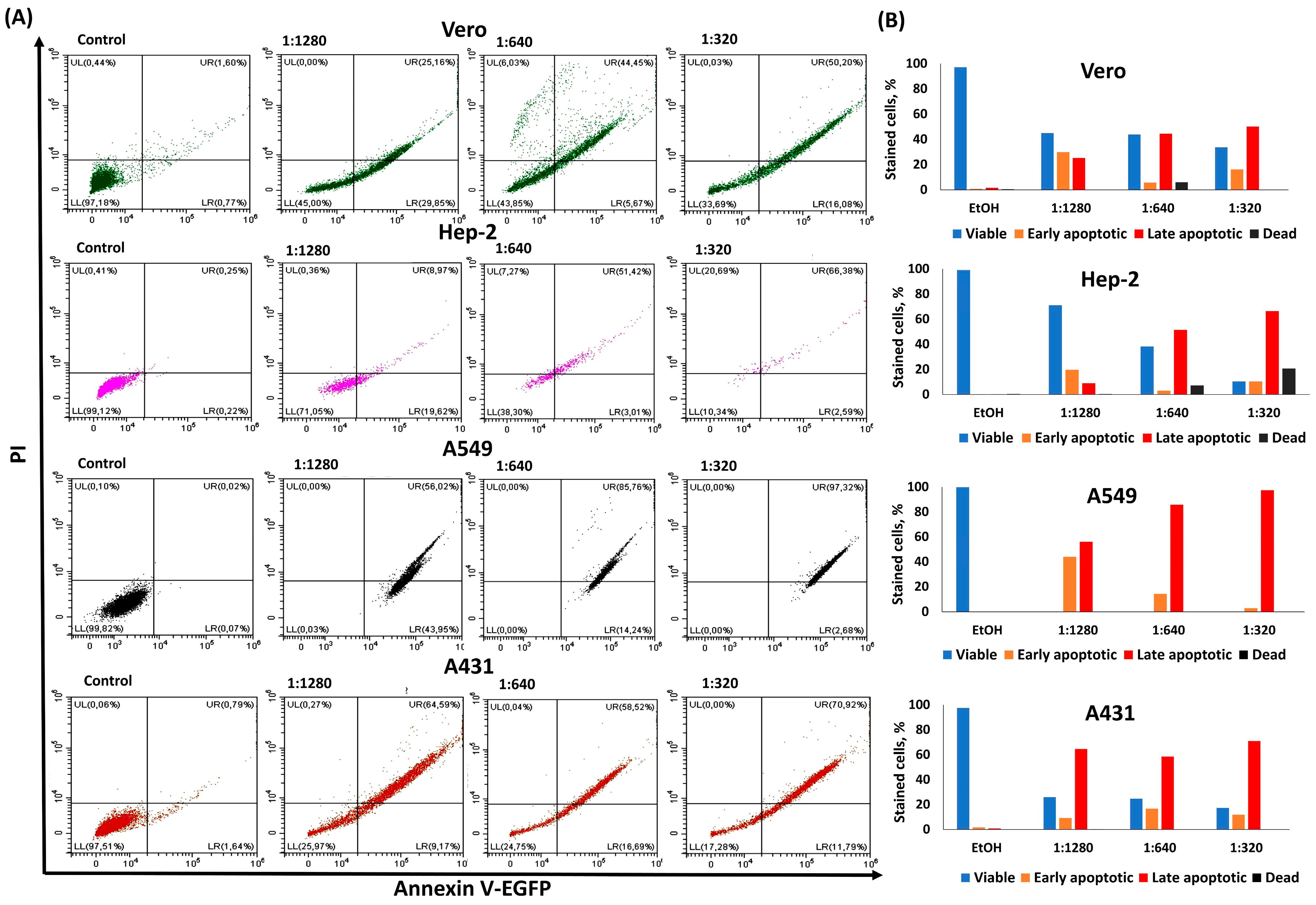
Given the strong performance shown by the anchorage-dependent MTT viability assay, which demonstrated the dose-dependent cytotoxic effect of the S. anulatus 89 biomass extract on respiratory metabolism, we next examined whether flow cytometry could be employed to validate these findings. To calculate the number of events corresponding to each cell subset and apoptotic stage, specific cell populations were gated on a bivariate dot plot, representing intercalation of propidium iodide (PI) into the nuclear DNA of damaged or dead cells versus Annexin V-EGFP binding to phosphatidylserine (PS) exposed on the outer membranes of apoptotic cells (Figure 8).
The double-staining system employed a fluorescent recombinant derivative of Annexin V from E. coli in combination with PI and has previously been validated using doxorubicin hydrochloride as a positive control on the cells employed in this study. In dynamic assessment, IC50 values were determined via the MTT assay for A431 and Vero cells. Moreover, through MTT and Annexin V–eGFP/PI confirmation tests, the drug Doxo-HCl was evaluated on a few different molecular subtypes of breast cancer cell lines—MCF-7 (luminal A, ER-positive, PR-positive, HER-2-negative, and Ki-67low) and MDA-MB-231 (basal-like and triple-negative) [32,33].
After 24 h of incubation, significant increases were observed in the proportions of total AnnV-EGFP(+)/PI(−) and AnnV-EGFP(+)/PI(+) cells (p < 0.05), whereas the proportion of double-negative viable cells decreased significantly (p < 0.05). The ethanolic biomass extract of S. anulatus 89 induced apoptosis in all cell lines tested, with comparatively milder effects observed in non-malignant Vero cells. Apoptosis was induced in the extract-treated tumor cells. The A549 and A431 cell lines were the most sensitive to secondary metabolites in the extract, starting from the highest dilution (1:1280), whereas pure ethanol did not affect any of the cells even at a 1:640 dilution. Significantly higher percentages of late apoptotic cells were recorded in A549 (lung adenocarcinoma) and A431 (epidermoid carcinoma) after 24 h of treatment with S. anulatus 89 at 1:1280 and more concentrated dilutions compared to the control. At the 1:1280 crude extract dilution, Hep-2 cells showed a higher proportion of viable cells (71%) than Vero cells (45%). In contrast, human laryngeal epidermoid carcinoma cells (Hep-2) exhibited a 1.85-fold reduction in viability at 1:640 (38.3%), while Vero cell viability remained similar (43.85%). At the 1:320 dilution, dramatic increases in late apoptotic cells were detected in both cell lines, accompanied by irreversible morphological and metabolic changes. At this dilution, only Hep-2 cells were stained exclusively with PI (dead cells). The analysis confirmed higher survival of Vero cells and a more favorable ratio of early to late apoptotic cells compared to all three malignant solid cell lines.

4. Discussion

The genome of S. anulatus 89 possesses broad adaptive potential supported by encoded mechanisms of resistance to diverse stress factors, including osmotic, oxidative, and periplasmic stress. For instance, the osmotically inducible protein OsmY contributes to the stabilization of proteins and membranes, thereby enhancing survival under saline conditions [31]. It also encoded osmoprotectants such as ectoine and the glycerol uptake facilitator protein, together with osmolyte transporters like the propanediol diffusion facilitator, which potentially regulate intracellular pressure. These features underpin the resilience of S. anulatus 89 to the coastal marine zone from which it was isolated.
In this study, the ability of S. anulatus 89 to synthesize several classes of antibiotics under submerged cultivation is reported, including pladienolide, an ability rarely described for Streptomyces species. The strain also produced nactins (monactin, dinactin, trinactin, and tetranactin), 21-hydroxyoligomycin, and pladienolide, as well as phenazinomycin. Although Streptomyces species are well known to synthesize diverse classes of antibiotics during cultivation [1], our results indicate, for the first time, that S. anulatus strain 89 is capable of simultaneously producing the listed bioactive metabolites when grown in GYM medium. This observation allows us to add this strain to the list of strains reported to produce anticancer compounds and suggests that further investigation of strain 89 may be valuable for pharmacological studies or for exploring aspects of respiratory metabolism. In addition to their antitumor potential, these compounds are known for their cytotoxicity, which likely contributed to the significant reduction in the viability of tumor cell lines at high dilutions under experimental conditions. Furthermore, the BGC associated with nactin biosynthesis was detected in the genome. Previously, the presence of the BGC for dynactin was reported in the genome of the Streptomyces sp. YINM00001 strain, which is closely related to S. anulatus NRRL B-2000T [34]. As previously reported, these compounds act as ionophores, transporting alkali metal cations across membranes and thereby compromising membrane integrity, which underlies their cytotoxicity. Their antitumor activity is linked to disruption of cellular energy metabolism through uncoupling of oxidative phosphorylation and induction of ATP hydrolysis. Monactin is particularly noteworthy because of its inhibitory effects on the P170 glycoprotein-mediated efflux of chemotherapeutic agents in multiple-drug-resistant cancer cells [35]. 21-Hydroxyoligomycin is also toxic to eukaryotic cells, and its bioactivity is associated with inhibition of mitochondrial ATP synthase, specifically the FoF1 complex, which is responsible for ATP production during oxidative phosphorylation [36]. It is known for its activity mainly against rapidly proliferating tumor cells that rely heavily on oxidative phosphorylation, including certain leukemia, lymphoma, and solid tumor cell lines (e.g., hepatocellular carcinoma, breast cancer, and colon carcinoma). However, its potential use as a therapeutic agent is limited due to the significant impact of mitochondrial ATP synthase inhibition on non-tumor cells [30]. Pladienolides were first reported in 2004 from S. platensis Mer-11107. Six of the seven compounds suppressed hypoxia-induced VEGF promoter activity (IC50 0.0018–2.89 μM) and inhibited growth of U251 glioma cells in vitro. They act as potent blockers of hypoxia signaling and tumor cell proliferation, underscoring their potential as antitumor agents [37]. The ability to synthesize pladienolide has also been reported for S. hygroscopicus A-9561, whereas such information has not previously been reported for S. anulatus [38,39]. Pladienolides inhibit the spliceosome (SF3b subunit), leading to defective mRNA splicing and apoptosis in cancer cells while simultaneously exhibiting cytotoxicity. In contrast, known derivatives such as E7107 have undergone certain clinical trials [40]. Phenazinomycin was previously reported only in Streptomyces sp. WK-2057 and S. iakyrus DSM 41873 [41]. This is one of the antitumor antibiotics, and it is a terpene with three consecutive isoprene units [42]. While each of these compounds has been reported in the literature to possess cytotoxic or specific antitumor properties, their combined presence may result in synergistic effects that differ from those described for individual antibiotics.
Given the demonstrated ability of S. anulatus 89 to synthesize a range of bioactive metabolites with known antitumor activity, the strain appears to be a promising candidate for further study as a producer of antitumor compounds. In vitro assays showed that the crude ethanolic biomass extract markedly suppressed the viability of cancer cell lines. Morphological alterations were observed in tumor cells, which lost their typical morphology and displayed rounding, shrinkage, increased granularity, elongation, loss of intercellular contacts, cytoplasmic vacuolization, and disruption of the cell monolayer. Minor changes were detected in normal Vero cells only at much higher extract concentrations. These findings indicate that the extract inhibits cell growth and induces apoptosis-mediated cancer cell death at specific dilutions, although its selectivity towards malignant cells is moderate rather than absolute. The A549 and A431 cell lines have been identified as the most sensitive. Therefore, S. anulatus 89 represents a promising source of antitumor antibiotics that can be simultaneously produced under the cultivation conditions applied in this study. The cytotoxic effects observed in this study may be associated with the identified antibiotics, although they are not necessarily restricted to these compounds.

5. Implications for Applications

The presence of several secondary metabolites with antitumor properties in the crude biomass extract highlights the need for rigorous downstream purification, analytical validation, and extract standardization for future applications. It will also be important to determine the bioactivity of individual compounds. Since fraction collection and purification were not performed in the present study, future work will require preparative HPLC fractionation. In addition, NMR analysis will be necessary to elucidate chemical structure of pladienolide.
Application of these antibiotics as antitumor agents is limited due to their cytotoxicity toward normal cells; however, these compounds represent a valuable field of research, particularly for the synthesis of derivatives with reduced cytotoxicity or for the investigation of respiratory metabolism in eukaryotic cells. Future studies should also include human non-malignant cell lines (e.g., HEK-293) to validate the selectivity profiles of the bioactive compounds identified in the extract.

6. Conclusions

S. anulatus 89 possesses broad, genomically determined adaptive potential, particularly for adaptation to saline environments similar to the habitat from which it was isolated. The strain synthesized a range of antibiotics, and its ethanolic biomass extract significantly reduced the number of viable tumor cells. The primary LC-MS data were considered reliable within the accuracy of instrument calibration (mass error: 0.7–3.9 ppm), providing consistent metabolite assignments at the putative identification level based on database matching. Additional confidence was supported by the manual identification of the nonactin BGC, whose atypical organization explains why automated pipelines such as antiSMASH fail to classify it as a canonical PKS/NRPS cluster. The cytotoxic properties of the crude extract were not highly selective; however, the extract exhibited greater cytotoxicity toward cancer cells than toward normal cells. Treatment of tumor cell lines with the extract resulted in the induction of apoptosis followed by cell death. The strain represents a promising candidate for further investigation as a producer of antitumor compounds, given its ability to simultaneously synthesize multiple classes of bioactive metabolites.

Author Contributions

Conceptualization, M.L.; methodology, A.S. (Andrii Sylchuk), M.L., I.R., A.S. (Andrii Siromolot), L.A. and O.P.; software, M.L., A.S. (Andrii Siromolot) and I.R.; validation, M.L., G.I., A.S. (Andrii Siromolot), S.Z. and R.M.; formal analysis, S.Z., O.P. and R.M.; investigation, M.L., A.S. (Andrii Siromolot), L.A. and G.I.; resources, R.M.; data curation, M.L., G.I. and R.M.; writing—original draft preparation, A.S. (Andrii Sylchuk), M.L., I.R., A.S. (Andrii Siromolot), G.I. and L.A.; writing—review and editing, M.L., I.R., G.I. and R.M.; visualization, M.L., I.R., A.S. (Andrii Siromolot) and L.A.; supervision, M.L.; project administration, R.M.; funding acquisition, M.L. and R.M. All authors have read and agreed to the published version of the manuscript.

Funding

This research was funded by budget funding from the National Academy of Sciences of Ukraine through the Department of General and Soil Microbiology of the D.K. Zabolotny Institute of Microbiology and Virology, National Academy of Sciences of Ukraine, under the project titled “Ecosystem functions of the Soil Microbiome in Restorative Agrophytocenoses”, for the 2025–2029 period (0125U000938), and through the Department of Reproduction of Viruses under the project “Innovative strategies for the use of broad-spectrum natural drugs to combat acute respiratory and persistent viral infections and modulate cellular defence systems”, with grant No. 0125U000643.

Informed Consent Statement

Not applicable.

Data Availability Statement

The original contributions presented in this study are included in the article. Further inquiries can be directed to the corresponding author.

Acknowledgments

The authors are grateful to Liudmyla Biliavska, who made a significant contribution to the development of this research direction at the D.K. Zabolotny Institute of Microbiology and Virology, National Academy of Sciences of Ukraine. The authors acknowledge the Summer School in Natural Products Research 2025 (LLC Explogen, Ukraine, in collaboration with the German–Ukrainian Core of Excellence in Natural Products Research, Germany) for the valuable training in LC-MS analysis and the Ministry of Education and Science of Ukraine for its grant support (BG-21NF). A.S. (Andrii Sylchuk) and M.L. are grateful to the Visegrad Scholarship Program for individual grant Nos. ID 52510474 and ID 52510471, respectively.

Conflicts of Interest

The authors declare no conflicts of interest.

Abbreviations

The following abbreviations are used in this manuscript:
BV-BRCBacterial and Viral Bioinformatics Resource Centre
RASTRapid Annotation using Subsystem Technology
DFAST—DDBJFast Annotation and Submission Tool
ANIAverage Nucleotide Identity
BGCBiosynthetic Gene Cluster
LC-MSLiquid Chromatography–Mass Spectrometry
LC-HRMSLiquid Chromatography–High-Resolution Mass Spectrometry
PIPropidium Iodide
PBSPhosphate-Buffered Saline
IC50Half-Maximal Inhibitory Concentration (50% Inhibition Concentration)

References

  1. Alam, K.; Mazumder, A.; Sikdar, S.; Zhao, Y.M.; Hao, J.; Song, C.; Wang, Y.; Sarkar, R.; Islam, S.; Zhang, Y.; et al. Streptomyces: The biofactory of secondary metabolites. Front. Microbiol. 2022, 13, 968053. [Google Scholar] [CrossRef] [PubMed]
  2. Nicault, M.; Zaiter, A.; Dumarcay, S.; Chaimbault, P.; Gelhaye, E.; Leblond, P.; Bontemps, C. Elicitation of antimicrobial active compounds by Streptomyces-fungus co-cultures. Microorganisms 2021, 9, 178. [Google Scholar] [CrossRef] [PubMed]
  3. Schütze, E.; Kothe, E. Heavy metal-resistant streptomycetes in soil. In Bio-Geo Interactions in Metal-Contaminated Soils; Kothe, E., Varma, A., Eds.; Springer: Berlin/Heidelberg, Germany, 2012; pp. 163–182. [Google Scholar] [CrossRef]
  4. Conrado, R.; Gomes, T.C.; Roque, G.S.; De Souza, A.O. Overview of bioactive fungal secondary metabolites: Cytotoxic and antimicrobial compounds. Antibiotics 2022, 11, 1604. [Google Scholar] [CrossRef] [PubMed]
  5. Sayed, A.M.; Hassan, M.H.A.; Alhadrami, H.A.; Hassan, H.M.; Goodfellow, M.; Rateb, M.E. Extreme environments: Microbiology leading to specialized metabolites. J. Appl. Microbiol. 2020, 128, 630–657. [Google Scholar] [CrossRef] [PubMed]
  6. Potapenko, K.; Lisiutin, G.; Vasylieva, N.; Strashnova, I.; Franke, R.; Petriv, N.; Duduyemi, O.P.; Baklan, K.; Korotaieva, N.; Gudzenko, T.; et al. Antimicrobial and anticancer activity of Streptomyces ambofaciens (Myt 8) and S. globisporus ONU 1019 (Myt 11) secondary metabolites isolated from the Odesa Bay, the Black Sea: An in vitro study. Biomed. Pharmacother. 2025, 186, 117981. [Google Scholar] [CrossRef] [PubMed]
  7. Nan, Y.; Su, H.; Zhou, B.; Liu, S. The function of natural compounds in important anticancer mechanisms. Front. Oncol. 2023, 12, 1049888. [Google Scholar] [CrossRef] [PubMed]
  8. Sznarkowska, A.; Kostecka, A.; Meller, K.; Bielawski, K.P. Inhibition of cancer antioxidant defense by natural compounds. Oncotarget 2016, 8, 15996. [Google Scholar] [CrossRef] [PubMed]
  9. Norte, D.M.; Avitia-Dominguez, L.A.; Rozen, D.E. Evolution and ecology of Streptomyces. Annu. Rev. Microbiol. 2025, 79, 383–403. [Google Scholar] [CrossRef] [PubMed]
  10. Fick, S.E.; Hijmans, R.J. WorldClim 2: New 1-km spatial resolution climate surfaces for global land areas. Int. J. Climatol. 2017, 37, 4302–4315. [Google Scholar] [CrossRef]
  11. Koblitz, J.; Halama, P.; Spring, S.; Thiel, V.; Baschien, C.; Hahnke, R.L.; Pester, M.; Overmann, J.; Reimer, L.C. MediaDive: The expert-curated cultivation media database. Nucl. Acids Res. 2023, 51, D1531–D1538. [Google Scholar] [CrossRef] [PubMed]
  12. Zerbino, D.R. Using the Velvet de novo assembler for short-read sequencing technologies. Curr. Protoc. Bioinform. 2010, 31, 11.5.1–11.5.12. [Google Scholar] [CrossRef] [PubMed]
  13. Carattoli, A.; Zankari, E.; García-Fernández, A.; Voldby, L.M.; Lund, O.; Villa, L.; Møller Aarestrup, F.; Hasman, H. In silico detection and typing of plasmids using PlasmidFinder and plasmid multilocus sequence typing. Antimicrob. Agents Chemother. 2014, 58, 3895–3903. [Google Scholar] [CrossRef] [PubMed]
  14. Parks, D.H.; Imelfort, M.; Skennerton, C.T.; Hugenholtz, P.; Tyson, G.W. CheckM: Assessing the quality of microbial genomes recovered from isolates, single cells, and metagenomes. Genome Res. 2015, 25, 1043–1055. [Google Scholar] [CrossRef] [PubMed]
  15. Shukla, M.; Wattam, A.R.; Aleman, A.; Bhattacharya, R.; Bowers, N.; Brettin, T.; Capria, A.; Chia, N.; Cucinell, C.; Davis, J.J.; et al. BV-BRC: A Unified Bacterial and Viral Bioinformatics Resource with expanded functionality and AI integration. Nucleic Acids Res. 2026, 54, D715–D723. [Google Scholar] [CrossRef] [PubMed]
  16. Katoh, K. MAFFT: A novel method for rapid multiple sequence alignment based on fast fourier transform. Nucleic Acids Res. 2002, 30, 3059–3066. [Google Scholar] [CrossRef] [PubMed]
  17. Stamatakis, A. RAxML Version 8: A tool for phylogenetic analysis and post-analysis of large phylogenies. Bioinformatics 2014, 30, 1312–1313. [Google Scholar] [CrossRef] [PubMed]
  18. Letunic, I.; Bork, P. Interactive tree of life (iTOL) v6: Recent updates to the phylogenetic tree display and annotation tool. Nucleic Acids Res. 2024, 52, W78–W82. [Google Scholar] [CrossRef] [PubMed]
  19. Elmanzalawi, M.; Fujisawa, T.; Mori, H.; Nakamura, Y.; Tanizawa, Y. DFAST_QC: Quality assessment and taxonomic identification tool for prokaryotic genomes. BMC Bioinform. 2025, 26, 3. [Google Scholar] [CrossRef] [PubMed]
  20. Brettin, T.; Davis, J.J.; Disz, T.; Edwards, R.A.; Gerdes, S.; Olsen, G.J.; Olson, R.; Overbeek, R.; Parrello, B.; Pusch, G.D.; et al. RASTtk: A modular and extensible implementation of the RAST algorithm for building custom annotation pipelines and annotating batches of genomes. Sci. Rep. 2015, 5, 8365. [Google Scholar] [CrossRef] [PubMed]
  21. Sun, J.; Lu, F.; Luo, Y.; Bie, L.; Xu, L.; Wang, Y. OrthoVenn3: An integrated platform for exploring and visualizing orthologous data across genomes. Nucleic Acids Res. 2023, 51, W397–W403. [Google Scholar] [CrossRef] [PubMed]
  22. Blin, K.; Shaw, S.; Vader, L.; Szenei, J.; Reitz, Z.L.; Augustijn, H.E.; Cediel-Becerra, J.D.D.; de Crécy-Lagard, V.; Koetsier, R.A.; Williams, S.E.; et al. antiSMASH 8.0: Extended gene cluster detection capabilities and analyses of chemistry, enzymology, and regulation. Nucleic Acids Res. 2025, 53, W32–W38. [Google Scholar] [CrossRef] [PubMed]
  23. Gilchrist, C.L.M.; Chooi, Y.-H. Clinker & Clustermap.Js: Automatic generation of gene cluster comparison figures. Bioinformatics 2021, 37, 2473–2475. [Google Scholar] [CrossRef] [PubMed]
  24. Loboda, M.; Iutynska, G.; Biliavska, L.; Tsygankova, V.; Sylchuk, A.; Blyuss, K.; Shysha, E.; Spivak, S.; Yemets, A.; Mariychuk, R.; et al. Metabolic bioproducts of soil streptomycetes and their effect on RNAi-mediated resistance of tomato plants against root-knot nematode Meloidogyne incognita. TOASJ 2026, 20, e18743315443780. [Google Scholar] [CrossRef]
  25. Abdel-Razik, M.A.; Azmy, A.F.; Dishisha, T.; El-Gendy, A.O.; Afzan, A.; Kamal, N.; Tawfike, A.; Sebak, M. Screening of Red Sea- and Mediterranean Sea-derived Actinomycetes for antimicrobial and antitumor activities: LC-ESI-HRMS-based metabolomics study. Microb. Cell Factories 2025, 24, 136. [Google Scholar] [CrossRef] [PubMed]
  26. Nowak, A.; Steglińska, A.; Gutarowska, B.; Kręgiel, D. Cyto- and genotoxicity of selected plant extracts and microbial metabolites with confirmed activity against phytopathogens of potato seed (Solanum puberosum L.). Molecules 2025, 30, 701. [Google Scholar] [CrossRef] [PubMed]
  27. Siromolot, A.; Zhukova, D.; Romaniuk, S.; Kolybo, D.; Komisarenko, S. Multiple strategies for HB-EGF inhibition: Interference with receptor binding, ectodomain shedding and matricrine signaling. Biochimie 2026, 244, 136–146. [Google Scholar] [CrossRef] [PubMed]
  28. Zhukova, D.; Katashynska, D.; Siromolot, A.; Romaniuk, S.; Kolybo, D.; Komisarenko, S. Nontoxic diphtheria toxin derivates CRM197 and B-fragment can serve as the means for targeted curcumin delivery into sensitive cancer cells. J. Drug Deliv. Sci. Technol. 2024, 96, 105673. [Google Scholar] [CrossRef]
  29. Kim, S.; Chen, J.; Cheng, T.; Gindulyte, A.; He, J.; He, S.; Li, Q.; Shoemaker, B.A.; Thiessen, P.A.; Yu, B.; et al. PubChem 2025 Update. Nucleic Acids Res. 2025, 53, D1516–D1525. [Google Scholar] [CrossRef] [PubMed]
  30. Wagenaar, M.M.; Williamson, R.T.; Ho, D.M.; Carter, G.T. Structure and absolute stereochemistry of 21-hydroxyoligomycin A. J. Nat. Prod. 2007, 70, 367–371. [Google Scholar] [CrossRef] [PubMed]
  31. Poynton, E.F.; van Santen, J.A.; Pin, M.; Contreras, M.M.; McMann, E.; Parra, J.; Showalter, B.; Zaroubi, L.; Duncan, K.R.; Linington, R.G. The Natural Products Atlas 3.0: Extending the database of microbially derived natural products. Nucleic Acids Res. 2025, 6, D691–D699, gkae1093. [Google Scholar] [CrossRef] [PubMed]
  32. Siromolot, A.A.; Loboda, M.I.; Romaniuk, S.I.; Labyntsev, A.J.; Klymkovych, I.-M.M.; Molozhava, O.S.; Kolybo, D.V. Curcumin boosts doxorubicin cytotoxicity in breast cancer cells. Ukr. Biochem. J. 2026, 98, 101–111. [Google Scholar] [CrossRef]
  33. Gamaliia, I.I.; Siromolot, A.A.; Kolybo, D.V. HB-EGF as a carrier for targeted delivery of drugs in certain types of cancer. Biotechnol. Acta 2025, 18, 39–44. [Google Scholar] [CrossRef]
  34. Liu, T.; Ren, Z.; Chunyu, W.-X.; Li, G.-D.; Chen, X.; Zhang, Z.-T.-L.; Sun, H.-B.; Wang, M.; Xie, T.-P.; Wang, M. Exploration of diverse secondary metabolites from Streptomyces sp. YINM00001, using genome mining and one strain many compounds approach. Front. Microbiol. 2022, 13, 831174. [Google Scholar] [CrossRef] [PubMed]
  35. Rawangkan, A.; Wongsirisin, P.; Pook-In, G.; Siriphap, A.; Yosboonruang, A.; Kiddee, A.; Chuerduangphui, J.; Reukngam, N.; Duangjai, A.; Saokaew, S.; et al. Dinactin: A new antitumor antibiotic with cell cycle progression and cancer stemness inhibiting activities in lung cancer. Antibiotics 2022, 11, 1845. [Google Scholar] [CrossRef] [PubMed]
  36. Ratajczak, K.; Lukasiak, A.; Grel, H.; Dworakowska, B.; Jakiela, S.; Stobiecka, M. Monitoring of dynamic atp level changes by oligomycin-modulated atp synthase inhibition in SW480 cancer cells using fluorescent “on-off” switching DNA aptamer. Anal. Bioanal. Chem. 2019, 411, 6899–6911. [Google Scholar] [CrossRef] [PubMed]
  37. Sakai, T.; Sameshima, T.; Matsufuji, M.; Kawamura, N.; Dobashi, K.; Mizui, Y. Pladienolides, new substances from culture of Streptomyces platensis Mer-11107 I. taxonomy, fermentation, isolation and screening. J. Antibiot. 2004, 57, 173–179. [Google Scholar] [CrossRef] [PubMed]
  38. Machida, K.; Aritoku, Y.; Tsuchida, T. One-pot fermentation of pladienolide D by Streptomyces platensis expressing a heterologous cytochrome P450 Gene. J. Biosci. Bioeng. 2009, 107, 596–598. [Google Scholar] [CrossRef] [PubMed]
  39. Smith, E.R.; Al-Smadi, D.H.; Dai, Y.; Khin, M.; Burdette, J.E.; Duggan, B.M.; La Clair, J.J.; Eustáquio, A.S.; Burkart, M.D. Engineering regiospecific methylation of the pladienolides. RSC Chem. Biol. 2025, 6, 1126–1134. [Google Scholar] [CrossRef] [PubMed]
  40. Dehm, S.M. Test-firing ammunition for spliceosome inhibition in cancer. Clin. Cancer Res. 2013, 19, 6064–6066. [Google Scholar] [CrossRef] [PubMed]
  41. Omura, S.; Eda, S.; Funayama, S.; Komiyama, K.; Takahashi, Y.; Woodruff, H.B. Studies on a novel antitumor antibiotic, phenazinomycin: Taxonomy, fermentation, isolation, and physico-chemical and biological characteristics. J. Antibiot. 1989, 42, 1037–1042. [Google Scholar] [CrossRef] [PubMed]
  42. Qin, Z.; Wang, X.; Rateb, M.E.; Ass’ad, L.A.; Jaspars, M.; Deng, Z.; Yu, Y.; Deng, H. Disruption of a methyltransferase gene in actinomycin g gene cluster in Streptomyces iakyrus increases the production of phenazinomycin. FEMS Microbiol. Lett. 2014, 352, 62–68. [Google Scholar] [CrossRef] [PubMed]
Figure 1. Maximum-likelihood phylogenetic tree of S. anulatus 89 based on 500 single-copy orthologous genes. The tree was constructed using the BV-BRC Phylogenetic Tree Building pipeline. Numbers at the nodes indicate bootstrap support values calculated using RAxML Fast Bootstrapping (100 bootstrap replicates). The genome of Micromonospora chalcea DSM 43026 (GCA_002926165.1) was used as an outgroup.
Applsci 16 06743 g001
Figure 2. Graphical annotation of the genome of S. anulatus 89 (A) and its plasmid (B).
Applsci 16 06743 g002
Figure 3. Clinker-generated gene cluster alignments of the nonactin biosynthetic gene cluster. Connecting lines indicate homologous genes, with color intensity reflecting sequence similarity.
Applsci 16 06743 g003
Figure 4. LC/MS profiles of antibiotics detected in the ethanolic biomass extract of S. anulatus 89, showing the following [M + H]+ ions: pladienolide, at m/z 553.3318, UV 220 nm (A); phenazinomycin, at m/z 401.2518, UV 222 nm (B); and 21-hydroxyoligomycin, at m/z 807.5264, UV 226 nm (C).
Applsci 16 06743 g004
Figure 5. LC/MS profiles of nactins, showing the [M + H]+ ion detected in the crude ethanolic biomass extract of S. anulatus 89 when grown in GYM medium (UV 226 nm): monactin, m/z 751.4584 (A); dynactin, m/z 765.4739 (B); trinactin, m/z 779.4891 (C); and tetranactin, 793.5041 (D).
Applsci 16 06743 g005
Figure 6. Viability of cancer cell lines A431, A549, and Hep-2, as well as the normal Vero cells, after 48 h of treatment with dilutions of the crude ethanolic biomass extract of S. anulatus 89 (A) and the solvent control (B), as determined via the MTT assay. Values represent the means ± SDs of three independent experiments. A statistically significant difference in the growth inhibition effect was observed at p < 0.05.
Applsci 16 06743 g006
Figure 7. Morphological changes in the cell lines after treatment with the ethanolic biomass extract of S. anulatus 89. Inverted microscope images of cells exposed to varying dilutions of the extract for 48 h (70× magnification): 1—disruption of the cell monolayer (cell detachment), 2—cell rounding, 3—vacuolization of the cytoplasm, and 4—cell shrinkage.
Applsci 16 06743 g007
Figure 8. Flow cytometry distribution. (A): Dot plots and graphical presentation (B) of apoptosis stages in Vero, Hep-2, A549, and A431 cells after 24 h of treatment with the S. anulatus 89 extract. LL: viable cells; LR: early apoptotic cells; UR: late apoptotic cells; UL: dead cells; PI: propidium iodide.
Applsci 16 06743 g008
Table 1. Biosynthetic gene clusters (BGCs) of secondary metabolites identified in the genome of S. anulatus 89 and predicted by antiSMASH 8.0.
RegionType 1Similarity ConfidenceVerified Streptomyces Producers
1.1ectoineHighectoine
2.1terpene precursor--
2.2lanthipeptide-class-iiiHighAmfS
2.3melaninHighmelanin
2.4azole-containing-RiPP--
2.5T1PKS, NRPS-like--
3.1T3PKSHighnaringenin
3.2NRP-metallophore, NRPSHighcoelichelin
3.3TerpeneHighgeosmin
3.4butyrolactone--
3.5terpene precursor, NRPSLowmarformycin A–F
5.1T3PKSHigh* phenol
5.2terpene precursor, melaninHighmelanin
5.3RiPP-likeMediumstreptamidine
5.4T1PKSLowlidamycin
5.5RiPP-like--
5.6NRPS, T1PKSHighSGR PTM Compound b–d
7.1terpeneMediumhopene
9.1NRPS--
10.1RiPP-likeLow14-hydroxyisochainin
10.2terpeneHigh2-methylisoborneol
11.1T2PKS, oligosaccharide, NRPSHighwarkmycin CS1/warkmycin CS2
11.2hydrogen cyanideLowaborycin
11.3NI-siderophoreLowkinamycin
14.1NI-siderophoreHighdesferrioxamin B
14.2lanthipeptide-class-ii + ііі--
16.1Terpene--
17.1MelaninMediumgrixazone A
22.1NRPS, T1PKS--
22.2terpene precursor--
22.3LinaridinLowsteffimycin D
25.1Terpene--
27.1NRPSLowskyllamycin D/skyllamycin E
33.1arylpolyene, terpene, NRPSHighisorenieratene
36.1terpene, hglE-KSHighectoine
1 Types: PKS—polyketide synthase; T1/T2/T3 PKS—type I/II or III polyketide synthase; NRPS—non-ribosomal peptide synthase; NRP—non-ribosomal peptide; RiPP—ribosomally produced and post-translationally modified peptide-like; hglE-KS—heterocyst glycolipid synthase-like PKS. * 2-2-methoxy-5-methyl-6-(13-methyltetradecyl)-1,4-benzoquinone/2-methoxy-5-methyl-6-(13-methyltetradecyl) phenol.
Table 2. Antibiotics identified in the ethanolic biomass extract of S. anulatus 89.
AntibioticRetention Time, minExact Mass, Dam/z Experimental, [M + H]+Mass Error, ppmKnown Producer [25]
Pladienolide4.0552.3298553.33183.6S. platensis Mer-11107
Phenazinomycin9.1400.2515401.25180.7Streptomyces sp. WK-2057
Monactin17.0750.4554751.45833.9Streptomyces sp., S. globisporus, S. araujoniae
Dinactin17.3764.4711765.47373.4Streptomyces sp., Actinomadura sp. SF2487
Trinactin17.8778.4867779.48913.0S. globisporus, S. araujoniae, S. griseus
Tetranactin18.3792.5023793.50412.3S. globisporus, S. araujoniae, S. griseus
21-Hydroxyoligomycin18.8806.5180807.52642.0S. cyaneogriseus ssp. noncyanogenus (LL-F28249)
Table 3. IC50 values (in dilution) calculated for the extract on the studied cells.
Test SampleCell Cultures
VeroA431SI *A549SI *Hep-2SI *
Extract1:3131:109041:111941:9143
Solvent control (96% EtOH)<1:20<1:20-<1:20-<1:20-
* SI—Selectivity index.
Disclaimer/Publisher’s Note: The statements, opinions and data contained in all publications are solely those of the individual author(s) and contributor(s) and not of MDPI and/or the editor(s). MDPI and/or the editor(s) disclaim responsibility for any injury to people or property resulting from any ideas, methods, instructions or products referred to in the content.