Article

Pyrroloquinoline Quinone Targets the Allosteric Activation Site of Nicotinamide Phosphoribosyltransferase (NAMPT): Structural Basis and Consequences for NAD+ Metabolism in Aging


by
Alessandro Medoro
1,
Sergio Davinelli
1,*,
Tassadaq Hussain Jafar
1,
Truong Tan Trung
2,
Ciro Costagliola
3,
Gemma Caterina Maria Rossi
4 and
Giovanni Scapagnini
1
1
Department of Medicine and Health Sciences “V. Tiberio”, University of Molise, 86100 Campobasso, Italy
2
Laboratory of Computation and Nanoscience, Dong Nai Technology University, Bien Hoa City 810000, Vietnam
3
Department of Neurosciences, Reproductive Sciences and Dentistry, University of Naples “Federico II”, 80131 Naples, Italy
4
Department of Surgical Sciences, University Eye Clinic, Fondazione IRCCS Policlinico S. Matteo, 27100 Pavia, Italy
*
Author to whom correspondence should be addressed.
Appl. Sci. 2026, 16(13), 6695; https://doi.org/10.3390/app16136695
Submission received: 4 June 2026 / Revised: 1 July 2026 / Accepted: 3 July 2026 / Published: 4 July 2026
(This article belongs to the Special Issue Biological Activities of Plant Extracts and Their Applications)

Abstract

NAD+ depletion is a defining feature of the aging cell, driven by a progressive decline in nicotinamide phosphoribosyltransferase (NAMPT) activity, the rate-limiting enzyme of the NAD+ salvage pathway. Pyrroloquinoline quinone (PQQ), a plant-derived redox-active quinone cofactor, elevates intracellular NAD+ by a mechanism that remains incompletely understood. We employed an integrated in silico approach combining molecular docking, density functional theory (DFT), and 100 ns molecular dynamics (MD) simulation to evaluate whether PQQ directly targets NAMPT. Docking against the NAMPT crystal structure (PDB: 7ENQ) yielded a binding free energy of −9.4 kcal/mol, with PQQ positioned in the allosteric activation site and forming hydrogen bonds at His191, Asp219, and Val242 together with π–π stacking at Tyr188, extending a known synthetic activator pharmacophore to a dietary ligand class. MM-GBSA analysis yielded binding free energy = −31.2 kcal/mol, confirming dominant electrostatic and van der Waals stabilization. In silico alanine mutagenesis of Tyr188 and Val242 reduced binding affinity to −7.2 and −7.0 kcal/mol respectively, with complete loss of allosteric-site contacts, validating the proposed mechanism computationally. DFT analysis revealed a HOMO–LUMO gap of 3.20 eV and electrophilicity index ω = 8.91 eV, consistent with non-covalent binding to nucleophilic residues. MD simulation confirmed retention of PQQ within the allosteric site over 100 ns. These data provide a structural and electronic framework for the NAD+-boosting activity of PQQ and a rationale for experimental validation.

1. Introduction

Aging is associated with a progressive decline in nicotinamide adenine dinucleotide (NAD+). NAD+ is not merely an electron carrier in oxidative phosphorylation. As an obligate co-substrate for sirtuins (SIRT1–7), poly(ADP-ribose) polymerases (PARPs), and cyclic ADP-ribose synthases (CD38, CD157), it couples bioenergetic status to the regulation of DNA repair, chromatin remodeling, mitochondrial biogenesis, and the inflammatory response [1,2]. The tissue NAD+ pool declines by 40–60% between young adulthood and old age in rodents and humans alike, a decline now recognized as a mechanistic driver of the aging phenotype, not a passive consequence of cellular senescence [3,4].
The mechanisms underlying age-associated NAD+ depletion are both catabolic and biosynthetic. On the consumption side, the accumulation of oxidative DNA lesions with age drives chronic PARP activation, which can reduce cellular NAD+ concentrations [5]. CD38 expression rises with the senescence-associated secretory phenotype (SASP), amplifying NAD+ hydrolysis in aging tissues [6]. On the biosynthetic side, the principal route of NAD+ replenishment in most mammalian tissues is the salvage pathway, which recycles nicotinamide (NAM) through nicotinamide mononucleotide (NMN) to NAD+. The flux through this pathway declines with age in skeletal muscle and adipose tissue, compounding the NAD+ deficit [7,8]. This biosynthetic failure is not confined to peripheral tissues: in retinal ganglion cells, salvage pathway capacity is further compromised by an age- and disease-dependent downregulation observed in glaucoma, the leading cause of irreversible blindness worldwide [9].
The rate-limiting step of the salvage pathway is catalyzed by nicotinamide phosphoribosyltransferase (NAMPT), a homodimeric enzyme whose catalytic sites are formed at the dimer interface, where residues from both monomers contribute to the condensation of NAM and 5-phosphoribosyl-1-pyrophosphate (PRPP) to generate NMN. Structural and biochemical studies have identified a functionally distinct allosteric activation site adjacent to the catalytic pocket, whose occupancy by small-molecule activators enhances NAMPT turnover, raises intracellular NMN and NAD+ concentrations, and confers neuroprotection in preclinical models of neurodegeneration and ischemia. The key residues lining this site, principally Tyr188, His191, Asp219, Val242, Ser275, and Ile309, have been identified by co-crystallography and mutagenesis as essential for activator binding, establishing that these positions mediate allosteric coupling between the activation site and the catalytic site. The suitability of this site for small-molecule activation has motivated the development of structurally diverse synthetic activators, yet no dietary or endogenous compound has been shown to engage it [10,11].
Pyrroloquinoline quinone (PQQ; 4,5-dihydro-4,5-dioxo-1H-pyrrolo[2,3-f]quinoline-2,7,9-tricarboxylic acid) is a water-soluble tricyclic orthoquinone found in plant foods and human breast milk that functions as a redox cofactor, performing continuous redox cycling with a catalytic efficiency estimated at 100–1000-fold greater than that of ascorbate [12]. PQQ is ubiquitous in the plant kingdom, with quantifiable concentrations detected in all plant species analyzed, ranging from 7 to 34 µg/kg in fruits and vegetables to 60–800 µg/kg in fermented plant-derived products such as natto, miso, and green tea [13]. In the plant environment, PQQ is produced by rhizosphere-associated bacteria of the genera Pseudomonas, Methylobacterium, and Rahnella, which secrete it into the soil where it functions as a growth-promoting signal: PQQ enables phosphate solubilization through glucose dehydrogenase-dependent gluconic acid production, stimulates seedling growth, and induces systemic resistance against pathogens, establishing it as a key mediator of plant–microbe interactions in the rhizosphere [14]. In mammalian systems, PQQ promotes mitochondrial biogenesis through peroxisome proliferator-activated receptor gamma coactivator 1-alpha (PGC-1α) upregulation and suppresses nuclear factor kappa B (NF-κB)-driven inflammation [15,16,17]. PQQ also raises intracellular NAD+ levels in HepG2 cell line, in NIH/3T3 fibroblasts, and in the superior colliculus and optic nerve of healthy mice following systemic administration [18,19,20].
This NAD+-elevating effect is particularly relevant to aging tissues. The cognitive and mitochondrial benefits of PQQ supplementation documented in clinical trials occur precisely in the population where NAMPT-dependent NAD+ biosynthesis is most compromised. A double-blind trial in healthy adults (aged 41–65 years) shows that oral PQQ (20 mg/day for 12 weeks) improves composite memory, verbal memory, and cognitive flexibility [21]. In elderly individuals with mild cognitive impairment, PQQ supplementation improved mitochondrial biomarkers and brain metabolic indices after six weeks [22]. PQQ supplementation has also demonstrated clinical benefit in glaucoma, with combined administration of citicoline, homotaurine, and PQQ improving retinal ganglion cell parameters, a finding mechanistically coherent with the age-dependent decline of NAMPT in the glaucomatous retina [9,23]. This raises the question of whether direct NAMPT engagement underlies the efficacy of PQQ in aging [7,8].
Several mechanisms have been proposed to account for the NAD+-elevating effect of PQQ. Inhibition of lactate dehydrogenase (LDH) by PQQ shifts the reduced nicotinamide adenine dinucleotide (NADH)/NAD+ ratio by reducing cytosolic NADH consumption, transiently increasing the oxidized pool [24]. PQQ also scavenges reactive oxygen species and reduces the oxidative DNA damage load that drives chronic PARP activation, thereby limiting one of the principal routes of NAD+ consumption in aged tissues [5]. A third proposed route is upregulation of NAMPT gene expression through PGC-1α-mediated transcriptional programs. In HepG2 cells, NAMPT mRNA increases within 18 h of PQQ exposure and NAD+ activity rises at 24 h, suggesting that biosynthetic capacity can be enhanced on a timescale longer than the acute LDH-dependent effect [20]. None of these mechanisms, however, predict a direct physical interaction between PQQ and NAMPT protein, and no study has examined whether PQQ engages the NAMPT allosteric activation site. The planar tricyclic scaffold of PQQ, with electron-withdrawing carbonyls and carboxylate substituents capable of hydrogen-bond donation and acceptance, is physicochemically compatible with the NAMPT activation site as defined by known activators [10,11].
We investigated this hypothesis using an integrated in silico approach combining molecular docking, density functional theory (DFT) analysis, and 100 ns molecular dynamics (MD) simulation. The study provides the first structural evidence that PQQ targets the NAMPT allosteric activation site and a quantum-chemical rationale for the selectivity of this interaction.

2. Materials and Methods

2.1. Protein Structure Retrieval and Preparation

The crystal structure of human NAMPT was retrieved from the RCSB Protein Data Bank (PDB ID: 7ENQ, resolution 2.6 Å). This structure was selected because it captures the active, activator-bound, open conformation of the homodimer, which differs substantially from the apo and inhibitor-bound forms in the geometry of the allosteric binding site. Use of an activator-bound template is standard practice in comparable docking studies targeting the NAMPT activation site [10,11]. The asymmetric unit was inspected for missing residues, which were modeled by loop refinement. Crystallographic water molecules, co-crystallized ligands, and non-crystallographic chains were removed. The structure was prepared using UCSF Chimera v1.12, as previously described. Hydrogen atoms were added and histidine protonation states were assigned according to the AMBER ff98 force field. The resulting assembly was subjected to energy minimization using the conjugate gradient method for 1000 steps [25,26,27]. Final energy minimization was performed with the Chiron online server to ensure structural stability prior to docking [28].

2.2. Ligand Preparation

The two-dimensional structure of PQQ was obtained from PubChem (CID: 1024015) and redrawn in ChemDraw Ultra v12.0. The structure was converted to three-dimensional coordinates (.pdb format) in Chem3D Pro and subjected to geometry optimization in UCSF Chimera v1.12 prior to docking.

2.3. Molecular Docking

Molecular docking was performed with AutoDock Vina v4.2, as previously described [27,29]. The docking grid was centered on the allosteric activation site of NAMPT, defined by the coordinates of key activator-binding residues. Grid dimensions were 75 × 75 × 75 Å with a spacing of 0.650 Å. Polar hydrogens were added to the receptor. One hundred independent docking runs were performed. The pose with the lowest binding free energy within the most populated cluster was selected for interaction analysis. Default parameters of AutoDock Vina v4.2 for electrostatic forces, Van der Waals forces, AMBER force field, and intermolecular forces for the docked complex were utilized, with the genetic algorithm performed as the main search protocol. Two-dimensional interaction diagrams were generated in Discovery Studio v2022, distinguishing hydrogen bonds, hydrophobic contacts, and π-stacking interactions.

2.4. Docking of the PQQ Tri-Amide Analog

To evaluate the contribution of the carboxylate groups of PQQ to NAMPT binding, a tri-amide analog (PQQ-NH2) was generated by replacing all three carboxylic acid groups with amide functionalities using ChemDraw Ultra v12.0. The three-dimensional geometry of PQQ-NH2 was optimized using Chem3D and the resulting structure was prepared for docking following the same protocol applied to native PQQ (Section 2.1). Molecular docking was performed with AutoDock Vina v4.2 using identical grid box dimensions (75 × 75 × 75 Å, spacing 0.650 Å), docking parameters, and number of independent runs as for native PQQ. Binding free energies and interaction profiles were compared between native PQQ and PQQ-NH2 to quantify the contribution of the carboxylate hydrogen-bond network to allosteric-site engagement.

2.5. In Silico Alanine Mutagenesis

Alanine substitutions at Tyr188 and Val242 were introduced in silico using the Rotamers tool in UCSF Chimera v1.12, applied to the same wild-type NAMPT structure used for docking (PDB: 7ENQ). Each substitution was introduced independently to generate two single-mutant structures (Tyr188Ala and Val242Ala), and a double-mutant structure (Tyr188Ala/Val242Ala) was generated by sequential application of both substitutions. Following each substitution, the mutated side chain and all residues within 5 Å were subjected to local energy minimization using the conjugate gradient method for 1000 steps in UCSF Chimera v1.12, with the remainder of the structure held fixed, to relieve steric clashes introduced by the truncation. The minimized mutant structures were prepared for docking following the same protocol applied to the wild-type receptor (Section 2.1). Molecular docking of PQQ to each mutant structure was performed with AutoDock Vina v4.2 using identical grid box dimensions (75 × 75 × 75 Å, spacing 0.650 Å), docking parameters, and number of independent runs as for the wild-type. Binding free energies and interaction profiles of the mutant complexes were compared with those of the wild-type to evaluate the contribution of Tyr188 and Val242 to ligand recognition at the allosteric activation site.

2.6. Density Functional Theory Calculations

Electronic structure calculations were performed in Gaussian 09W using the B3LYP method. Geometry optimization was carried out at the B3LYP/6-311++G(d,p) level in both gas-phase and aqueous-phase using the integral equation formalism polarizable continuum model. Frontier molecular orbital (FMO) analysis was performed at the B3LYP/def2TZVP level using natural bond orbital (NBO) 5.0 analysis in Gaussian 09W. Global reactivity descriptors were computed, comprising ionization potential (IP = −EHOMO), electron affinity (EA = −ELUMO), chemical hardness (η), chemical potential (μ), and electrophilicity index (ω = μ2/2η). The molecular electrostatic potential (MEP) map was calculated at the same level of theory on the optimized geometry to identify regions of nucleophilic and electrophilic reactivity, and results were visualized in GaussView 6.0.

2.7. Molecular Dynamics Simulation

The PQQ–NAMPT docked complex was prepared for MD simulation using the Protein Preparation Wizard in Maestro v12.5. The simulation system was constructed with the System Builder tool in Desmond v6.0 (Schrödinger LLC, New York, NY, USA). The complex was solvated in an orthorhombic simulation box with a minimum buffer of 10 Å using the transferable intermolecular potential with 3 points (TIP3P) water model. Counter-ions were added to neutralize the system, and the ionic strength was adjusted to 0.15 M NaCl. The optimized potentials for liquid simulations (OPLS) 2005 force field was applied to all atoms. A 100 ns isothermal-isobaric (NPT) ensemble simulation was performed at 300 K and 1 atm. Simulation stability was assessed by root mean square deviation (RMSD) of backbone Cα atoms and of the ligand heavy atoms relative to the initial frame. Per-residue root mean square fluctuation (RMSF) was calculated to identify regions of differential flexibility. Secondary structure element (SSE) composition was monitored throughout the trajectory. Protein–ligand interaction fingerprints, including hydrogen bonds, hydrophobic contacts, ionic interactions, and water bridges, were computed with the Desmond Simulation Interactions Diagram module. Ligand properties were monitored throughout the trajectory, comprising the radius of gyration (rGyr), intramolecular hydrogen bonds, molecular surface area (MolSA), solvent-accessible surface area (SASA), and polar surface area (PSA).

2.8. MM-GBSA Binding Free Energy Calculation

The binding free energy of the PQQ–NAMPT complex was estimated using the MM-GBSA module (Schrödinger LLC, New York, NY, USA) with the VSGB 2.0 implicit solvation model. Prior to calculation, the docked complex was prepared using the Protein Preparation Wizard in Maestro, with steps including assignment of bond orders, addition of hydrogen atoms, optimization of protonation states and hydrogen-bonding networks, correction of structural issues where necessary, and restrained energy minimization using the OPLS-2005 force field. The MM-GBSA calculation was performed on the single, energy-minimized structure corresponding to the optimal docking pose of the PQQ–NAMPT complex. This value represents an enthalpy-dominated potential score lacking the full solute conformational entropy contribution and serves primarily as a relative ranking validation rather than an absolute affinity estimate. The total binding free energy was decomposed into van der Waals, Coulomb, covalent, lipophilic, hydrogen-bond, packing and generalized-Born solvation contributions. Molecular dynamics simulations were conducted independently as a separate validation step to assess the structural stability and interaction persistence of the docked complex.

3. Results and Discussion

3.1. Molecular Docking of PQQ to the NAMPT Allosteric Activation Site

The structural basis for NAMPT activation by small molecules has been elucidated in detail by X-ray crystallography and mutagenesis. Activators bind at a site running along the dimer interface, distinct from but adjacent to the NAM/PRPP catalytic pocket, and their binding stabilizes the active conformation of the enzyme by reinforcing interdimer contacts and reducing the entropic cost of substrate binding [10,11]. These residues were identified by co-crystallography and mutagenesis as the pharmacophoric anchors of established synthetic NAMPT activators and are not in themselves novel; the present study evaluates whether PQQ, a dietary compound never previously linked to direct NAMPT engagement, recapitulates this same pharmacophoric network. Mutation of either residue abolishes the activating response without impairing basal NAMPT activity, demonstrating that they are required specifically for allosteric coupling between the allosteric and catalytic sites [11]. The three hydrogen bonds identified in the PQQ docking pose at His191, Asp219, and Val242, together with the π–π stacking contact at Tyr188, recapitulate the pharmacophoric interactions reported for multiple structurally distinct NAMPT activators. Yao et al. established the structural basis for this pharmacophore through the co-crystal structure of NAMPT with NAT (PDB: 7ENQ), identifying Val242, Asp219, and Tyr188 as principal contacts engaged through water-mediated hydrogen bonds, and demonstrated by mutagenesis that Tyr188Ala and Val242Ala substitutions abolish allosteric activation without affecting basal NAMPT activity; compounds engaging this network achieve micromolar EC50 values in neuronal NAD+ assays [11]. Wang et al. reported that His191 and Val242 are engaged through water-mediated hydrogen bonds in the NAMPT-NAT co-crystal structure, and that optimization of contacts within the hydrophobic cavity of the activation site drives in vivo neuroprotective efficacy [30]. Tang et al. extended this analysis to non-pyridyl scaffolds and demonstrated that NAMPT activators sharing the urea-class binding mode retain activity through water-mediated interactions within the active site, independent of pyridine warhead identity [10]. These authors additionally showed that compounds lacking the canonical pyridine ring but retaining shape complementarity with the activation site maintain measurable NAMPT activation, establishing that scaffold identity is less critical than geometric fit within the binding pocket [10]. PQQ satisfies all these structural requirements. Its planar tricyclic quinone provides the π-stacking platform, its carbonyl oxygens and pyridine nitrogen supply the hydrogen-bond acceptors for His191 and Asp219, and its carboxylate at C14 donates the hydrogen bond to Val242 that is conserved across all activator classes.
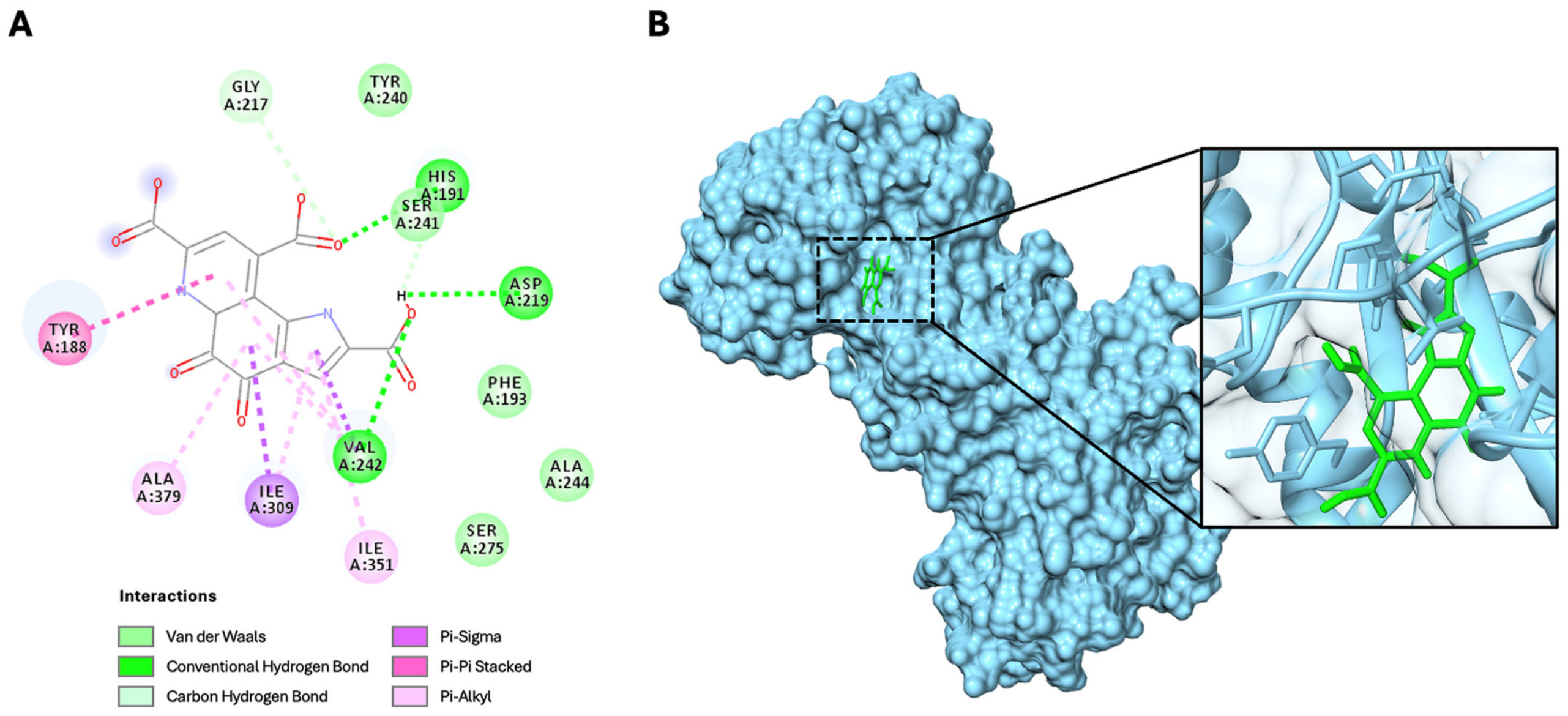
Molecular docking of PQQ to the allosteric activation site of human NAMPT (PDB: 7ENQ) was performed to identify the preferred binding geometry and key interacting residues. PQQ docked to NAMPT with a binding free energy of −9.4 kcal/mol and the optimal pose positions PQQ in the allosteric activation site at the dimer interface. The quinone scaffold is positioned between Phe193 of one monomer and Tyr188 of the opposing chain, forming π–π stacking with Tyr188, the residue essential for activator binding. Three hydrogen bonds stabilize the complex. The quinone O8 carbonyl accepts a proton from the His191 imidazole. The carboxylate at C14 forms a hydrogen bond with the Val242 backbone amide NH. The Asp219 carboxylate side chain donates a hydrogen bond to the quinone N17. These three contacts (His191, Asp219, Val242) are consistent across all lowest-energy docking poses. Hydrophobic and Van der Waals contacts involve Phe193, Tyr240, Ser241, Ala244, Ser275, Ile309, Ile351, and Ala379 (Figure 1).
The docking geometry also places the PQQ carboxylate at C22 in close proximity to Gly217, a residue whose mutation abrogates binding of the reference NAMPT inhibitor FK866, suggesting that this position is sterically constrained and accessible to different chemical classes of ligand. PQQ overlaps with the footprint of both activators and inhibitors at this site. Its planar quinone scaffold lacks the pyridine amide and piperidine linker of known NAMPT inhibitors, making an inhibitory mode of action structurally implausible [10,11,31]. The binding free energy of −9.4 kcal/mol is within the range reported for synthetic NAMPT activators in comparable docking studies and corresponds qualitatively to the low-micromolar to sub-micromolar EC50 values documented for structurally diverse activators in enzymatic and cellular assays [10,11,30,31,32].
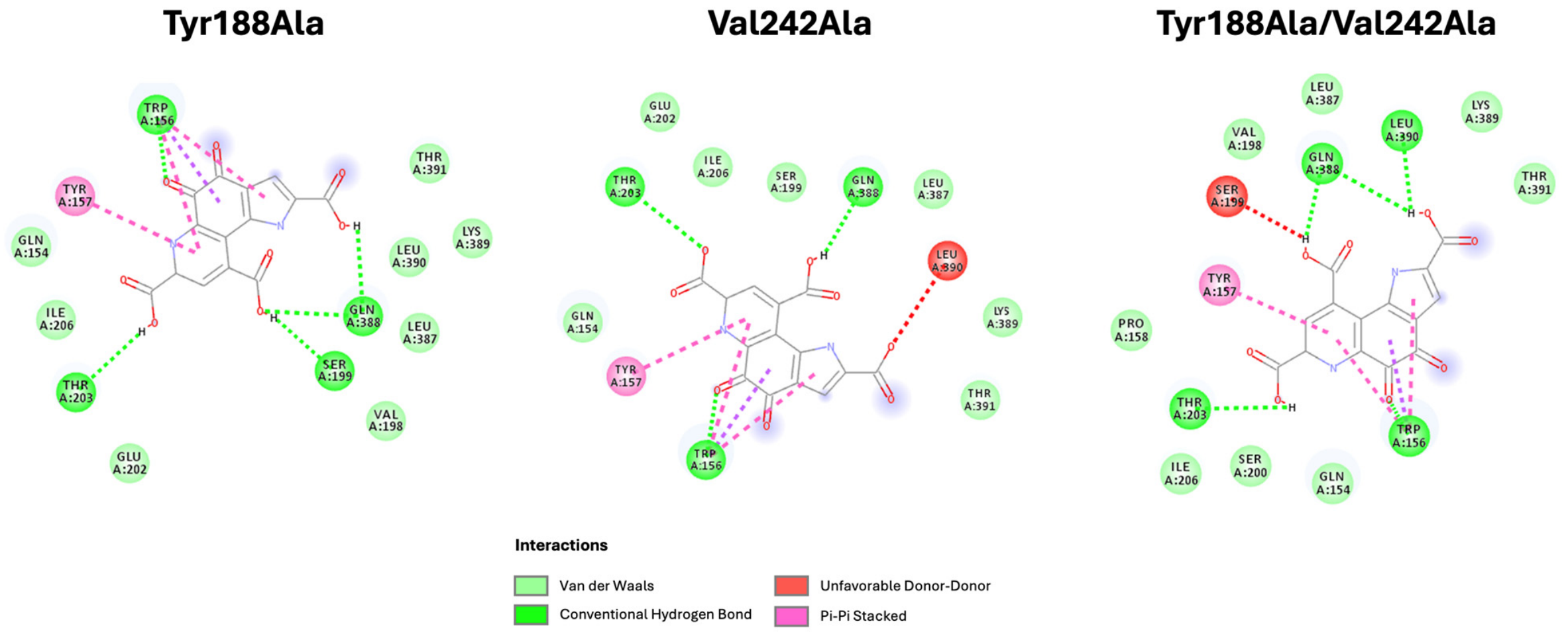
To provide direct computational validation of the role of Tyr188 and Val242 as pharmacophoric anchors, alanine substitutions were generated in silico and subjected to molecular docking under identical conditions. The Tyr188Ala mutant exhibited a binding free energy of −7.2 kcal/mol, and the Val242Ala mutant −7.0 kcal/mol, compared with −9.4 kcal/mol for the wild-type (reductions of 2.2 and 2.4 kcal/mol, respectively). The double mutant Tyr188Ala/Val242Ala showed a further reduction to −6.8 kcal/mol, consistent with an additive contribution of both residues to binding affinity. Critically, both single mutants show complete loss of the pharmacophoric contact network that defines the allosteric activation site: the hydrogen bonds at His191, Asp219, and Val242 and the π–π stacking interaction at Tyr188 are absent in all mutant poses, replaced by contacts with residues distal from the canonical activation site, including Trp156, Thr203, and Gln388. Both mutant poses additionally display unfavorable donor–donor interactions absent in the wild-type complex, Leu390 in Val242Ala and Ser199 in the double mutant, providing a further energetic rationale for the observed loss of affinity. These findings are consistent with the experimental mutagenesis data for synthetic NAMPT activators, in which alanine substitution at Tyr188 and Val242 abolishes the activating response without impairing basal NAMPT activity [11], and confirm that the pharmacophoric specificity of PQQ for the allosteric site depends critically on these two residues (Figure 2).
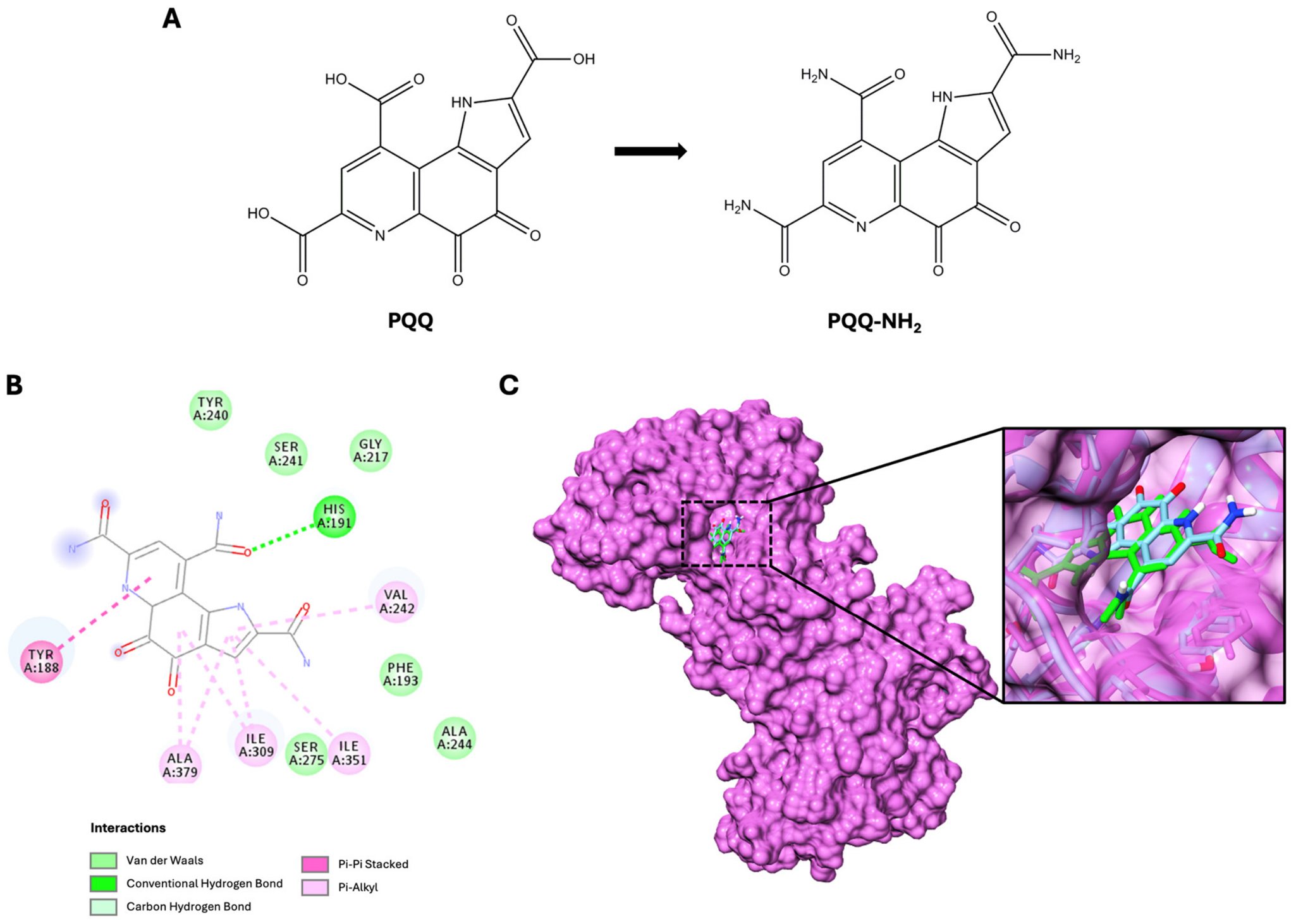
To address the structural basis of PQQ carboxylate contributions to NAMPT binding, the tri-amide analog of PQQ (PQQ-NH2), in which all three carboxylic acid groups are replaced by amide functionalities, was docked to the wild-type NAMPT allosteric site using the same protocol applied to native PQQ. The amide analog exhibited a binding free energy of −8.0 kcal/mol, representing a reduction of 1.4 kcal/mol relative to native PQQ (−9.4 kcal/mol). The reduced affinity is attributable to the selective loss of hydrogen bonds at Asp219 and Val242, which are present in the native PQQ pose but absent in the amide analog docking pose. His191 retains a single hydrogen bond with the amide analog, confirming that this contact is independent of carboxylate ionization. The π–π stacking interaction with Tyr188 is preserved, and the van der Waals contact network (Tyr240, Ser241, Gly217, Phe193, Ala244, Ser275, Ile309, Ile351, Ala379) is largely maintained, consistent with the conserved orientation of the pyrroloquinoline quinone scaffold within the allosteric pocket as confirmed by superimposition of the two docking poses. These data indicate that the carboxylate groups of native PQQ contribute favorably but not exclusively to NAMPT binding: the amide analog retains productive engagement with the allosteric site, while the 1.4 kcal/mol reduction in binding free energy quantifies the net hydrogen-bonding contribution of the three carboxylate groups to allosteric-site affinity (Figure 3).

3.2. Molecular Geometry and Electronic Structure of PQQ

The electronic properties of PQQ provide a quantum-chemical rationale for the binding mode identified by docking and complemented by MD simulation. The high electrophilicity index (ω = 8.91 eV) and the concentration of negative MEP over the quinone carbonyls and pyridine nitrogen predict strong electron-accepting interactions with the nucleophilic imidazole of His191, a contact type consistent with the established reactivity of electrophilic ortho-quinones toward histidine imidazoles [33]. These parameters are theoretical descriptors of reactivity potential and do not imply a confirmed covalent or irreversible interaction. PQQ–NAMPT engagement is modeled throughout this study as a reversible, non-covalent allosteric process, consistent with the moderate kinetic stability indicated by the HOMO–LUMO gap. Geometry optimization at the B3LYP/6-311++G(d,p) level confirmed a planar, fully conjugated tricyclic structure for PQQ in both gas and aqueous phases, with C1 point group symmetry. The quinone C=O bond lengths (C5=O6: 1.207 Å; C7=O8: 1.218 Å in water) and the pyridine-ring C–N bond lengths (1.325–1.376 Å) are consistent with an extensively delocalized π system with partial resonance contribution from the carboxylate groups. The dipole moment increases from 7.999 D in the gas phase to 11.713 D in aqueous phase, indicating significant polarization by the solvent, consistent with favorable electrostatic interactions with charged protein residues. This high dipole moment maintains a favorable electrostatic environment for solvent-mediated charge transfer even without direct atomic contact, a pattern documented in related in silico analyses from our group [27,34].
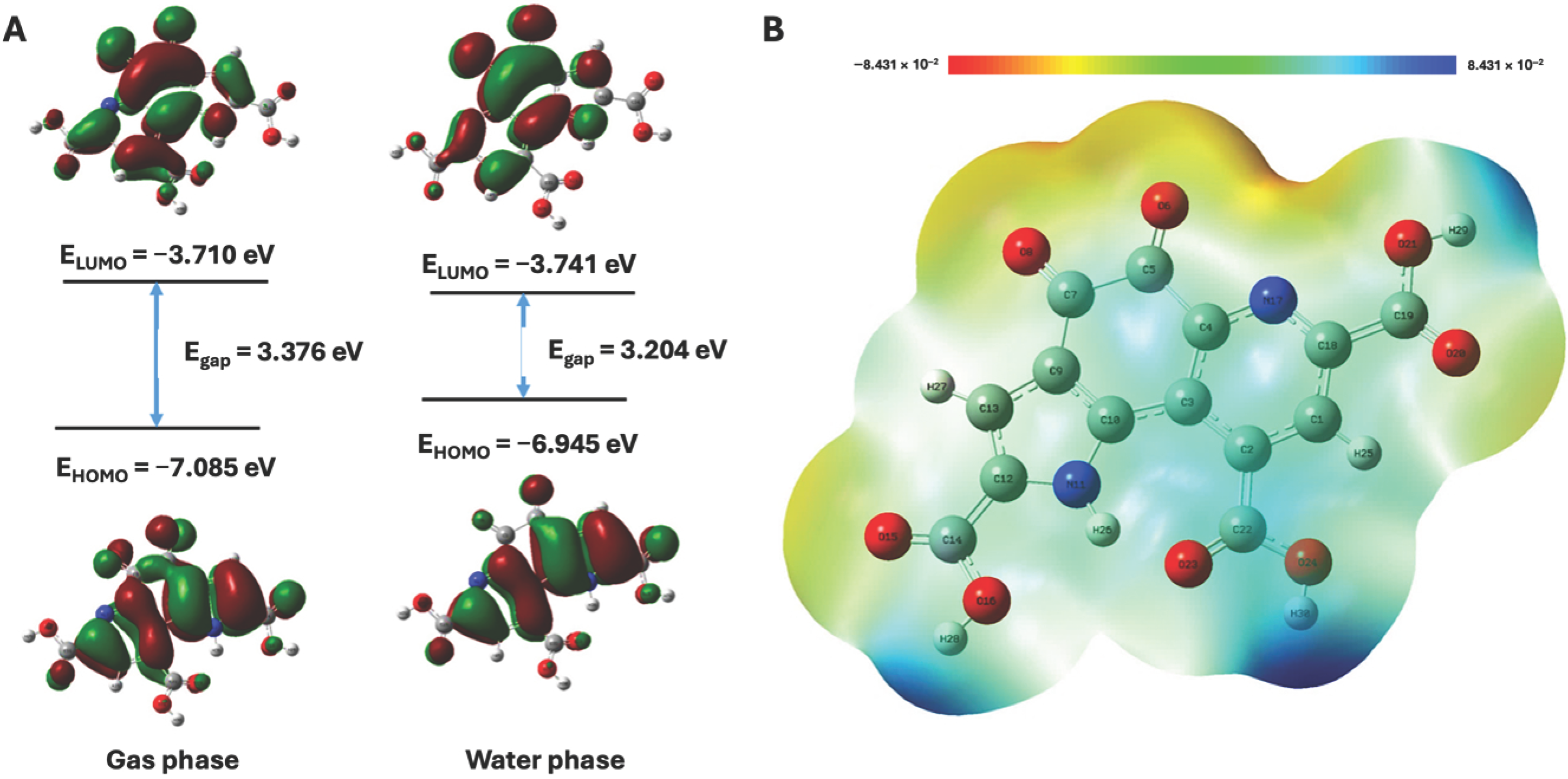
FMO analysis at the B3LYP/def2TZVP level yielded HOMO and LUMO energies of −6.945 eV and −3.741 eV in aqueous phase, respectively, corresponding to an energy gap (Egap) of 3.204 eV (Table 1).
This Egap in water indicates moderate kinetic stability, consistent with reversible, geometry-dependent interactions and not covalent modification, a property that is mechanistically appropriate for an allosteric modulator required to associate and dissociate reversibly without consuming the enzyme. The computed global reactivity descriptors identify PQQ as a potent electrophile. The electrophilicity index ω = 8.91 eV substantially exceeds the 1.5 eV threshold used to classify strong electrophiles, and the maximum charge transfer index Nmax = 3.335 eV indicates a high capacity to accept electron density from nucleophilic molecules. The MEP map localizes the highest negative potential over the quinone carbonyl oxygens O6, O8, O15, and O20 and the pyridine nitrogen N17. These atoms serve as the primary hydrogen-bond acceptors. Positive potential concentrates over the carboxylate hydrogens H28–H30 (Figure 4).
These features collectively distinguish PQQ from non-specific antioxidants and support a mode of NAMPT engagement that is both geometrically and electronically selective.

3.3. Molecular Dynamics Simulation

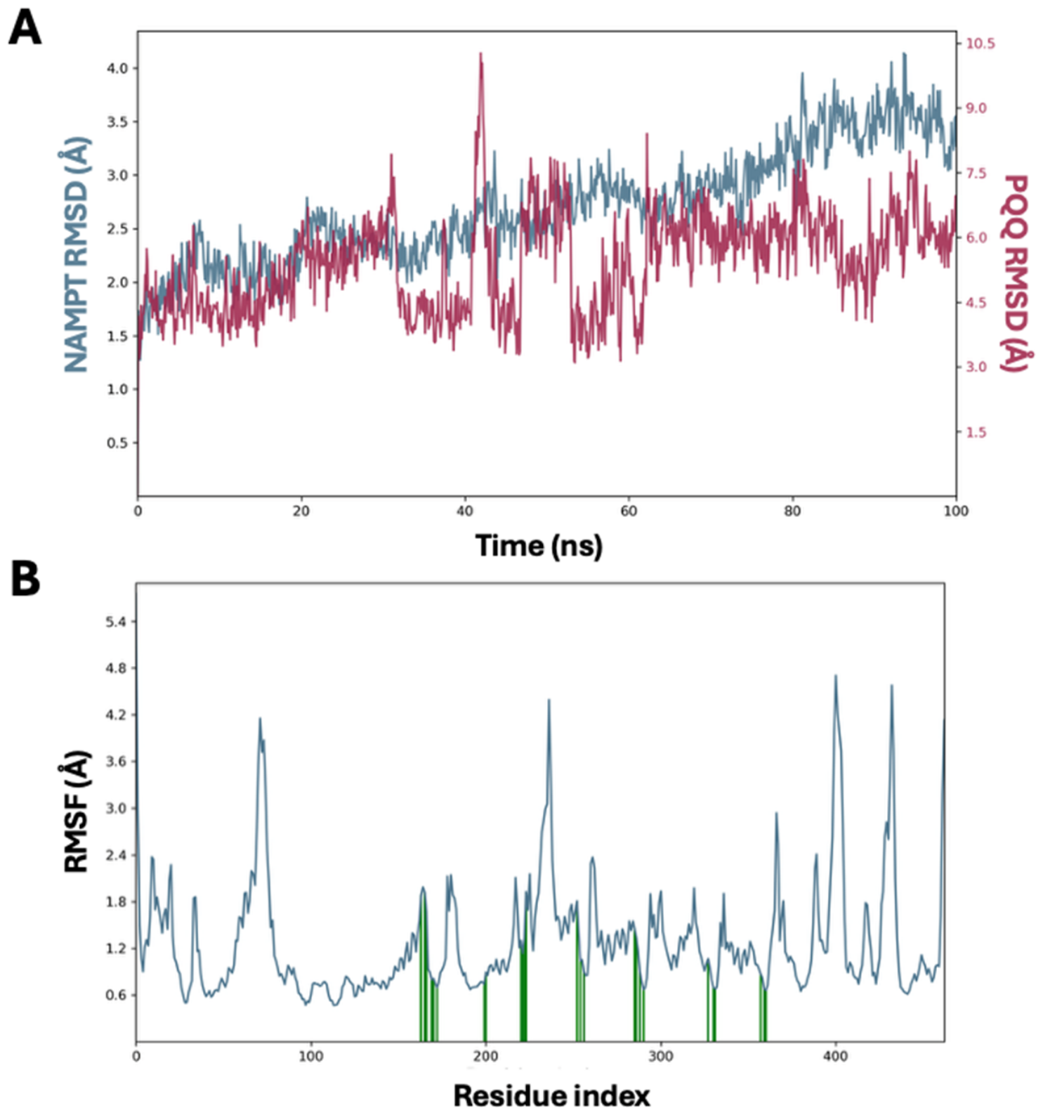
Over the 100 ns simulation, the PQQ–NAMPT complex maintained structural integrity consistent with a stable binding mode. The backbone Cα RMSD of NAMPT rose gradually throughout the simulation, reaching 2.5–3.5 Å by 50 ns and up to 4.1 Å by the end of the trajectory, consistent with the conformational flexibility of the homodimer at equilibrium. The RMSD of PQQ heavy atoms fluctuated between ~4 and ~7 Å throughout the simulation (mean 5.4 Å), with a transient peak of 10.3 Å at approximately 42 ns, consistent with the conformational flexibility inherent to allosteric binding sites and reflecting dynamic sampling of the activation pocket rather than ligand dissociation (Figure 5A).
RMSF analysis identified loop regions and terminal segments as the most flexible elements of the protein, with a peak of ~4.2 Å at residues 60–70. The core of the binding pocket (positions 185–260) exhibited fluctuations below 1.5 Å, consistent with its structural rigidity (Figure 5B).
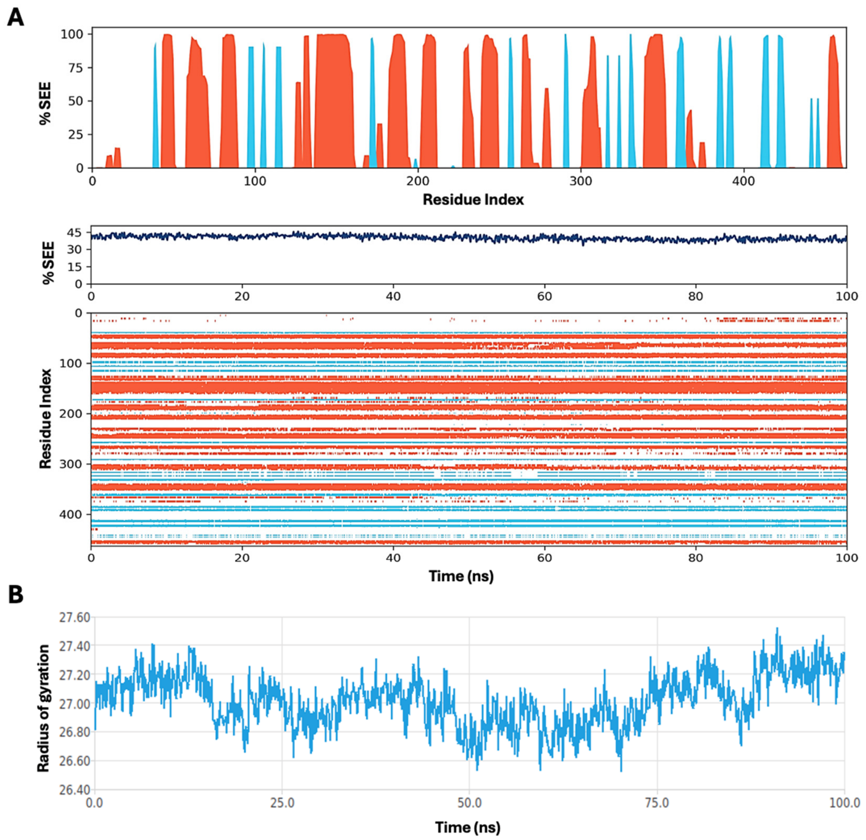
SSE analysis showed that NAMPT maintained a stable secondary structure composition of 29.4% α-helix and 10.8% β-strand throughout the trajectory, with no remodeling attributable to ligand binding (Figure 6A). The radius of gyration of the backbone oscillated within 26.52–27.52 Å with no systematic drift, confirming preservation of the overall homodimer architecture (Figure 6B).
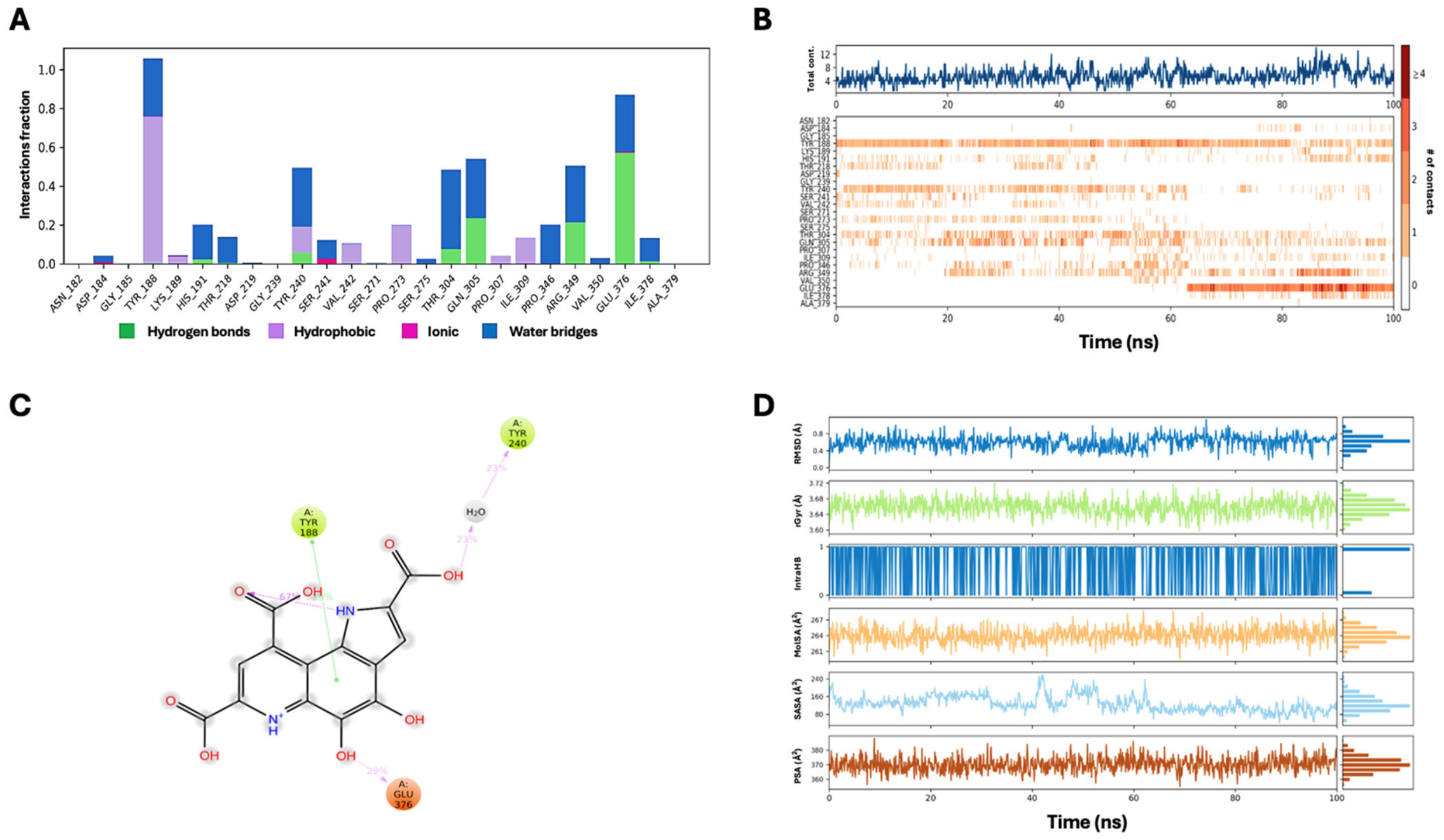
Protein–ligand interaction fingerprint analysis over the full trajectory identified six residues with interaction fractions exceeding 0.20 (Figure 7A,B). Tyr188 displayed the highest total interaction fraction (~1.05), comprising π–π stacking (54.7% of frames), water bridges (29.9%), and hydrophobic contacts (15.6%), consistent with its role as the π-stacking clamp at the entrance of the activation site and confirming the primary docking contact. Glu376 and Gln305 showed interaction fractions of ~0.87 and ~0.55, respectively, attributable largely to water bridges and hydrophobic contacts. Thr304 and Arg349 contributed water-mediated contacts at fractions of ~0.49 and ~0.51, respectively. Tyr240 maintained a mixed hydrophobic/water-mediated interaction at ~0.50. These residues were not all identified as principal contacts in the static docking pose, indicating that dynamic sampling of the site exposes additional stabilizing contacts consistent with an induced-fit binding mode. His191 contributed a water-mediated contact at ~0.20, with direct hydrogen bond occupancy of 2.3% and water-mediated contact of 17.9% of trajectory frames. The predominance of solvent-mediated over direct contact is consistent with the dynamic flexibility of histidine imidazoles at physiological pH and supports a reversible, non-covalent binding rather than an irreversible nucleophilic reaction. Val242 showed a markedly reduced interaction fraction in the MD fingerprint relative to the docking pose, with no direct hydrogen bond detected in any trajectory frame (0% occupancy) and hydrophobic contact present in 10.4% of frames. This finding reflects the sensitivity of this position to local loop dynamics (residues 217–242) and indicates that, in solution, the Val242 contact is highly dynamic and relies predominantly on water-bridge architectures rather than direct hydrogen bonding. Critically, however, Val242 remains a structural anchor of the allosteric site: the Val242Ala substitution reduces binding affinity by 2.4 kcal/mol and abolishes the allosteric-site-specific contact network in silico, consistent with the loss of activating response observed experimentally for synthetic NAMPT activators carrying this mutation [11]. The dissociation between static docking pose and dynamic MD fingerprint at this position therefore reflects solvent-mediated adjustment of a functionally essential contact rather than its absence (Figure 7C) [11,35]. MM-GBSA analysis of the energy-minimized PQQ–NAMPT complex yielded a total binding free energy of −31.2 kcal/mol (van der Waals −38.9, Coulomb −83.6, covalent +2.6, lipophilic −16.2, hydrogen-bond −1.2, packing −0.5, generalized-Born solvation +106.5 kcal/mol), substantially more favorable than the docking score (−9.4 kcal/mol) and consistent with the dominant electrostatic and van der Waals contributions identified in the MD interaction fingerprint. This value, calculated on a single energy-minimized structure, represents an enthalpy-dominated potential score lacking the full solute conformational entropy contribution and should be interpreted as a relative ranking validation rather than an absolute affinity estimate. PQQ ligand properties, including intermittent intramolecular hydrogen bonding, radius of gyration (3.60–3.72 Å), molecular surface area (259.5–268.7 Å2), solvent-accessible surface area, and polar surface area (354.8–388.2 Å2), remained stable throughout the simulation trajectory, confirming that the internal geometry of the ligand was preserved despite its conformational sampling within the allosteric site (Figure 7D).
These computational data provide the first structural evidence that PQQ engages the allosteric activation site of NAMPT with binding geometry and key contact residues that are quantitatively consistent with those of established synthetic NAMPT activators, extending a known structural pharmacophore to a novel dietary ligand class. The convergence of molecular docking, DFT reactivity analysis, and MD simulation on a coherent picture of the PQQ–NAMPT interaction strengthens the hypothesis that direct NAMPT engagement contributes to the NAD+-elevating effect of PQQ observed in cell-based and in vivo systems. The biological implications of these findings must be considered against the existing experimental evidence for PQQ-dependent NAD+ elevation. PQQ upregulates NAMPT mRNA within 18 h and raises NAD+ at 24 h in HepG2 hepatocytes, establishing a transcriptional route to increased biosynthetic capacity [20]. In NIH/3T3 fibroblasts, PQQ raises cellular NAD+ without altering NAMPT protein levels, indicating a post-translational mechanism independent of new enzyme synthesis [18]. These findings are mechanistically compatible, not discordant. Allosteric activation of NAMPT by direct ligand binding would increase enzymatic turnover without requiring any change in protein abundance, a distinction that standard immunoblotting cannot resolve. Direct NAMPT activation would amplify NMN synthesis from the NAM salvage pathway, because higher NAD+ drives SIRT1-mediated deacetylation of PGC-1α, which in turn upregulates NAMPT transcription, creating a feed-forward loop that sustains the NAD+ pool over time [7].
The aging context gives this mechanism particular significance. NAMPT activity declines in adipose tissue and skeletal muscle during aging, contributing to the progressive NAD+ deficit that underlies mitochondrial dysfunction, accumulation of oxidative DNA lesions, and increased SASP [8]. Direct NAMPT engagement at nutritional concentrations provides a biochemical rationale for PQQ supplementation to counteract age-related NAD+ depletion, complementary to NMN or nicotinamide riboside (NR) supplementation strategies that bypass NAMPT by providing NMN directly or via nicotinamide riboside kinase (NRK)-mediated phosphorylation [36]. The glaucomatous retina represents an additional tissue context in which this mechanism may be operationally relevant. NAMPT is downregulated in retinal ganglion cells in an age- and disease-dependent manner, contributing to the progressive NAD+ deficit that underlies RGC metabolic vulnerability, and PQQ supplementation has improved retinal ganglion cell parameters in glaucoma patients, raising the possibility that direct NAMPT engagement contributes to the neuroprotective activity of PQQ in the visual system [9,23,37].
Several limitations of the present study warrant explicit acknowledgment. The present work is explicitly framed as a computational hypothesis-generating study. The mechanistic links to NAD+ metabolism in aging are proposed as a testable hypothesis generated from converging computational evidence, not as an experimentally established mechanism. The predicted interactions require validation by surface plasmon resonance or isothermal titration calorimetry to establish affinity and stoichiometry, and by X-ray crystallography or cryo-EM to confirm the binding geometry. The contact with Gly217, whose mutation abolishes FK866 binding, raises the question of whether PQQ could exert mixed activator/inhibitor effects depending on NAMPT conformational state. This cannot be excluded computationally and requires testing in enzymatic activity assays across a concentration range. The OPLS 2005 force field may not fully capture the electronic polarizability of the PQQ system. The elevated ligand RMSD and the absence of full trajectory convergence within 100 ns indicate that longer simulations or independent replicas are required to formally characterize the bound-state ensemble; these are identified as priorities for future work. Replication with a polarizable force field or QM/MM approach would additionally strengthen the simulation conclusions. Plasma PQQ concentrations following oral supplementation at 20 mg/day are in the low nanomolar range, whereas the binding free energy of −9.4 kcal/mol corresponds qualitatively to a low-micromolar KD, raising the question of whether direct NAMPT engagement is thermodynamically relevant at physiological PQQ concentrations. This discrepancy could reflect local PQQ accumulation in mitochondria-rich tissues, repeated redox cycling that amplifies its effective concentration, or cooperative effects at the homodimer interface not captured by rigid-receptor docking. Concentration-response enzymatic assays must address this empirically [31]. The MM-GBSA binding free energy reported here was calculated on a single energy-minimized structure; a trajectory-averaged estimate with mean ± SD computed over a representative set of frames would provide a more statistically complete measure of binding free energy variance and is identified as a natural extension of the present computational framework. Finally, the present study examined only NAMPT as a potential direct target of PQQ among NAD+-biosynthetic enzymes. PQQ is a chemically reactive ortho-quinone that interacts with multiple nucleophilic protein residues [33]. Other enzymes in the NAD+ salvage or de novo pathways may be engaged with comparable or greater affinity, a possibility that the present data cannot exclude.
The present trajectory captures a dynamic conformational sampling of the bound-state ensemble rather than a single static binding geometry; this ensemble will be refined through multi-replica simulations in future experimental validation studies. The following experimental roadmap represents the required next step for formal validation of the computational hypothesis presented here: a NAMPT enzymatic activity assay with PQQ across a concentration range, with parallel testing against the Tyr188Ala and Val242Ala activation-site mutants to confirm allosteric and not non-specific stimulation. Binding affinity and stoichiometry should be determined by isothermal titration calorimetry or surface plasmon resonance. The binding geometry at atomic resolution should be resolved by co-crystallization or cryo-EM of the PQQ–NAMPT complex. If validated, the structural overlap of PQQ with the pharmacophore of synthetic NAMPT activators would position it as a dietary scaffold for rational optimization of quinone-based NAD+-boosting agents with potential application in aging and neurodegeneration [10,11,31].

Author Contributions

Conceptualization, A.M., S.D. and G.S.; methodology, A.M., S.D. and G.S.; software, T.H.J. and T.T.T.; formal analysis, A.M., S.D., T.H.J. and T.T.T.; data curation, A.M., T.H.J. and T.T.T.; writing—original draft preparation, A.M., S.D. and T.H.J.; writing—review and editing, C.C., G.C.M.R. and G.S.; supervision, S.D. and G.S. All authors have read and agreed to the published version of the manuscript.

Funding

This research received no external funding.

Institutional Review Board Statement

Not applicable.

Informed Consent Statement

Not applicable.

Data Availability Statement

Data generated during the current study are available from the corresponding author on reasonable request.

Acknowledgments

The authors would like to express their gratitude to FB Vision S.r.l. (Italy) and Advanced Nutra Research S.r.l. (Italy) for the scientific support.

Conflicts of Interest

The authors declare no conflicts of interest.

References

  1. Imai, S.; Guarente, L. NAD+ and Sirtuins in Aging and Disease. Trends Cell Biol. 2014, 24, 464–471. [Google Scholar] [CrossRef]
  2. Verdin, E. NAD+ in Aging, Metabolism, and Neurodegeneration. Science 2015, 350, 1208–1213. [Google Scholar] [CrossRef] [PubMed]
  3. Gomes, A.P.; Price, N.L.; Ling, A.J.Y.; Moslehi, J.J.; Montgomery, M.K.; Rajman, L.; White, J.P.; Teodoro, J.S.; Wrann, C.D.; Hubbard, B.P.; et al. Declining NAD+ Induces a Pseudohypoxic State Disrupting Nuclear-Mitochondrial Communication during Aging. Cell 2013, 155, 1624–1638. [Google Scholar] [CrossRef] [PubMed]
  4. Mouchiroud, L.; Houtkooper, R.H.; Moullan, N.; Katsyuba, E.; Ryu, D.; Cantó, C.; Mottis, A.; Jo, Y.S.; Viswanathan, M.; Schoonjans, K.; et al. The NAD+/Sirtuin Pathway Modulates Longevity through Activation of Mitochondrial UPR and FOXO Signaling. Cell 2013, 154, 430–441. [Google Scholar] [CrossRef] [PubMed]
  5. Bai, P.; Cantó, C.; Oudart, H.; Brunyánszki, A.; Cen, Y.; Thomas, C.; Yamamoto, H.; Huber, A.; Kiss, B.; Houtkooper, R.H.; et al. PARP-1 Inhibition Increases Mitochondrial Metabolism through SIRT1 Activation. Cell Metab. 2011, 13, 461–468. [Google Scholar] [CrossRef] [PubMed]
  6. Camacho-Pereira, J.; Tarragó, M.G.; Chini, C.C.S.; Nin, V.; Escande, C.; Warner, G.M.; Puranik, A.S.; Schoon, R.A.; Reid, J.M.; Galina, A.; et al. CD38 Dictates Age-Related NAD Decline and Mitochondrial Dysfunction through an SIRT3-Dependent Mechanism. Cell Metab. 2016, 23, 1127–1139. [Google Scholar] [CrossRef] [PubMed]
  7. Imai, S.; Guarente, L. It Takes Two to Tango: NAD+ and Sirtuins in Aging/Longevity Control. npj Aging Mech. Dis. 2016, 2, 16017. [Google Scholar] [CrossRef] [PubMed]
  8. Yoshino, J.; Mills, K.F.; Yoon, M.J.; Imai, S. Nicotinamide Mononucleotide, a Key NAD+ Intermediate, Treats the Pathophysiology of Diet- and Age-Induced Diabetes in Mice. Cell Metab. 2011, 14, 528–536. [Google Scholar] [CrossRef] [PubMed]
  9. Tribble, J.R.; Hagström, A.; Jusseaume, K.; Lardner, E.; Wong, R.C.B.; Stålhammar, G.; Williams, P.A. NAD Salvage Pathway Machinery Expression in Normal and Glaucomatous Retina and Optic Nerve. Acta Neuropathol. Commun. 2023, 11, 18. [Google Scholar] [CrossRef] [PubMed]
  10. Tang, S.; Sanz, M.G.; Smith, O.; Krämer, A.; Egbase, D.; Caton, P.W.; Knapp, S.; Butterworth, S. Chemistry-Led Investigations into the Mode of Action of NAMPT Activators, Resulting in the Discovery of Non-Pyridyl Class NAMPT Activators. Acta Pharm. Sin. B 2023, 13, 709–721. [Google Scholar] [CrossRef] [PubMed]
  11. Yao, H.; Liu, M.; Wang, L.; Zu, Y.; Wu, C.; Li, C.; Zhang, R.; Lu, H.; Li, F.; Xi, S.; et al. Discovery of Small-Molecule Activators of Nicotinamide Phosphoribosyltransferase (NAMPT) and Their Preclinical Neuroprotective Activity. Cell Res. 2022, 32, 570–584. [Google Scholar] [CrossRef] [PubMed]
  12. Jonscher, K.R.; Chowanadisai, W.; Rucker, R.B. Pyrroloquinoline-Quinone Is More Than an Antioxidant: A Vitamin-like Accessory Factor Important in Health and Disease Prevention. Biomolecules 2021, 11, 1441. [Google Scholar] [CrossRef] [PubMed]
  13. Kumazawa, T.; Sato, S.; Seno, H.; Ishii, A.; Suzuki, O. Levels of Pyrroloquinoline Quinone in Various Foods. Biochem. J. 1995, 307, 331. [Google Scholar] [CrossRef] [PubMed]
  14. Carreño-López, R.; Alatorre-Cruz, J.M.; Marín-Cevada, V. Pyrroloquinoline Quinone (PQQ): Role in Plant-Microbe Interactions. In Secondary Metabolites of Plant Growth Promoting Rhizomicroorganisms: Discovery and Applications; Springer: Singapore, 2019; pp. 169–184. [Google Scholar] [CrossRef]
  15. Chowanadisai, W.; Bauerly, K.A.; Tchaparian, E.; Wong, A.; Cortopassi, G.A.; Rucker, R.B. Pyrroloquinoline Quinone Stimulates Mitochondrial Biogenesis through CAMP Response Element-Binding Protein Phosphorylation and Increased PGC-1α Expression. J. Biol. Chem. 2010, 285, 142–152. [Google Scholar] [CrossRef] [PubMed]
  16. Harris, C.B.; Chowanadisai, W.; Mishchuk, D.O.; Satre, M.A.; Slupsky, C.M.; Rucker, R.B. Dietary Pyrroloquinoline Quinone (PQQ) Alters Indicators of Inflammation and Mitochondrial-Related Metabolism in Human Subjects. J. Nutr. Biochem. 2013, 24, 2076–2084. [Google Scholar] [CrossRef] [PubMed]
  17. Yang, C.; Yu, L.; Kong, L.; Ma, R.; Zhang, J.; Zhu, Q.; Zhu, J.; Hao, D. Pyrroloquinoline Quinone (PQQ) Inhibits Lipopolysaccharide Induced Inflammation in Part via Downregulated NF-ΚB and P38/JNK Activation in Microglial and Attenuates Microglia Activation in Lipopolysaccharide Treatment Mice. PLoS ONE 2014, 9, e109502. [Google Scholar] [CrossRef] [PubMed]
  18. Saihara, K.; Kamikubo, R.; Ikemoto, K.; Uchida, K.; Akagawa, M. Pyrroloquinoline Quinone, a Redox-Active o-Quinone, Stimulates Mitochondrial Biogenesis by Activating the SIRT1/PGC-1α Signaling Pathway. Biochemistry 2017, 56, 6615–6625. [Google Scholar] [CrossRef] [PubMed]
  19. Canovai, A.; Tribble, J.R.; Jöe, M.; Westerlund, D.Y.; Amato, R.; Trounce, I.A.; Dal Monte, M.; Williams, P.A. Pyrroloquinoline Quinone Drives ATP Synthesis in Vitro and in Vivo and Provides Retinal Ganglion Cell Neuroprotection. Acta Neuropathol. Commun. 2023, 11, 146. [Google Scholar] [CrossRef] [PubMed]
  20. Zhang, J.; Meruvu, S.; Bedi, Y.S.; Chau, J.; Arguelles, A.; Rucker, R.; Choudhury, M. Pyrroloquinoline Quinone Increases the Expression and Activity of Sirt1 and -3 Genes in HepG2 Cells. Nutr. Res. 2015, 35, 844–849. [Google Scholar] [CrossRef] [PubMed]
  21. Tamakoshi, M.; Suzuki, T.; Nishihara, E.; Nakamura, S.; Ikemoto, K. Pyrroloquinoline Quinone Disodium Salt Improves Brain Function in Both Younger and Older Adults. Food Funct. 2023, 14, 2496–2501. [Google Scholar] [CrossRef] [PubMed]
  22. Baltic, S.; Nedeljkovic, D.; Todorovic, N.; Ranisavljev, M.; Korovljev, D.; Cvejic, J.; Ostojic, J.; LeBaron, T.W.; Timmcke, J.; Stajer, V.; et al. The Impact of Six-Week Dihydrogen-Pyrroloquinoline Quinone Supplementation on Mitochondrial Biomarkers, Brain Metabolism, and Cognition in Elderly Individuals with Mild Cognitive Impairment: A Randomized Controlled Trial. J. Nutr. Health Aging 2024, 28, 100287. [Google Scholar] [CrossRef] [PubMed]
  23. Rossi, G.C.M.; Rinaldi, M.; Matarazzo, F.; Strianese, D.; Campagna, G.; La Ragione, M.; Esposito Veneruso, P.; Scapagnini, G.; Costagliola, C. Randomized, Crossover, Multicenter, Single-Blind Study Comparing Citicoline 500 Mg/Homotaurine 50 Mg/Vitamin B3 54 Mg/Pyrroloquinoline Quinone 5 Mg and Citicoline 800 Mg on Pattern Electroretinogram (PERG) and Quality of Life in Patients with Primary Open-Angle Glaucoma with Well-Controlled Intraocular Pressure. J. Clin. Med. 2025, 14, 3774. [Google Scholar] [CrossRef] [PubMed]
  24. Akagawa, M.; Minematsu, K.; Shibata, T.; Kondo, T.; Ishii, T.; Uchida, K. Identification of Lactate Dehydrogenase as a Mammalian Pyrroloquinoline Quinone (PQQ)-Binding Protein. Sci. Rep. 2016, 6, 26723. [Google Scholar] [CrossRef] [PubMed]
  25. Pettersen, E.F.; Goddard, T.D.; Huang, C.C.; Couch, G.S.; Greenblatt, D.M.; Meng, E.C.; Ferrin, T.E. UCSF Chimera—A Visualization System for Exploratory Research and Analysis. J. Comput. Chem. 2004, 25, 1605–1612. [Google Scholar] [CrossRef] [PubMed]
  26. Medoro, A.; Jafar, T.H.; Ali, S.; Trung, T.T.; Sorrenti, V.; Intrieri, M.; Scapagnini, G.; Davinelli, S. In Silico Evaluation of Geroprotective Phytochemicals as Potential Sirtuin 1 Interactors. Biomed. Pharmacother. 2023, 161, 114425. [Google Scholar] [CrossRef] [PubMed]
  27. Medoro, A.; Jafar, T.H.; Sallustio, F.; Scapagnini, G.; Saso, L.; Davinelli, S. Computational Assessment of Carotenoids as Keap1-Nrf2 Protein-Protein Interaction Inhibitors: Implications for Antioxidant Strategies. Biochemistry 2024, 89, 1681–1691. [Google Scholar] [CrossRef] [PubMed]
  28. Ramachandran, S.; Kota, P.; Ding, F.; Dokholyan, N.V. Automated Minimization of Steric Clashes in Protein Structures. Proteins 2011, 79, 261–270. [Google Scholar] [CrossRef] [PubMed]
  29. Trott, O.; Olson, A.J. AutoDock Vina: Improving the Speed and Accuracy of Docking with a New Scoring Function, Efficient Optimization, and Multithreading. J. Comput. Chem. 2010, 31, 455–461. [Google Scholar] [CrossRef] [PubMed]
  30. Wang, L.; Liu, M.; Zu, Y.; Yao, H.; Wu, C.; Zhang, R.; Ma, W.; Lu, H.; Xi, S.; Liu, Y.; et al. Optimization of NAMPT Activators to Achieve in Vivo Neuroprotective Efficacy. Eur. J. Med. Chem. 2022, 236, 114260. [Google Scholar] [CrossRef] [PubMed]
  31. Gardell, S.J.; Hopf, M.; Khan, A.; Dispagna, M.; Sessions, E.H.; Falter, R.; Kapoor, N.; Brooks, J.; Culver, J.; Petucci, C.; et al. Boosting NAD+ with a Small Molecule That Activates NAMPT. Nat. Commun. 2019, 10, 3241. [Google Scholar] [CrossRef] [PubMed]
  32. Almeida, K.H.; Avalos-Irving, L.; Berardinelli, S.; Chauvin, K.; Yanez, S. Novel Carbon Skeletons Activate Human Nicotinamide Phosphoribosyl Transferase (NAMPT) Enzyme in Biochemical Assay. PLoS ONE 2023, 18, e0283428. [Google Scholar] [CrossRef] [PubMed]
  33. Monks, T.J.; Jones, D.C. The Metabolism and Toxicity of Quinones, Quinonimines, Quinone Methides, and Quinone-Thioethers. Curr. Drug Metab. 2002, 3, 425–438. [Google Scholar] [CrossRef] [PubMed]
  34. Medoro, A.; Scapagnini, G.; Brogi, S.; Jafar, T.H.; Trung, T.T.; Saso, L.; Davinelli, S. Carotenoid Interactions with PCSK9: Exploring Novel Cholesterol-Lowering Strategies. Pharmaceuticals 2024, 17, 1597. [Google Scholar] [CrossRef] [PubMed]
  35. Zhang, S.-L.; Xu, T.-Y.; Yang, Z.-L.; Han, S.; Zhao, Q.; Miao, C.-Y. Crystal Structure-Based Comparison of Two NAMPT Inhibitors. Acta Pharmacol. Sin. 2018, 39, 294–301. [Google Scholar] [CrossRef] [PubMed]
  36. Belenky, P.; Racette, F.G.; Bogan, K.L.; McClure, J.M.; Smith, J.S.; Brenner, C. Nicotinamide Riboside Promotes Sir2 Silencing and Extends Lifespan via Nrk and Urh1/Pnp1/Meu1 Pathways to NAD+. Cell 2007, 129, 473–484. [Google Scholar] [CrossRef] [PubMed]
  37. Williams, P.A.; Harder, J.M.; John, S.W.M. Glaucoma as a Metabolic Optic Neuropathy: Making the Case for Nicotinamide Treatment in Glaucoma. J. Glaucoma 2017, 26, 1161–1168. [Google Scholar] [CrossRef] [PubMed]
Figure 1. Two-dimensional protein–ligand interaction diagram of the PQQ–NAMPT docked complex. (A) Two-dimensional interaction diagram of the optimal AutoDock Vina pose (binding free energy −9.4 kcal/mol) generated by Discovery Studio Visualizer. Conventional hydrogen bonds are formed with His191, Asp219, and Val242; a carbon hydrogen bond involves Gly217; π–π stacking is observed at Tyr188; hydrophobic and Van der Waals contacts involve Phe193, Tyr240, Ser241, Ala244, Ser275, Ile309, Ile351, and Ala379. Residue numbering refers to the human NAMPT crystal structure (PDB: 7ENQ). (B) Three-dimensional representation of PQQ (green sticks) positioned within the allosteric activation site of NAMPT (surface representation, cyan). The inset shows an enlarged view of the binding site.
Applsci 16 06695 g001
Figure 2. Comparative docking analysis of native PQQ and its tri-amide analog (PQQ-NH2) at the NAMPT allosteric activation site. (A) Chemical structures of native PQQ (left) and PQQ-NH2 (right), showing replacement of all three carboxylic acid groups with amide functionalities. (B) Two-dimensional protein–ligand interaction diagram of PQQ-NH2 docked to NAMPT (binding free energy −8.0 kcal/mol). His191 retains a conventional hydrogen bond; contacts with Asp219 and Val242 present in native PQQ are absent. π–π stacking at Tyr188 and Van der Waals contacts with Phe193, Gly217, Tyr240, Ser241, Ala244, Ser275, Ile309, Ile351, and Ala379 are preserved. (C) Superimposition of native PQQ (green sticks) and PQQ-NH2 (cyan sticks) docking poses within the NAMPT allosteric site, showing conservation of the overall scaffold orientation. Residue numbering refers to the human NAMPT crystal structure (PDB: 7ENQ).
Applsci 16 06695 g002
Figure 3. In silico alanine mutagenesis of the NAMPT allosteric activation site. Two-dimensional protein–ligand interaction diagrams of PQQ docked to the Tyr188Ala (left), Val242Ala (center), and Tyr188Ala/Val242Ala double mutant (right) structures of NAMPT, generated by Discovery Studio Visualizer. Binding free energies: Tyr188Ala, −7.2 kcal/mol; Val242Ala, −7.0 kcal/mol; Tyr188Ala/Val242Ala, −6.8 kcal/mol (wild-type: −9.4 kcal/mol). In all mutant poses, the pharmacophoric contacts defining the allosteric activation site in the wild-type complex, hydrogen bonds at His191, Asp219, and Val242 and π–π stacking at Tyr188, are absent, replaced by contacts with residues distal from the canonical activation site (Trp156, Thr203, Gln388). Red residues indicate unfavorable donor–donor interactions. Residue numbering refers to the human NAMPT crystal structure (PDB: 7ENQ).
Applsci 16 06695 g003
Figure 4. Frontier molecular orbital analysis and molecular electrostatic potential map of pyrroloquinoline quinone (PQQ). (A) HOMO and LUMO orbital surfaces computed at the B3LYP/def2TZVP level shown for gas phase and aqueous phase. HOMO energies: −7.085 eV (gas), −6.945 eV (water); LUMO energies: −3.710 eV (gas), −3.741 eV (water); Eeap: 3.376 eV (gas), 3.204 eV (water). The HOMO is delocalized over the quinone ring system; the LUMO is concentrated on the carbonyl oxygens and the pyridine nitrogen, consistent with the high electrophilicity index (ω = 8.91 eV). (B) Molecular electrostatic potential (MEP) map projected onto the van der Waals surface of the optimized PQQ geometry. Negative potential (red) is localized over the carbonyl oxygens and the pyridine nitrogen, identifying the principal hydrogen-bond acceptor sites. Positive potential (blue) over the carboxylate hydrogens identifies the hydrogen-bond donor sites.
Applsci 16 06695 g004
Figure 5. Structural stability of the PQQ–NAMPT complex during 100 ns molecular dynamics simulation. (A) Root mean square deviation (RMSD) of backbone Cα atoms of NAMPT (blue) and PQQ heavy atoms (red) relative to the initial frame as a function of simulation time. The ligand RMSD fluctuated between ~4 and ~7 Å throughout the simulation, with a transient peak of 10.3 Å at approximately 42 ns, consistent with the conformational flexibility of the allosteric binding site. (B) Per-residue root mean square fluctuation (RMSF) of backbone Cα atoms. Green bars indicate key residues of the allosteric activation site (positions 185–260). The peak at residues ~60–70 (~4.2 Å) corresponds to a surface loop distal from the binding site.
Applsci 16 06695 g005
Figure 6. Secondary structure and global conformation of NAMPT during molecular dynamics simulation. (A) Secondary structure element (SSE) analysis: upper panel shows SSE composition per residue (α-helix, orange; β-strand, cyan; coil, white); lower panel shows SSE content as a function of simulation time. The overall composition remained stable at 29.4% α-helix and 10.8% β-strand throughout the trajectory. (B) Radius of gyration (Rg) of the NAMPT backbone as a function of simulation time, oscillating within 26.52–27.52 Å with no systematic drift, confirming preservation of the overall homodimer architecture.
Applsci 16 06695 g006
Figure 7. Protein–ligand interaction fingerprint of the PQQ–NAMPT complex over the 100 ns simulation trajectory. (A) Bar chart showing the interaction fraction (proportion of trajectory frames) for each NAMPT residue contacting PQQ. Colors indicate interaction type: green, hydrogen bonds; purple, hydrophobic; pink, ionic; blue, water bridges. (B) Timeline heatmap of per-residue contacts over the 100 ns trajectory; color intensity indicates the number of contacts per frame. (C) Two-dimensional interaction diagram of PQQ within the NAMPT binding site. (D) Time-series plots of PQQ ligand properties over the 100 ns trajectory.
Applsci 16 06695 g007
Table 1. Global electronic reactivity descriptors for PQQ calculated at the B3LYP/def2TZVP level.
ParameterGas PhaseAqueous Phase
EHOMO (eV)−7.085−6.945
ELUMO (eV)−3.710−3.741
Egap (eV)3.3763.204
Ionization potential IP (eV)7.0856.945
Electron affinity EA (eV)3.7103.741
Chemical hardness η (eV)1.6881.602
Chemical potential μ (eV)−5.398−5.343
Electrophilicity index ω (eV)8.6318.910
Maximum charge transfer Nmax (eV)3.1983.335
Disclaimer/Publisher’s Note: The statements, opinions and data contained in all publications are solely those of the individual author(s) and contributor(s) and not of MDPI and/or the editor(s). MDPI and/or the editor(s) disclaim responsibility for any injury to people or property resulting from any ideas, methods, instructions or products referred to in the content.