Article

Subepithelial Connective Tissue Graft Versus De-Epithelialized Free Gingival Graft with the Modified Coronally Advanced Tunnel Technique: A Split-Mouth Pilot Randomized Trial


1
Independent Researcher, Pune 411001, India
2
Department of Periodontology, Sinhgad Dental College & Hospital, Pune 411001, India
3
CL Dentistry, 00037 Segni, Italy
4
Independent Researcher, Mumbai 400706, India
5
Department of Periodontology, M A Rangoonwala College of Dental Sciences & Research Centre, Pune 411001, India
6
Independent Researcher, 84127 Salerno, Italy
*
Author to whom correspondence should be addressed.
Appl. Sci. 2026, 16(11), 5445; https://doi.org/10.3390/app16115445 (registering DOI)
Submission received: 18 April 2026 / Revised: 20 May 2026 / Accepted: 28 May 2026 / Published: 30 May 2026

Featured Application

This exploratory pilot study provides preliminary data suggesting that the de-epithelialized free gingival graft, when combined with the modified coronally advanced tunnel technique, may yield root coverage outcomes comparable to those of the subepithelial connective tissue graft while showing a potentially different morbidity profile. The findings are hypothesis-generating and should be confirmed in adequately powered trials before clinical recommendations can be formulated.

Abstract

This single-center, split-mouth, single-blind pilot randomized trial compared patient morbidity, healing and root coverage between the subepithelial connective tissue graft (SCTG) and the de-epithelialized free gingival graft (D-FGG) when combined with the modified coronally advanced tunnel (MCAT) technique in multiple adjacent gingival recessions. Sixteen systemically healthy patients with bilateral Miller Class I/II (Cairo RT1) recessions were enrolled, and contralateral sides were randomly allocated to MCAT + SCTG (control) or MCAT + D-FGG (test) by means of sequentially numbered, opaque, sealed envelopes (SNOSE). Patient-reported outcomes (pain, chewing discomfort, bleeding) and the Landry Healing Index were assessed at 1 and 2 weeks; recession depth, mean root coverage (mRC) and Root Coverage Esthetic Score (RES) were evaluated at 6 months. Both techniques achieved high mRC (test: 94.22% ± 10.28%; control: 94.60% ± 9.99%) and RES (test: 8.02 ± 0.63; control: 8.14 ± 0.77), with no statistically significant intergroup differences. Donor-site pain was significantly lower in the D-FGG group, whereas early chewing discomfort and bleeding were higher. Because the trial was not powered for equivalence or non-inferiority, these preliminary findings are hypothesis-generating and require confirmation in adequately powered trials.

1. Introduction

Gingival recession is defined as the apical displacement of the gingival margin with exposure of the root surface [1,2]. Its etiology is multifactorial and typically involves a thin periodontal phenotype, traumatic tooth-brushing, orthodontic movements beyond the alveolar envelope and localized plaque-induced inflammation [3,4]. These factors often interact, making clinical management demanding.
Beyond its esthetic impact, recession may lead to dentine hypersensitivity, non-carious cervical lesions, root caries, attachment loss and difficulty with plaque control [5,6]. Root coverage procedures aim to restore soft-tissue architecture and reduce sensitivity and to increase soft-tissue thickness in order to facilitate plaque control and prevent further periodontal breakdown [7].
The coronally advanced flap (CAF) combined with a connective tissue graft (CTG) is currently regarded as the reference technique for both single and multiple recession defects [7]. The treatment of multiple adjacent gingival recessions (MAGRs) poses additional challenges, including a larger avascular recipient bed, variations in recession depth and tooth position, extended surgical time and greater postoperative morbidity [8]. To address these issues, minimally invasive tunnel techniques (TUN) have gained attention; a meta-analysis by Tavelli et al. reported that TUN combined with a subepithelial connective tissue graft (SCTG) achieved a higher Root Coverage Esthetic Score (RES) than CAF [9].
The modified coronally advanced tunnel (MCAT) technique, proposed by Aroca et al. [8], is a minimally invasive surgical approach for MAGRs. It involves a subperiosteal tunnel without vertical releasing incisions, preserves the interdental papillae and enhances vascular supply. The procedure is, however, highly technique-sensitive and requires experienced clinicians to achieve reproducible outcomes [10].
SCTG harvesting has been associated with increased patient morbidity, longer surgical time and postoperative complications such as donor-site bleeding and transient numbness [11]. De-epithelialized free gingival grafts (D-FGG) have been proposed as an autogenous alternative with the potential to improve patient comfort and reduce donor-site morbidity [12,13]. A recent meta-analysis suggested that CAF combined with D-FGG may achieve root coverage outcomes comparable to, or better than, CAF + SCTG [14]. Direct evidence comparing D-FGG and SCTG within the MCAT framework, however, remains limited.
From a biological standpoint, SCTG and D-FGG differ in histological composition. SCTG yields a graft rich in dense collagen bundles from the deep lamina propria, whereas D-FGG, obtained through extra-oral removal of the epithelial layer, retains a higher proportion of collagen-rich superficial lamina propria with comparatively less fatty or glandular tissue [12,13]. These compositional differences may influence vascularization dynamics, early graft stabilization and subsequent tissue remodeling. In addition, the open palatal wound produced during free gingival graft harvesting is larger than that resulting from the single-incision SCTG technique, which may generate a distinct postoperative morbidity profile. The MCAT framework provides a particularly informative setting to test these biological distinctions, since the closed, papilla-sparing tunnel restricts plasmatic imbibition to a thin film of fibrin trapped between the graft and the overlying flap and limits revascularization to the lateral and apical margins of the recipient bed. In this constrained environment, even subtle differences in graft fibrous density, residual fatty/glandular content and tissue stiffness may be amplified in terms of early stabilization, marginal contraction and final tissue thickness, in a way that more vascularized open-flap designs may attenuate. Clarifying whether SCTG and D-FGG behave equivalently under these biologically demanding conditions is therefore not redundant with the existing CAF-based literature, but addresses a distinct and clinically relevant question. Whether these biological distinctions translate into clinically meaningful differences in outcomes within the MCAT framework—where graft survival depends critically on rapid revascularization through the overlying flap—has not yet been directly investigated.
The aim of the present split-mouth pilot trial was therefore to compare patient morbidity, healing and root coverage outcomes following MCAT performed with either SCTG or D-FGG in the treatment of MAGRs. The null hypothesis was that no difference exists between the two graft types in terms of mean root coverage at 6 months.

2. Materials and Methods

The study was designed as a single-center, split-mouth, single-blind randomized controlled trial and was reported in accordance with the CONSORT 2010 statement. The trial was conducted between September 2024 and March 2025 at the Department of Periodontology and was prospectively registered with the Clinical Trial Registry–India (CTRI/2024/08/072598).

2.1. Ethical Considerations

The study protocol was approved by the Sinhgad Dental College and Hospital Institutional Ethics Committee (SDCH/IEC/OUT/2022/21) and by the Scientific Advisory Committee of the same institution. Written informed consent was obtained from all participants after a detailed explanation of the study procedures, benefits and potential risks. The investigation was conducted in accordance with the principles of the Declaration of Helsinki.

2.2. Study Population

Consecutive patients with bilateral MAGRs were screened for eligibility.

2.2.1. Inclusion Criteria

  • At least two adjacent Miller Class I/II (Cairo RT1) recession defects in the esthetic area (second premolar to second premolar) of the maxilla or mandible;
  • Gingival recession depth ≥ 2 mm;
  • Age 18–60 years;
  • Systemically healthy subjects with no untreated periodontal disease;
  • Full-mouth plaque score (FMPS) < 25% and full-mouth bleeding score (FMBS) < 20%;
  • Willingness to participate and to sign informed consent.

2.2.2. Exclusion Criteria

  • Pregnancy or lactation;
  • Current tobacco smokers;
  • Systemic conditions affecting periodontal healing;
  • Systemic antibiotic therapy within the previous 3 months;
  • Medications known to affect gingival tissues (e.g., inducing gingival overgrowth).

2.3. Randomization, Allocation Concealment and Blinding

Within each patient, the two contralateral sides were randomly assigned to the control group (MCAT + SCTG) or to the test group (MCAT + D-FGG) using a computer-generated random-number table. Allocation was performed by an independent investigator not otherwise involved in the study and was concealed through the sequentially numbered, opaque, sealed envelope (SNOSE) method. Allocation was disclosed on the day of surgery. Patients and the statistician were blinded to the assigned intervention; the operator could not be blinded due to the intrinsic nature of the surgical procedure. Both surgical sites were treated in a single operative session.

2.4. Surgical Procedures

2.4.1. Pre-Surgical Protocol

All patients received thorough oral prophylaxis and oral-hygiene instructions 3–4 weeks prior to surgery [15]. Compliance with plaque control was monitored, and surgery was postponed if FMPS and FMBS thresholds were not met. Baseline clinical measurements were recorded using a UNC-15 color-coded periodontal probe (Hu-Friedy, Chicago, IL, USA).

2.4.2. Recipient Site Preparation

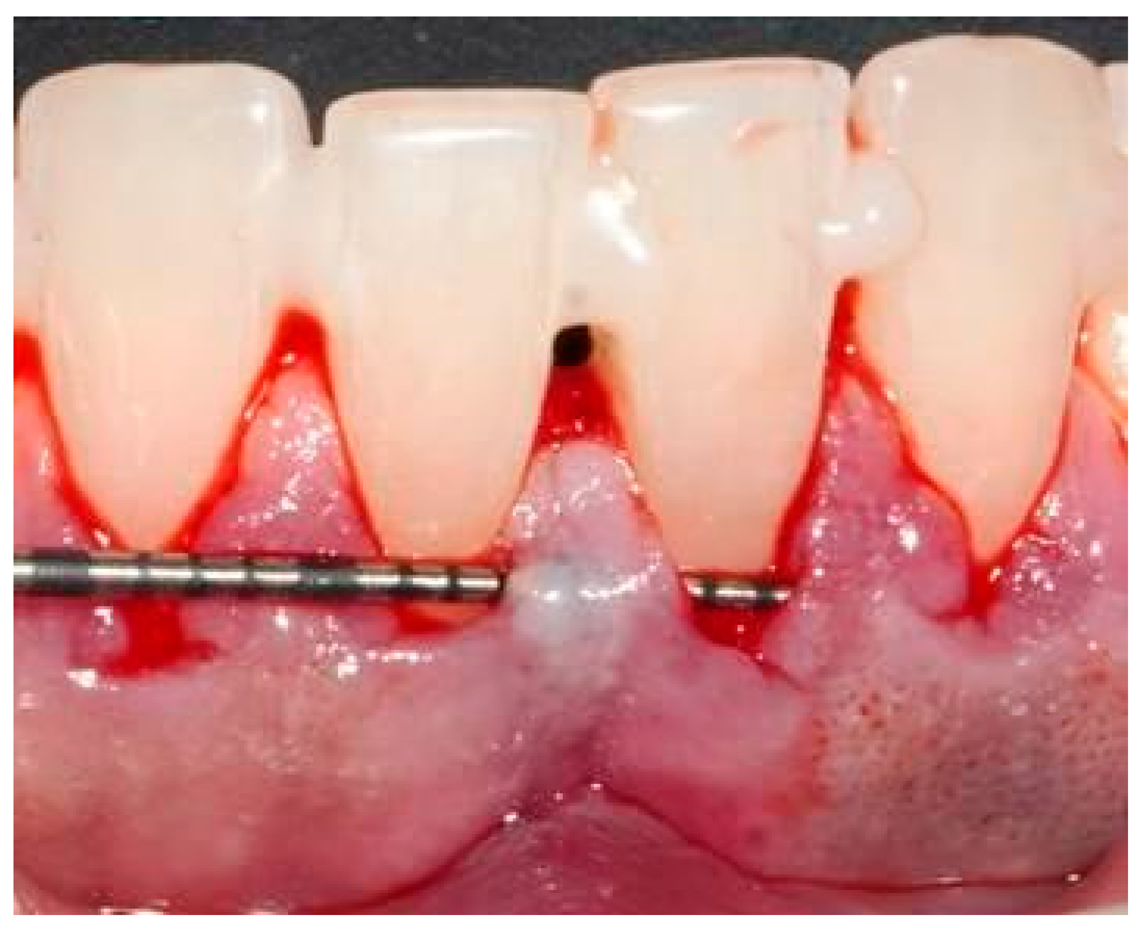
After local infiltration anesthesia (2% lignocaine with 1:80,000 epinephrine; Septodont, Saint-Maur-des-Fossés, France), exposed root surfaces were carefully root-planed with Gracey curettes (Hu-Friedy). Intrasulcular incisions were performed with a 15° microsurgical blade (Lance tip, Deo Surgicals, Mumbai, India) and a supraperiosteal tunnel was prepared using dedicated tunneling knives (TKN1, TKN2; GDC, Mumbai, India). The tunnel was full-thickness up to the mucogingival junction and split-thickness beyond, allowing adequate tissue flexibility and graft accommodation. The interdental papillae were gently undermined with a tunneling knife to permit tension-free coronal advancement [16].

2.4.3. Donor Site—Control Group (SCTG)

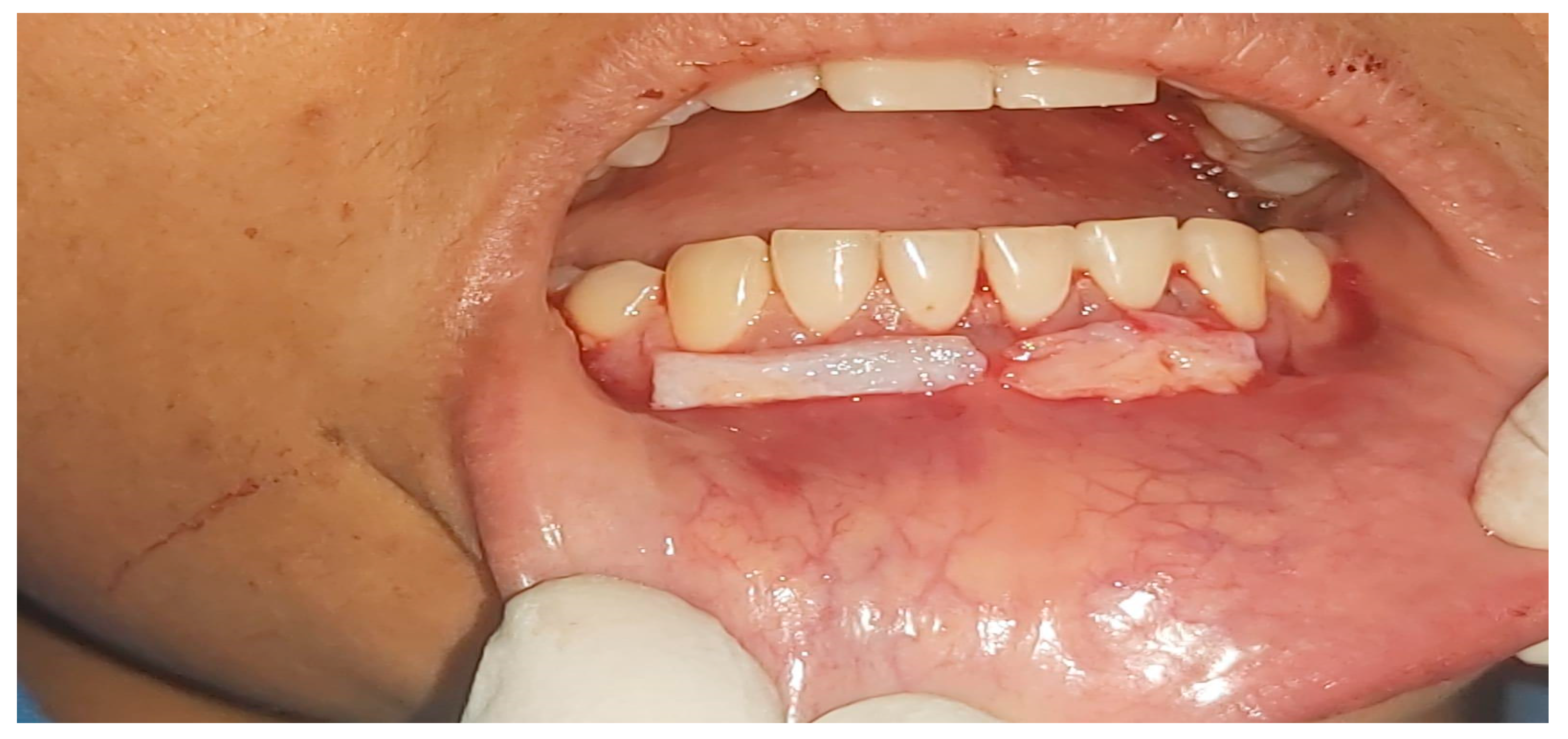
An SCTG was harvested from the hard palate using a standardized single-incision technique. The graft was trimmed to match the recipient bed dimensions and secured to the de-epithelialized papillae with 6–0 monofilament poliglecaprone sutures (Mendmono, Meril, Vapi, India), ensuring close adaptation to the prepared root surfaces.

2.4.4. Donor Site—Test Group (D-FGG)

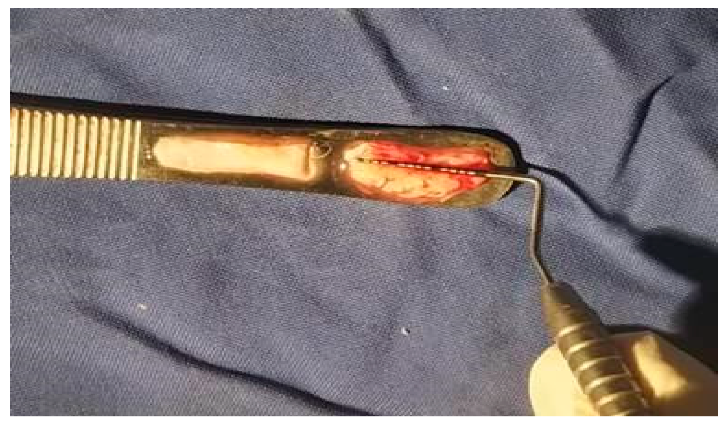
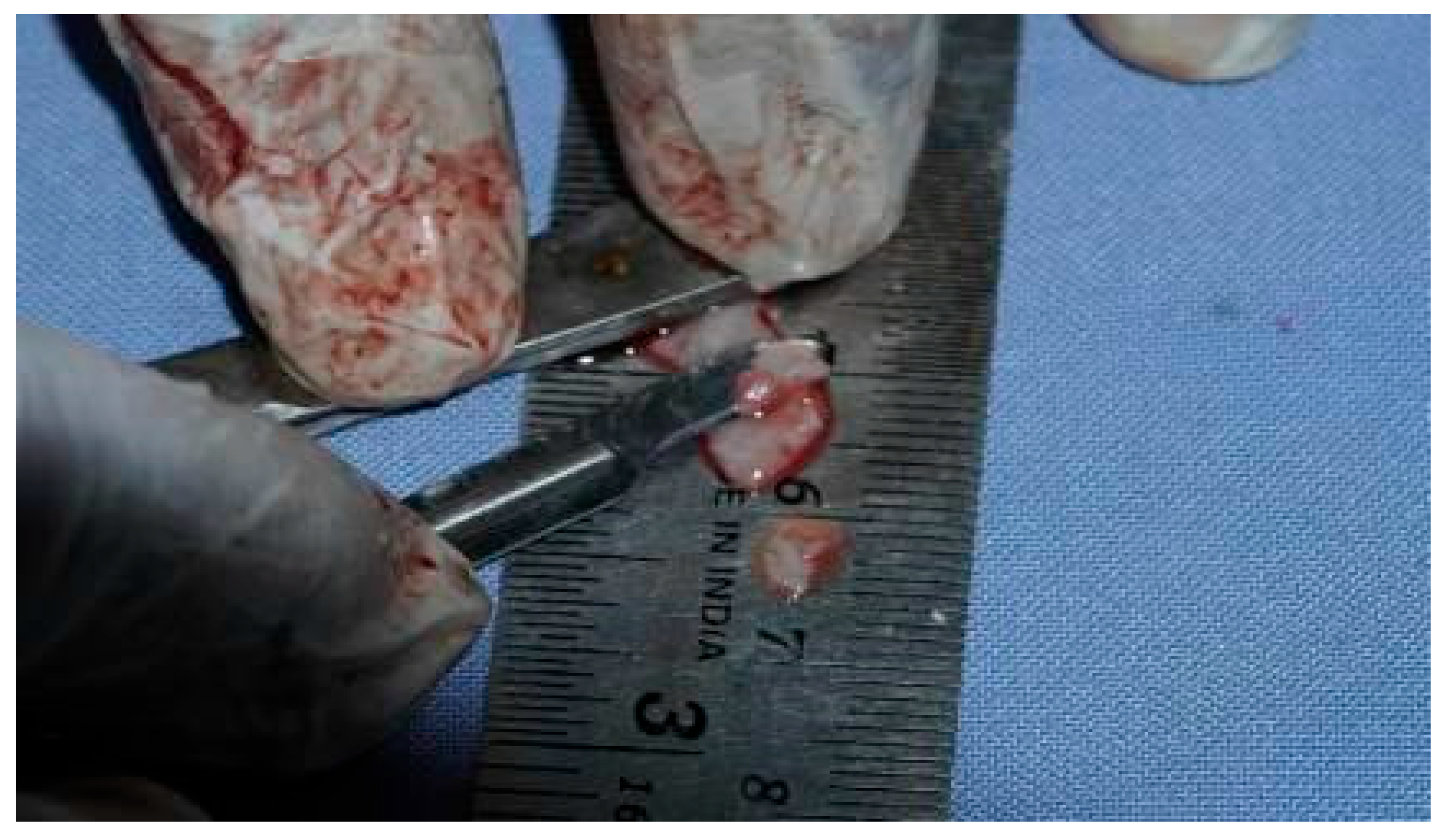
A free gingival graft was harvested from the palate, and the epithelial layer was removed extra-orally with a #15 blade to isolate the connective tissue component [12]. The resulting D-FGG was shaped and sutured into the recipient tunnel with 6–0 monofilament poliglecaprone sutures (Mendmono).

2.4.5. Donor-Site Management

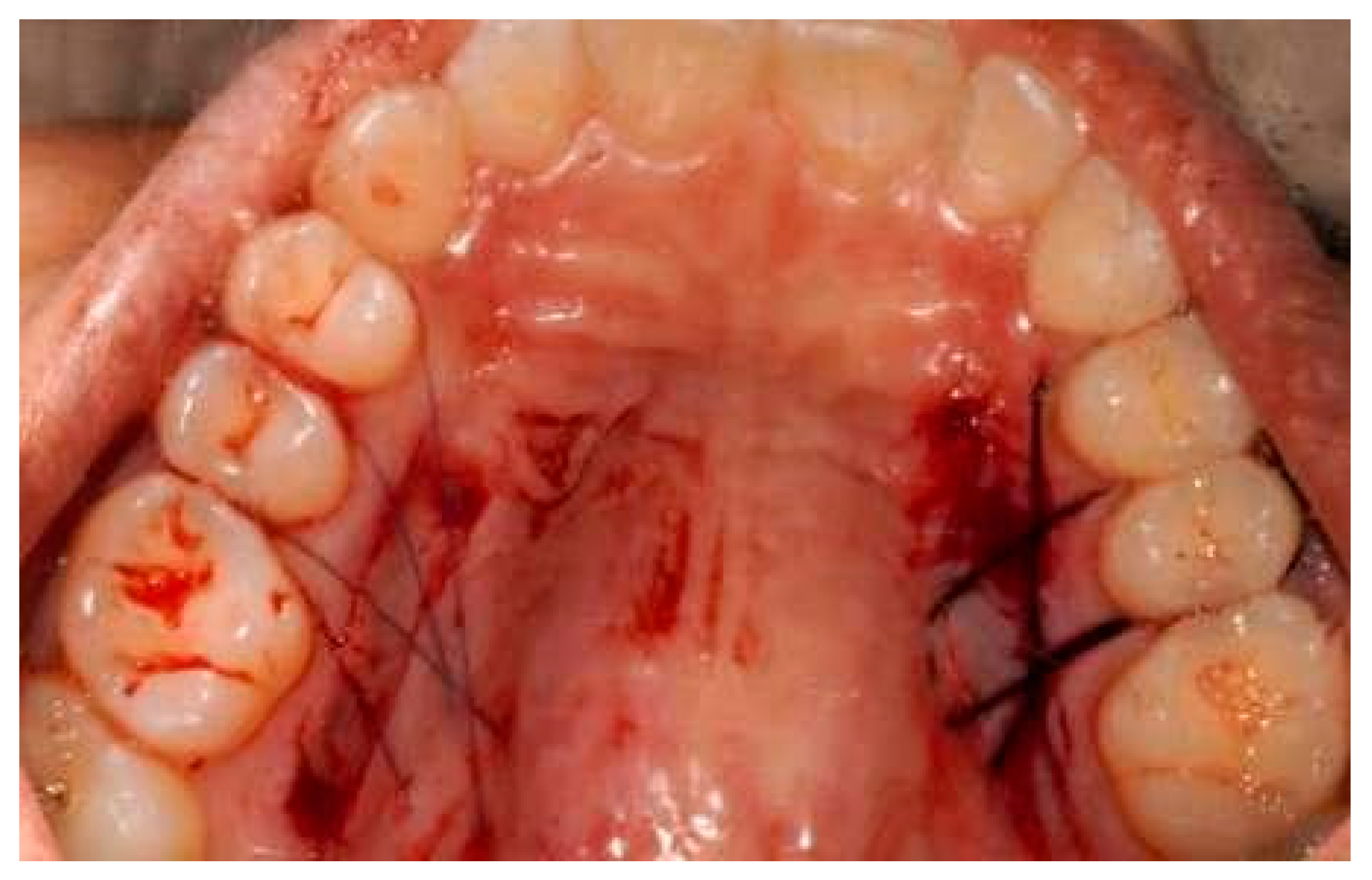
In both groups, hemostasis of the palatal wound was achieved with a collagen-based hemostatic agent (HAEMOCOL; Advanced Biotech Products, Fremont, CA, USA). In the test group, the palate was additionally covered with a bovine collagen membrane (SURGICOLL-MESH; Advanced Biotech Products) and stabilized with sutures, whereas in the control group, the palatal wound was closed by primary intention without a membrane. It is explicitly acknowledged that the non-identical management of the donor sites between groups introduces a methodological confounder for patient-reported morbidity outcomes (pain, chewing discomfort, bleeding), as the larger open palatal wound left after D-FGG harvesting and the use of an additional bovine collagen membrane in the test group cannot be disentangled from the intrinsic biological behavior of the two grafts. Donor-site morbidity findings must therefore be interpreted as a property of the entire harvesting protocol rather than of the graft type alone.

2.4.6. Flap Advancement and Composite Stops

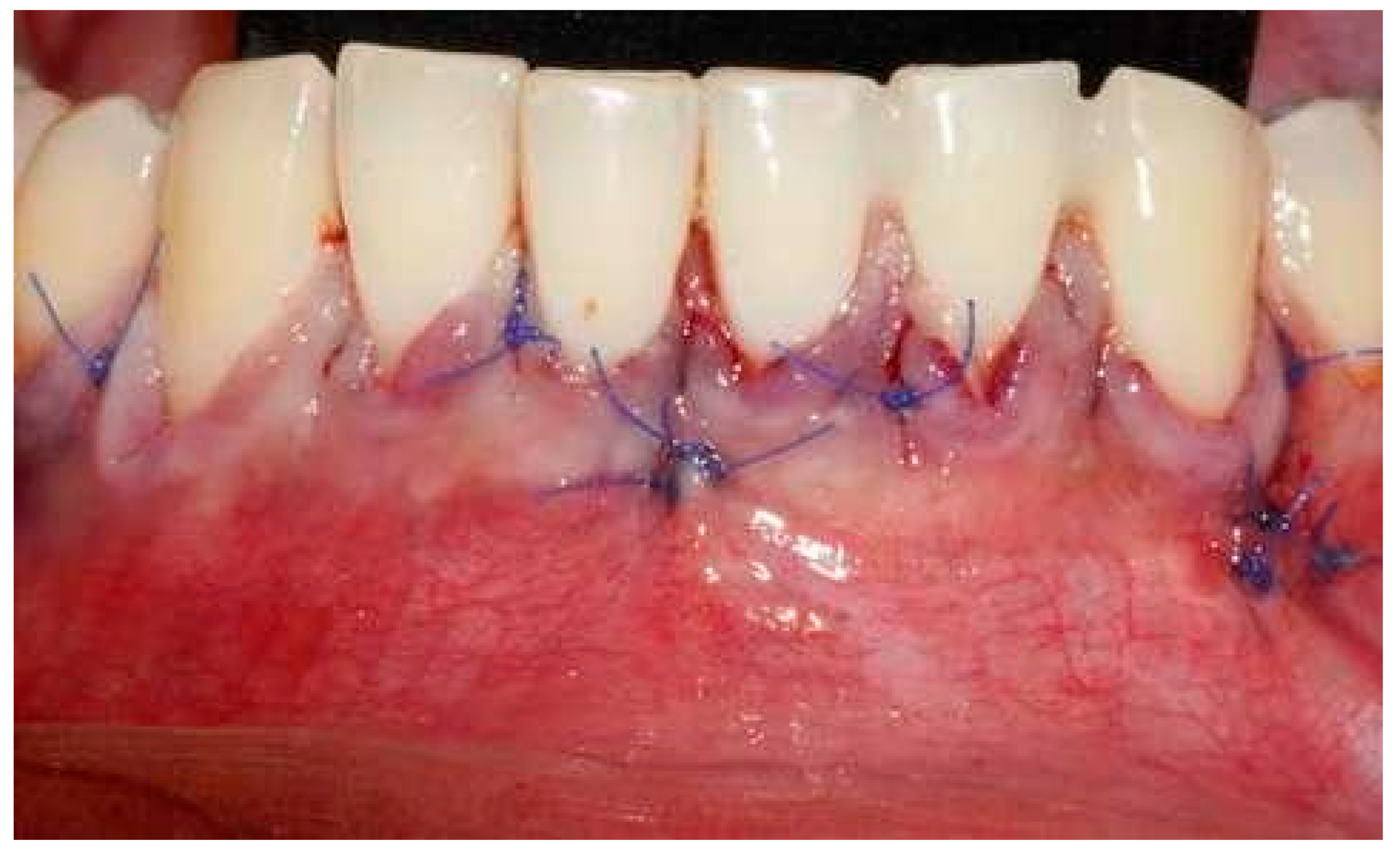
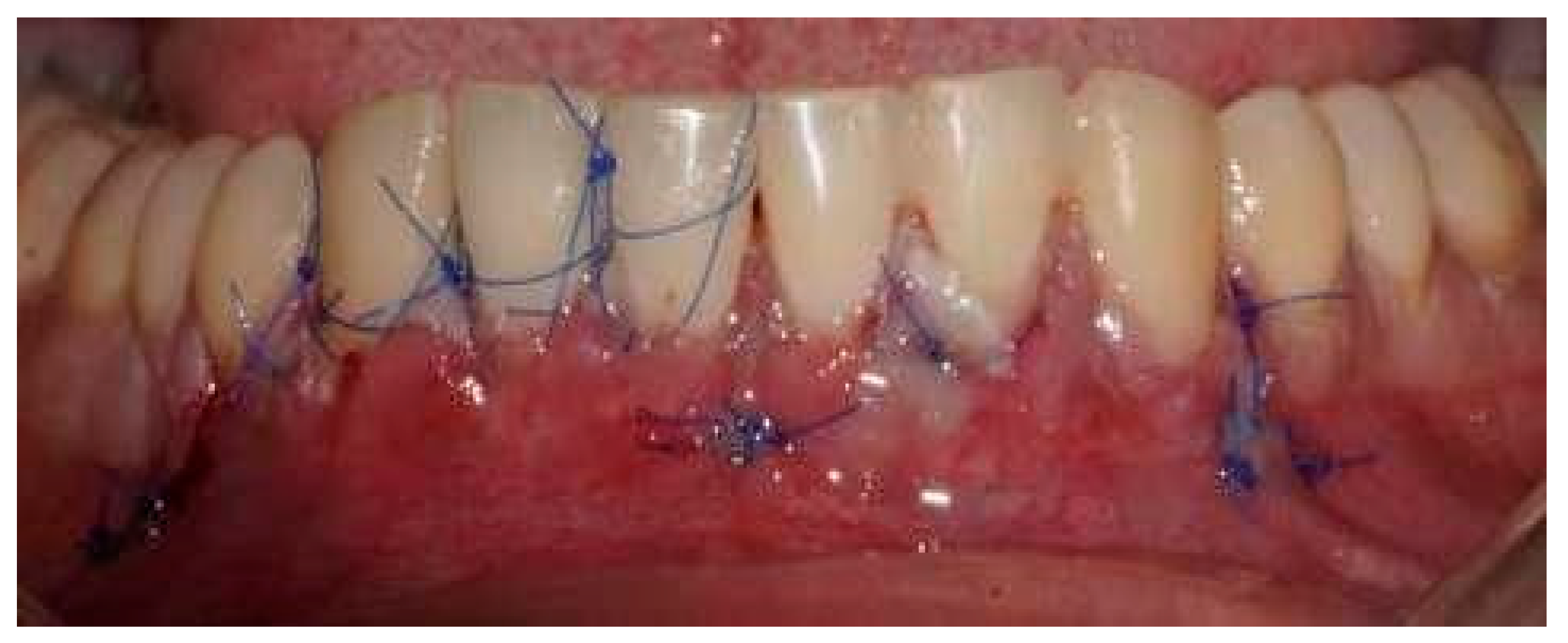
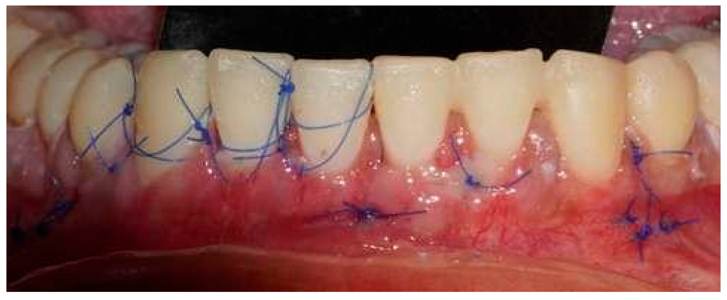
The overlying flap was coronally repositioned over the graft and secured with a sling suture technique using 6–0 monofilament polyamide sutures (Seamlon, Bengaluru, India), providing uniform tension and wound stability. Flowable composite stops (3M, St. Paul, MN, USA) were placed interdentally to maintain the coronal position of the flap during early healing. A representative sequence of the surgical steps is shown in Figure 1, Figure 2, Figure 3, Figure 4, Figure 5, Figure 6, Figure 7 and Figure 8.

2.5. Postoperative Care

Patients were prescribed analgesics for 7 days and were instructed to refrain from mechanical cleaning of the surgical area for 14 days. A 0.2% chlorhexidine gluconate mouth rinse (Clohex ADS, Dr. Reddy’s Laboratories, Hyderabad, India) was recommended twice daily for 21 days. Palatal sutures were removed at 7 days and recipient-site sutures between 2 and 3 weeks. Follow-up visits were scheduled at 1 week, 2 weeks, 1 month, 3 months and 6 months, with the primary endpoint assessment at 6 months.

2.6. Outcome Variables

The primary outcome was the percentage of mean root coverage (mRC) at 6 months. Secondary outcomes included the following:
  • Recession depth reduction from baseline to 6 months;
  • Root Coverage Esthetic Score (RES) at 6 months [17];
  • Patient-reported pain and discomfort, assessed using 100 mm visual analog scales (VAS), with 0 indicating ‘no pain/discomfort’ and 100 ‘worst imaginable pain/discomfort.’ Patients received standardized written and verbal instructions for VAS completion, with explicit definitions of pain (sharp, aching or throbbing sensations requiring analgesic medication) and discomfort (functional impairment, tenderness or awareness without frank pain). VAS assessments were administered at 24 h, 7 days and 14 days postoperatively. Although VAS is a widely validated instrument with established psychometric properties in periodontal surgery, no study-specific calibration or validation procedures were performed. VAS forms were completed independently by patients without examiner influence;
  • Chewing discomfort, recorded on a VAS at 1 and 2 weeks;
  • Postoperative bleeding at the donor site, recorded on a VAS at 1 and 2 weeks;
  • Early wound healing, assessed with the Landry Healing Index [18] at 1 and 2 weeks.

2.7. Examiner Calibration and Measurement Reproducibility

All clinical measurements were performed by a single calibrated examiner (S.J.) who was not involved in the surgical procedures and was blinded to the side allocation. Prior to study initiation, the examiner underwent a calibration session against a senior investigator (N.D.) on ten non-study patients presenting with multiple gingival recessions in the esthetic zone, in which recession depth was recorded twice at each site at a one-hour interval. Agreement was considered acceptable when at least 90% of duplicate measurements fell within ±1 mm. Intra-examiner reliability was further quantified by computing the intra-class correlation coefficient (ICC, two-way mixed-effects, absolute agreement, single measure) for recession depth, with values ≥ 0.75 considered indicative of good reproducibility. The Landry Healing Index and the Root Coverage Esthetic Score (RES) were assessed using the published scoring rubrics, and reference clinical images were reviewed with the senior examiner before the study started to align the interpretation of borderline cases. Visual analog scale (VAS) forms were administered to all patients using identical 100 mm horizontal lines on a standardized paper template, with anchor descriptors printed at both ends; written and verbal instructions were given in a quiet, private setting by an investigator not involved in the surgical phase, and patients were explicitly asked to mark donor-site and recipient-site outcomes on separate, clearly labeled lines to limit cross-contamination between the two outcomes.

2.8. Statistical Analysis

Statistical analyses were performed using SPSS v21.0 (IBM Corp., Armonk, NY, USA). The significance level was set at α = 0.05 (two-sided). Normality of continuous variables was tested with the Shapiro–Wilk test. Continuous data are presented as mean ± standard deviation and categorical data as absolute numbers and percentages.
Because of the split-mouth design, the two sides of each patient were treated as paired observations to account for within-subject clustering. Intra-group changes over time and intergroup comparisons at each time point were analyzed with the paired t-test when the normality assumption was met or with the Wilcoxon signed-rank test otherwise. Ninety-five percent confidence intervals of the intergroup differences were reported in the format [lower; upper] when appropriate, and effect sizes were computed as Cohen’s d for parametric comparisons. Multiple recessions within the same patient and side were treated as repeated measures nested within the subject; the residual correlation introduced by this hierarchical structure could not be fully modeled with the available summary dataset and is explicitly addressed in the Section 5.
As the investigation was conceived as an exploratory pilot trial, no formal a priori sample-size calculation was performed; a convenience sample of 16 patients (32 sides, 74 recessions) was therefore enrolled. Two consequences of this design are explicitly acknowledged. First, the study was not powered to detect small or even moderate intergroup differences in mRC or RES, and the absence of statistically significant differences must therefore not be interpreted as evidence of equivalence or non-inferiority between the two grafts; the results are reported as preliminary point estimates rather than as inferential conclusions. Second, the analysis of multiple recessions as repeated measures nested within subject and side carries an inherent risk of pseudo-replication when the residual within-cluster correlation cannot be fully modeled; this may, in principle, narrow variance estimates and inflate type I error for intergroup comparisons performed at the recession level. To partially mitigate this risk, the primary outcome (mRC) was also evaluated descriptively at the side level (mean across recessions per side), and the corresponding paired comparison was used as the principal reference for inference, with recession-level statistics provided only as supportive information.

3. Results

3.1. Participant Flow and Baseline Characteristics

Twenty-eight patients were screened; twelve did not meet the inclusion criteria or declined to participate, so that sixteen patients (32 sides, 74 recessions: 36 control, 38 test) were finally enrolled and randomized. No patient was lost to follow-up, and healing was uneventful in all cases.
The study sample comprised 12 males and 4 females, with a mean age of 43 ± 13.07 years (most patients aged between 41 and 50 years). Among the 36 control defects, 16 were located in the maxilla and 20 in the mandible; among the 38 test defects, 20 were in the maxilla and 18 in the mandible. Of the 74 defects overall, 35 involved incisors, 15 canines and 24 premolars. Baseline recession depth was comparable between groups. Demographic data are summarized in Table 1.

3.2. Patient Morbidity

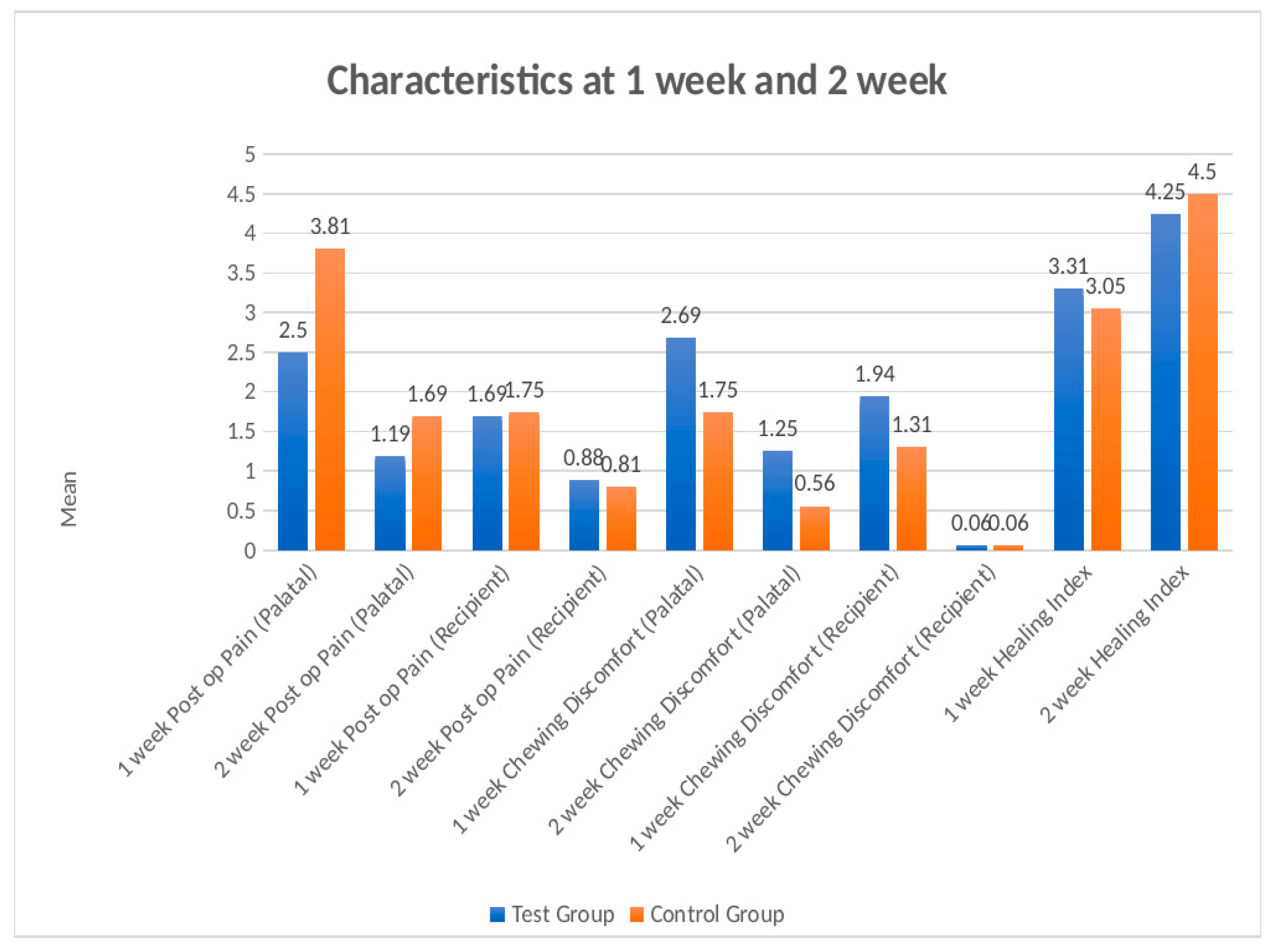
Postoperative VAS scores and healing indices are summarized in Table 2 and graphically represented in Figure 9.

3.2.1. Postoperative Pain

In the test group, donor-site pain decreased from 2.50 ± 0.73 at 1 week to 1.18 ± 0.54 at 2 weeks (p < 0.001), and recipient-site pain decreased from 1.68 ± 0.70 to 0.87 ± 0.61 (p < 0.01). In the control group, donor-site pain decreased from 3.81 ± 0.83 to 1.68 ± 0.70 (p < 0.001) and recipient-site pain from 1.75 ± 0.68 to 0.81 ± 0.65 (p < 0.01).
Intergroup comparison showed significantly lower donor-site pain in the test group at both 1 week (2.50 ± 0.73 vs. 3.81 ± 0.83; Cohen’s d = 1.68; p < 0.001) and 2 weeks (1.19 ± 0.54 vs. 1.69 ± 0.70; Cohen’s d = 0.80; p < 0.05), whereas recipient-site pain did not differ at either time-point.

3.2.2. Chewing Discomfort

Chewing discomfort decreased significantly in both groups between 1 and 2 weeks (test: 2.69 ± 0.79 → 1.25 ± 0.58, p < 0.001; control: 1.75 ± 0.68 → 0.56 ± 0.63, p < 0.001). Intergroup comparison showed significantly greater chewing discomfort in the test group at both 1 week (Cohen’s d = 1.28; p < 0.01) and 2 weeks (Cohen’s d = 1.14; p < 0.05).

3.2.3. Postoperative Bleeding

Bleeding scores decreased significantly between 1 and 2 weeks in both groups (test: 1.94 ± 0.85 → 0.06 ± 0.25; control: 1.31 ± 0.60 → 0.06 ± 0.25; both p < 0.001). At 1 week, bleeding was significantly greater in the test group than in the control group (Cohen’s d = 0.86; p < 0.05), whereas no difference was observed at 2 weeks.

3.3. Root Coverage

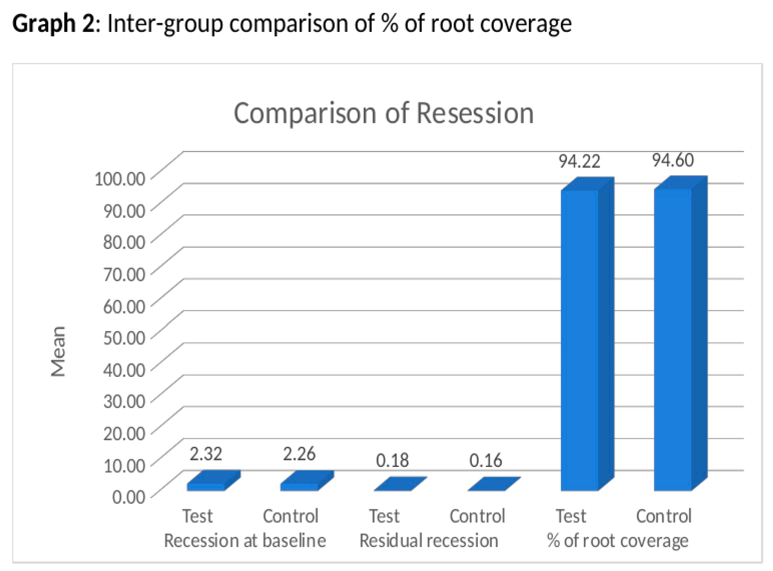
Baseline recession depth was 2.32 ± 0.82 mm in the test group and 2.26 ± 0.74 mm in the control group. At 6 months, residual recession was 0.18 ± 0.32 mm and 0.16 ± 0.29 mm, respectively, with a highly significant within-group reduction in both groups (p < 0.001). Intra-group and inter-group comparisons are detailed in Table 3 and Table 4; a graphical summary is shown in Figure 10.
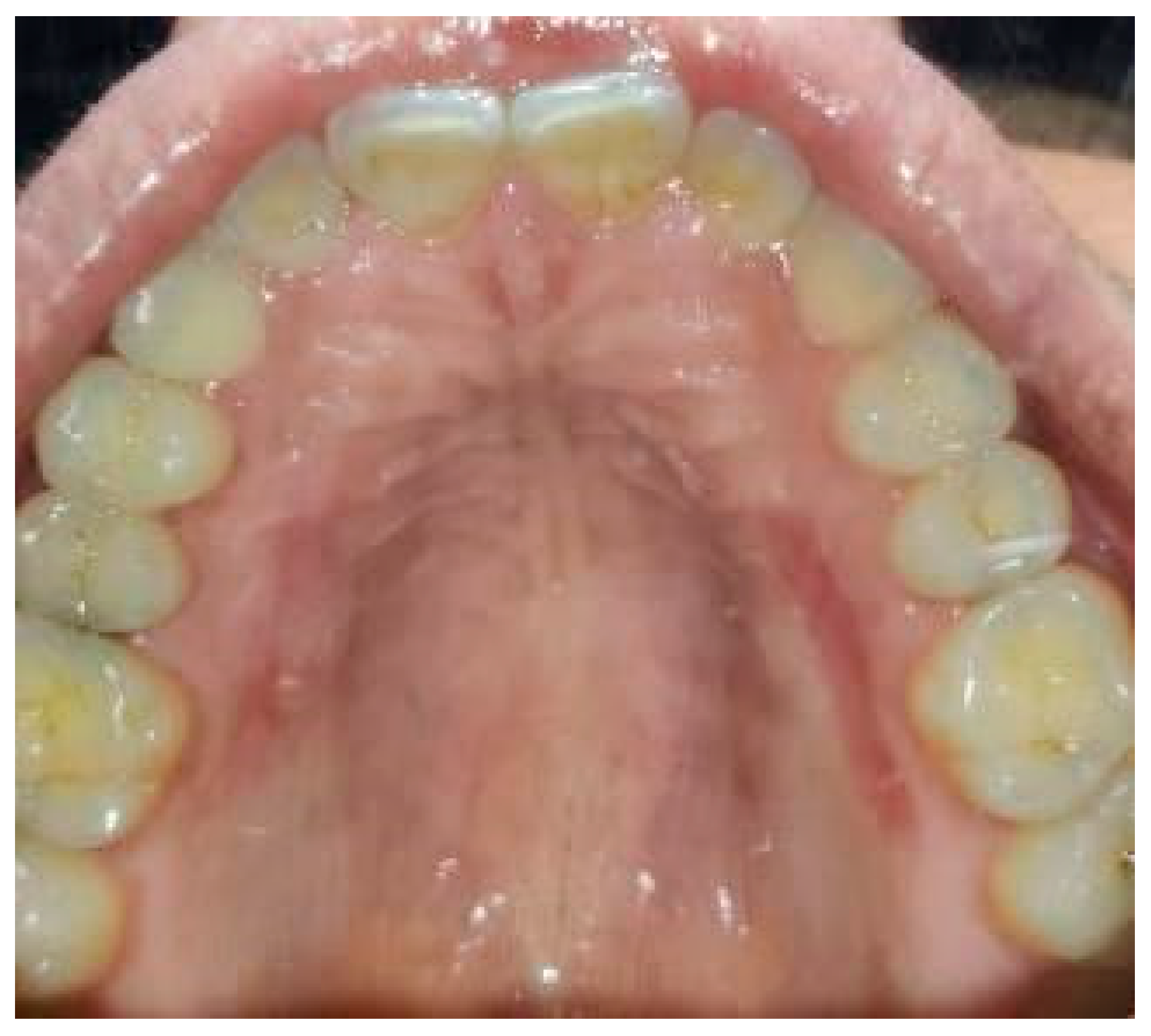
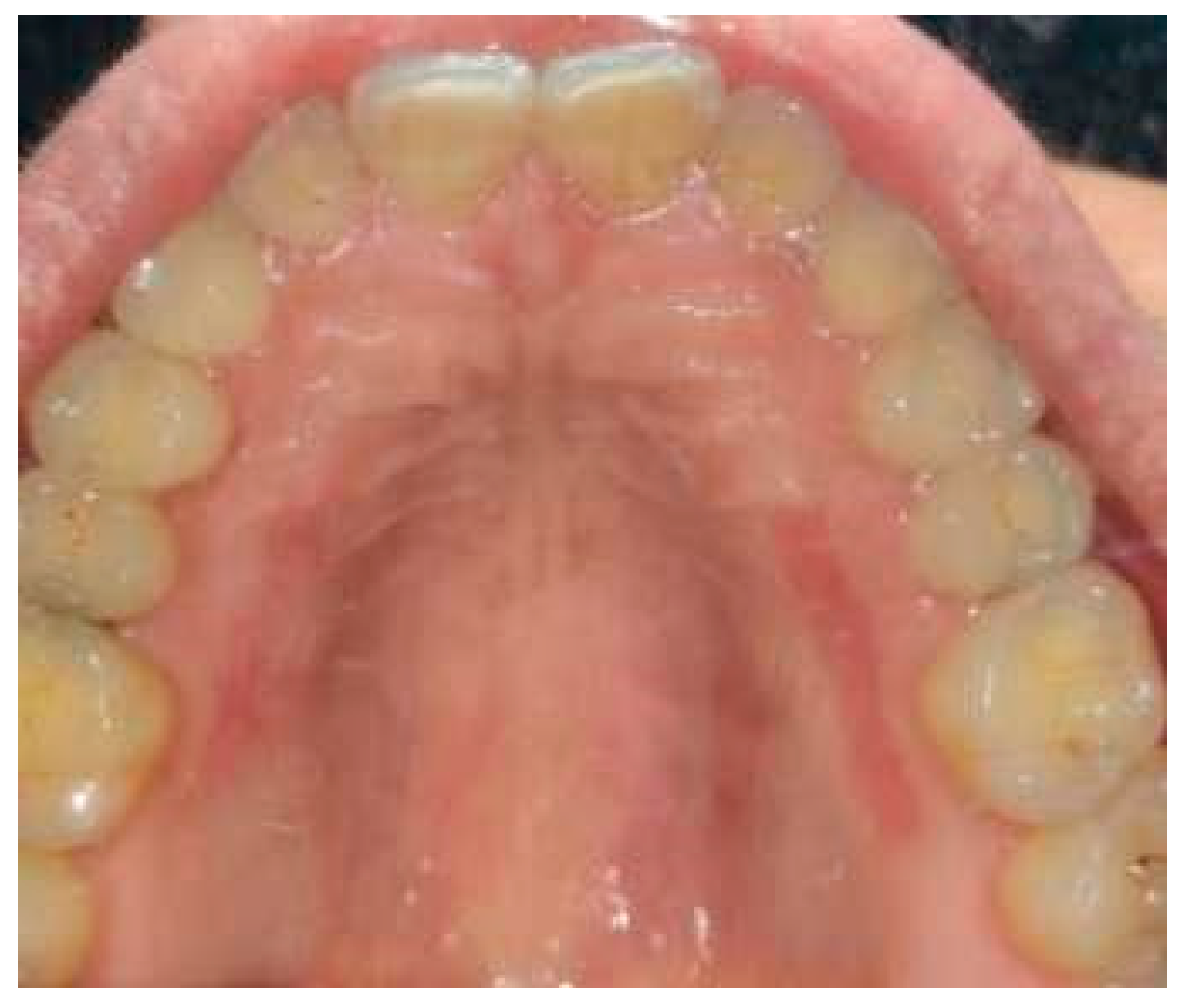
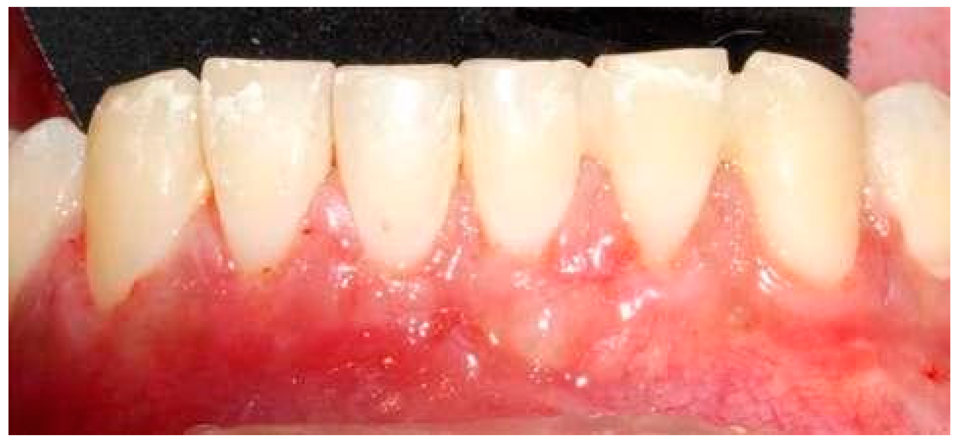
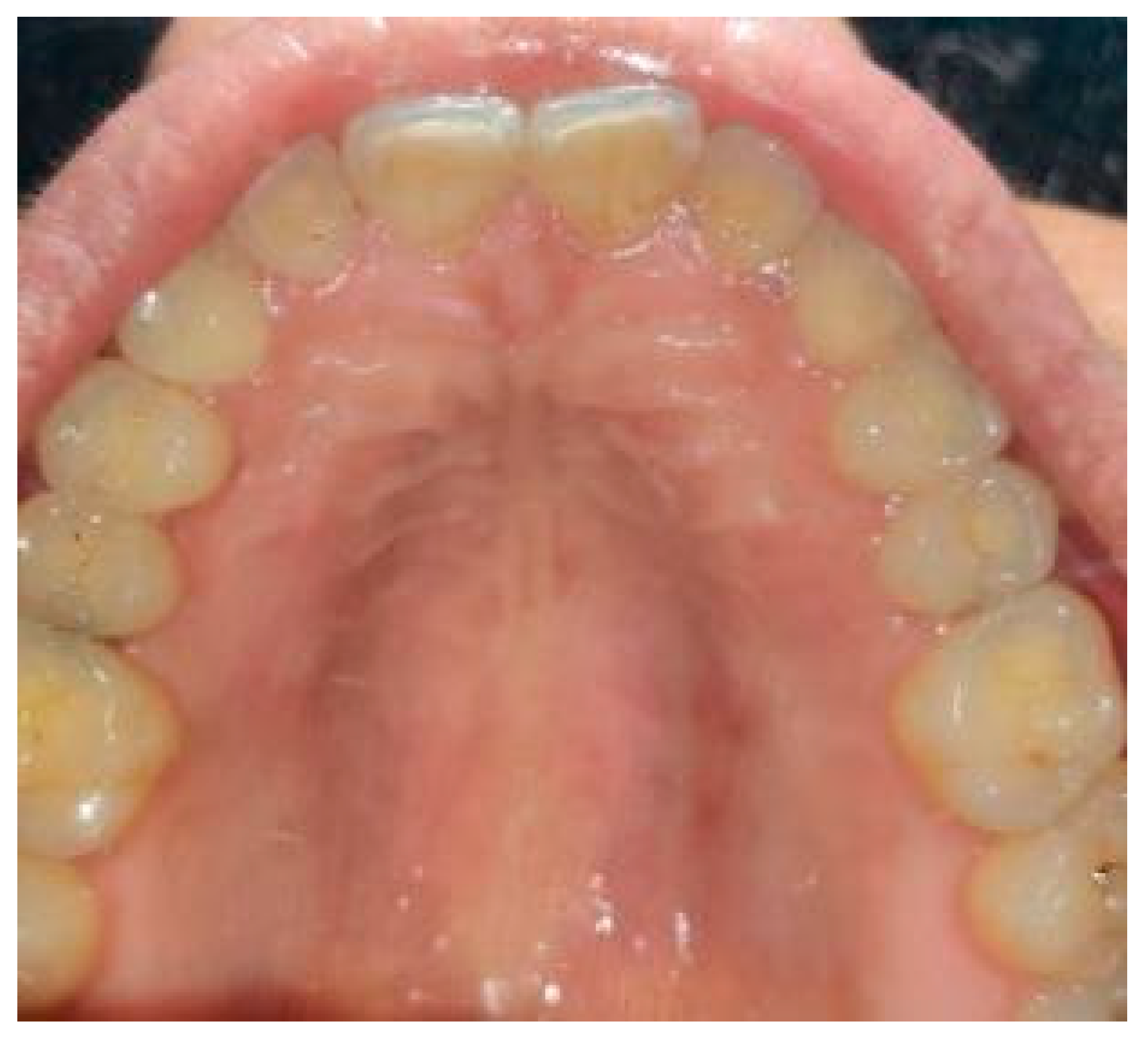
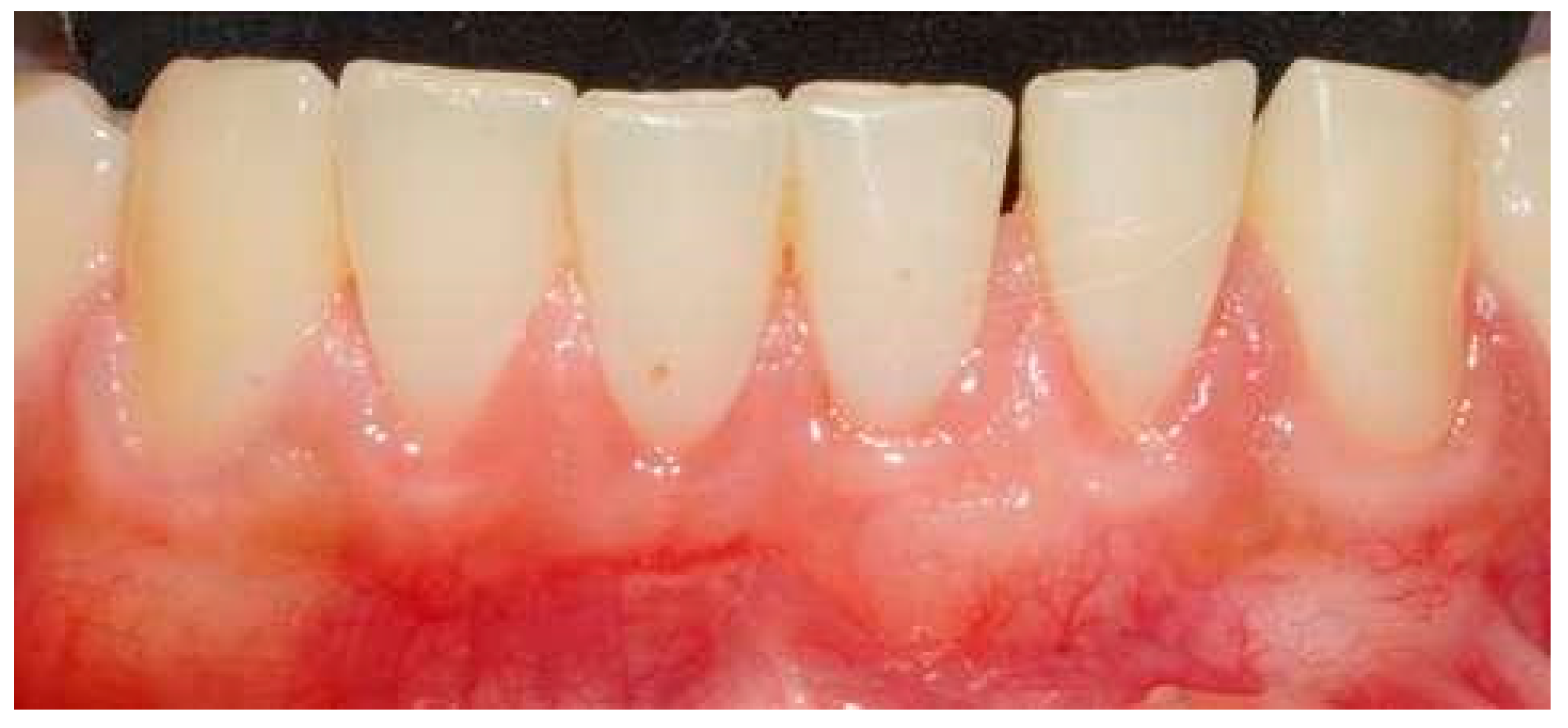
Mean root coverage at 6 months was 94.22% ± 10.28% in the test group and 94.60% ± 9.99% in the control group, with no statistically significant intergroup difference (Cohen’s d = −0.04; p > 0.05). Given the limited sample size, this absence of a statistically significant difference should be interpreted strictly as a non-rejection of the null hypothesis at the present sample size, and not as a positive demonstration that the two grafts are clinically equivalent in terms of root coverage. Representative follow-up photographs at 1 week, 2 weeks, 1 month and 6 months are provided in Figure 11, Figure 12, Figure 13, Figure 14, Figure 15, Figure 16 and Figure 17.

3.4. Root Coverage Esthetic Score

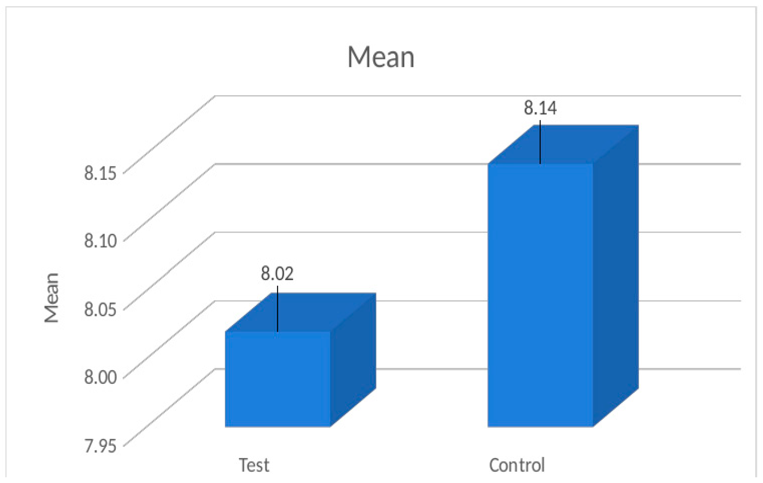
The mean RES was 8.02 ± 0.63 in the test group and 8.14 ± 0.77 in the control group, with no statistically significant intergroup difference (Cohen’s d = −0.17; p > 0.05). The highest score (9) was recorded at 8 test and 13 control sites, while the lowest score (7) was observed at 7 test and 8 control sites (Figure 18).

3.5. Early Healing

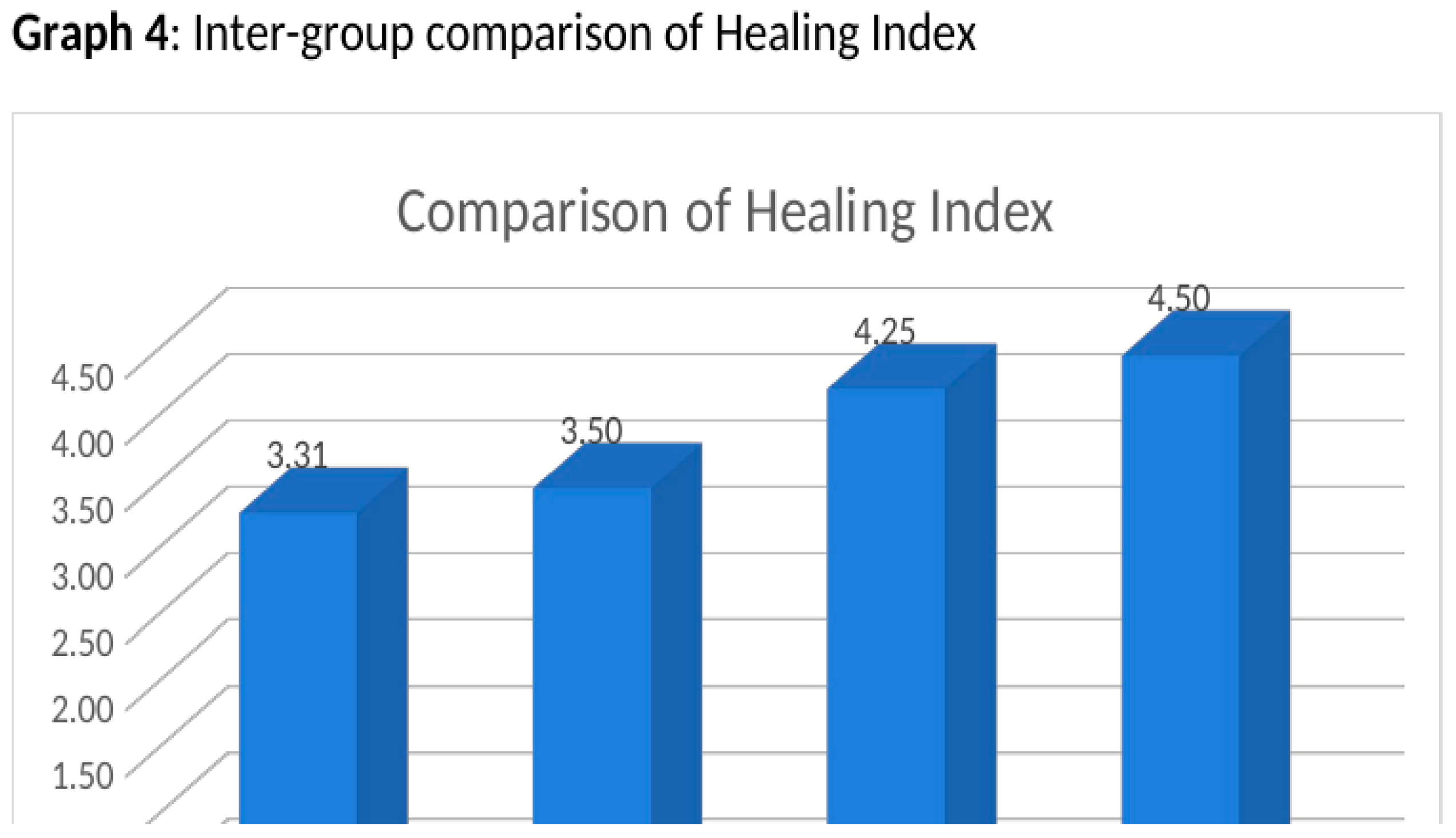
The Landry Healing Index improved significantly between 1 and 2 weeks in both groups (test: 3.31 ± 0.48 → 4.25 ± 0.68; control: 3.50 ± 0.63 → 4.50 ± 0.52; both p < 0.001). Intergroup comparison showed no statistically significant difference at either time-point (Figure 19).

4. Discussion

The present split-mouth pilot trial compared the clinical efficacy and postoperative morbidity of MCAT performed with either D-FGG or SCTG in the treatment of MAGRs. The success of periodontal plastic procedures depends on several factors, including surgical technique, operator experience and patient compliance; soft-tissue management in MAGRs is particularly demanding because of the limited blood supply over the avascular root surfaces [19]. Both graft materials provided clinically meaningful outcomes in terms of mean root coverage, esthetic score and early wound healing, thereby supporting the null hypothesis for the primary outcome. Most of the available literature has compared different surgical techniques with a single graft material, while randomized trials comparing different graft materials within the same surgical protocol remain scarce.
The demographic distribution of the present cohort also deserves comment. The sample comprised 12 males and only 4 females, a marked sex imbalance that represents an additional limitation. Because sex-related differences in pain perception and in wound healing are well recognized, the predominantly male composition may limit the generalizability of the morbidity findings to female patients, and balanced sex representation should be ensured in future larger trials.
Mucogingival surgery has been associated with greater postoperative pain than other periodontal procedures [20], with pain assessment typically focused on the donor palatal site; patient-centered outcomes following the tunnel technique remain comparatively under-investigated relative to other root-coverage approaches [21]. In the present study, a standardized assessment of donor- and recipient-site pain, chewing discomfort and bleeding was performed at 1 and 2 weeks.
The mean root coverage observed in the present cohort (94.22% ± 10.28% in the test group and 94.60% ± 9.99% in the control group) was in line with, or higher than, that reported in previous trials on MCAT. Bakhishov et al. reported an mRC of 91.72% ± 16.59% for D-FGG + TUN and 84.72% ± 19.72% for SCTG + TUN [22], concluding that graft type was not a significant predictor of mRC, whereas baseline recession depth, keratinized tissue height and gingival thickness were. Aroca et al. reported an mRC of 82% and 83% with MCAT + SCTG with and without enamel matrix derivative (EMD), respectively [8]. Molnár et al. observed that 9-year outcomes after MCAT + CTG or MCAT + collagen matrix were characterized by substantial relapse of the gingival margin [23], while Stähli et al., comparing MCAT + SCTG with and without EMD, reported a 6-month mRC of 78 ± 26% and 77 ± 18% [24]. Pietruska et al. described superior mRC and complete root coverage for D-FGG + TUN compared with collagen matrix + TUN [25].
These comparisons should be interpreted with caution. The higher mRC values observed in the present trial may reflect not only graft- and technique-related factors but also baseline recession depth, recipient-site anatomy and operator experience, which jointly influence outcomes after root coverage procedures. Sculean et al. [26,27] demonstrated that MCAT combined with different graft materials provides a predictable treatment for Miller Class I and II recessions, and several authors [16,28,29] have confirmed the role of CTG as the reference graft within tunnel-based approaches. CTG, however, has known limitations, including limited palatal donor tissue, greater postoperative pain and the risk of palatal flap necrosis or dehiscence [12]; D-FGG has been proposed to overcome these limitations, particularly when palatal thickness is ≤2.5 mm [30], and offers theoretical advantages such as reduced postoperative shrinkage and a higher proportion of collagen-rich lamina propria with comparatively less fatty or glandular tissue [12,13].
Tavelli et al. [14] suggested that the histological composition of D-FGG may enhance gingival recession reduction. McLeod et al. [31] reported that D-FGG + TUN allowed easier recipient-site manipulation and less postoperative morbidity than CTG, whereas Zucchelli et al. [12], in a CAF-based randomized trial, found no significant difference in postoperative pain or root coverage between SCTG and D-FGG. Direct evidence comparing D-FGG and SCTG within the TUN/MCAT framework is, however, still limited, which provided the rationale for the present trial.
In the present cohort, maxillary teeth showed greater mean root coverage than mandibular teeth, in agreement with previous reports [14,25,32], although Chaparro et al. [33] did not observe significant maxillary–mandibular differences. Mandibular recession treatment is intrinsically more demanding because of unfavorable anatomy, reduced vascularization, narrower interdental papillae, shallow vestibule and lip-muscle activity, all of which limit coronal displacement of the flap [32]. Moreover, multiple adjacent defects require longer operative time and carry a higher risk of complications than single recessions [34].
Mucogingival surgery has been reported to be 3.5 times more likely to cause pain than osseous surgery, and soft-tissue graft surgery has been identified as the periodontal procedure producing the greatest postoperative discomfort [35]. Despite the introduction of alternative biomaterials [36], autogenous soft-tissue grafts remain the reference in periodontal and peri-implant plastic surgery [37], and patient-reported outcome measures (PROMs) are now recognized as key endpoints in clinical trials in this field. In the present study, perceived pain peaked in the first postsurgical days and returned towards baseline by 2 weeks, consistent with Burkhardt et al. [38], who described the first three days as the acute phase; the progressive pain reduction has been attributed to the stabilization of the blood clot over the highly innervated palatal periosteum [39].
Intergroup comparison showed more sustained donor-site pain in the SCTG group, with no difference at the recipient site. Conversely, greater chewing discomfort was observed in the D-FGG group at both 1 and 2 weeks, together with greater early bleeding at 1 week. Cortellini et al. [40] linked the duration of grafting procedures to postsurgical pain, which may partly explain the increased pain in the control group, while the better chewing ability in the SCTG group is in line with Zangrando et al. [41]: the open palatal wound left after D-FGG harvesting may contribute to patient anxiety and to voluntarily reduced chewing aimed at protecting the surgical site [12]. The harvesting location and the composition of the CTG have also been suggested to influence both root coverage and donor morbidity [12,13], and thicker palatal flaps have been associated with greater postoperative pain, providing a plausible explanation for the greater donor-site pain observed in the control group.
Two methodological aspects deserve explicit acknowledgment in the interpretation of these morbidity findings. First, donor-site management was not identical across groups: the D-FGG palatal wound received a bovine collagen membrane, whereas the SCTG wound was closed by primary intention without a membrane. This differential management represents a confounding variable that limits the attribution of observed morbidity differences exclusively to graft type, and future trials should standardize donor-site coverage across groups to allow unconfounded comparisons. Second, the split-mouth design may have introduced a carry-across behavioral bias in patient-reported outcomes: subjects experiencing notable discomfort on one side may have modified mastication bilaterally, thereby influencing scores at both surgical sites simultaneously and potentially confounding intergroup comparisons of chewing discomfort.
Not all previous studies are in agreement with the present findings. Zucchelli et al. [12] reported similar patient morbidity for FGG and CTG harvesting, with increased analgesic intake only in the presence of dehiscence or necrosis, and Bakhishov et al. [22] found no significant difference in pain or discomfort between D-FGG + TUN and SCTG + TUN. Differences in surgical protocols, pain-assessment tools and individual pain perception may account for these discrepancies [42].
The RES system, introduced by Cairo et al. [17], integrates complete root coverage (60% of the total score) with soft-tissue contour, texture, keloid formation, gingival color and mucogingival alignment (40%). In the present study, the mean RES was 8.02 ± 0.63 in the test group and 8.14 ± 0.77 in the control group, with no significant intergroup difference, in agreement with previous reports of favorable esthetic outcomes after the tunnel procedure with CTG [43]. Postoperative healing was uneventful in all patients, with no significant intergroup difference in Landry Healing Index at 1 and 2 weeks and no adverse events such as abscess, graft necrosis or infection. Taken together, these preliminary findings suggest that D-FGG + MCAT may warrant further investigation as a potential alternative to SCTG + MCAT; however, given the exploratory nature of the present pilot study and its acknowledged statistical limitations, definitive conclusions regarding comparative efficacy cannot be drawn from the present data.
The present findings should be interpreted within the broader context of evidence-based periodontal plastic surgery. Autogenous soft-tissue grafts remain the reference standard for root coverage and peri-implant soft-tissue augmentation, with a stable band of keratinized and attached tissue contributing to favorable long-term outcomes [44,45,46]. The minimally invasive, papilla-sparing logic of the MCAT technique is consistent with the contemporary shift toward less traumatic surgical approaches [47,48,49], and the present results, although obtained on a small sample, are coherent with the high mRC and RES values reported by these works when soft-tissue grafting is performed under minimally invasive conditions. The temporal pattern of healing observed in this cohort—rapid early stabilization followed by progressive maturation of the marginal soft tissues—is also in keeping with the broader literature on biomechanical adaptation of grafted sites [50], and the close integration between refined surgical execution and graft selection [51] remains, in our view, the most plausible explanation for the comparable outcomes obtained here with two histologically distinct autogenous grafts.

5. Limitations

  • The 6-month observation period does not allow conclusions on the long-term stability of the gingival margin;
  • The sample size was based on convenience, and the study was not formally powered to detect small intergroup differences in mRC or RES; the results are therefore exploratory and should not be interpreted as evidence of equivalence between the two grafts;
  • Multiple recessions within the same patient and side were treated as repeated observations, and the available summary data did not allow a formal hierarchical model (mixed-effects or generalized estimating equations) accounting for the full correlation structure of the split-mouth design;
  • No histological or immunohistochemical analysis of the donor or recipient sites was performed; differences in tissue composition between SCTG and D-FGG could only be inferred indirectly;
  • Additional clinical parameters such as recession width, keratinized tissue width, width of attached gingiva and clinical attachment level were not collected;
  • Postoperative outcomes were assessed through self-reported VAS, which is inherently subject to inter-individual variability in pain perception and tolerance.
  • Donor-site management was not standardized across groups (additional bovine collagen membrane in the D-FGG arm only), which represents a methodological confounder for the interpretation of patient-reported morbidity outcomes;
  • The cohort showed a marked sex imbalance (12 males vs. 4 females), which may limit the generalizability of the morbidity findings, particularly given known sex-related differences in pain perception and wound healing.

6. Conclusions

Within the limitations of this pilot trial—most notably the small convenience sample, the absence of an a priori power calculation, the lack of a formal hierarchical statistical model and the non-identical donor-site management between groups—both MCAT + SCTG and MCAT + D-FGG were associated with high mRC and RES values at 6 months, and no statistically significant intergroup differences were detected for any of the measured outcomes. D-FGG showed a trend toward lower donor-site pain but greater early chewing discomfort and bleeding. These observations should be regarded as preliminary and hypothesis-generating only; they do not support any inference of clinical equivalence, non-inferiority or superiority between the two grafts, nor do they justify a change in current clinical practice. Adequately powered, hierarchically modeled randomized trials with standardized donor-site management are required before D-FGG can be recommended as an alternative to SCTG within the MCAT framework.

Author Contributions

Conceptualization, S.J., N.D., P.C., R.J., B.J., A.D. and C.L.; Methodology, S.J., R.J., B.J., A.D. and P.L.; Software, B.J. and P.L.; Validation, N.D., R.J., B.J., P.L. and A.P.; Formal analysis, P.C. and B.J.; Investigation, S.J. and N.D.; Resources, S.J. and N.D.; Data curation, N.D. and A.D.; Writing—original draft, N.D., R.J. and P.L.; Writing—review & editing, P.C., B.J., A.P. and C.L.; Visualization, S.J., N.D. and P.L.; Supervision, S.J.; Funding acquisition, P.L. All authors have read and agreed to the published version of the manuscript.

Funding

This research received no external funding.

Institutional Review Board Statement

The study was conducted in accordance with the Declaration of Helsinki and approved by the Institutional Ethics Committee of Sinhgad Dental College and Hospital (protocol code SDCH/IEC/OUT/2022/21). The trial was prospectively registered with the Clinical Trial Registry–India (CTRI/2024/08/072598).

Informed Consent Statement

Written informed consent was obtained from all subjects involved in the study.

Data Availability Statement

The data presented in this study are available on reasonable request from the corresponding author. The data are not publicly available due to privacy restrictions.

Conflicts of Interest

The authors declare no conflicts of interest.

Abbreviations

The following abbreviations are used in this manuscript:
CAFCoronally Advanced Flap
CRCComplete Root Coverage
CTGConnective Tissue Graft
D-FGGDe-epithelialized Free Gingival Graft
EMDEnamel Matrix Derivative
FMBSFull-Mouth Bleeding Score
FMPSFull-Mouth Plaque Score
MAGRsMultiple Adjacent Gingival Recessions
MCATModified Coronally Advanced Tunnel
mRCMean Root Coverage
PROMsPatient-Reported Outcome Measures
RESRoot Coverage Esthetic Score
SCTGSubepithelial Connective Tissue Graft
SNOSESequentially Numbered Opaque Sealed Envelopes
TUNTunnel Technique
VASVisual Analog Scale

References

  1. Armitage, G.C. Development of a classification system for periodontal diseases and conditions. Ann. Periodontol. 1999, 4, 1–6. [Google Scholar] [CrossRef]
  2. Wennström, J.L. Mucogingival therapy. Ann. Periodontol. 1996, 1, 671–701. [Google Scholar] [CrossRef]
  3. Rios, F.S.; Costa, R.S.A.; Wagner, T.P.; Christofoli, B.R.; Goergen, J.; Izquierdo, C.; Jardim, J.J.; Maltz, M.; Haas, A.N. Incidence and progression of gingival recession over 4 years: A population-based longitudinal study. J. Clin. Periodontol. 2021, 48, 114–125. [Google Scholar] [CrossRef]
  4. Daprile, G.; Gatto, M.R.; Checchi, L. The evolution of buccal gingival recessions in a student population: A 5-year follow-up. J. Periodontol. 2007, 78, 611–614. [Google Scholar] [CrossRef] [PubMed]
  5. Serino, G.; Wennström, J.L.; Lindhe, J.; Eneroth, L. The prevalence and distribution of gingival recession in subjects with a high standard of oral hygiene. J. Clin. Periodontol. 1994, 21, 57–63. [Google Scholar] [CrossRef] [PubMed]
  6. Chambrone, L.; de Castro Pinto, R.C.N.; Chambrone, L.A. The concepts of evidence-based periodontal plastic surgery: Application of the principles of evidence-based dentistry for the treatment of recession-type defects. Periodontology 2000 2019, 79, 81–106. [Google Scholar] [CrossRef]
  7. Cairo, F. Periodontal plastic surgery of gingival recessions at single and multiple teeth. Periodontology 2000 2017, 75, 296–316. [Google Scholar] [CrossRef]
  8. Aroca, S.; Keglevich, T.; Nikolidakis, D.; Gera, I.; Nagy, K.; Azzi, R.; Etienne, D. Treatment of class III multiple gingival recessions: A randomized-clinical trial. J. Clin. Periodontol. 2010, 37, 88–97. [Google Scholar] [CrossRef]
  9. Cairo, F.; Pagliaro, U.; Buti, J.; Baccini, M.; Graziani, F.; Tonelli, P.; Pagavino, G.; Tonetti, M.S. Root coverage procedures improve patient aesthetics. A systematic review and Bayesian network meta-analysis. J. Clin. Periodontol. 2016, 43, 965–975. [Google Scholar] [CrossRef] [PubMed]
  10. Gaikwad, I.; Lele, P.; Dodwad, V.; Mariam, S. Modified Coronally Advanced Tunnel (MCAT) Technique with Subepithelial Connective Tissue Graft (SCTG): A Case Report. J. Young Pharm. 2023, 15, 764–766. [Google Scholar] [CrossRef]
  11. Puri, K.; Kumar, A.; Khatri, M.; Bansal, M.; Rehan, M.; Siddeshappa, S.T. 44-year journey of palatal connective tissue graft harvest: A narrative review. J. Indian Soc. Periodontol. 2019, 23, 395–408. [Google Scholar] [CrossRef] [PubMed]
  12. Zucchelli, G.; Mele, M.; Stefanini, M.; Mazzotti, C.; Marzadori, M.; Montebugnoli, L.; De Sanctis, M. Patient morbidity and root coverage outcome after subepithelial connective tissue and de-epithelialized grafts: A comparative randomized-controlled clinical trial. J. Clin. Periodontol. 2010, 37, 728–738. [Google Scholar] [CrossRef]
  13. Bertl, K.; Pifl, M.; Hirtler, L.; Rendl, B.; Nürnberger, S.; Stavropoulos, A.; Ulm, C. Relative Composition of Fibrous Connective and Fatty/Glandular Tissue in Connective Tissue Grafts Depends on the Harvesting Technique but not the Donor Site of the Hard Palate. J. Periodontol. 2015, 86, 1331–1339. [Google Scholar] [CrossRef]
  14. Tavelli, L.; Barootchi, S.; Nguyen, T.V.N.; Tattan, M.; Ravidà, A.; Wang, H.L. Efficacy of tunnel technique in the treatment of localized and multiple gingival recessions: A systematic review and meta-analysis. J. Periodontol. 2018, 89, 1075–1090. [Google Scholar] [CrossRef]
  15. Mashaly, M.; Ghallab, N.A.; Elbattawy, W.; Elarab, A.E. Soft Tissue Augmentation Using De-Epithelialized Free Gingival Graft Compared to Single-line Incision Subepithelial Connective Tissue Graft in the Management of Miller Class I and II Gingival Recession: A Randomized Controlled Clinical Trial. Contemp. Clin. Dent. 2022, 13, 227–235. [Google Scholar] [CrossRef] [PubMed]
  16. Aroca, S.; Molnár, B.; Windisch, P.; Gera, I.; Salvi, G.E.; Nikolidakis, D.; Sculean, A. Treatment of multiple adjacent Miller class I and II gingival recessions with a Modified Coronally Advanced Tunnel (MCAT) technique and a collagen matrix or palatal connective tissue graft: A randomized, controlled clinical trial. J. Clin. Periodontol. 2013, 40, 713–720. [Google Scholar] [CrossRef]
  17. Cairo, F.; Rotundo, R.; Miller, P.D.; Pini Prato, G.P. Root coverage esthetic score: A system to evaluate the esthetic outcome of the treatment of gingival recession through evaluation of clinical cases. J. Periodontol. 2009, 80, 705–710. [Google Scholar] [CrossRef]
  18. Landry, R.G.; Turnbull, R.S.; Howley, T. Effectiveness of benzydamine HCl in the treatment of periodontal post-surgical patients. Res. Clin. Forums 1988, 10, 105–118. [Google Scholar]
  19. Harris, R.J.; Miller, R.; Miller, L.H.; Harris, C. Complications with surgical procedures utilizing connective tissue grafts: A follow-up of 500 consecutively treated cases. Int. J. Periodontics Restor. Dent. 2005, 25, 449–459. [Google Scholar]
  20. Chambrone, L.; Tatakis, D.N. Periodontal soft tissue root coverage procedures: A systematic review from the AAP Regeneration Workshop. J. Periodontol. 2015, 86, S8–S51. [Google Scholar] [CrossRef]
  21. Wessel, J.R.; Tatakis, D.N. Patient outcomes following subepithelial connective tissue graft and free gingival graft procedures. J. Periodontol. 2008, 79, 425–430. [Google Scholar] [CrossRef]
  22. Bakhishov, H.; Isler, S.C.; Bozyel, B.; Yıldırım, B.; Tekindal, M.A.; Ozdemir, B. De-epithelialized gingival graft versus subepithelial connective tissue graft in the treatment of multiple adjacent gingival recessions using the tunnel technique: 1-year results of a randomized clinical trial. J. Clin. Periodontol. 2021, 48, 970–983. [Google Scholar] [CrossRef]
  23. Molnár, B.; Aroca, S.; Dobos, A.; Orbán, K.; Szabó, J.; Windisch, P.; Stähli, A.; Sculean, A. Treatment of multiple adjacent RT 1 gingival recessions with the modified coronally advanced tunnel (MCAT) technique and a collagen matrix or palatal connective tissue graft: 9-year results of a split-mouth randomized clinical trial. Clin. Oral Investig. 2022, 26, 7135–7142. [Google Scholar] [CrossRef] [PubMed]
  24. Stähli, A.; Imber, J.C.; Raptis, E.; Salvi, G.E.; Eick, S.; Sculean, A. Effect of enamel matrix derivative on wound healing following gingival recession coverage using the modified coronally advanced tunnel and subepithelial connective tissue graft: A randomised, controlled, clinical study. Clin. Oral Investig. 2020, 24, 1043–1051. [Google Scholar] [CrossRef]
  25. Pietruska, M.; Skurska, A.; Podlewski, Ł.; Milewski, R.; Pietruski, J. Clinical evaluation of Miller class I and II recessions treatment with the use of modified coronally advanced tunnel technique with either collagen matrix or subepithelial connective tissue graft: A randomized clinical study. J. Clin. Periodontol. 2019, 46, 86–95. [Google Scholar] [CrossRef] [PubMed]
  26. Sculean, A.; Cosgarea, R.; Stähli, A.; Katsaros, C.; Arweiler, N.B.; Brecx, M.; Deppe, H. The modified coronally advanced tunnel combined with an enamel matrix derivative and subepithelial connective tissue graft for the treatment of isolated mandibular Miller Class I and II gingival recessions: A report of 16 cases. Quintessence Int. 2014, 45, 829–835. [Google Scholar] [PubMed]
  27. Sculean, A.; Cosgarea, R.; Stähli, A.; Katsaros, C.; Arweiler, N.B.; Miron, R.J.; Deppe, H. Treatment of multiple adjacent maxillary Miller Class I, II, and III gingival recessions with the modified coronally advanced tunnel, enamel matrix derivative, and subepithelial connective tissue graft: A report of 12 cases. Quintessence Int. 2016, 47, 653–659. [Google Scholar]
  28. Dembowska, E.; Drozdzik, A. Subepithelial connective tissue graft in the treatment of multiple gingival recession. Oral Surg. Oral Med. Oral Pathol. Oral Radiol. Endod. 2007, 104, e1–e7. [Google Scholar] [CrossRef]
  29. Nart, J.; Valles, C. Subepithelial Connective Tissue Graft in Combination with a Tunnel Technique for the Treatment of Miller Class II and III Gingival Recessions in Mandibular Incisors: Clinical and Esthetic Results. Int. J. Periodontics Restor. Dent. 2016, 36, 591–598. [Google Scholar] [CrossRef]
  30. Zuhr, O.; Rebele, S.F.; Cheung, S.L.; Hürzeler, M.B. Research Group on Oral Soft Tissue Biology and Wound Healing. Surgery without papilla incision: Tunneling flap procedures in plastic periodontal and implant surgery. Periodontology 2000 2018, 77, 123–149. [Google Scholar] [CrossRef]
  31. McLeod, D.E.; Reyes, E.; Branch-Mays, G. Treatment of multiple areas of gingival recession using a simple harvesting technique for autogenous connective tissue graft. J. Periodontol. 2009, 80, 1680–1687. [Google Scholar] [CrossRef]
  32. Aroca, S.; Barbieri, A.; Clementini, M.; Renouard, F.; De Sanctis, M. Treatment of class III multiple gingival recessions: Prognostic factors for achieving a complete root coverage. J. Clin. Periodontol. 2018, 45, 861–868. [Google Scholar] [CrossRef] [PubMed]
  33. Chaparro, A.; De la Fuente, M.; Albers, D.; Hernandez, D.; Villalobos, A.M.; Gaedechens, D. Root Coverage of Multiple Miller Class I and II Recession Defects Using Acellular Dermal Matrix and Tunneling Technique in Maxilla and Mandible: A 1-Year Report. Int. J. Periodontics Restor. Dent. 2015, 35, 639–645. [Google Scholar] [CrossRef] [PubMed]
  34. Clauser, C.; Nieri, M.; Franceschi, D.; Pagliaro, U.; Pini-Prato, G. Evidence-based mucogingival therapy. Part 2: Ordinary and individual patient data meta-analyses of surgical treatment of recession using complete root coverage as the outcome variable. J. Periodontol. 2003, 74, 741–756. [Google Scholar] [CrossRef]
  35. Matthews, D.C.; McCulloch, C.A. Evaluating patient perceptions as short-term outcomes of periodontal treatment: A comparison of surgical and non-surgical therapy. J. Periodontol. 1993, 64, 990–997. [Google Scholar] [CrossRef] [PubMed]
  36. Wang, H.L.; Romanos, G.E.; Geurs, N.C.; Sullivan, A.; Suárez-López Del Amo, F.; Eber, R.M. Comparison of two differently processed acellular dermal matrix products for root coverage procedures: A prospective, randomized multicenter study. J. Periodontol. 2014, 85, 1693–1701. [Google Scholar] [CrossRef]
  37. Tavelli, L.; Barootchi, S.; Di Gianfilippo, R.; Kneifati, A.; Majzoub, J.; Stefanini, M.; Zucchelli, G.; Wang, H.L. Patient experience of autogenous soft tissue grafting has an implication for future treatment: A 10- to 15-year cross-sectional study. J. Periodontol. 2021, 92, 637–647. [Google Scholar] [CrossRef]
  38. Burkhardt, R.; Hammerle, C.H.; Lang, N.P. Research Group on Oral Soft Tissue Biology & Wound Healing. Self-reported pain perception of patients after mucosal graft harvesting in the palatal area. J. Clin. Periodontol. 2015, 42, 281–287. [Google Scholar]
  39. Weisel, J.W. Structure of fibrin: Impact on clot stability. J. Thromb. Haemost. 2007, 5, 116–124. [Google Scholar] [CrossRef]
  40. Cortellini, P.; Tonetti, M.; Baldi, C.; Francetti, L.; Rasperini, G.; Rotundo, R.; Nieri, M.; Franceschi, D.; Labriola, A.; Prato, G.P. Does placement of a connective tissue graft improve the outcomes of coronally advanced flap for coverage of single gingival recessions in upper anterior teeth? A multi-centre, randomized, single-blind, clinical trial. J. Clin. Periodontol. 2009, 36, 68–79. [Google Scholar] [CrossRef]
  41. Zangrando, M.S.; Eustachio, R.R.; de Rezende, M.L.; Sant’ana, A.C.; Damante, C.A.; Greghi, S.L. Clinical and Patient-Centered outcomes using two types of subepithelial connective tissue grafts: A Split-Mouth randomized clinical trial. J. Periodontol. 2021, 92, 814–822. [Google Scholar] [CrossRef] [PubMed]
  42. Gobbato, L.; Nart, J.; Bressan, E.; Mazzocco, F.; Paniz, G.; Lops, D. Patient morbidity and root coverage outcomes after the application of a subepithelial connective tissue graft in combination with a coronally advanced flap or via a tunneling technique: A randomized controlled clinical trial. Clin. Oral Investig. 2016, 20, 2191–2202. [Google Scholar] [CrossRef] [PubMed]
  43. Sordahl, A.J.; Verket, A. Root coverage of gingival recession defects with a modified coronally advanced tunnel technique: A case series. Clin. Exp. Dent. Res. 2022, 8, 140–147. [Google Scholar]
  44. Carosi, P.; Ferrigno, N.; Arcuri, C.; Laureti, M. Computer-Aided Surgery and Immediate Loading to Rehabilitate Complete Arch with Four Dental Implants and Fixed Screw-Retained Prosthesis Up to 4 Years in Function: A Retrospective Study. Int. J. Oral Maxillofac. Implant. 2021, 36, 1180–1187. [Google Scholar] [CrossRef] [PubMed]
  45. Carosi, P.; Ottria, L.; Lio, F.; Laureti, A.; Papi, P. The health of soft tissues around four dental implants loaded immediately supporting a 4-year-old fixed screw-retained prosthesis. J. Biol. Regul. Homeost. Agents 2021, 35, 57–66. [Google Scholar] [CrossRef]
  46. Carosi, P.; Lorenzi, C.; Di Gianfilippo, R.; Papi, P.; Laureti, A.; Wang, H.L.; Arcuri, C. Immediate vs. Delayed Placement of Immediately Provisionalized Self-Tapping Implants: A Non-Randomized Controlled Clinical Trial with 1 Year of Follow-Up. J. Clin. Med. 2023, 12, 489. [Google Scholar] [CrossRef]
  47. Leggeri, A.; Carosi, P.; Mazzetti, V.; Arcuri, C.; Lorenzi, C. Techniques to Improve the Accuracy of Intraoral Digital Impression in Complete Edentulous Arches: A Narrative Review. Appl. Sci. 2023, 13, 7068. [Google Scholar] [CrossRef]
  48. Pozzi, A.; Carosi, P.; Lorenzi, C.; Chow, J.; Wang, H.L.; Gallucci, G.O. In Vivo Accuracy of Autonomous Dental Implant Robotic Surgery: Systematic Review and Meta-analysis. Int. J. Oral Maxillofac. Implant. 2025, 40, 683–690. [Google Scholar] [CrossRef]
  49. Pozzi, A.; Carosi, P.; Lorenzi, C.; Arcuri, L. Dynamic navigation accuracy for implant placement in the anterior aesthetic area: A prospective clinical study. J. Dent. 2026, 168, 106560. [Google Scholar] [CrossRef]
  50. Cardaropoli, D.; Tamagnone, L.; Roffredo, A.; Costanzo, L. Effect of time and local factors on the stability of hydrophilic self-tapping tissue-level implants: 1-year prospective study. Int. J. Oral Maxillofac. Implant. 2025; in press.
  51. Pinto, A.; Mazzetti, V.; Carosi, P.; Lorenzi, C. Cortical Laminar Bone Membrane in Implant Dentistry: Biological Basis, Clinical Protocols, and Outcomes. Appl. Sci. 2025, 15, 12243. [Google Scholar] [CrossRef]
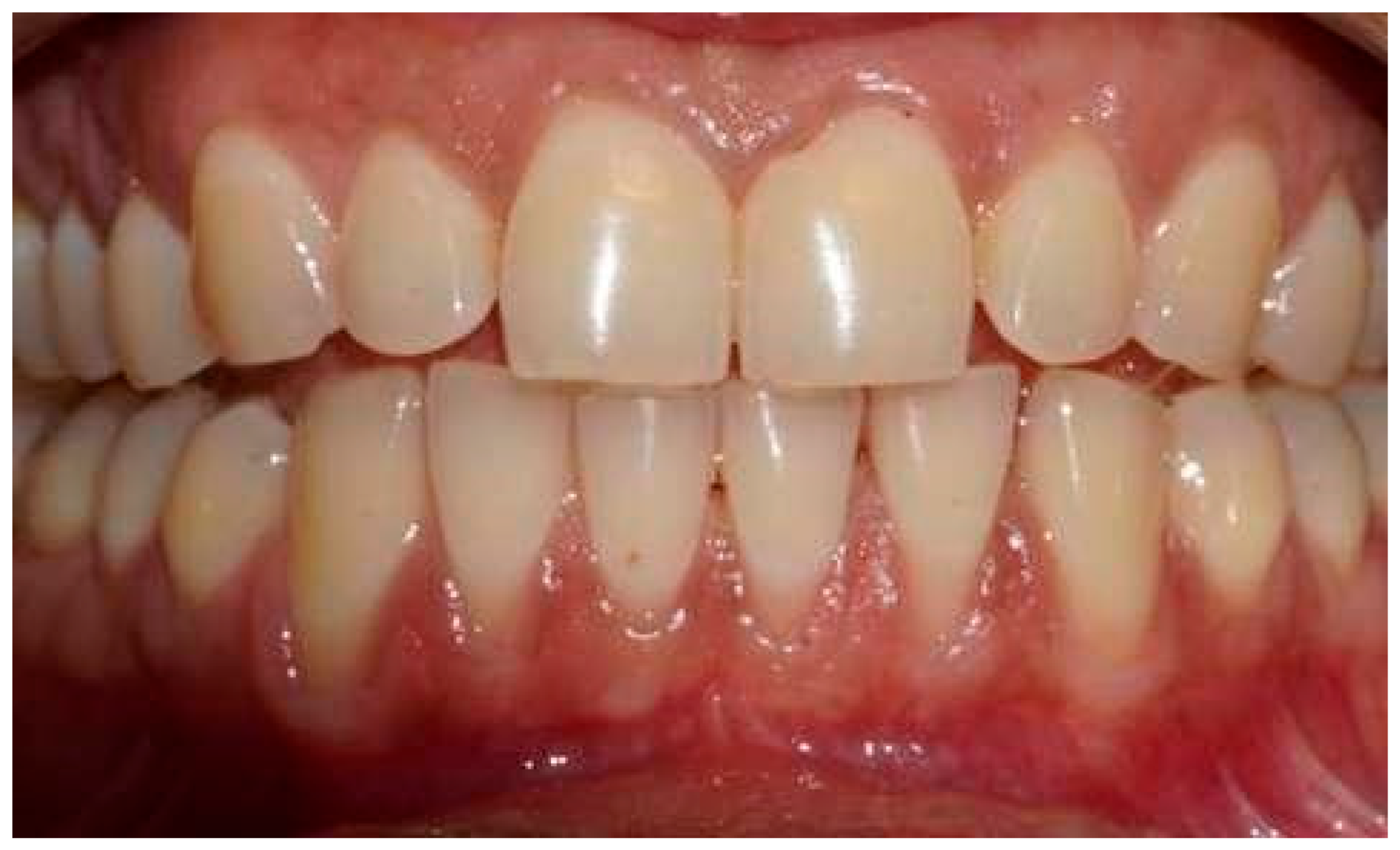
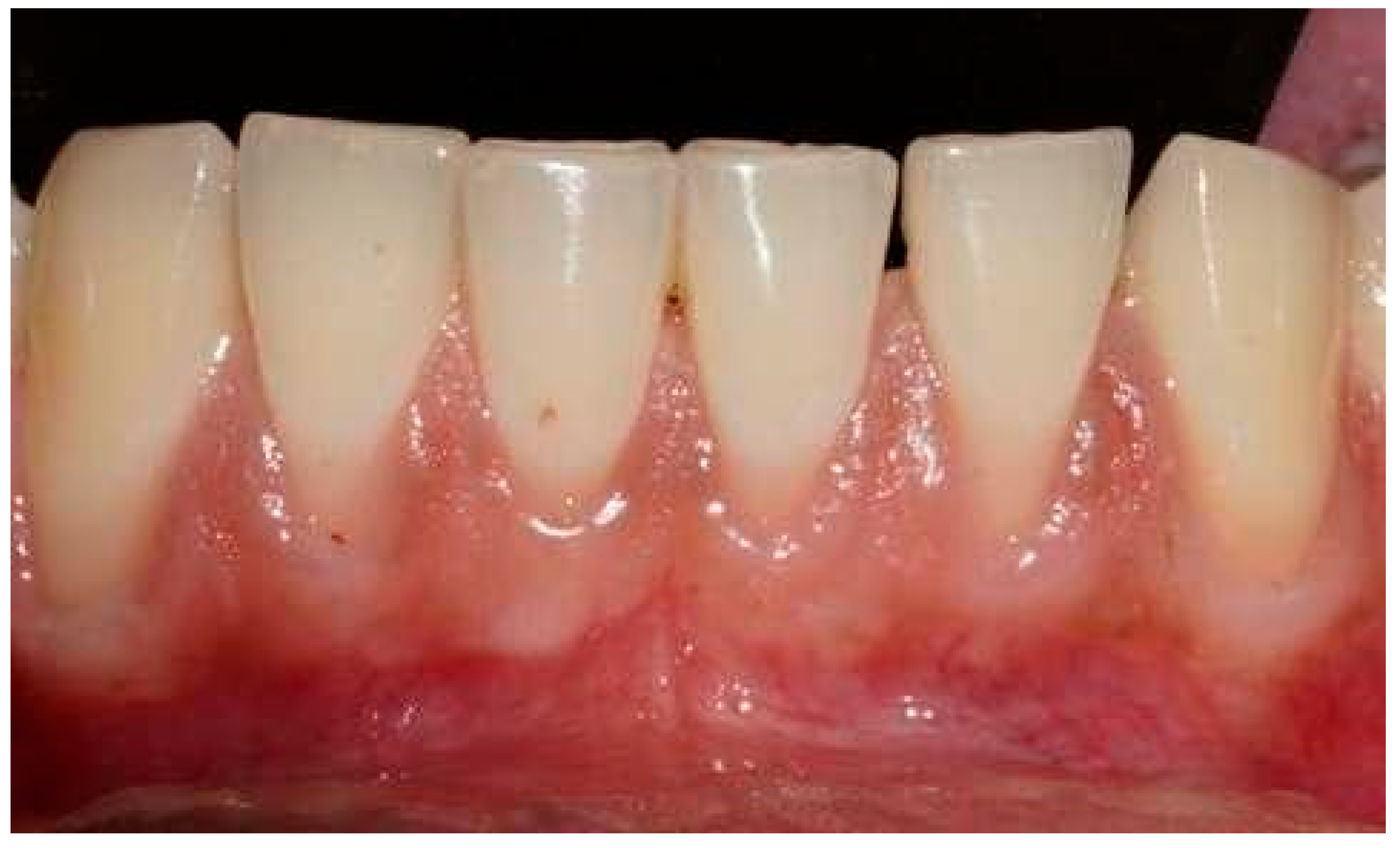
Figure 1. Pre-operative clinical view.
Applsci 16 05445 g001
Figure 2. Pre-operative clinical view, additional perspective.
Applsci 16 05445 g002
Figure 3. Intra-operative view showing the subperiosteal tunnel preparation.
Applsci 16 05445 g003
Figure 4. Harvested soft tissue grafts: subepithelial connective tissue graft (SCTG) and free gingival graft (FGG).
Applsci 16 05445 g004
Figure 5. De-epithelialization of the free gingival graft to obtain the D-FGG.
Applsci 16 05445 g005
Figure 6. Adaptation of the soft tissue graft within the prepared tunnel.
Applsci 16 05445 g006
Figure 7. Donor-site suturing after graft harvesting.
Applsci 16 05445 g007
Figure 8. Recipient-site suturing and coronal flap advancement.
Applsci 16 05445 g008
Figure 9. Inter-group comparison of patient morbidity measures at 1 and 2 weeks.
Applsci 16 05445 g009
Figure 10. Inter-group comparison of recession depth at baseline, residual recession and percentage of root coverage at 6 months.
Applsci 16 05445 g010
Figure 11. One-week follow-up, recipient site.
Applsci 16 05445 g011
Figure 12. One-week follow-up, donor site.
Applsci 16 05445 g012
Figure 13. Two-week follow-up, recipient site.
Applsci 16 05445 g013
Figure 14. Two-week follow-up, donor site.
Applsci 16 05445 g014
Figure 15. One-month follow-up, recipient site.
Applsci 16 05445 g015
Figure 16. One-month follow-up, donor site.
Applsci 16 05445 g016
Figure 17. Six-month follow-up, recipient site.
Applsci 16 05445 g017
Figure 18. Inter-group comparison of Root Coverage Esthetic Score (RES) at 6 months.
Applsci 16 05445 g018
Figure 19. Inter-group comparison of Landry Healing Index at 1 and 2 weeks.
Applsci 16 05445 g019
Table 1. Demographic profile of the study sample.
Test Group (n = 38)Control Group (n = 36)
Age (range) 43 ± 13.07 (25–68)43 ± 13.07 (25–68)
Tooth locationMaxilla2016
Mandible1820
Type of teethIncisors1817
Canines87
Premolars1212
Table 2. Intra-group comparison of patient morbidity measures in the test and control groups.
VariableTimeMeanSDSEt-Valuep-Value
Test Group
Post-op pain (donor)1 week2.50.730.18310.967<0.001
2 weeks1.1880.5440.136
Post-op pain (recipient)1 week1.6880.7040.1768.062<0.001
2 weeks0.8750.6190.155
Chewing discomfort (donor)1 week2.6880.7930.1987.064<0.001
2 weeks1.250.5770.144
Post-op bleeding (donor)1 week1.9380.8540.2138.474<0.001
2 weeks0.0630.250.063
Control Group
Post-op pain (donor)1 week3.8130.8340.2098.295<0.001
2 weeks1.6880.7040.176
Post-op pain (recipient)1 week1.750.6830.1715.514<0.001
2 weeks0.8130.6550.164
Chewing discomfort (donor)1 week1.750.6830.1717.251<0.001
2 weeks0.5630.6290.157
Post-op bleeding (donor)1 week1.3130.6020.1518.66<0.001
2 weeks0.0630.250.063
Table 3. Intra-group comparison of Landry Healing Index and recession depth in the test and control groups.
VariableTimeMeanNSDSEt-Valuep-Value
Test Group
Healing Index1 week3.313160.4790.12−8.474<0.001
2 weeks4.25160.6830.171
Recession (mm)Baseline2.342380.8150.13219.384<0.001
Residual0.171380.3140.051
Control Group
Healing Index1 week3.5160.6320.158−10.954<0.001
2 weeks4.5160.5160.129
Recession (mm)Baseline2.289380.7320.11920.796<0.001
Residual0.145380.2830.046
Table 4. Inter-group comparison of root coverage outcomes.
VariableGroupNMeanSDSEt-Valuep-Value
Recession at baseline (mm)Test382.320.820.130.3640.717
Control362.260.740.13
Residual recession (mm)Test380.180.320.050.2580.797
Control360.160.290.05
Root coverage (%)Test3894.2210.281.69−0.1600.873
Control3694.69.991.69
SD: standard deviation; SE: standard error; p < 0.05 considered statistically significant.
Disclaimer/Publisher’s Note: The statements, opinions and data contained in all publications are solely those of the individual author(s) and contributor(s) and not of MDPI and/or the editor(s). MDPI and/or the editor(s) disclaim responsibility for any injury to people or property resulting from any ideas, methods, instructions or products referred to in the content.