Article

Effects of Abutment Screw Preload on Peri-Implant Bone Tissue Under Dynamic Loading: A Preliminary In Vivo Rabbit Study


1
Division of Bio-Prosthodontics, Faculty of Dentistry & Graduate School of Medical and Dental Sciences, Niigata University, Niigata 951-8514, Japan
2
Department of Advanced Prosthodontics, Graduate School of Medical and Dental Sciences, Institute of Science Tokyo, Tokyo 113-8510, Japan
*
Author to whom correspondence should be addressed.
Appl. Sci. 2026, 16(11), 5227; https://doi.org/10.3390/app16115227
Submission received: 23 April 2026 / Revised: 16 May 2026 / Accepted: 20 May 2026 / Published: 23 May 2026
(This article belongs to the Section Applied Dentistry and Oral Sciences)

Abstract

This study evaluated how abutment screw preload affects peri-implant bone under vertical dynamic loading using an in vivo rabbit tibia model. Eight Japanese white rabbits received two implants in each tibia. After 8 weeks of healing, implants were assigned to a control group without abutment connection or to abutment-connected groups tightened to 35 Ncm or 70 Ncm. The abutment groups were further divided into loading and non-loading subgroups. In the loading groups, vertical dynamic loading (50 N, 3 Hz, 1800 cycles) was applied twice weekly for 4 weeks. Peri-implant bone responses were assessed by micro-computed tomography, histology, and histomorphometry. Under loading conditions, the 35 Ncm group showed significantly higher bone volume, bone-to-implant contact, and bone area fraction than the 70 Ncm group (p < 0.05). Histologically, the 35 Ncm group exhibited more continuous cortical bone and new bone formation, whereas the 70 Ncm group more frequently showed cortical discontinuity and enlarged marrow spaces. Within the limitations of this animal study, abutment screw preload influenced peri-implant bone adaptation under repeated loading, and the manufacturer-recommended torque of 35 Ncm was associated with more favorable bone parameters than the 70 Ncm condition.

1. Introduction

The long-term success of dental implants depends on the maintenance of stable peri-implant bone under functional loading conditions [1,2]. Peri-implant bone remodeling is influenced by multiple factors, including biological conditions, implant design, and mechanical loading. While physiologic loading can promote bone formation and remodeling, excessive or unfavorable mechanical stress may lead to bone resorption and compromised implant stability [3,4,5].
Abutment screw preload, generated during tightening, is essential for maintaining the stability of the implant–abutment connection. However, preload also induces internal stress within the implant system and may influence stress distribution in the peri-implant bone [6]. Previous studies using finite element analysis and in vitro models have suggested that higher preload levels can increase stress concentrations around the implant–abutment interface and in the adjacent bone [7,8]. Despite these findings, the biologic response of peri-implant bone to preload has not been sufficiently investigated in vivo [9].
In a previous animal study, we demonstrated that abutment screw preload alone could affect peri-implant bone morphology in the absence of functional loading [10]. However, in clinical situations, implants are exposed to repeated occlusal forces after prosthetic loading. Dynamic loading has been reported to stimulate bone remodeling and, depending on the magnitude and frequency of the applied force, may enhance peri-implant bone formation [11,12,13,14]. Therefore, the interaction between preload and dynamic loading is likely to play an important role in peri-implant bone adaptation.
To date, few in vivo studies have evaluated the combined effects of abutment screw preload and dynamic loading on peri-implant bone. Furthermore, although manufacturer-recommended torque values are widely used in clinical practice, their biologic relevance remains unclear [9]. In contrast, excessive torque may represent a worst-case mechanical condition that could adversely affect peri-implant bone.
Therefore, the purpose of this study was to evaluate the effect of abutment screw preload on peri-implant bone under vertical dynamic loading in a rabbit tibia model. The null hypotheses were that (1) abutment screw preload would not affect peri-implant bone under dynamic loading and (2) excessive preload would not result in differences in peri-implant bone compared with the manufacturer-recommended preload.

2. Materials and Methods

2.1. Implants

Thirty-two titanium implants (8 mm in length and 3.3 mm in diameter; Roxolid SLActive Bone Level, Institut Straumann AG, Basel, Switzerland) and 24 abutments (Narrow Connection Anatomic Straight Abutment; Institut Straumann AG, Basel, Switzerland) were used.

2.2. Animals

Eight male Japanese White rabbits (16–18 weeks old; body weight, 3.0–3.5 kg) were used. The animals were housed individually under controlled environmental conditions (22–25 °C, 55% humidity, and a 12 h light/dark cycle) with free access to food and water. All procedures were approved by the Institutional Animal Care and Use Committee of Niigata University (Approval No. SA01093) and were conducted in accordance with the ARRIVE guidelines.

2.3. Surgical Procedures and Experimental Design

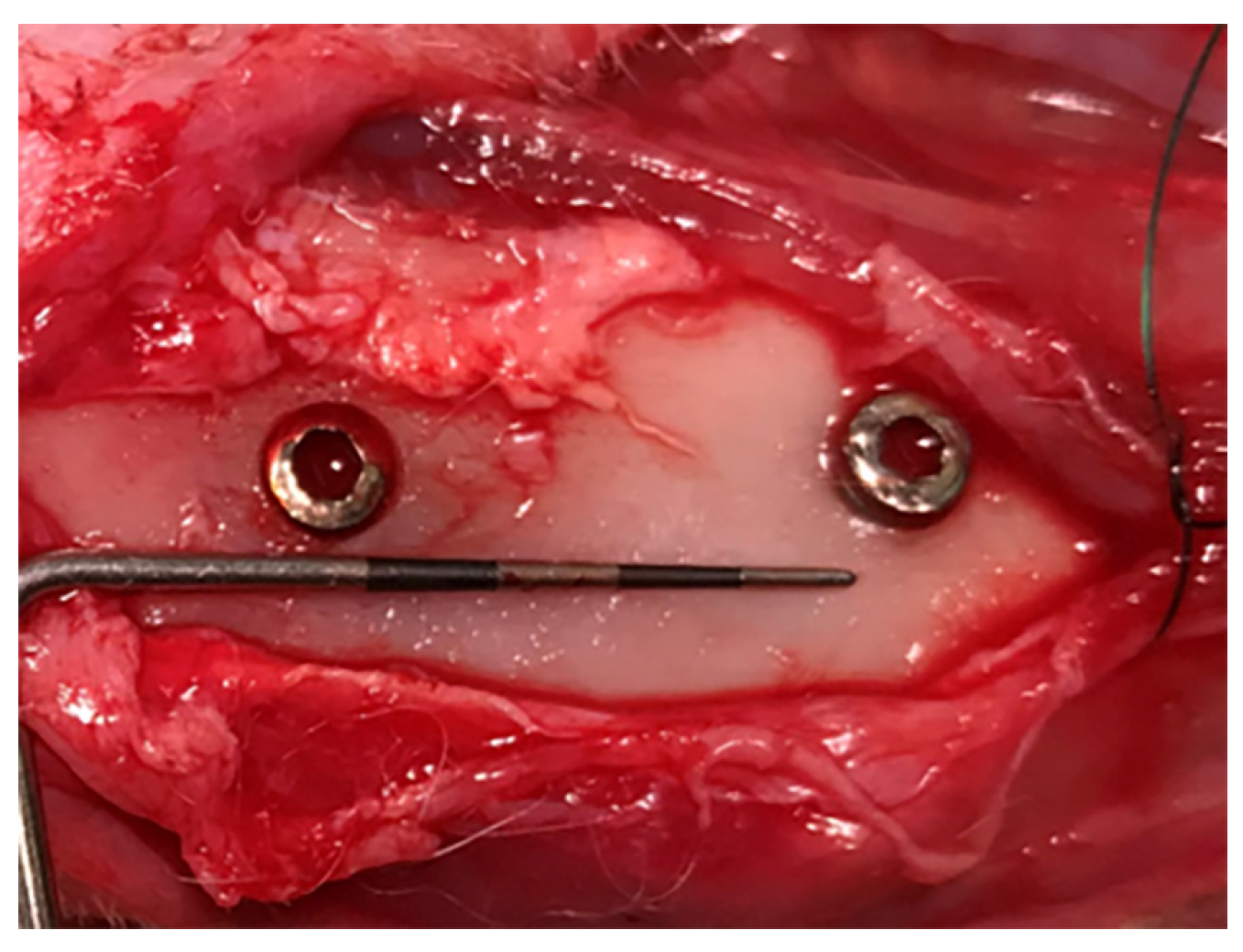
Each rabbit received two implants in each tibia (four implants per animal), which were placed on the medial surface with an inter-implant distance of 12 mm (Figure 1).
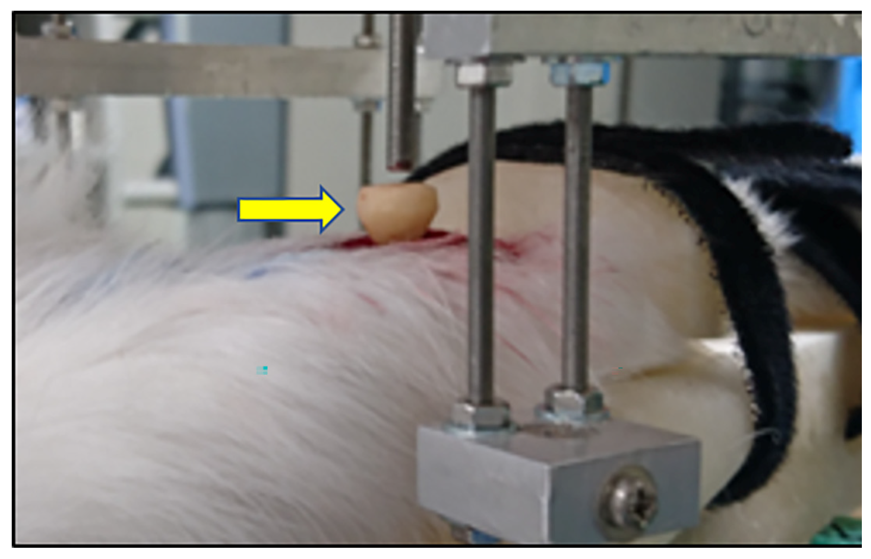
Implant placement was performed under general anesthesia induced by intramuscular administration of medetomidine (0.5 mg/kg; Domitor; Nippon Zenyaku Kogyo, Fukushima, Japan), midazolam (2.0 mg/kg; Sando, Yamagata, Japan), and butorphanol (0.5 mg/kg; Meiji Seika Pharma, Tokyo, Japan), together with local anesthesia using 2% lidocaine with epinephrine (Sandoz, Tokyo, Japan). Standardized drilling procedures were performed according to the manufacturer’s instructions under saline irrigation. The drilling sequence consisted of a 1.4 mm round bur followed by a 2.2 mm drill and was completed with a 2.8 mm drill. A cover screw was placed to seal the abutment screw access hole. Ketoprofen (3 mg/kg; Kissei Pharmaceutical Co., Ltd., Nagano, Japan) was administered perioperatively and postoperatively for analgesia. After 8 weeks of healing, which was selected to allow initial osseointegration in the rabbit tibia model based on previous related animal studies [10], the implants were randomly assigned to three groups: (1) no abutment connection (control, n = 6), (2) abutments tightened to 35 Ncm (manufacturer-recommended torque, n = 12), and (3) abutments tightened to 70 Ncm (high-torque condition, n = 12). Each abutment-connected group (35 Ncm and 70 Ncm) was further divided into loading and non-loading subgroups (n = 6 each). Abutments were tightened to the designated torque values using a digital torque meter (Newton-1; Kyoto Tool Co., Ltd., Kyoto, Japan). In the loading groups, vertical dynamic loading (50 N, 3 Hz, 1800 cycles) was applied twice weekly for 4 weeks using a custom-designed loading device (Figure 2) [15]. The 4-week loading period was selected to evaluate early peri-implant bone adaptation to repeated mechanical stimulation while minimizing excessive animal burden and potential complications during a prolonged experimental period. The loading frequency and magnitude were determined with reference to previous dynamic loading studies in small animal models and to the reported masticatory frequency and strain environment in rabbits [12,16,17,18,19]. All loading procedures were performed under general anesthesia. At the end of the experimental period, the animals were euthanized by intravenous administration of potassium chloride (Fujifilm, Tokyo, Japan) under general anesthesia, and tibial specimens containing the implants were harvested for subsequent analysis.

2.4. Micro-Computed Tomography and Histomorphometric Analysis

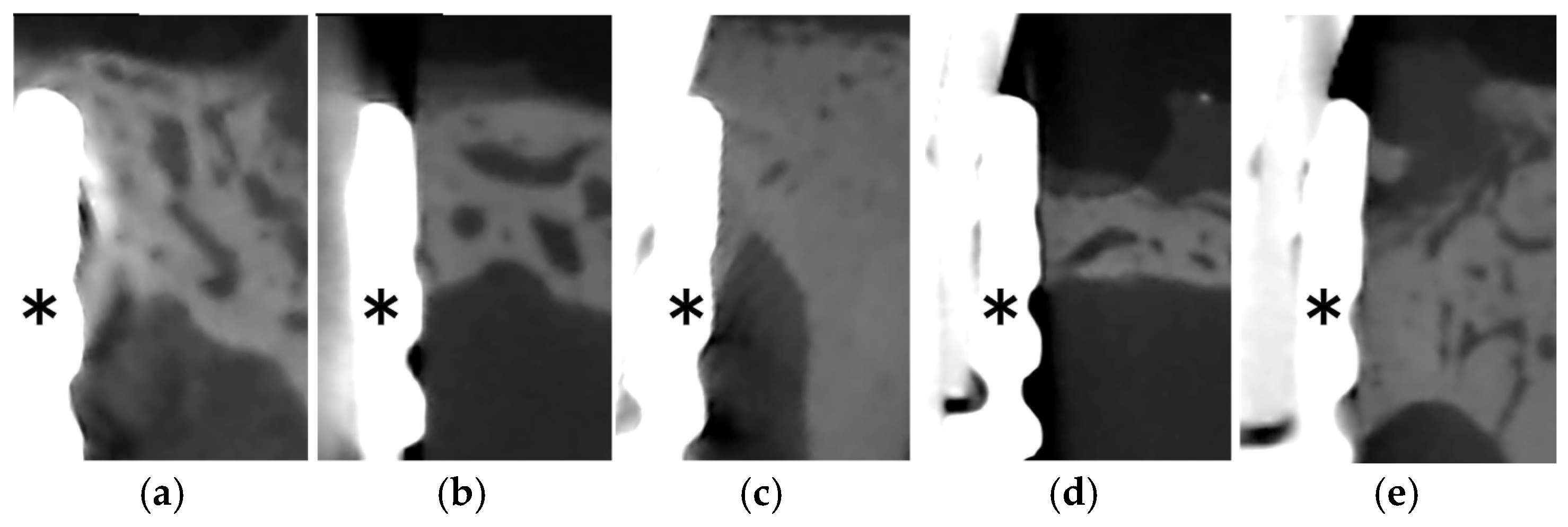
Specimens were fixed in 10% neutral buffered formalin for 3 days and then scanned using a micro-computed tomography system (CosmoScan GX; Rigaku, Tokyo, Japan) at a voxel resolution of 14 μm and a tube voltage of 80 kV (Figure 3). The acquired images were reconstructed using the manufacturer’s reconstruction software under the same reconstruction settings for all specimens. Micro-CT datasets were analyzed using TRI/3D-Bon software (Ratoc System Engineering, Tokyo, Japan), following the same basic procedure as in our previous related animal study. Bone around the implant was reconstructed and analyzed by manual contouring. A standardized region of interest (ROI) was defined around each implant, extending 1000 μm radially from the implant surface and 2500 μm vertically from the implant platform. The implant body was excluded from the ROI to avoid metal-related artifacts. Extracortical bone and periosteal reaction outside the predefined ROI were also excluded from the analysis. Bone segmentation and quantitative analysis were performed in TRI/3D-Bon, and the same procedure was applied to all specimens. Bone volume (BV, %) and bone marrow cavity fraction (BMC, %) were quantified within the defined ROI. BV (%) was calculated as (bone volume/total tissue volume) × 100, and BMC (%) was calculated as (bone marrow cavity volume/total tissue volume) × 100.
After dehydration in ascending concentrations of ethanol, the non-decalcified specimens were embedded in polymethyl methacrylate resin (Fujifilm, Tokyo, Japan). The specimens were sectioned using a diamond saw (MiniCut; SCAN-DIA GmbH, Hagen, Germany) and polished with waterproof abrasive paper to a thickness of 50–70 μm. The sections were stained with toluidine blue (Fujifilm, Tokyo, Japan) and examined under a light microscope (BX53; Olympus, Tokyo, Japan).
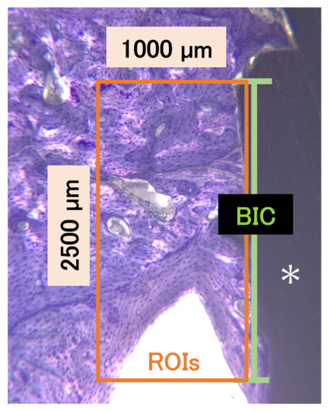
Histomorphometric analysis was performed using ImageJ software (version 1.54; National Institutes of Health, Bethesda, MD, USA). The histomorphometric parameters included bone-to-implant contact (BIC, %) and bone area fraction (BAF, %). BIC (%) was defined as (length of direct bone-to-implant contact/total evaluated implant surface length [2500 μm]) × 100 and was measured longitudinally from the implant shoulder (green line). BAF (%) was defined as (peri-implant bone area/total ROI area [2.5 mm2]) × 100 within a standardized region (2500 μm × 1000 μm) extending apically from the implant shoulder (orange box) (Figure 4).

2.5. Statistical Analysis

Because reliable preliminary data for the combined preload and dynamic loading model were not available, a formal a priori sample size calculation was not performed. Therefore, the expected effect size, statistical power, and correction for sphericity could not be prespecified. The sample size was determined with reference to previous related animal studies and was set to allow balanced allocation across all experimental groups while minimizing the number of animals used in accordance with ethical considerations for animal experiments. Statistical analyses were performed using SPSS version 28 (IBM Corp., Armonk, NY, USA). Because multiple implants were placed in the same animal, potential within-animal correlation was considered in the statistical analysis. For the abutment-connected groups, linear mixed-effects models were used to evaluate the effects of preload, loading, and their interaction, with preload and loading included as fixed effects and animal included as a random effect. In addition, group comparisons including the control group were performed using a mixed-effects model with group as a fixed effect and animal as a random effect. Pairwise comparisons were conducted based on estimated marginal means. Normality and homogeneity of variance were assessed using the Shapiro–Wilk test and Levene’s test, respectively. A significance level of p < 0.05 was used.

3. Results

3.1. Sample Distribution and Clinical Observations

A total of 32 implants were placed in eight rabbits. During the experimental period, two implants were removed because of infection and were excluded from further analysis. The remaining 30 implants were included in the final analysis and were allocated as follows: control group (n = 6), 35 Ncm without loading group (n = 6), 35 Ncm with loading group (n = 6), 70 Ncm without loading group (n = 6), and 70 Ncm with loading group (n = 6). The general condition of the animals was monitored throughout the experimental period. No major complications other than implant infection were observed in the included samples.

3.2. Micro-Computed Tomography Findings

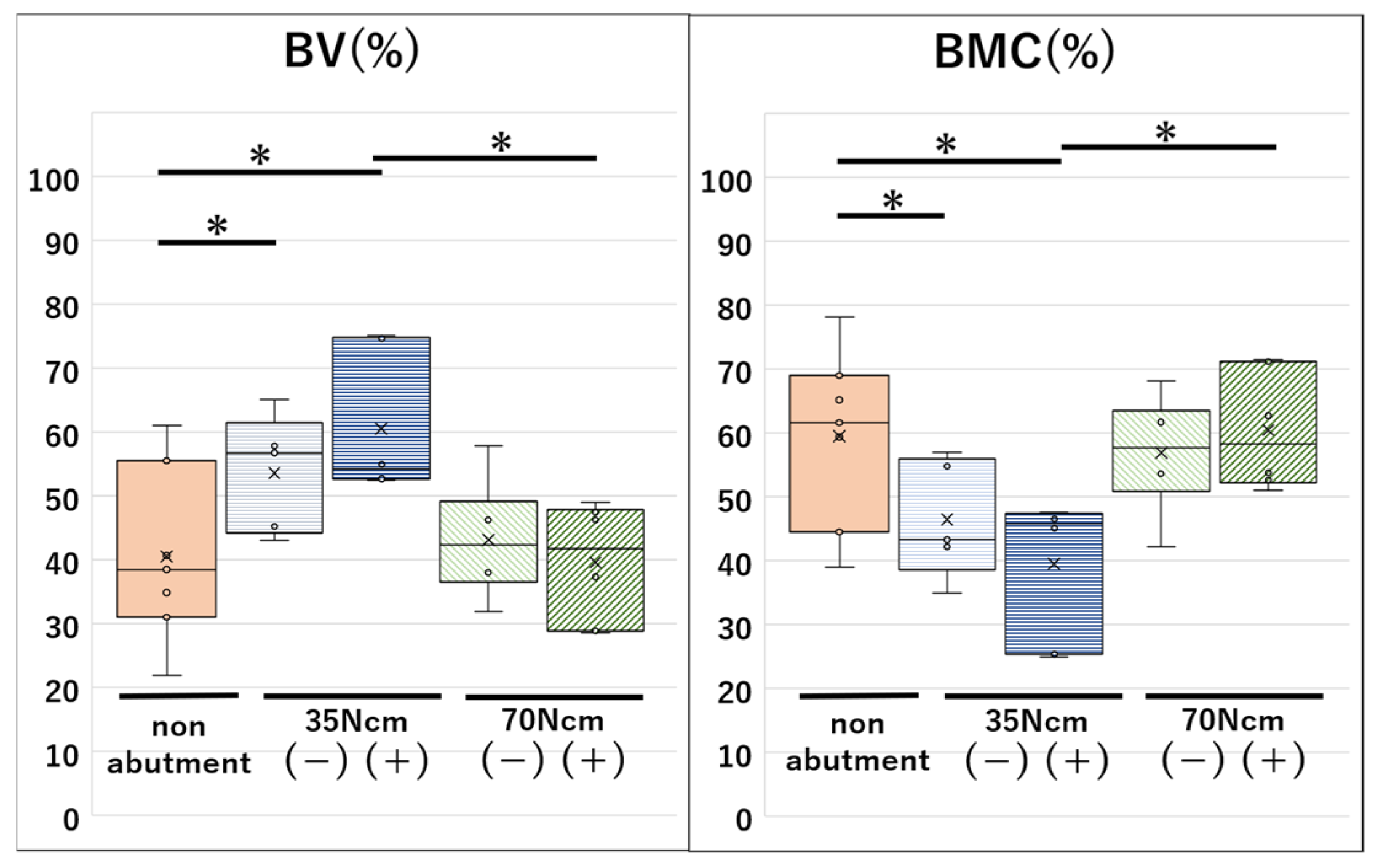
Bone volume differed among the groups. In the linear mixed-effects analysis of the abutment-connected groups, the 35 Ncm with loading group showed significantly higher BV than the 70 Ncm with loading group (p = 0.00047). In contrast, BMC was significantly lower in the 35 Ncm with loading group than in the 70 Ncm with loading group (p = 0.00047). In the supplementary analysis including the control group, both 35 Ncm groups showed significantly higher BV and lower BMC than the control group, with significant differences observed for the 35 Ncm with loading group (p = 0.00048) and the 35 Ncm without loading group (p = 0.0399). Overall, the 35 Ncm groups tended to show more favorable micro-CT parameters than the 70 Ncm groups (Figure 5 and Table 1).

3.3. Histological Observations

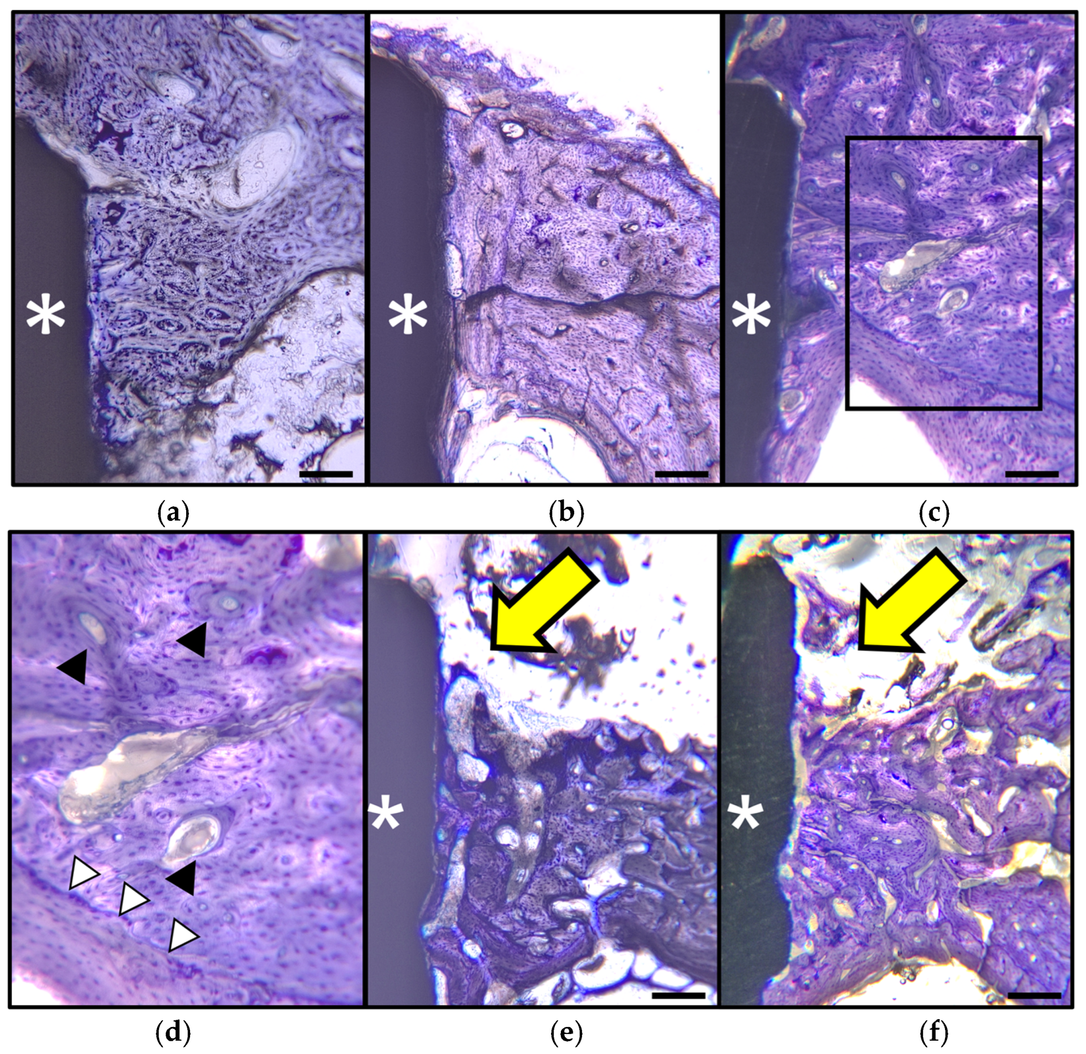
Osseointegration was maintained in all analyzed groups at the end of the experimental period. In the control group, compact cortical bone was observed adjacent to the implant shoulder, and there was virtually no bone resorption (Figure 6a). In the 35 Ncm without loading group, the histologic appearance was similar to that in the control group, although localized new bone formation was observed on the cortical bone surface (Figure 6b). In the 35 Ncm with loading group, osseointegration was preserved, and the cortical bone appeared more mature, with dense Haversian systems (black arrowhead) and newly formed bone along the inner cortical surface (white arrowhead) (Figure 6c,d). In contrast, both 70 Ncm groups more frequently showed discontinuities of the cortical bone surface and enlargement of marrow spaces. In addition, peri-implant bone levels were often located slightly apical to the implant shoulder, and bone adjacent to the implant shoulder appeared reduced (yellow arrow) compared with that in the 35 Ncm groups (Figure 6e,f).

3.4. Histomorphometric Findings

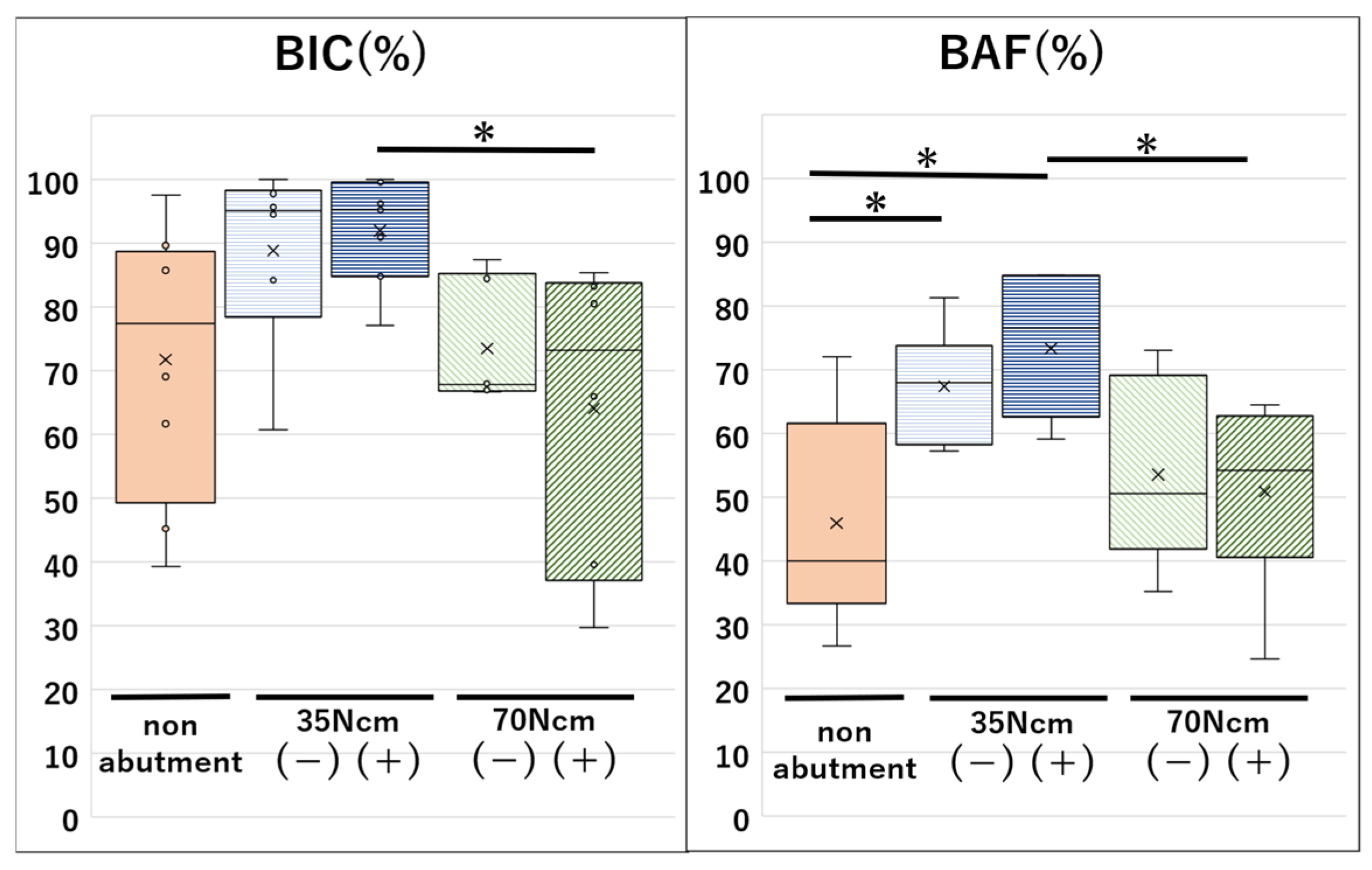
In the linear mixed-effects analysis of the abutment-connected groups, BIC was significantly higher in the 35 Ncm with loading group than in the 70 Ncm with loading group (p = 0.031). The 35 Ncm without loading group also showed significantly higher BIC than the 70 Ncm without loading group (p = 0.034). For BAF, the 35 Ncm with loading group showed a significantly higher value than the 70 Ncm with loading group (p = 0.0011), and the 35 Ncm without loading group showed a significantly higher value than the 70 Ncm without loading group (p = 0.039). In the supplementary analysis including the control group, both 35 Ncm groups showed significantly higher BAF than the control group (p = 0.00038 and p = 0.0045, respectively), whereas BIC showed no significant differences. Overall, the 35 Ncm groups showed more favorable histomorphometric parameters than the 70 Ncm groups (Figure 7 and Table 1).

4. Discussion

Preload is indispensable for maintaining the stability of the implant–abutment connection, but it also generates internal stress within the implant complex [20]. Previous in vitro and finite element studies have suggested that increasing tightening torque can alter load transfer at the implant–abutment junction, reduce micro gap formation, and increase stress within implant components, with potential effects on stress distribution in the surrounding bone [21]. The present findings extend that concept to the in vivo setting by suggesting that excessive preload may be associated with less favorable peri-implant bone conditions, particularly when combined with repeated loading. At the same time, the present results should not be interpreted as defining a universal biologic threshold for acceptable torque. Recent in vitro studies by Lorusso et al. and Lee et al. showed that tightening torque, abutment screw design, and cyclic loading can influence implant–abutment joint behavior, including micro gap formation and torque loss [22,23]. A recent systematic review also reported that implant–abutment connection geometry and cyclic loading influence preload retention and screw stability, supporting the clinical relevance of preload control under repeated loading conditions [24]. Unlike these mechanical studies, the present study evaluated peri-implant bone responses in vivo. Therefore, our findings extend previous biomechanical evidence by suggesting that preload-related mechanical differences may also be reflected in peri-implant bone adaptation. Rather, they indicate that, under the conditions tested in this model, the 35 Ncm condition was associated with more favorable peri-implant bone outcomes than the 70 Ncm condition.
Dynamic loading is known to exert both beneficial and detrimental effects on peri-implant bone, depending on the magnitude, direction, frequency, and duration of the applied force [25,26]. Implant-bearing rabbit tibiae have been reported to share similarities with the human mandible in terms of bone quality, bone density, and bone type [16,27]. The occlusal force in the human premolar region has been estimated to range from 20 to 35 N [28,29], and the chewing frequency of rabbits has been reported to be approximately 3.3 to 4.0 Hz [17,18,30]. With these considerations in mind, and taking animal tolerance into account, the dynamic loading protocol in the present study was set at 50 N, 3 Hz, and 1800 cycles. It has been reported that application of a 50 N force to the rabbit tibia produces a strain of 1661 ± 123 με [16]. Strain in the range of 1500 to 3000 με has been reported to promote osteogenesis [19] and increase cell number [31]. In the present study, the 35 Ncm with loading group showed the most favorable bone-related findings among the tested conditions. Histologically, osseointegration was maintained, dense Haversian systems were observed, and newly formed bone was identified along the cortical bone surface. These findings are consistent with previous animal studies showing that controlled dynamic or repetitive loading can promote peri-implant bone remodeling and bone formation [32,33,34,35]. In contrast, a similar favorable pattern was not observed in the 70 Ncm with loading group. This difference suggests that the biological effect of dynamic loading may depend on the preload condition of the implant–abutment assembly and that excessive preload may alter the local biomechanical environment in a manner that limits a favorable bone response to loading.
The non-abutment control group requires careful interpretation. This condition was not intended to serve as a true clinical negative control, because implants in clinical function are restored through an implant–abutment assembly. Instead, the non-abutment condition was included as a reference condition to examine the relative effect of abutment screw tightening on peri-implant bone. The most meaningful comparison in the present study is therefore between the 35 Ncm and 70 Ncm groups under the same loading condition, because these groups more directly reflect the influence of preload magnitude. This point is important when considering the biologic interpretation of the present findings.
The rationale for including the 70 Ncm condition also requires clarification. This torque value was not intended to represent a clinically recommended torque. Rather, it was established as the maximum torque that could be physically applied to the present abutment in order to model excessive preload and test whether an increase in tightening force would adversely affect peri-implant bone. Therefore, the 70 Ncm group should be interpreted as an experimental overload condition. The less favorable bone parameters observed in this group support the possibility that excessive tightening may have biologic consequences in addition to its mechanical effects. Previous reports have suggested that mechanical overload and prosthetic factors may contribute to unfavorable peri-implant bone responses [36,37,38]. In addition, finite element analyses have shown that implant–abutment connection design, abutment material, and loading conditions can influence stress and strain distribution in peri-implant bone, particularly in the crestal cortical region [39]. Nevertheless, finite element studies cannot directly demonstrate biological tissue responses such as bone remodeling, new bone formation, or changes in bone-to-implant contact. The present in vivo findings therefore provide complementary biological evidence suggesting that an excessive preload condition may be associated with less favorable peri-implant bone adaptation under repeated loading.
Several limitations of this study should be acknowledged. First, the loading protocol was limited to vertical dynamic loading and therefore did not reproduce the multidirectional complexity of clinical occlusal forces. Second, the observation period after loading was relatively short, which limited the assessment of long-term peri-implant bone adaptation. Third, the sample size was small, and two implants were excluded because of infection during the experimental period. Although balanced group sizes were achieved in the final analysis, the limited number of samples reduced statistical robustness and restricted the generalizability of the findings. Fourth, the study was performed in a rabbit tibia model rather than an oral implant model, and only one implant system was evaluated. These factors limited the direct translation of the present findings to clinical prosthodontic treatment. For these reasons, the results should be interpreted as preclinical evidence supporting a biologic influence of preload on peri-implant bone rather than as definitive evidence for an optimal clinical torque value.
Despite these limitations, the present study has an important strength. To the authors’ knowledge, few in vivo studies have specifically investigated peri-implant bone responses to abutment screw preload under dynamic loading conditions. By combining histology, histomorphometry, and micro-computed tomography, this study provided convergent evidence that preload magnitude may influence peri-implant bone behavior under repeated loading. These findings suggest, from a biologic perspective, that the effects of preload generated by tightening torque should be considered during routine clinical procedures and that the prescribed torque should be applied accurately [20].
Further studies with larger sample sizes, longer observation periods, multidirectional loading protocols, and different implant systems are needed to clarify the biologic range of preload compatible with stable peri-implant bone. Future studies should also improve transparency in sample allocation and exclusion reporting and evaluate whether similar bone responses are observed under more clinically relevant restorative conditions.

5. Conclusions

The present study evaluated the influence of abutment screw preload on peri-implant bone under vertical dynamic loading in a rabbit tibia model. Within the limitations of this experimental design, the main finding was that implants tightened to the manufacturer-recommended torque of 35 Ncm showed more favorable peri-implant bone parameters under loading than those tightened to 70 Ncm. In particular, the 35 Ncm with loading group demonstrated higher BV, BIC, and BAF values than the 70 Ncm with loading group, whereas the 70 Ncm groups showed less favorable histologic features, including cortical discontinuities and enlarged marrow spaces. These findings suggest that preload magnitude may influence peri-implant bone response under dynamic loading. Excessive preload may adversely affect peri-implant bone; however, further studies are needed before definitive clinical recommendations can be made.

Author Contributions

Conceptualization, M.N. and K.U.; methodology, Y.Y., M.N., T.Z., K.N. and K.U.; software, Y.Y. and M.N.; validation, M.N. and K.U.; formal analysis, Y.Y. and M.N.; investigation, Y.Y., M.N. and T.Z.; resources, M.N. and K.U.; data curation, Y.Y. and M.N.; writing—original draft preparation, Y.Y. and M.N.; writing—review and editing, M.N. and K.U.; visualization, Y.Y. and M.N.; supervision, M.N. and K.U.; project administration, M.N. and K.U.; funding acquisition, M.N. and K.U. All authors have read and agreed to the published version of the manuscript.

Funding

This work was supported by Grant-in-Aid for Scientific Research from the Japan Society for the Promotion of Science (JSPS) (23K09271).

Institutional Review Board Statement

Animal care and experimental procedures were performed in accordance with the Guidelines for Animal Experimentation of Niigata University School of Dentistry, with approval from the Intramural Animal Care and Use Committee, Niigata, Japan (Approval no. SA01093). The study protocol was designed in accordance with all recommendations of the ARRIVE (Animal Research: Reporting In Vivo Experiments) guidelines for conducting experimental animal studies.

Data Availability Statement

The data presented in this study are available on request from the corresponding author. The data are not publicly available due to institutional and animal experiment data management restrictions.

Acknowledgments

Straumann Japan Co., Ltd., Tokyo, Japan partially supported this experiment.

Conflicts of Interest

The authors declare no conflicts of interest associated with this manuscript.

Abbreviations

The following abbreviations are used in this manuscript:
ANOVAAnalysis of variance
BAF (%)Bone area fraction, (peri-implant bone area/total ROI area) × 100
BIC (%)Bone-to-implant contact, (length of direct bone-to-implant contact/total evaluated implant surface length) × 100
BMC (%)Bone marrow cavity, (bone marrow cavity volume/total tissue volume) × 100
BV (%)Bone volume, (bone volume/total tissue volume) × 100
Micro-CTMicro-computed tomography
ROIRegion of interest

References

  1. Albrektsson, T.; Brånemark, P.I.; Hansson, H.A.; Lindström, J. Osseointegrated Titanium Implants. Requirements for Ensuring a Long-Lasting, Direct Bone-to-Implant Anchorage in Man. Acta Orthop. Scand. 1981, 52, 155–170. [Google Scholar] [CrossRef]
  2. Albrektsson, T.; Zarb, G.; Worthington, P.; Eriksson, A.R. The Long-Term Efficacy of Currently Used Dental Implants: A Review and Proposed Criteria of Success. Int. J. Oral Maxillofac. Implants 1986, 1, 11–25. [Google Scholar]
  3. Heitz-Mayfield, L.J.; Lang, N.P. Comparative Biology of Chronic and Aggressive Periodontitis vs. Peri-Implantitis. Periodontology 2000 2010, 53, 167–181. [Google Scholar] [CrossRef]
  4. Nagasawa, M.; Takano, R.; Maeda, T.; Uoshima, K. Observation of the Bone Surrounding an Overloaded Implant in a Novel Rat Model. Int. J. Oral Maxillofac. Implants 2013, 28, 109–116. [Google Scholar] [CrossRef]
  5. Papantonopoulos, G.; Gogos, C.; Housos, E.; Bountis, T.; Loos, B.G. Peri-Implantitis: A Complex Condition with Non-Linear Characteristics. J. Clin. Periodontol. 2015, 42, 789–798. [Google Scholar] [CrossRef]
  6. Khraisat, A. Influence of Abutment Screw Preload on Stress Distribution in Marginal Bone. Int. J. Oral Maxillofac. Implants 2012, 27, 303–307. [Google Scholar]
  7. Ziaie, B.; Khalili, S.M.R. Evaluation of Fatigue Life for Dental Implants Using FEM Analysis. Prosthesis 2021, 3, 300–313. [Google Scholar] [CrossRef]
  8. Sun, M.; Shui, Y.; Zhang, Y.; Ma, R.; Zhao, Y.; Chen, H.; Yu, P.; Li, Z.; Wu, T.; Yu, H. Influence of Abutment Screw-Tightening Methods on the Screw Joint: Immediate and Long-Term Stability. Int. J. Dent. 2024, 2024, 5768318. [Google Scholar] [CrossRef] [PubMed]
  9. Goheen, K.L.; Vermilyea, S.G.; Vossoughi, J.; Agar, J.R. Torque Generated by Handheld Screwdrivers and Mechanical Torquing Devices for Osseointegrated Implants. Int. J. Oral Maxillofac. Implants 1994, 9, 149–155. [Google Scholar]
  10. Al-Omari, F.A.; Nagasawa, M.; Suliman, M.; Khraisat, A.; Uoshima, K. Peri-Implant Bone Alterations Under the Influence of Abutment Screw Preload Stress. A Preclinical Vivo Study. Clin. Oral Implants Res. 2010, 31, 1232–1242. [Google Scholar] [CrossRef]
  11. Leung, K.S.; Shi, H.F.; Cheung, W.H.; Qin, L.; Ng, W.K.; Tam, K.F.; Tang, N. Low-Magnitude High-Frequency Vibration Accelerates Callus Formation, Mineralization, and Fracture Healing in Rats. J. Orthop. Res. 2009, 27, 458–465. [Google Scholar] [CrossRef]
  12. Yagihara, A.; Kawasaki, R.; Mita, A.; Takakuda, K. Impact of Dynamic and Static Load on Bone Around Implants: An Experimental Study in a Rat Model. Int. J. Oral Maxillofac. Implants 2016, 31, e49–e56. [Google Scholar] [CrossRef] [PubMed]
  13. Irandoust, S.; Müftü, S. The Interplay Between Bone Healing and Remodeling Around Dental Implants. Sci. Rep. 2020, 10, 4335. [Google Scholar] [CrossRef] [PubMed]
  14. Okawara, H.; Arai, Y.; Matsuno, H.; Marcián, P.; Borák, L.; Aoki, K.; Wakabayashi, N. Effect of Load-Induced Local Mechanical Strain on Peri-Implant Bone Cell Activity Related to Bone Resorption and Formation in Mice: An Analysis of Histology and Strain Distributions. J. Mech. Behav. Biomed. Mater. 2021, 116, 104370. [Google Scholar] [CrossRef] [PubMed]
  15. Nozaki, K.; Kaku, M.; Yamashita, Y.; Yamauchi, M.; Miura, H. Effect of Cyclic Mechanical Loading on Osteoclast Recruitment in Periodontal Tissue. J. Periodontal Res. 2010, 45, 8–15. [Google Scholar] [CrossRef]
  16. Kuroshima, S.; Yasutake, M.; Tsuiki, K.; Nakano, T.; Sawase, T. Structural and Qualitative Bone Remodeling Around Repetitive Loaded Implants in Rabbits. Clin. Implant Dent. Relat. Res. 2015, 17, e699–e710. [Google Scholar] [CrossRef]
  17. Morimoto, T.; Inoue, T.; Nakamura, T.; Kawamura, Y. Characteristics of Rhythmic Jaw Movements of the Rabbit. Arch. Oral Biol. 1985, 30, 673–677. [Google Scholar] [CrossRef]
  18. Weijs, W.A.; de Jongh, H.J. Strain in Mandibular Alveolar Bone During Mastication in the Rabbit. Arch. Oral Biol. 1977, 22, 667–675. [Google Scholar] [CrossRef]
  19. Frost, H.M. Bone’s Mechanostat: A 2003 Update. Anat. Rec. A Discov. Mol. Cell. Evol. Biol. 2003, 275, 1081–1101. [Google Scholar] [CrossRef]
  20. Honório Tonin, B.S.; He, Y.; Ye, N.; Chew, H.P.; Fok, A. Effects of Tightening Torque on Screw Stress and Formation of Implant–Abutment Microgaps: A Finite Element Analysis. J. Prosthet. Dent. 2022, 127, 882–889. [Google Scholar] [CrossRef]
  21. Kwan, J.C. An In Vitro Study of Torque Application in Abutment Screw Tightening at the Implant-Abutment Junction in Simulated Osseointegrated Implants. Int. J. Oral Maxillofac. Implants 2026, 41, 171–180. [Google Scholar] [CrossRef]
  22. Lorusso, F.; Scarano, A.; Tari, S.R.; Singhal, I.; Goker, F.; Soldini, M.C.; Tartaglia, G.M.; Del Fabbro, M. The Effect of Different Tightening Torques of Implant Cone Morse Abutment Connection Under Dynamic Fatigue Loading: An In Vitro Study. Biomimetics 2025, 10, 511. [Google Scholar] [CrossRef]
  23. Lee, J.-T.; Lee, J.-C.; Han, D.-W.; Kim, B. Effect of Abutment Screw Design on Torque Loss Under Cyclic Fatigue Loading: A Comparison of TSIII and KSIII Implant Systems. Bioengineering 2025, 12, 1131. [Google Scholar] [CrossRef]
  24. Sammour, R.R.; Ziqi, X.; Ogawa, T.; Yoda, N. Impact of Implant Abutment Connection Designs and Cyclic Loading on Screw Stability in Dental Implants: A Systematic Review and Meta-Analysis. J. Prosthet. Dent. 2025, 135, 317.e1–317.e14. [Google Scholar] [CrossRef]
  25. Kadow-Romacker, A.; Duda, G.N.; Bormann, N.; Schmidmaier, G.; Wildemann, B. Slight Changes in the Mechanical Stimulation Affects Osteoblast- and Osteoclast-Like Cells in Co-Culture. Transfus. Med. Hemother. 2013, 40, 441–447. [Google Scholar] [CrossRef] [PubMed]
  26. Yan, Y.-X.; Gong, Y.-W.; Guo, Y.; Lv, Q.; Guo, C.; Zhuang, Y.; Zhang, Y.; Li, R.; Zhang, X.-Z. Mechanical Strain Regulates Osteoblast Proliferation Through Integrin-Mediated ERK Activation. PLoS ONE 2012, 7, e35709. [Google Scholar] [CrossRef]
  27. Slotte, C.; Lundgren, D.; Sennerby, L.; Lundgren, A.K. Surgical Intervention in Enchondral and Membranous Bone: Intraindividual Comparisons in the Rabbit. Clin. Implant Dent. Relat. Res. 2003, 5, 263–268. [Google Scholar] [CrossRef] [PubMed]
  28. Hattori, Y.; Satoh, C.; Kunieda, T.; Endoh, R.; Hisamatsu, H.; Watanabe, M. Bite Forces and Their Resultants During Forceful Intercuspal Clenching in Humans. J. Biomech. 2009, 42, 1533–1538. [Google Scholar] [CrossRef] [PubMed]
  29. Shiota, M.; Ohno, S. Clinical Study of Occlusal Forces on Dental Implants. Kokubyo Gakkai Zasshi 1997, 64, 405–412. (In Japanese) [Google Scholar] [CrossRef]
  30. Peptan, A.I.; Lopez, A.; Kopher, R.A.; Mao, J.J. Responses of Intramembranous Bone and Sutures Upon In Vivo Cyclic Tensile and Compressive Loading. Bone 2008, 42, 432–438. [Google Scholar] [CrossRef]
  31. Kaspar, D.; Seidl, W.; Neidlinger-Wilke, C.; Beck, A.; Claes, L.; Ignatius, A. Proliferation of Human-Derived Osteoblast-Like Cells Depends on the Cycle Number and Frequency of Uniaxial Strain. J. Biomech. 2002, 35, 873–880. [Google Scholar] [CrossRef] [PubMed]
  32. Hu, X.; Fan, Y.; Chen, X. RANKL, OPG, and CTS-K Release in Bone Response to Immediate Nonfunctional Loading of a Single Implant in Mandibular Molar Sites During Osseointegration Establishment. Clin. Exp. Dent. Res. 2025, 11, e70193. [Google Scholar] [CrossRef]
  33. Duyck, J.; Vandamme, K. The Effect of Loading on Peri-Implant Bone: A Critical Review of the Literature. J. Oral Rehabil. 2014, 41, 783–794. [Google Scholar] [CrossRef]
  34. Leucht, P.; Kim, J.-B.; Wazen, R.; Currey, J.A.; Nanci, A.; Brunski, J.B.; Helms, J.A. Effect of Mechanical Stimuli on Skeletal Regeneration Around Implants. Bone 2007, 40, 919–930. [Google Scholar] [CrossRef]
  35. Yorioka, H.; Otsu, Y.; Suzuki, R.; Matsunaga, S.; Nakano, T.; Abe, S.; Sasaki, H. The Influence of Immediate Occlusal Loading on Micro/Nano-Structure of Peri-Implant Jaw Bone in Rats. Int. J. Implant Dent. 2024, 10, 24. [Google Scholar] [CrossRef]
  36. Lin, G.; Lee, E.; Barootchi, S.; Rosen, P.S.; Curtis, D.; Kan, J.; Wang, H. The Influence of Prosthetic Designs on Peri-Implant Bone Loss: An AO/AAP Systematic Review and Meta-Analysis. J. Periodontol. 2025, 96, 634–651. [Google Scholar] [CrossRef] [PubMed]
  37. Isidor, F. Histological Evaluation of Peri-Implant Bone at Implants Subjected to Occlusal Overload or Plaque Accumulation. Clin. Oral Implants Res. 1997, 8, 1–9. [Google Scholar] [CrossRef]
  38. Kozlovsky, A.; Tal, H.; Laufer, B.; Leshem, R.; Rohrer, M.D.; Weinreb, M.; Artzi, Z. Impact of Implant Overloading on the Peri-Implant Bone in Inflamed and Non-Inflamed Peri-Implant Mucosa. Clin. Oral Implants Res. 2007, 18, 601–610. [Google Scholar] [CrossRef] [PubMed]
  39. Ziada, A.; Beleidy, M. Influence of Different Customized Abutment Materials on Stress Distribution of Different Internal Implant-Abutment Connections: Finite Element Analysis. BMC Oral Health 2025, 25, 1820. [Google Scholar] [CrossRef]
Figure 1. Implants placed on the medial surface of the rabbit tibia.
Applsci 16 05227 g001
Figure 2. Custom-designed device used to apply vertical dynamic loading. The arrow indicates the loading jig attached to the implant fixture.
Applsci 16 05227 g002
Figure 3. Micro-computed tomography images of the analyzed area. (a) Non abutment connection (Control), (b) 35 Ncm without loading group, (c) 35 Ncm with loading group, (d) 70 Ncm without loading group, (e) 70 Ncm with loading group. *; Implant body.
Applsci 16 05227 g003
Figure 4. Cross-section of the analyzed specimen including the implant body. BAF was measured within the region indicated by the orange box (ROI). BIC was measured along the green line corresponding to the evaluated implant surface. *; Implant body.
Applsci 16 05227 g004
Figure 5. Box-and-whisker plots of micro-computed tomography measurements of BV and BMC. 35 Ncm (−); 35 Ncm without loading group, 35 Ncm (+); 35 Ncm with loading group, 70 Ncm (−); 70 Ncm without loading group, 70 Ncm (+); 70 Ncm with loading group, * p < 0.05.
Applsci 16 05227 g005
Figure 6. Non-decalcified sections of the analyzed areas including the implant body. (a) Non abutment connection (Control group). (b) 35 Ncm without loading group. (c) 35 Ncm with loading group. (d) Magnified view of the area enclosed by the rectangle in panel (c). (e) 70 Ncm without loading group. (f) 70 Ncm with loading group. Panels (ac,e,f), ×50; panel (d), ×100. Scale bar = 500 μm. *; Implant body. Toluidine blue.
Applsci 16 05227 g006
Figure 7. Box-and-whisker plots of histomorphometric measurements of BIC and BAF. 35 Ncm (−); 35 Ncm without loading group, 35 Ncm (+); 35 Ncm with loading group, 70 Ncm (−); 70 Ncm without loading group, 70 Ncm (+); 70 Ncm with loading group, * p < 0.05.
Applsci 16 05227 g007
Table 1. Mean values and standard deviations (SDs) of the peri-implant bone parameters evaluated in each group.
NumberBV (%)BMC (%)BIC (%)BAF (%)
Non-abutment640.50 ± 13.6459.50 ± 13.6471.72 ± 21.5245.93 ± 16.00
35 Ncm
without loading
653.57 ± 9.2246.43 ± 9.2288.80 ± 14.8067.38 ± 8.80
35 Ncm
with loading
660.53 ± 11.0839.47 ± 11.0891.95 ± 8.4173.33 ± 10.43
70 Ncm
without loading
643.09 ± 9.0756.91 ± 9.0773.49 ± 9.6653.55 ± 14.64
70 Ncm
with loading
639.57 ± 9.3160.43 ± 9.3164.05 ± 23.9850.90 ± 15.27
Disclaimer/Publisher’s Note: The statements, opinions and data contained in all publications are solely those of the individual author(s) and contributor(s) and not of MDPI and/or the editor(s). MDPI and/or the editor(s) disclaim responsibility for any injury to people or property resulting from any ideas, methods, instructions or products referred to in the content.