Article

Protective Effects of Violaxanthin on Cellular Oxidative Stress via NRF2 Activation in H2O2-Stimulated Human Keratinocytes


1
Department of Cosmetics Engineering, Konkuk University, 120 Neungdong-ro, Gwangjin-gu, Seoul 05029, Republic of Korea
2
Dermato Bio, Inc., 25-23 Dasanjungang-ro, 19beon-gil, Namyangju 12248, Republic of Korea
3
Department of Biological Engineering, Konkuk University, 120 Neungdong-ro, Gwangjin-gu, Seoul 05029, Republic of Korea
4
Cell Factory Research Center, Korea Research Institute of Bioscience and Biotechnology (KRIBB), Daejeon 34141, Republic of Korea
5
Department of Bioresource and Environmental Engineering, KRIBB School of Biotechnology, University of Science and Technology, Daejeon 34113, Republic of Korea
6
ASK LABS Co., Ltd., KRIBB BVC Center 109, Daejeon 34141, Republic of Korea
*
Author to whom correspondence should be addressed.
These authors contributed equally to this work.
Appl. Sci. 2026, 16(10), 5132; https://doi.org/10.3390/app16105132
Submission received: 23 April 2026 / Revised: 15 May 2026 / Accepted: 18 May 2026 / Published: 21 May 2026

Abstract

Excessive accumulation of reactive oxygen species from exogenous and endogenous stressors can cause cellular damage. Chlorella contains diverse bioactive compounds, and violaxanthin, a major carotenoid pigment found in Chlorella sp. HS-V, has been reported to possess anti-inflammatory, anticancer, and antioxidant properties. We investigated the effect of violaxanthin on hydrogen peroxide (H2O2)-induced oxidative stress in human keratinocytes. Chlorella sp. HS-V extract significantly restored the H2O2-induced decrease in cell viability. Similarly, violaxanthin reduced H2O2-induced cytotoxicity and intracellular reactive oxygen species levels, which was associated with the upregulation of antioxidant enzyme expression. Under H2O2-induced oxidative stress conditions, violaxanthin may enhance cellular antioxidant defense by promoting nuclear factor erythroid 2-related factor 2 (NRF2) translocation through the phosphoinositide 3-kinase/protein kinase B/glycogen synthase kinase 3β (PI3K/AKT/GSK3β) signaling pathway. Additionally, violaxanthin improved H2O2-impaired wound healing in HaCaT human keratinocyte cells and reduced senescence-associated beta-galactosidase-positive normal human epidermal keratinocytes. Overall, these findings suggest that violaxanthin may serve as a potential therapeutic agent for mitigating oxidative stress-induced cellular dysfunction.

1. Introduction

The skin is the outermost organ of the body and acts as the first line of defense against bacteria and other organisms [1,2]. Keratinocytes are the primary cellular constituents of the epidermis and play a crucial role in maintaining skin barrier function [3]. Skin can be stimulated by reactive oxygen species (ROS) resulting from various factors, such as exposure to ultraviolet rays and the intrinsic aging process [4]. ROS are inherently unstable and react strongly with surrounding substances, resulting in various types of damage [5,6]. Many studies have revealed that the excessive accumulation of ROS induces oxidative stress, which can damage DNA and other biomolecules, thereby contributing to inflammation, aging, and tumorigenesis [7,8,9,10]. ROS can also lead to dysfunction and damage in keratinocytes, which play a key role in skin barrier function. O’Toole et al. showed that the migration of human keratinocytes, which is an essential process in wound healing, was inhibited by hydrogen peroxide (H2O2) [11]. Other studies have suggested that increased intracellular ROS production and deficiencies in antioxidant defense systems within keratinocytes contribute to the pathogenesis of keratinocyte-related inflammatory diseases, such as psoriasis [12,13]. Cellular defense against oxidative stress is regulated by multiple signaling pathways that coordinate ROS detoxification, antioxidant enzyme expression, inflammatory responses, and cell survival. Among these, the NRF2/antioxidant response element (ARE) pathway is a central defense system that induces cytoprotective and antioxidant genes, including HO-1, NQO1, SOD, CAT, and GPx [14,15]. In addition, upstream pathways such as PI3K/AKT/GSK3β, mitogen-activated protein kinases (MAPKs; ERK, JNK, and p38), and AMP-activated protein kinase (AMPK) have been reported to modulate oxidative stress responses by regulating NRF2 stabilization, nuclear translocation, antioxidant enzyme induction, and stress-adaptive cell survival [16,17,18]. Therefore, elucidating and activating these antioxidant defense mechanisms may be an important strategy for protecting keratinocytes against H2O2-induced oxidative stress.
Microalgae are photosynthetic microorganisms that possess diverse defense systems to survive under a variety of environmental conditions, enabling the synthesis of numerous bioactive metabolites [19]. Numerous studies have reported that microalgae contain a wide range of bioactive compounds, including proteins, fatty acids, phenolic compounds, vitamins, and photosynthetic pigments [20,21]. These microalgae-derived metabolites have attracted considerable attention for their potential applications in the cosmeceutical [22,23,24,25], pharmaceutical [24,25,26,27,28], and food industries [25,27,28,29]. Among various microalgae, Chlorella is a freshwater unicellular green alga [30]. Chlorella has been reported to contain vitamins, lipids, and carotenoids, including zeaxanthin, violaxanthin, and astaxanthin [19,31,32]. Due to its rich bioactive composition, Chlorella has been reported to exhibit diverse physiological activities [33,34,35]. Recent studies have demonstrated that Chlorella extract exhibits anticancer activity, including the suppression of apoptosis-related pathways, in various cancer cell lines [36,37,38]. Additionally, Chlorella extract has been shown to possess anti-inflammatory and immunomodulatory properties through the downregulation of several inflammatory cytokines [39,40,41,42]. Among the carotenoids found in Chlorella, violaxanthin is a major carotenoid that can be isolated from this microalga [38,43,44,45,46,47]. Previous studies have reported that violaxanthin exhibits higher antioxidant capacity compared to β-carotene and lutein [45]. Previous studies have indicated that violaxanthin exhibits antiproliferative activity in cancer cells and triggers biochemical changes indicative of early apoptosis [38,48]. Violaxanthin has also been shown to exert anti-inflammatory effects by inhibiting the nuclear factor kappa B (NF-κB) pathway in LPS-stimulated RAW 264.7 mouse macrophages [44]. Furthermore, violaxanthin derived from Nannochloropsis oceanica was found to protect human dermal fibroblasts against UVB-induced damage, notably restoring decreased collagen expression in UVB-mediated cellular senescence [49]. These studies suggest that violaxanthin has potential as a therapeutic agent in cosmetic and pharmaceutical applications. However, the protective effects and underlying mechanisms of violaxanthin against oxidative stress in keratinocytes remain unexplored. In this study, we investigated the protective effects of violaxanthin against H2O2-induced oxidative stress in human keratinocytes and explored the underlying molecular mechanisms.

2. Materials and Methods

2.1. Cell Culture and Reagents

For this study, the immortalized human keratinocyte cell line (HaCaT) was selected, since it is commonly applied to evaluate protective mechanisms against skin oxidative stress [17,50]. HaCaT cells were obtained from Cell Line Service (DKFZ, Eppelheim, Germany). The cells were cultured in Dulbecco’s modified Eagle’s medium (DMEM, WelGene, Inc., Daegu, Republic of Korea), supplemented with 10% fetal bovine serum (FBS, Corning Inc., Corning, NY, USA) and 1% penicillin/streptomycin (Thermo Fisher Scientific, Waltham, MA, USA). nHEK cells, primary normal human epidermal keratinocytes, were obtained from CELLnTEC (Bern, Switzerland). nHEK cells were cultured in Keratinocyte Basal Medium-2 (Lonza, Basel, Switzerland), supplemented with bovine pituitary extract, epinephrine, gentamicin sulfate-amphotericin, hydrocortisone, human epidermal growth factor, insulin, and transferrin, as recommended by the manufacturer. All cell lines were maintained in a humidified incubator at 37 °C with 5% CO2. Dimethyl sulfoxide (DMSO), violaxanthin, ascorbic acid, N-acetylcysteine (NAC), and hydrogen peroxide (H2O2) were purchased from Sigma-Aldrich (St. Louis, MO, USA). LY294002 was purchased from MedChemExpress (Monmouth Junction, NJ, USA). Violaxanthin was dissolved in DMSO, and the final concentration of DMSO in the culture medium did not exceed 0.1% (v/v).

2.2. Microalgal Strain and Culture Conditions

Chlorella sp. HS-V (KCTC 13850BP; Korean Collection for Type Cultures, Jeongeup, Republic of Korea) was cultivated in 2 L Erlenmeyer flasks with a working volume of 500 mL. The culture medium consisted of 1 g/L S-feed fertilizer (Farmhannong, Seongnam, Republic of Korea), 3 g/L yeast extract (BD Biosciences, Franklin Lakes, NJ, USA), and 15 g/L glucose, with the initial pH adjusted to 7.0. Cultures were incubated at 30 °C with continuous shaking at 120 rpm for 72 h under continuous illumination at an intensity of 150 μmol photons m−2 s−1. After cultivation, the biomass was harvested by centrifugation and subsequently lyophilized for further analysis.

2.3. Preparation of Chlorella sp. HS-V Extract

Bioactive compounds were extracted from the lyophilized microalgal biomass via saponification followed by solvent fractionation. Briefly, 2 g of lyophilized biomass was combined with 8 mL of 60% (w/v) KOH aqueous solution and incubated at 40 °C for 1 h. The saponified mixture was then extracted with 60 mL of hexane/ethyl acetate (1:1, v/v) at 40 °C for 1 h with continuous stirring. Phase separation was induced by the addition of 30 mL of distilled water followed by vigorous agitation. The upper organic phase was collected and concentrated under reduced pressure using a rotary evaporator to yield the solid crude extract. The resulting extract was dissolved in medium-chain triglyceride (MCT) oil and filtered to remove insoluble particulates, yielding the final oil-based formulation.

2.4. Cell Viability Assay

HaCaT cells (4.0 × 10⁴) were seeded in 24-well plates and incubated for 24 h, followed by treatment with the indicated concentrations of Chlorella sp. HS-V extract, violaxanthin or H2O2 for 24 h. Before each subsequent treatment, the cells were washed with DPBS to remove residual compounds. Subsequently, MTT [3-(4,5-dimethylthiazol-2-yl)-2,5-diphenyltetrazolium bromide] solution (0.5 mg/mL) was added to each well, and cells were incubated for an additional 2 h. The culture medium was removed, and DMSO was added to each well to dissolve the formazan crystals. Cell viability was determined by measuring the absorbance at 520 nm using a Synergy™ HTX Multi-Mode Microplate Reader (BioTek, Winooski, VT, USA), and was calculated using the following formula: cell viability (%) = [(absorbance of treated cells − absorbance of blank)/(absorbance of control cells − absorbance of blank)] × 100, with the untreated control set at 100%.

2.5. High-Performance Liquid Chromatography (HPLC) Analysis

The chemical profile of the ethanol-dissolved Chlorella sp. HS-V extract was analyzed by HPLC using an Agilent 1260 Infinity Binary LC system coupled with a diode array detector (DAD; Agilent Technologies, Santa Clara, CA, USA). Chromatographic separation was performed on a Waters Spherisorb® S5 ODS1 column (4.6 mm × 250 mm, 5 μm; Waters Corporation, Milford, MA, USA) maintained at 40 °C. The injection volume was 20 μL, and the flow rate was set at 1.2 mL/min. Solvent A consisted of acetonitrile/methanol/0.1 M Tris-HCl buffer at 84:2:14 (v/v/v), while solvent B consisted of methanol/ethyl acetate at 68:32 (v/v). The gradient program was set as follows: 100% A from 0 to 15 min, a linear change to 100% B from 15 to 19 min, to 100% B from 19 to 25 min, and a return to 100% A for re-equilibration from 25 to 30 min. The detection wavelength was 445.4 nm.

2.6. ABTS Radical Scavenging Assay

The ABTS radical cation solution was prepared using 2,2′-azino-bis(3-ethylbenzothiazoline-6-sulfonic acid) (ABTS; MedChemExpress, Monmouth Junction, NJ, USA). Briefly, ABTS was reacted with potassium peroxydisulfate (Supelco, Bellefonte, PA, USA) to obtain a final potassium peroxydisulfate concentration of 2.45 mM. The mixture was then allowed to stand for 16 h at room temperature under light-protected conditions to generate ABTS radical cations. Before use, the resulting ABTS•+ solution was adjusted with distilled water to an absorbance of 0.70 ± 0.02 at 734 nm. This diluted solution was then mixed with ascorbic acid (28.4 µM) and violaxanthin (2, 5, and 10 µM), followed by shaking for 5 min in a dark room. The ABTS radical scavenging activity was analyzed by measuring the absorbance at 734 nm using a SynergyTM HTX Multi-Mode Microplate Reader (BioTek Instruments, Winooski, VT, USA).

2.7. Intracellular Reactive Oxygen Species (ROS) Measurement Assay

HaCaT cells (8.0 × 10³) were seeded in 96-well plates and incubated for 24 h. Following 24 h of pre-treatment with violaxanthin (2, 5, and 10 µM) or NAC (5 mM), cells were washed twice with Dulbecco’s phosphate-buffered saline (DPBS, WelGene) and incubated with 10 µM 2′,7′-dichlorofluorescein diacetate (DCF-DA; Sigma-Aldrich) for 30 min in the dark. The DCF-DA solution was aspirated, and cells were treated with H2O2 (1 mM) for 20 min to induce a ROS response. Fluorescence intensity was quantified at an excitation wavelength of 485 nm and an emission wavelength of 520 nm using a Synergy™ HTX Multi-Mode Microplate Reader.

2.8. Reverse Transcription–Polymerase Chain Reaction (RT-PCR) and Reverse Transcription–Quantitative Polymerase Chain Reaction (RT-qPCR)

Total mRNA expression levels of antioxidant-related genes were assessed by RT-PCR and RT-qPCR. HaCaT cells (6.0 × 10⁵) were seeded in 100 mm dishes and treated with violaxanthin (2, 5, and 10 µM) and/or H2O2 (500 µM) for 24 h. Total RNA was extracted using RiboEx reagent (GeneAll Biotechnology, Seoul, Republic of Korea). Complementary DNA (cDNA) was synthesized using oligo-dT primers, 2.5 mM dNTP, and Moloney murine leukemia virus reverse transcriptase (M-MLV RT; Thermo Fisher Scientific). The RT-PCR mixture contained 10X reaction buffer with MgCl2, 2.5 mM dNTP, DMSO, and Taq DNA polymerase (Bioneer, Daejeon, Republic of Korea). For RT-qPCR, the EvaGreen qPCR master mix (Solis BioDyne, Tartu, Estonia) was used, along with the StepOnePlus™ Real-Time PCR System (Applied Biosystems, Waltham, MA, USA). All primer sequences were designed using the NCBI Primer-BLAST online tool (National Center for Biotechnology Information, Bethesda, MD, USA) based on reference sequences deposited in the NCBI database. The primer sequences used in this study are listed in Table 1.

2.9. Immunoblotting

HaCaT cells (6.0 × 10⁵) and nHEK cells (8.0 × 10⁵) were seeded in 100 mm dishes. For Western blot analysis, reagent-treated cells were harvested and subjected to protein extraction using a RIPA lysis buffer containing a PhosSTOP phosphatase inhibitor cocktail (Roche, Basel, Switzerland). The lysis buffer was composed of 150 mM NaCl, 25 mM Tris-HCl, 5 mM EDTA, 1% sodium deoxycholate, 1% NP-40, and 0.025% sodium dodecyl sulfate. The total protein in each lysate was determined using a BCA Protein Assay Kit (Thermo Fisher Scientific).
Equivalent amounts of protein were loaded onto SDS-polyacrylamide gels, separated by electrophoresis, and transferred to 0.2 µm nitrocellulose membranes (Cytiva, Amersham, UK). Non-specific binding was minimized by blocking the membranes with 2% skim milk in TBST for 2 h at room temperature. The membranes were incubated with primary antibodies at 4 °C overnight and then reacted with HRP-conjugated secondary antibodies for 2 h at room temperature. The protein signals were developed using Clarity Western ECL Substrate (Bio-Rad, Hercules, CA, USA).
The anti-actin antibody (sc-47778) was purchased from Santa Cruz Biotechnology (Dallas, TX, USA). The antibodies against nuclear factor erythroid 2-related factor 2 (NRF2; #12721), lamin A/C (#4777), β-tubulin (#2128), AKT (#9272), phosphorylated AKT at S473 (#9271), glycogen synthase kinase 3β (GSK3β; #9315), phosphorylated GSK3β at S9 (#9323), p16 (#18769), p21 (#2947), p53 (#9282), phosphorylated p53 at S15 (#9284), anti-rabbit IgG, and anti-mouse IgG were obtained from Cell Signaling Technology (Beverly, MA, USA).

2.10. Cell Fractionation Assay

To examine the subcellular localization of NRF2, nuclear and cytoplasmic extracts were isolated from HaCaT cells using NE-PER™ Nuclear and Cytoplasmic Extraction Reagents (Thermo Fisher Scientific). All extraction steps were performed following the manufacturer’s protocol. In brief, after the designated treatments, cells were harvested, rinsed twice with ice-cold DPBS, and pelleted by centrifugation at 500× g for 5 min.
For cytoplasmic extraction, the cell pellet was first suspended in ice-cold Cytoplasmic Extraction Reagent I (CER I) via vigorous vortexing and then incubated for 10 min. CER II was subsequently added, and the samples were subjected to additional vortexing and brief incubation on ice. After centrifugation at 16,000× g for 5 min at 4 °C, the supernatant was recovered as the cytoplasmic extract. The residual pellet was further processed with ice-cold nuclear extraction reagent (NER) through repeated vortexing and ice-incubation cycles to extract nuclear proteins. The samples were then centrifuged at 16,000× g for 10 min, and the supernatant was collected as the nuclear extract.
The obtained fractions were analyzed by immunoblotting to assess NRF2 protein distribution. Lamin A/C and β-tubulin served as nuclear and cytoplasmic fractionation controls, respectively.

2.11. Wound-Healing Assay

HaCaT cells (4.0 × 10⁵) were seeded in 60 mm dishes and grown to approximately 90% confluence. Cells were pretreated with 500 µM H2O2 for 24 h, after which a linear scratch wound was created across the cell monolayer using a sterile 1000 µL pipette tip. Cells were then washed with DPBS to remove debris and treated with the indicated concentrations of violaxanthin. Wound closure was quantified using ImageJ software (ImageJ 1.52a, National Institutes of Health, Bethesda, MD, USA) and is expressed as the percentage of wound closure relative to the initial wound area at 0 h.

2.12. Senescence-Associated Beta (SA-β)-Galactosidase Staining Assay

Cellular senescence was assessed by SA-β-gal staining based on a modified protocol from Debacq-Chainiaux et al. [51]. nHEK cells (8.0 × 10⁵) were seeded and incubated at 37 °C for 24 h. Cells were pretreated with violaxanthin (5 and 10 µM) or NAC (5 mM) for 24 h, followed by exposure to 200 µM H2O2 for 4 h to induce oxidative stress-mediated cellular senescence. Subsequently, cells were fixed for 5 min and incubated in a staining solution for 8 h at 37 °C without CO2. SA-β-gal-positive cells were visualized under a bright-field microscope and quantified by counting the cells.

2.13. Statistical Analysis

All quantitative data are presented as the mean ± standard deviation (SD) of at least three independent experiments. Statistical analyses and graphical representations were performed using GraphPad Prism software (version 8.0.1, GraphPad Software, San Diego, CA, USA). The statistical significance of differences between multiple groups was evaluated using one-way analysis of variance (ANOVA), followed by Tukey’s test. A p-value of less than 0.05 was considered to indicate statistical significance, and specific significance levels are denoted in the respective figure legends.

3. Results

3.1. Chlorella Extract Restores H2O2-Induced Cytotoxicity in HaCaT Cells

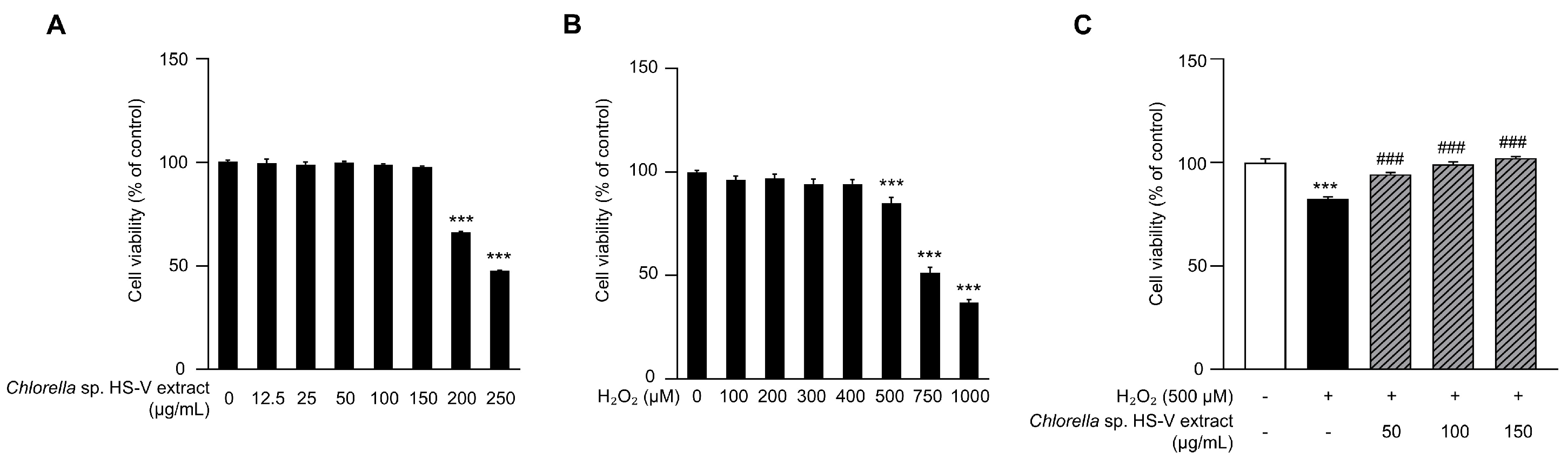
Previous studies have reported that Chlorella exerts protective effects against H2O2-induced cellular damage through its antioxidant capacity [52,53]. We therefore investigated whether Chlorella extract exerts a similar protective effect against H2O2-induced cytotoxicity in HaCaT cells. As shown in Figure 1A, cells were treated with Chlorella sp. HS-V extract at 12.5, 25, 50, 100, 150, 200, and 250 µg/mL for 24 h. Cell viability decreased to approximately 66% at 200 µg/mL, whereas no significant cytotoxicity was observed at concentrations up to 150 µg/mL. To determine the optimal H2O2 concentration for inducing cytotoxicity, cells were treated with H2O2 at 100, 200, 300, 400, 500, 750, and 1000 µM for 24 h (Figure 1B). Treatment with 500 µM H2O2 reduced cell viability to approximately 80%. A minimal viability reduction may be insufficient to assess the recovery effect of protective compounds, whereas excessive H2O2-induced cytotoxicity may shift the experimental readout toward irreversible cell death, such as apoptosis or necrosis, rather than a recoverable oxidative stress response [54,55,56]. Consistent with this approach, previous HaCaT oxidative stress studies have selected H2O2 concentrations that reduced cell viability to approximately 70–80% for evaluating antioxidant or cytoprotective effects [57,58]. Therefore, the Chlorella sp. HS-V extract at 50, 100, and 150 µg/mL and 500 µM H2O2 were selected for subsequent experiments. To evaluate the protective effect of the Chlorella sp. HS-V extract against H2O2-induced cytotoxicity, cells were pretreated with the extract (50, 100, and 150 µg/mL) for 24 h, followed by treatment with 500 µM H2O2 for 24 h. As shown in Figure 1C, the Chlorella sp. HS-V extract restored the H2O2-induced decrease in cell viability in a dose-dependent manner. These results suggest that the Chlorella sp. HS-V extract attenuates H2O2-induced cytotoxicity in HaCaT cells.

3.2. Violaxanthin, a Major Carotenoid in Chlorella Extract, Protects HaCaT Cells from H2O2-Induced Cytotoxicity

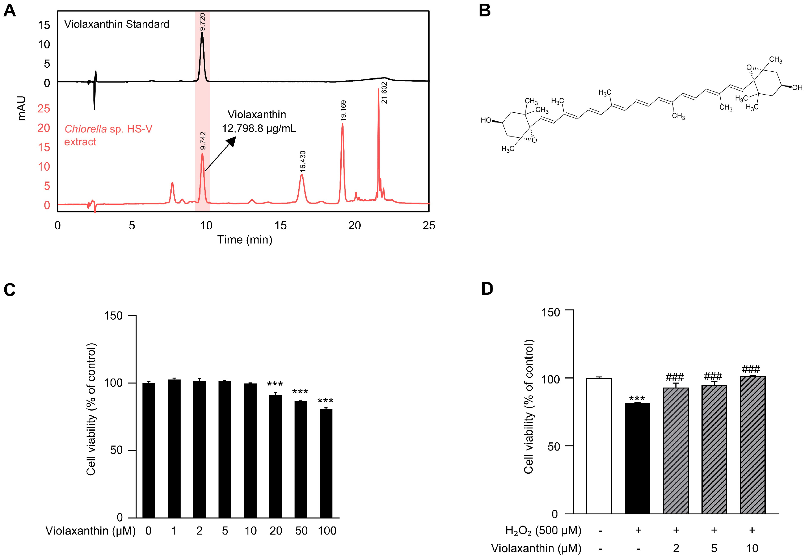
Given that the Chlorella sp. HS-V extract exhibited a protective effect against H2O2-induced cytotoxicity (Figure 1), we performed HPLC analysis to identify the major active component responsible for this activity (Figure 2A). HPLC analysis showed a distinct peak corresponding to violaxanthin in the Chlorella sp. HS-V extract, and the violaxanthin content was 12,798.8 µg/g of evaporated extract. These results indicate that violaxanthin is a major carotenoid present in the Chlorella sp. HS-V extract among the detected pigments. To evaluate whether violaxanthin itself exerts cytotoxic effects, HaCaT cells were treated with violaxanthin at concentrations ranging from 1 to 100 µM for 24 h, which revealed no significant cytotoxicity at concentrations up to 10 µM (Figure 2C). Therefore, subsequent experiments were conducted using 2, 5, and 10 µM violaxanthin to evaluate its protective effects within a non-cytotoxic concentration range. Based on these findings, we next investigated whether violaxanthin exerts a similar protective effect against H2O2-induced cytotoxicity, as observed with the Chlorella sp. HS-V extract (Figure 1C). Cells were pretreated with violaxanthin (2, 5, and 10 µM) for 24 h, and then treated with 500 µM of H2O2. As a result, violaxanthin restored the H2O2-induced decrease in cell viability in a dose-dependent manner. Notably, pretreatment with 10 µM violaxanthin restored cell viability to levels comparable to those of the untreated control (Figure 2D). These data show that violaxanthin, the major carotenoid in the Chlorella sp. HS-V extract, protects HaCaT cells against H2O2-induced cytotoxicity.

3.3. Violaxanthin Decreases Intracellular Reactive Oxygen Species Levels in HaCaT Cells

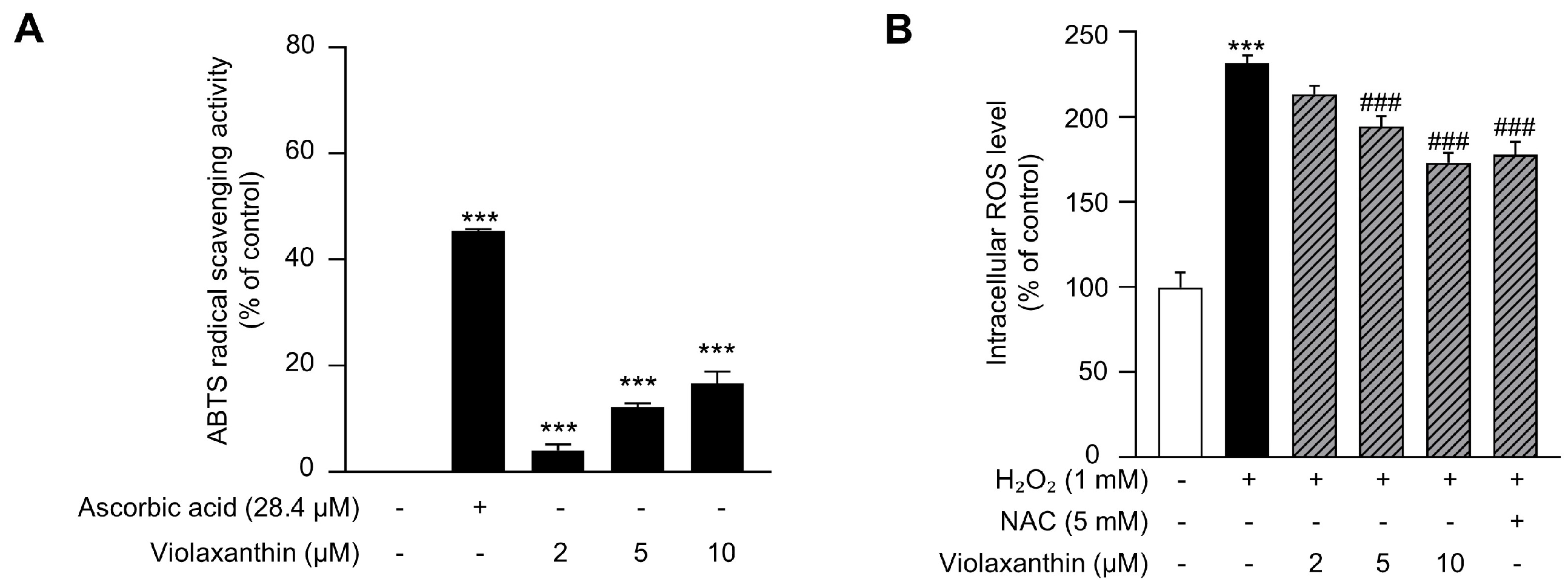
Violaxanthin has previously been shown to exhibit antioxidant properties comparable to those of other carotenoids, including β-carotene and zeaxanthin [47]. Therefore, we next examined whether the protective effect of violaxanthin against H2O2-induced cellular damage was associated with its radical-scavenging activity using an ABTS assay. As shown in Figure 3A, violaxanthin showed concentration-dependent ABTS radical scavenging activity. Although its activity was weaker than that of ascorbic acid, which was used as a representative antioxidant control [59], these results indicate that violaxanthin possesses measurable antioxidant activity. To determine whether violaxanthin reduces intracellular ROS levels, cells were pretreated with violaxanthin (2, 5, and 10 µM) or NAC (5 mM) for 24 h, followed by exposure to 1 mM H2O2 for 20 min to induce intracellular ROS accumulation. As shown in Figure 3B, violaxanthin significantly reduced intracellular DCF fluorescence intensity in a dose-dependent manner. Notably, treatment with 10 µM violaxanthin reduced ROS levels to a degree comparable to those of NAC. Collectively, these results suggest that violaxanthin exerts a protective effect against H2O2-induced oxidative stress by reducing intracellular ROS levels in HaCaT cells.

3.4. Violaxanthin Increases the mRNA Expression of Antioxidant Enzymes in HaCaT Cells

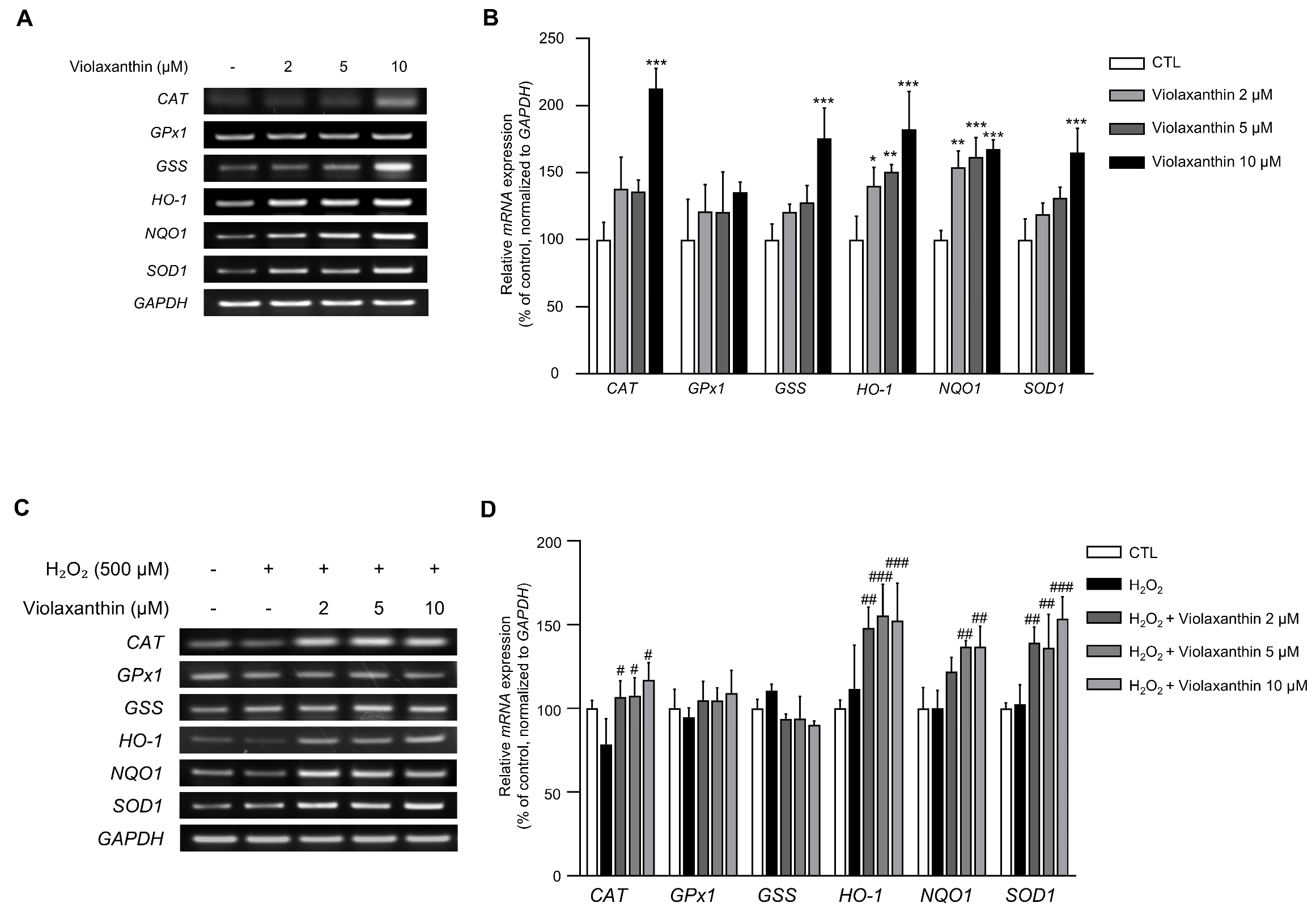
To alleviate oxidative stress caused by ROS, cells regulate antioxidant defense systems through antioxidant enzymes such as catalase (CAT), superoxide dismutase (SOD), glutathione peroxidases (GPxs), glutathione synthetase (GSS), heme oxygenase-1 (HO-1), and NAD(P)H quinone oxidoreductase (NQO1). We therefore examined whether the ROS-scavenging effect of violaxanthin is associated with the upregulation of antioxidant enzyme expression. HaCaT cells were treated with violaxanthin (2, 5, and 10 µM) for 3 h, and the mRNA expression levels of CAT, GPx1, GSS, HO-1, NQO1, and SOD1 were assessed by RT-PCR and RT-qPCR. Violaxanthin increased the mRNA expression of CAT, GSS, HO-1, NQO1, and SOD1 relative to the untreated control (Figure 4A,B). To examine whether violaxanthin still upregulates antioxidant enzyme expression under oxidative stress conditions, RT-PCR and RT-qPCR were performed. Cells were pretreated with violaxanthin, followed by treatment with 500 µM H2O2. Even in the presence of H2O2, violaxanthin significantly upregulated the mRNA expression of CAT, HO-1, NQO1, and SOD1 (Figure 4C,D). Collectively, these results indicate that violaxanthin reduces intracellular ROS levels by upregulating the mRNA expression of antioxidant enzymes, not only under basal conditions but also under H2O2-induced oxidative stress.

3.5. Violaxanthin Promotes the Nuclear Translocation of NRF2 in HaCaT Cells

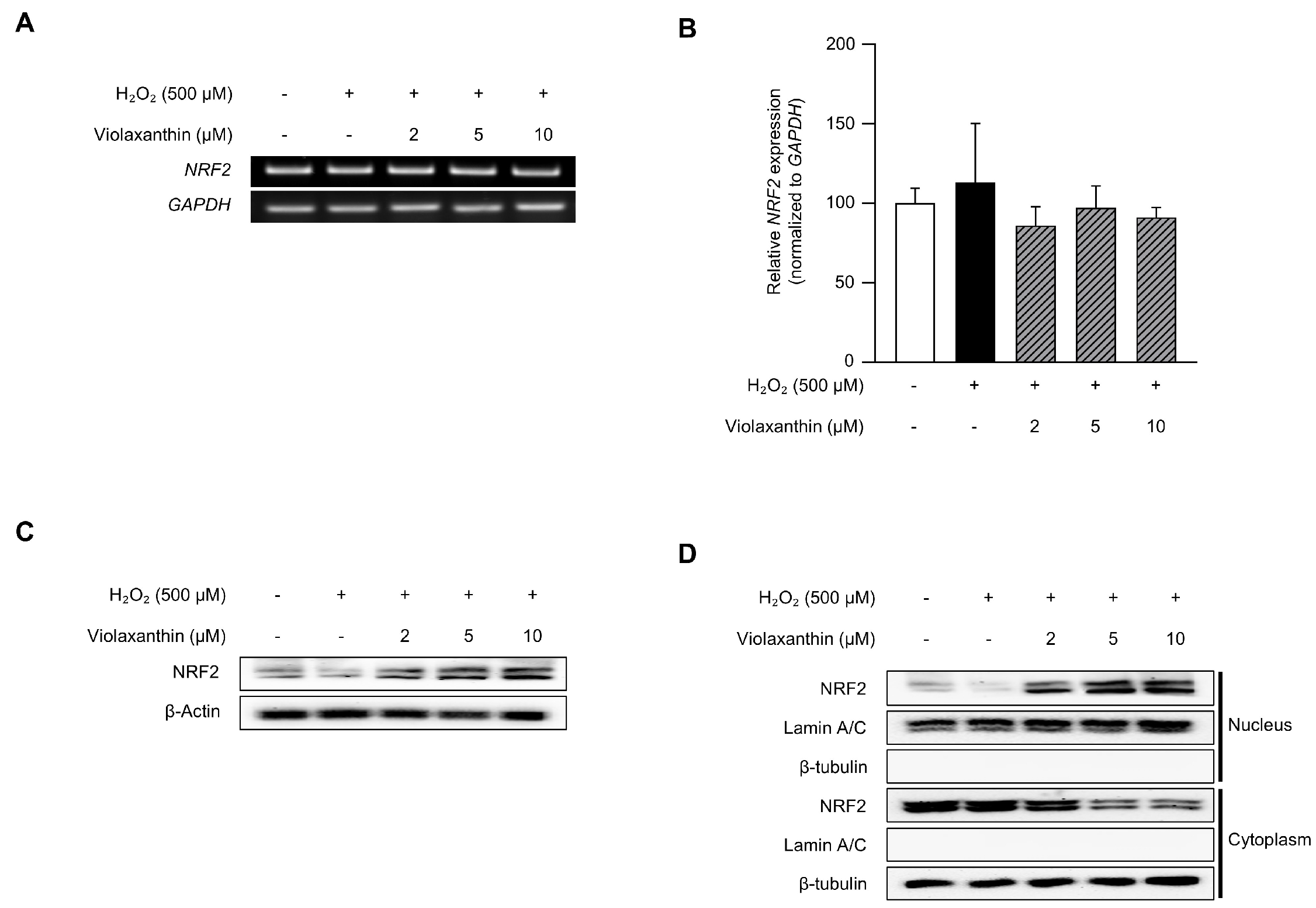
As shown in Figure 4, violaxanthin increased the mRNA expression of antioxidant enzymes. Previous studies have reported that nuclear factor erythroid 2-related factor 2 (NRF2) is a key transcription factor that regulates cellular antioxidant responses by upregulating the expression of antioxidant enzymes [60,61,62]. To determine whether violaxanthin affects the activation of NRF2, immunoblotting was performed. As shown in Figure 5C, the expression of NRF2 was increased by violaxanthin. This finding raised the possibility that NRF2 activation by violaxanthin may contribute to the observed upregulation of antioxidant enzymes. RT-PCR and RT-qPCR analyses were performed to determine whether the increase in NRF2 protein was attributable to enhanced transcription, but violaxanthin did not significantly alter NRF2 mRNA expression (Figure 5A,B). These results suggest that violaxanthin may regulate NRF2 at a post-translational level rather than enhancing its transcription. As NRF2 requires nuclear translocation for its transcriptional activity, and the expression of its downstream target genes, including CAT, SOD, and GPxs, is regulated by this process [63], we examined whether violaxanthin promotes the nuclear translocation of NRF2. To this end, cytoplasmic and nuclear fractions were separated, and the NRF2 protein levels in each fraction were assessed by immunoblotting (Figure 5D). Treatment with violaxanthin increased nuclear NRF2 levels while decreasing cytoplasmic NRF2 levels. Consequently, these results suggest that violaxanthin may promote antioxidant response by promoting nuclear translocation for the activation of NRF2 rather than by transcriptional expression.

3.6. Violaxanthin Activates NRF2 via the AKT/GSK3β Signaling Pathway in HaCaT Cells

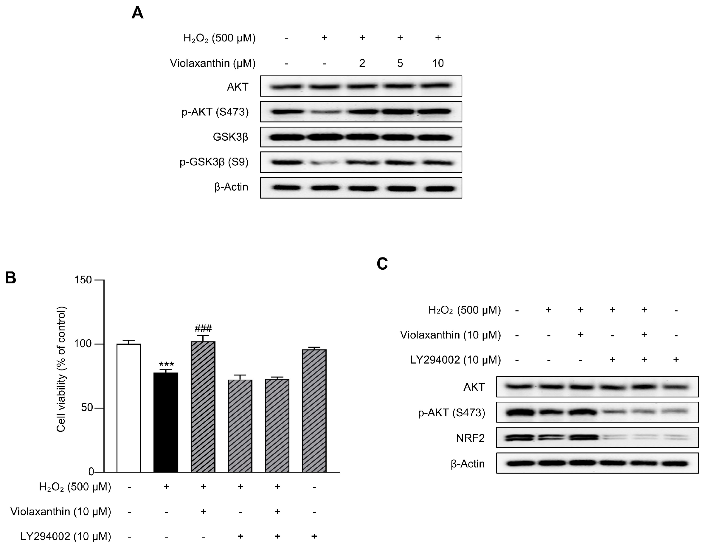
Previous studies have reported that the PI3K/AKT/GSK3β signaling pathway acts as an upstream regulator of NRF2 nuclear translocation [64,65,66]. To examine whether violaxanthin itself affects AKT/GSK3β signaling under basal conditions, HaCaT cells were treated with violaxanthin in the absence of H2O2. Violaxanthin did not markedly alter the total protein levels of AKT or GSK3β, but it increased the phosphorylation levels of AKT (Ser473) and GSK3β (Ser9) (Supplementary Figure S1). These results suggest that violaxanthin may activate AKT/GSK3β signaling under basal conditions without changing the total expression levels of AKT and GSK3β. To further investigate whether this pathway is involved in violaxanthin-induced NRF2 nuclear translocation, immunoblotting was performed (Figure 6A). The treatment with H2O2 (500 µM), which was performed to induce oxidative stress, decreased the phosphorylation levels of AKT (S473) and GSK3β (S9), whereas pretreatment with violaxanthin restored these levels. To further verify that the AKT/GSK3β pathway mediates the effect of violaxanthin on NRF2 translocation, the cells were pretreated with the PI3K/AKT inhibitor LY294002 prior to violaxanthin and H2O2 treatment. The MTT analysis showed that the recovery effect of violaxanthin on H2O2-induced cytotoxicity was inhibited by LY294002 (Figure 6B). Furthermore, LY294002 suppressed violaxanthin-induced AKT (Ser473) phosphorylation and NRF2 protein expression (Figure 6C). These data suggest that the violaxanthin-induced translocation of NRF2 may be mediated by the AKT/GSK3β signaling pathway.

3.7. Violaxanthin Promotes Wound Healing in H2O2-Treated HaCaT Cells

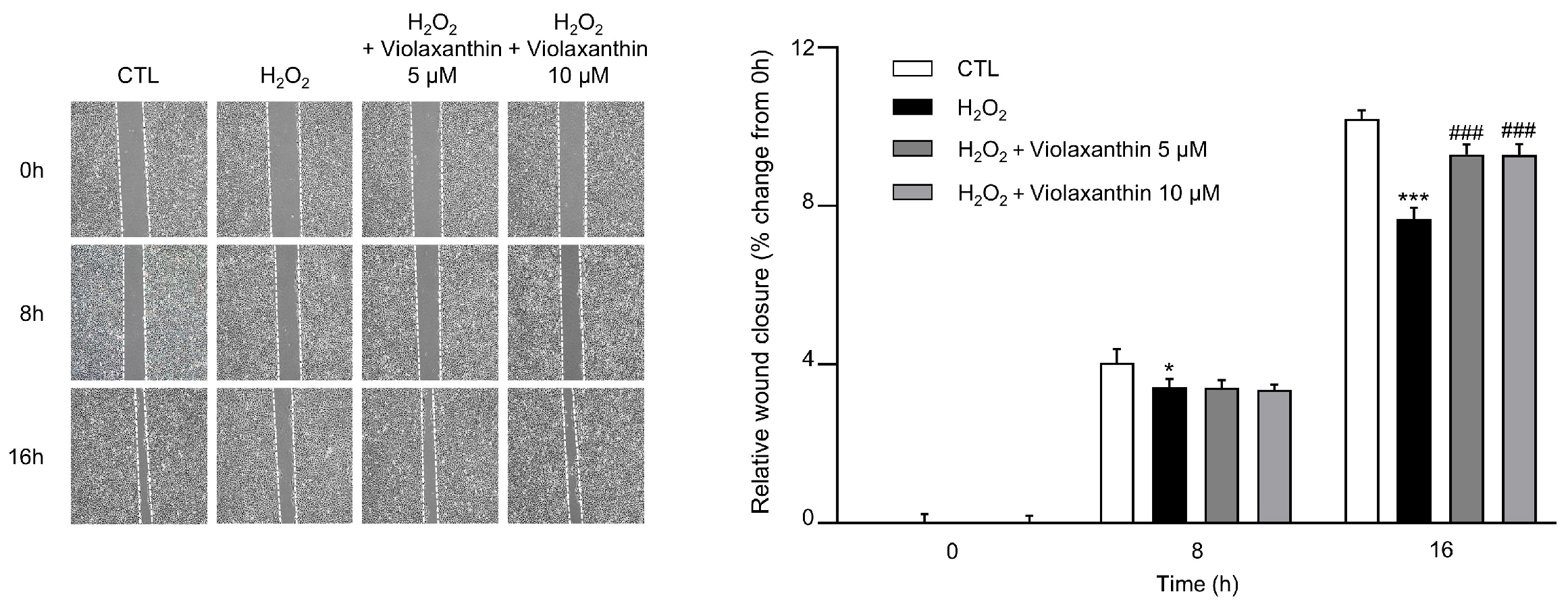
Accumulating evidence has implied that excessive cellular ROS levels cause impaired function in fibroblasts and keratinocytes by modifying and degrading extracellular matrix proteins or increasing cell death [67,68]. It has been reported that one therapeutic approach to delayed wound-healing is the regulation of excessive ROS [69]. NAC has also been suggested as a potential wound-healing agent by promoting cell proliferation and migration through the antioxidant defense systems [70]. Since 10 μM of violaxanthin demonstrated a comparable ROS-scavenging ability to that of NAC, we hypothesized that violaxanthin would have an effect on H2O2-induced wound-healing delay. As shown in Figure 7, the results of the wound healing assay showed that violaxanthin induced faster scratch closure than in H2O2 treatment alone at 16 h. Consequently, these data indicated that violaxanthin might have a protective effect on wound healing delayed by ROS.

3.8. Violaxanthin Suppresses H2O2-Induced Premature Senescence in nHEK Cells

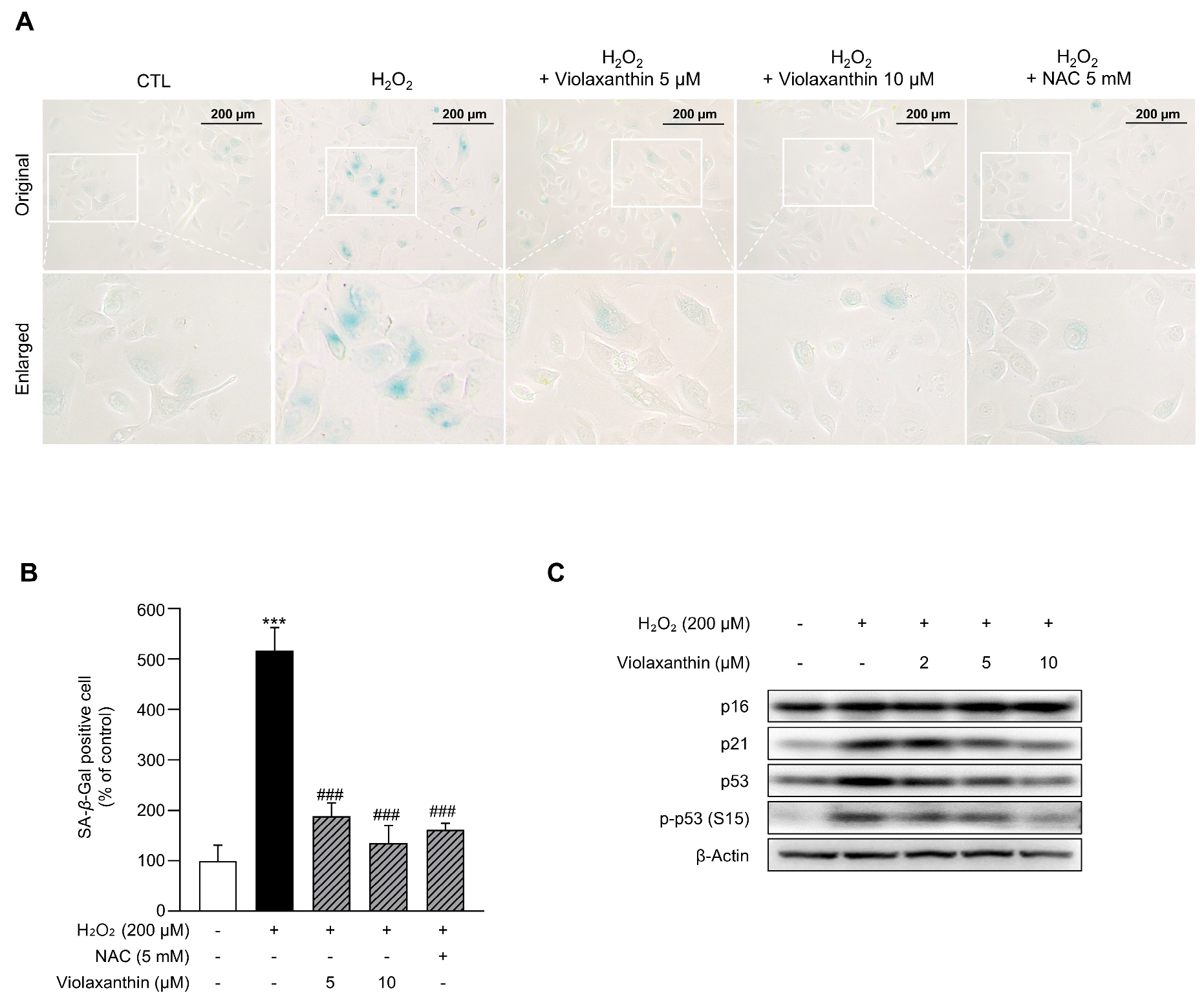
Extensive studies have reported that intracellular ROS, produced by endogenous and exogenous stressors, induce the senescence of keratinocytes and also delay wound healing [71]. It has been revealed that exposure to low doses of H2O2 in keratinocytes leads to premature senescence by decreasing population doublings and increasing SA-β-gal-positive cells [72]. Since HaCaT cells have been reported to not express SA-β-galactosidase [73,74], SA-β-gal staining was performed in nHEK cells to determine whether violaxanthin can inhibit the senescence of keratinocytes induced by H2O2. After pretreatment with violaxanthin (5 and 10 µM), SA-β-gal-positive cells were significantly decreased compared to the H2O2 treatment alone, suggesting that violaxanthin alleviated cellular senescence under oxidative stress conditions (Figure 8A,B). In addition, the expression of p16, p21, and p53, which were involved in the senescence signaling pathway [75], was analyzed by immunoblotting, as shown in Figure 8C. The expression of p21 and p53 and the phosphorylation of p53 (S15) were increased by H2O2, effectively inducing cellular senescence in nHEK cells. Interestingly, violaxanthin (2, 5, and 10 µM) significantly reduced the expression of p21 and p53, and the phosphorylation of p53 (S15) in a dose-dependent manner. These results suggest that violaxanthin can suppress H2O2-induced senescence.

4. Discussion

Microalgae are rich sources of physiologically active compounds with diverse biological activities, such as anticancer, antioxidant, and anti-inflammatory properties [32,76,77]. Chlorella, a unicellular green microalga of Chlorophyta [78], is one of the most widely cultivated microalgal species owing to its broad biological activities, rapid growth rate, and resilience to adverse environmental conditions [21]. It has been reported that peptides extracted from Chlorella have protective effects against oxidative stress caused by free radicals [79]. Bae MJ et al. indicated that a hydrothermal extract from Chlorella inhibited histamine release, suggesting its potential as an antiallergic agent [80]. In the present study, the Chlorella sp. HS-V extract significantly recovered the H2O2-induced cytotoxicity in a dose-dependent manner (Figure 1). Based on these findings, the Chlorella sp. HS-V extract is suggested to exert antioxidant activity in keratinocytes.
Previous studies have indicated that Chlorella contains numerous active ingredients such as vitamins, lipids, proteins, and carotenoids [31,32]. As shown in Figure 2A, HPLC analysis indicated that a major component of the Chlorella sp. HS-V extract was violaxanthin. Violaxanthin is a carotenoid that can be isolated from microalgae such as Chlorella ellipsoidea and Nannochloropsis oceanica, as well as orange-colored fruits and green vegetables [38,43,44,46]. Violaxanthin has been reported to possess antioxidant, anti-inflammatory, and antiproliferative properties [38,44,45]. Furthermore, violaxanthin has been demonstrated to inhibit UVB-induced cellular senescence in human dermal fibroblasts, highlighting its potential as an anti-photoaging agent [49]. However, the effects of violaxanthin on keratinocytes remain limited. In the present study, we demonstrated for the first time that violaxanthin exerts protective effects against H2O2-induced oxidative damage in keratinocytes.
The primary function of the skin is to form an effective barrier between the body and the external environment [81]. ROS are generated in the skin upon exposure to external stressors or as byproducts of intracellular metabolic processes [82]. Excessive ROS not only cause skin aging and inflammation but also inhibit the proliferation of keratinocytes, which act as a primary barrier against the penetration of exogenous chemicals and microorganisms [82]. Furthermore, ROS have been reported to affect various intracellular signaling pathways and molecular processes in keratinocytes [83]. Accumulating evidence suggests that H2O2-induced oxidative stress triggers autophagy and apoptosis in keratinocytes [84,85,86]. In order to maintain keratinocyte homeostasis and overall skin integrity, excessive ROS need to be controlled through cellular antioxidant defense systems [83]. Several studies have explored the use of exogenous antioxidants to activate intracellular antioxidant defense systems and protect keratinocytes from oxidative stress [87,88]. Zheng J et al. showed that americanin B protected HaCaT keratinocytes from H2O2 by reducing the level of ROS and alleviating DNA damage [87]. Consistent with these findings, both the Chlorella sp. HS-V extract and violaxanthin restored the H2O2-induced decrease in cell viability in a dose-dependent manner in the present study (Figure 1 and Figure 2). Moreover, as shown in Figure 3, treatment with 10 µM violaxanthin reduced intracellular ROS levels to a degree comparable to that of NAC. Collectively, these results suggest that violaxanthin reduces intracellular ROS levels and modulates cellular antioxidant defense systems, thereby attenuating H2O2-induced cytotoxicity. Additionally, while 10 μM of violaxanthin demonstrated near-complete recovery of cell viability without causing cytotoxicity, the absolute optimal concentration may lie between 10 and 20 μM. Further fine-tuning of the dosage within this narrow window will be necessary to establish the exact therapeutic index for future formulation and clinical applications.
Antioxidant enzymes serve as key components of cellular antioxidant defense systems, playing a central role in maintaining redox homeostasis and protecting against oxidative damage [62]. These enzymes include SOD, CAT, and the GPx family [89]. However, both acute and chronic H2O2 exposure have been reported to reduce their activities, thereby inducing oxidative stress [90]. Accordingly, various studies have focused on alleviating oxidative stress by enhancing antioxidant enzyme activity [86,91,92]. As shown in Figure 4, violaxanthin alone increased the expression of CAT, GSS, HO-1, NQO1, and SOD1. Moreover, in H2O2-treated cells, violaxanthin significantly upregulated the expression of CAT, NQO1, and SOD1. Interestingly, the regulatory effects of violaxanthin on the mRNA levels of individual antioxidant enzymes showed subtle differences depending on the presence or absence of H2O2. Similar differential regulation has been reported in previous studies, and this pattern may reflect the gene-specific and condition-dependent nature of antioxidant defense systems. For example, in normal human dermal fibroblasts, avenanthramide C treatment alone dose-dependently increased HO-1 expression via NRF2 activation, whereas the expression of other antioxidant genes remained unchanged or even decreased. However, under H2O2-stimulated conditions, avenanthramide C reduced the H2O2-induced increase in antioxidant gene transcripts, including HO-1, GSS, SOD1, GPx1, and CAT [93]. In primary rat hepatocytes, antioxidant enzyme expression also changed differently depending on the culture condition and H2O2 exposure; catalase expression decreased during basal culture, whereas H2O2 exposure prevented this decrease and induced catalase mRNA expression, indicating that antioxidant enzyme regulation can be strongly influenced by oxidative conditions [94]. Pilat et al. showed that oxidative stress did not uniformly regulate all antioxidant enzymes in ARPE-19 cells, where HO-1 was induced by sublethal H2O2 treatment, whereas GPx and catalase showed distinct or limited responses depending on the stress condition [95]. These findings suggest that antioxidant enzymes do not necessarily respond uniformly to antioxidant compounds or oxidative stimuli, but may be selectively regulated according to cellular redox status, stress intensity, and enzyme-specific physiological roles. Thus, the differential responses observed in Figure 4 may represent adaptive fine-tuning of antioxidant defense pathways under basal versus H2O2-induced oxidative stress conditions. Further studies examining time-dependent changes in antioxidant enzyme activity, and NRF2 target gene activation would help clarify the gene-specific regulatory effects of violaxanthin.
Many studies have shown that NRF2 regulates ROS homeostasis by modulating the cellular antioxidant defense system through various mechanisms [62,96,97,98]. The NRF2–ARE signaling pathway is a major transcriptional regulatory pathway controlling the expression of antioxidant enzymes [99,100]. NRF2 is known to be regulated by Kelch-like ECH-associated protein 1 (Keap1), as well as by GSK3β-mediated beta-transducin repeat-containing protein (β-TrCP)-dependent degradation [101]. Under normal conditions, Keap1 forms homodimers that bind to NRF2, thereby promoting its cytoplasmic sequestration and ubiquitin-dependent proteasomal degradation [102,103]. In addition to Keap1, NRF2 is also subject to degradation through GSK3β-mediated phosphorylation and β-TrCP-dependent ubiquitin ligase activity, providing an additional layer of regulation [104]. Therefore, the inhibition of GSK3β activity may stabilize NRF2 protein by preventing its β-TrCP-dependent degradation. Under oxidative stress conditions, NRF2 is released from Keap1, translocates to the nucleus, and binds to ARE sequences to drive the transcription of antioxidant enzyme genes [105]. As shown in Figure 5, violaxanthin enhanced NRF2 protein stability and promoted its nuclear translocation under H2O2-induced oxidative stress conditions, suggesting that violaxanthin may activate the NRF2–ARE signaling axis to reinforce cellular antioxidant defense systems.
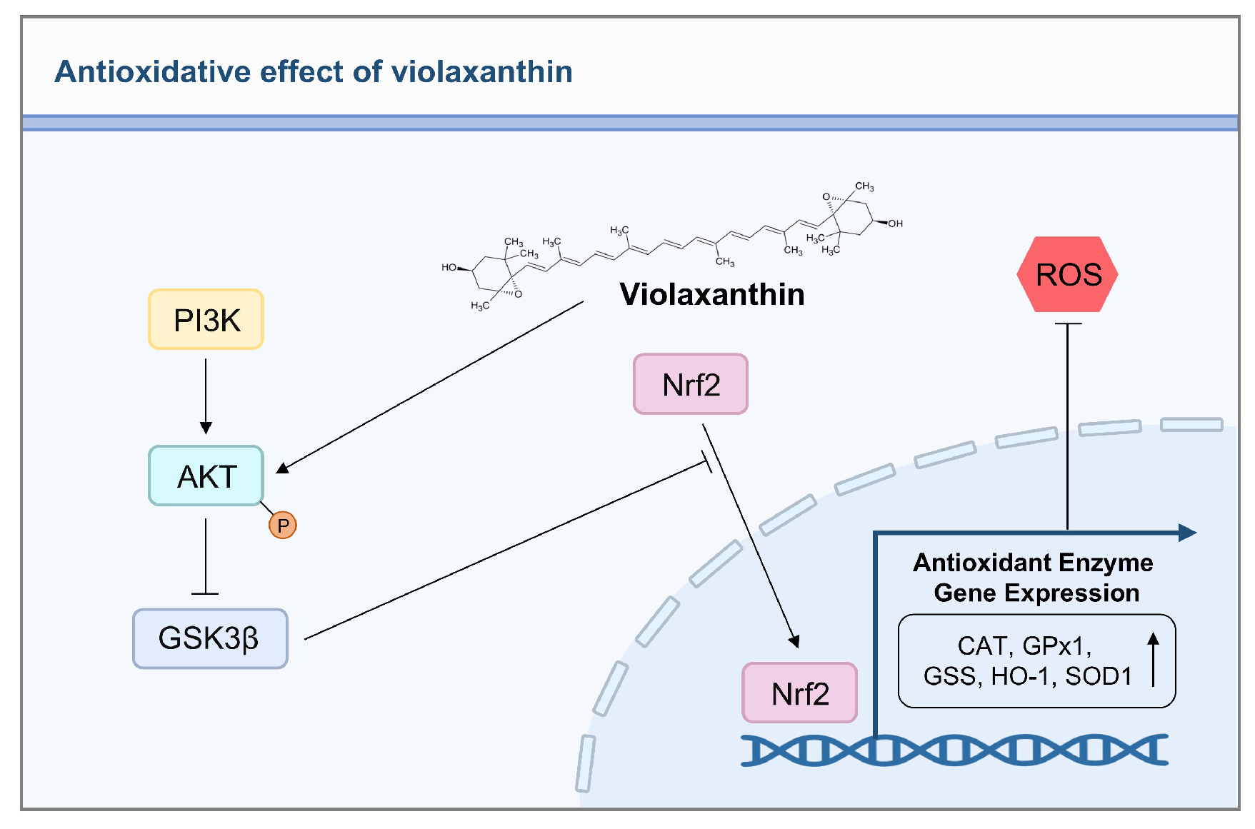
Previous studies have demonstrated that NRF2 can be upregulated by the activation of AKT, as well as by the inactivation of GSK3β [104,106]. It has been well established that PI3K-mediated phosphorylation of AKT at Ser473 leads to the inactivation of GSK3β [107]. Consistent with this, protective effects mediated by PI3K/AKT–NRF2 activation against oxidative stress have been reported in various cell types, including human trabecular meshwork cells, 661W cells, and keratinocytes [108,109,110,111,112]. For instance, fisetin has been shown to activate the PI3K/AKT/NRF2 signaling pathway, thereby inhibiting H2O2-induced apoptosis in HaCaT keratinocytes [113]. Based on the observed increase in NRF2 protein expression and nuclear accumulation by violaxanthin, we next investigated the upstream signaling mechanism underlying these effects. Immunoblotting analysis revealed that H2O2 treatment significantly reduced the phosphorylation of both AKT (Ser473) and GSK3β (Ser9), whereas pretreatment with violaxanthin dose-dependently restored these phosphorylation levels (Figure 6A). To confirm the involvement of AKT/GSK3β signaling in violaxanthin-mediated NRF2 activation, cells were treated with the PI3K/AKT inhibitor LY294002 prior to violaxanthin treatment. LY294002 reduced the cytoprotective effect of violaxanthin against H2O2-induced cytotoxicity (Figure 6B) and suppressed violaxanthin-induced AKT phosphorylation (Figure 6C); however, it only partially inhibited NRF2 protein expression. This partial suppression suggests that while the PI3K/AKT pathway is an important mediator, it may not be solely responsible for NRF2 regulation in this context. Previous studies have established that NRF2 activation is governed by a complex network of upstream kinases; in addition to the PI3K/AKT axis, mitogen-activated protein kinases (MAPKs), particularly p38 and ERK, are known to facilitate NRF2 nuclear translocation and the subsequent induction of antioxidant enzymes in response to various chemicals and oxidative stimuli [114,115]. Therefore, further studies are required to investigate the potential involvement of other upstream signaling pathways, including p38 MAPK or ERK, in violaxanthin-mediated NRF2 regulation. Taken together, these results suggest that violaxanthin activates NRF2 signaling, at least in part, through the AKT/GSK3β pathway, thereby reinforcing cellular antioxidant defense against H2O2-induced oxidative stress (Figure 9).
Beyond their role in keratinocyte homeostasis, ROS also play an important role in wound healing [116]. A controlled ROS level is required for the transition from the inflammatory to the proliferation phase [117], but prolonged or excessive ROS exposure can result in tissue damage and delay the wound healing process [118]. Therefore, proper regulation of ROS levels through cellular antioxidant defense systems is essential for normal wound healing [116]. Resveratrol, for example, has been shown to accelerate wound healing by restoring oxidative-stress-impaired cell proliferation and migration [119]. Similarly, in the present study, wound-healing analysis revealed that violaxanthin restored H2O2-induced impairment of wound closure in HaCaT cells (Figure 7). This effect is likely attributable, at least in part, to the antioxidant capacity of violaxanthin. Furthermore, the observed changes in AKT/GSK3β and NRF2 signaling may also be associated with the wound-healing-promoting effects of violaxanthin. Collectively, these findings suggest that violaxanthin may serve as a potential candidate for alleviating the oxidative-stress-induced impairment of wound-healing in keratinocytes.
Accumulating evidence indicates that cellular senescence and skin aging are promoted by increased ROS production resulting from harmful environmental factors and chronic ultraviolet (UV) exposure [120,121]. Violaxanthin, which is abundantly found in the microalga Nannochloropsis oceanica, has been shown to inhibit UVB-induced senescence in normal human dermal fibroblasts by suppressing G1 phase arrest, ERK phosphorylation, and the upregulation of p16 and p21 [49]. Based on these findings, we hypothesized that violaxanthin may similarly inhibit H2O2-induced senescence in keratinocytes. Cellular senescence is characterized by increased senescence-associated β-galactosidase (SA-β-gal) activity, which is widely used as a marker of senescent cells [51]. Liu L et al. reported that although HaCaT cells may exhibit senescence-associated phenotypes upon H2O2 exposure, they do not express SA-β-galactosidase activity [73]. In the present study, nHEK cells were therefore used to evaluate H2O2-induced senescence rather than HaCaT cells. As shown in Figure 8, violaxanthin reduced the number of SA-β-gal-positive cells and downregulated the expression of p21 and p53, as well as the phosphorylation of p53 at Ser15, all of which are key mediators of the senescence signaling pathway. These results suggest that violaxanthin may inhibit H2O2-induced cellular senescence in nHEK cells primarily through suppression of the p53/p21 signaling pathway. This finding is partially consistent with a previous study showing that violaxanthin suppresses cellular senescence [49]; however, the present study extends these findings to epidermal keratinocytes and provides additional mechanistic and functional evidence under H2O2-induced oxidative stress conditions. Moreover, while the previous study mainly focused on ERK phosphorylation, G1 phase arrest, and ECM-related photoaging responses, the present study differs in that it suggests the possible involvement of the AKT/GSK3β–NRF2 axis as an upstream signaling pathway involved in violaxanthin-mediated antioxidant defense, as supported by NRF2 nuclear translocation and antioxidant enzyme induction. Another notable finding is that p16 expression was not significantly altered by violaxanthin treatment under the present experimental conditions. This result does not necessarily exclude the involvement of p16 in cellular senescence, but suggests that p16 may not be the major senescence-associated target regulated by violaxanthin in this model. Consistent with our findings, Ido et al. reported that H2O2 treatment in primary human keratinocytes induced p21 expression but not p16 expression, and suggested that low-dose H2O2-induced premature senescence was mainly associated with activation of the p53/p21 pathway rather than the p38MAPK/p16 pathway [122]. Similarly, in a stress-induced premature senescence model using human skin fibroblasts, low-dose H2O2 exposure did not significantly increase p16 expression compared with untreated control cells, whereas p16 upregulation became more evident under higher-dose or more sustained senescence conditions [123]. Mechanistically, the p53/p21 and p16/RB pathways are two major but partially distinct regulatory axes of senescence-associated cell cycle arrest. The p53/p21 pathway is generally associated with the early DNA damage and oxidative stress response, whereas the p16/RB pathway is more closely related to the maintenance and stabilization of senescence [124]. Taken together, these findings indicate that p16 expression is not always uniformly increased in H2O2-induced senescence models and may depend on cell type, stress intensity, and the stage of senescence. Further studies using other senescence induction conditions or additional p16/RB pathway markers, such as phosphorylated RB and CDK4/6 activity, would be needed to clarify whether the p16/RB pathway contributes to the anti-senescent effects of violaxanthin. In addition, we acknowledge that the present study was conducted using 2D monolayer cell culture models, which lack the complex structural organization and multi-cellular interactions of actual human skin. To better understand the physiological relevance and translational potential of violaxanthin as a cosmeceutical or pharmacological agent, future studies should evaluate its protective efficacy and tissue penetration using 3D human skin equivalents or in vivo animal models of oxidative skin damage.

5. Conclusions

In this study, we investigated the protective effects of violaxanthin against H2O2-induced oxidative damage in human keratinocytes. Violaxanthin, a major bioactive component in the Chlorella sp. HS-V extract, attenuated H2O2-induced cytotoxicity and reduced intracellular ROS levels. Furthermore, it upregulated the mRNA expression of key antioxidant enzymes, including CAT, NQO1 and SOD1, both in the presence and absence of H2O2, suggesting its capacity to reinforce cellular antioxidant defense systems. Mechanistically, violaxanthin promoted nuclear translocation of NRF2 via the AKT/GSK3β signaling pathway. In addition, violaxanthin restored H2O2-impaired wound healing in HaCaT cells and suppressed oxidative-stress-induced senescence in nHEK cells. In conclusion, violaxanthin may serve as a potential therapeutic agent for alleviating ROS-induced damage in keratinocytes, with its cytoprotective effects mediated, at least in part, through activation of the AKT/GSK3β–NRF2 antioxidant signaling axis.

Supplementary Materials

The following supporting information can be downloaded at: https://www.mdpi.com/article/10.3390/app16105132/s1, Figure S1: Effect of violaxanthin on AKT/GSK3β signaling under basal conditions in HaCaT cells. HaCaT cells were treated with violaxanthin (2, 5, and 10 µM) for 24 h. The total protein levels of AKT and GSK3β and the phosphorylation levels of AKT (Ser473) and GSK3β (Ser9) were analyzed by immunoblotting. β-Actin was used as a loading control.

Author Contributions

Conceptualization, J.-S.K., H.S.K., J.W.K., S.-B.P., H.-S.K., Y.J.L. and S.B.; methodology, J.-S.K., H.S.K. and S.B.; software, J.-S.K., H.S.K., H.-J.S. and S.P.; validation, J.-S.K., H.S.K., H.-J.S., S.P. and S.B.; formal analysis, J.-S.K., H.S.K., H.-J.S., S.P., J.W.K., S.-B.P., H.-S.K., Y.J.L. and S.B.; investigation, J.-S.K., H.S.K., J.W.K., S.-B.P., H.-S.K. and Y.J.L.; resources, J.-S.K., H.S.K., H.-J.S., S.P., J.W.K., S.-B.P., H.-S.K., Y.J.L. and S.B.; data curation, J.-S.K., H.S.K., H.-J.S., S.P. and S.B.; writing—original draft preparation, J.-S.K. and H.S.K.; writing—review and editing, J.-S.K., H.-J.S. and S.B.; visualization, J.-S.K. and H.S.K.; supervision, S.B.; project administration, S.B.; funding acquisition, S.B. All authors have read and agreed to the published version of the manuscript.

Funding

This paper was supported by Konkuk University Researcher Fund in 2024.

Data Availability Statement

The data presented in this study are available in the article and Supplementary Materials. Additional data are available from the corresponding author upon request.

Conflicts of Interest

Author Ji-Seon Kim and Seokmuk Park were employed by the company Dermato Bio, Inc. Author Hee-Sik Kim was employed by the company ASK LABS Co., Ltd. The remaining authors declare that the research was conducted in the absence of any commercial or financial relationships that could be construed as a potential conflict of interest.

References

  1. Lee, H.J.; Kim, M. Skin Barrier Function and the Microbiome. Int. J. Mol. Sci. 2022, 23, 13071. [Google Scholar] [CrossRef]
  2. Guan, J.; Wu, C.; He, Y.; Lu, F. Skin-associated adipocytes in skin barrier immunity: A mini-review. Front. Immunol. 2023, 14, 1116548. [Google Scholar] [CrossRef] [PubMed]
  3. Piipponen, M.; Li, D.; Landén, N.X. The Immune Functions of Keratinocytes in Skin Wound Healing. Int. J. Mol. Sci. 2020, 21, 8790. [Google Scholar] [CrossRef] [PubMed]
  4. Papaccio, F.; D′Arino, A.; Caputo, S.; Bellei, B. Focus on the Contribution of Oxidative Stress in Skin Aging. Antioxidants 2022, 11, 1121. [Google Scholar] [CrossRef]
  5. Beckman, K.B.; Ames, B.N. The free radical theory of aging matures. Physiol. Rev. 1998, 78, 547–581. [Google Scholar] [CrossRef] [PubMed]
  6. Frippiat, C.; Chen, Q.M.; Zdanov, S.; Magalhaes, J.P.; Remacle, J.; Toussaint, O. Subcytotoxic H2O2 stress triggers a release of transforming growth factor-beta 1, which induces biomarkers of cellular senescence of human diploid fibroblasts. J. Biol. Chem. 2001, 276, 2531–2537. [Google Scholar] [CrossRef]
  7. Aruoma, O.I. Free radicals, oxidative stress, and antioxidants in human health and disease. J. Am. Oil Chem. Soc. 1998, 75, 199–212. [Google Scholar] [CrossRef]
  8. Cross, C.E.; Halliwell, B.; Borish, E.T.; Pryor, W.A.; Ames, B.N.; Saul, R.L.; McCord, J.M.; Harman, D. Oxygen radicals and human disease. Ann. Intern. Med. 1987, 107, 526–545. [Google Scholar] [CrossRef]
  9. Girsang, E.; Ginting, C.N.; Lister, I.N.E.; Gunawan, K.Y.; Widowati, W. Anti-inflammatory and antiaging properties of chlorogenic acid on UV-induced fibroblast cell. PeerJ 2021, 9, e11419. [Google Scholar] [CrossRef]
  10. Guo, W.; An, Y.; Jiang, L.; Geng, C.; Zhong, L. The protective effects of hydroxytyrosol against UVB-induced DNA damage in HaCaT cells. Phytother. Res. PTR 2010, 24, 352–359. [Google Scholar] [CrossRef]
  11. O’Toole, E.A.; Goel, M.; Woodley, D.T. Hydrogen peroxide inhibits human keratinocyte migration. Dermatol. Surg. 1996, 22, 525–529. [Google Scholar] [CrossRef]
  12. Bickers, D.R.; Athar, M. Oxidative stress in the pathogenesis of skin disease. J. Investig. Dermatol. 2006, 126, 2565–2575. [Google Scholar] [CrossRef]
  13. Briganti, S.; Picardo, M. Antioxidant activity, lipid peroxidation and skin diseases. What’s new. J. Eur. Acad. Dermatol. Venereol. 2003, 17, 663–669. [Google Scholar] [CrossRef]
  14. Saha, S.; Buttari, B.; Panieri, E.; Profumo, E.; Saso, L. An Overview of Nrf2 Signaling Pathway and Its Role in Inflammation. Molecules 2020, 25, 5474. [Google Scholar] [CrossRef]
  15. Boo, Y.C. Natural Nrf2 Modulators for Skin Protection. Antioxidants 2020, 9, 812. [Google Scholar] [CrossRef]
  16. Zhang, B.; Zhao, Z.; Meng, X.; Chen, H.; Fu, G.; Xie, K. Hydrogen ameliorates oxidative stress via PI3K-Akt signaling pathway in UVB-induced HaCaT cells. Int. J. Mol. Med. 2018, 41, 3653–3661. [Google Scholar] [CrossRef]
  17. Zhang, J.; Wang, W.; Mao, X. Chitopentaose protects HaCaT cells against H2O2-induced oxidative damage through modulating MAPKs and Nrf2/ARE signaling pathways. J. Funct. Foods 2020, 72, 104086. [Google Scholar] [CrossRef]
  18. Bajpai, V.K.; Alam, M.B.; Quan, K.T.; Kwon, K.-R.; Ju, M.-K.; Choi, H.-J.; Lee, J.S.; Yoon, J.-I.; Majumder, R.; Rather, I.A. Antioxidant efficacy and the upregulation of Nrf2-mediated HO-1 expression by (+)-lariciresinol, a lignan isolated from Rubia philippinensis, through the activation of p38. Sci. Rep. 2017, 7, 46035. [Google Scholar] [CrossRef] [PubMed]
  19. Mobin, S.M.; Chowdhury, H.; Alam, F. Commercially important bioproducts from microalgae and their current applications–A review. Energy Procedia 2019, 160, 752–760. [Google Scholar] [CrossRef]
  20. Ismail, M.M.; Diab, M.H.; Elkomy, R.G. Algal Bioactive Compounds and Their biological activities. Int. J. Pharm. Res. 2021, 13, 1440–1452. [Google Scholar] [CrossRef]
  21. Zhao, C.; Wu, Y.; Yang, C.; Liu, B.; Huang, Y. Hypotensive, hypoglycaemic and hypolipidaemic effects of bioactive compounds from microalgae and marine micro-organisms. Int. J. Food Sci. Technol. 2015, 50, 1705–1717. [Google Scholar] [CrossRef]
  22. Anbuchezhian, R.; Karuppiah, V.; Li, Z. Prospect of marine algae for production of industrially important chemicals. In Algal Biorefinery: An Integrated Approach; Springer: Cham, Switzerland, 2015; pp. 195–217. [Google Scholar]
  23. Paniagua-Michel, J.d.J.; Morales-Guerrero, E.; Olmos Soto, J. Microalgal biotechnology: Biofuels and bioproducts. In Springer Handbook of Marine Biotechnology; Springer: Berlin/Heidelberg, Germany, 2015; pp. 1355–1370. [Google Scholar]
  24. Zhuang, D.; He, N.; Khoo, K.S.; Ng, E.P.; Chew, K.W.; Ling, T.C. Application progress of bioactive compounds in microalgae on pharmaceutical and cosmetics. Chemosphere 2022, 291, 132932. [Google Scholar] [CrossRef]
  25. Vieira, M.V.; Pastrana, L.M.; Fucinos, P. Microalgae Encapsulation Systems for Food, Pharmaceutical and Cosmetics Applications. Mar. Drugs 2020, 18, 644. [Google Scholar] [CrossRef]
  26. Fu, W.; Nelson, D.R.; Yi, Z.; Xu, M.; Khraiwesh, B.; Jijakli, K.; Chaiboonchoe, A.; Alzahmi, A.; Al-Khairy, D.; Brynjolfsson, S. Bioactive compounds from microalgae: Current development and prospects. Stud. Nat. Prod. Chem. 2017, 54, 199–225. [Google Scholar] [CrossRef]
  27. Mobin, S.; Alam, F. Some promising microalgal species for commercial applications: A review. Enrgy Proced. 2017, 110, 510–517. [Google Scholar] [CrossRef]
  28. Suganya, T.; Varman, M.; Masjuki, H.H.; Renganathan, S. Macroalgae and microalgae as a potential source for commercial applications along with biofuels production: A biorefinery approach. Renew. Sust. Energ. Rev. 2016, 55, 909–941. [Google Scholar] [CrossRef]
  29. Sun, H.; Wang, Y.; He, Y.; Liu, B.; Mou, H.; Chen, F.; Yang, S. Microalgae-Derived Pigments for the Food Industry. Mar. Drugs 2023, 21, 82. [Google Scholar] [CrossRef]
  30. Chen, C.L.; Liou, S.F.; Chen, S.J.; Shih, M.F. Protective effects of Chlorella-derived peptide on UVB-induced production of MMP-1 and degradation of procollagen genes in human skin fibroblasts. Regul. Toxicol. Pharmacol. 2011, 60, 112–119. [Google Scholar] [CrossRef] [PubMed]
  31. Yuan, Q.; Li, H.; Wei, Z.; Lv, K.; Gao, C.; Liu, Y.; Zhao, L. Isolation, structures and biological activities of polysaccharides from Chlorella: A review. Int. J. Biol. Macromol. 2020, 163, 2199–2209. [Google Scholar] [CrossRef]
  32. Khavari, F.; Saidijam, M.; Taheri, M.; Nouri, F. Microalgae: Therapeutic potentials and applications. Mol. Biol. Rep. 2021, 48, 4757–4765. [Google Scholar] [CrossRef]
  33. Widyaningrum, D.; Prianto, A.D. Chlorella as a source of functional food ingredients: Short review. IOP Conf. Ser. Earth Environ. Sci. 2021, 794, 012148. [Google Scholar] [CrossRef]
  34. Plaza, M.; Santoyo, S.; Jaime, L.; Avalo, B.; Cifuentes, A.; Reglero, G.; Reina, G.G.B.; Señoráns, F.J.; Ibáñez, E. Comprehensive characterization of the functional activities of pressurized liquid and ultrasound-assisted extracts from Chlorella vulgaris. Lwt-Food Sci. Technol. 2012, 46, 245–253. [Google Scholar] [CrossRef]
  35. Bito, T.; Okumura, E.; Fujishima, M.; Watanabe, F. Potential of Chlorella as a Dietary Supplement to Promote Human Health. Nutrients 2020, 12, 2524. [Google Scholar] [CrossRef]
  36. El-Fayoumy, E.A.; Shanab, S.M.M.; Gaballa, H.S.; Tantawy, M.A.; Shalaby, E.A. Evaluation of antioxidant and anticancer activity of crude extract and different fractions of Chlorella vulgaris axenic culture grown under various concentrations of copper ions. Bmc Complement. Med. 2021, 21, 51. [Google Scholar] [CrossRef] [PubMed]
  37. Sawasdee, N.; Jantakee, K.; Wathikthinnakon, M.; Panwong, S.; Pekkoh, J.; Duangjan, K.; Yenchitsomanus, P.T.; Panya, A. Microalga Chlorella sp. extract induced apoptotic cell death of cholangiocarcinoma via AKT/mTOR signaling pathway. Biomed. Pharmacother. 2023, 160, 114306. [Google Scholar] [CrossRef]
  38. Cha, K.H.; Koo, S.Y.; Lee, D.U. Antiproliferative effects of carotenoids extracted from Chlorella ellipsoidea and Chlorella vulgaris on human colon cancer cells. J. Agric. Food Chem. 2008, 56, 10521–10526. [Google Scholar] [CrossRef]
  39. Cheng, F.C.; Lin, A.; Feng, J.J.; Mizoguchi, T.; Takekoshi, H.; Kubota, H.; Kato, Y.; Naoki, Y. Effects of chlorella on activities of protein tyrosine phosphatases, matrix metalloproteinases, caspases, cytokine release, B and T cell proliferations, and phorbol ester receptor binding. J. Med. Food 2004, 7, 146–152. [Google Scholar] [CrossRef]
  40. Cherng, J.Y.; Liu, C.C.; Shen, C.R.; Lin, H.H.; Shih, M.F. Beneficial effects of Chlorella-11 peptide on blocking LPS-induced macrophage activation and alleviating thermal injury-induced inflammation in rats. Int. J. Immunopathol. Pharmacol. 2010, 23, 811–820. [Google Scholar] [CrossRef]
  41. Kang, H.; Lee, C.H.; Kim, J.R.; Kwon, J.Y.; Seo, S.G.; Han, J.G.; Kim, B.G.; Kim, J.E.; Lee, K.W. Chlorella vulgaris Attenuates Dermatophagoides Farinae-Induced Atopic Dermatitis-Like Symptoms in NC/Nga Mice. Int. J. Mol. Sci. 2015, 16, 21021–21034. [Google Scholar] [CrossRef]
  42. Suárez, E.R.; Kralovec, J.A.; Grindley, T.B. Isolation of phosphorylated polysaccharides from algae: The immunostimulatory principle of Chlorella pyrenoidosa. Carbohydr. Res. 2010, 345, 1190–1204. [Google Scholar] [CrossRef] [PubMed]
  43. Park, S.B.; Yun, J.H.; Ryu, A.J.; Yun, J.; Kim, J.W.; Lee, S.; Choi, S.; Cho, D.H.; Choi, D.Y.; Lee, Y.J.; et al. Development of a novel nannochloropsis strain with enhanced violaxanthin yield for large-scale production. Microb. Cell Factories 2021, 20, 43. [Google Scholar] [CrossRef] [PubMed]
  44. Soontornchaiboon, W.; Joo, S.S.; Kim, S.M. Anti-inflammatory effects of violaxanthin isolated from microalga Chlorella ellipsoidea in RAW 264.7 macrophages. Biol. Pharm. Bull. 2012, 35, 1137–1144. [Google Scholar] [CrossRef]
  45. Fu, H.F.; Xie, B.J.; Ma, S.J.; Zhu, X.R.; Fan, G.; Pan, S.Y. Evaluation of antioxidant activities of principal carotenoids available in water spinach (Ipomoea aquatica). J. Food Compos. Anal. 2011, 24, 288–297. [Google Scholar] [CrossRef]
  46. Kimura, M.; Rodriguez-Amaya, D.B. A scheme for obtaining standards and HPLC quantification of leafy vegetable carotenoids. Food Chem. 2002, 78, 389–398. [Google Scholar] [CrossRef]
  47. Wang, F.; Huang, L.; Gao, B.; Zhang, C. Optimum Production Conditions, Purification, Identification, and Antioxidant Activity of Violaxanthin from Microalga Eustigmatos cf. polyphem (Eustigmatophyceae). Mar. Drugs 2018, 16, 190. [Google Scholar] [CrossRef]
  48. Pasquet, V.; Morisset, P.; Ihammouine, S.; Chepied, A.; Aumailley, L.; Berard, J.B.; Serive, B.; Kaas, R.; Lanneluc, I.; Thiery, V.; et al. Antiproliferative activity of violaxanthin isolated from bioguided fractionation of Dunaliella tertiolecta extracts. Mar. Drugs 2011, 9, 819–831. [Google Scholar] [CrossRef]
  49. Kim, H.M.; Jung, J.H.; Kim, J.Y.; Heo, J.; Cho, D.H.; Kim, H.S.; An, S.; An, I.S.; Bae, S. The Protective Effect of Violaxanthin from Nannochloropsis oceanica Against Ultraviolet B-Induced Damage in Normal Human Dermal Fibroblasts. Photochem. Photobiol. 2019, 95, 595–604. [Google Scholar] [CrossRef]
  50. Kim, G.; Han, D.W.; Lee, J.H. The Cytoprotective Effects of Baicalein on H2O2-Induced ROS by Maintaining Mitochondrial Homeostasis and Cellular Tight Junction in HaCaT Keratinocytes. Antioxidants 2023, 12, 902. [Google Scholar] [CrossRef]
  51. Debacq-Chainiaux, F.; Erusalimsky, J.D.; Campisi, J.; Toussaint, O. Protocols to detect senescence-associated beta-galactosidase (SA-betagal) activity, a biomarker of senescent cells in culture and in vivo. Nat. Protoc. 2009, 4, 1798–1806. [Google Scholar] [CrossRef] [PubMed]
  52. Lin, C.Y.; Huang, P.J.; Chao, C.Y. Chlorella protects against hydrogen peroxide-induced pancreatic beta-cell damage. J. Med. Food 2014, 17, 1273–1280. [Google Scholar] [CrossRef] [PubMed]
  53. Savvidou, M.G.; Georgiopoulou, I.; Antoniou, N.; Tzima, S.; Kontou, M.; Louli, V.; Fatouros, C.; Magoulas, K.; Kolisis, F.N. Extracts from Chlorella vulgaris Protect Mesenchymal Stromal Cells from Oxidative Stress Induced by Hydrogen Peroxide. Plants 2023, 12, 361. [Google Scholar] [CrossRef]
  54. Kohshour, M.O.; Najafi, L.; Heidari, M.; Sharaf, M.G. Antiproliferative effect of H2O2 against human acute myelogenous leukemia KG1 cell line. J. Acupunct. Meridian Stud. 2013, 6, 134–141. [Google Scholar] [CrossRef]
  55. Chidawanyika, T.; Supattapone, S. Hydrogen Peroxide-Induced Cell Death in Mammalian Cells. J. Cell Signal. 2021, 2, 206–211. [Google Scholar] [CrossRef]
  56. Saito, Y.; Nishio, K.; Ogawa, Y.; Kimata, J.; Kinumi, T.; Yoshida, Y.; Noguchi, N.; Niki, E. Turning point in apoptosis/necrosis induced by hydrogen peroxide. Free Radic. Res. 2006, 40, 619–630. [Google Scholar] [CrossRef]
  57. Lee, Y.; Jeong, D.Y.; Jeun, Y.C.; Choe, H.; Yang, S. Preventive and ameliorative effects of potato exosomes on UVB-induced photodamage in keratinocyte HaCaT cells. Mol. Med. Rep. 2023, 28, 167. [Google Scholar] [CrossRef]
  58. Han, J.M.; Song, H.Y.; Jung, J.H.; Lim, S.; Seo, H.S.; Kim, W.S.; Lim, S.T.; Byun, E.B. Deinococcus radiodurans-derived membrane vesicles protect HaCaT cells against H2O2-induced oxidative stress via modulation of MAPK and Nrf2/ARE pathways. Biol. Proced. Online 2023, 25, 17. [Google Scholar] [CrossRef]
  59. Gegotek, A.; Skrzydlewska, E. Ascorbic acid as antioxidant. Vitam. Horm. 2023, 121, 247–270. [Google Scholar] [CrossRef]
  60. Baird, L.; Yamamoto, M. The Molecular Mechanisms Regulating the KEAP1-NRF2 Pathway. Mol. Cell Biol. 2020, 40, e00099-20. [Google Scholar] [CrossRef]
  61. Tonelli, C.; Chio, I.I.C.; Tuveson, D.A. Transcriptional Regulation by Nrf2. Antioxid. Redox Signal. 2018, 29, 1727–1745. [Google Scholar] [CrossRef]
  62. Zhang, H.; Davies, K.J.A.; Forman, H.J. Oxidative stress response and Nrf2 signaling in aging. Free Radic. Biol. Med. 2015, 88, 314–336. [Google Scholar] [CrossRef]
  63. Ulasov, A.V.; Rosenkranz, A.A.; Georgiev, G.P.; Sobolev, A.S. Nrf2/Keap1/ARE signaling: Towards specific regulation. Life Sci. 2022, 291, 120111. [Google Scholar] [CrossRef]
  64. Bryan, H.K.; Olayanju, A.; Goldring, C.E.; Park, B.K. The Nrf2 cell defence pathway: Keap1-dependent and -independent mechanisms of regulation. Biochem. Pharmacol. 2013, 85, 705–717. [Google Scholar] [CrossRef]
  65. Wang, L.; Zhang, X.; Xiong, X.; Zhu, H.; Chen, R.; Zhang, S.; Chen, G.; Jian, Z. Nrf2 Regulates Oxidative Stress and Its Role in Cerebral Ischemic Stroke. Antioxidants 2022, 11, 2377. [Google Scholar] [CrossRef]
  66. Niture, S.K.; Khatri, R.; Jaiswal, A.K. Regulation of Nrf2-an update. Free Radic. Biol. Med. 2014, 66, 36–44. [Google Scholar] [CrossRef]
  67. Moseley, R.; Stewart, J.E.; Stephens, P.; Waddington, R.J.; Thomas, D.W. Extracellular matrix metabolites as potential biomarkers of disease activity in wound fluid: Lessons learned from other inflammatory diseases? Br. J. Dermatol. 2004, 150, 401–413. [Google Scholar] [CrossRef]
  68. Ponugoti, B.; Xu, F.; Zhang, C.; Tian, C.; Pacios, S.; Graves, D.T. FOXO1 promotes wound healing through the up-regulation of TGF-beta1 and prevention of oxidative stress. J. Cell Biol. 2013, 203, 327–343. [Google Scholar] [CrossRef] [PubMed]
  69. Dunnill, C.; Patton, T.; Brennan, J.; Barrett, J.; Dryden, M.; Cooke, J.; Leaper, D.; Georgopoulos, N.T. Reactive oxygen species (ROS) and wound healing: The functional role of ROS and emerging ROS-modulating technologies for augmentation of the healing process. Int. Wound J. 2017, 14, 89–96. [Google Scholar] [CrossRef] [PubMed]
  70. Tsai, M.L.; Huang, H.P.; Hsu, J.D.; Lai, Y.R.; Hsiao, Y.P.; Lu, F.J.; Chang, H.R. Topical N-acetylcysteine accelerates wound healing in vitro and in vivo via the PKC/Stat3 pathway. Int. J. Mol. Sci. 2014, 15, 7563–7578. [Google Scholar] [CrossRef]
  71. Sasaki, M.; Kajiya, H.; Ozeki, S.; Okabe, K.; Ikebe, T. Reactive oxygen species promotes cellular senescence in normal human epidermal keratinocytes through epigenetic regulation of p16(INK4a.). Biochem. Biophys. Res. Commun. 2014, 452, 622–628. [Google Scholar] [CrossRef] [PubMed]
  72. Bernard, D.; Gosselin, K.; Monte, D.; Vercamer, C.; Bouali, F.; Pourtier, A.; Vandenbunder, B.; Abbadie, C. Involvement of Rel/nuclear factor-kappaB transcription factors in keratinocyte senescence. Cancer Res. 2004, 64, 472–481. [Google Scholar] [CrossRef]
  73. Liu, L.; Xie, H.; Chen, X.; Shi, W.; Xiao, X.; Lei, D.; Li, J. Differential response of normal human epidermal keratinocytes and HaCaT cells to hydrogen peroxide-induced oxidative stress. Clin. Exp. Dermatol. 2012, 37, 772–780. [Google Scholar] [CrossRef]
  74. Song, S.; Lee, Y.M.; Lee, Y.Y.; Yeum, K.J. Oat (Avena sativa) Extract Against Oxidative Stress-Induced Apoptosis in Human Keratinocytes. Molecules 2021, 26, 5564. [Google Scholar] [CrossRef] [PubMed]
  75. Wagner, K.D.; Wagner, N. The Senescence Markers p16INK4A, p14ARF/p19ARF, and p21 in Organ Development and Homeostasis. Cells 2022, 11, 1966. [Google Scholar] [CrossRef]
  76. Barkia, I.; Saari, N.; Manning, S.R. Microalgae for High-Value Products Towards Human Health and Nutrition. Mar. Drugs 2019, 17, 304. [Google Scholar] [CrossRef] [PubMed]
  77. Balasubramaniam, V.; Gunasegavan, R.D.; Mustar, S.; Lee, J.C.; Mohd Noh, M.F. Isolation of Industrial Important Bioactive Compounds from Microalgae. Molecules 2021, 26, 943. [Google Scholar] [CrossRef]
  78. Eckardt, N.A. The Chlorella genome: Big surprises from a small package. Plant Cell 2010, 22, 2924. [Google Scholar] [CrossRef]
  79. Ko, S.C.; Kim, D.; Jeon, Y.J. Protective effect of a novel antioxidative peptide purified from a marine Chlorella ellipsoidea protein against free radical-induced oxidative stress. Food Chem. Toxicol. 2012, 50, 2294–2302. [Google Scholar] [CrossRef]
  80. Bae, M.J.; Shin, H.S.; Chai, O.H.; Han, J.G.; Shon, D.H. Inhibitory effect of unicellular green algae (Chlorella vulgaris) water extract on allergic immune response. J. Sci. Food Agric. 2013, 93, 3133–3136. [Google Scholar] [CrossRef]
  81. Proksch, E.; Brandner, J.M.; Jensen, J.M. The skin: An indispensable barrier. Exp. Dermatol. 2008, 17, 1063–1072. [Google Scholar] [CrossRef]
  82. Nakai, K.; Tsuruta, D. What Are Reactive Oxygen Species, Free Radicals, and Oxidative Stress in Skin Diseases? Int. J. Mol. Sci. 2021, 22, 10799. [Google Scholar] [CrossRef] [PubMed]
  83. Bito, T.; Nishigori, C. Impact of reactive oxygen species on keratinocyte signaling pathways. J. Dermatol. Sci. 2012, 68, 3–8. [Google Scholar] [CrossRef]
  84. Yoon, J.Y.; Jeon, H.O.; Kim, E.J.; Kim, C.H.; Yoon, J.U.; Park, B.S.; Yu, S.B.; Kwak, J.W. Propofol protects human keratinocytes from oxidative stress via autophagy expression. J. Dent. Anesth. Pain. Med. 2017, 17, 21–28. [Google Scholar] [CrossRef][Green Version]
  85. Yao, C.W.; Piao, M.J.; Kim, K.C.; Zheng, J.; Cha, J.W.; Hyun, J.W. 6′-o-galloylpaeoniflorin protects human keratinocytes against oxidative stress-induced cell damage. Biomol. Ther. 2013, 21, 349–357. [Google Scholar] [CrossRef]
  86. Park, G.; Kim, H.G.; Kim, Y.O.; Park, S.H.; Kim, S.Y.; Oh, M.S. Coriandrum sativum L. protects human keratinocytes from oxidative stress by regulating oxidative defense systems. Ski. Pharmacol. Physiol. 2012, 25, 93–99. [Google Scholar] [CrossRef]
  87. Zheng, J.; Piao, M.J.; Kim, K.C.; Yao, C.W.; Cha, J.W.; Kim, S.M.; Hyun, C.L.; Chae, S.; Ahn, Y.S.; Hyun, J.W. Americanin B protects cultured human keratinocytes against oxidative stress by exerting antioxidant effects. Vitr. Cell. Dev. Biol.-Anim. 2014, 50, 766–777. [Google Scholar] [CrossRef]
  88. Lee, J.H.; Moon, J.H.; Nazim, U.M.; Lee, Y.J.; Seol, J.W.; Eo, S.K.; Lee, J.H.; Park, S.Y. Melatonin protects skin keratinocyte from hydrogen peroxide-mediated cell death via the SIRT1 pathway. Oncotarget 2016, 7, 12075–12088. [Google Scholar] [CrossRef]
  89. Lei, X.G.; Zhu, J.H.; Cheng, W.H.; Bao, Y.; Ho, Y.S.; Reddi, A.R.; Holmgren, A.; Arner, E.S. Paradoxical Roles of Antioxidant Enzymes: Basic Mechanisms and Health Implications. Physiol. Rev. 2016, 96, 307–364. [Google Scholar] [CrossRef]
  90. Miguel, F.; Augusto, A.C.; Gurgueira, S.A. Effect of acute vs chronic H2O2-induced oxidative stress on antioxidant enzyme activities. Free Radic. Res. 2009, 43, 340–347. [Google Scholar] [CrossRef] [PubMed]
  91. Piao, M.J.; Yoo, E.S.; Koh, Y.S.; Kang, H.K.; Kim, J.; Kim, Y.J.; Kang, H.H.; Hyun, J.W. Antioxidant effects of the ethanol extract from flower of Camellia japonica via scavenging of reactive oxygen species and induction of antioxidant enzymes. Int. J. Mol. Sci. 2011, 12, 2618–2630. [Google Scholar] [CrossRef] [PubMed]
  92. Wang, Z.; Wang, Q.; Zhong, W.; Liang, F.; Guo, Y.; Wang, Y.; Wang, Z. Moisturizing and Antioxidant Effects of Artemisia argyi Essence Liquid in HaCaT Keratinocytes. Int. J. Mol. Sci. 2023, 24, 6809. [Google Scholar] [CrossRef] [PubMed]
  93. Wang, C.; Eskiw, C.H. Cytoprotective effects of Avenathramide C against oxidative and inflammatory stress in normal human dermal fibroblasts. Sci. Rep. 2019, 9, 2932. [Google Scholar] [CrossRef] [PubMed]
  94. Röhrdanz, E.; Kahl, R. Alterations of antioxidant enzyme expression in response to hydrogen peroxide. Free Radic. Biol. Med. 1998, 24, 27–38. [Google Scholar] [CrossRef]
  95. Pilat, A.; Herrnreiter, A.M.; Skumatz, C.M.B.; Sarna, T.; Burke, J.M. Oxidative Stress Increases HO-1 Expression in ARPE-19 Cells, But Melanosomes Suppress the Increase When Light Is the Stressor. Investig. Ophthalmol. Vis. Sci. 2013, 54, 47–56. [Google Scholar] [CrossRef][Green Version]
  96. Ma, Q. Role of nrf2 in oxidative stress and toxicity. Annu. Rev. Pharmacol. Toxicol. 2013, 53, 401–426. [Google Scholar] [CrossRef]
  97. Esteras, N.; Abramov, A.Y. Nrf2 as a regulator of mitochondrial function: Energy metabolism and beyond. Free Radic. Biol. Med. 2022, 189, 136–153. [Google Scholar] [CrossRef]
  98. Huang, Y.; Li, W.; Su, Z.Y.; Kong, A.N. The complexity of the Nrf2 pathway: Beyond the antioxidant response. J. Nutr. Biochem. 2015, 26, 1401–1413. [Google Scholar] [CrossRef]
  99. Shaw, P.; Chattopadhyay, A. Nrf2-ARE signaling in cellular protection: Mechanism of action and the regulatory mechanisms. J. Cell. Physiol. 2020, 235, 3119–3130. [Google Scholar] [CrossRef]
  100. Amaral, J.H.; Rizzi, E.S.; Alves-Lopes, R.; Pinheiro, L.C.; Tostes, R.C.; Tanus-Santos, J.E. Antioxidant and antihypertensive responses to oral nitrite involves activation of the Nrf2 pathway. Free Radic. Biol. Med. 2019, 141, 261–268. [Google Scholar] [CrossRef]
  101. Itoh, K.; Tong, K.I.; Yamamoto, M. Molecular mechanism activating Nrf2-Keap1 pathway in regulation of adaptive response to electrophiles. Free Radic. Biol. Med. 2004, 36, 1208–1213. [Google Scholar] [CrossRef]
  102. Yamamoto, M.; Kensler, T.W.; Motohashi, H. The KEAP1-NRF2 System: A Thiol-Based Sensor-Effector Apparatus for Maintaining Redox Homeostasis. Physiol. Rev. 2018, 98, 1169–1203. [Google Scholar] [CrossRef]
  103. Taguchi, K.; Motohashi, H.; Yamamoto, M. Molecular mechanisms of the Keap1-Nrf2 pathway in stress response and cancer evolution. Genes. Cells Devoted Mol. Cell. Mech. 2011, 16, 123–140. [Google Scholar] [CrossRef]
  104. Salazar, M.; Rojo, A.I.; Velasco, D.; de Sagarra, R.M.; Cuadrado, A. Glycogen synthase kinase-3beta inhibits the xenobiotic and antioxidant cell response by direct phosphorylation and nuclear exclusion of the transcription factor Nrf2. J. Biol. Chem. 2006, 281, 14841–14851. [Google Scholar] [CrossRef]
  105. Villavicencio Tejo, F.; Quintanilla, R.A. Contribution of the Nrf2 Pathway on Oxidative Damage and Mitochondrial Failure in Parkinson and Alzheimer’s Disease. Antioxidants 2021, 10, 1069. [Google Scholar] [CrossRef]
  106. Cuadrado, A.; Kugler, S.; Lastres-Becker, I. Pharmacological targeting of GSK-3 and NRF2 provides neuroprotection in a preclinical model of tauopathy. Redox Biol. 2018, 14, 522–534. [Google Scholar] [CrossRef]
  107. Case, N.; Thomas, J.; Sen, B.; Styner, M.; Xie, Z.; Galior, K.; Rubin, J. Mechanical regulation of glycogen synthase kinase 3beta (GSK3beta) in mesenchymal stem cells is dependent on Akt protein serine 473 phosphorylation via mTORC2 protein. J. Biol. Chem. 2011, 286, 39450–39456. [Google Scholar] [CrossRef]
  108. Liu, Y.; Liu, P.; Wang, Q.; Sun, F.; Liu, F. Sulforaphane Attenuates H2O2-induced Oxidant Stress in Human Trabecular Meshwork Cells (HTMCs) via the Phosphatidylinositol 3-Kinase (PI3K)/Serine/Threonine Kinase (Akt)-Mediated Factor-E2-Related Factor 2 (Nrf2) Signaling Activation. Med. Sci. Monit. 2019, 25, 811–818. [Google Scholar] [CrossRef]
  109. Zhuang, Y.; Wu, H.R.; Wang, X.X.; He, J.Y.; He, S.P.; Yin, Y.L. Resveratrol Attenuates Oxidative Stress-Induced Intestinal Barrier Injury Through PI3K/Akt-Mediated Nrf2 Signaling Pathway. Oxid. Med. Cell Longev. 2019, 2019, 7591840. [Google Scholar] [CrossRef]
  110. Lin, C.W.; Yang, C.M.; Yang, C.H. Protective Effect of Astaxanthin on Blue Light Light-Emitting Diode-Induced Retinal Cell Damage via Free Radical Scavenging and Activation of PI3K/Akt/Nrf2 Pathway in 661W Cell Model. Mar. Drugs 2020, 18, 387. [Google Scholar] [CrossRef]
  111. Ryu, M.J.; Kang, K.A.; Piao, M.J.; Kim, K.C.; Zheng, J.; Yao, C.W.; Cha, J.W.; Chung, H.S.; Kim, S.C.; Jung, E.; et al. 7,8-Dihydroxyflavone protects human keratinocytes against oxidative stress-induced cell damage via the ERK and PI3K/Akt-mediated Nrf2/HO-1 signaling pathways. Int. J. Mol. Med. 2014, 33, 964–970. [Google Scholar] [CrossRef]
  112. Cheng, S.; Jia, H.; Zhang, Y.; Zhou, J.; Chen, X.; Wu, L.; Wang, J. Geniposidic Acid from Eucommia ulmoides Oliver Staminate Flower Tea Mitigates Cellular Oxidative Stress via Activating AKT/NRF2 Signaling. Molecules 2022, 27, 8568. [Google Scholar] [CrossRef]
  113. Seo, S.H.; Jeong, G.S. Fisetin inhibits TNF-alpha-induced inflammatory action and hydrogen peroxide-induced oxidative damage in human keratinocyte HaCaT cells through PI3K/AKT/Nrf-2-mediated heme oxygenase-1 expression. Int. Immunopharmacol. 2015, 29, 246–253. [Google Scholar] [CrossRef]
  114. Zipper, L.M.; Mulcahy, R.T. Inhibition of ERK and p38 MAP kinases inhibits binding of Nrf2 and induction of GCS genes. Biochem. Biophys. Res. Commun. 2000, 278, 484–492. [Google Scholar] [CrossRef]
  115. Diniyah, N.; Alam, M.B.; Choi, H.J.; Lee, S.H. Lablab purpureus Protects HaCaT Cells from Oxidative Stress-Induced Cell Death through Nrf2-Mediated Heme Oxygenase-1 Expression via the Activation of p38 and ERK1/2. Int. J. Mol. Sci. 2020, 21, 8583. [Google Scholar] [CrossRef] [PubMed]
  116. Khalid, K.A.; Nawi, A.F.M.; Zulkifli, N.; Barkat, M.A.; Hadi, H. Aging and Wound Healing of the Skin: A Review of Clinical and Pathophysiological Hallmarks. Life 2022, 12, 2142. [Google Scholar] [CrossRef]
  117. Gould, L.; Abadir, P.; Brem, H.; Carter, M.; Conner-Kerr, T.; Davidson, J.; DiPietro, L.; Falanga, V.; Fife, C.; Gardner, S.; et al. Chronic wound repair and healing in older adults: Current status and future research. Wound Repair Regen. 2015, 23, 1–13. [Google Scholar] [CrossRef] [PubMed]
  118. Zouboulis, C.C.; Makrantonaki, E.; Nikolakis, G. When the skin is in the center of interest: An aging issue. Clin. Dermatol. 2019, 37, 296–305. [Google Scholar] [CrossRef] [PubMed]
  119. Zhou, X.; Ruan, Q.; Ye, Z.; Chu, Z.; Xi, M.; Li, M.; Hu, W.; Guo, X.; Yao, P.; Xie, W. Resveratrol accelerates wound healing by attenuating oxidative stress-induced impairment of cell proliferation and migration. Burns 2021, 47, 133–139. [Google Scholar] [CrossRef]
  120. Ganceviciene, R.; Liakou, A.I.; Theodoridis, A.; Makrantonaki, E.; Zouboulis, C.C. Skin anti-aging strategies. Derm.-Endocrinol. 2012, 4, 308–319. [Google Scholar] [CrossRef]
  121. Masaki, H. Role of antioxidants in the skin: Anti-aging effects. J. Dermatol. Sci. 2010, 58, 85–90. [Google Scholar] [CrossRef]
  122. Ido, Y.; Duranton, A.; Lan, F.; Cacicedo, J.M.; Chen, T.C.; Breton, L.; Ruderman, N.B. Acute activation of AMP-activated protein kinase prevents H2O2-induced premature senescence in primary human keratinocytes. PLoS ONE 2012, 7, e35092. [Google Scholar] [CrossRef]
  123. Gerasymchuk, M.; Robinson, G.I.; Kovalchuk, O.; Kovalchuk, I. Modeling of the Senescence-Associated Phenotype in Human Skin Fibroblasts. Int. J. Mol. Sci. 2022, 23, 7124. [Google Scholar] [CrossRef] [PubMed]
  124. Yoon, J.H.; Kim, Y.H.; Jeong, E.Y.; Lee, Y.H.; Byun, Y.; Shin, S.S.; Park, J.T. Senescence Rejuvenation through Reduction in Mitochondrial Reactive Oxygen Species Generation by Polygonum cuspidatum Extract: In Vitro Evidence. Antioxidants 2024, 13, 1110. [Google Scholar] [CrossRef] [PubMed]
Figure 1. Protective effect of Chlorella sp. HS-V extract on cell viability against H2O2-induced cytotoxicity in HaCaT cells. (A) Cells (4.0 × 10⁴) were seeded in 24-well plates for 24 h. After treatment with the Chlorella sp. HS-V extract (0, 12.5, 25, 50, 100, 150, 200, and 250 µg/mL) for 24 h, the cell viability was examined using the 3-(4,5-dimethylthiazol-2-yl)-2,5-diphenyltetrazolium bromide (MTT) assay. (B) Cells (4.0 × 10⁴) were seeded in 24-well plates for 24 h, and then treated with H2O2 (0, 100, 200, 300, 400, 500, 750, and 1000 µM) for 24 h in serum-free Dulbecco’s modified Eagle medium (DMEM). The cell viability was determined using the MTT assay. (C) Cells were pretreated with the Chlorella sp. HS-V extract (50, 100, and 150 µg/mL) for 24 h and then treated with H2O2 (500 µM) in serum-free medium. After 24 h, the MTT assay was performed to analyze the cell viability percentage relative to the control. All experiments were repeated three times. Significant differences were calculated using one-way analysis of variance (ANOVA). *** p < 0.001 compared with the control group and ### p < 0.001 compared with the H2O2-treated group were considered statistically significant.
Applsci 16 05132 g001
Figure 2. Protective effect of violaxanthin on cell viability against H2O2-induced cytotoxicity in HaCaT cells. (A) HPLC/DAD chromatogram at 445.4 nm corresponding to violaxanthin standard (upper black graph) and the Chlorella sp. HS-V extract (lower red graph). The highlighted area indicates the violaxanthin-specific peak. (B) Chemical structure of violaxanthin. (C) Cells (4.0 × 10⁴) were seeded in 24-well plates for 24 h. After treatment with violaxanthin (0, 1, 2, 5, 10, 20, 50, and 100 µM) for 24 h, the cell viability was examined using the MTT assay. (D) Cells were pretreated with violaxanthin (2, 5, and 10 µM) for 24 h and then treated with H2O2 (500 µM) in serum-free medium. After 24 h, the MTT assay was performed to analyze the cell viability percentage relative to the control. (C,D) The experiments were repeated three times. Significant differences were calculated using one-way analysis of variance (ANOVA). *** p < 0.001 compared with the control group and ### p < 0.001 compared with the H2O2-treated group were considered statistically significant.
Applsci 16 05132 g002
Figure 3. Effect of violaxanthin on intracellular reactive oxygen species-scavenging capacity. (A) ABTS radical scavenging activity of violaxanthin. Ascorbic acid (28.4 µM) was used as a positive control. (B) HaCaT cells (8.0 × 10³) were seeded in 96-well plates for 24 h and then pretreated with violaxanthin (2, 5, and 10 µM) and N-acetylcysteine (NAC, 5 mM). After 24 h, cells were washed twice with Dulbecco’s phosphate-buffered saline (DPBS). Cells were stained with 10 μM of 2′,7′-dichlorofluorescein diacetate (H2DCFDA) for 30 min, and then treated with H2O2 (1 mM) for 20 min. Fluorescence intensity was analyzed by measuring excitation at 485 nm and emission at 520 nm using a fluorescence microplate reader. All experiments were repeated three times. Significant differences were calculated using one-way analysis of variance (ANOVA). *** p < 0.001 compared with the control group and ### p < 0.001 compared with the H2O2-treated group were considered statistically significant.
Applsci 16 05132 g003
Figure 4. Effect of violaxanthin on mRNA expression of antioxidant enzymes in HaCaT cells. (A,B) Cells (2.0 × 10⁵) were seeded in 60 mm plates for 24 h and then treated with violaxanthin (2, 5, and 10 μM) for 3 h. The mRNA expressions of CAT, GPx1, GSS, HO-1, NQO1, and SOD1 were analyzed through RT-PCR and RT-qPCR. GAPDH was used as a loading control. (C,D) Cells were pretreated with violaxanthin (2, 5, and 10 μM) for 24 h and then washed with DPBS. After treatment with 500 μM of H2O2 for 24 h in serum-free DMEM, the mRNA expression of CAT, GPx1, GSS, HO-1, NQO1, and SOD1 was analyzed through RT-PCR and RT-qPCR. GAPDH was used as a loading control. CAT, catalase; GPX1, glutathione peroxidase 1; GSS, glutathione synthetase; HO-1, heme oxygenase 1; NQO1, NAD(P)H quinone dehydrogenase 1; SOD1, superoxide dismutase 1; GAPDH, glyceraldehyde-3-phosphate dehydrogenase. Significant differences were calculated using one-way analysis of variance (ANOVA). * p < 0.05, ** p < 0.01 and *** p < 0.001 compared with the control group and # p < 0.05, ## p < 0.01 and ### p < 0.001 compared with the H2O2-treated group were considered statistically significant.
Applsci 16 05132 g004
Figure 5. Effect of violaxanthin on NRF2 activation in HaCaT cells. Cells (6.0 × 10⁵) were seeded in 100 mm plates for 24 h and then pretreated with violaxanthin (2, 5, and 10 μM). After 24 h of treatment with violaxanthin, cells were washed with DPBS and then treated with 500 μM H2O2 for 24 h in serum-free media. (A,B) The mRNA expression of NRF2 was analyzed through RT-PCR and RT-qPCR. GAPDH was used as a loading control. (C) Immunoblotting was performed to analyze the expression of NRF2. Actin was used as a loading control. (D) Proteins in the cells were separated into the nucleus and cytoplasm, and immunoblotting was conducted to examine the expression of NRF2 in the nucleus and cytoplasm. Lamin A/C was used as a loading control for the nucleus, and β-tubulin was used as a loading control for the cytoplasm. Significant differences were calculated using one-way analysis of variance (ANOVA).
Applsci 16 05132 g005
Figure 6. Effect of violaxanthin on NRF2 activation via the AKT/GSK3β signaling pathway in HaCaT cells. (A) Cells (6.0 × 10⁵) were seeded in 100 mm plates for 24 h and then pretreated with violaxanthin (2, 5, and 10 μM). After 24 h of treatment with violaxanthin, cells were washed with DPBS and then treated with 500 μM H2O2 for 24 h in serum-free media. After harvesting all the cells, AKT, GSK3β, and their phosphorylation levels were analyzed by immunoblotting. Actin was used as a loading control. Cells were pretreated with violaxanthin (10 μM) or LY294002 (10 μM) for 24 h and then treated with H2O2 (500 μM) for 24 h in serum-free media. (B) The cell viability was measured using the MTT assay. (C) Immunoblotting was conducted to analyze the expression of AKT, NRF2, and the phosphorylation levels of AKT (S473). Actin was used as a loading control. Significant differences were calculated using one-way analysis of variance (ANOVA). *** p < 0.001 compared with the control group and ### p < 0.001 compared with the H2O2-treated group were considered statistically significant.
Applsci 16 05132 g006
Figure 7. Effect of violaxanthin on wound healing in H2O2-treated HaCaT cells. Cells (4.0 × 10⁵) were seeded in 60 mm plates for 24 h and then treated with H2O2 (500 μM) for 24 h in serum-free media. Cells were scratched with a micropipette tip (1000 μL) and treated with violaxanthin (5 and 10 μM). The areas of scratch were observed via microscopy at 0, 8, and 16 h. Relative wound closure was indicated by comparing the changed scratch area with the control scratch at 0 h. Significant differences were calculated using one-way analysis of variance (ANOVA). * p < 0.05, *** p < 0.001 compared with the control group, and ### p < 0.001 compared with the H2O2-treated group were considered statistically significant.
Applsci 16 05132 g007
Figure 8. Effect of violaxanthin on H2O2-induced premature senescence in normal human epidermal keratinocytes (nHEKs). Cells (2.0 × 10⁵) were seeded in 60 mm plates for 24 h. Following 24 h of pretreatment with violaxanthin (5 and 10 μM) and NAC (5 mM), cells were treated with H2O2 (200 μM) for 4 h. After 40 h of incubation, cellular senescence was determined by (A,B) senescence-associated β-galactosidase (SA-β-gal) staining, and (C) the expression of p16, p21, and p53 and the phosphorylation of p53 (S15) were analyzed by immunoblotting. The number of SA-β-gal-positive cells was quantified by microscopy. Significant differences were calculated using one-way analysis of variance (ANOVA). *** p < 0.001 compared with the control group and ### p < 0.001 compared with the H2O2-treated group were considered statistically significant. Actin was used as an immunoblotting loading control.
Applsci 16 05132 g008
Figure 9. Violaxanthin exerts protective effects against H2O2-induced oxidative stress in human keratinocytes by reducing intracellular ROS levels and enhancing antioxidant defense systems. Mechanistically, violaxanthin may enhance NRF2 protein stability and promote its nuclear translocation via activation of the AKT/GSK3β signaling pathway, leading to the upregulation of antioxidant enzymes such as CAT, NQO1, and SOD1. Consequently, violaxanthin attenuates oxidative-stress-induced cytotoxicity, impaired wound healing, and cellular senescence.
Applsci 16 05132 g009
Table 1. List of primers used for RT-PCR and RT-qPCR analysis.
Target mRNAPrimer Sequences
CATF_5′-GGCCAGTCCTGACAAAATGC-3′
R_5′-TGGGGTAGTAATTTGGAGCA-3′
GPx1F_5′-GCAGCTCGTTCATCTGGGTG-3′
R_5′-ATGTGTGCTGCTCGGCTAGC-3′
GSSF_5′-TAGATGCCCCACGTGCTTGT-3′
R_5′-ATCCTCATGGAGAAGATCGAACC-3′
HO-1F_5′-GCCCTTCAGCATCCTCAGTTCC-3′
R_5′-AGTGGTCATGGCCGTGTCAAC-3′
NQO1F_5′-GGGAGACAGCCTCTTACTTGCC-3′
R_5′-AACACCCAGCCGTCAGCTATTG-3′
SOD1F_5′-CCAGTGCAGGGCATCATCA-3′
R_5′-TTGGCCCACCGTGTTTTCT-3′
NRF2F_5′-ATAGCTGAGCCCAGTATC-3′
R_5′-CATGCACGTGAGTGCTCT-3′
GAPDHF_5′-TCCAAAATCAAGTGGGGCGATGC-3′
R_5′-GCCAGTAGAGGCAGGGATGATGT-3′
Disclaimer/Publisher’s Note: The statements, opinions and data contained in all publications are solely those of the individual author(s) and contributor(s) and not of MDPI and/or the editor(s). MDPI and/or the editor(s) disclaim responsibility for any injury to people or property resulting from any ideas, methods, instructions or products referred to in the content.