Article

Alkali-Treated, Nanostructured-Micro-Porous Titanium Surfaces Enhance Osteogenic Differentiation of Adipose Derived Stem Cells

by
Aniruddha Vijay Savargaonkar
1,
Emma Holloway
2 and
Ketul C. Popat
3,*
1
Department of Mechanical Engineering, Colorado State University, Fort Collins, CO 80523, USA
2
School of Biomedical Engineering, Colorado State University, Fort Collins, CO 80523, USA
3
Department of Bioengineering, George Mason University, Fairfax, VA 22030, USA
*
Author to whom correspondence should be addressed.
Appl. Sci. 2025, 15(9), 5061; https://doi.org/10.3390/app15095061
Submission received: 26 March 2025 / Revised: 28 April 2025 / Accepted: 29 April 2025 / Published: 2 May 2025
(This article belongs to the Special Issue Titanium and Its Compounds: Properties and Innovative Applications)

Abstract

:
Ensuring effective integration between the material of an implant and bone is critical to orthopedic implants’ success in the long term. A major issue with dense materials is the mechanical mismatch between them and the bone, which leads to improper osseointegration. Porous implants have presented a solution to this issue as they are able to retain material properties in addition to decreasing mismatches. In order to make implants more biomimetic and to match the micro-/nano hierarchy of bone, several surface modifications have been explored in the literature. Hydrothermal treatment in an alkali media on dense titanium has demonstrated higher differentiation of adipose-derived stem cells to osteogenic lineages. In this study, we fabricated nanostructures using hydrothermal treatment in an alkali medium on micro-porous titanium surfaces and evaluated the adhesion, proliferation, and differentiation of adipose derived stem cells to osteoblasts. The nanostructured-micro-porous titanium surfaces displayed enhanced osteogenic differentiation of adipose derived stem cells. Therefore, they have the potential to be used as surfaces for the fabrication of orthopedic implants.

1. Introduction

Titanium and its alloys like Ti-6Al-4V have become a material of interest to design orthopedic implants due to their wonderful mechanical properties, great corrosion resistance, and biocompatibility, as well as their non-toxic nature [1]. For the long-term success of titanium implants, it is critical for the material surface to integrate with the bone tissue, which is termed osseointegration. However, the bioinert nature of titanium leads to an extended osseointegration time [2]. This osseointegration time can be decreased by appropriately modifying implant surface topography, chemistry, wettability, and roughness [3]. Furthermore, bone also has a micro-nano level hierarchy that must be considered when designing implant surfaces [4]. Several approaches have been applied in research to mimic this hierarchy to achieve increased osseointegration. At the micro-level, surface topography has been modified by the fabrication of structures such as micro-pits, groove formation, and honeycomb-shapes [5,6,7], in addition to modifying the surface chemistry with trace elements like copper (Cu), silver (Ag), zinc (Zn), and strontium (Sr) together with groups like carboxyl (-COOH) and amino (-NH2) and compounds like hydroxyapatite [8,9,10,11,12,13]. Nanostructures, such as nanotubes, nanowires, nanoflowers, nanoribbons, etc. [14,15,16,17], are fabricated with the goal of complementing micro-scale modification to achieve a hierarchy.
Even after fabricating surfaces which mirror the micro-nano hierarchy of the bone, implants with dense surfaces also have to overcome a bio-mechanical mismatch of the elastic modulus and structural difference with the surface of bone [18]. The mismatch is due to the inherent mechanical properties of bulk titanium being significantly higher to than that of bone [19]. This leads to bone resorption and implant loosening [20]. Porous material surfaces offer a solution to both of these issues as porosity brings the elastic modulus closer to that of the bone and also mimics the inherent porous structure present in bone. Porosity also serves as a natural scaffold, allowing cell growth and differentiation [18]. The differentiation of cells is influenced by the three-dimensional structure, which provides channels for nutrient transport [20,21]. However, the bioinertness of titanium also needs to be countered; hence, some of the surface modifications mentioned before have also been applied to porous titanium surfaces in other studies [22,23] to increase their bioactivity.
In this study, nanostructures were fabricated on micro-porous titanium surfaces using hydrothermal treatment in an alkali medium to fabricate a micro-nano hierarchy. Micro-porous surfaces have displayed enhanced differentiation of mesenchymal stem cells to osteogenic lineages through enhanced expression of the RUNX2 gene and osteocalcin [20]. Hydrothermal treatment is a simple, one-step technique which fabricates titania nanostructures, which have demonstrated enhanced osteoblast response when fabricated on dense titanium surfaces [24,25]. Cell interactions with these surfaces were investigated by seeding adipose derived stem cells on them and characterizing cell growth using fluorescence microscopy and SEM. Cell differentiation was characterized using immunofluorescence microscopy, SEM, and protein expression assays. The results of this study indicate that nanostructured, micro-porous titanium demonstrates enhanced osteogenesis compared to titanium surfaces, thus making them a prospective surface for fabricating orthopedic implants.

2. Materials and Methods

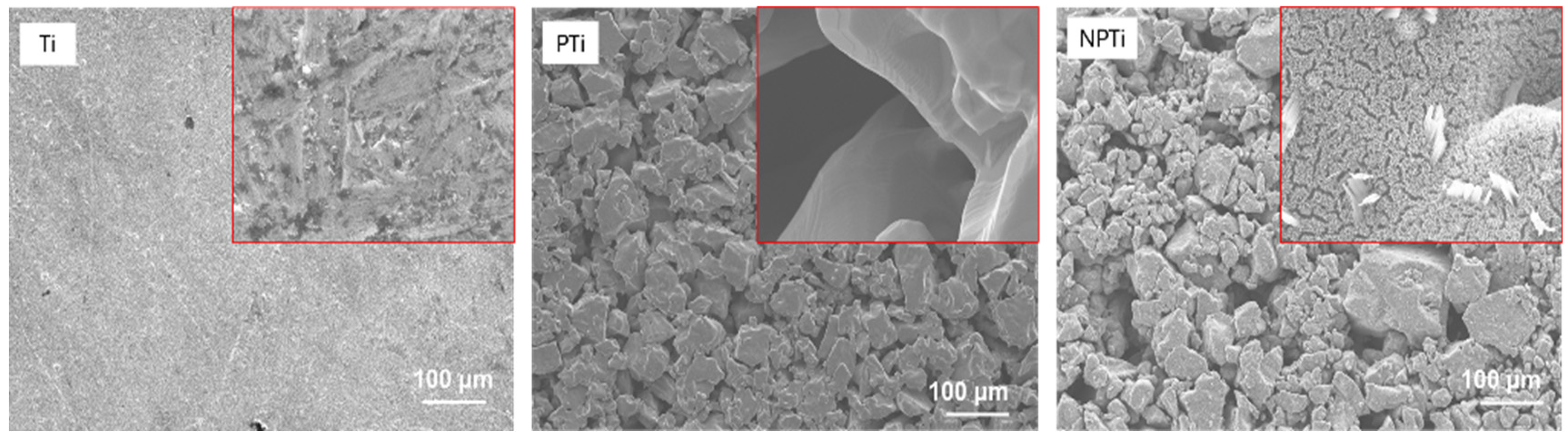
2.1. Fabrication of Nanostructured-Micro-Porous Titanium Surfaces

Commercially available micro-porous titanium disks (PTi) were modified using a hydrothermal treatment (HT) to fabricate nanostructured-micro-porous titanium surfaces (NPTi). PTi surfaces were cleaned in deionized (DI) water and subjected to HT in an alkali medium of 25% NaOH solution with the HT parameters set at 60 °C for 24 h. After HT, the surfaces were rinsed with DI water and annealed at 530 °C for 3 h in an oxygen-ambient environment where the temperature increment rate was 15 °C/min at the beginning of the annealing process.

2.2. Characterization of Nanostructured-Micro-Porous Titanium Surfaces

The surface morphology was characterized using JEOL 6500 field emission scanning electron microscopy (SEM) (JEOL, Tokyo, Japan). Prior to the imaging, surfaces were coated with a 10 nm layer of gold using a sputter coater in order to enhance conductivity. The SEM parameters were optimized and chosen as: accelerating voltage of 15 kV, working distance of 10 mm, and vacuum pressure below 3 × 10⁻⁴ Pa. Images were captured at magnifications of 100x and 5000x.
The surface chemistry was characterized using a PHI-5600 X-ray photoelectron spectroscopy (XPS) (IRDQ, Montreal, QC, Canada) probe equipped with an AI Kα X-ray source. The survey spectra were collected to evaluate the elemental composition of the surfaces at a pass energy of 187 eV and 0.05 eV step for 16 cycles. The data were analyzed using CASA XPS software (Version 2.3.24PR1.0), and the percentage of elements was calculated.

2.3. Adipose Derived Stem Cell (ADSC) Culture

Human ADSCs were obtained from the laboratory of the late Dr. Kimberly Cox-York at Colorado State University. The ADSC isolation protocol was approved by the Institutional Review Board at Colorado State University. The cells were cultured in a media comprising of α-MEM media, 10% (v/v) fetal bovine serum (FBS), and 1% (v/v) penicillin/streptomycin [26] at 37 °C and 5% CO2. The cells were cultured on a tissue culture flask, and the media was changed every other day until the confluency of the cells reached >80%. Following expansion, cells were detached using TrypLE (Thermo Fisher Scientific, Waltham, MA, USA) and suspended in growth media. All cells used in these studies were at or below passage 4. Prior to the seeding of cells, surfaces were sterilized through incubation in 70% ethanol solution for 10 min, followed by ultraviolet light exposure for 30 min before being rinsed three times in phosphate-buffered saline (PBS) solution. Surfaces were kept in 48-well plates, and 2 × 10⁴ cells were seeded per well.

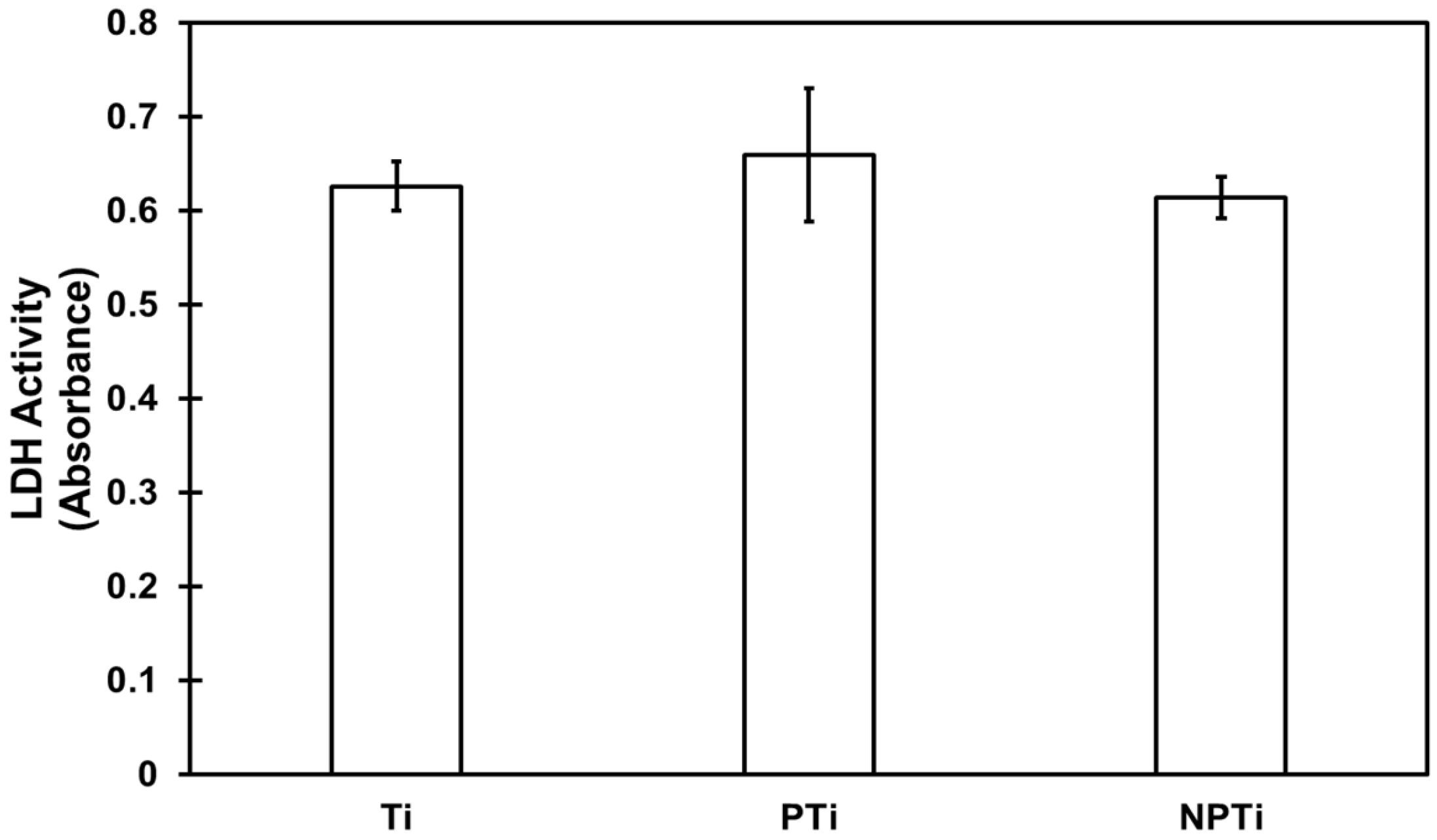
2.4. Cytotoxicity of Different Surfaces

The cytotoxicity of different surfaces was evaluated using a commercially available lactate dehydrogenase (LDH) assay from CyQUANTTM (Thermo Fisher Scientific, Waltham, MA, USA). After reaching 80% confluency, 20 × 104 cells/mL were transferred to each well of a 48-well plate containing surfaces. The cells were grown for 24 h at 37 °C and 5% CO2. Cells were lysed using a lysis buffer for the positive control, and cells grown in wells were used as the negative control. In total, 50 μL of the solution were transferred from each well to a 96-well plate, and the manufacturer’s protocol was followed to determine the cytotoxicity.

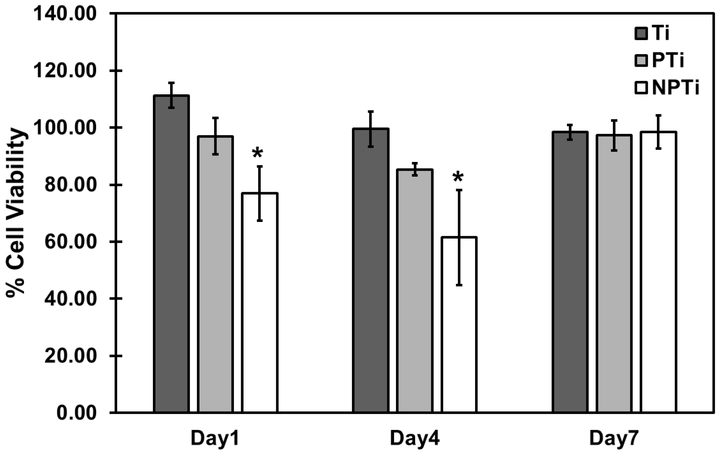
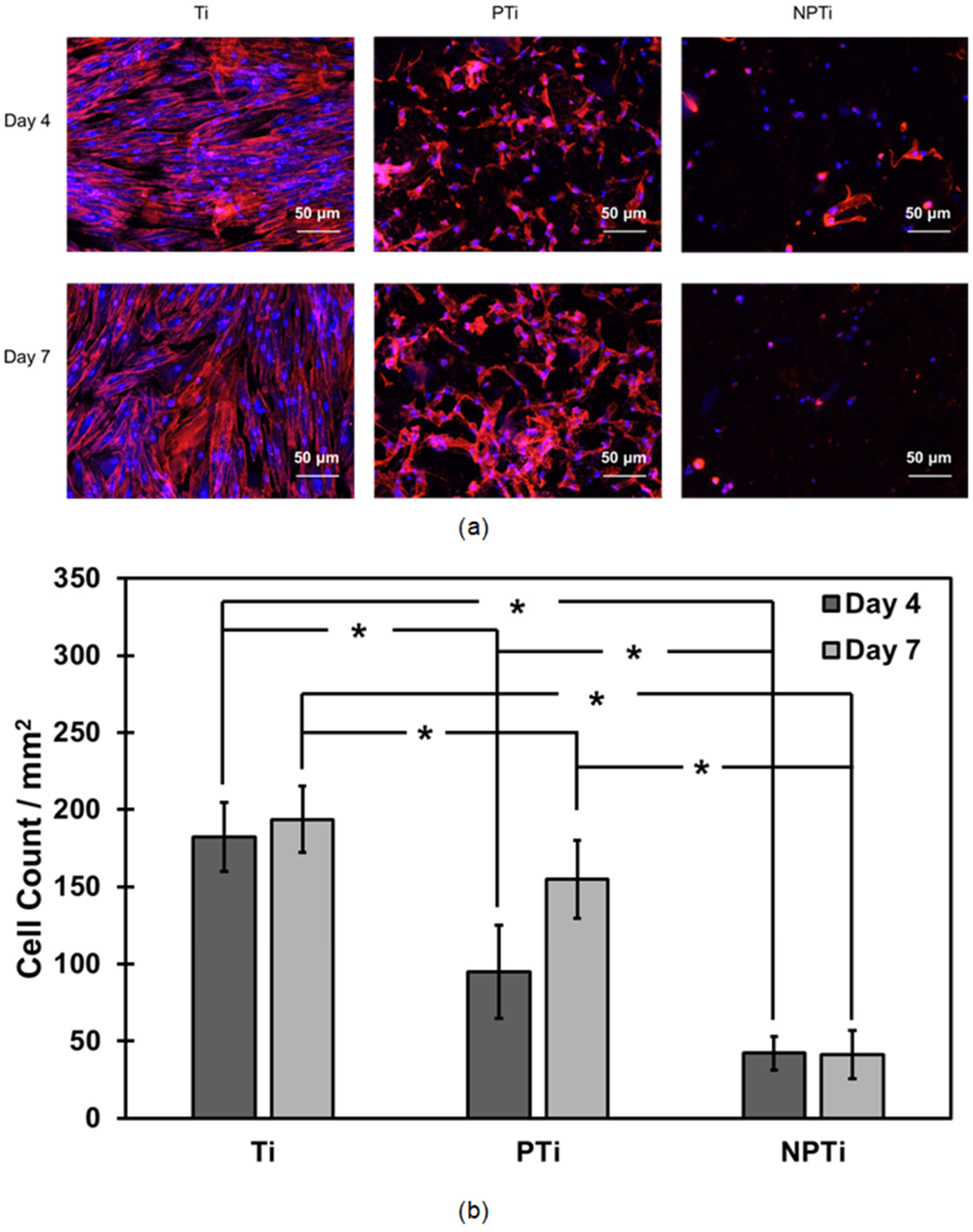
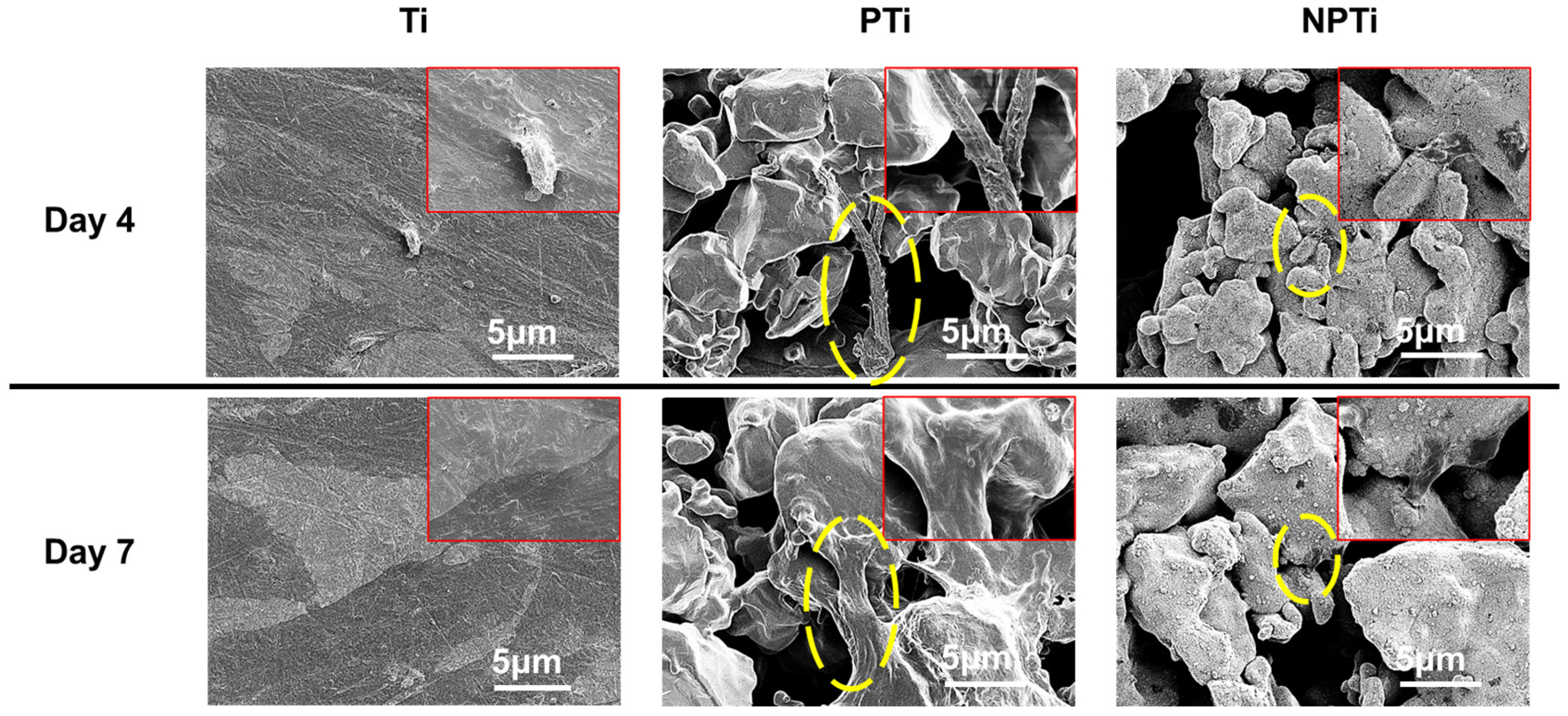
2.5. ADSC Adhesion and Proliferation on Different Surfaces

Cell viability on the surfaces was evaluated using a CellTiter-Blue assay (Promega, Madison, WI, USA) after 1, 4, and 7 days of culture. The manufacturer’s protocol was followed [27], and the cells were incubated in a solution of 10% (v/v) CellTiter-Blue reagent in growth media for 4 h at 37 °C and 5% CO2. After the incubation period, 150 μL solution were taken from each well and placed in a 96-well plate. The absorbance was read at 570 nm and 600 nm.
Cell adhesion and proliferation were evaluated using fluorescence microscopy. After days 4 and 7 of culture, the media was removed, and surfaces were rinsed three times in PBS. The cells were then fixed on the surfaces using a 3.7% (v/v) formaldehyde solution in PBS for 15 min. Surfaces were then rinsed three times in PBS before the cells were permeabilized using a 1% Triton X-100 solution in PBS for 3 min and rinsed twice with PBS. The cells were then stained using rhodamine phalloidin stain in PBS for 20 min to stain the cytoskeleton, and DAPI was added for the final 5 min to stain the nucleus. The surfaces were then rinsed twice with PBS and kept in PBS until imaging under a fluorescence microscope was completed. The cells were counted from the fluorescence microscopy images via the number of nuclei adhered per mm2 of surfaces.
Cell morphology on the surfaces was evaluated using SEM. After 4 and 7 days of culture, the media was removed, and surfaces were rinsed thrice in PBS. Cells were fixed on the surfaces for 45 min with a fixative solution of 3% glutaraldehyde, 0.1 M sucrose, and 0.1 M sodium cacodylate in DI water. After the cells were fixed, the surfaces were allowed to sit for 10 min in a buffer solution (fixative solution except glutaraldehyde). The surfaces were then immersed successively for 10 min each in 35%, 50%, 70%, and 100% ethanol solution before being stored in a desiccator. Prior to imaging, surfaces were coated with a 10 nm layer of chromium (Cr) to increase conductivity. SEM images were captured at different magnifications ranging from 250x to 5000x. SEM parameters were set as follows: accelerating voltage of 3 kV, working distance of 10 mm, and vacuum pressure below 3 × 10⁻⁴ Pa.

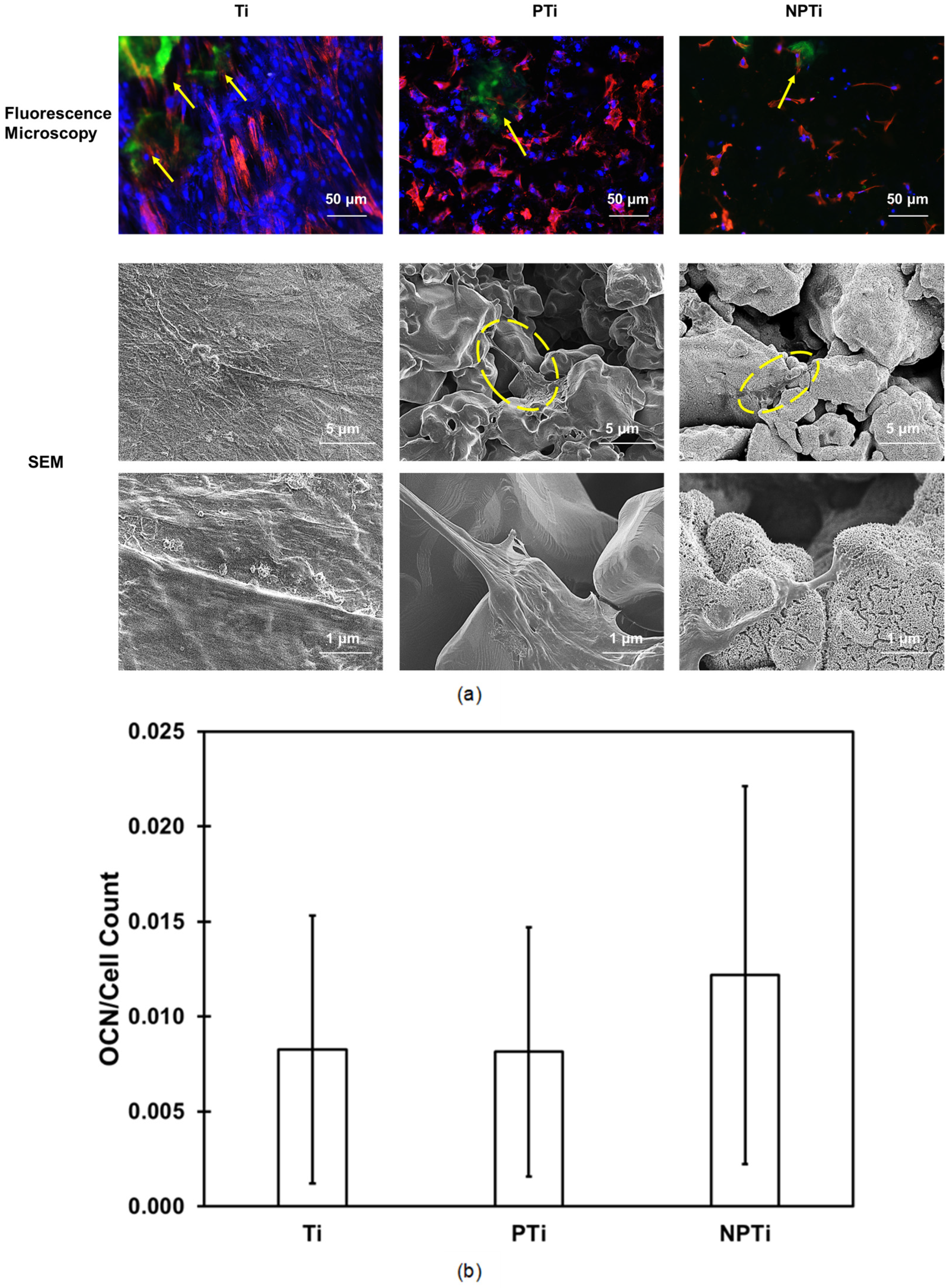
2.6. ADSC Differentiation on Different Surfaces

Cell differentiation to osteogenic lineages was initiated after 7 days of culture. The cells were exposed to a differentiation media (growth media plus 10−8 M dexamethasone, 50 μg/mL β-glycerol phosphate, and 6 mM ascorbic acid) to induce osteogenesis. The differentiation media was changed every other day from day 7 to 14. After 14 days of culture, the media was removed from the wells and the surfaces were rinsed twice with PBS. Subsequently, 500 μL 0.2% (v/v) Triton X-100 solution in PBS were added to each well, and the surfaces were shaken at 150 rpm for 20 min to remove the proteins from the cells. Osteogenic differentiation was then evaluated using the following assays: total protein content, alkaline phosphatase (ALP) activity, and calcium concentration (Ca):
  • The total protein content was determined using a commercially available microBCA assay. In total, 150 μL of working reagent were generated from the assay and 150 μL of protein supernatant were added to a 96-well plate and incubated for 2 h at 37 °C and 5% CO2. After the incubation period, the absorbance was read at 562 nm. The total protein concentration was determined from a standard absorbance curve versus the known albumin standard provided by the manufacturer.
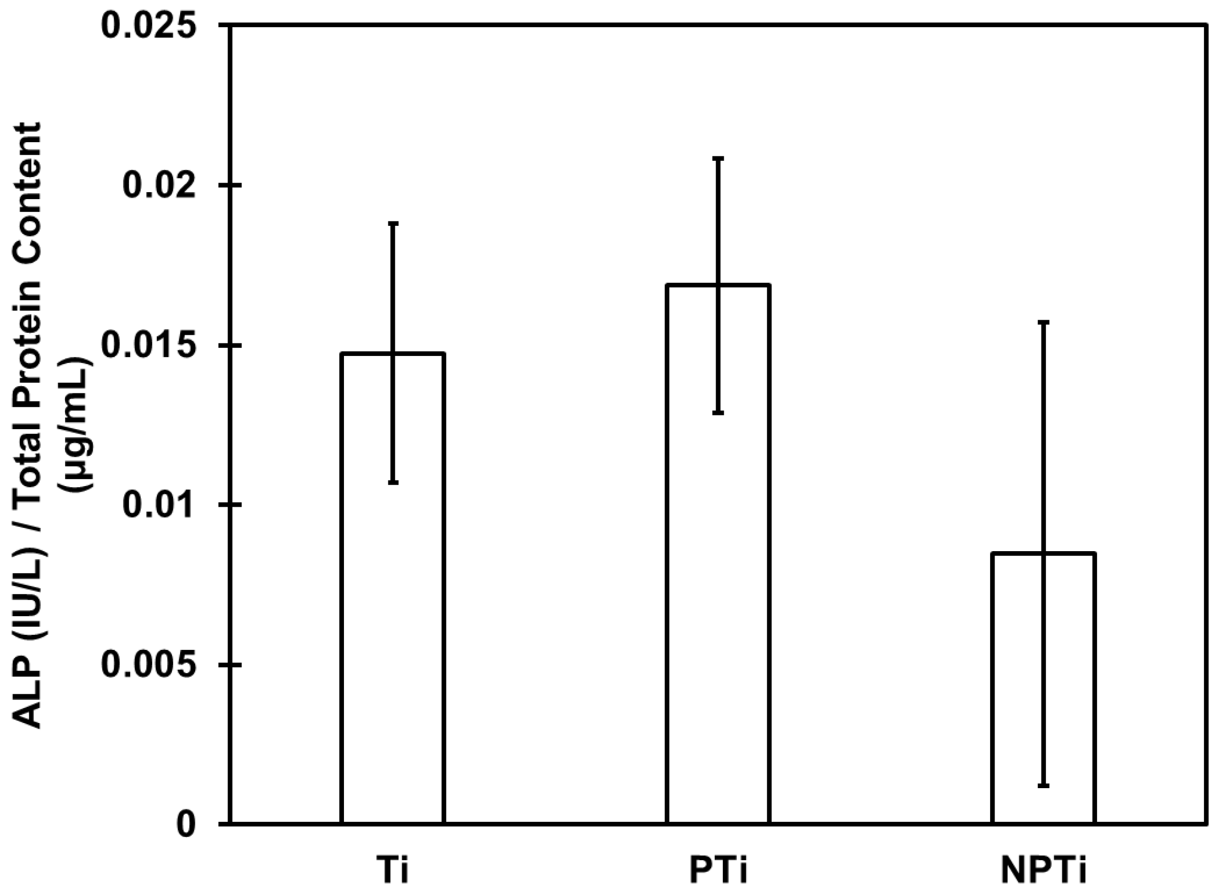
  • ALP activity was determined using a commercially available ALP assay kit from QuantiChromTM. In total, 150 μL of working reagent were prepared from the assay kit, and 50 μL of protein supernatant were added to a 96-well plate. The absorbance was read at 405 nm and repeated after 4 min. Absorbance was converted to concentration using an ALP standard, and data were normalized using the total protein content.
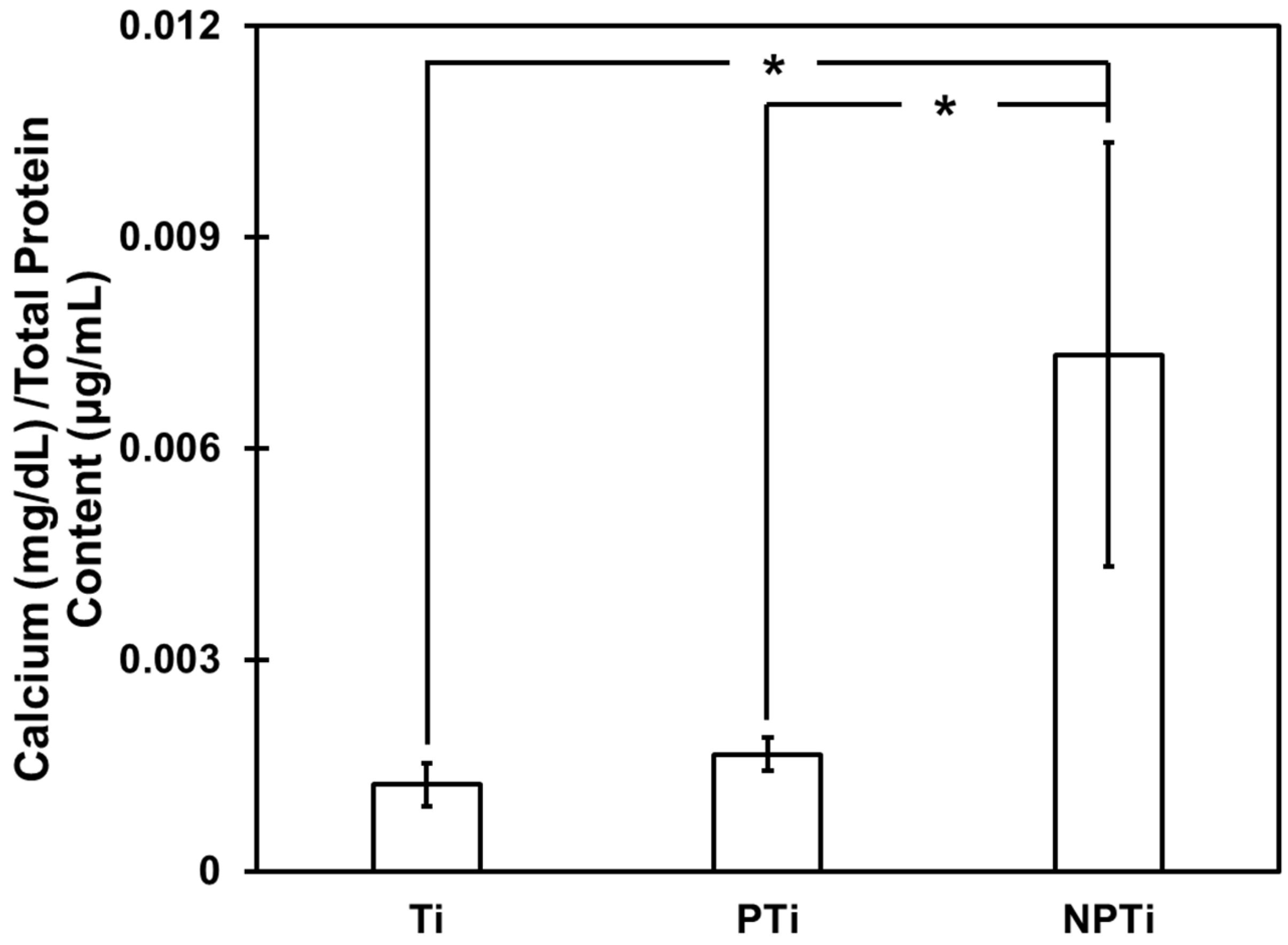
  • Calcium deposition was determined using a commercially available calcium reagent test from Teco Diagnostics (Anaheim, CA, USA). The protein supernatant was removed, and surfaces were rinsed with DI water. In total, 6 M HCl (Hydrochloric acid) solution were added to the wells, and the surfaces were placed in a shaker for 12 h at 100 rpm to ensure all the calcium dissolved in the solution. After 12 h, 1 mL of working reagent was prepared from the test kit, and 20 μL of HCl–calcium solution were added to a 24-well plate. The absorbance was read at 570 nm and was converted to concentration using the calcium standard provided by the manufacturer and the data was normalized using total protein content.
The differentiation was also characterized using immunofluorescence microscopy for osteocalcin. After 14 and 28 days of culture, the media was removed, and the surfaces were rinsed thrice in PBS. Cells were then fixed using a 3.7% (v/v) formaldehyde solution in PBS for 15 min. The surfaces were rinsed three times in PBS before the cells were permeabilized using a 1% Triton X-100 solution in PBS for 3 min and rinsed twice with PBS. The surfaces were immersed in a 10% bovine serum albumin (BSA) solution in PBS for 30 min to block non-specific binding sites in the cells. After BSA was removed, surfaces were washed twice with PBS and an osteocalcin (OCN) primary antibody in a 1% BSA solution at a dilution of 1:100 for 60 min. Surfaces were then washed with PBS thrice, followed by the addition of a secondary antibody, FITC, in a 1% BSA solution at a dilution of 1:200 for 45 min. Following washing with PBS, surfaces were stained with rhodamine-phalloidin and DAPI, the process for which was explained in the previous section. The surfaces were then washed twice with PBS and left in PBS until being imaged under a fluorescence microscope.

2.7. Statistical Analysis

Surface characterization was repeated for at least six different samples on each surface. Cell studies were performed two times with at least three different samples in each group (nmin = 6). The quantitative results were analyzed using JMP software(Version 17). Two-way analysis of variance (ANOVA), and Tukey’s honestly significant difference (HSD) test with significant results considered when p < 0.05. The data presented here are derived from one of the studies, and similar trends were observed in other cell studies as well.

3. Results and Discussion

In a previous study [28], nano-structured, micro-porous titanium surfaces were fabricated via hydrothermal treatment in a sodium hydroxide solution and evaluated for their blood-clotting properties and antibacterial properties against staphylococcus aureus and pseudomonas aeruginosa. The platelet adhesion and activation and whole blood-clotting characteristics were evaluated, and the adhesion, growth, and bacteria morphology were evaluated. Results indicated that NPTi surfaces demonstrated significantly decreased bacteria adhesion, proliferation, and biofilm formation (p < 0.05). NPTi surfaces displayed a reduction in growth of over 20% for Gram-positive bacteria and over 10% for Gram-negative bacteria. This reduction is a result of the surface topography of NPTi surfaces, which deforms the membrane of the bacteria cells and hence prevents their adhesion and subsequent growth. The blood-clotting characteristics (e.g., platelet adhesion and activation and whole blood clotting) were also evaluated for the surfaces. As compared to titanium, PTi and NPTi had higher rates of whole blood clotting after 15 min of clotting. However, after 45 min, all the surfaces had equivalent clotting. PTi and NPTi surfaces had significantly lower adhesion of platelets than Ti; however, the platelets had higher spreading for PTi and NPTi surfaces. Cell adhesion, proliferation, and differentiation are also influenced by surface properties like surface topography, surface chemistry, etc. Hence, in this study, cell adhesion, proliferation, and osteogenic differentiation of ADSCs were investigated.
The surface topography of the surfaces was characterized using SEM (Figure 1) and is discussed elsewhere [28]. To explain in brief, Ti surfaces did not display any distinct features. The roughness could be due to the process used to clean the Ti surfaces. In the micro-porous surfaces, no topography change was observed at lower magnifications due to the HT process between unmodified (PTi) and modified (NPTi) surfaces. However, at higher magnifications, the effect of HT is visible in the form of nano-petal-like features and a “web”-like structure, which is formed because of the dissolution and precipitation mechanisms in the HT [25]. The HT parameters were determined to fabricate nanostructures while retaining the inherent micro-porous structure.
XPS was performed to characterize the surface chemical composition (Table 1), which is explained in detail elsewhere [28]. In brief, titanium (Ti 2p3/2), oxygen (O1s), and carbon (C1s) were present on all the surfaces. Carbon presence was attributed to contamination from the XPS chamber and other surface impurities. NPTi surfaces had sodium (Na1s) present due to the HT in NaOH solution, which forms sodium titanate on the surfaces.
For the long-term success of implants, it is crucial for the material to be conducive to cell growth and, hence, not be toxic towards them. A commercially available LDH assay was used to evaluate the cytotoxicity of different surfaces. LDH is an enzyme found in cells which, when released, is a sign of cell damage in the form of plasma membrane damage or lysis [11]. Higher LDH activity is an indicator of high cell damage, which shows the material is toxic. The manufacturer protocol was followed, and the absorbance was measured to calculate the LDH activity in the wells (Figure 2). Ti surfaces have been well established as a biocompatible material that is not cytotoxic. As expected, all the surfaces have similar LDH activity, demonstrating that they are not toxic to cells. NPTi surfaces are even a little less toxic than Ti surfaces, which indicates that HT is not only non-toxic towards cells but also removes some toxic matter from the surface. Similar behavior has been reported in studies involving HT treatment in an alkali medium [29].
After being certain that surfaces are not cytotoxic, it is important to investigate whether they will allow for cell viability, adhesion, and proliferation as they are the most important for osseointegration. A commercially available CellTiter-Blue assay (Promega) was used to assess cell viability after 1, 4, and 7 days of cell culture (Figure 3). Cell viability was determined by calculating the reduction percentage of the reagent from the CellTiter-Blue assay. The reagent solution contains resazurin, which is reduced to resorufin by living cells through dehydrogenase enzymes. Therefore, the higher the expression of resorufin, the higher cell viability [30]. Cells grown on the tissue culture polystyrene well plate were used as a positive control for the assay, and results were normalized with the same. After 1 and 4 days of cell culture, Ti surfaces displayed the highest cell viability, with a slight decrease in PTi surfaces. NPTi surfaces displayed significantly lower viability compared to Ti surfaces. However, after 7 days of culture, all the surfaces displayed equivalent cell viability.
The adhesion and proliferation of ADSCs were investigated using fluorescence microscopy. After 4 and 7 days of cell culture (Figure 4), as expected, Ti surfaces displayed the most cell adhesion, followed by PTi and NPTi, respectively. NPTi surfaces displayed significantly lower cell counts than both Ti and PTi surfaces. There was a drastic growth of cells on PTi surfaces between 4 and 7 days of culture. However, the cell functionality of the adhered cells was important. The cell growth on Ti surfaces was unidirectional, whereas on PTi and NPTi surfaces, it was more elongated. Not much growth was observed on NPTi surfaces, which also corroborated the quantified results. The lower adhesion could be attributed to the microporosity of the surfaces for PTi and NPTi, with similar studies showing porous surfaces can lead to lower cell proliferation yet higher differentiation [31,32]. Hence, even though PTi and NPTi surfaces displayed lower adhesion, they have a micro-porous structure that has been established as one that promotes osseointegration.
SEM was used to evaluate the morphology of adhered cells (Figure 5). As expected, the cell morphology for Ti surfaces was comparable to that reported in similar studies [33]. As seen with fluorescence microscopy images, an increase was observed in the proliferation of cells from day 4 to 7 of culture. The growth of cells on PTi and NPTi surfaces was elongated, as was seen in the fluorescence microscopy. The elongations are actin-based protrusions called filopodia, and they play a critical role in the cell adhesion process and in sensing their environment for contact guidance [34]. Filopodia are important for the cell response to nanoscale topography [35]. The spreading is also different from Ti surfaces to PTi and NPTi surfaces. Cells are constricted on PTi and NPTi surfaces compared to the spreading seen on Ti surfaces.
After 7 days of cell culture, differentiation to osteogenic lineage was induced by providing osteogenic media to cells. The osteogenic media was prepared by supplementing the cell culture growth media, which included β-glycerophosphate, dexamethasone, and ascorbic acid. Supplementing the growth media is necessary as α-MEM media enhances cell proliferation but disables differentiation markers [36]. Each of the additives has a specific role in influencing the osteogenic differentiation of ADSCs. RUNX2 expression, a key transcription factor for osteoblast differentiation, is induced and regulated by dexamethasone. Phosphate for bone mineral (hydroxyapatite (HAp)) formation is provided by β-glycerophosphate. Ascorbic acid facilitates an increase in collagen type 1 secretion, which ensures osteogenic differentiation [37]. It is important for osteogenesis for successful osseointegration of implants [26]. ADSC differentiation to osteogenic lineages was evaluated through immunofluorescence microscopy and protein-based assays. Deposition of osteocalcin—a marker protein for osseointegration, was evaluated through immunofluorescence microscopy. Additionally, calcium deposition and ALP secretion were evaluated through assays to understand the differentiation level of cells.
ALP plays a crucial role in the mineralization of the bone matrix and is an early indicator of immature osteoblast activity [38]. As it is an early indicator, ALP activity usually reaches its peak when differentiation is in its early stages and then decreases before increasing again [39]. The ALP results for different surfaces were normalized with the total protein (microBCA) content after 7 days of inducing differentiation (Figure 6). Normalized ALP activity was higher for Ti and PTi surfaces than for NPTi surfaces. The lower expression could be attributed to the cyclic nature of the ALP and might be due to the micro/nano topography, which alleviates the rate of differentiation to osteogenic lineages.
Hap is an integral inorganic component of the bone and is made up of two major components: calcium and phosphorous. Hap is released during the mineralization process; hence, it is necessary to quantify it to get an idea of the differentiation of cells. Phosphorous is released by cells in the form of ALP. Hap crystals are deposited on the surfaces by cells. HCl was used to dissolve calcium deposited on surfaces and quantified by a colorimetric assay, and the results were normalized with the microBCA content (Figure 7). Ti and Pti surfaces displayed similar calcium deposition; however, NPTi surfaces displayed significantly higher (almost three times) calcium deposition.
As the differentiation of stem cells to osteogenic lineage begins, some matrix proteins that are required for mineralization are released. One of those proteins is osteocalcin (OCN), a late marker in osteoblast differentiation that is involved in the formation of the bone matrix [40]. OCN deposition was evaluated on different surfaces using immunofluorescence microscopy 7 days after differentiation was induced (Figure 8). OCN was present on all surfaces, hence indicating the differentiation of ADSCs to osteoblasts. The OCN area was calculated using ImageJ software (Version 1.53) and normalized by the number of nuclei on the surfaces. Immunofluorescence images show the presence of OCN on all the surfaces. Despite a lower cell count, NPTi surfaces displayed a higher normalized OCN expression. These results, combined with calcium deposition results, demonstrate a higher differentiation of ADSCs to osteoblasts on NPTi surfaces. Lower cell proliferation yet higher differentiation has been observed in similar studies and is attributed to the promoted Runx2 on nanostructured surfaces [41,42]. These studies also demonstrated increased OCN deposition and ALP activity [42].
The calcium deposition and OCN are higher for PTi surfaces than for the Ti surface, hence demonstrating that micro-porosity enhances differentiation. The higher differentiation between PTi and NPTi surfaces can be clearly attributed to HT. Stem cell morphology was evaluated using SEM (Figure 8). As expected, there was a higher growth of cells on Ti surfaces as compared to PTi and NPTi surfaces. PTi and NPTi surfaces, however, displayed long filopodia. The cells were also more elongated in length as compared to 4 and 7 days after cell culture. The higher values of OCN and calcium deposition on NPTi surfaces can be attributed to the surface topography, as well as the formation of sodium titanate on the NPTi surfaces. Higher OCN and calcium deposition have been observed in other studies where the surface was modified using sodium hydroxide [43].

4. Conclusions

In this work, NPTi surfaces were fabricated and adhesion, growth, and osteogenic differentiation of ADSCs were investigated. NPTi surfaces were characterized for their surface topography and surface chemistry using SEM and XPS, respectively. SEM displayed the presence of nano-petal-like features and a “web”-like structure in addition to the micro-porous structure. The XPS survey showed the presence of sodium, which was expected due to the formation of sodium titanate because of HT. The LDH assay was used to evaluate the cytotoxicity of the surfaces, which showed the surfaces were not toxic towards cells, and LDH activity was marginally lower than Ti. Cell viability, adhesion, proliferation, and differentiation on the surfaces were evaluated by seeding ADSCs on the surfaces. Cell viability for the surfaces was evaluated using a CellTiter-Blue assay, and it showed that even though the NPTi surface had lower cell viability on day 1, by day 7, it was equivalent to Ti surfaces. NPTi surfaces displayed significantly lowered cell adhesion and proliferation compared to Ti and PTi surfaces after days 4 and 7 of culture. After 7 days, osteogenesis was induced by the introduction of supplements in addition to growth media. Differentiation to osteogenic lineages was evaluated with immunofluorescence staining for a marker protein (OCN) along with protein assays to evaluate the ALP activity and calcium deposition. After 7 days of introducing differentiation media, NPTi surfaces displayed significantly higher OCN expression and calcium deposition amongst all groups. These results displayed that alkali-treated, nanostructured-micro-porous surfaces demonstrated enhanced osteogenic differentiation of ADSCs and have the potential to be used as surfaces for the fabrication of orthopedic implants.

Author Contributions

Conceptualization, A.V.S. and K.C.P.; methodology, A.V.S. and K.C.P.; validation, A.V.S. and E.H.; formal analysis, A.V.S.; investigation, A.V.S. and E.H.; resources, A.V.S. and K.C.P.; data curation, A.V.S.; writing—original draft preparation, A.V.S.; writing—review and editing, A.V.S. and K.C.P.; visualization, A.V.S.; supervision, K.C.P.; project administration, A.V.S.; funding acquisition, K.C.P. All authors have read and agreed to the published version of the manuscript.

Funding

This research was funded by the National Heart, Lung, and Blood Institute of the National Institute of Health, grant number R21EB033511.

Institutional Review Board Statement

Not applicable.

Informed Consent Statement

Not applicable.

Data Availability Statement

The datasets used and analyzed during the current study are available from the corresponding authors upon reasonable request.

Acknowledgments

The authors acknowledge Analytical Resources Core (ARC, RRID: SCR_021758) at the Colorado State University, Fort Collins, for their help in SEM and XPS data acquisition and analysis.

Conflicts of Interest

The authors declare no conflicts of interest. The funders had no role in the design of the study; in the collection, analyses, or interpretation of data; in the writing of the manuscript; or in the decision to publish the results.

References

  1. Sarraf, M.; Ghomi, E.R.; Alipour, S.; Ramakrishna, S.; Sukiman, N.L. A state-of-the-art review of the fabrication and characteristics of titanium and its alloys for biomedical applications. Bio-Des. Manuf. 2021, 5, 371–395. [Google Scholar] [CrossRef]
  2. Wang, Q.; Zhou, P.; Liu, S.; Attarilar, S.; Ma, R.L.-W.; Zhong, Y.; Wang, L. Multi-Scale Surface Treatments of Titanium Implants for Rapid Osseointegration: A Review. Nanomaterials 2020, 10, 1244. [Google Scholar] [CrossRef] [PubMed]
  3. Mendonça, G.; Mendonça, D.B.S.; Aragão, F.J.L.; Cooper, L.F. Advancing dental implant surface technology—From micron- to nanotopography. Biomaterials 2008, 29, 3822–3835. [Google Scholar] [CrossRef] [PubMed]
  4. Wu, Y.; Gao, G.; Wu, G. Self-assembled three-dimensional hierarchical porous V2O5/graphene hybrid aerogels for supercapacitors with high energy density and long cycle life. J. Mater. Chem. A Mater. 2015, 3, 1828–1832. [Google Scholar] [CrossRef]
  5. Maleki, H.; Shahbazi, M.-A.; Montes, S.; Hosseini, S.H.; Eskandari, M.R.; Zaunschirm, S.; Verwanger, T.; Mathur, S.; Milow, B.; Krammer, B.; et al. Mechanically Strong Silica-Silk Fibroin Bioaerogel: A Hybrid Scaffold with Ordered Honeycomb Micromorphology and Multiscale Porosity for Bone Regeneration. ACS Appl. Mater. Interfaces 2019, 11, 17256–17269. [Google Scholar] [CrossRef]
  6. Kenar, H.; Köse, G.T.; Hasirci, V. Tissue engineering of bone on micropatterned biodegradable polyester films. Biomaterials 2006, 27, 885–895. [Google Scholar] [CrossRef]
  7. Wilkinson, A.; Hewitt, R.N.; McNamara, L.E.; McCloy, D.; Meek, R.D.; Dalby, M.J. Biomimetic microtopography to enhance osteogenesis in vitro. Acta Biomater. 2011, 7, 2919–2925. [Google Scholar] [CrossRef]
  8. Yu, T.T.; Cui, F.Z.; Meng, Q.Y.; Wang, J.; Wu, D.C.; Zhang, J.; Kou, X.X.; Yang, R.L.; Liu, Y.; Zhang, Y.S.; et al. Influence of Surface Chemistry on Adhesion and Osteo/Odontogenic Differentiation of Dental Pulp Stem Cells. ACS Biomater. Sci. Eng. 2017, 3, 1119–1128. [Google Scholar] [CrossRef]
  9. Lu, R.J.; Wang, X.; He, H.X.; E, L.-L.; Li, Y.; Zhang, G.L.; Li, C.J.; Ning, C.Y.; Liu, H.C. Tantalum-incorporated hydroxyapatite coating on titanium implants: Its mechanical and in vitro osteogenic properties. J. Mater. Sci. Mater. Med. 2019, 30, 111. [Google Scholar] [CrossRef]
  10. Rajendran, A.; Kapoor, U.; Jothinarayanan, N.; Lenka, N.; Pattanayak, D.K. Effect of Silver-Containing Titania Layers for Bioactivity, Antibacterial Activity, and Osteogenic Differentiation of Human Mesenchymal Stem Cells on Ti Metal. ACS Appl. Bio Mater. 2019, 2, 3808–3819. [Google Scholar] [CrossRef]
  11. Savargaonkar, A.V.; Munshi, A.H.; Soares, P.; Popat, K.C. Antifouling Behavior of Copper-Modified Titania Nanotube Surfaces. J. Funct. Biomater. 2023, 14, 413. [Google Scholar] [CrossRef] [PubMed]
  12. Wen, J.; Li, J.; Pan, H.; Zhang, W.; Zeng, D.; Xu, L.; Wu, Q.; Zhang, X.; Liu, X.; Jiang, X. Strontium delivery on topographical titanium to enhance bioactivity and osseointegration in osteoporotic rats. J. Mater. Chem. B 2015, 3, 4790–4804. [Google Scholar] [CrossRef] [PubMed]
  13. Wolf-Brandstetter, C.; Beutner, R.; Hess, R.; Bierbaum, S.; Wagner, K.; Scharnweber, D.; Gbureck, U.; Moseke, C. Multifunctional calcium phosphate based coatings on titanium implants with integrated trace elements. Biomed. Mater. 2020, 15, 025006. [Google Scholar] [CrossRef] [PubMed]
  14. Yao, C.; Slamovich, E.B.; Webster, T.J. Enhanced osteoblast functions on anodized titanium with nanotube-like structures. J. Biomed. Mater. Res. A 2008, 85A, 157–166. [Google Scholar] [CrossRef]
  15. Porter, J.R.; Henson, A.; Popat, K.C. Biodegradable poly(ε-caprolactone) nanowires for bone tissue engineering applications. Biomaterials 2009, 30, 780–788. [Google Scholar] [CrossRef]
  16. Park, J.K.; Kim, Y.; Yeom, J.; Jeon, J.H.; Yi, G.; Je, J.H.; Hahn, S.K. The Topographic Effect of Zinc Oxide Nanoflowers on Osteoblast Growth and Osseointegration. Adv. Mater. 2010, 22, 4857–4861. [Google Scholar] [CrossRef]
  17. Oliveira, F.C.; Carvalho, J.O.; Gusmão, S.B.S.; Gonçalves, L.d.S.; Mendes, L.M.S.; Freitas, S.A.P.; Gusmão, G.O.d.M.; Viana, B.C.; Marciano, F.R.; Lobo, A.O. High loads of nano-hydroxyapatite/graphene nanoribbon composites guided bone regeneration using an osteoporotic animal model. Int. J. Nanomed. 2019, 14, 865–874. [Google Scholar] [CrossRef]
  18. Wen, C.E.; Yamada, Y.; Hodgson, P.D. Fabrication of novel TiZr alloy foams for biomedical applications. Mater. Sci. Eng. C 2006, 26, 1439–1444. [Google Scholar] [CrossRef]
  19. Barbas, A.; Bonnet, A.-S.; Lipinski, P.; Pesci, R.; Dubois, G. Development and mechanical characterization of porous titanium bone substitutes. J. Mech. Behav. Biomed. Mater. 2012, 9, 34–44. [Google Scholar] [CrossRef]
  20. Yang, J.; Wang, J.; Yuan, T.; Zhu, X.D.; Xiang, Z.; Fan, Y.J.; Zhang, X.D. The enhanced effect of surface microstructured porous titanium on adhesion and osteoblastic differentiation of mesenchymal stem cells. J. Mater. Sci. Mater. Med. 2013, 24, 2235–2246. [Google Scholar] [CrossRef]
  21. Gao, C.; Deng, Y.; Feng, P.; Mao, Z.; Li, P.; Yang, B.; Deng, J.; Cao, Y.; Shuai, C.; Peng, S. Current Progress in Bioactive Ceramic Scaffolds for Bone Repair and Regeneration. Int. J. Mol. Sci. 2014, 15, 4714–4732. [Google Scholar] [CrossRef]
  22. Habibovic, P.; Li, J.; van der Valk, C.M.; Meijer, G.; Layrolle, P.; van Blitterswijk, C.A.; de Groot, K. Biological performance of uncoated and octacalcium phosphate-coated Ti6Al4V. Biomaterials 2005, 26, 23–36. [Google Scholar] [CrossRef] [PubMed]
  23. Fujibayashi, S.; Neo, M.; Kim, H.-M.; Kokubo, T.; Nakamura, T. Osteoinduction of porous bioactive titanium metal. Biomaterials 2004, 25, 443–450. [Google Scholar] [CrossRef] [PubMed]
  24. Rani, V.V.D.; Manzoor, K.; Menon, D.; Selvamurugan, N.; Nair, S.V. The design of novel nanostructures on titanium by solution chemistry for an improvedosteoblast response. Nanotechnology 2009, 20, 195101. [Google Scholar] [CrossRef] [PubMed]
  25. Vishnu, J.; Manivasagam, V.K.; Gopal, V.; Garcia, C.B.; Hameed, P.; Manivasagam, G.; Webster, T.J. Hydrothermal treatment of etched titanium: A potential surface nano-modification technique for enhanced biocompatibility. Nanomedicine 2019, 20, 102016. [Google Scholar] [CrossRef]
  26. Sabino, R.M.; Mondini, G.; Kipper, M.J.; Martins, A.F.; Popat, K.C. Tanfloc/heparin polyelectrolyte multilayers improve osteogenic differentiation of adipose-derived stem cells on titania nanotube surfaces. Carbohydr. Polym. 2021, 251, 117079. [Google Scholar] [CrossRef]
  27. CellTiter-Blue® Cell Viability Assay. Available online: https://www.promega.com/products/cell-health-assays/cell-viability-and-cytotoxicity-assays/celltiter_blue-cell-viability-assay/?catNum=G8080 (accessed on 26 October 2023).
  28. Savargaonkar, A.V.; Holloway, E.; Madruga, L.Y.C.; Pereira, B.L.; Soares, P.; Popat, K.C. Anti-Bacterial Properties and Hemocompatibility of Alkali Treated Nano-Structured Micro-Porous Titanium Surfaces. Biomimetics 2025, 10, 115. [Google Scholar] [CrossRef]
  29. Manivasagam, V.K.; Popat, K.C. Endothelial and smooth muscle cell interaction with hydrothermally treated titanium surfaces. Vitr. Models 2024, 3, 109–123. [Google Scholar] [CrossRef]
  30. Madruga, L.Y.; Sabino, R.M.; Santos, E.C.; Popat, K.C.; Balaban, R.d.C.; Kipper, M.J. Carboxymethyl-kappa-carrageenan: A study of biocompatibility, antioxidant and antibacterial activities. Int. J. Biol. Macromol. 2020, 152, 483–491. [Google Scholar] [CrossRef]
  31. Pałka, K.; Pokrowiecki, R. Porous Titanium Implants: A Review. Adv. Eng. Mater. 2018, 20, 1700648. [Google Scholar] [CrossRef]
  32. Chang, B.; Song, W.; Han, T.; Yan, J.; Li, F.; Zhao, L.; Kou, H.; Zhang, Y. Influence of pore size of porous titanium fabricated by vacuum diffusion bonding of titanium meshes on cell penetration and bone ingrowth. Acta Biomater. 2016, 33, 311–321. [Google Scholar] [CrossRef] [PubMed]
  33. Dias-Netipanyj, M.F.; Cowden, K.; Sopchenski, L.; Cogo, S.C.; Elifio-Esposito, S.; Popat, K.C.; Soares, P. Effect of crystalline phases of titania nanotube arrays on adipose derived stem cell adhesion and proliferation. Mater. Sci. Eng. C 2019, 103, 109850. [Google Scholar] [CrossRef] [PubMed]
  34. Albuschies, J.; Vogel, V. The role of filopodia in the recognition of nanotopographies. Sci. Rep. 2013, 3, srep01658. [Google Scholar] [CrossRef]
  35. McNamara, L.E.; Sjöström, T.; Seunarine, K.; Meek, R.D.; Su, B.; Dalby, M.J. Investigation of the limits of nanoscale filopodial interactions. J. Tissue Eng. 2014, 5, 2041731414536177. [Google Scholar] [CrossRef]
  36. Watchrarat, K.; Korchunjit, W.; Buranasinsup, S.; Taylor, J.; Ritruechai, P.; Wongtawan, T. MEM α Promotes Cell Proliferation and Expression of Bone Marrow Derived Equine Mesenchymal Stem Cell Gene Markers but Depresses Differentiation Gene Markers. J. Equine Veter. Sci. 2017, 50, 8–14. [Google Scholar] [CrossRef]
  37. Langenbach, F.; Handschel, J. Effects of dexamethasone, ascorbic acid and β-glycerophosphate on the osteogenic differentiation of stem cells in vitro. Stem Cell Res. Ther. 2013, 4, 1–7. [Google Scholar] [CrossRef]
  38. de Souza, R.F.B.; de Souza, F.C.B.; Thorpe, A.; Mantovani, D.; Popat, K.C.; Moraes, A.M. Phosphorylation of chitosan to improve osteoinduction of chitosan/xanthan-based scaffolds for periosteal tissue engineering. Int. J. Biol. Macromol. 2020, 143, 619–632. [Google Scholar] [CrossRef]
  39. Huang, Z.; Nelson, E.R.; Smith, R.L.; Goodman, S.B. The Sequential Expression Profiles of Growth Factors from Osteroprogenitors to Osteoblasts In Vitro. Tissue Eng. 2007, 13, 2311–2320. [Google Scholar] [CrossRef]
  40. Cowden, K.; Dias-Netipanyj, M.F.; Popat, K.C. Effects of titania nanotube surfaces on osteogenic differentiation of human adipose-derived stem cells. Nanomedicine 2019, 17, 380–390. [Google Scholar] [CrossRef]
  41. Huang, Y.Z.; He, S.K.; Guo, Z.J.; Pi, J.K.; Deng, L.; Dong, L.; Zhang, Y.; Su, B.; Da, L.C.; Zhang, L.; et al. Nanostructured titanium surfaces fabricated by hydrothermal method: Influence of alkali conditions on the osteogenic performance of implants. Mater. Sci. Eng. C 2019, 94, 1–10. [Google Scholar] [CrossRef]
  42. Manivasagam, V.K.; Popat, K.C. Hydrothermally treated titanium surfaces for enhanced osteogenic differentiation of adipose derived stem cells. Mater. Sci. Eng. C 2021, 128, 112315. [Google Scholar] [CrossRef] [PubMed]
  43. Fujino, T.; Taguchi, Y.; Komasa, S.; Sekino, T.; Tanaka, M. Cell Differentiation on Nanoscale Features of a Titanium Surface: Effects of Deposition Time in NaOH Solution. J. Hard Tissue Biol. 2014, 23, 63–70. [Google Scholar] [CrossRef]
Figure 1. SEM images of different surfaces at 100x and 5000x magnifications.
Applsci 15 05061 g001
Figure 2. LDH assay results showing the cytotoxicity of surfaces.
Applsci 15 05061 g002
Figure 3. Normalized cell viability results after 4 and 7 days of culture (* denotes p < 0.05).
Applsci 15 05061 g003
Figure 4. (a) Fluorescence microscopy images showing ADSCs stained with DAPI (blue) and rhodamine phalloidin (red) on days 4 and 7 of culture. (b) Quantification of cell count on days 4 and 7 (* indicates p < 0.05).
Applsci 15 05061 g004
Figure 5. ADSC SEM images on surfaces after 4 and 7 days of culture at 500× and 2500× magnification.
Applsci 15 05061 g005
Figure 6. Normalized ALP activity on surfaces after 7 days of inducing differentiation.
Applsci 15 05061 g006
Figure 7. Normalized calcium deposition on surfaces after 7 days of inducing differentiation (* denotes p < 0.05).
Applsci 15 05061 g007
Figure 8. (a) SEM and fluorescence microscopy images of ADSCs stained with DAPI (blue), osteocalcin (green), and rhodamine phalloidin (red) after 7 days of differentiation induction. (b) Osteocalcin area coverage percentage normalized to the number of nuclei after 7 days of differentiation.
Applsci 15 05061 g008
Table 1. Surface elemental composition for different surfaces [28].
%Ti 2p3/2%C 1s%O 1s%Na 1s
Ti7.3857.4335.20-
PTi14.1538.5647.30-
NPTi3.6227.5344.7824.07
Disclaimer/Publisher’s Note: The statements, opinions and data contained in all publications are solely those of the individual author(s) and contributor(s) and not of MDPI and/or the editor(s). MDPI and/or the editor(s) disclaim responsibility for any injury to people or property resulting from any ideas, methods, instructions or products referred to in the content.

