Article

Theoretical Prediction of the Color Expression of Malvidin 3-Glucoside by In Silico Tristimulus Colorimetry: Effects of Structure Conformational Changes and Molecular Interactions

by
Francisco Chamizo-González
,
Francisco J. Heredia
*,
María Fernanda López-Molina
,
Francisco J. Rodríguez-Pulido
,
M. Lourdes González-Miret
and
Belén Gordillo
Food Colour and Quality Laboratory, Facultad de Farmacia, Universidad de Sevilla, 41012 Sevilla, Spain
*
Author to whom correspondence should be addressed.
Appl. Sci. 2025, 15(8), 4238; https://doi.org/10.3390/app15084238
Submission received: 12 February 2025 / Revised: 8 April 2025 / Accepted: 9 April 2025 / Published: 11 April 2025
(This article belongs to the Special Issue Wine Technology and Sensory Analysis)

Abstract

:
The development of in silico methods for accurately predicting the perceived color of wine pigments is still a challenge for current theoretical approaches. In this work, computational tools (docking and molecular dynamics) in conjunction with TD-DFT calculations and the basis of tristimulus colorimetry in the CIELAB color space were applied to study the molecular mechanisms involved in the color expression of malvidin 3-glucoside. The 3D structure of different malvidin 3-glucoside conformers was obtained, and the theoretical visible spectra were computed, providing insight into the effects of the internal distortions of the flavylium chromophore (involving the dihedral angle) and non-covalent interactions (with a grape seed peptide) on the predicted color due to copigmentation phenomena. The results show a strong relationship between the conformational changes of the flavylium cation and the predicted CIELAB parameters, mainly the hue. The simulated malvidin 3-glucoside–grape seed peptide interactions allowed a good hue prediction of energetically favorable conformations of anthocyanin complexes as part of the comprehensive appearance in wines.

1. Introduction

Anthocyanins are flavonoid pigments of great interest as they provide color and possess antioxidant properties [1]. Foods of economic importance rich in anthocyanins include red grapes and derived beverages such as red wines in which color is one of the most important quality parameters that plays a key role in the acceptability of consumers.
The color of anthocyanins is the result of the resonant structure of the flavylium chromophore, which is composed of two aromatic rings (A: benzopyrilium ring, B: phenolic ring) linked by a chain of three carbon atoms (Figure 1). This chromophore undergoes reversible structural transformations in aqueous solutions with pH modifications, consisting in a variety of species having different colorations [2]. Under acidic conditions (pH < 3), the colored flavylium cation is the predominant form of anthocyanins, and it loses its vivid coloration at near-neutral pH values. The color (red, orange, purple, or blue), intensity, and stability of the flavylium cation are highly variable depending on its chemical structure [3] and environmental factors such as interactions with other compounds [4,5].
The most common anthocyanins described in fruits, vegetables, and derived food products are 3-glucosides of the anthocyanidins cyanidin, delphinidin, malvidin, peonidin, petunidin, and pelargonidin. Aglycones differ from each other in the methoxyl and hydroxyl substitution pattern of ring B and in the sugar substitutions and acylation (number, type, and position in the aglycone skeleton). The small conformational distortions of the aglycone (stretching, bending, or torsion) related to the molecular substitutions modify the π-delocalization of the chromophore, resulting in perceptible changes in hues [5,6].
Anthocyanins are highly unstable but can interact with other compounds, affecting their color expression and stability through the copigmentation phenomenon. The copigmentation phenomenon consists of non-covalent interactions between the planar polarizable nuclei of anthocyanins’ colored forms (mainly the flavylium cation) and diverse colorless organic molecules (copigments) forming molecular complexes [7]. Biopolymers such as polysaccharides or proteins have been demonstrated to modulate the color of anthocyanins such as malvidin 3-glucoside (mb3glc) by weak molecular forces (hydrogen bond interaction and hydrophobic effects such as π-π stacking) similar to copigmentation [8,9], but sometimes unpredictably [10].
Thus, the use of proteins from different plants and agricultural by-products as anthocyanin stabilizers has been studied with great interest [11]. Accordingly, grape seed globulins interact with mv3glc through a static binding mechanism, contributing to positively modulating its color, which has been confirmed by fluorescence techniques and colorimetric investigation [12].
Furthermore, soluble protein hydrolysates or peptides obtained through enzymatic methods or by separation techniques may harbor similar applications [13]. Peptides rich in aromatic or hydrophobic amino acids could establish interactions with anthocyanins, playing a role in their chemical stability and color expression [14,15].
UV-vis spectroscopy is the most common tool used to study the spectral changes of anthocyanins due to variations in their conformation, chemical structure, or molecular interactions. The colorimetric behavior of the anthocyanin pigments can be assessed with an accurate description of the bands over the whole UV-vis range (360–780 nm) and by applying the basis of tristimulus colorimetry [16,17]. The perceptually uniform CIE 1976 (L*a*b*) color space (CIELAB), based on tristimulus colorimetry, is the most recommended for color specifications [18].
To reduce repetitive synthesis/experimental testing of compounds and chemical waste [19], computational (in silico) methods such as density functional theory (DFT) and its time-dependent extension (TD-DFT) are suitable tools for accurately reproducing conformational, stacking, chemical, and optical features of anthocyanins, alone or involved in non-covalent interactions [20]. For the TD-DFT method, a wide class of functionals and different basis sets are used to theoretically calculate the excitation energies, structures, and UV-vis absorption spectra of polyphenols up to approximately 200 atoms [21,22]. In particular, DFT calculations based on hybrid functionals are considered a valuable tool in the case of medium-sized molecules having π-conjugations such as anthocyanins. Of the hybrid functionals, B3LYP with the 6-31+G(d,p) double-ζ basis set is one of the most adopted for assessing the thermodynamic and photophysical properties of flavonoids due to its favorable accuracy and reasonable computational cost ratio [23,24]. For anthocyanins involved in copigmentation systems, the use of classical hybrid functionals (uncorrected) is not sufficient to reproduce non-covalent interactions (mainly dispersive effects) and their electronic and optical properties properly.
The theoretical investigation of the binding and thermodynamic parameters of anthocyanin complexes provides useful information for predicting if complexation processes are favored, the nature of the interactions, and their thermodynamic stability under different simulated conditions [24]. In addition, predicting the color changes of anthocyanins due to structural factors and molecular interactions is very useful for developing strategies aimed at modulating and stabilizing anthocyanins.
Despite the recent advances in this field, knowledge of the suitability of in silico methods for accurately predicting the perceived color of anthocyanins (rather than its spectral features) is still scarce, representing one of the challenges for current theoretical approaches. Nevertheless, further studies are needed to gain insight into the modulation and prediction of the color of important natural pigments such as anthocyanins.
Therefore, the purpose of this study was to propose an in silico colorimetric protocol based on a combination of computational tools (docking and molecular dynamics), TD-DFT calculations (using the dispersion-corrected hybrid functional B3LYP with the double-ζ Pople-type basis set), and tristimulus colorimetry to predict color variations of anthocyanin pigments in the CIELAB color space. The main interest was focused on studying the effect of conformational changes on the flavylium cation structure of malvidin 3 glucoside (torsion of the dihedral angle of B-ring, Figure 1) and its molecular interaction with a grape seed peptide (GSP).

2. Materials and Methods

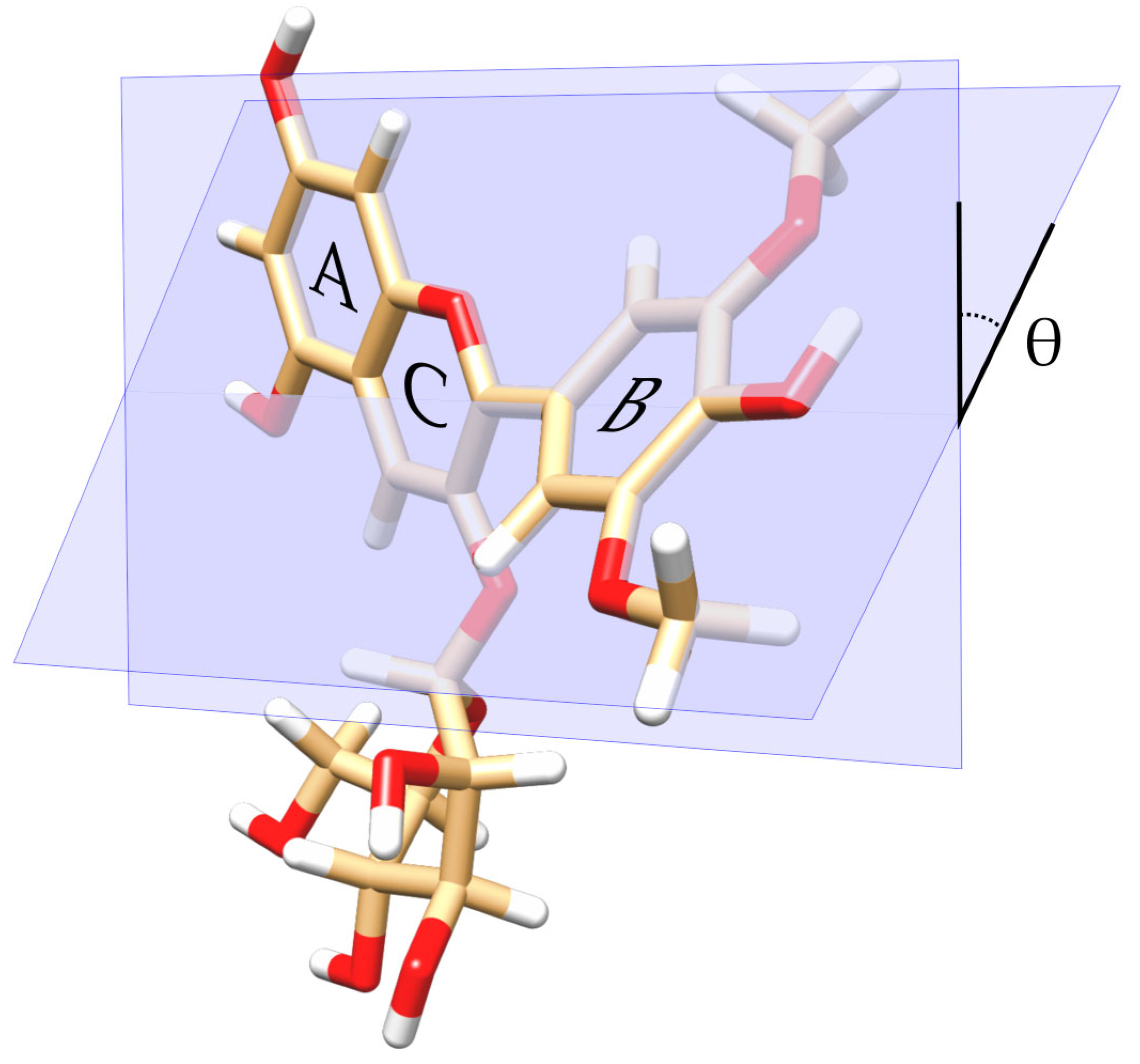
2.1. Electronic Structure Method for Modeling the 3D Structure of Mv3glc Conformers at Different Dihedral Angles

To build the three-dimensional (3D) structure of mv3glc, a frequency analysis was considered, which confirmed the absence of imaginary frequencies in ground state properties. The hydrogens of the framework were then adjusted to a pH of 3.5 by using the Avogadro software 1.2.0 [12]. Ground and excited state properties were computed in the Gaussian09 computational package and its GaussView 5.0 visualizer by applying the density functional theory (DFT) and its time-dependent (TD-DFT) extension, through the DFT-D correction [25]. Calculations were based on the dispersion-corrected hybrid functional B3LYP in conjunction with the double-ζ Pople basis set, 6-31+G(d,p), since the polarized and diffuse functions consider the effects of substituents [23].
The molecular distortion that most affects the optical properties of anthocyanins is the torsion between the single (B) and double (AC) rings, described by the dihedral angle θ [26]. Based on previous quantum chemical calculations, the 3D structure of mv3glc was computed considering the dihedral angle of the B-ring θ at 0° (planar structure). The additional 3D conformers of mv3glc were obtained by modifying the torsion of the dihedral angle θ of the B-ring to positive orientations (to 10°, 20°, 30°, 40°, 50°, 60°, 70°, 80°, and 90°).
In this study, the integral equation formalism polarizable continuum model (IE-FPCM) was applied [27], using water as a solvent (ε = 78.5 at room temperature). The PCM-TD-DFT calculations applied did not consider the solvent relaxation as a function of time but provided a reasonable accuracy in the prediction of the spectral properties [23,28].

2.2. Modeling of the 3D Structure of the Grape Seed Peptide

The 3D structure of the grape seed peptide was obtained using UCSF Chimera software 1.17.3 [29]. The modeled peptide (R.ADVYTPR.G) was identified by mass spectroscopy AB Sciex TOF/TOF™ 5800 system (Framingham, MA, USA) in a previous study [30] from the sequences of Vitis vinifera provided by the database of the National Center for Biotechnology Information (NCBI), using the MASCOT search engine (v.2.5.1, Matrix Science Inc., Boston, MA, USA).
The 3D model was subjected to energy minimization by molecular dynamics (MD) simulation to assess its conformation and stability in the solvation process, using GROMACS 2021.7 with the AMBER99SB-ILDN force field. For this purpose, the modeled peptide was dissolved in 1.0 nm cubic boxes using single-point-charge water molecules, which were then replaced by counterions for electroneutrality. Energy minimization was carried out in 50,000 iteration steps. Two equilibration steps (100 ps each) were carried out to reach the optimal pressure and temperature conditions. The reference pressure and temperature values were 1 bar and 300 K, respectively. Once the equilibrium stages were completed, the system was set to the desired temperature and pressure. Then, a 50 ns MD simulation was carried out with a 2 fs time step algorithm.

2.3. Mv3glc–Grape Seed Peptide Docking Study

Molecular docking studies were performed to study the molecular binding of mv3glc as a ligand of the grape seed peptide (R.ADVYTPR.G) and to compute the deriving UV-vis absorbance spectra based on quantum chemical calculations.
For this purpose, the optimized 3D structures of mv3glc (at 0° dihedral angle) and the grape seed peptide were docked with AutoDock Vina v1.2.x [31]. The mv3glc–grape seed peptide complex was subjected to an energy minimization process by DFT-based quantum calculations using the dispersion-corrected hybrid functional B3LYP in conjunction with the double-ζ Pople-type basis set, 6-31+G(d,p).
AutoDockTools (version 1.5.6) was used to assign the charges of all Kollman atoms. A docking grid dimension was selected with 43 × 44 × 30 points for the peptide–mv3glc interaction, and nine complexes were obtained with different docking energies. Discovery Studio was used as a visualizer to assess the type of molecular interactions in the complex.

2.4. Computing the Theoretical Visible Absorbance Spectrum of Mv3glc Under Different Simulated Environments

The molecular docking protocol with TD-DFT calculations provides an accurate characterization of excited states and the evaluation of absorption properties of mv3glc in the UV–visible range due to π→π* electronic transitions, in relation to conformational variations (torsion of the dihedral angle θ of the B-ring) and non-covalent interaction effects with the grape seed peptide.
The spectrum of mv3glc under different simulated environments was evaluated by averaging the TD-DFT spectra computed for each conformer. The theoretical visible absorption spectra (360–780 nm) output files of mv3glc obtained by Gaussian functions contain the molar absorptivities and calculated oscillator strengths, which represent their intensities as a function of the wavelengths. Due to the approximately symmetrical nature of the main absorption band characteristic of anthocyanins, its maximum excitation energy (λmax) can be identified directly with the calculated vertical transition of the first excited singlet state [23]. It exhibits the highest oscillator strength among all electronic transitions in the visible region in this study.

2.5. In Silico Colorimetric Methodology in the CIELAB Color Space

The predicted color characteristics of each theoretical 3D structure of mv3glc were calculated in the CIELAB color space from the TD-DFT-based calculations. The theoretical visible spectra were transformed into absorbance values as a function of the wavelengths (360–780 nm) based on the Lambert–Beer law (Aλ = ελ × d × c). To apply the formula, the concentration of mv3glc used in the calculus was 5 × 10⁻⁵ M.
The CIELAB parameters were then calculated from the absorption spectra (360–780 nm; Δλ = 2 nm; 1 cm path length cell) by using the original software CromaLab® v2.0 [32] following the recommendations of the Commission Internationale de L’Eclairage: the CIE 1964 10° Standard Observer and the Standard Illuminant D65 [18].
The CIELAB parameters obtained were L* (lightness, ranging from 0 = black to 100 = white) and two rectangular coordinates, a* (which takes positive values for reddish colors and negative values for greenish ones) and b* (positive for yellowish colors and negative for the bluish ones). From a* and b*, polar coordinates are defined as follows:
  • Hue angle (chromatic tonality): hab = arctan (b*/a*);
  • Chroma (color intensity or saturation): C*ab = [(a*)2 + (b*)2]1/2.
The color variations between the different mv3glc-simulated conformations were evaluated by Differential Colorimetry according to Gordillo et al. (2012) [16]. Color differences were computed as Euclidean distances in CIELAB (CIE76 color difference formulae): ΔE*ab = [(ΔL*)2 + (Δa*)2 + (Δb*)2]1/2.
The weight of the three color attributes (lightness, chroma, and hue) was calculated as the relative contribution of each one to the color difference value (ΔE*ab), as follows:
  • Relative contribution (%) of lightness: %ΔL = [(ΔL*)2/(ΔE*ab)2] × 100;
  • Relative contribution (%) of chroma: %ΔC = [(ΔC*ab)2/(ΔE*ab)2] × 100;
  • Relative contribution (%) of hue: %ΔH = [(ΔH)2/(ΔE*ab)2] × 100, with ΔH being mathematically deduced from ΔH = [(ΔE*ab)2 − (ΔL)2 − (ΔC)2)]1/2.

3. Results and Discussion

3.1. In Silico Analysis of Conformational Effects on Mv3glc Predicted Color

The visible absorbance spectra for the different mv3glc conformers computed by the time-dependent density functional theory (in the positively charged flavylium state) with the dispersion-corrected hybrid functional B3LYP are shown in Figure 2, while their corresponding simulated CIELAB color parameters (L*, a*, b*, C*ab, and hab) are presented in Table 1. Mv3glc conformers differ from each other in the torsion of the dihedral angle θ, which was modified from 0° to 90° to cover a wide range of conformations. Considering the planar geometry of the flavylium chromophore (θ = 0°), the computed spectra for mv3glc in the visible range included a single strong absorption peak between 470 and 530 nm (λmax = 498 nm; Figure 2), which corresponds in qualitative terms to the spectrum characteristics obtained for malvin by Ge et al. [33], who used the B3PYL XC functional, and for mv3glc by Rustioni et al. [34] based on the IEFPCM-B3P86/6-311+G(d,p) theoretical level.
In this study, a deviation from planarity in the calculations between 0° and 60° does not affect the global shape or the number of simulated spectral peaks of mv3glc conformers but reflects important variations in the position of the main absorption band peak and its intensity. The theoretical results reproduced a progressive bathochromic effect in the mv3glc computed spectra by 58 nm at increasing molecular torsions from 0° to 60° (λmax = 498 nm and 556 nm, respectively) simultaneously with a notably hypochromic effect. A greater deviation from planarity (θ torsion larger than 60°) reproduces a suppression of the absorption in the visible range of the flavylium chromophore. Previous computational studies with cyanidin-3-glucoside have shown that changing the dihedral angle significantly affects its optical behavior, depending on several competing effects such as the orientation of the hydroxyl groups in the aglycone structure or the relative position of the sugar in the 3′ position [26,35].
By applying the tristimulus colorimetry algorithms to the computed spectra, the in silico methodology allowed the prediction of the color of the different mv3glc conformers, both in quantitative (L* and C*ab) and qualitative (hab) colorimetric terms (Table 1). Considering no torsion effect in the mv3gcl structure (θ = 0°), the computed CIELAB parameters were L* = 50.27, C*ab = 50.41, and hab = 1.47°, which would correspond to a vivid red hue. These results predicted by computational tools match the color exhibited by mv3glc in its flavylium cation form in solutions observed in previous experimental studies [16].
Modifying the torsion of the dihedral angle from 0° to 40° led to different mv3glc conformers having similar predicted values of lightness and chroma (L* ~ 50 and C*ab = 50–54), similar in darkness and color intensity, respectively. However, they notably differ from each other regarding their predicted hue values (hab decreased from 1.5° to −8.1°), indicating that the in silico methodology applied reproduced important qualitative color variations (Figure 3). As the dihedral angle increases up to 50°, simulated mv3glc conformers show an increase in bluish tonalities.
These predicted colorimetric effects based on the B3LYP/6-31+G(d,p) theoretical level reflect the strong structure–color relationship of anthocyanin pigments. Accordingly, the colorimetric results provided an adequate color prediction for mv3glc according to conformational variations. It is worth mentioning that these simulated conformations could result from structural changes or interactions between mv3glc with other molecular species present in wine. This suggests that a particular conformation would result in a specific color in a food matrix that can be predicted by in silico colorimetry. Higher torsions of the dihedral angles (θ = 75°, 80°, and 90°) (Table 1) led to mv3gcl conformers with notably higher values of lightness and lower chroma values (L* > 90 and C*ab < 10). Thus, under these conformations, mv3glc would appear almost colorless (Figure 3), as previously detected with the in silico spectral effects.
The color differences (ΔE*ab) between the different mv3glc conformations when the dihedral angles were increased (from 10° to 90°) compared to the initial conformation (0°) were calculated alongside the relative lightness, chroma, and hue differences (%ΔL, %ΔC, and %ΔH) for each ΔE*ab value (Table 2), which allows assessing the color attribute most influenced in the prediction. ΔE*ab values varied from 4 to 67 units, confirming that conformational changes based on θ torsion result in color variations of the mv3glc flavylium cation, and in most cases, these changes can be considered visually perceptible. According to Martínez et al. [36], ΔE*ab values around 3–5 units are considered close to the visual threshold of clearly perceived color differences by average non-trained observers. Thus, the theoretical results showed that the greater θ torsion, the more evident the chromatic effects are. By comparing the relative contributions of lightness (%ΔL), chroma (%ΔC), and hue (%ΔH), it was confirmed that the ΔE*ab calculated between the mv3glc conformers corresponded to the predicted qualitative changes (%ΔH = 99–84 from 10 to 60°).

3.2. In Silico Colorimetric Analysis of the Grape Seed Peptide–Mv3glc Interaction

Table 3 shows the results of the nine mv3glc–grape seed peptide (mv3glc-GSP) conformations (Figure S1) based on the obtained docking parameters (binding energy, mv3glc dihedral angle, and type of intermolecular interaction), spectral parameters (λmax and A520 nm), and CIELAB color parameters (L*, a*, b*, C*ab, hab).
The energy values of the nine mv3glc-GSP conformations obtained in the docking analysis (Figure 2) ranged from −4.1 to −3.9 kcal/mol, indicating that all of them are energetically probable. As observed in Table 3, the protein–anthocyanin complexes were characterized by the presence of intermolecular forces established between the aromatic rings of the anthocyanin and the peptide (Figure 1), including hydrogen bonding, π-alkyl, and π-π interactions. Among them, the π-alkyl forces are common in all the complexes (Table 3).
The colorimetric results showed qualitative and quantitative differences for the re-produced colors between the nine systems. The predicted colors for the conformations 2, 4, 6, 8, and 9, as indicated by the reproduced hab values, corresponded to unusual tonalities of the flavylium cation in acidic conditions. Particularly, the hab values of 45° and 90° of these conformations correspond to orange and yellow tonalities, respectively. Thus, although all these conformations are energetically probable, the in silico colorimetric methodology applied in these conformers inaccurately reproduced the resulting color from the molecular interaction with the peptide. In addition, the predicted color of conformations 2, 8, and 9 corresponded to very pale tonalities according to the predicted high values of lightness (L* > 90).
In comparison, conformations 1, 3, 5, and 7 closely reproduced the wide range of tonalities based on their predicted hab values, distinctive of the flavylium cation in acid conditions. Figure S2 shows the absorption spectra calculated with the B3LYP/6-31+G(d,p) theoretical level for the isolated mv3glc molecule corresponding to these minimum-energy configurations.
Notably, conformations 1 and 7 with predicted hab values of 20° and 28°, respectively, reproduced red-orange tonalities, while conformations 3 and 5 with hab values of 333.4° and 342.2° accurately reproduced red-bluish tonalities. In all cases, the C*ab values ranged from 11 to 35 units, suggesting vivid tonalities.
Regarding the conformations that reproduced the red-bluish colorations properly (3 and 5), the docking results indicate that mv3gcl interacts with the grape seed peptide through π-π and π-alkyl interactions, which has been described in copigmentation interactions involving the flavylium cation form. Moreover, from a structural perspective, the dihedral angles of these conformations (3 and 5) corresponded to 66.5°; therefore, this parameter could provide a structural reference of the flavylium cation’s torsion caused by the malvidin–GSP interaction. Thus, B3LYP combined with 6-31+G(d,p) has also been demonstrated to be effective in the prediction of spectroscopic and colorimetric features of anthocyanins related to interaction effects, offering a satisfactory balance between accuracy and computational efficiency. However, its performance has also limitations such as the systematic underestimation of λmax, suggesting the importance of exploring additional approaches. In this context, the B3P86 functional may be a solid alternative offering good performance in dealing with molecule substitutions, especially in systems with functional groups such as OH and OCH3 [23]. In copigmentation systems, where molecular interactions produce significant bathochromic shifts, other approaches such as ωB97X-D [37] or a hybrid functional with Grimme dispersion TD-CAM-B3LYP-D3, the cc-pVDZ basis set, and a state-specific PCM (SS-PCM) [38] have achieved greater accuracy in predicting spectral shifts.

4. Conclusions

The combination of quantum calculations and subsequent spectral analysis through tristimulus colorimetry in the CIELAB color space has allowed for an accurate prediction of the color modulation of mv3glc in its flavylium cation form, related to torsions in the chromophore and non-covalent interactions. It was confirmed that the PCM-TD-DFT calculations based on the B3LYP/6-31+G(d,p) theoretical level reflect the strong structure–color relationship of anthocyanin pigments by means of the prediction of quantitative and qualitative color variations considered visually perceptible and dependent on the θ torsion. Colorimetric changes (red-orange or bluish effects) in mv3glc due to non-covalent interactions (π-alkyl and π-π) with a grape seed peptide have also been predicted by using the theoretical methodologies described above, which could support experimental observations. Nevertheless, from a technological standpoint, future research should aim to supplement the findings with experimental data and cross-validate them with alternative theoretical approaches to significantly enhance the reliability and robustness of the predictions, ensuring that conclusions drawn from molecular modeling are validated. It is essential to improve the integration of each functional to comprehensively address the spectral and structural properties, optimizing the strengths already established by the method used and complementing them with the capabilities of other functionals.

Supplementary Materials

The following supporting information can be downloaded at https://www.mdpi.com/article/10.3390/app15084238/s1: Figure S1: Nine different conformations of the interaction between mv3gcl and the grape seed peptide (R.ADVYTPR.G) obtained by docking studies; Figure S2: Theoretical visible absorption spectra of the mv3glc-GSP complexes (docking conformations 1, 3, 5, and 7) calculated with the dispersion-corrected hybrid functional B3LYP.

Author Contributions

Conceptualization: F.J.H.; methodology: F.C.-G.; investigation: F.C.-G.; data curation: F.C.-G.; writing—original draft preparation: F.C.-G.; writing—review and editing: M.F.L.-M., F.J.R.-P., M.L.G.-M. and B.G.; validation: F.J.R.-P. and B.G.; image design: F.J.R.-P.; supervision: B.G.; project administration: F.J.H. and M.L.G.-M.; funding acquisition: F.J.H. and M.L.G.-M. All authors have read and agreed to the published version of the manuscript.

Funding

This research was financially supported by Project PID2021-127126OB-C22 funded by MICIU/AEI/10.13039/501100011033 and FEDER, UE. Consejería de Universidad, Investigación e Innovación de la Junta de Andalucía, Spain (Project PAIDI2021 PROYEXCEL_00578). F.C.G acknowledges the PhD grant PRE2018-087184 funded by ESF Investing in your future. M.F.L.M acknowledges the PhD grant PRE2022-104543 funded by MICIU/AEI/10.13039/501100011033 and by “ESF+”.

Data Availability Statement

The raw data supporting the conclusions of this article will be made available by the authors on request.

Acknowledgments

Authors thank the assistance of the Biology Service (SGI, Universidad de Sevilla, Spain) and the Scientific Computation Center of Andalusia (CICA) for the computing services they provided.

Conflicts of Interest

The authors declare no conflicts of interest.

Abbreviations

The following abbreviations are used in this manuscript:
CIECommission Internationale de L’Eclairage
DFTdensity functional theory
GSPgrape seed peptide
MDmolecular dynamics
mv3glcmalvidin 3-glucoside
TD-DFTtime-dependent density functional theory

References

  1. He, J.; Giusti, M.M. Anthocyanins: Natural Colorants with Health-Promoting Properties. Annu. Rev. Food Sci. Technol. 2010, 1, 163–187. [Google Scholar] [CrossRef] [PubMed]
  2. Pina, F.; Melo, M.J.; Laia, C.A.T.; Parola, A.J.; Lima, J.C. Chemistry and Applications of Flavylium Compounds: A Handful of Colours. Chem. Soc. Rev. 2012, 41, 869–908. [Google Scholar] [CrossRef] [PubMed]
  3. De Freitas, V.; Mateus, N. Chemical Transformations of Anthocyanins Yielding a Variety of Colours. Environ. Chem. Lett. 2006, 4, 175–183. [Google Scholar] [CrossRef]
  4. Basílio, N.; Mendoza, J.; Seco, A.; Oliveira, J.; De Freitas, V.; Pina, F. Strategies Used by Nature to Fix the Red, Purple and Blue Colours in Plants: A Physical Chemistry Approach. Phys. Chem. Chem. Phys. 2021, 23, 24080–24101. [Google Scholar] [CrossRef]
  5. Sigurdson, G.T.; Robbins, R.J.; Collins, T.M.; Giusti, M.M. Impact of Location, Type, and Number of Glycosidic Substitutions on the Color Expression of o-Dihydroxylated Anthocyanidins. Food Chem. 2018, 268, 416–423. [Google Scholar] [CrossRef]
  6. Malcıoğlu, O.B.; Calzolari, A.; Gebauer, R.; Varsano, D.; Baroni, S. Dielectric and Thermal Effects on the Optical Properties of Natural Dyes: A Case Study on Solvated Cyanin. J. Am. Chem. Soc. 2011, 133, 15425–15433. [Google Scholar] [CrossRef]
  7. Trouillas, P.; Sancho-García, J.C.; De Freitas, V.; Gierschner, J.; Otyepka, M.; Dangles, O. Stabilizing and Modulating Color by Copigmentation: Insights from Theory and Experiment. Chem. Rev. 2016, 116, 4937–4982. [Google Scholar] [CrossRef]
  8. Fernandes, A.; Brás, N.F.; Oliveira, J.; Mateus, N.; De Freitas, V. Impact of a Pectic Polysaccharide on Oenin Copigmentation Mechanism. Food Chem. 2016, 209, 17–26. [Google Scholar] [CrossRef]
  9. He, Z.; Zhu, H.; Xu, M.; Zeng, M.; Qin, F.; Chen, J. Complexation of Bovine β-Lactoglobulin with Malvidin-3-O-Glucoside and Its Effect on the Stability of Grape Skin Anthocyanin Extracts. Food Chem. 2016, 209, 234–240. [Google Scholar] [CrossRef]
  10. Buchweitz, M.; Speth, M.; Kammerer, D.R.; Carle, R. Impact of Pectin Type on the Storage Stability of Black Currant (Ribes Nigrum, L.) Anthocyanins in Pectic Model Solutions. Food Chem. 2013, 139, 1168–1178. [Google Scholar] [CrossRef]
  11. Marangon, M.; Vincenzi, S.; Curioni, A. Wine Fining with Plant Proteins. Molecules 2019, 24, 2186. [Google Scholar] [CrossRef] [PubMed]
  12. Chamizo-González, F.; Estévez, I.G.; Gordillo, B.; Manjón, E.; Escribano-Bailón, M.T.; Heredia, F.J.; González-Miret, M.L. First Insights into the Binding Mechanism and Colour Effect of the Interaction of Grape Seed 11S Globulin with Malvidin 3-O-Glucoside by Fluorescence Spectroscopy, Differential Colorimetry and Molecular Modelling. Food Chem. 2023, 413, 135591. [Google Scholar] [CrossRef] [PubMed]
  13. Mora-Garrido, A.B.; Escudero-Gilete, M.L.; González-Miret, M.L.; Heredia, F.J.; Cejudo-Bastante, M.J. Effect of the Addition of Protein Hydrolysates from Grape Seed Meal Residue to Red Wines in Warm Regions in the Stabilization Stage. LWT 2024, 205, 116554. [Google Scholar] [CrossRef]
  14. Yao, L.; Xu, J.; Zhang, L.; Zheng, T.; Liu, L.; Zhang, L. Physicochemical Stability-Increasing Effects of Anthocyanin via a Co-Assembly Approach with an Amphiphilic Peptide. Food Chem. 2021, 362, 130101. [Google Scholar] [CrossRef]
  15. López-Molina, M.F.; Rodríguez-Pulido, F.J.; Mora-Garrido, A.B.; González-Miret, M.L.; Heredia, F.J. New Approaches for Screening Grape Seed Peptides as Colourimetric Modulators by Malvidin-3-O-Glucoside Stabilisation. Food Chem. 2025, 464, 141708. [Google Scholar] [CrossRef]
  16. Gordillo, B.; Rodríguez-Pulido, F.J.; Escudero-Gilete, M.L.; González-Miret, M.L.; Heredia, F.J. Comprehensive Colorimetric Study of Anthocyanic Copigmentation in Model Solutions. Effects of pH and Molar Ratio. J. Agric. Food Chem. 2012, 60, 2896–2905. [Google Scholar] [CrossRef]
  17. Heredia, F.J.; Francia-Aricha, E.M.; Rivas-Gonzalo, J.C.; Vicario, I.M.; Santos-Buelga, C. Chromatic Characterization of Anthocyanins from Red Grapes—I. pH Effect. Food Chem. 1998, 63, 491–498. [Google Scholar] [CrossRef]
  18. CIE (Ed.) Colorimetry, 3rd. ed.; Technical report/CIE; CIE, Central Bureau: Vienna, Austria, 2004; ISBN 978-3-901906-33-6. [Google Scholar]
  19. Chen, Q.; Xiao, Y.; Zhang, W.; Mu, W. Current Methods and Applications in Computational Protein Design for Food Industry. Crit. Rev. Food Sci. Nutr. 2020, 60, 3259–3270. [Google Scholar] [CrossRef]
  20. Trouillas, P.; Di Meo, F.; Gierschner, J.; Linares, M.; Sancho-Garcia, J.C.; Otyepka, M. Optical Properties of Wine Pigments: Theoretical Guidelines with New Methodological Perspectives. Tetrahedron 2015, 71, 3079–3088. [Google Scholar] [CrossRef]
  21. Anouar, E.H.; Osman, C.P.; Weber, J.-F.F.; Ismail, N.H. UV/Visible Spectra of a Series of Natural and Synthesised Anthraquinones: Experimental and Quantum Chemical Approaches. SpringerPlus 2014, 3, 233. [Google Scholar] [CrossRef]
  22. Soto-Rojo, R.; Baldenebro-López, J.; Flores-Holguín, N.; Glossman-Mitnik, D. Comparison of Several Protocols for the Computational Prediction of the Maximum Absorption Wavelength of Chrysanthemin. J. Mol. Model. 2014, 20, 2378. [Google Scholar] [CrossRef] [PubMed]
  23. Anouar, E.H.; Gierschner, J.; Duroux, J.-L.; Trouillas, P. UV/Visible Spectra of Natural Polyphenols: A Time-Dependent Density Functional Theory Study. Food Chem. 2012, 131, 79–89. [Google Scholar] [CrossRef]
  24. Sinopoli, A.; Calogero, G.; Bartolotta, A. Computational Aspects of Anthocyanidins and Anthocyanins: A Review. Food Chem. 2019, 297, 124898. [Google Scholar] [CrossRef]
  25. Grimme, S.; Antony, J.; Schwabe, T.; Mück-Lichtenfeld, C. Density Functional Theory with Dispersion Corrections for Supramolecular Structures, Aggregates, and Complexes of (Bio)Organic Molecules. Org Biomol Chem 2007, 5, 741–758. [Google Scholar] [CrossRef]
  26. Rusishvili, M.; Grisanti, L.; Laporte, S.; Micciarelli, M.; Rosa, M.; Robbins, R.J.; Collins, T.; Magistrato, A.; Baroni, S. Unraveling the Molecular Mechanisms of Color Expression in Anthocyanins. Phys. Chem. Chem. Phys. 2019, 21, 8757–8766. [Google Scholar] [CrossRef]
  27. Tomasi, J.; Mennucci, B.; Cammi, R. Quantum Mechanical Continuum Solvation Models. Chem. Rev. 2005, 105, 2999–3094. [Google Scholar] [CrossRef]
  28. Li, Y.; Prejanò, M.; Toscano, M.; Russo, N. Oenin and Quercetin Copigmentation: Highlights from Density Functional Theory. Front. Chem. 2018, 6, 245. [Google Scholar] [CrossRef]
  29. Pettersen, E.F.; Goddard, T.D.; Huang, C.C.; Couch, G.S.; Greenblatt, D.M.; Meng, E.C.; Ferrin, T.E. UCSF Chimera—A Visualization System for Exploratory Research and Analysis. J. Comput. Chem. 2004, 25, 1605–1612. [Google Scholar] [CrossRef]
  30. Chamizo-González, F.; Heredia, F.J.; Rodríguez-Pulido, F.J.; González-Miret, M.L.; Gordillo, B. Proteomic and Computational Characterisation of 11S Globulins from Grape Seed Flour By-Product and Its Interaction with Malvidin 3-Glucoside by Molecular Docking. Food Chem. 2022, 386, 132842. [Google Scholar] [CrossRef]
  31. Trott, O.; Olson, A.J. AutoDock Vina: Improving the Speed and Accuracy of Docking with a New Scoring Function, Efficient Optimization, and Multithreading. J. Comput. Chem. 2010, 31, 455–461. [Google Scholar] [CrossRef]
  32. Ramirez, A.; Heredia, F.J.; González-Miret, M.; Lourdes, M.; Moro, Á.; Corsino, J. CromaLab, 2.0; Sevilla, Spain, 2004.
  33. Ge, X.; Calzolari, A.; Baroni, S. Optical Properties of Anthocyanins in the Gas Phase. Chem. Phys. Lett. 2015, 618, 24–29. [Google Scholar] [CrossRef]
  34. Rustioni, L.; Di Meo, F.; Guillaume, M.; Failla, O.; Trouillas, P. Tuning Color Variation in Grape Anthocyanins at the Molecular Scale. Food Chem. 2013, 141, 4349–4357. [Google Scholar] [CrossRef] [PubMed]
  35. Deineka, V.I.; Kulchenko, Y.Y. Quantum-Chemical Calculations for the Electronic Absorption Spectra of Certain Anthocyanidins. Russ. J. Phys. Chem. A 2021, 95, 1378–1385. [Google Scholar] [CrossRef]
  36. Martínez, J.A.; Melgosa, M.; Pérez, M.M.; Hita, E.; Negueruela, A.I. Note. Visual and Instrumental Color Evaluation in Red Wines. Food Sci. Technol. Int. 2001, 7, 439–444. [Google Scholar] [CrossRef]
  37. Di Meo, F.; Sancho Garcia, J.C.; Dangles, O.; Trouillas, P. Highlights on Anthocyanin Pigmentation and Copigmentation: A Matter of Flavonoid π-Stacking Complexation To Be Described by DFT-D. J. Chem. Theory Comput. 2012, 8, 2034–2043. [Google Scholar] [CrossRef]
  38. Wu, L.; Zhang, Y.; Prejanò, M.; Marino, T.; Russo, N.; Tao, Y.; Li, Y. Gallic Acid Improves Color Quality and Stability of Red Wine via Physico-Chemical Interaction and Chemical Transformation as Revealed by Thermodynamics, Real Wine Dynamics and Benchmark Quantum Mechanical Calculations. Food Res. Int. 2024, 188, 114510. [Google Scholar] [CrossRef]
Figure 1. Three-dimensional structure of flavylium chromophore, indicating the notation of the aromatic rings and the dihedral angle.
Applsci 15 04238 g001
Figure 2. Theoretical visible absorption spectra of mv3glc conformers calculated with the dispersion-corrected hybrid functional B3LYP.
Applsci 15 04238 g002
Figure 3. Location of the predicted color of mv3glc conformers in the CIELAB (a*b*) diagram.
Applsci 15 04238 g003
Table 1. In silico color parameters of the mv3glc conformations at increasing torsions of the dihedral angle.
Mv3glc
Conformation
Dihedral Angle (θ)L*a*b*C*abhab
1050.2750.391.3050.411.47°
21050.2450.73−2.7250.80356.94°
32049.7150.33−4.7350.55354.63°
43049.6850.11−12.9251.75345.54°
54050.8348.33−25.8954.83331.82°
65055.9239.87−37.6854.86316.61°
76067.9020.21−35.4940.84299.66°
87089.801.68−9.9110.05279.63°
98093.612.08−4.855.28293.77°
109097.612.670.482.7110.23°
Table 2. Color differences (ΔE*ab) calculated between the simulated colors of the different mv3glc conformations at increasing dihedral angles (from 10° to 90°) respect to mv3glc conformation at 0°, together to the relative differences in lightness, chroma, and hue (%ΔL, %ΔC, and %ΔH) that make up each ΔE*ab.
Dihedral Angle (θ)ΔE*abLCH
0–104.030.010.9499.05
0–206.050.850.0699.09
0–3014.240.170.8998.94
0–4027.270.042.6297.34
0–5040.771.921.1996.89
0–6050.7512.073.5684.38
0–7063.7238.4740.1221.41
0–8065.1944.1947.927.89
0–9067.2249.5850.350.07
Table 3. Results obtained for the nine docking conformations: chemical interaction parameters (mv3glc dihedral angle, binding energy, and type of intermolecular interaction), spectral parameters (λmax and A520 nm), and CIELAB color parameters.
Quantum Chemical CalculationsSpectral
Parameters
CIELAB Color Parameters
Dihedral
Angle (θ)
E Docking (kcal/mol)Molecular
Interaction
λmaxA520L*a*b*C*abhab
167.0°−4.1π-π/π-alkyl4920.3679.420.07.321.320.2º
2−85.3°−4.1hydrogen bond/π-alkyl4240.0894.4−2.528.628.794.9º
366.5°−4.1π-π/π-alkyl5420.3277.110.4−5.211.6333.4º
471.3°−4.0hydrogen bond/π-alkyl4760.2982.514.212.318.840.9º
566.5°−4.0π-π/π-alkyl5120.4572.822.8−7.323.9342.2°
658.0°−4.0π-π/π-alkyl4720.4377.321.722.431.245.9º
760.0°−4.0hydrogen bond/π-alkyl4840.5474.430.816.434.928.1º
8−83.7°−4.0π-π/π-alkyl4280.0893.4−1.015.215.693.9º
9−85.1°−3.9π-π/π-alkyl4280.0893.9−1.116.216.293.9º
Disclaimer/Publisher’s Note: The statements, opinions and data contained in all publications are solely those of the individual author(s) and contributor(s) and not of MDPI and/or the editor(s). MDPI and/or the editor(s) disclaim responsibility for any injury to people or property resulting from any ideas, methods, instructions or products referred to in the content.

