Article

Force Expressed by 3D-Printed Aligners with Different Thickness and Design Compared to Thermoformed Aligners: An in Vitro Study

by
Francesca Cremonini
,
Carolina Pancari
,
Luca Brucculeri
,
Ariyan Karami Shabankare
and
Luca Lombardo
*
Postgraduate School of Orthodontics, University of Ferrara, Via Luigi Borsari 46, 44121 Ferrara, Italy
*
Author to whom correspondence should be addressed.
Appl. Sci. 2025, 15(6), 2911; https://doi.org/10.3390/app15062911
Submission received: 7 December 2024 / Revised: 16 February 2025 / Accepted: 6 March 2025 / Published: 7 March 2025
(This article belongs to the Special Issue Orthodontics: Advanced Techniques, Methods and Materials)

Abstract

:
(1) Background: Clear aligners are favored for their aesthetics in orthodontics, with newer 3D-printed technologies allowing the design of aligners with differential thicknesses and materials, offering advantages in terms of force distribution on the teeth, thereby optimizing treatment biomechanics. This study aimed to compare the initial and final forces of three types of 3D-printed aligners (with different thickness gradients and gingival margins) and traditional thermoformed aligners (with different gingival margins), evaluating stress relaxation and force consistency to determine which material and configuration may be optimal for better force distribution; (2) Methods: Twenty-seven 3D-printed aligners with three design variations and 18 thermoformed aligners were analyzed. Customized models were used to assess force at specific points on the upper incisor (1.1) and molar (2.6). A 3 h stress-relaxation test was conducted at 37 °C, and force data were recorded every second using a motorized compression stand. Statistical analysis was performed using ANOVA, post hoc tests, and Kruskal–Wallis tests for comparisons; (3) Results and Conclusions: Aligners with vertical and horizontal thickness gradients and a gingival margin trimmed 2 mm above the gingival contour exerted the highest forces, particularly at incisal/occlusal points. No significant differences in stress relaxation were observed. The force applied to the molars was consistently higher than the force applied to the incisors. These 3D-printed aligners with both horizontal and vertical gradients may offer a viable alternative to thermoformed aligners.

1. Introduction

In recent decades, aesthetics has become increasingly central to orthodontic treatment, leading patients to favor discreet options such as clear aligners, which improve the smile without interfering with daily life [1,2,3]. The evolution of aligners can be traced back to the 1940s when Kesling proposed the use of a “positioner” to move teeth incrementally [4]. Introduced in the 1990s, clear aligners represented a significant advancement in orthodontics by offering removable and nearly invisible devices that allowed for intermittent application of orthodontic forces, which cease during oral hygiene routines and meals. Despite their advantages, traditional aligners made from PET-G (Polyethylene Terephthalate Glycol-modified) materials tend to experience stress relaxation, resulting in a progressive reduction in force exerted on the teeth, thereby impacting the predictability of tooth movement [5].
With technological advances, aligner production has shifted from thermoforming to direct 3D printing, which allows greater control over structure, thickness, and material properties, while also reducing manufacturing steps and waste, improving both precision and production time [6,7]. Among the 3D printing technologies employed, Direct Light Processing (DLP) and Selective Laser Sintering (SLS) enable the creation of customized aligners with variable thicknesses and optimized configurations tailored to clinical requirements [8,9]. Recent studies suggest that by varying the design and thickness of the aligners, it is possible to influence the force exerted on the teeth, thereby optimizing treatment biomechanics and reducing the need for visible attachments [10,11].
The introduction of rapid prototyping (RP) technologies in recent years has further transformed aligner manufacturing. Originally developed in the 1980s, RP—also known as additive manufacturing—has advanced significantly, finding applications in various fields, including medicine, dentistry, and orthodontics [6]. In orthodontics, the use of digital impressions and 3D printing to produce aligners has greatly reduced errors associated with traditional impression methods and the fabrication of plaster models, which are susceptible to environmental damage and storage issues [12]. Studies have confirmed that 3D-printed models offer high precision and minimal measurement errors, making them a reliable alternative to plaster models [13].
Traditional aligner fabrication involved the thermoforming of plastic sheets over 3D-printed models of planned tooth movements. However, the direct 3D printing of aligners has emerged as a groundbreaking innovation. This approach simplifies the workflow, reduces production time, and allows for better control over aligner properties such as thickness and material composition [6]. Technologies like DLP and SLS play a pivotal role in this evolution. DLP uses photopolymerization to cure entire layers of liquid resin in a single exposure, while SLS relies on a laser to sinter polymer powder, enabling the creation of highly detailed and complex objects [9]. These methods not only enhance production efficiency but also reduce material waste and support the development of on-demand aligner treatments [6,7].
A notable innovation in this field is the NOXI aligner (Sweden & Martina, Due Carrare, Padova, Italy), which uses advanced polyamide material and CAD-CAM technology for direct printing. While aligners are more aesthetically acceptable than traditional fixed metal appliances, they cannot be considered completely invisible. The NOXI aligner features a design optimized for 12 h daily wear, incorporating differential thickness gradients that may influence force distribution. These thickness variations increase from the incisal to the gingival margin and from anterior to posterior teeth, influencing fit and force distribution across different dental elements, thus presenting a compelling alternative to traditional thermoformed aligners [14,15]. By incorporating these thickness variations, the NOXI aligner is better able to address specific biomechanical needs, reducing the necessity for additional attachments or auxiliary devices.
However, further research is required to determine the impact of stress relaxation on the efficacy of NOXI aligners compared to traditional thermoformed alternatives. Previous studies have indicated that stress relaxation is a major limiting factor in aligner therapy, as it influences both the duration and the predictability of force application. For instance, materials such as PET-G and polyurethane have been found to exhibit different rates of stress decay, with PET-G showing a faster decline in force retention [16].
Previous studies have shown that stress relaxation occurs primarily within the first 8 h, reaching a plateau thereafter [16]. As a result, this study concentrated on the early phase of force decay, which is critical for assessing aligner performance.
The objective of this study was to compare the initial and final force values exerted on the upper central incisor and the upper first molar at three specific points using three types of NOXI aligners, differentiated by gingival margin cut and thickness. These results were compared with those from thermoformed aligners with different gingival margins. Additionally, the percentage of stress relaxation over time was analyzed for each type of aligner, aiming to identify which material and configuration may optimize force application for specific areas.

2. Materials and Methods

2.1. Sample Preparation

A master scan of a maxillary arch from a patient previously treated with orthodontics was acquired using an intraoral scanner, Trios scanner (3Shape, Copenhagen, Denmark), and selected for the study (Figure 1). The same upper model was used to design and print all aligners. The test model for thermoforming the aligners was printed using the high-speed Nexa 3D Xip printer (Nexa3D, Ventura, CA, USA) with XDent201 Grey resin (Nexa3D, Ventura, CA, USA). Printing was performed with a layer thickness of 100 microns, and the total print time was approximately 30 min.

2.2. Aligners Selected

For this study, a total of three types of NOXI aligners and two types of thermoformed aligners were chosen. A total of 27 NOXI aligners with three different characteristics were analyzed (Figure 2):
  • Nine NOXI aligners made of Polyammide (PA) with a vertical and horizontal thickness gradient (vertical: 0.65 mm at the incisal margin, gradually increasing to 0.95 mm at the gingival margin; horizontal: 0.65 mm at the incisors, gradually increasing to 0.95 mm at the molars). The gingival margin of the aligner is trimmed 2 mm above the gingival contour (NHVH).
  • Nine NOXI aligners made of Polyammide (PA) with a horizontal thickness gradient (0.65 mm at the incisors, increasing to 0.95 mm at the molars) and a gingival margin trimmed 2 mm above the gingival contour (NHH).
  • Nine NOXI aligners made of Polyammide (PA) with a horizontal thickness gradient (0.65 mm at the incisors, increasing to 0.95 mm at the molars) and a gingival margin trimmed at the zenith of the tooth (NHZ).
Figure 2. (a) NHVH (NOXI Horizontal Vertical High); (b) NHH (NOXI Horizontal High); (c) NHZ (NOXI Horizontal Zenith).
Applsci 15 02911 g002
These aligners were compared with six thermoformed aligners with two different characteristics (Figure 3):
  • Nine F22 aligners with a straight gingival margin (cut at the zenith) made of EvoFlex (Bay Materials LLC, Fremont, CA, USA) with a thickness of 0.76 mm (F22).
  • Nine aligners with a scalloped gingival margin made of EvoFlex with a thickness of 0.76 mm (AS).
Figure 3. (a) F22 (straight with gingival margin); (b) AS (scalloped gingival margin).
Applsci 15 02911 g003

2.2.1. Mechanical Properties of Polyamide

Polyamide (PA) is a biocompatible linear, semi-crystalline, 3D-printable thermoplastic composite with excellent mechanical properties. Its modulus of elasticity (Young’s modulus) ranges from 1.5 to 2.1 GPa, depending on the specific processing conditions and testing parameters. PA exhibits high tensile strength and flexibility, which contribute to its resilience under cyclic loads common in orthodontic applications. The flexural modulus of PA aligns well with its ability to retain shape under stress, making it a prime choice for aligners. Additionally, PA has a low density (1.01–1.03 g/cm3), enhancing comfort for patients during prolonged use. PA’s thermal stability (up to 185 °C) allows for sterilization before clinical use. This is complemented by its low moisture absorption (<1%), ensuring minimal dimensional changes in humid oral environments, which is critical for maintaining precise aligner fit [17,18].

2.2.2. Mechanical Properties of EvoFlex

EvoFlex is a multi-layered material introduced in 2019. It consists of three layers: two external layers of polyurethane and an internal elastomer layer, with a total thickness of 0.75 mm. Designed to improve the elasticity and shape-memory properties of F22 aligners while enhancing stability over time, EvoFlex exhibits unique mechanical characteristics. Its combination of layers provides high elastic deformation capacity, enabling an effective and consistent force application throughout orthodontic treatment. In stress-relaxation tests conducted at elevated temperatures to simulate 15 days of usage, EvoFlex demonstrated superior final stress retention compared to other aligner materials, with a final stress value of 6.5 MPa. The stress relaxation at 5 days was 49.7% and 60.8% at 15 days, markedly lower than traditional F22 aligners (84.3%) and materials such as PET-G (96.1%) and Durasoft (95.4%). This indicates that EvoFlex retains its orthodontic force longer, contributing to predictable tooth movement over extended periods. Moreover, the triple-layer structure of EvoFlex enhances its modulus of elasticity and yield point compared to monolayer materials like PET-G and traditional polyurethane aligners. While EvoFlex shares the benefits of low thickness (0.75 mm) and flexibility with F22 aligners, it demonstrates greater durability and a reduced rate of stress decay under prolonged use [19].

2.3. Preparation and Testing Model Creation

Two test models were created based on the scan used to fabricate the aligners. The first model was generated by importing the file into Rhinoceros 8 software (Robert McNeel & Associates, Seattle, CA, USA) where the model was segmented to remove element 1.1 up to 1 mm from the gingival margin near the position where force would be applied. The same process was applied to the second model, removing element 2.6. The test models were printed using a high-speed 3D printer, Nexa 3D Xip, with Xdent201 resin (Nexa3D, Ventura, CA, USA) and supported vertically to ensure stability and space for material testing.

2.4. Testing Procedure

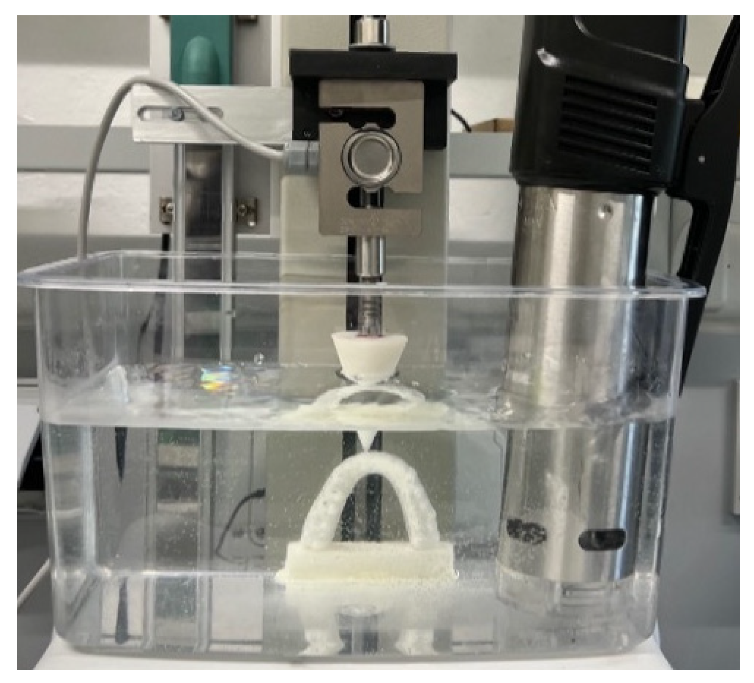
A motorized vertical test stand, TVO-S (AstraLab, Mariano Comense, Italy), was used to perform a single-point vertical compression, applying a constant, preset load. Models were placed in a container (24 cm × 12 cm × 14 cm) serving as horizontal support to ensure that the force application occurred at the same location and remained perpendicular to the plane. This container was filled with distilled water at a constant temperature of 37 °C and positioned under the load. An Isw-100W immersion heater (INKBIRD, Shenzhen, China) was used to maintain the water temperature at 37 °C (Figure 4). Each sample underwent a 3 h stress-relaxation test. After placing the aligner correctly on the model, a speed of 1 mm/min was set, achieving the desired deflection within one minute of testing. A constant deflection force of 1N was maintained, with data collected every second to obtain a precise stress-relaxation curve for each material. An external force gauge holder, Sauter TVO-A01, was connected to a computer equipped with Sauter AFH Fast data transmission software (AstraLab, Mariano Comense, Italy), providing a relaxation curve (force [N]/time [s]) for each observation period. Each aligner type was tested at three points (gingival, middle, and incisal/occlusal) on elements 1.1 and 2.6. Three equivalent trials were conducted for each point using different aligners for each. To compare stress decay across samples, normalized stress was calculated using the following equation to determine stress decay percentage:
Stress Decay % = σ/σmax × 100
where σ represents the initial stress of the material, and σmax represents the maximum stress reached during observation.

2.5. Statistical Analysis

2.5.1. Repeatability Test

Statistical analysis was based on the mean of three tests conducted at each point (gingival, middle, and occlusal). Repeatability tests were initially performed on elements 1.1 and 2.6, calculating the Intraclass Correlation Coefficient (ICC) to ensure high measurement repeatability.

2.5.2. Descriptive Statistics

Descriptive statistics, including mean, standard deviation, median, and 25th and 75th percentiles, were calculated for each aligner group and measurement point on elements 1.1 and 2.6.

2.5.3. Statistical Tests

Levene’s test assessed homoscedasticity, revealing a violation of the homogeneity of variances assumption (p < 0.001). Consequently, the Brown–Forsythe robust ANOVA was used, ensuring that comparisons remained valid despite differences in variability across groups. This method was chosen specifically to account for variance heterogeneity and improve the reliability of statistical results. For each measurement point (gingival, middle, incisal), ANOVA compared the mean force values across the five aligner groups for both elements 1.1 and 2.6. Results were considered significant at a 95% confidence level with p < 0.05. If significance was found, Tamhane’s post hoc test was applied, suitable for data with heterogeneous variances.

2.5.4. Force Analysis Among Points

A further comparison of force at the three measurement points (gingival, middle, and incisal/occlusal) for each aligner was conducted using the Brown–Forsythe robust ANOVA.

2.5.5. Force Decay (Stress Relaxation)

To evaluate differences in stress relaxation among aligners, the non-parametric Kruskal–Wallis test was applied, given the non-normal data distribution and relatively small sample size per group.

2.5.6. Effect Measurements

To quantify the magnitude of differences between groups, eta-squared and Cohen’s d were calculated, both indicative of the proportion of variance explained by the differences between aligners.

2.5.7. Comparison of Force Between Incisor and Molar

For each measurement point and for each type of aligner, an independent-samples t-test was used to determine whether the force applied differed significantly between molars and incisors. Before conducting the t-test, the assumption of homoscedasticity (equality of variances) was assessed using Levene’s test. If Levene’s test was significant, indicating heteroscedasticity (unequal variances), a robust version of the t-test was applied, which adjusts the analysis to account for variance differences between groups. This procedure ensured the statistical validity of the comparisons between different tooth types regarding the force applied by the aligners.

2.5.8. Sample Size

The adequacy of the sample size was evaluated using a t-test for independent samples, based on the lowest observed effect size (0.197), a significance level of 0.05, and a sample size of 10,752 per group. This resulted in a statistical power greater than 0.99, confirming its sufficiency to detect significant differences. However, this sample size does not represent independent specimens but derives from repeated measurements on each aligner. Each aligner was tested at three measurement points (gingival, middle, and occlusal) with three repeated tests per point to refine force estimation and account for intra-aligner variability. Statistical analyses accounted for data non-independence by applying appropriate methods that consider within-subject variability, ensuring precision in force estimation without artificially inflating statistical power.

2.5.9. Software

Statistical analyses were conducted with a significance level of 0.05, using IBM SPSS Statistics version 29.

3. Results

The repeatability of the measurements was evaluated using the Intraclass Correlation Coefficient (ICC) for the three measurement points (gingival, middle, and incisal/occlusal) on both the incisors and molars. The ICC values were all higher than 0.75 for each point and each tooth, confirming the reliability of the repeated measurements analyses (Table 1). This allowed for the use of the average of the measurements in the subsequent.

3.1. Incisor (1.1)

The ANOVA test is performed to verify, for each point, whether the mean force differs significantly among the aligners. In the case of a significant test (p-value < 0.05), it is concluded that at least one aligner has a mean value different from the others. Before the test, the homoscedasticity assumption was checked, and since it was not satisfied, the robust version of the ANOVA test by Brown–Forsythe was chosen. For all points, it was found that at least one aligner exhibits a significant difference in force compared to the others (p-value < 0.001), with very large eta-squared values indicating highly pronounced differences among the aligners. Post hoc comparisons conducted using Tamhane’s test reveal that all aligners, for each point, have significantly different force values.
  • At the gingival point, the NHVH aligner exerted the highest average force, followed by NHH, F22, NHZ, and AS;
  • At the middle point, NHVH again showed the highest force, followed by NHH, NHZ, F22, and AS;
  • At the incisal point, NHH had the highest force, followed by NHVH, NHZ, F22, and AS (Table 2).
Table 2. Comparison of strength at each point among aligners in 1.1. Robust One-Way ANOVA Brown–Forsythe 1.
PointAlignernMeanSDF (df1, df2)p-ValueEta-Squared
GingivalAS10,7521.40.2297,934.011
(4, 37,236.278)
<0.001 *0.957
F2210,7523.20.4
NHH10,7523.20.4
NHVH10,7527.20.6
NHZ10,7522.80.3
MiddleAS10,7524.30.598,293.072
(4, 43,204.643)
<0.001 *0.878
F2210,7525.30.7
NHH10,7525.90.4
NHVH10,7528.70.6
NHZ10,7525.50.4
IncisalAS10,7526.30.853,640.504
(4, 49,636.352)
<0.001 *0.797
F2210,7527.80.7
NHH10,75210.10.8
NHVH10,7529.80.6
NHZ10,7528.00.6
1 NHVH (NOXI Horizontal Vertical High); NHH (NOXI Horizontal High), NHZ (NOXI Horizontal Zenith), F22 (straight gingival margin—cut at the zenith); AS (scalloped gingival margin); N, sample size; SD, standard deviation; F (df1, df2), F-distribution; * p-value < 0.05 (statistically significant).
Additionally, when comparing the force across the three measurement points, it was found that for all aligners, the highest average force occurred at the incisal point, followed by the middle point, and finally the gingival point. This was consistent across all aligners, as confirmed by the robust ANOVA test and post hoc Tamhane comparisons, which showed significant differences between the points for each aligner.
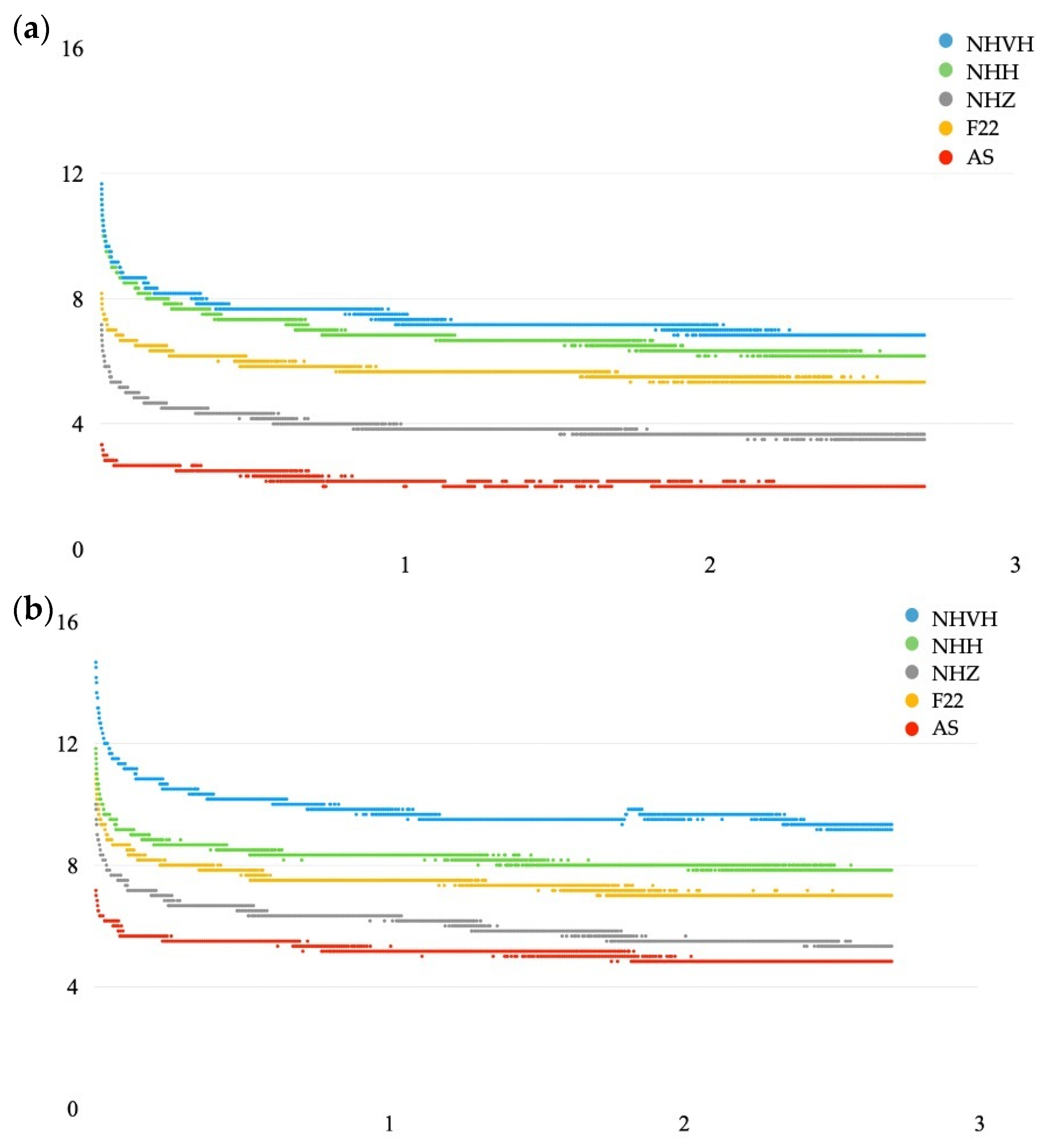
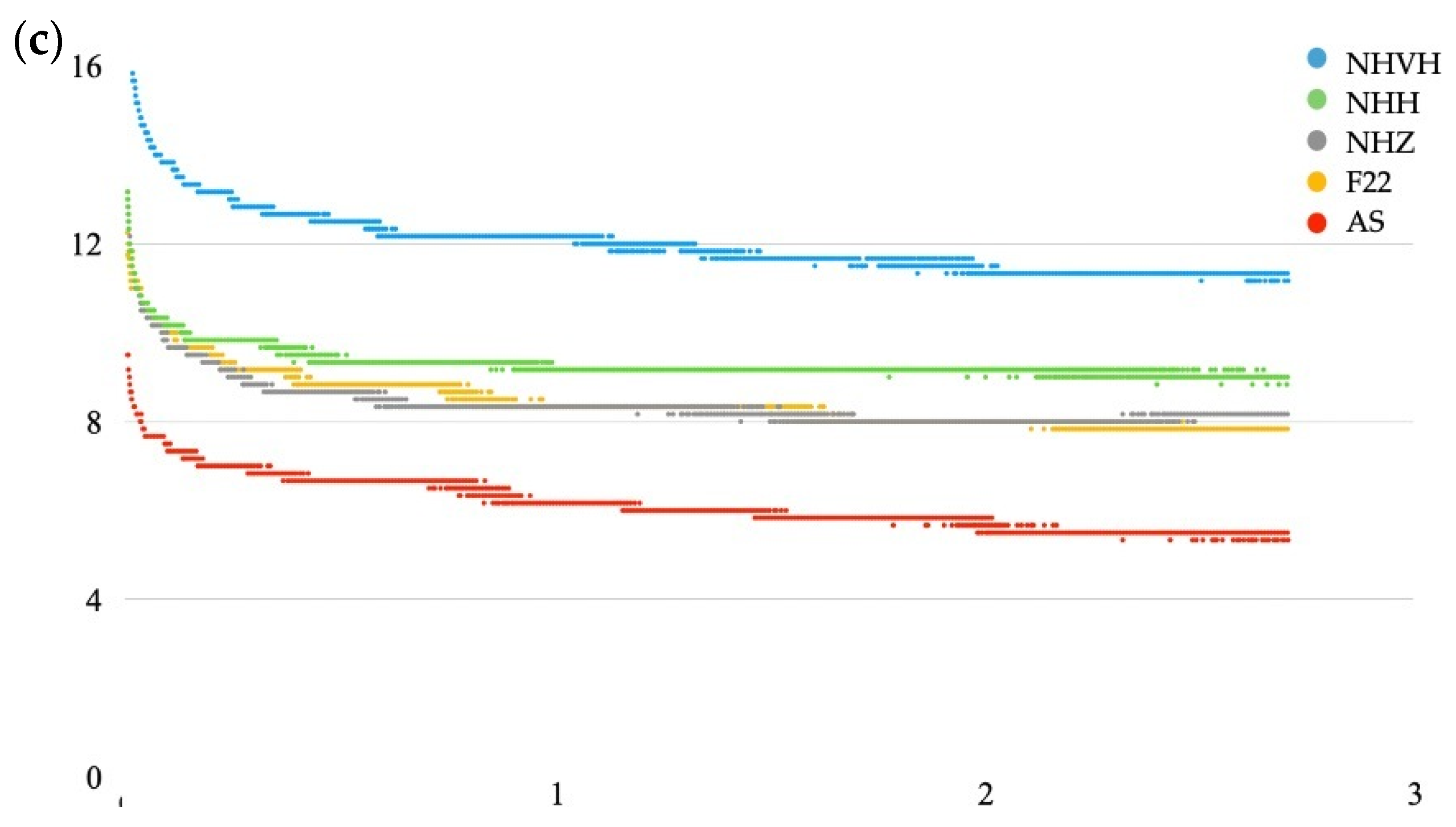
Regarding the initial and final force values, as well as the average percentage of stress decay, the mean values for each of the five aligners were calculated for the incisors (1.1). As for stress decay, the Kruskal–Wallis non-parametric test indicated no significant differences (p-value > 0.05) in stress relaxation between the aligners on the 1.1 tooth (Table 3). In Figure 5, the analysis of the force applied to the upper central incisor shows that aligners exhibited significantly different forces at each measured point. The highest forces at the gingival and middle points were observed with the NHVH model, followed by the NHH, F22, NHZ, and finally AS, which recorded much lower values. This highlights the effectiveness of the thickness gradients in stabilizing force in the gingival areas, where dissipation naturally occurs, confirming the design’s efficacy. At the incisal point, the NHH generated the highest force (16.3 N), followed closely by the NHVH (14.7 N), with the AS showing significantly lower values. Despite the statistical significance, the difference between NHH and NHVH is minimal and within acceptable limits. In summary, NHVH achieves the highest forces at the gingival and middle points while maintaining adequate force at the incisal point. The highest average force was recorded at the incisal point, followed by the middle and gingival points, confirming natural force dissipation. Additionally, the NHZ aligners showed lower forces, particularly at the gingival point, likely due to their zenith cut geometry. This trimming effect is also seen in thermoformed aligners, with straight-trimmed models exerting much higher forces compared to scalloped-trimmed ones.

3.2. Molar (2.6)

The same statistical analyses performed on 1.1 were also conducted on 2.6. The robust version of the ANOVA test by Brown–Forsythe was conducted and for all points on 2.6, it was found that at least one aligner exhibits a significant difference in force compared to the others (p-value < 0.001). Additionally, very large eta-squared values were observed, indicating highly pronounced differences among the aligners. Tamhane’s post hoc test was used to determine where the significant differences between aligners lie (Table 4). The results show that, for each point, all aligners are statistically different in terms of force. Specifically, comparisons revealed, in all the measurement points (gingival, middle, and occlusal), that NHVH exerted the highest force, followed by NHH, F22, NHZ, and AS. Since the values are very similar, Cohen’s d was calculated for the occlusal point between the NHZ and F22 aligners. The result obtained was quite low (0.075), indicating that the relationship is statistically significant but negligible. Therefore, it can be concluded that the force exerted by F22 and NHZ is equivalent.
When comparing the force across the three points on the 2.6 tooth, the robust ANOVA test confirmed that all aligners showed significant differences (p-value < 0.001) between the points. For each aligner, the occlusal point had the highest average force, followed by the middle point, and finally the gingival point, as revealed by post hoc comparisons using the Tamhane test.
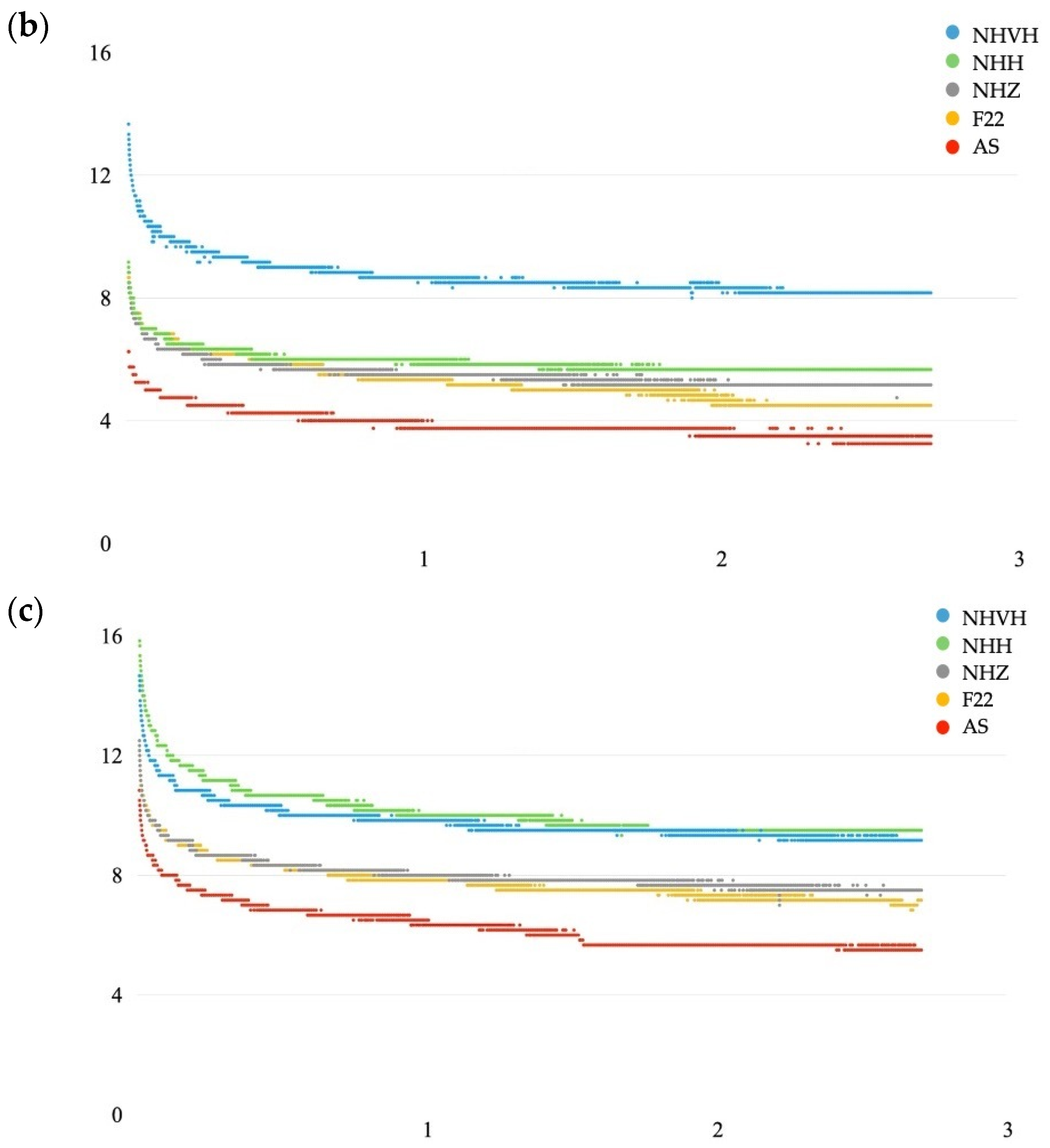
From all the data collected, an average value of initial and final stress was identified, along with the corresponding average percentage of stress decay for each of the five aligners, both on 2.6. Regarding stress relaxation, similar to 1.1, no significant differences (p-value > 0.05) were found between the aligners on the 2.6 tooth (Table 5). In Figure 6, the results show that at the gingival and middle levels, the NHVH aligner exerts the highest forces, followed by the NHH, F22, NHZ, and finally AS, which shows the lowest values. While the molar thickness is uniform at 0.95 mm across all Noxi aligners, the forces differ between models. This is due to the unique structural design of each aligner, where differential thickness in the anterior region affects force distribution in the posterior areas. For example, despite identical molar thickness and trimming, the NHVH exerts significantly higher forces than the NHH. Trimming also plays a key role, as seen with the NHZ aligner, which exerts lower forces compared to models with higher gradient designs. Similarly, the scalloped-trimmed thermoformed aligner exerts much lower forces than other aligners tested.
A comparison of force between the incisors (1.1) and molars (2.6) revealed significant differences (p-value < 0.001) for all measurement points and aligners. The effect size of these differences, measured using Cohen’s d, was strong (d > 0.8) in most cases. However, there were two exceptions where the force on the incisors was greater than on the molars: the incisal/occlusal point with the “festooned cut” aligner and the incisal/occlusal point with the “high horizontal gradient” NOXI aligner.
The comparison using the independent samples t-test between incisors and molars is taken as a reference, utilizing the lowest effect size (0.197). Given this value, a significance level of 0.05 and a sample size of 10,752 per group, a statistical power of >0.99 is achieved.

4. Discussion

Orthodontic aligners provide an aesthetic alternative to fixed braces, although they are not completely invisible. Among the latest innovations is NOXI (Sweden & Martina, Due Carrare, Padova, Italy), made of polyamide (PA) by 3D printing, designed to be used only 12 h a day, especially at night. This feature improves compliance, patient comfort, and reduces the social and functional problems associated with continuous use [20]. Studies such as that of Kameyama T. et al. highlight the effectiveness of intermittent orthodontic forces, suggesting that breaks of 4–9 h do not compromise tooth movement and reduce root resorption [21,22]. A recent study compared two different aligner usage protocols and their effect on crevicular fluid. Ten patients participated in a split-mouth study, where one side was treated with the conventional protocol of 20 h per day while the other hemiarch followed a protocol of only 12 h per day for 14 days. Inflammation and crevicular fluid levels were measured, showing an increase in both groups immediately after applying orthodontic force. No statistically significant differences in inflammation or fluid quantity were observed between the two hemiarches. Additionally, overlaying the models using Geomagic X Control software revealed no statistically significant differences in linear measurements (mm) between the two hemiarches. This was one of the first studies to compare protocols with reduced hours while maintaining the same duration in days. The findings showed that the accuracy of the NOXI aligner was slightly lower (64.2%) compared to the F22 aligners (65.4%). Mesiodistal and vestibulolingual tipping movements were found to be the most predictable in both groups. For these movements, as well as for the increase in inter-canine and inter-molar diameters, no statistically significant differences were observed between the groups. Therefore, the use of the NOXI aligner proves to be a valid alternative to the traditional F22 aligner [23].
The selection of gingival margin designs and thicknesses for the aligners in this study was carefully considered to ensure clinical relevance and facilitate meaningful comparisons across commonly used configurations in orthodontic practice. The chosen designs and thicknesses were guided by both clinical evidence and the need to explore biomechanically effective aligner configurations.
For NHVH and NHH, the straight margin cut 2 mm above the gingival zenith was selected based on the findings of Cowley et al. [24]. Their study demonstrated that this margin design offers the highest retention forces among different margin styles. Specifically, they concluded that “the most retentive aligner design uses a straight margin cut 2 mm above the gingival zenith” and emphasized that “straight gingival margins reduce the flexibility of a thermoformed aligner, thus improving retention and the ability to perform more complex movements and to express more tooth movement”. This design ensures enhanced mechanical stability and facilitates effective and precise orthodontic forces.
The NHZ aligner, as well as the F22 and AS thermoformed aligners, were selected to represent scalloped and zenith-cut geometries, which are among the most frequently used designs in commercially available aligners. Including these margin styles allows for a meaningful comparison of biomechanical performance across widely adopted configurations in clinical practice.
Regarding thickness, the AS and F22 aligners were fabricated with a uniform thickness of 0.76 mm. For the NOXI aligners, however, the thickness is not uniform but ranges from 0.65 mm to 0.96 mm. This range was specifically designed to provide differential thickness gradients, optimizing force distribution across the dental arch. Despite this variability, the average thickness of 0.76 mm serves as a central reference.
The thickness of 0.76 mm was chosen after conducting several preliminary in vitro studies to determine the most suitable thickness. These studies evaluated various configurations and identified 0.76 mm as the optimal choice, as it provides a balance between maintaining appropriate orthodontic forces and ensuring patient comfort. This conclusion is supported by the findings of Cremonini et al. [25] who demonstrated that this thickness allows for effective stress relaxation properties while retaining sufficient force application over time.
By designing the NOXI aligners with thickness gradients centered around this 0.76 mm average, the study aimed to explore the advantages of differential thickness in achieving biomechanically optimal force distributions. This approach complements the use of uniform thickness in the AS and F22 aligners, enabling a robust comparison of different design strategies.
The NOXI design incorporates differentiated thicknesses and the configuration chosen takes into account the anchorage value, which varies according to the root surface and the natural dissipation of force in the terminal portions of the arch [16]. The increased thickness at the gingival edge amplifies the force where needed, ensuring effective and customizable distribution. Three-dimensional printing and the properties of PA allow the force to be calibrated as needed, eliminating the need for attachments and improving aesthetics. The material maintains a constant force intensity due to reduced stress relaxation, making NOXI an aligner that combines targeted forces, aesthetics, and limited daily use, overcoming the limitations of traditional aligners [25].
The introduction of NOXI represents an innovative solution addressing some of the main challenges highlighted in the current literature on clear aligners. A recent study analyzing Instagram content explored patient experiences with clear aligner therapy, emphasizing that key discomforts are associated with aesthetic issues (e.g., visibility of attachments), difficulties in speaking and eating, and the pain related to continuous aligner wear [26].
With its design tailored for nightly use, limited to 12 h per day, NOXI significantly reduces the social and functional impact of treatment compared to traditional 20–22 h daily protocols. This feature directly addresses patients’ needs for comfort and quality of life, minimizing disruptions in daily activities. Additionally, the attachment-free design, made possible by the advanced mechanical properties of polyamide (PA), enhances aesthetics and increases patient acceptance of the treatment—a critical factor for compliance.
The Instagram study also highlighted how the initial phase of treatment is often accompanied by high aesthetic and functional expectations, which may diminish during the treatment phase due to pain or operational difficulties [26]. The use of materials such as PA in NOXI, with high elastic memory and stress stability, helps mitigate these issues by ensuring consistent and predictable orthodontic forces even after extended periods. These clinically relevant aspects of NOXI, combined with reduced wear time and the elimination of attachments, demonstrate its potential to significantly improve patient comfort and satisfaction, making orthodontic treatment less invasive and more aligned with individuals’ social and personal needs.
The aim of this in vitro study was to compare the initial and final force values of three types of NOXI aligners, characterized by different thicknesses and gingival margin cuts, with two types of thermoformed aligners made of the same material and thickness but with different gingival margin cuts. In this study, to simulate oral cavity conditions, the aligners were fully immersed in a humid environment at 37 °C and subjected to constant loading for 3 h. The stress within thermoplastic materials induced by the initial deformation tends to cause a deterioration of the mechanical properties over time, as these materials are viscoelastic [27]. This phenomenon, known as stress relaxation, leads to a further loss of a thermoplastic’s ability to move a tooth over time, making the prediction of orthodontic forces and tooth movement much more challenging [5].
The force analysis on the upper central incisors showed significant differences between the various points analyzed. At the gingival and middle points, NHVH recorded the highest forces, followed by NHH, F22, NHZ, and finally AS, which showed significantly lower values. The design with horizontal and vertical thickness gradients of NHVH proved particularly effective in concentrating the force in the gingival areas, where it tends to dissipate the most. At the incisal point, NHH exerted the highest force (16.3 N), followed by NHVH (14.7 N), a small but statistically significant difference. The NHZ and AS aligners showed lower forces, with AS being significantly less effective. The aligner shear also influences these results: the NHZ aligners exert lower forces than the other NOXI aligners, particularly at the gingival point, probably due to the geometry of the zenithal shear, which, while maintaining a uniform force, does not reach the initial intensity of other models. The cutting difference is also evident when comparing the two thermoformed aligners, as the straight-cut aligner exerts significantly higher forces than the scalloped-cut aligner.
To better understand the force dissipation observed in the gingival regions and the differences between aligners, it is important to consider the biomechanical behavior of aligners during thermoforming. As highlighted in the study by Palone et al. [28], aligners typically exhibit a greater gap at gingival areas compared to occlusal or incisal points after thermoforming procedures. This phenomenon can be attributed to the stretching behavior of the plastic material, which is more pronounced in the tapered anterior regions and at the gingival level during manufacturing. Consequently, the force transmission in these areas is less efficient due to greater dissipation across the larger gap. Conversely, at the occlusal and incisal points, where the aligner fits more closely to the tooth surface, forces are more concentrated and thus more effective in inducing tooth movement. These findings align with the results of the present study, which confirmed that the highest average forces are observed at the incisal/occlusal points, followed by the middle point, and finally the gingival point, further demonstrating the greater dissipation of force in the gingival region.
Comparing the results with the study by Hertan et al. [29], it appears that 3D-printed aligners made of polyamide (PA) perform very differently from those made of Tera Harz TC-85. Hertan found that 3D-printed aligners produce 77% lower forces than thermoformed ones for peak force and almost 90% lower for stabilized force. The superiority of PA over Tera Harz TC-85 in this study suggests that the mechanical properties of the material significantly influence the ability to generate and maintain orthodontic forces.
In the present study, at the gingival and middle points of the molar, the NHVH aligner exerts a significantly higher force, followed by NHH, F22, NHZ and finally AS, which shows the lowest force values. Given the similar values, Cohen’s D was calculated at the occlusal point between the NHZ aligner and the F22 aligner. The resulting value is relatively low (0.075), indicating that the relationship is statistically significant but negligible. Therefore, it can be concluded that the force exerted by F22 and NHZ is almost equivalent.
An important observation concerns the force distribution between the different aligners. While the thickness of the incisors varies between the NOXI models, they all show a uniform thickness of 0.95 mm in the molars. Despite this uniformity, the models show different forces on the molars, which can be explained by the structural design: different thicknesses in the anterior section also influence the force in the posterior areas. For example, NHVH generates greater forces than NHH, despite having the same thickness in the molars, due to its horizontal and vertical gradients. NHZ, on the other hand, despite having the same thickness as the other models in the molars, exerts lower forces, such as thermoformed scalloped aligners, which produce significantly lower forces. In summary, the NOXI configuration responds to the biomechanical requirements of incisors and molars, ensuring effective force distribution for orthodontic movement.
The average force was higher on molars than on incisors for all aligners and measuring points, with some exceptions: scalloped cut models and NOXI models with a high horizontal gradient showed lower values on molars. This finding is consistent with that described by Proffit et al. [21], according to whom, molars, having a larger root surface, require more intense forces for orthodontic movement. The NOXI configuration, with increased thicknesses of up to 1 mm in molars, addresses this biomechanical need.
With the advent of 3D printing, concerns about mechanical material properties, chemical stability, and technical processes have emerged [30,31]. Can et al. [32] found that the mechanical properties of 3D-printed aligners did not deteriorate after one week of intraoral use, while Lee et al. [33] observed more stress relaxation than aligners thermoformed in PET-g at 37 °C. A subsequent study on 3D-printed materials, Material X (Envisiontec, Inc; Dearborn, MI, USA), and OD-Clear TF (3DResyns, Barcelona, Spain) showed that the residual stress after 2 h of 2% deformation was less than 10%, indicating a significant reduction in the initial stress, speculating that longer tests would further reduce stress [34]. Cremonini et al. [23] compared thermoformed aligners, Zendura FLX (Bay Materials LLC, Fremont, CA, USA) and Duran (SCHEU, Iserlohn, Germany), and 3D-printed aligners, Tera Hartz TC-85 (Graphy Inc., Seoul, Republic of Korea) and polyamide (PA, Sweden&Martina, Due Carrare, Padova, Italy), by subjecting them to stress relaxation tests for 8 h at 37 °C and controlled humidity. The results showed significant differences between Tera Hartz TC-85 and PA; the former showed the lowest stress relaxation values, with a decay from 90% to 100% in both tests. The PA aligners were characterized by a greater ability to maintain strength, with 32% relaxation in test 1 and 23% in test 2, compared to the thermoformed aligners, values that were, however, lower than Duran and Tera Hartz TC-85. In the current study, the different aligners showed no significant differences in stress relaxation, maintaining stable and constant forces comparable to those of existing aligners but with higher initial forces. This stability ensures controlled orthodontic movement, reduces the risk of microtrauma, and improves patient comfort [20]. Discrepancies with previous studies can be attributed to differences in materials, molds, and methodologies used.
This study provides valuable insights into the force application of different aligners, particularly NOXI, on the upper central incisor and first molar. Although NOXI exhibited higher force values in vitro, further clinical studies are needed to determine its actual effectiveness in patient treatment.
The introduction of PA material and differentiated thicknesses enabled the development of aligners capable of exerting greater forces than traditional thermoformed aligners. While this could potentially compensate for the reduced wear time, its impact on tooth movement remains to be clinically validated. Additionally, aligner performance in real-world conditions may differ from in vitro simulations. This study was conducted at a constant temperature of 37 °C, without considering potential temperature fluctuations in the oral cavity.
Moreover, while repeated measurements improved force estimation, they do not replace the need for a larger number of independent aligners. Future research should focus on increasing the sample size and assessing force retention over extended periods to enhance generalizability. Longitudinal in vivo studies will be essential to further understand the long-term mechanical behavior of NOXI aligners.

5. Conclusions

The analysis of the initial and final force values from the three types of NOXI aligners, each characterized by different thicknesses and cutting margins, alongside the thermoformed aligners with varying cutting configurations, led to the following findings:
  • The NOXI aligner with both horizontal and vertical gradients and a high margin exerted the greatest overall force, particularly at the gingival point where force dissipation was most evident. It was followed by the NOXI with a horizontal gradient and a high margin.
  • No statistically significant differences in stress relaxation were observed among the five aligners tested.
  • For aligners of the same thickness, the height of the margin significantly influenced the force exerted.
  • The force applied was greater at the upper first molar than at the upper central incisor.
These results demonstrate the effectiveness of the different aligners in generating orthodontic forces on both the upper central incisor and first molar. The comparison between various NOXI models reveals the importance of structural features such as thickness gradients and margin types in optimizing force application for specific areas. The NOXI aligner exhibits a different force application profile, which may influence mechanical performance compared to conventional aligners worn for 20–22 h per day.

Author Contributions

Conceptualization, F.C., C.P. and L.B.; methodology, C.P. and L.B.; software, C.P.; validation, F.C., C.P. and L.L.; formal analysis, C.P.; investigation, C.P.; resources, C.P.; data curation, C.P.; writing—original draft preparation, A.K.S.; writing—review and editing, A.K.S.; visualization, C.P.; supervision, L.L.; project administration, F.C.; funding acquisition, L.L. All authors have read and agreed to the published version of the manuscript.

Funding

This research received no external funding.

Institutional Review Board Statement

Not applicable.

Informed Consent Statement

Not applicable.

Data Availability Statement

The raw data supporting the conclusions of this article will be made available by the authors on request.

Conflicts of Interest

The authors declare no conflicts of interest.

References

  1. Bonnick, A.M.; Nalbandian, M.; Siewe, M.S. Technological advances in nontraditional orthodontics. Dent. Clin. 2011, 55, 571–584. [Google Scholar] [CrossRef] [PubMed]
  2. Favero, L.; Cortelazzo, V.; Arreghini, A. Efficacia e vantaggi di una metodica invisibile a rivalutazione sequenziale: Il sistema Clear Aligner. Mondo Ortod. 2010, 35, 87–94. [Google Scholar] [CrossRef]
  3. Proffit, W.R.; Fields, H.W.; Sarver, D.M. Contemporary Orthodontics, 5th ed.; Elsevier: Filadelfia, PA, USA, 2013. [Google Scholar]
  4. Kesling, H.D. Coordinating the predetermined pattern and tooth positioner with conventional treatment. Am. J. Orthod. Oral Surg. 1946, 32, 285–293. [Google Scholar] [CrossRef] [PubMed]
  5. Fang, D.; Zhang, N.; Chen, H.; Bai, Y. Dynamic Stress Relaxation of Orthodontic Thermoplastic Materials in a Simulated Oral Environment. Dent. Mater. J. 2013, 32, 946–951. [Google Scholar] [CrossRef]
  6. Tartaglia, G.M.; Mapelli, A.; Maspero, C.; Santaniello, T.; Serafin, M.; Farronato, M.; Caprioglio, A. Direct 3D Printing of Clear Orthodontic Aligners: Current State and Future Possibilities. Materials 2021, 14, 1799. [Google Scholar] [CrossRef]
  7. Voet, V.S.D.; Guit, J.; Loos, K. Sustainable Photopolymers in 3D Printing: A Review on Biobased, Biodegradable, and Recyclable Alternatives. Macromol. Rapid Commun. 2021, 42, 2000475. [Google Scholar] [CrossRef]
  8. Cremonini, F.; Vianello, M.; Bianchi, A.; Lombardo, L. A Spectrophotometry Evaluation of Clear Aligners Transparency: Comparison of 3D-Printers and Thermoforming Disks in Different Combinations. Appl. Sci. 2022, 12, 11964. [Google Scholar] [CrossRef]
  9. Gueche, Y.A.; Sanchez-Ballester, N.M.; Cailleaux, S.; Bataille, B.; Soulairol, I. Selective Laser Sintering (SLS), a New Chapter in the Production of Solid Oral Forms (SOFs) by 3D Printing. Pharmaceutics 2021, 13, 1212. [Google Scholar] [CrossRef]
  10. Hertan, E.; Lewis, H.G.; Kharlamov, A. Advancements in Clear Aligner Therapy: Direct 3D Printing and Biomechanical Innovations. J. Clin. Orthod. 2020, 54, 205–212. [Google Scholar]
  11. Eliades, G.; Zinelis, S.; Papageorgiou, S.N. Mechanical Properties and Clinical Performance of Clear Aligners and Fixed Appliances. J. Clin. Orthod. 2021, 53, 102–110. [Google Scholar]
  12. Jindal, P.; Juneja, M.; Siena, F.L.; Bajaj, D.; Breedon, P. Mechanical and geometric properties of thermoformed and 3D printed clear dental aligners. Am. J. Orthod. Dentofac. Orthop. 2019, 56, 694–701. [Google Scholar] [CrossRef] [PubMed]
  13. Hazeveld, A.; Slater, J.J.H.; Ren, Y. Accuracy and reproducibility of dental replica models reconstructed by different rapid prototyping techniques. Am. J. Orthod. Dentofac. Orthop. 2014, 145, 108–115. [Google Scholar] [CrossRef] [PubMed]
  14. Chuchulska, B.; Zlatev, S. Linear Dimensional Change and Ultimate Tensile Strength of Polyamide Materials for Denture Bases. Polymers 2021, 13, 3446. [Google Scholar] [CrossRef] [PubMed]
  15. Lyu, X.; Cao, X.; Yan, J.; Zeng, R.; Tan, J. Biomechanical Effects of Clear Aligners with Different Thicknesses and Gingival-Margin Morphology for Appliance Design Optimization. Am. J. Orthod. Dentofac. Orthop. 2023, 164, 239–252. [Google Scholar] [CrossRef]
  16. Lombardo, L.; Martines, E.; Mazzanti, V.; Arreghini, A.; Mollica, F.; Siciliani, G. Stress Relaxation Properties of Four Orthodontic Aligner Materials: A 24-Hour In Vitro Study. Angle Orthod. 2016, 87, 11–18. [Google Scholar] [CrossRef]
  17. Beretta, M.; Federici Canova, F.; Gianolio, A.; Mangano, A.; Paglia, M.; Colombo, S.; Cirulli, N. ZeroExpander: Metal-free automatic palatal expansion for special-needs patients. Eur. J. Paediatr. Dent. 2021, 22, 151–154. [Google Scholar] [CrossRef]
  18. Campobasso, A.; Battista, G.; Lo Muzio, E.; Colombo, S.; Paglia, M.; Federici Canova, F.; Gianolio, A.; Beretta, M. New 3D printed polymers in orthodontics: A scoping review. Eur. J. Paediatr. Dent. 2023, 24, 224–228. [Google Scholar] [CrossRef]
  19. Albertini, P.; Mazzanti, V.; Mollica, F.; Pellitteri, F.; Palone, M.; Lombardo, L. Stress Relaxation Properties of Five Orthodontic Aligner Materials: A 14-Day In-Vitro Study. Bioengineering 2022, 9, 349. [Google Scholar] [CrossRef]
  20. Samuels, R.H.; Rudge, S.J.; Mair, L.H. The Effect of Orthodontic Forces on the Dental Pulp. J. Orthod. 2016, 43, 73–81. [Google Scholar]
  21. Proffit, W.R.; Fields, H.W.; Larson, B.E.; Sarver, D.M.; Cozza, P. Ortodonzia Moderna, 6th ed.; Edra: Perignano, Italy, 2020. [Google Scholar]
  22. Kameyama, T.; Warita, H.; Matsumoto, Y.; Otsubo, K. A Mechanical Stress Model Applied to the Rat Periodontium: Using Controlled Magnitude and Direction of Orthodontic Force with an Absolute Anchorage. Oral Med. Pathol. 2002, 7, 1–7. [Google Scholar] [CrossRef]
  23. Cremonini, F.; Pavan, F.; Pellitteri, F.; Palone, M.; Lombardo, L. Variation of gingival crevicular fluid volume in early orthodontic tooth movement with clear aligner: A pilot study. Sci. Rep. 2023, 14, 42–47. [Google Scholar] [CrossRef]
  24. Cowley, D.P.; Mah, J.; O’Toole, B. The Effect of Gingival-Margin Design on the Retention of Thermoformed Aligners. J. Clin. Orthod. 2012, 46, 697–702; quiz 705. [Google Scholar] [PubMed]
  25. Cremonini, F.; Brucculeri, L.; Pepe, F.; Palone, M.; Lombardo, L. Comparison of Stress Relaxation Properties between 3-Dimensional Printed and Thermoformed Orthodontic Aligners: A Pilot Study of In Vitro Simulation of Two Consecutive 8-Hours Force Application. APOS Trends Orthod. 2024, 14, 225–234. [Google Scholar] [CrossRef]
  26. Grassia, V.; d’Apuzzo, F.; Alansari, R.A.; Jamilian, A.; Sayahpour, B.; Adel, S.M.; Nucci, L. Instagram and Clear Aligner Therapy: A Content Analysis of Patient Perspectives. Semin. Orthod. 2024, 30, 123–130. [Google Scholar] [CrossRef]
  27. Sheridan, J.J.; Ledoux, W.; McMinn, R. Essix Appliances: Minor Tooth Movement with Divots and Windows. J. Clin. Orthod. 1994, 28, 659–663. [Google Scholar]
  28. Palone, M.; Longo, M.; Arveda, N.; Nacucchi, M.; Pascalis, F.; Spedicato, G.A.; Siciliani, G.; Lombardo, L. Micro-computed tomography evaluation of general trends in aligner thickness and gap width after thermoforming procedures involving six commercial clear aligners: An in vitro study. Korean J. Orthod. 2021, 51, 135–141. [Google Scholar] [CrossRef]
  29. Hertan, E.; McCray, J.; Bankhead, B.; Kim, K.B. Force Profile Assessment of Direct-Printed Aligners Versus Thermoformed Aligners and the Effects of Non-Engaged Surface Patterns. Prog. Orthod. 2022, 23, 49. [Google Scholar] [CrossRef]
  30. Edelmann, A.; English, J.D.; Chen, S.J.; Kasper, F.K. Analysis of the Thickness of 3-Dimensional-Printed Orthodontic Aligners. Am. J. Orthod. Dentofac. Orthop. 2020, 158, e91–e98. [Google Scholar] [CrossRef]
  31. McCarty, M.C.; Chen, S.J.; English, J.D.; Kasper, F. Effect of Print Orientation and Duration of Ultraviolet Curing on the Dimensional Accuracy of a 3-Dimensionally Printed Orthodontic Clear Aligner Design. Am. J. Orthod. Dentofac. Orthop. 2020, 158, 889–897. [Google Scholar] [CrossRef]
  32. Can, E.; Panayi, N.; Polychronis, G.; Papageorgiou, S.N.; Zinelis, S.; Eliades, G.; Eliades, T. In-House 3D-Printed Aligners: Effect of In Vivo Ageing on Mechanical Properties. Eur. J. Orthod. 2022, 44, 51–55. [Google Scholar] [CrossRef]
  33. Lee, S.Y.; Kim, H.; Kim, H.J.; Chung, C.J.; Choi, Y.J.; Kim, S.J.; Cha, J.Y. Thermo-Mechanical Properties of 3D Printed Photocurable Shape Memory Resin for Clear Aligners. Sci. Rep. 2022, 12, 6246. [Google Scholar] [CrossRef] [PubMed]
  34. Shirey, N.; Mendonca, G.; Groth, C.; Kim-Berman, H. Comparison of Mechanical Properties of 3-Dimensional Printed and Thermoformed Orthodontic Aligners. Am. J. Orthod. Dentofac. Orthop. 2023, 163, 720–728. [Google Scholar] [CrossRef] [PubMed]
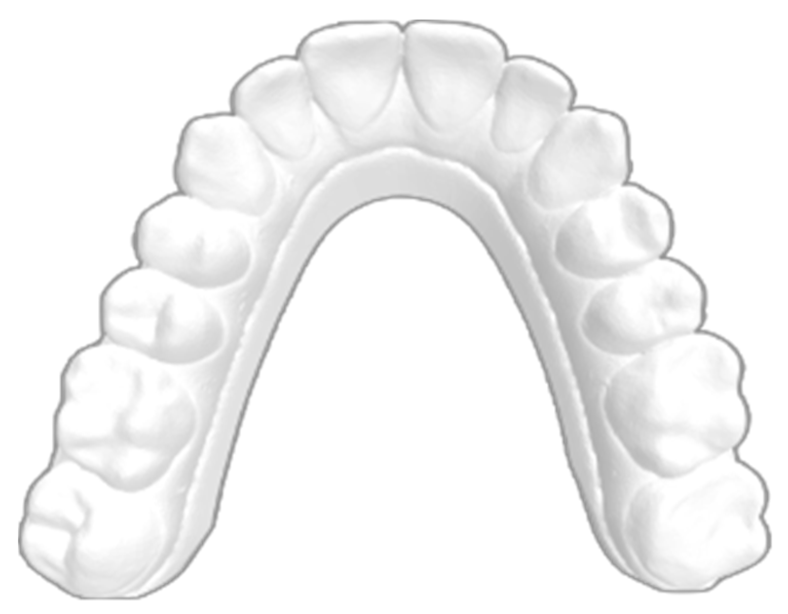
Figure 1. Master model used to print all aligners.
Applsci 15 02911 g001
Figure 4. The aligner sample immersed in the bath and positioned beneath the load cell.
Applsci 15 02911 g004
Figure 5. (a) Mean decay curve recorded at the gingival level of tooth 1.1; (b) mean decay curve recorded at the middle level of tooth 1.1; (c) mean decay curve recorded at the incisal level of tooth 1.1 The X-axis represents time in hours, and the Y-axis represents force in Newtons.
Applsci 15 02911 g005aApplsci 15 02911 g005b
Figure 6. (a) Mean decay curve recorded at the gingival level of tooth 2.6; (b) mean decay curve recorded at the middle level of tooth 2.6; (c) mean decay curve recorded at the incisal level of tooth 2.6. The X-axis represents time in hours, and the Y-axis represents force in Newtons.
Applsci 15 02911 g006aApplsci 15 02911 g006b
Table 1. Inter-Item Correlation Matrix.
Force (N)Force (N)Force (N)
Inter-Item Correlation Matrix Gingival Incisor1.000.900.96
0.901.000.92
0.960.921.00
Inter-Item Correlation Matrix Middle Incisor1.000.940.94
0.941.000.93
0.940.931.00
Inter-Item Correlation Matrix Incisal Incisor1.000.940.96
0.941.000.92
0.960.921.00
Inter-Item Correlation Matrix Gingival Molar1.000.970.92
0.971.000.93
0.920.931.00
Inter-Item Correlation Matrix Middle Molar1.000.910.91
0.911.000.92
0.910.921.00
Inter-Item Correlation Matrix Occlusal Molar1.000.960.96
0.961.000.96
0.960.961.00
Table 3. Initial stress values, final stress values, stress decay, and a comparison of strength decay among aligners tested on element 1.1, assessed using the independent sample Kruskal–Wallis test.
MaterialPointInitial Force (N)Final Force (N)Stress
Relaxation (%)
MeanMedianSD25th
Percentile
75th
Percentile
p-Value
ASGingival2.01.050.050.350.01.349.251.70.212
Middle6.63.551.7
Incisal10.85.549.2
F22Gingival5.22.051.946.847.65.541.051.9
Middle6.63.547.6
Incisal5.5841.0
NHHGingival6.02.853.344.742.97.838.053.3
Middle9.25.638.0
Incisal16.39.542.9
NHVHGingival11.76.544.440.740.13.537.444.4
Middle13.78.740.1
Incisal14.79.737.4
NHZGingival4.32.546.542.540.93.540.046.5
Middle8.85.240.9
Incisal12.57.540.0
NHVH (NOXI Horizontal Vertical High); NHH (NOXI Horizontal High), NHZ (NOXI Horizontal Zenith), F22 (straight gingival margin—cut at the zenith); AS (scalloped gingival margin); SD, standard deviation.
Table 4. Comparison of strength at each point among aligners in 2.6. Robust One-Way ANOVA Brown–Forsythe 1.
PointAlignernMeanSDF (df1, df2)p-ValueEta-Squared
GingivalAS10,7522.20.2209,619.977
(4, 39,266.816)
<0.001 *0.940
F2210,7525.70.4
NHH10,7527.40.6
NHVH10,7526.80.7
NHZ10,7524.00.4
MiddleAS10,7525.20.3140,950.949
(4, 45,639.097)
<0.001 *0.913
F2210,7527.40.5
NHH10,7529.80.6
NHVH10,7528.30.4
NHZ10,7526.10.6
OcclusalAS10,7526.10.6127,554.367
(4, 46,685.640)
<0.001 *0.905
F2210,7528.40.7
NHH10,75212.00.8
NHVH10,7529.30.4
NHZ10,7528.40.6
1 NHVH (NOXI Horizontal Vertical High); NHH (NOXI Horizontal High), NHZ (NOXI Horizontal Zenith), F22 (straight gingival margin—cut at the zenith); AS (scalloped gingival margin); N, sample size; SD, standard deviation; F (df1, df2), F-distribution; * p-value < 0.05 (statistically significant).
Table 5. Initial stress values, final stress values, stress decay, and a comparison of strength decay among aligners tested on element 2.6, assessed using the independent sample Kruskal–Wallis test.
MaterialPointInitial Force (N)Final Force (N)Stress
Relaxation (%)
MeanMedianSD25th
Percentile
75th
Percentile
p-Value
ASGingival3.32.039.439.039.45.533.344.20.284
Middle7.24.833.3
Incisal9.55.344.2
F22Gingival8.25.334.635.936.41.134.636.6
Middle11.07.036.4
Incisal12.37.836.6
NHHGingival11.26.244.639.038.52.636.741.9
Middle11.87.834.4
Incisal13.28.833.3
NHVHGingival11.76.841.937.434.46.233.344.6
Middle14.79.236.7
Incisal18.211.338.5
NHZGingival7.23.551.445.447.06.937.951.4
Middle10.05.347.0
Incisal13.28.237.9
NHVH (NOXI Horizontal Vertical High); NHH (NOXI Horizontal High), NHZ (NOXI Horizontal Zenith), F22 (straight gingival margin—cut at the zenith); AS (scalloped gingival margin); SD, standard deviation.
Disclaimer/Publisher’s Note: The statements, opinions and data contained in all publications are solely those of the individual author(s) and contributor(s) and not of MDPI and/or the editor(s). MDPI and/or the editor(s) disclaim responsibility for any injury to people or property resulting from any ideas, methods, instructions or products referred to in the content.