Article

Characterization and Selection of Lactobacillus Strains with Potential Probiotic Applications

by
Yulian Tumbarski
1,*,
Ivelina Peykova-Shapkova
2,
Mihaela Ivanova
2,
Remzi Cholakov
3,
Agata Dutkiewicz
4 and
Krzysztof Grzymajło
4
1
Department of Microbiology and Biotechnology, University of Food Technologies, 4002 Plovdiv, Bulgaria
2
Department of Milk and Dairy Products, University of Food Technologies, 4002 Plovdiv, Bulgaria
3
Özgazi B. V. Dairy Foods, 4879 AP Etten-Leur, The Netherlands
4
Department of Biochemistry and Molecular Biology, Wrocław University of Environmental and Life Sciences, 50-375 Wrocław, Poland
*
Author to whom correspondence should be addressed.
Appl. Sci. 2025, 15(6), 2902; https://doi.org/10.3390/app15062902
Submission received: 1 February 2025 / Revised: 26 February 2025 / Accepted: 4 March 2025 / Published: 7 March 2025
(This article belongs to the Special Issue New Insights into Food Ingredients for Human Health Promotion)

Abstract

:
In the present study, four Lactobacillus strains (Lacticaseibacillus casei ATCC 393, Lacticaseibacillus casei RC-1, Lactobacillus acidophilus RC-2 and Lacticaseibacillus rhamnosus RC-10) were investigated to determine their probiotic potential. The strains were evaluated based on their resistance to simulated upper gastrointestinal conditions, tolerance to bile salts, antimicrobial activity, antibiotic resistance and ability to adhere to intestinal cells. All the strains were resistant to simulated upper gastrointestinal conditions (pH 2.5 + pepsin and pH 7.4 + pancreatin) and exhibited cell recovery rates varying between 74.24 and 87.50% (pH 2.5 + pepsin) and between 93.95 and 98.15% (pH 7.4 + pancreatin) at the 24th h of cultivation. The strains demonstrated resistance to various concentrations of bile salts (0.25, 0.5, 0.75 and 1%) with survival rates > 50% (L. casei ATCC 393 and L. casei RC-1) and >60% (L. acidophilus RC-2 and L. rhamnosus RC-10) in bile salt concentration of 0.25% at the 24th h of incubation. The four Lactobacillus components—Lactobacillus culture (LC), cell biomass (CB) and cell-free supernatant (CFS) exhibited moderate to high antimicrobial activity against six Gram-positive and six Gram-negative bacteria; however, they failed to inhibit the growth of yeasts and fungi tested in the antimicrobial screening. In addition, the neutralized to pH = 7.0 cell-free supernatant (NCFS) of the four strains completely lost its antimicrobial effect. The results for antibiotic susceptibility of four Lactobacillus strains showed that L. casei ATCC 393 was resistant to 11, L. casei RC-1 was resistant to 9, L. acidophilus RC-2 was resistant to 8, and L. rhamnosus RC-10 was resistant to 11 of the total 23 antibiotics tested. The highest degree of adhesion of the studied isolates to the intestinal cell line IPEC-J2 was observed in L. casei RC-1 (39.45%), L. rhamnosus RC-10 (23.38%) and L. acidophilus RC-2 (19.25%) in comparison with the reference probiotic strain L. casei ATCC 393. Based on the results obtained, the strains studied can be considered as having probiotic potential.

1. Introduction

In recent decades, lactic acid bacteria (LAB) have been the most investigated group for application as probiotics. According to the generally adopted definition, probiotics represent “live microbial food supplements which, when administered in adequate amounts confer a health benefit on the host” [1]. In 2014, the International Scientific Association for Probiotics and Prebiotics (ISAPP) confirmed and accepted minor grammatical corrections to the previously announced by FAO/WHO definition, retaining its scientific meaning. As defined by ISAPP, the term “probiotic” refers to “many types of microorganisms, which demonstrate health benefits for the host, while remaining alive” [2].
Although the exploration of probiotic strains is not a new concept, they continue to attract the attention of both the scientific society and the food industry sector. Amongst the most studied bacteria for probiotic potential are the members of the genera Lactobacillus and Bifidobacterium, which are known as non-pathogenic inhabitants of animal and human gastrointestinal and female genital tracts [3,4]. In addition, as a part of the normal microflora, they are frequently isolated from various dairy products and non-dairy sources [5,6]. Lactic acid bacteria, in particular lactobacilli isolated from dairy products, are generally recognized as safe (GRAS) and extensively used as starter cultures and/or adjunct cultures to produce various fermented milk products, alcoholic beverages, sourdough and silage preparations [7]. Numerous studies have revealed that some other members of the LAB group, such as Lactococcus, Pediococcus and Leuconostoc, as well as strains belonging to the genera Saccharomyces, Enterococcus, Streptococcus, Bacillus, and Escherichia, have also exhibited promising probiotic properties [8].
The selection of probiotic strains is associated with certain important criteria for effectiveness, such as the capacity to survive the adverse upper gastrointestinal tract conditions (low pH, bile toxicity, digestive enzymes), the production of metabolites with antimicrobial activity (organic acids, bacteriocins, H2O2), antibiotic resistance and the ability to adhere to the epithelial intestinal cells, thus colonizing the intestines and exerting their health-promoting effects [5,9]. The beneficial effects of probiotic LAB strains are reported to involve antimicrobial, gastroprotective, anti-mutagenic, anti-carcinogenic, immunomodulating, cholesterol-reducing [10] and antioxidant [11] activities. These therapeutic features are species- and even strain-dependent. Undoubtedly, the crucial role of Lactobacillus spp. is associated with the immunity and gastrointestinal health of the host, which are closely related to intestinal barrier integrity. In vitro studies have revealed that some Lactobacillus species, with their metabolites, stimulate mucin production by the enterocytes, thus enhancing the intestinal barrier. The short-chain fatty acids (acetate, propionate and butyrate), H2O2 and antimicrobial peptides (bacteriocins) produced by the probiotic strains or secreted by the organism after stimulation are also important factors in improvement (stabilization) of the intestinal cell junction and play a key role in defense against pathogenic microorganisms. An important property of some Lactobacillus species is the stimulation of the secretion of immunoglobulin A (IgA) by intestinal B-cells (located in Peyer’s patches and lamina propria), resulting in an increase in local immunity [12]. Apart from that, probiotic microorganisms, with their physiological functions, contribute to maintaining gastrointestinal health. It is known that Lactobacillus species positively affect various conditions related to microbial imbalance and inflammation, such as enteric infections, antibiotic-associated diarrhea, necrotizing enterocolitis, inflammatory bowel disease (IBD), irritable bowel syndrome (IBS) and colorectal cancer (CRC), as well as non-digestive disorders such as urogenital diseases, bacterial vaginosis in women, atopic dermatitis, food hypersensitivity and dental caries, which emphasize their multiple therapeutic applications [13,14,15].
The aim of the present research was to evaluate the in vitro probiotic potential (resistance to bile salts, gastrointestinal tolerance, antimicrobial activity, antibiotic susceptibility and intestinal cell adhesion ability) of four Lactobacillus strains to determine future applications in the development of new probiotic products, especially dairy-based ones.

2. Materials and Methods

Lactobacillus strains
The strain Lacticaseibacillus casei RC-1 was isolated from homemade cheese from the town of Erzurum, Turkey; the strain Lactobacillus acidophilus RC-2 was isolated from homemade cheese from the town of Edirne, Turkey; and the strain Lacticaseibacillus rhamnosus RC-10 was isolated from fermented dairy beverage from the town of Etten-Leur, The Netherlands. The strain Lacticaseibacillus casei ATCC 393 was purchased from the National Bank for Industrial Microorganisms and Cell Cultures (NBIMCC), Sofia, Bulgaria.
The molecular genetic analyses (isolation of total DNA, 16S rDNA PCR amplification, purification of the 16S rDNA and the sequencing of the 16S rRNA gene) of the first three strains (L. casei RC-1, L. acidophilus RC-2 and L. rhamnosus RC-10) were conducted at the “Macrogen Europe Laboratory”, The Netherlands. The identification of the strains was performed using the CLC Sequence Viewer software v. 8.0.0. The entire sequence of the 16S rRNA gene was obtained, and the resulting whole sequence was compared with the on-line database sequences via the BLASTn algorithm. Thus, the strains studied were identified to the species level with the corresponding confidence level. The phylogenetic analysis revealed 99% pairwise similarity of the sequence of 16S rDNA of L. casei RC-1 to the partial sequence of 16S rDNA of L. casei strain CAU 6470, 99% similarity of L. acidophilus RC-2 to L. acidophilus strain FSI4, and 99% similarity of L. rhamnosus RC-10 with L. rhamnosus strain JCM 1136. All strains were cultured in MRS broth under microaerophilic conditions.
Test microorganisms
Twenty microorganisms, including six Gram-positive bacteria (Bacillus subtilis ATCC 6633, Bacillus cereus NCTC 11145, Staphylococcus aureus ATCC 25923, Listeria monocytogenes NBIMCC 8632, Enterococcus faecalis ATCC 19433, Micrococcus luteus 2YC-YT), six Gram-negative bacteria (Salmonella enteritidis ATCC 13076, Klebsiella pneumoniae RC-20, Escherichia coli ATCC 25922, Proteus vulgaris ATCC 6380, Pseudomonas aeruginosa ATCC 9027, Acinetobacter baumanii RC-22), three yeasts (Candida albicans NBIMCC 74, Saccharomyces cerevisiae ATCC 9763, Pichia membranaefaciens) and five fungi (Aspergillus niger ATCC 1015, Aspergillus ochraceus, Penicillium chrysogenum, Fusarium moniliforme ATCC 38932, Fusarium oxysporum) from the collection of the Department of Microbiology and Biotechnology at the University of Food Technologies, Plovdiv, Bulgaria, were selected for the antimicrobial activity test.
Culture media
Luria–Bertani agar medium with glucose
Luria–Bertani agar medium with glucose (LBG) agar (Condalab SL Madrid, Spain) was used for cultivation of test bacteria. A quantity of 50 g of LBG–solid substance mixture (containing 10 g tryptone, 5 g yeast extract, 10 g NaCl, 10 g glucose and 15 g agar) was dissolved in 1 L of deionized water, pH 7.5 ± 0.2.
Malt extract agar
Malt extract agar (MEA) (Scharlab SL, Barcelona, Spain) was used for cultivation of test yeasts and fungi. A quantity of 50 g of the MEA–solid substance mixture (containing 30 g malt extract, 5 g mycological peptone and 15 g agar) was dissolved in 1 L of deionized water, pH 5.4 ± 0.2.
De Man, Rogosa and Sharpe (MRS) broth and MRS agar
These media (Chemsolute, Renningen, Germany) were used for cultivation of lactobacilli. A quantity of 52 g of MRS broth (containing 10 g proteose peptone, 8 g meat extract, yeast 4 g extract, 20 g D(+)-glucose, 5 g sodium acetate, 2 g triammonium citrate, 0.2 g magnesium sulfate, 0.05 g manganese sulfate, 2 g dipotassium phosphate, 1 g polysorbate 80) was dissolved in 1 L of deionized water, pH 6.2 ± 0.2. MRS agar was prepared by dissolving 15 g/L of agar (Chemsolute) in MRS broth.
The culture media were prepared according to the manufacturer’s instructions and autoclaved at 121 °C for 20 min before use.
The effect of temperature on the growth of Lactobacillus strains
Determination of viable cell counts
In order to determine the growth characteristics, fresh, 24 h cultures of the Lactobacillus strains (2%) were inoculated in MRS broth. The inoculated medium was transferred in equal portions into sterile tubes, which were incubated at 30 °C and 37 °C. Aliquots for determination of the number of viable cells (by spread plating onto MRS agar) and titratable acidity at 0 h, 4 h, 8 h, 12 h, 16 h, 20 h, 24 h, 36 h and 48 h were taken. The number of viable cells was expressed as log CFU/mL.
Determination of titratable acidity
The titratable acidity was assessed by titration of 2 mL of sample (diluted with 10 mL of distilled water) with 0.1 N NaOH using phenolphthalein as an indicator until the appearance of a pale pink color persisting over 1 min. The results from three consecutive experiments were expressed as % lactic acid [16].
Resistance to bile salts
The resistance to bile salts was implemented according to García-Hernández et al. [17] with some modifications. Fresh, 24 h culture of the Lactobacillus strain (2%) was inoculated in MRS broth supplemented with 0.25, 0.5, 0.75 and 1% (w/v) of bile salts (Oxoid, Basingstoke, UK). Samples without addition of bile salts (0%) were used as controls. The samples were transferred in quantity of 200 μL per well in a sterile 96-well microplate with a lid (Sartorius, Helsinki, Finland) and incubated at 37 °C in an orbital incubator Stuart SI500 (Keison, Chelmsford, UK) with slow shaking (60 rpm). The optical density (OD) was measured at 600 nm at 0 h, 2 h, 4 h, 6 h, 8 h and 24 h of incubation using a microplate spectrophotometer system SpectroStar Nano (BMG Labtech, Ortenberg, Germany). Survival rate (S) was calculated using the following equation:
%   S = O D s t r a i n + b i l e   s a l t s O D c o n t r o l × 100
Resistance to simulated upper gastrointestinal tract conditions
The resistance of the strains to the gastrointestinal tract conditions was performed by combining the methods of García-Hernández et al. [17] and Caggia et al. [3]. Fresh, 24 h cultures of the Lactobacillus strains were pelleted by centrifugation at 5000 rpm for 10 min, triple-washed with phosphate buffer saline (PBS) (pH 7.4) and resuspended in 3 mL of PBS. These suspensions were inoculated at a 1/10 ratio (v/v) in artificial gastric juice prepared by dissolving pepsin (Fisher Scientific, Geel, Belgium) at concentration of 2 g/L in 0.5% sterile saline previously adjusted to pH 2.5 and artificial small intestinal juice prepared by dissolving pancreatin (Niksen Ltd., Gabrovo, Bulgaria) at concentration of 0.25 g/L in PBS with pH 7.4. The treated suspensions were incubated at 37 °C for 3 h and then centrifuged, triple-washed with PBS and resuspended in MRS broth. Next, aliquots of 200 μL per well were transferred in a sterile 96-well microplate with a lid (Sartorius). Recovery of the cells was assessed by OD at 600 nm at 0 h, 1 h, 2 h, 3 h, 4 h, 5 h, 6 h and 24 h of incubation at 37 °C with slow shaking (60 rpm) using a microplate spectrophotometer system SpectroStar Nano (BMG Labtech).
Antimicrobial activity assay
Antimicrobial activity was determined using the agar well diffusion method as described in our previous research [18]. The antimicrobial activity of the four Lactobacillus strains components—whole Lactobacillus culture (LC); cell biomass (CB) centrifuged at 4500 rpm for 10 min, triple-washed with PBS and resuspended in PBS; cell-free supernatant (CFS) obtained after the centrifugation and filtered through a sterile syringe filter (pore size = 0.45 μm, Aijiren, China); and neutralized cell-free supernatant (NCFS) to pH = 7.0 with sterile 10% NaOH—was assessed by measuring the diameter of the inhibition zones (IZs) around the wells on the 24th and 48th hour of incubation. Test microorganisms with IZ ≥ 18 mm were considered sensitive (high antimicrobial activity); moderately sensitive were those with IZ between 12 and 18 mm (moderate antimicrobial activity); and resistant were those with IZ < 12 mm (low antimicrobial activity) or completely missing.
Antibiotic susceptibility test
Antibiotic susceptibility test was performed by the disk diffusion method [18] with impregnated paper disks of 23 antibiotics (Bul Bio—NCIPD Ltd., Sofia, Bulgaria). The strain suspensions (0.1 mL) were spread plated onto MRS agar medium, and then four disks of different antibiotics per Petri dish (d = 90 mm, Isolab GmbH, Germany) were put on the agar medium surface. The dishes were incubated at 37 °C for 48 h. Zones of inhibition were measured and recorded at 24th and 48th hour of incubation. Strains with no inhibition zones were considered resistant (R), strains with inhibition zones from 7 to 16 mm were considered intermediate sensitive (SR), and strains with zones > 16 mm were considered sensitive (S).
Adhesion ability to intestinal cells
Adhesion test was performed as previously described [19] with minor modifications.
Cell line
To determine the adhesion ability of the Lactobacillus strains to intestinal cells, 24 h monolayers of IPEC-J2 cell line (intestinal porcine enterocytes isolated from the jejunum of a neonatal unsuckled piglet) grown in Dulbecco’s Modified Eagle Medium (DMEM) supplemented with fetal bovine serum (FBS) and antibiotics (Penicillin, Streptomycin) at 37 °C under 5% CO2 atmosphere were used.
Preparation of the Lactobacillus strains
A quantity of 1 mL from each Lactobacillus culture in MRS broth was centrifuged at 2500 rpm for 5 min. The supernatant was discarded, and the pellet was suspended in 1 mL of sterile PBS. The optical density (OD) was determined at 600 nm, and each sample was adjusted to 2 × 10⁸ CFU/mL (by the drop plate method) in the infection media.
Preparation of cell-Lactobacillus culture
The monolayer cell culture in a 24-well plate with a lid was washed with 0.5 mL of sterile PBS per well. DMEM (without FBS and antibiotics) and MRS, each 200 μL, were added to the wells. Then, 100 μL of bacterial suspensions (2 × 10⁸ CFU/mL) was added to the appropriate wells, and the plate was incubated at 37 °C with 5% CO2 for 2 h. After the incubation, the medium was aspirated from the infected wells, and then the monolayers were washed gently three times with PBS (0.5 mL/well). A 1% Triton X-100 in PBS solution, 200 μL per well, was added, and the plates were incubated on a rocker at 37 °C for 10 min. Next, 0.8 mL/well of PBS was added and pipetted up and down 10–15 times to remove all attached cells.
The samples were serially diluted using a 96-well microplate (180 μL of PBS per well was added and then mixed with 20 μL from each repetition to a well in the first row of the plate). A quantity of 5 μL of each dilution was dropped on pre-prepared plates containing MRS agar, incubated at 37 °C for 48, and then counted for CFU calculation.
Statistical analysis
Data from triplicate experiments were processed with MS Office Excel 2010 software using statistical functions to determine the standard deviation (±SD) and maximum estimation error at significance levels p < 0.05. A multiple comparison test was performed by ANOVA using the LSD (Least Significant Difference) method.

3. Results and Discussion

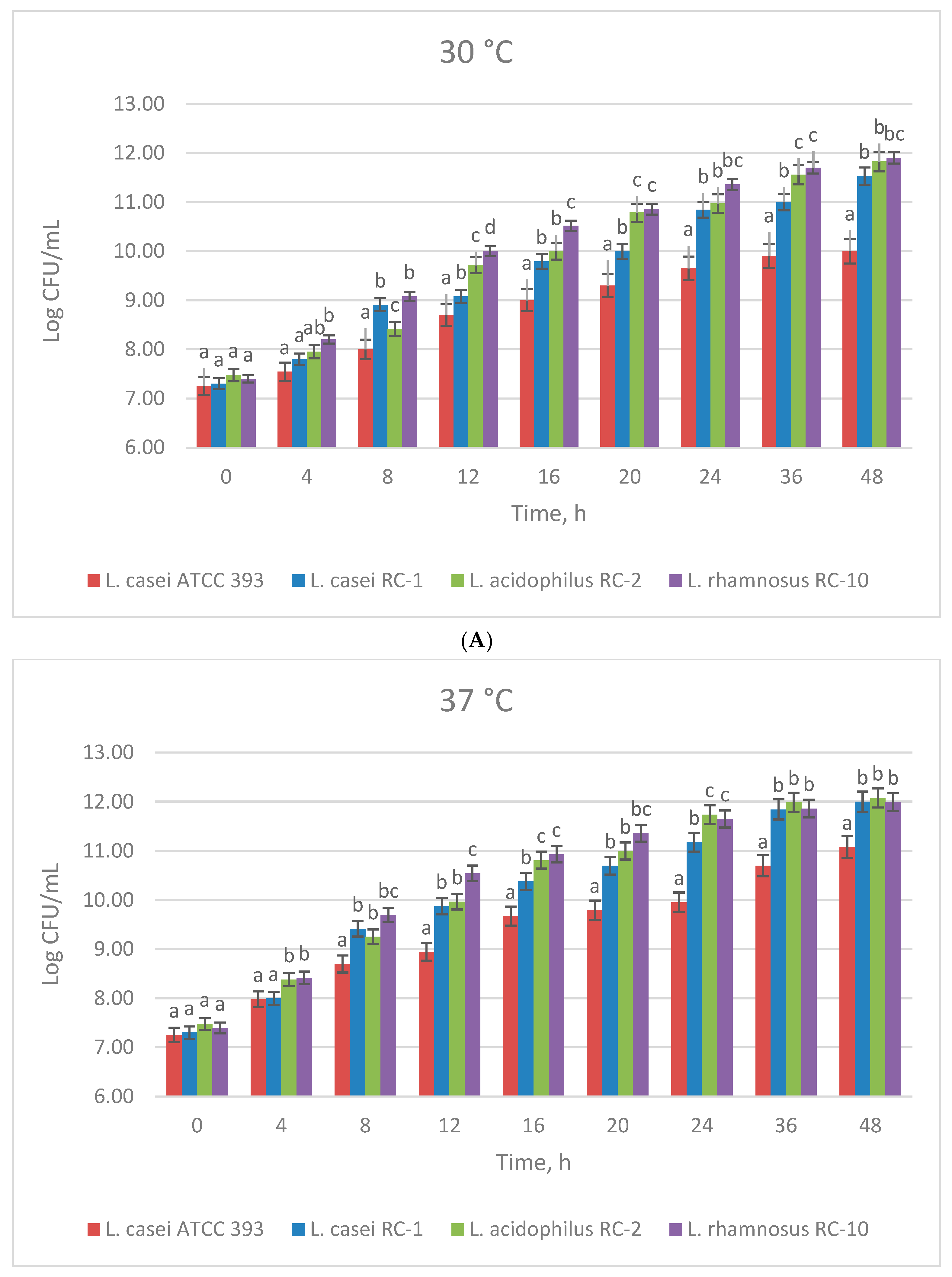
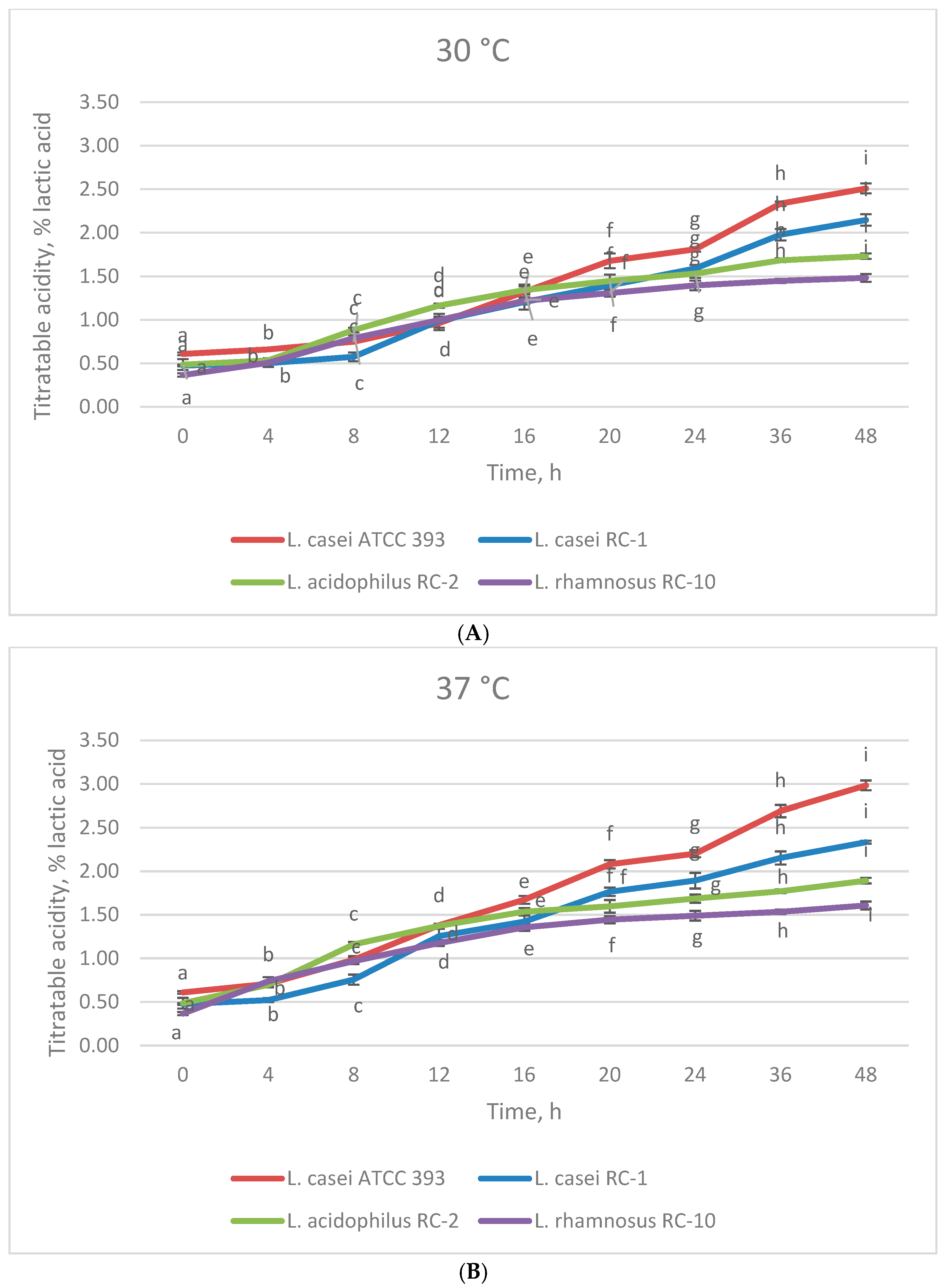
3.1. The Effect of Temperature on the Growth of Lactobacillus Strains

Lactobacillus spp. growth can be influenced by various external factors, such as pH, temperature, oxygen concentration, water activity and the composition of the culture medium. The optimal temperature for lactobacilli varies between 30 and 40 °C, whereas the optimal pH values for their growth vary between 5.5 and 6.2 [20]. The determination of optimal growth conditions of Lactobacillus strains is an important step from a technological point of view in the development of probiotic products.
In the first stage of a series of experiments, the growth characteristics of Lactobacillus strains under two different regimes of cultivation (30 °C and 37 °C) were determined. The two parameters (number of viable cells and titratable acidity) were determined at different time intervals (0 h, 4 h, 8 h, 12 h, 16 h, 20 h, 24 h, 36 h and 48 h). The temperature impact on the growth and lactic acid production of the four studied strains is illustrated in Figure 1A,B and Figure 2A,B.
As seen from the results, the Lactobacillus strains demonstrated the ability to adapt, grow and produce metabolites in both temperature regimes of cultivation (30 °C and 37 °C). However, better proliferation expressed by higher viable cell counts and higher lactic acid production were observed when the four strains were cultivated at 37 °C, reaching the highest values at the end of the cultivation process (Figure 1A,B and Figure 2A,B). At the 48th hour, the strain L. casei ATCC 393 exhibited the lowest viable cell counts, 10 log CFU/mL and 11.2 log CFU/mL, but the highest lactic acid production, 2.5% and 3% (at 30 °C and 37 °C, respectively), compared to the other strains. In contrast, at the same hour of cultivation, the strain L. rhamnosus RC-10 reached the highest viable cell count (11.8 log CFU/mL) at 30 °C (but lower than that at 37 °C—11.98 log CFU/mL), and the lowest lactic acid production—1.5% and 1.65% at 30 °C and 37 °C, respectively.

3.2. Resistance of Lactobacillus Strains to Simulated Upper Gastrointestinal Tract Conditions

As already mentioned, bacterial strains, in particular Lactobacillus strains, should possess several important properties to be considered as probiotic. One of the major criteria for effectiveness is that strains retain their viability and activity under unfavorable gastrointestinal tract conditions and during the manufacture and storage of probiotic products. Therefore, one of the first steps of the evaluation of new probiotic strains is the determination of their survival in in vitro simulated upper gastrointestinal conditions [21].
The growth of the four Lactobacillus strains after exposure to pH 2.5 + pepsin and pH 7.4 + pancreatin (containing lipase, protease and amylase) for 3 h is presented in Figure 3A–D. As seen from the obtained results, in all treatments and the controls, after the 5th hour of incubation, the number of viable cells gradually began to increase, which was more pronounced in the controls and the treatments at pH 7.4 + pancreatin. In terms of OD600 nm, at the end of incubation (24th h), all treatments reached their maximum growth. In the L. casei ATCC 393 and L. casei RC-1 strains, the cell recovery in the treatment with pH 7.4 + pancreatin was more intensive and showed OD values close to the controls, whereas in the treatment with pH 2.5 + pepsin, the cell recovery was less pronounced. It demonstrated that these two strains were more susceptible to low pH and the presence of pepsin compared to the other two strains. In the L. acidophilus RC-2 and L. rhamnosus RC-10 strains, the cells in both treatments (pH 2.5 + pepsin and pH 7.4 + pancreatin) recovered at almost the same rate as the controls. The cell recovery, summarized in Table 1, demonstrated that all of the studied strains were resistant to the simulated upper gastrointestinal tract conditions and can be considered to have probiotic potential.
Several studies have assessed the resistance of different Lactobacillus species to simulated gastrointestinal tract conditions. Das et al. [22] investigated the probiotic potential of three L. casei strains (SB71, SB73 and SB93) and stated that the cell viability of the strain SB71 in gastric juice reduced significantly after 3 h of exposure, while intestinal juice reduced its viability between 3 and 24 h. A similar decreasing trend was observed for strain SB73. The viability of the strain SB93 was unaffected by the gastric juice but significantly reduced by the intestinal juice (between 6 and 24 h). The percentage survival of the strain SB93 after incubation in intestinal juice for 24 h was significantly higher than the other two isolates studied. Research conducted by Nagyzbekkyzy et al. [23] showed that the percentage of cell recovery of four studied strains from the L. casei group isolated from Kazakh dairy foods ranged between 35% and 63% after 1 h of exposure under simulated gastric conditions. Various technological approaches can be applied to improve the viability of Lactobacillus species in the gastrointestinal tract and during the manufacture of fermented products. In this regard, Dimitrellou et al. [24,25] evaluated the survival of spray-dried microencapsulated and alginate-encapsulated cells of L. casei ATCC 393 under simulated gastrointestinal conditions and during the production and storage of fermented milk, finding that these methods significantly improved survival rate and retained a significantly higher number of viable cells during fermented milk production.
The members of L. acidophilus group are normal inhabitants of the gastrointestinal tract of healthy humans and are widely documented as LAB with probiotic potential. Farid et al. [26] assessed the probiotic features of three L. acidophilus strains, namely WFA1 (KU877440), WFA2 (KU877441) and WFA3 (KU877442) isolated from Pakistan Dahi yogurt. The authors reported that simulated gastric conditions had a more severe impact on their survival compared to the effect of simulated intestinal conditions. The survival rates of the studied strains under gastric conditions ranged between 43.12% and 50.45%, while those under intestinal conditions varied between 57.45% and 71.36% after 24 h of incubation, which was lower in comparison with our results for L. acidophilus RC-2 strain at the same time of incubation. Han et al. [27] compared the probiotic properties of various LAB and reported that survival rates of the strain L. acidophilus AD1 in simulated gastric juice (pH 3.0) were 91.21%, 88.48% and 83.41% at the 1st, 2nd and 3rd h, whereas the survival rates in simulated intestinal juice (pH 8.0) were 99.15%, 96.90% and 84.67% at the 2nd, 4th and 8th h of incubation, respectively. Similar to L. casei, the various encapsulation methods (ionic gelation, extrusion, emulsification and spray drying) are also considered effective means for improving the survival and viability of L. acidophilus cells in harsh environments during the digestion process or manufacture of different products [28].
L. rhamnosus GG is a strain known for its high tolerance to adverse gastrointestinal tract conditions. Zhao et al. [29] established that the survival rates of the strains L. rhamnosus X253 and L. rhamnosus GG after incubation in artificial gastric fluid (pH 3.0) for 3 h remained above 90%. The survivability of L. rhamnosus GG after exposure to artificial intestinal fluid (pH 8.0) for 5 h was 80%, whereas the survival rate of L. rhamnosus X253 declined markedly to 55%. Vougiouklaki et al. [30] stated that the strain L. rhamnosus GG ATCC 53103 exhibited promising survival capacity after exposure to simulated gastric juice for 3 h (98.86%) and simulated intestinal juice for 12 h (88.12%), which was consistent with our results obtained for L. rhamnosus RC-10. According to previously reported data, strains must exhibit ≥ 50% resistance at pH = 3.0 to be considered acid tolerant [31].

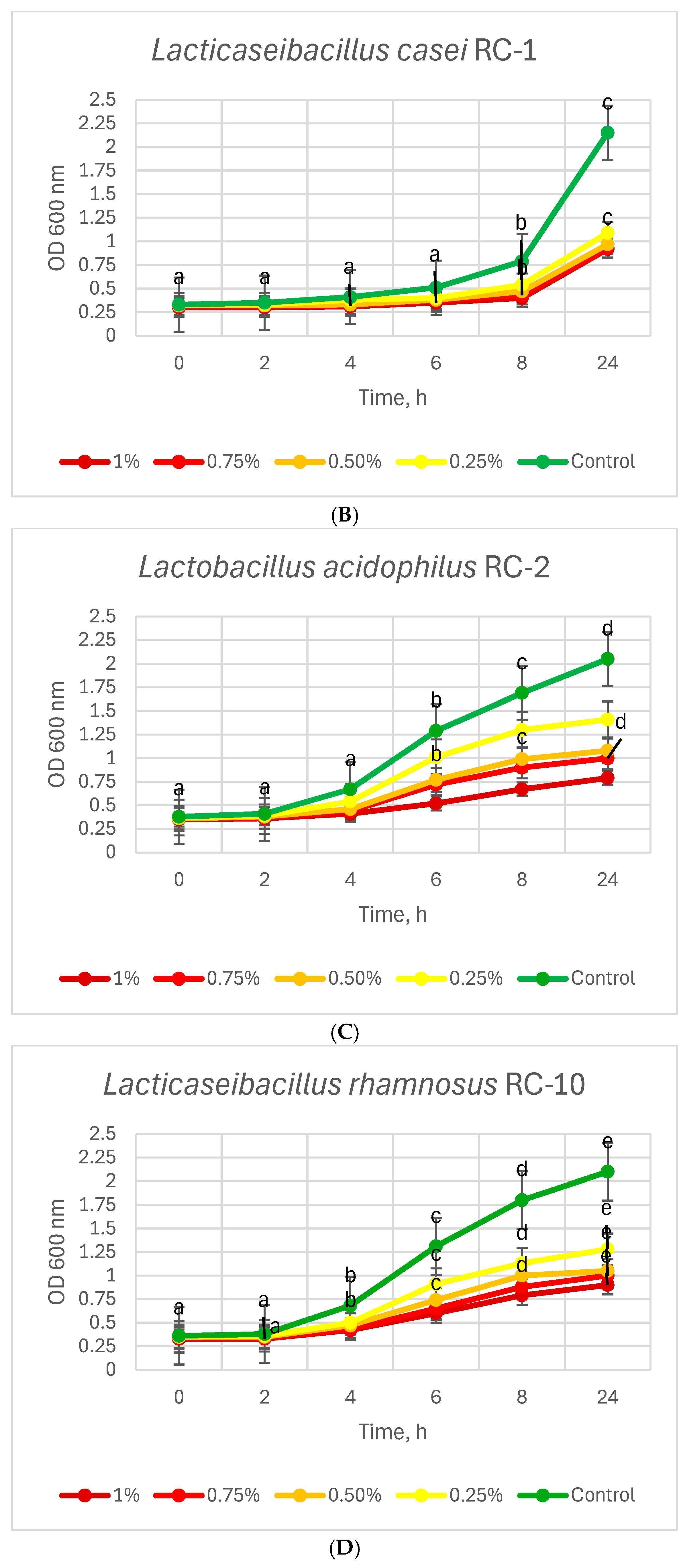
3.3. Resistance of Lactobacillus Strains to Bile Salts

Similar to that through the stomach, the passage of probiotics through the small intestine can result in a significant decrease in viable cells due to the presence of bile acids, salts and pancreatic enzymes, while the neutral pH (6.1 to 7.8) does not possess an inhibitory effect. Bile acids (derivatives of cholic acid) attend in the dispersion and absorption of lipids, thus playing an important role in fat digestion and microbial ecology in humans’ small and large intestines. It has been found that bile acids are secreted by the liver, whereas bile salts (bile acid conjugates with glycine or taurine) are produced in the duodenum [21,32]. Although tolerance to intestinal juice is a strain-dependent property, it has been reported that lactobacilli can progressively adapt to the presence of bile salts and other components of the intestinal juice due to global cellular responses such as preservation of internal pH, cell membrane integrity, and activation of bile salt efflux pumps [33].
The results presented in Table 2 and Figure 4A–D showed that the survival rates of the Lactobacillus strains decreased with increasing bile salt concentration and prolongation of the cultivation time. It is noteworthy that all of the strains retained their survival from 61.19% (L. acidophilus RC-2) to 82.5% (L. casei ATCC 393) at the highest bile salt concentration (1%) at the 4th hour of incubation. All of the strains retained over 50% survival rates at the lowest bile salt concentration (0.25%) at the 24th hour of cultivation. It should be noted here that the average concentration of bile salts in duodenal juice during the fasted state is up to 0.15 mM (approximately 0.6–0.7%) [34]. On the other hand, the time required for food to pass through the upper gastrointestinal tract (including small intestines—duodenum, jejunum and ileum) varies between 1.5 and 9 h, depending on the nature of the food [35]. Based on the results obtained and the above-mentioned physiological points, it can be concluded that the investigated strains possess probiotic potential.
As previously noted, the resistance of LAB to bile salts is a feature depending on the species, strain and even the experimental conditions. A study conducted by Das et al. [22] demonstrated that the viability of L. casei SB71 reduced in the presence of 0.5% bile salts, compared to the control, whereas L. casei SB73 and L. casei SB93 at the same concentration remained unaffected (L. casei SB93 exhibited 100% survival). In the presence of 1% bile salts, the authors observed a significant reduction in the survival rate of L. casei SB73 compared to the control. Although L. casei SB93 tolerated up to 1.8% bile, its survival was significantly reduced. Masalam et al. [36] observed an insignificant reduction in viable cells of the strains L. casei BgShn3, L. casei Dwan5, L. casei MSJ1, L. casei MasaLam7, L. acidophilus Hadhramaut4 and L. acidophilus Musallam2 isolated from raw and fermented milk from Saudi Arabia after treatment with 0.5% bile for 4 h. The results showed that survival rates ranged between 97.4% and 99.3% for the L. casei strains and between 90.7% and 99.45% for the L. acidophilus strains; these values were higher in comparison with our results for the same hour of cultivation and the same bile salt concentration. Nawaz et al. [37] investigated the probiotic properties of five L. casei strains isolated from camel milk from Pakistan and determined that the strains tolerated a concentration of 1.5% bile salts (exposure of 5–7 h) with survival rates varying between 77.78% and 81.35%. The strain L. acidophilus SAM1, isolated from human breast milk, showed a viability rate of 67.4% after 24 h of incubation in MRS broth supplemented with 3% bile salts [38]. A study conducted by Oh et al. [39] revealed that L. rhamnosus 4B15 isolated from infant feces exhibited significant capability of growth with a survival rate of 99.4% after incubation for 6 h with 1% bile salts.
In contrast, Hu et al. [40] found a significant decrease in survivability of L. rhamnosus ATCC 53103 (51.89%), L. acidophilus ATCC 43121 (44.09%), L. casei ATCC 334 (36.25%) and L. casei ATCC 393 (18.03%) in the presence of 0.3% oxgall for 24 h, and these values were lower compared to our results. The same trend was observed by Malilay et al. [41], who investigated the functional properties of L. acidophilus BIOTECH 1900 and stated that the strain viability decreased progressively with the increase in the bile salt concentration. At the 24th h of cultivation, the authors established resistance rates of 93.91%, 89%, 45.98%, 14.73 and 10.53% for bile salt concentrations of 0.1, 0.3, 0.5, 0.7, and 0.9%, respectively. According to some works [7,17,41], the standard for probiotics selection was set at 0.3% bile concentration with a resistance rate of more than 50%.

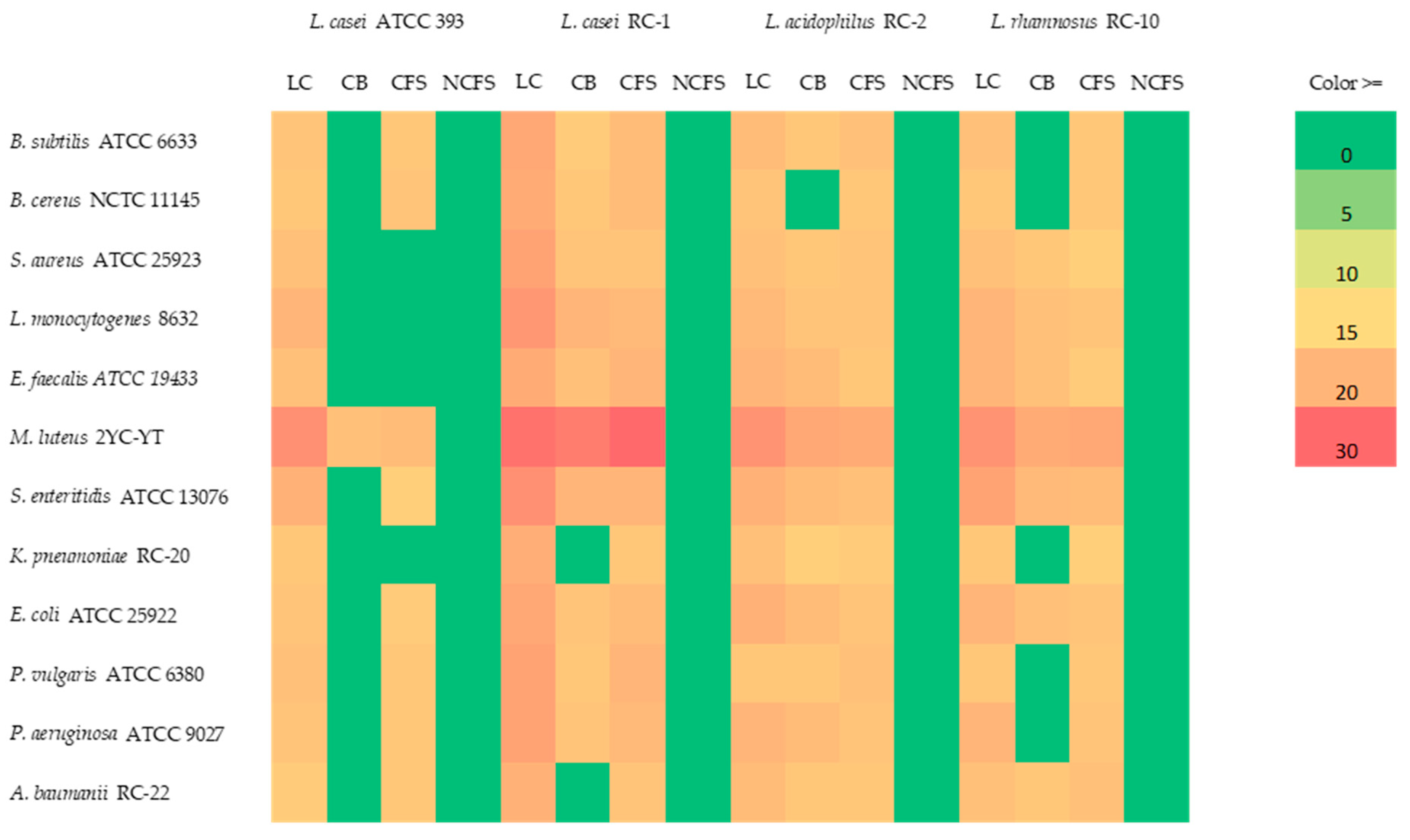
3.4. Antimicrobial Activity of Lactobacillus Strains

Antimicrobial activity is an important criterion for the selection of probiotic strains as natural antagonists of pathogenic and spoilage microorganisms. To assess whether the inhibitory activity was due to lactic acid production or cell-bound components, each strain was pretreated and tested in the form of Lactobacillus culture (LC), cell biomass (CB), cell-free supernatant (CFS) and neutralized cell-free supernatant to pH = 7.0 (NCFS).
As seen from the results illustrated in Figure 5, the four Lactobacillus strains inhibited the growth of Gram-positive and Gram-negative bacteria (at varying degrees) but did not exhibit antifungal activity against the tested yeasts C. albicans NBIMCC 74, S. cerevisiae ATCC 9763 and P. membranaefaciens, and fungi A. niger ATCC 1015, A. ochraceus, P. chrysogenum, F. moniliforme ATCC 38932 and F. oxysporum (data not presented). In addition, L. casei ATCC 393 showed the lowest antimicrobial potential in comparison to the other three Lactobacillus strains.
The results obtained (Figure 5) demonstrated that the neutralized cell-free supernatant (NCFS) to pH = 7.0 in all of the tested Lactobacillus strains completely lost its inhibitory effect, indicating that one of the reasons for their antimicrobial activity is the production of lactic acid in the culture medium. On the other hand, the highest lactic acid production was not always associated with the highest antimicrobial activity as in the case of L. casei ATCC 393 (Figure 2A,B and Figure 5). Consequently, the antagonistic activity was also due to the presence of cell-associated components possessing an antimicrobial effect.
Nawaz et al. [37] investigated the antimicrobial activity of five L. casei strains isolated from camel milk from Pakistan and determined that the strains inhibited the growth of the multidrug-resistant E. coli AZ1 (diameter of IZ from 13.67 to 15.33 mm) and methicillin-resistant S. aureus Saba-1 (IZ from 15 to 16.33 mm). Barzegar et al. [42] investigated seven Lactobacillus strains, namely L. brevis (B2 and B3), L. acidophilus (B14, B15 and B17), L. plantarum (B20) and L. casei (B22) isolated from Iranian cheese, as well as L. rhamnosus GG as a reference strain. The authors reported that the neutral cell-free supernatant (NCFS) of the first seven strains inhibited the growth of test bacterial strains—E. coli, S. aureus, P. aeruginosa and Salmonella typhimurium at varying degrees. The strain L. acidophilus B14 showed the highest inhibitory activity against the studied pathogens, with an IZ ranging from 14.6 to 23.2 mm, while the strain L. casei B22 exhibited the lowest inhibitory activity, with an IZ ranging between 7.2 and 16.5 mm. George-Okafor et al. [43] studied the efficacy of cell-free supernatant (CFS) obtained from L. acidophilus ATCC 314 and determined that the inhibitory activity of concentrated CFS (dialyzed with 5 M sucrose solution at 4 °C) was most pronounced against S. aureus (IZ = 12.85 mm), followed by E. coli (IZ = 8.15 mm), A. niger (IZ = 6.98 mm) and Aspergillus flavus (IZ = 5.6 mm). The untreated and precipitated (with 4 °C acetone) CFSs showed lower antimicrobial activity. Using the agar well diffusion assay, Ali et al. [44] examined the antimicrobial activity of CFSs of Lactobacillus strains isolated from traditional fermented dairy products and determined that L. acidophilus (LAB 2) and L. rhamnosus (LAB 5) inhibited the growth of test bacteria P. aeruginosa (IZs = 15.7 and 15.6 mm), E. coli (IZs = 15.3 and 16.9 mm), E. coli MC1400 (IZs = 14.5 and 15.5 mm), S. aureus (IZs = 15.3 and 17.7 mm) and Listeria ivanovii (IZs = 11.3 and 12.4 mm). In contrast to our results regarding antifungal activity, the CFSs of the two strains exhibited inhibitory effect on the yeasts C. albicans (IZs = 14.7 and 14.3 mm) and fungi A. niger, A. flavus, Aspergillus fumigatus and P. chrysogenum. A study conducted by Bohora et al. [45] demonstrated that L. rhamnosus ATCC 7469 effectively inhibited the growth of E. faecalis ATCC 29212 and C. albicans ATCC 10231 (used as experimental models of endodontic pathogens) as the tested strain exhibited IZs of 19.03–19.77 mm and 18.52–19.48 mm, respectively; these values were higher in comparison with the results for our L. rhamnosus RC-10 strain.
Many scientific reports have been focused on the antimicrobial activity of strains of the genus Lactobacillus against clinical isolates of various pathogens. The effectiveness of L. casei ATCC 393 and L. acidophilus ATCC 4356 against 100 clinical isolates of E. coli was shown in a study conducted by Soltani et al. [46], who determined that the diameter of inhibition zones of Lactobacillus extractions varied between 15 and 17 mm, and between 16 and 19 mm, respectively. The screening for probiotic properties of Lactobacillus clinical isolates performed by Stivala et al. [47] revealed that the strain L. rhamnosus AD3 possessed high antimicrobial activity against E. faecalis ATCC 29212, P. aeruginosa ATCC 27853 and Candida glabrata 14, and moderate inhibitory activity against K. pneumoniae ATCC 700603, C. albicans 10 and Streptococcus agalactiae (a clinical isolate). A significant inhibitory effect of L. acidophilus and L. rhamnosus against K. pneumoniae clinical isolates was confirmed by Anil et al. [48].

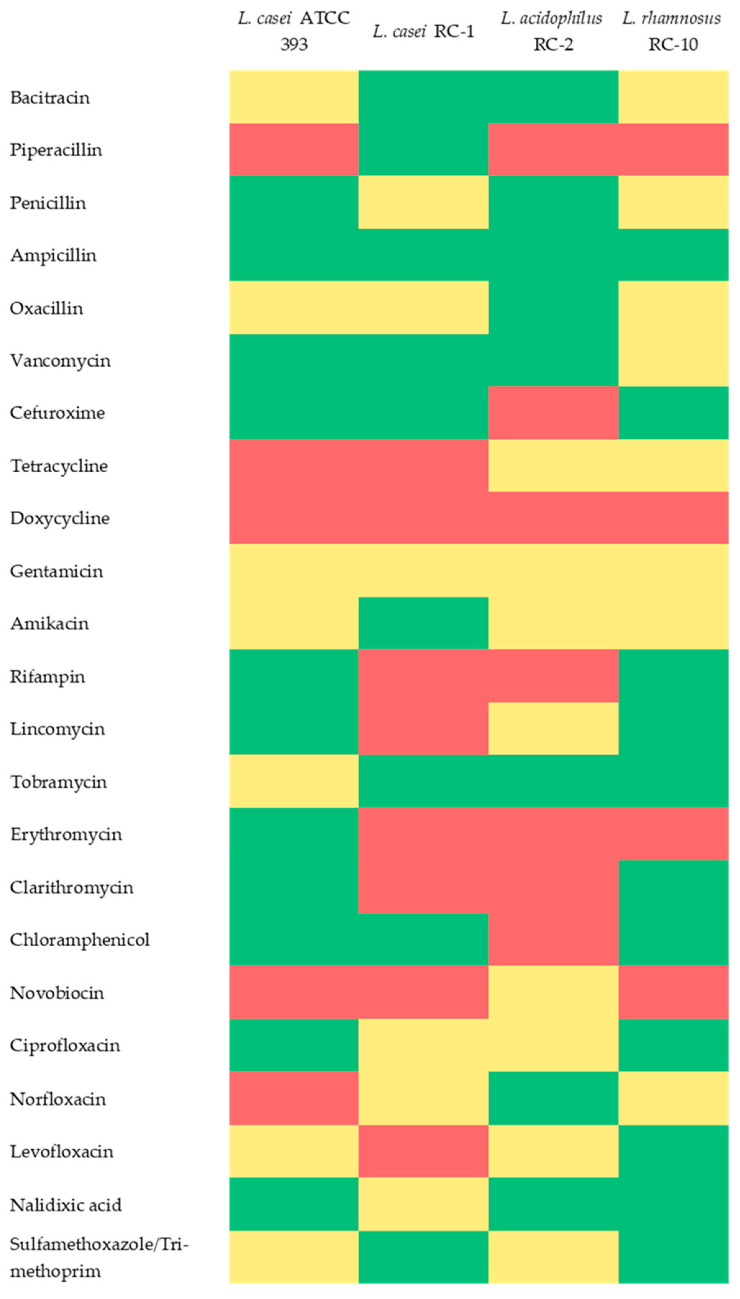
3.5. Antibiotic Susceptibility of Lactobacillus Strains

Antibiotic resistance is accepted as a main characteristic of probiotic bacteria. Therefore, an important step in the design of successful technological schemes for manufacturing various probiotic products and functional foods is the selection of suitable Lactobacillus strains that are resistant to the conventional antibiotics used in medical/clinical practice.
The investigated Lactobacillus strains were sensitive, moderately sensitive or resistant to the 23 antibiotics used in the study (Figure 6). The antibiotics used belonged to three main groups depending on their mechanism of action, namely inhibitors of the cell wall synthesis (bacitracin, piperacillin, penicillin, ampicillin, oxacillin, vancomycin, cefuroxime), inhibitors of the protein synthesis (tetracycline, doxycycline, gentamicin, amikacin, rifampin, lincomycin, tobramycin, erythromycin, clarithromycin, chloramphenicol) and inhibitors of the DNA synthesis or cell division (novobiocin, ciprofloxacin, norfloxacin, levofloxacin, nalidixic acid, sulfamethoxazole/trimethoprim).
According to data existing in the literature [49], Lactobacillus species are generally sensitive to inhibitors of protein synthesis (chloramphenicol, erythromycin, clindamycin, tetracycline and others) and resistant to the antibiotics belonging to the aminoglycoside group, such as neomycin, kanamycin, streptomycin, gentamicin and others. However, our results were partially consistent with these findings because three of the strains (L. casei ATCC 393, L. casei RC-1 and L. rhamnosus RC-10) were resistant to chloramphenicol; L. casei ATCC 393 was resistant to erythromycin; L. acidophilus RC-2 and L. rhamnosus RC-10 were determined as moderately susceptible (SR) to tetracycline; and all of the studied strains were moderately susceptible to gentamicin.
The degree of antibiotic susceptibility is a feature depending on the origin of the strains. Anisimova and Yarullina [50] examined the antibiotic susceptibility of 20 Lactobacillus strains and stated that L. rhamnosus B-8238 was resistant to vancomycin, amikacin, kanamycin, streptomycin and gentamicin. Similar results were reported by De Souza et al. [51], who investigated the probiotic potential of twelve L. casei strains (SJRP38, SJRP39, SJRP48, SJRP135, SJRP136, SJRP143, SJRP144, SJRP147, SJRP148, SJRP150, SJRP151 and SJRP166) isolated from water buffalo mozzarella cheese, and determined that all of the strains were resistant to vancomycin and moderately susceptible to gentamicin. Ten of these isolates were resistant to kanamycin, while the other two were moderately susceptible to this antibiotic. The strain L. rhamnosus isolated from commercial dietary supplements was resistant to erythromycin and ceftazidime, while the strain L. acidophilus (isolated from the same sources) was resistant only to ceftazidime out of the total of five antibiotics tested [52]. Shao et al. [53] investigated the antibiotic susceptibility of 17 L. casei isolates from fermented cow’s and yak’s milk from Tibet and stated the number of resistant strains as follows: all of these were resistant to vancomycin, 12 of these to chloramphenicol, 10 of these to streptomycin, 4 of these to kanamycin, 1 of these to neomycin and 1 of these to tetracycline.

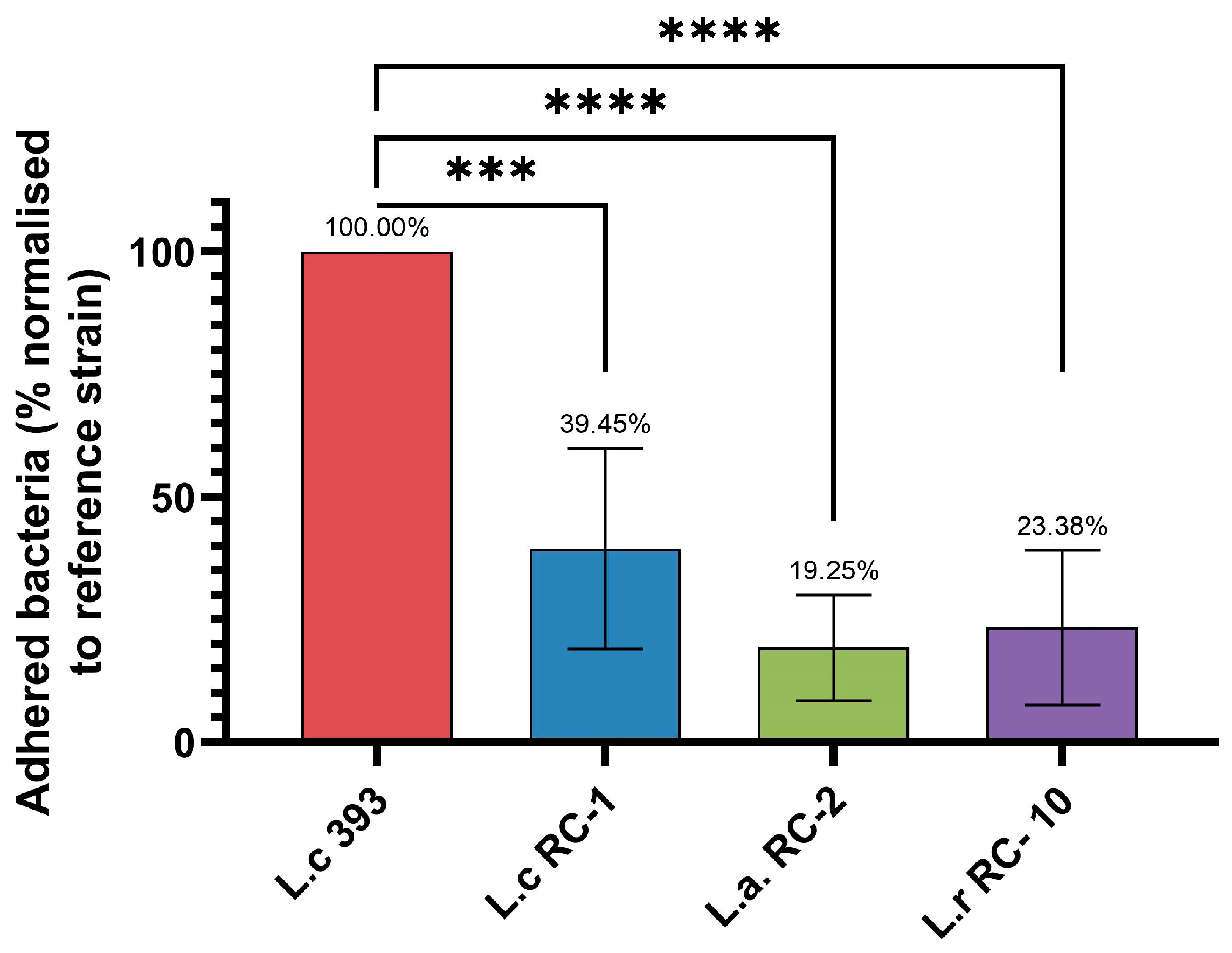
3.6. Adhesion Ability of Lactobacillus Strains to Intestinal Cells

The ability of potential probiotic strains to adhere to intestinal cells is regarded as an essential criterion for probiotic selection. This biological feature allows them to colonize the mucous membrane of the intestines and exert their immunomodulatory effects. Adhesion is a complex process that is mediated by nonspecific physico-chemical interactions between the bacterial and host cell surfaces or through specific interactions between cell components, such as exopolysaccharides, mucus-binding proteins (MUBs), surface layer proteins (Slps) and surface layer-associated proteins (Slaps). The adhesion of lactobacilli is a strain- and species-dependent property and can be influenced by various factors, including the cell line type [54].
In order to investigate how the isolated Lactobacillus strains adhered to intestinal cells in comparison to the reference strain, they were cultured overnight with shaking and analyzed via an adhesion assay. In all strains, we observed lower adhesion to swine intestinal cells IPEC-J2 compared to the reference strain L. casei ATCC 393, which is known as an important probiotic strain widely used in the dairy sector [55]. The highest degree of adhesion of the studied isolates was observed in L. casei RC-1—39.45%, followed by L. rhamnosus RC-10—23.38%. The lowest degree of adhesion was observed in L. acidophilus RC-2—19.25% in comparison with the reference probiotic strain (Figure 7).
Barzegar et al. [42] investigated the probiotic properties of seven Lactobacillus strains, including L. brevis (B2 and B3), L. acidophilus (B14, B15 and B17), L. plantarum (B20) and L. casei (B22) isolated from Iranian cheese, and used L. rhamnosus GG as a reference strain. The results from the adhesion test demonstrated that all of the studied isolates were able to adhere to the human Caco-2 cell line with percentage rates varying from 2.5% (L. brevis B3) to 14.6% (L. acidophilus B14). The strain L. rhamnosus GG showed an adhesion rate of 15%. These values were lower in comparison with our results. In another study by Soltani et al. [46], L. casei ATCC 393 showed an adhesion rate of 51.8% to the human intestinal epithelial cell line HT-29, whereas the adhesion value for the strain L. acidophilus ATCC 4356 was 29.3%. Using the same cell line (HT-29), Zhang et al. [56] determined that the adhesion rates of four L. rhamnosus strains (LR013, LR019, LR087 and LR093) isolated from cheese were between 4 and 9.5%. Das et al. [22] investigated the adhesive capacity of three L. casei strains (SB71, SB73 and SB93) and found that all strains adhered well to Caco-2 cells, with ratios of 32%, 38% and 45%, respectively. In contrast, Sophatha et al. [57] determined the high adhesion ability of the strain L. rhamnosus SD4 to Caco-2 and human oral keratinocyte cell line H357, which showed adhesion rates of 76% and 89%, respectively. Feng et al. [58] reported that the adhesion capacity of three L. rhamnosus strains (WEI-17, WEI-30 and WEI-41) isolated from the intestinal mucosa of healthy piglets to the IPEC-J2 cell line varied between 54% and 78%. Considering the adhesion ability and the results of additional tests, the authors concluded that the studied strains possessed probiotic potential. The adhesion affinity to epithelial cells is a property typical not only for the gut lactobacilli but also for those inhabiting the female urogenital tract (vagina). In this regard, Stojanov et al. [59] proved the adhesion ability of four vaginal Lactobacillus strains, namely Lactobacillus crispatus, Lactobacillus gasseri, Lactobacillus jensenii and Lactiplantibacillus plantarum, to the Caco-2 cell line.

4. Conclusions

The results in the present study revealed that the four investigated Lactobacillus strains, namely Lacticaseibacillus casei ATCC 393, Lacticaseibacillus casei RC-1, Lactobacillus acidophilus RC-2 and Lacticaseibacillus rhamnosus RC-10, exhibited promising probiotic potential. Therefore, these strains could be successfully applied in designing novel functional dairy products or used for further in vitro and in vivo investigations to elucidate their additional health benefits. In order to determine the most suitable strains to use in our future work, some additional factors related to probiotic dairy production will be investigated, such as the type of dairy technology applied, synergism or antagonism with classical starter cultures, and others.

Author Contributions

Conceptualization, Y.T.; methodology, Y.T., I.P.-S., M.I., R.C., A.D. and K.G.; software, M.I. and A.D.; validation, Y.T. and M.I.; formal analysis, Y.T., M.I. and A.D.; investigation, Y.T., I.P.-S., M.I., R.C., A.D. and K.G.; resources, Y.T., R.C. and K.G.; data curation, Y.T., M.I. and A.D.; writing—original draft preparation, Y.T., I.P.-S. and M.I.; writing—review and editing, K.G.; visualization, Y.T., M.I. and A.D.; supervision, Y.T. and K.G.; project administration, Y.T.; funding acquisition, Y.T. All authors have read and agreed to the published version of the manuscript.

Funding

This research received no external funding.

Institutional Review Board Statement

Not applicable.

Informed Consent Statement

Not applicable.

Data Availability Statement

The raw data supporting the conclusions of this article will be made available by the authors on request.

Conflicts of Interest

Author Remzi Cholakov was employed by the company Özgazi B. V. Dairy Foods. The remaining authors declare that the research was conducted in the absence of any commercial or financial relationships that could be construed as a potential conflict of interest.

References

  1. FAO; WHO. Guidelines for the Evaluation of Probiotics in Food; WHO: Geneva, Switzerland, 2002; pp. 1–11. [Google Scholar]
  2. Zielinska, D.; Kolohyn-Krajewska, D. Food-Origin Lactic Acid Bacteria May Exhibit Probiotic Properties: Review. BioMed. Res. Int. 2018, 2018, 5063185. [Google Scholar] [CrossRef] [PubMed]
  3. Caggia, C.; De Angelis, M.; Pitino, I.; Pino, A.; Randazzo, C.L. Probiotic features of Lactobacillus strains isolated from Ragusano and Pecorino Siciliano cheeses. Food Microbiol. 2015, 50, 109–117. [Google Scholar] [CrossRef] [PubMed]
  4. Bertuccini, L.; Russo, R.; Iosi, F.; Superti, F. Effects of Lactobacillus rhamnosus and Lactobacillus acidophilus on bacterial vaginal pathogens. Int. J. Immunopathol. Pharmacol. 2017, 30, 163–167. [Google Scholar] [CrossRef] [PubMed]
  5. Li, Y.; Liu, T.; Zhao, M.; Zhong, H.; Luo, W.; Feng, F. In vitro and in vivo investigations of probiotic properties of lactic acid bacteria isolated from Chinese traditional sourdough. Appl. Microbiol. Biotechnol. 2019, 103, 1893–1903. [Google Scholar] [CrossRef]
  6. Shehata, M.G.; El Sohaimy, S.A.; El-Sahn, M.A.; Youssef, M.M. Screening of isolated potential probiotic lactic acid bacteria for cholesterol lowering property and bile salt hydrolase activity. Ann. Agric. Sci. 2016, 61, 65–75. [Google Scholar] [CrossRef]
  7. Kumar, A.; Kumar, D. Characterization of Lactobacillus isolated from dairy samples for probiotic properties. Anaerobe 2015, 33, 117–123. [Google Scholar] [CrossRef]
  8. Fijan, S. Microorganisms with Claimed Probiotic Properties: An Overview of Recent Literature. Int. J. Environ. Res. Public Health 2014, 11, 4745–4767. [Google Scholar] [CrossRef]
  9. Chaudhary, A.; Saharan, B.S. Probiotic Properties of Lactobacillus plantarum. J. Pure Appl. Microbiol. 2019, 13, 933–948. [Google Scholar] [CrossRef]
  10. Song, M.; Yun, B.; Moon, J.-H.; Park, D.-J.; Lim, K.; Oh, S. Characterization of Selected Lactobacillus Strains for Use as Probiotics. Korean J. Food Sci. Anim. Resour. 2015, 35, 551–556. [Google Scholar] [CrossRef]
  11. Xu, S.; Liu, T.; Radji, C.A.I.; Yang, J.; Chen, L. Isolation, Identification, and Evaluation of New Lactic Acid Bacteria Strains with Both Cellular Antioxidant and Bile Salt Hydrolase Activities In Vitro. J. Food Prot. 2016, 79, 1919–1928. [Google Scholar] [CrossRef]
  12. Dempsey, E.; Corr, S.C. Lactobacillus spp. for Gastrointestinal Health: Current and Future Perspectives. Front. Immunol. 2022, 13, 840245. [Google Scholar] [CrossRef] [PubMed]
  13. Mu, Q.; Tavella, V.J.; Luo, X.M. Role of Lactobacillus reuteri in Human Health and Diseases. Front. Microbiol. 2018, 9, 757. [Google Scholar] [CrossRef] [PubMed]
  14. Zhang, Z.; Lv, J.; Pan, L.; Zhang, Y. Roles and applications of probiotic Lactobacillus strains. Appl. Microbiol. Biotechnol. 2018, 102, 8135–8143. [Google Scholar] [CrossRef]
  15. Liu, Y.; Li, Y.; Yu, X.; Yu, L.; Tian, F.; Zhao, J.; Zhang, H.; Zhai, Q.; Chen, W. Physiological Characteristics of Lactobacillus casei Strains and Their Alleviation Effects against Inflammatory Bowel Disease. J. Microbiol. Biotechnol. 2021, 31, 92–103. [Google Scholar] [CrossRef]
  16. Tumbarski, Y.; Todorova, M.; Topuzova, M.; Georgieva, P.; Ganeva, Z.; Mihov, R.; Yanakieva, V. Antifungal Activity of Carboxymethyl Cellulose Edible Films Enriched with Propolis Extracts and Their Role in Improvement of the Storage Life of Kashkaval Cheese. Curr. Res. Nutr. Food Sci. 2021, 9, 487–499. [Google Scholar] [CrossRef]
  17. García-Hernández, Y.; Pérez-Sánchez, T.; Boucourt, R.; Balcázar, J.L.; Nicoli, J.R.; Moreira-Silva, J.; Rodríguez, Z.; Fuertes, H.; Nuñez, O.; Albelo, N.; et al. Isolation, characterization and evaluation of probiotic lactic acid bacteria for potential use in animal production. Res. Vet. Sci. 2016, 108, 125–132. [Google Scholar] [CrossRef]
  18. Tumbarski, Y.; Yanakieva, V.; Denkova, R.; Denkova, Z. Isolation, Identification and Comparison of Some Properties of Lactobacillus delbrueckii subsp. bulgaricus Strains from Traditional Bulgarian and Italian Yogurts. Carpathian J. Food Sci. Technol. 2021, 13, 38–54. [Google Scholar] [CrossRef]
  19. Grzymajło, K.; Dutkiewicz, A.; Czajkowska, J.; Carolak, E.; Aleksandrowicz, A.; Waszczuk, W. Salmonella adhesion is decreased by hypoxia due to adhesion and motility structure crosstalk. Vet. Res. 2023, 54, 99. [Google Scholar] [CrossRef]
  20. Śliżewska, K.; Chlebicz-Wójcik, A. Growth Kinetics of Probiotic Lactobacillus Strains in the Alternative, Cost-Efficient Semi-Solid Fermentation Medium. Biology 2020, 9, 423. [Google Scholar] [CrossRef]
  21. Ayyash, M.M.; Abdalla, A.K.; AlKalbani, N.S.; Baig, M.A.; Turner, M.S.; Liu, S.-Q.; Shah, N.P. Characterization of new probiotics from dairy and nondairy products—Insights into acid tolerance, bile metabolism and tolerance, and adhesion capability. J. Dairy Sci. 2021, 104, 8363–8379. [Google Scholar] [CrossRef]
  22. Das, P.; Khowala, S.; Biswas, S. In vitro probiotic characterization of Lactobacillus casei isolated from marine samples. LWT—Food Sci. Technol. 2016, 73, 383–390. [Google Scholar] [CrossRef]
  23. Nagyzbekkyzy, E.; Abitayeva, G.; Anuarbekova, S.; Shaikhina, D.; Li, K.; Shaikhin, S.; Almagambetov, K.; Abzhalelov, A.; Saduakhassova, S.; Kushugulova, A.; et al. Investigation of acid and bile tolerance, antimicrobial activity and antibiotic resistance of Lactobacillus strains isolated from kazakh dairy foods. Asian J. Appl. Sci. 2016, 9, 143–158. [Google Scholar] [CrossRef]
  24. Dimitrellou, D.; Kandylis, P.; Petrović, T.; Dimitrijević-Branković, S.; Lević, S.; Nedović, V.; Kourkoutas, Y. Survival of spray dried microencapsulated Lactobacillus casei ATCC 393 in simulated gastrointestinal conditions and fermented milk. LWT—Food Sci. Technol. 2016, 71, 169–174. [Google Scholar] [CrossRef]
  25. Dimitrellou, D.; Kandylis, P.; Lević, S.; Petrović, T.; Ivanović, S.; Nedović, V.; Kourkoutas, Y. Encapsulation of Lactobacillus casei ATCC 393 in alginate capsules for probiotic fermented milk production. LWT—Food Sci. Technol. 2019, 116, 108501. [Google Scholar] [CrossRef]
  26. Farid, W.; Masud, T.; Sohail, A.; Ahmad, N.; Saqlan Naqvi, S.M.; Khan, S.; Ali, A.; Khalifa, S.A.; Hussain, A.; Ali, A.; et al. Gastrointestinal transit tolerance, cell surface hydrophobicity, and functional attributes of Lactobacillus acidophilus strains isolated from Indigenous Dahi. Food Sci. Nutr. 2021, 9, 5092–5102. [Google Scholar] [CrossRef]
  27. Han, Q.; Kong, B.; Chen, Q.; Sun, F.; Zhang, H. In vitro comparison of probiotic properties of lactic acid bacteria isolated from Harbin dry sausages and selected probiotics. J. Funct. Foods 2017, 32, 391–400. [Google Scholar] [CrossRef]
  28. Altamirano-Rios, A.V.; Guadarrama-Lezama, A.Y.; Arroyo-Maya, I.J.; Hernandez-Alvarez, A.-J.; Orozco-Villafuerte, J. Effect of encapsulation methods and materials on the survival and viability of Lactobacillus acidophilus: A review. Int. J. Food Sci. Technol. 2022, 57, 4027–4040. [Google Scholar] [CrossRef]
  29. Zhao, L.; Zhang, Y.; Liu, Y.; Zhong, J.; Zhang, D. Assessing the Safety and Probiotic Characteristics of Lacticaseibacillus rhamnosus X253 via Complete Genome and Phenotype Analysis. Microorganisms 2023, 11, 140. [Google Scholar] [CrossRef]
  30. Vougiouklaki, D.; Tsironi, T.; Tsantes, A.G.; Tsakali, E.; Van Impe, J.F.M.; Houhoula, D. Probiotic Properties and Antioxidant Activity In Vitro of Lactic Acid Bacteria. Microorganisms 2023, 11, 1264. [Google Scholar] [CrossRef]
  31. Sieladie, D.V.; Zambou, N.F.; Kaktcham, P.M.; Cresci, A.; Fonteh, F. Probiotic properties of Lactobacilli strains isolated from raw cow milk in the western highlands of Cameroon. Innov. Rom. Food Biotechnol. 2011, 9, 12–28. [Google Scholar]
  32. Thaweesang, S.; Leenanon, B. Survival of Lactobacillus acidophilus TISTR1338 in bile salt stress conditions. Asian J. Microbiol. Biotech. Env. Sci. 2016, 18, 569–579. [Google Scholar]
  33. Tarrah, A.; da Silva Duarte, V.; de Castilhos, J.; Pakroo, S.; Lemos Junior, W.J.F.; Luchese, R.H.; Fioravante Guerra, A.; Rossi, R.C.; Righetto Ziegler, D.; Corich, V.; et al. Probiotic potential and biofilm inhibitory activity of Lactobacillus casei group strains isolated from infant feces. J. Funct. Foods 2019, 54, 489–497. [Google Scholar] [CrossRef]
  34. Vinarov, Z.; Abdallah, M.; Agundez, J.A.G.; Allegaert, K.; Basit, A.W.; Braeckmans, M.; Ceulemans, J.; Corsetti, M.; Griffin, B.T.; Grimm, M.; et al. Impact of gastrointestinal tract variability on oral drug absorption and pharmacokinetics: An UNGAP review. Eur. J. Pharm. Sci. 2021, 162, 105812. [Google Scholar] [CrossRef] [PubMed]
  35. Sensoy, I. A review on the food digestion in the digestive tract and the used in vitro models. Curr. Res. Food Sci. 2021, 4, 308–319. [Google Scholar] [CrossRef]
  36. Masalam, M.S.B.; Bahieldin, A.; Alharbi, M.G.; Al-Masaudi, S.; Al-Jaouni, S.K.; Harakeh, S.M.; Al-Hindi, R.R. Isolation, Molecular Characterization and Probiotic Potential of Lactic Acid Bacteria in Saudi Raw and Fermented Milk. Evid. Based Complement. Alternat. Med. 2018, 2018, 7970463. [Google Scholar] [CrossRef]
  37. Nawaz, Z.; Zahoor, M.K.; Shafique, M.; Athar, R.; Yasmin, A.; Zahoor, M.A. In vitro assessment of probiotic properties of lactic acid bacteria isolated from camel milk: Enhancing sustainable foods. Front. Sustain. Food Syst. 2024, 8, 1437201. [Google Scholar] [CrossRef]
  38. El-Sayed, A.I.M.; El-Borai, A.M.; Akl, S.H.; EL-Aassar, S.A.; Abdel-Latif, M.S. Identification of Lactobacillus strains from human mother milk and cottage cheese revealed potential probiotic properties with enzymatic activity. Sci Rep. 2022, 12, 22522. [Google Scholar] [CrossRef]
  39. Oh, N.S.; Joung, J.Y.; Lee, J.Y.; Kim, Y. Probiotic and anti-inflammatory potential of Lactobacillus rhamnosus 4B15 and Lactobacillus gasseri 4M13 isolated from infant feces. PLoS ONE 2018, 13, e0192021. [Google Scholar] [CrossRef]
  40. Hu, P.-L.; Yuan, Y.-H.; Yue, T.-L.; Guo, C.-F. A new method for the in vitro determination of the bile tolerance of potentially probiotic lactobacilli. Appl. Microbiol. Biotechnol. 2018, 102, 1903–1910. [Google Scholar] [CrossRef]
  41. Malilay, J.K.T.; Oliveros, M.C.R.; Bautista, J.A.N.; Castillo-Israel, K.A.T. Functional, safety and technological properties of Lactobacillus acidophilus BIOTECH 1900. Philipp. J. Vet. Anim. Sci. 2019, 45, 11–21. [Google Scholar]
  42. Barzegar, H.; Behbahani, B.A.; Falah, F. Safety, probiotic properties, antimicrobial activity, and technological performance of Lactobacillus strains isolated from Iranian raw milk cheeses. Food Sci Nutr. 2021, 9, 4094–4107. [Google Scholar] [CrossRef] [PubMed]
  43. George-Okafor, U.; Ozoani, U.; Tasie, F.; Mba-Omeje, K. The efficacy of cell-free supernatants from Lactobacillus plantarum Cs and Lactobacillus acidophilus ATCC 314 for the preservation of home-processed tomato-paste. Sci. Afr. 2020, 8, e00395. [Google Scholar] [CrossRef]
  44. Ali, F.S.; Zayed, G.; Saad, O.A.O.; Salwa, A.; Gharib, H. Antimicrobial activity and Probiotic Properties of Lactic Acid Bacteria Isolated From Traditional Fermented Dairy Products. J. Mod. Res. 2020, 2, 40–48. [Google Scholar] [CrossRef]
  45. Bohora, A.A.; Kokate, S.R.; Khedkar, S.; Vankudre, A. Antimicrobial activity of probiotics against endodontic pathogens:- A preliminary study. Indian J. Med. Microbiol. 2019, 37, 5–11. [Google Scholar] [CrossRef]
  46. Soltani, N.; Abbasi, S.; Baghaeifar, S.; Taheri, E.; Jadid, M.F.S.; Emami, P.; Abolhasani, K.; Aslanshirzadeh, F. Antibacterial and antibiofilm activity of Lactobacillus strains secretome and extraction against Escherichia coli isolated from urinary tract infection. Biotechnol. Rep. 2022, 36, e00760. [Google Scholar] [CrossRef]
  47. Stivala, A.; Carota, G.; Fuochi, V.; Furneri, P.M. Lactobacillus rhamnosus AD3 as a Promising Alternative for Probiotic Products. Biomolecules 2021, 11, 94. [Google Scholar] [CrossRef]
  48. Anil, A.; Raj, A.; Azhar, M.M.; Kuriakose, N.; Murali, R.; Kumar, H.K.S. Evaluation of antimicrobial activity of Lactobacillus acidophilus and Lactobacillus rhamnosus against clinically isolated Klebsiella pneumoniae: An in vitro study. Int. J. Res. Med. Sci. 2024, 12, 1140–1146. [Google Scholar] [CrossRef]
  49. Darsanaki, R.K.; Aliabadi, M.A.; Chakoosari, M.M.D. Antibiotic resistance of lactic acid bacteria. Sci. J. Microbiol. 2013, 2, 201–206. [Google Scholar]
  50. Anisimova, E.A.; Yarullina, D.R. Antibiotic Resistance of LACTOBACILLUS Strains. Curr Microbiol. 2019, 76, 1407–1416. [Google Scholar] [CrossRef]
  51. De Souza, B.M.S.; Borgonovi, T.F.; Casarotti, S.N.; Todorov, S.D.; Penna, A.L.B. Lactobacillus casei and Lactobacillus fermentum Strains Isolated from Mozzarella Cheese: Probiotic Potential, Safety, Acidifying Kinetic Parameters and Viability under Gastrointestinal Tract Conditions. Probiotics Antimicrob. Proteins 2019, 11, 382–396. [Google Scholar] [CrossRef]
  52. Selvin, J.; Maity, D.; Sajayan, A.; Kiran, G.S. Revealing antibiotic resistance in therapeutic and dietary probiotic supplements. J. Glob. Antimicrob. Resist. 2020, 22, 202–205. [Google Scholar] [CrossRef] [PubMed]
  53. Shao, Y.; Zhang, W.; Guo, H.; Pan, L.; Zhang, H.; Sun, T. Comparative studies on antibiotic resistance in Lactobacillus casei and Lactobacillus plantarum. Food Control 2015, 50, 250–258. [Google Scholar] [CrossRef]
  54. Celebioglu, H.U.; Svensson, B. Dietary Nutrients, Proteomes, and Adhesion of Probiotic Lactobacilli to Mucin and Host Epithelial Cells. Microorganisms 2018, 6, 90. [Google Scholar] [CrossRef] [PubMed]
  55. Abdel-Hamid, M.; Romeih, E.; Gamba, R.R.; Nagai, E.; Suzuki, T.; Koyanagi, T.; Enomoto, T. The biological activity of fermented milk produced by Lactobacillus casei ATCC 393 during cold storage. Int. Dairy J. 2019, 91, 1–8. [Google Scholar] [CrossRef]
  56. Zhang, X.; Esmail, G.A.; Alzeer, A.F.; Arasu, M.V.; Vijayaraghavan, P.; Choi, K.C.; Al-Dhabi, N.A. Probiotic characteristics of Lactobacillus strains isolated from cheese and their antibacterial properties against gastrointestinal tract pathogens. Saudi J. Biol. Sci. 2020, 27, 3505–3513. [Google Scholar] [CrossRef]
  57. Sophatha, B.; Piwat, S.; Teanpaisan, R. Adhesion, anti-adhesion and aggregation properties relating to surface charges of selected Lactobacillus strains: Study in Caco-2 and H357 cells. Arch. Microbiol. 2020, 202, 1349–1357. [Google Scholar] [CrossRef]
  58. Feng, Y.; Qiao, L.; Liu, R.; Yao, H.; Gao, C. Potential probiotic properties of lactic acid bacteria isolated from the intestinal mucosa of healthy piglets. Ann. Microbiol. 2017, 67, 239–253. [Google Scholar] [CrossRef]
  59. Stojanov, S.; Plavec, T.V.; Zupančič, Š.; Berlec, A. Modified vaginal lactobacilli expressing fluorescent and luminescent proteins for more effective monitoring of their release from nanofibers, safety and cell adhesion. Microb. Cell Fact. 2024, 23, 333. [Google Scholar] [CrossRef]
Figure 1. Viable cells productivity of Lactobacillus strains during cultivation at 30 °C (A) and 37 °C (B) for 48 h. Values are expressed as means ± SD. Letters a–d indicate significant differences (p < 0.05) between the samples, n = 3.
Applsci 15 02902 g001
Figure 2. Production of lactic acid of Lactobacillus strains during cultivation at 30 °C (A) and 37 °C (B) for 48 h. Values are expressed as means ± SD. Letters a–i indicate significant differences (p < 0.05) between the samples, n = 3.
Applsci 15 02902 g002
Figure 3. Cell recovery of L. casei ATCC 393 (A), L. casei RC-1 (B), L. acidophilus RC-2 (C), L. rhamnosus RC-10 (D) in MRS broth after exposure to pH 2.5 + pepsin and pH 7.4 + pancreatin for 3 h during cultivation at 37 °C for 24 h. Values are expressed as means ± SD. Letters a–c indicate significant differences (p < 0.05) between the samples, n = 3.
Applsci 15 02902 g003aApplsci 15 02902 g003b
Figure 4. Growth of L. casei ATCC 393 (A), L. casei RC-1 (B), L. acidophilus RC-2 (C), L. rhamnosus RC-10 (D) in MRS broth supplemented or not with various concentrations of bile salts during cultivation at 37 °C for 24 h. Values are expressed as means ± SD. Letters a–e indicate significant differences (p < 0.05) between the samples, n = 3.
Applsci 15 02902 g004aApplsci 15 02902 g004b
Figure 5. Heat map analysis of the antimicrobial activity of the Lactobacillus strains. The different colors represent the diameter of the inhibition zones (IZs), with green indicating the absence or the smallest diameter and red indicating the largest. The transition from green to red illustrates the increase in the diameter of the IZs. LC—whole Lactobacillus culture; CB—cell biomass; CFS—cell-free supernatant; NCFS—neutralized cell-free supernatant.
Applsci 15 02902 g005
Figure 6. Heat map analysis of the antibiotic susceptibility of the Lactobacillus strains. The different colors represent the diameter of the inhibition zones (IZs), with green indicating the absence or the smallest diameter (resistant), yellow indicating IZ = 7–16 mm (moderately resistant) and red indicating the largest IZ > 16 mm (sensitive).
Applsci 15 02902 g006
Figure 7. Adhesion of Lactobacillus strains to the IPEC-J2 cell line. All strains were grown O/N and incubated for two hours with IPEC-J2 cell monolayers in MRS+DMEM in MOI 100. All results were normalized to reference strain L. casei ATCC 393 adhesion. Statistical differences between the strains were analyzed by one-way ANOVA and presented as geometric means. *** p < 0.001, **** p < 0.0001, n = 3.
Applsci 15 02902 g007
Table 1. Percentage of cell recovery of Lactobacillus strains after exposure to pH 2.5 + pepsin and pH 7.4 + pancreatin for 3 h.
StrainConditionsCell Recovery, %
6 h24 h
L. casei ATCC 393pH 2.5 + pepsin65.45 ± 0.02 aA75.35 ± 0.06 aB
pH 7.4 + pancreatin80.91 ± 0.03 bA93.95 ± 0.07 bB
L. casei RC-1pH 2.5 + pepsin60.25 ± 0.01 aA74.24 ± 0.03 aB
pH 7.4 + pancreatin86.96 ± 0.05 bA96.07 ± 0.03 bB
L. acidophilus RC-2pH 2.5 + pepsin66.67 ± 0.01 aA87.50 ± 0.15 aB
pH 7.4 + pancreatin87.10 ± 0.02 bA98.15 ± 0.02 bB
L. rhamnosus RC-10pH 2.5 + pepsin70.33 ± 0.12 aA87.30 ± 0.06 aB
pH 7.4 + pancreatin81.32 ± 0.07 bA96.83 ± 0.02 bB
Values are expressed as means ± SD. Letters a–b indicate significant differences (p < 0.05) within a sample exposed to different conditions, while letters A–B denote significant differences (p < 0.05) between the cultivation hours, n = 3.
Table 2. Survival rates of Lactobacillus strains after exposure to different concentrations of bile salts at the 4th, 8th and 24th hour of cultivation at 37 °C.
StrainBile Salts
Concentration
Survival Rate, %
4 h8 h24 h

L. casei ATCC 393
1%82.50 ± 0.02 aA60.00 ± 0.02 aB36.72 ± 0.06 aC
0.75%87.50 ± 0.01 bA61.67 ± 0.02 bB38.98 ± 0.04 bC
0.5%90.00 ± 0.02 cA65.00 ± 0.04 cB41.81 ± 0.06 cC
0.25%92.50 ± 0.03 dA68.33 ± 0.02 dB50.85 ± 0.08 dC

L. casei RC-1
1%75.61 ± 0.08 aA50.63 ± 0.08 aB42.79 ± 0.08 aC
0.75%78.05 ± 0.06 bA54.43 ± 0.08 bB43.26 ± 0.12 bC
0.5%80.49 ± 0.04 cA59.49 ± 0.06 cB45.12 ± 0.09 cC
0.25%92.68 ± 0.02 dA68.35 ± 0.03 dB50.70 ± 0.03 dC

L. acidophilus RC-2
1%61.19 ± 0.01 aA40.24 ± 0.04 aB38.54 ± 0.08 aC
0.75%65.67 ± 0.01 bA53.25 ± 0.09 bB48.78 ± 0.04 bC
0.5%68.66 ± 0.03 cA58.58 ± 0.06 cB52.68 ± 0.10 cC
0.25%80.60 ± 0.09 dA76.92 ± 0.19 dB68.78 ± 0.04 dC

L. rhamnosus RC-10
1%61.76 ± 0.01 aA43.89 ± 0.10 aB42.86 ± 0.06 aC
0.75%63.24 ± 0.08 bA48.89 ± 0.11 bB47.62 ± 0.05 bC
0.5%69.12 ± 0.03 cA55.56 ± 0.12 cB50.00 ± 0.12 cC
0.25%73.53 ± 0.14 dA62.78 ± 0.11 dB60.95 ± 0.10 dC
Values are expressed as means ± SD. Letters a–d indicate significant differences (p < 0.05) within a sample exposed to different conditions, while letters A–C denote significant differences (p < 0.05) between the cultivation hours, n = 3.
Disclaimer/Publisher’s Note: The statements, opinions and data contained in all publications are solely those of the individual author(s) and contributor(s) and not of MDPI and/or the editor(s). MDPI and/or the editor(s) disclaim responsibility for any injury to people or property resulting from any ideas, methods, instructions or products referred to in the content.

