Article

An Efficient Method for Lung Lesions Classification Using Automatic Vascularization Evaluation on Color Doppler Ultrasound


1
Automation Department, Technical University of Cluj-Napoca, 400114 Cluj-Napoca, Romania
2
Department of Internal Medicine, “Iuliu Hatieganu” University of Medicine and Pharmacy, 400347 Cluj-Napoca, Romania
3
Emergency Clinical County Hospital Cluj-Napoca, 400347 Cluj-Napoca, Romania
*
Authors to whom correspondence should be addressed.
Appl. Sci. 2025, 15(5), 2851; https://doi.org/10.3390/app15052851
Submission received: 25 January 2025 / Revised: 25 February 2025 / Accepted: 4 March 2025 / Published: 6 March 2025
(This article belongs to the Special Issue Advances in Diagnostic Radiology)

Featured Application

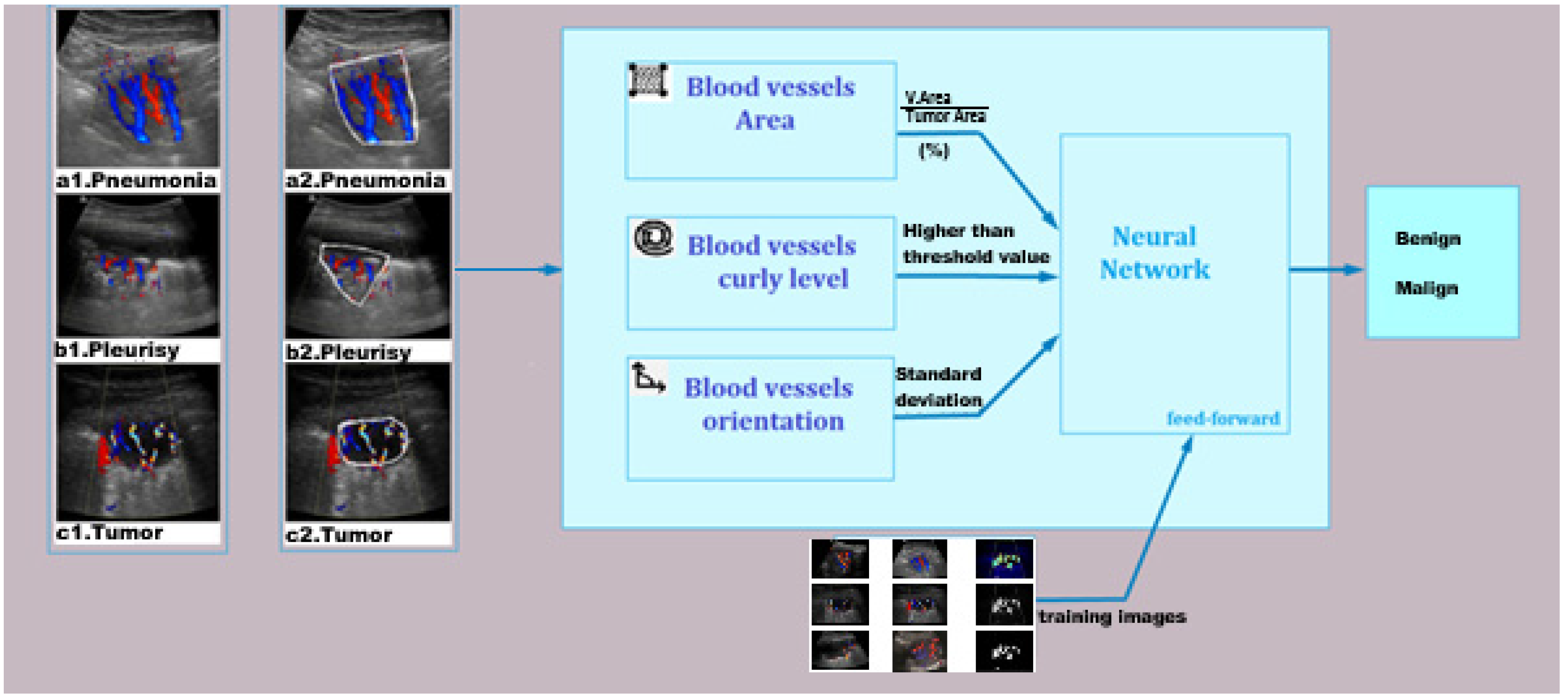
The proposed system offers an innovative, non-invasive, and economical approach for classifying lung lesions through the integration of Doppler ultrasound imaging and machine learning methodologies. The modular architecture facilitates the detailed assessment of vascularization parameters, hence improving diagnostic accuracy. This approach is especially appropriate for resource-constrained clinical environments, where conventional imaging techniques such as CT scans may be inaccessible or less available. It also possesses potential for incorporation into portable diagnostic instruments, enhancing early identification and monitoring of lung cancer.

Abstract

Lung cancer still represents one of the main causes of cancer-related mortality, highlighting the necessity for precise, effective, and minimally intrusive diagnostic methods. This research presents an innovative approach to classifying lung lesions using Doppler ultrasound imagery combined with a feed-forward neural network (FNN). This study integrates Doppler mode ultrasound vascularization features—blood vessel area, tortuosity index, and orientation—into an FNN to classify lung lesions as benign or malignant. A dataset of 565 Doppler ultrasound pictures was extended using augmentation techniques to enhance robustness, yielding a training dataset of 3390 images. The FNN architecture was trained utilizing the Levenberg–Marquardt algorithm, achieving a classification accuracy of 98%, demonstrating its potential as a diagnostic aid. The results indicate that integrating all three vascularization factors significantly improves diagnosis accuracy compared with individual modules. This method offers a non-invasive and cost-effective complementary tool to conventional techniques such as CT scans, with the potential to improve early detection and treatment planning for lung cancer patients.
Keywords: lung cancer; computer-aided diagnosis; lung vascularization; Doppler ultrasound lung cancer; computer-aided diagnosis; lung vascularization; Doppler ultrasound